Embed Size (px)
Citation preview
British Journal of Ophthalmology, 1987, 71, 273-278
Responses to tuberculin in the guinea-pig eye as amodel of cell mediated immune responses in theexternal eye
R ST C DWYER, S DAROUGAR, AND MARJORIE A MONNICKENDAM
From the Subdepartment of Virology, Institute of Ophthalmology, Judd Street, London WCJ H 9QS
SUMMARY The clinical and histological findings in an animal model of ocular cell mediatedimmune responses are described. These were evoked in sensitised guinea-pigs by droppingtuberculin into the conjunctival sac or injecting it under the palpebral conjunctiva. Whentuberculin was dropped into the conjunctival sac, higher doses were required to evoke a strongresponse than when it was injected subconjunctivally. When high doses of antigen were used forchallenge, a mild response was observed in unchallenged contralateral eyes. The cellular responseat low doses of tuberculin consisted predominantly of mononuclear cells. At higher doses thecellular infiltrate was more pronounced, and polymorphonuclear cells tended to predominate24 hours after challenge, while at 48 hours mononuclear cells predominated.
It has been suggested that cell mediated immuneresponses are important in the pathogenesis of eyediseases including trachoma, herpes simplex virusinfection, chronic staphylococcal infection, andvernal keratoconjunctivitis. However, there is as yetlittle direct evidence to support this hypothesis. Mostin-vivo studies of cell mediated immune responseshave been carried out in the skin, and there arerelatively few studies in other tissues. We havedeveloped an animal model using tuberculin to studycell mediated responses in the outer eye in theabsence of infection or immediate-type reactions.This is similar to the diagnostic test in humans,introduced in 1909, in which tuberculin in saline orpetroleum jelly was instilled into the conjunctival sacand ocular reactions. were observed.' Previousstudies of the reaction to tuberculin in the guinea-pigeye have involved injection of tuberculin into theguinea-pig cornea. In our model, briefly describedin an earlier paper,5 guinea-pigs sensitised to tuber-culin are challenged by dropping antigen on to theconjunctival sac or injecting it into the palpebralconjunctiva. We here report further studies on thismodel.
Material and methods
Animals. Female albino guinea-pigs of the Dunkin-Hartley strain weighing 250 to 500 g were used.Correspondcnce to Profcssor S Darougar.
Antigens. Freund's complete adjuvant was used forsensitisation (Bacto Adjuvant Complete (H27Ra),Difco Laboratories); tuberculin, mammalian puri-fied protein derivative (PPD) of tuberculin, orhuman PPD, lyophilised and phenol-free, was usedfor challenge (Ministry of Agriculture, Fisheries andFood, Central Veterinary Laboratories, Weybridge,Surrey).
Sensitisation. Animals were sensitised with foursimultaneous intradermal injections of 0-2 ml of a50/50 v/v emulsion of Freund's complete adjuvant inphosphate buffered saline. Animals were skin-testedfive to 10 weeks after sensitisation, and only animalswith induration of at least 18 mm diameter werechosen for eye challenge.
Ocular challenge. Right eyes were challenged byone of two methods: (1) mammalian PPD (0-02 ml)was instilled into the conjunctival sac and the lidsclosed with gentle massage to distribute the antigen;(2) human phenol-free PPD (2 [ug in 0-01 ml) wasinjected subconjunctivally midway in the upperpalpebral conjunctiva of the right eye with a 30 gneedle. Eyes were examined and were removed forhistological examinations at various times afterchallenge.
Clinical examination. The methods of clinicalexamination of the conjunctiva and cornea were asdescribed previously.' Eyes were examined 4, 8, 18,24, and 48 hours after challenge.
Histological examination. The methods used for273
on October 22, 2021 by guest. P
rotected by copyright.http://bjo.bm
j.com/
Br J O
phthalmol: first published as 10.1136/bjo.71.4.273 on 1 A
pril 1987. Dow
nloaded from
R St C Dwyer, S Darougar, and MarjorieA Monnickendam
histological examination were as described pre-viously.'
Results
CLINICAL FINDINGSChallenge with drops ofPPDSensitised and control animals with clinically normaleyes were challenged in the conjunctival sac of theright eye with drops containing 0-004, 0 04, 0-4, 4 0,or 40 [tg of mammalian PPD. No inflammatoryresponses were observed at any time in the eyes ofunsensitised control animals. In sensitised animalsthere were no inflammatory responses four andeight hours after challenge.At 24 hours three of 10 animals challenged with
0.004 Rg and all 10 animals challenged with 0.04 jigPPD showed slight hyperaemia of the right palpebralconjunctiva, but at 48 hours all these eyes appearednormal.At 18 hours three out of five animals challenged
with 0-4 ,ug PPD and all animals challenged with 4-0,ug PPD showed mild hyperaemia of the right palpe-bral conjunctiva, and at 24 hours all animals had mildhyperaemia and oedema of the palpebral conjunctivaand mild erythema of the right lid margin. Mildhyperaemia and oedema of the bulbar conjunctivawere observed at 24 and 48 hours in the animalschallenged with 4 [ig PPD, but there was no bulbarresponse in the animals challenged with 0-4 [tg PPD.At 72 hours all eyes appeared normal.
All animals challenged with 40 [tg PPD showedmild to moderate hyperaemia and oedema of thepalpebral and bulbar conjunctiva, slight congestionof limbal vessels, erythema of lid margins, andmucoid discharge at 18, 24, and 48-hours. At 72 hoursthe intensity of these clinical signs had diminished. At72 hours five guinea-pigs were killed for histologicalexamination and the lids of the right eyes shavedbefore dissection. After shaving, the eyelid skin wasseen to be moderately erythematous. At five daysthere was only mild hyperaemia of the palpebralconjunctiva, and, after shaving, the skin of the righteyelids was seen to be slightly bruised in appearance.At seven days all eyes appeared normal, but the skinof the right eyelids still appeared slightly bruisedwhen shaved.The left (unchallenged) eyes of about half of the
animals challenged with 0-4 [ig PPD or more hadmild hyperaemia of the palpebral conjunctiva at 24hours.
Challenge with drops ofPPD after mild trauma to theconjunctivaFive guinea-pigs were challenged with 4 [g PPD inthe conjunctival sac of the right eye immediately after
the upper palpebral conjunctiva had been rubbedfirmly several times with a cotton wool swab. At fourhours there was mild hyperaemia of the upperpalpebral conjunctiva, which had dis feared ateight hours. The clinical findings at 24 and 48 hourswere the same as in eyes which had been challengedwithout swabbing.
Secondary challenge with drops ofPPDFive guinea-pigs which had been challenged 10 dayspreviously were rechallenged with 4 [tg*p.PD in thesame eye. The inflammatory responses in the 48hours following challenge were very similar to thoseseen in animals given a primary challen with 4 RgPPD.
Challenge with subconjunctival injection ofPPDGuinea-pigs were challenged in the right eye. Inunsensitised animals there was mild hyperaemia ofthe right upper palpebral conjunctiva at four andeight hours, while the left eyes were normal. At 24hours all eyes were normal. When sensitised animalswere challenged, there was mild hyperaemia of theupper palpebral conjunctiva at four hours, but ateight hours all eyes appeared normal. At 18, 24, and48 hours there was moderate to severe oedema anderythema of the lid margins and purulent discharge atthe site of injection. There was mild to moderatehyperaemia and severe oedema of the palpebralconjunctiva, and mild to moderate hyperaemia andoedema of the bulbar conjunctiva, with mild tomoderate limbal oedema and congestion of limbalvessels and mild stromal keratitis. The left, unchal-lenged eyes, had mild hyperaemia and oedema of thepalpebral conjunctiva, and some had mild oedema ofthe bulbar conjunctiva at 24 hours, but at 48 hours theleft eyes appeared normal. At 24 hours some animalswere killed for histological examination. When theeyelids were shaved the right eyelids showed severeoedema and erythema, while the left eyelids werenormal in appearance. At 72 hours right eyelids weremoderately erythematous, and at five days the skin ofthe lids appeared to be slightly bruised.
HISTOLOGICAL FINDINGSChallenge with drops ofPPDNo significant histological changes were seen at 24 or48 hours in any of the eyes of animals challenged with0-004 or 0-04 [tg PPD. In some eyes challenged with0-4 [ig PPD there were small patches of mononuclearcells infiltrating the conjunctival epithelium near thelid margins and in and under the palpebral conjunc-tival epithelium.
In most eyes challenged with 4 Rg PPD there weregroups of mononuclear cells with a few polymorpho-nuclear cells in all parts of the palpebral conjunctiva
274
on October 22, 2021 by guest. P
rotected by copyright.http://bjo.bm
j.com/
Br J O
phthalmol: first published as 10.1136/bjo.71.4.273 on 1 A
pril 1987. Dow
nloaded from
Responses to tuberculin in the guinea-pig eye
*E' ~', wi *'-it -;4,e*'.4%
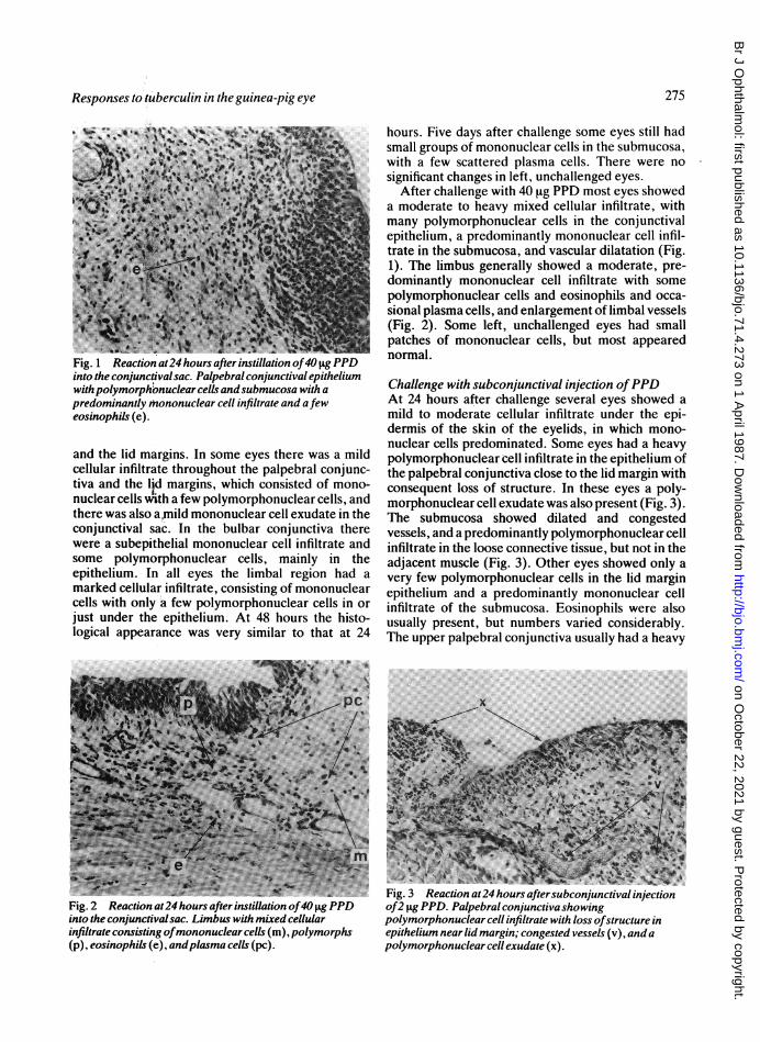
Fig. 1 Reaction at24 hours after instillation of40 jigPPDinto the conjunctival sac. Palpebral conjunctival epitheliumwithpolymorphbnuclear cells andsubmucosa with apredominantly Mnononuclear cell infiltrate and afeweosinophils (e).
and the lid margins. In some eyes there was a mildcellular infiltrate throughout the palpebral conjunc-tiva and the l~d margins, which consisted of mono-nuclear cells with a few polymorphonuclear cells, andthere was also atmild mononuclear cell exudate in theconjunctival sac. In the bulbar conjunctiva therewere a subepithelial mononuclear cell infiltrate andsome polymorphonuclear cells, mainly in theepithelium. In all eyes the limbal region had amarked cellular infiltrate, consisting of mononuclearcells with only a few polymorphonuclear cells in orjust under the epithelium. At 48 hours the histo-logical appearance was very similar to that at 24
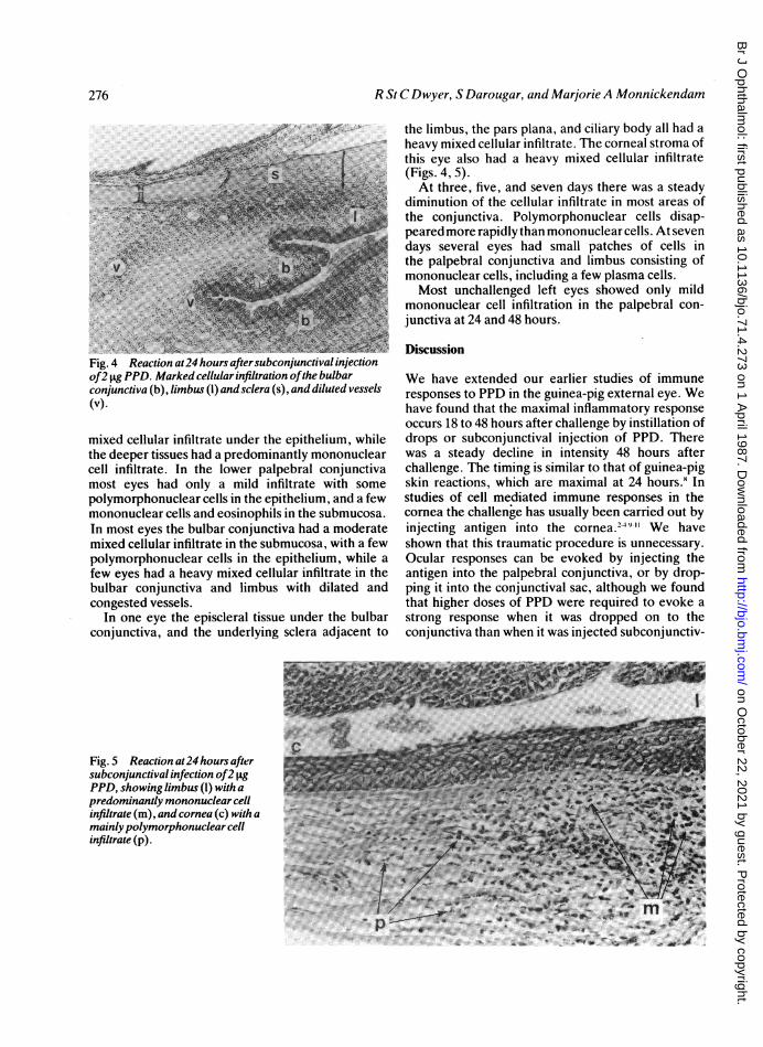
Fig. 2 Reaction at24 hours afterinstillation of40 tgPPDinto the conjunctival sac. Limbus with mixed cellularinfiltrate consisting ofmononuclear cells (m), polymorphs(p), eosinophils (e), andplasma cells (pc).
hours. Five days after challenge some eyes still hadsmall groups of mononuclear cells in the submucosa,with a few scattered plasma cells. There were nosignificant changes in left, unchallenged eyes.
After challenge with 40 ig PPD most eyes showeda moderate to heavy mixed cellular infiltrate, withmany polymorphonuclear cells in the conjunctivalepithelium, a predominantly mononuclear cell infil-trate in the submucosa, and vascular dilatation (Fig.1). The limbus generally showed a moderate, pre-dominantly mononuclear cell infiltrate with somepolymorphonuclear cells and eosinophils and occa-sional plasma cells, and enlargement of limbal vessels(Fig. 2). Some left, unchallenged eyes had smallpatches of mononuclear cells, but most appearednormal.
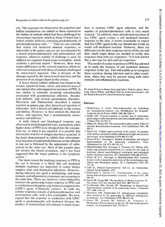
Challenge with subconjunctival injection ofPPDAt 24 hours after challenge several eyes showed amild to moderate cellular infiltrate under the epi-dermis of the skin of the eyelids, in which mono-nuclear cells predominated. Some eyes had a heavypolymorphonuclear cell infiltrate in the epithelium ofthe palpebral conjunctiva close to the lid margin withconsequent loss of structure. In these eyes a poly-morphonuclear cell exudate was also present (Fig. 3).The submucosa showed dilated and congestedvessels, and a predominantly polymorphonuclear cellinfiltrate in the loose connective tissue, but not in theadjacent muscle (Fig. 3). Other eyes showed only avery few polymorphonuclear cells in the lid marginepithelium and a predominantly mononuclear cellinfiltrate of the submucosa. Eosinophils were alsousually present, but numbers varied considerably.The upper palpebral conjunctiva usually had a heavy
'C
Fig. 3 Reaction at24 hours aftersubconjunctival injectionof2 Itg PPD. Palpebral conjunctivashowingpolymorphonuclear cell infiltrate with loss ofstructure inepithelium near lid margin; congested vessels (v), and apolymorphonuclear cell exudate (x).
275
on October 22, 2021 by guest. P
rotected by copyright.http://bjo.bm
j.com/
Br J O
phthalmol: first published as 10.1136/bjo.71.4.273 on 1 A
pril 1987. Dow
nloaded from
R St C Dwyer, S Darougar, and Marjorie A Monnickendam
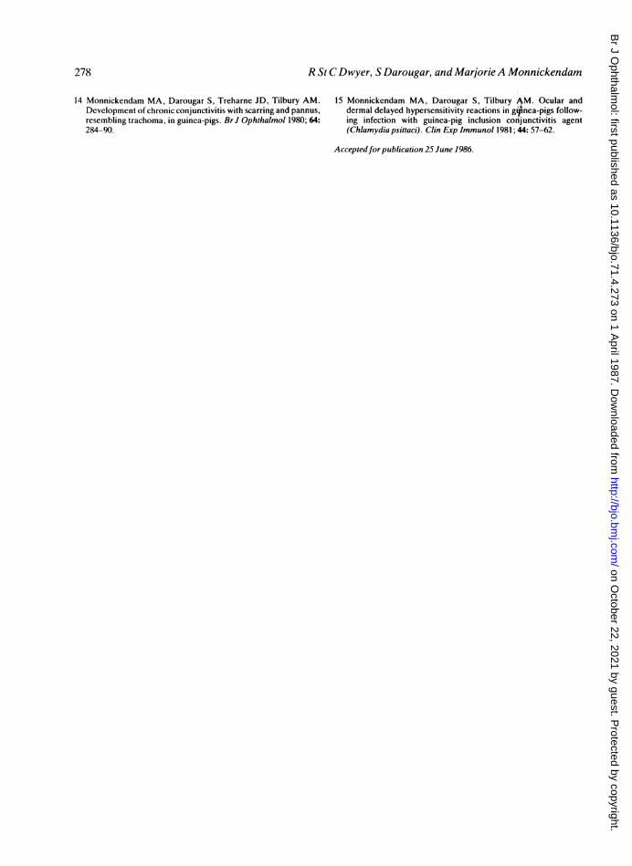
Fig. 4 Reaction at24 hours aftersubconjunctival injectionof2 pFg PPD. Marked cellularinfi Itration ofthe bulbarconjunctiva (b), limbus (1) and sclera (s), and diluted vessels(v).
mixed cellular infiltrate under the epithelium, whilethe deeper tissues had a predominantly mononuclearcell infiltrate. In the lower palpebral conjunctivamost eyes had only a mild infiltrate with somepolymorphonuclear cells in the epithelium, and a fewmononuclear cells and eosinophils in the submucosa.In most eyes the bulbar conjunctiva had a moderatemixed cellular infiltrate in the submucosa, with a fewpolymorphonuclear cells in the epithelium, while afew eyes had a heavy mixed cellular infiltrate in thebulbar conjunctiva and limbus with dilated andcongested vessels.
In one eye the episcleral tissue under the bulbarconjunctiva, and the underlying sclera adjacent to
the limbus, the pars plana, and ciliary body all had aheavy mixed cellular infiltrate. The corneal stroma ofthis eye also had a heavy mixed cellular infiltrate(Figs. 4, 5).At three, five, and seven days there was a steady
diminution of the cellular infiltrate in most areas ofthe conjunctiva. Polymorphonuclear cells disap-peared more rapidly than mononuclear cells. At sevendays several eyes had small patches of cells inthe palpebral conjunctiva and limbus consisting ofmononuclear cells, including a few plasma cells.Most unchallenged left eyes showed only mild
mononuclear cell infiltration in the palpebral con-junctiva at 24 and 48 hours.
Discussion
We have extended our earlier studies of immuneresponses to PPD in the guinea-pig external eye. Wehave found that the maximal inflammatory responseoccurs 18 to 48 hours after challenge by instillation ofdrops or subconjunctival injection of PPD. Therewas a steady decline in intensity 48 hours afterchallenge. The timing is similar to that of guinea-pigskin reactions, which are maximal at 24 hours8 Instudies of cell mediated immune responses in thecornea the challenge has usually been carried out byinjecting antigen into the cornea.- 9' We haveshown that this traumatic procedure is unnecessary.Ocular responses can be evoked by injecting theantigen into the palpebral conjunctiva, or by drop-ping it into the conjunctival sac, although we foundthat higher doses of PPD were required to evoke astrong response when it was dropped on to theconjunctiva than when it was injected subconjunctiv-
Fig. 5 Reaction at24 hours aftersubconjunctival infection of2 IgPPD, showing limbus (1) with apredominantly mononuclear cellinfiltrate (m), and cornea (c) with amainlypolymorphonuclear cellinfiltrate (p).
"W4 PT 6w ''''4,_4
276
on October 22, 2021 by guest. P
rotected by copyright.http://bjo.bm
j.com/
Br J O
phthalmol: first published as 10.1136/bjo.71.4.273 on 1 A
pril 1987. Dow
nloaded from
Responses to tmberculin in the guinea-pig eye
ally. The responses we observed in the palpebral andbulbar conjunctivae are similar to those reported inthe limbus of animals which had been challenged byintracorneal injection, which at about 24 hours had alimbal cellular infiltrate consisting predominantly ofpolymorphonuclear cells.' 4 " " Thus we can concludethat ocular cell mediated immune responses totuberculin in the guinea-pig eye are accompanied bya mixed polymorphonuclear and mononuclear cellresponse, whatever routine of challenge is used. Inaddition we regularly found some eosinophils, whichconfirms a previous report." However, there weremajor differences in the corneal responses which weobserved compared with those of animals challengedby intracorneal injection. This is because of thedamage caused by the intracorneal injection and thepresence of an antigen depot in the cornea.A heavy mixed cellular infiltrate was found in the
cornea, episcleral tissue, sclera, and anterior uvea ofone animal after subconjunctival injection of PPD. Itwas similar to naturally occurring sclerokeratitisassociated with granulomatous infection, rheuma-toid arthritis, and several autoimmune disorders.Silverstein and Zimmerman described a similarreaction in guinea-pigs after intravitreal injection oftuberculin, with a mixed cell infiltrate in the corneaand limbus. However, in their animals the choroid,sclera, and episclera had a predominantly mono-nuclear cell infiltrate.'A mild clinical and histological response was
observed in unchallenged left eyes, particularly whenhigher doses of PPD were dropped into the conjunc-tival sac, or when it was injected. It is possible thatinterocular transfer of antigen may have occurred. Ithas been demonstrated in rabbits that subconjunc-tival injection of radiolabelled human serum albuminin one eye is followed by the appearance of radio-activity in the other eye. Most of this transfer doesnot involve the blood circulation, and it has beensuggested that the major pathway is the lymphaticsystem. 13
Our main reason for studying responses to PPD inthe eye is because it is likely that cell mediatedimmune responses are important in ocular herpessimplex virus and chlamydial infections. However,during infection the agent is multiplying, and manyimmune and inflammatory responses are occurring atthe same time. There are, however, some similaritiesbetween the guinea-pig ocular response to PPD andto reinfection with guinea-pig inclusion conjunctivitis(GPIC) agent (Chlamydia psittaci). In both thecellular response consists of polymorphonuclear andmononuclear cells, with a few eosinophils. We haveargued that the response to reinfection with GPICagent is predominantly cell mediated because thenumber of mononuclear cells present is much larger
than in primary GPIC agent infection, and thenumber of polymorphonuclear cells is very muchreduced.'4 In addition, since intradermal injection oflive GPIC agent evokes a cell mediated immuneresponse in the skin, it seems reasonable to expectthat live GPIC agent in the eye should also evoke anocular cell mediated reaction. However, there aredifferences in the dose response curves of the eye andskin; much larger doses are needed to evoke skinresponses than with eye responses.'9 It is now knownthis is also true for skin and eye responses.
This model of ocular responses to PPD has allowedus to study the features of cell mediated immuneresponses in the eye. This will enable us to recognisethese reactions during infection and in other condi-tions, when they may be present along with otherimmune and inflammatory reactions.
We thank Professors Barrie Jones and John L Turk for advice, MaryLang, Eileen Wilson, and Maria Pink for technical assistance, andthe Medical Research Council for financial support.
References
1 Wolff-Eisner A. Ueber Tuberkulinvaseline zur Austellungder Konjunktival-reaktion, eine Modifikation der Konjunk-tivalprobe fur die Praxis. MMW 1909; 44: 2266-8.
2 Holley SW. Corneal reactions of normal and of tuberculousguinea-pigs to tuberculo-protein and tuberculophosphatide. AmJ Pathol 1935; 11: 937-51.
3 Milner JE, Weiser RS. The corneal tuberculin reaction. Effect ofsystemic desensitization treatment. Arch Ophthalmol 1965; 74:845-6.
4 Howes EL. Cellular hypersensitivity in the cornea. An analysisof the limbus and limbal cellular infiltration by light and electronmicroscopy. Arch Ophthalmol 197t); 83: 475-88.
5 Dwyer R St C, Darougar S. Models of immediate and delayedhypersensitivity in the guinea-pig conjunctiva. TransOphthalmol Soc UK 1971; 91: 451-61.
6 Monnickendam MA, Darougar S, Treharne JD, Tilbury AM.Guinea-pig inclusion conjunctivitis as a model for the study oftrachoma: clinical. microbiological, serological, and cytologicalstudies of primary infection. BrJ Ophthalmol 1980; 64: 279-83.
7 Dwyer R St C, Darougar S, Monnickendam MA. Unusualfeatures in the conjunctiva and cornea of the normal guinea-pig:clinical and histological studies. Br J Ophthalmol 1983; 67:737-41.
8 Turk JL. Delayed hypersensitivity. 2nd- rev. ed. Amsterdam,Oxford: North-Holland, 1975.
9 Schlossman S, Stetson CA. Vascularization of the corneaduring delayed hypersensitivity reactions. J Immunol 1957; 79:2)X8 1t.
10 Salvin SB, Gregg MB. The specificity of allergic reactions. IV.The cornea. Proc Soc Exp Biol Med 1961; 107: 478-85.
11 Friedlaender MH, Dvorak HF. Morphology of delayed-typehypersensitivity reactions in the guinea-pig cornea. J Immunol1977: 118: 1558-63.
12 Silverstein AM, Zimmerman LE. Immunogenic endophthal-mitis produced in the guinea-pig by different pathogeneticmechanisms. A]nJ Ophthalmol 1959; 48: 435-46.
13 Jenscn PR, Aronson SB, Pollycovc M, Yamamoto E.Mechanisms of host response in the eye. III. Interocular proteintransfer. Arch Ophthalmol 1967; 77: 814-7.
277
on October 22, 2021 by guest. P
rotected by copyright.http://bjo.bm
j.com/
Br J O
phthalmol: first published as 10.1136/bjo.71.4.273 on 1 A
pril 1987. Dow
nloaded from
R St C Dwyer, S Darougar, and MarjorieA Monnickendam
14 Monnickendam MA, Darougar S. Treharne JD, Tilbury AM.Development of chronic conjunctivitis with scarring and pannus,resembling trachoma, in guinea-pigs. Br J Ophthalmol 1980; 64:284-90.
15 Monnickendam MA, Darougar S. Tilbury AM. Ocular anddermal delayed hypersensitivity reactions in g4'inea-pigs follow-ing infection with guinea-pig inclusion conjunctivitis agent(Chlamydia psittaci). Clin Exp Immunol 1981; 44: 57-62.
Acceptedfor publication 25 June 1986.
278
on October 22, 2021 by guest. P
rotected by copyright.http://bjo.bm
j.com/
Br J O
phthalmol: first published as 10.1136/bjo.71.4.273 on 1 A
pril 1987. Dow
nloaded from