Embed Size (px)
Citation preview
ANTIMICROBIAL AGENTS AND CHEMOTHERAPY,0066-4804/01/$04.00�0 DOI: 10.1128/AAC.45.12.3409–3415.2001
Dec. 2001, p. 3409–3415 Vol. 45, No. 12
Copyright © 2001, American Society for Microbiology. All Rights Reserved.
In Vitro Activities of Two Antimitotic Compounds, Pancratistatinand 7-Deoxynarciclasine, against Encephalitozoon intestinalis,
a Microsporidium Causing Infections in HumansMERYEM OUARZANE-AMARA,1 JEAN-FRANCOIS FRANETICH,1 DOMINIQUE MAZIER,1
GEORGE R. PETTIT,2 LAURENT MEIJER,3 CHRISTIAN DOERIG,1
AND ISABELLE DESPORTES-LIVAGE1*
INSERM U511, Immunobiologie Cellulaire et Moleculaire des Infections Parasitaires, CHU Pitie-Salpetriere,75643 Paris Cedex 13,1 and Centre National de la Recherche Scientifique, Station Biologique,
29682 Roscoff Cedex,3 France, and Cancer Research Institute, Arizona State University,Tempe, Arizona 85287-16042
Received 9 April 2001/Returned for modification 4 June 2001/Accepted 31 August 2001
The antiparasitic effect of a collection of compounds with antimitotic activity has been tested on a mam-malian cell line infected with Encephalitozoon intestinalis, a microsporidian causing intestinal and systemicinfection in immunocompromised patients. The antiparasitic effect was evaluated by counting the number ofparasitophorous vacuoles detected by immunofluorescence. Out of 526 compounds tested, 2 (pancratistatinand 7-deoxynarciclasine) inhibited the infection without affecting the host cell. The 50% inhibitory concentra-tions (IC50s) of pancratistatin and 7-deoxynarciclasine for E. intestinalis were 0.18 �M and 0.2 �M, respec-tively, approximately eightfold lower than the IC50s of these same compounds against the host cells. Electronmicroscopy confirmed the gradual decrease in the number of parasitophorous vacuoles and showed that of thetwo life cycle phases, sporogony was more sensitive to the inhibitors than merogony. Furthermore, the per-sistence of meronts in some cells apparently devoid of sporonts and spores indicated that the inhibitors blockdevelopment rather than entry of the parasite into the host cell. The occurrence of binucleate sporoblasts andspores suggests that these inhibitors blocked a specific phase of cell division.
Microsporidia are widespread obligatory intracellular para-sites, apparently able to invade any cell in animals and humans(4, 16). These unicellular parasites have been increasingly rec-ognized as opportunistic pathogens of immunodeficient pa-tients (24). Two species cause diarrhea, malabsorption, andweight loss in AIDS patients (6). Enterocytozoon bieneusi is themost prevalent cause of these symptoms and is occasionallyassociated with hepatobiliary disease or infection of the upperrespiratory tract (24). The second prevalent species, Encepha-litozoon (Septata) intestinalis, is responsible for nephritis, bron-chitis, and lytic mandibular lesions (11, 16). Rational strategiesfor the development of chemotherapeutic agents against mi-crosporidia require a better understanding of the mechanismscontrolling the proliferation of these parasites.
The life cycle of Encephalitozoon intestinalis (Fig. 1) consistsof two successive developmental sequences, merogony andsporogony, both of which occur in a parasitophorous vacuole(PV) within host cells. During merogony, proliferative stagesknown as meronts are produced. After multiple divisions, mer-onts are transformed into sporonts. During sporogony, eachsporont divides into two sporoblasts, which mature into sporeswhich are approximately 2.0 by 1.2 �m. They contain a com-plex extrusion apparatus which ensures inoculation of the in-fective sporoplasm into a host cell. Meronts and sporonts aremononucleate cells which replicate by binary fission. Some-
times, the karyokinetic process is repeated before cytokinesisoccurs, resulting in a ribbon-like cell containing two to fournuclei. The production of tetranucleate meronts and sporontssuggests some variability in the timing of cytokinetic cycles inE. intestinalis (3). Sporoblasts and spores are exclusively mono-nucleate, an indication that the regulation of developmentmust be linked to the control of the cell cycle. Although verylittle is known about cell cycle control in microsporidia, a geneencoding a putative homologue of the cyclin-dependent kinase1 (CDK1) has been recently identified in Encephalitozoon cu-niculi (20), and a similar gene from E. intestinalis is beingcharacterized in our laboratory (unpublished data). CDKs aremajor players in the progression of the eukaryotic cell cycle.Their activity is regulated by their phosphorylation status andby the association with negative (cyclin kinase inhibitors) orpositive (cyclins) regulators, and by intracellular transloca-tions. The temporary association of kinase subunits with dif-ferent cyclins define time windows during which kinase activityis directed at distinct sets of substrates at the appropriatephase of the cell cycle.
Because of the importance of CDKs and their regulators inthe multiplication and development of eukaryotes, these en-zymes represent attractive potential targets for antiparasiticchemotherapy. The phylogenetic divergence between the par-asite and its host is likely to result in divergences in the struc-ture of their regulatory genes, as has been shown in otherparasite-host systems (9, 10, 15); such divergences might conferto parasite and host differential susceptibilities to a given in-hibitor. Therefore, we decided to evaluate the effect of a col-lection of antimitotic compounds (many but not all of which
* Corresponding author. Mailing address: INSERM Unit 511, CHUPitie-Salpetriere, 91 Bd de l’hopital, 75643 Paris Cedex 13, France.Phone: (33) 1 40 77 81 05. Fax: (33) 1 45 83 88 58. E-mail: [email protected].
3409

are CDK inhibitors) on the course of cellular infection by E.intestinalis.
MATERIALS AND METHODS
Inhibitors. Pancratistatin and 7-deoxynarciclasine were extracted and purifiedfrom Pancratium (Hymenocallis) littorale in Hawaii (17).
Parasites. E. intestinalis spores were collected from monolayers of rabbitkidney cells (RK13) as described by Van Gool et al. (22). Spores were harvestedevery 3 days, and suspensions of parasites were centrifuged at 350 � g for 5 minto eliminate cellular fragments. Spores were then pelleted by centrifugation at2,000 � g for 20 min and washed twice. Spores were counted with a Malassezslide and used immediately for infection of cultured cells.
Culturing of parasites and treatment. RK13 culture cells were cultivated inLab-Tek slides, in RPMI 1640 medium (Gibco BRL, Cergy Potoise, France)supplemented with 8% heat-inactivated fetal calf serum (56°C for 30 min) (Sig-ma, St. Quentin-Fallavier, France), streptomycin (100 �g/ml), penicillin (100U/ml) and L-glutamine (2 mM). Cells were adjusted to 105 cells/well and sporesfrom E. intestinalis were added to cultured cells at a ratio of 1 spore/10 cells. Thecompounds were added to cultured cells at infection time. The effects of thecompounds were determined by counting the PVs 48 h after infection andtreatment. The detection of PVs was performed by immunofluorescence assay(IFA).
IFA. The Lab-Tek slides were fixed in ethanol at �20°C for 10 min and thenincubated for 1 h at 37°C with the monoclonal antibody (MAb) M1.6C1.2C11 (1)at a dilution of 1/500. This MAb is directed against a coat protein of sporogonic
FIG. 1. Life cycle of E. intestinalis. Only the spores survive in theextracellular medium. The inoculation of the sporoplasm into the hostcell is the initial step of the intracellular development.
FIG. 2. Structures of pancratistatin and 7-deoxynarciclasine.
FIG. 3. (a) In vitro effect of inhibitors on E. intestinalis multi-plication. RK13 cells infected with E. intestinalis spores were incu-bated for 48 h, in the presence of various concentrations of inhib-itors. The number of PVs was determined by IFA. The datarepresent means � standard deviations (error bars) of triplicatecultures. �, significant difference between the PV number obtainedin treated culture and nontreated culture, as determined by Stu-dent’s test (P � 0.05). (b) In vitro effect of inhibitors on host cells(RK13). Microculture plates prepared with RK13 cells were treatedwith six concentrations of inhibitors. One microcurie of [3H]thymi-dine (5 Ci/mmol) was added to each well. Plates were incubated for48 h, and [3H]thymidine incorporation was measured. The datarepresent means � standard deviations (error bars) of triplicatecultures. �, significant difference between the 3H incorporation bythe treated culture and nontreated culture, as determined by Stu-dent’s test (P � 0.05).
3410 OUARZANE-AMARA ET AL. ANTIMICROB. AGENTS CHEMOTHER.
stages generated in the PVs. The slides were then washed in phosphate-bufferedsaline (PBS) and incubated with fluorescein-conjugated anti-mouse immuno-globulin G, diluted to 1/100 in Evans blue (1/1,000). After several washes withPBS, the slides were mounted in PBS-glycerol (50:50, vol/vol), and PVs werecounted using an epifluorescence microscope. The entire surface of each wellwas examined at a magnification of �500.
Determination of IC50s on parasite. The concentration of inhibitor required toinhibit parasite growth by 50% (IC50) was determined by IFA. The data wereplotted and the IC50s were determined using Cricket Graph software.
Determination of IC50s on host cells. Microculture plates (96-well flat-bottomplates; Falcon) were prepared with RK13 cells. Six concentrations of each in-hibitor were tested in triplicate. One microcurie of [3H] thymidine (5 Ci/mmol)was added to each well. Plates were incubated for 48 h and [3H]thymidineincorporation was measured with a scintillation counter (Beckman). The datawere plotted and the IC50s were determined using Cricket Graph software.
Electron microscopy. The effect of inhibitors on the morphology of E. intes-tinalis and RK13 cells was examined by electron microscopy. Infected monolay-ers were fixed at 48 h postinfection in 2.5% glutaraldehyde in 0.1 M Na caco-dylate buffer (pH 7.2) for 1 h. They were rinsed in the same buffer and postfixedin ferriosmium [OsO4 and K3Fe(CN)6 (1%, wt/vol) in cacodylate buffer] for 1 hat room temperature. After ethanolic dehydration, the samples were embeddedin Spurr’s resin. Thin sections were stained with uranyl acetate and lead citrateand then examined with a JEOL TEM 100CX transmission electron microscope.
RESULTS
Effect of inhibitors on parasite growth. RK13 cells infectedwith E. intestinalis spores were incubated for 48 h in the pres-ence of various concentrations of compounds. Since the stocksof inhibitors were dissolved in dimethyl sulfoxide (DMSO),controls were run simultaneously with the highest concentra-tion of DMSO (0.03 �l/ml) without inhibitor. The effect of theinhibitors on parasite multiplication was evaluated from thenumber of PVs detected by IFA. Microsporidia are fast-grow-ing organisms, and 48 h postinfection, PVs were detectable byIFA due to the occurrence of sporogonic stages cross-reactingwith the MAb.
In the first round of screening, 526 compounds were testedat concentrations of 2 and 5 �M. As expected, most com-pounds were toxic to the host cells and caused cytopathiceffects. The 51 compounds that showed little or no cytopathiceffect on the host cells were tested again at a concentrationrange of 0.1 to 5 �M for antiparasitic effect. This allowed us toselect two structurally related molecules with a definite effecton the number of PVs but with no apparent effect on the hostcells: pancratistatin and 7-deoxynarciclasine (Fig. 2). IC50s ofpancratistatin and 7-deoxynarciclasine, determined using arange of concentrations between 0.01 and 3 �M (Fig. 3a),were, respectively, 0.18 and 0.2 �M. No parasite growth couldbe detected at the highest pancratistatin concentration (3 �M).Low concentrations of this inhibitor (0.01 �M) also signifi-cantly reduced the parasite development. Likewise, in infectedcultures treated with 7-deoxynarciclasine, the number of par-asites decreased markedly at higher concentrations. Thus, pan-cratistatin and 7-deoxynarciclasine showed very good antipar-asitic activity.
Host cell IC50s. To determine whether any of these twomolecules may represent a useful lead compound, we nextmeasured their effect on host cells in a standard [3H]thymidineincorporation assay. RK13 cells were treated with the twoinhibitors (concentrations ranging from 0.01 to 10 �M), andincorporated radiolabel was then measured (see Materials andMethods). Again, cells treated with DMSO diluted in RPMIand RPMI alone were used as controls. The IC50s of pancra-tistatin and 7-deoxynarciclasine obtained from the graphs (Fig.3b) were both 1.5 �M and thus were 8- and 7.5-fold higherthan the parasite IC50s, respectively.
Morphological effects of the inhibitors. Examination of in-fected cultures by electron microscopy confirmed that both
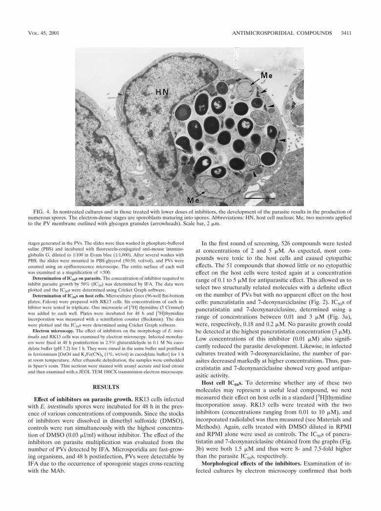
FIG. 4. In nontreated cultures and in those treated with lower doses of inhibitors, the development of the parasite results in the production ofnumerous spores. The electron-dense stages are sporoblasts maturing into spores. Abbreviations: HN, host cell nucleus; Me, two meronts appliedto the PV membrane outlined with glycogen granules (arrowheads). Scale bar, 2 �m.
VOL. 45, 2001 ANTIMICROSPORIDIAL COMPOUNDS 3411
compounds caused a gradual resorption of the infection. Incultures treated with the lower doses, most cells were infected.Normally fusiform, these cells became rounded due to thepresence of a large PV (Fig. 4). Two or three PVs could beseen in some cells. All developmental stages of the parasitewere present in these vacuoles, which contained up to 20spores. At a concentration of 0.5 �M, flat cells containing smallPVs with a concomitant decrease in the number of maturespores were observed. In some cells, PVs contained meronts aswell as a small number of sporoblasts and spores that werelarger (3 to 4 �m) than those produced in the absence ofinhibitors. Additionally, some alterations were observed in the
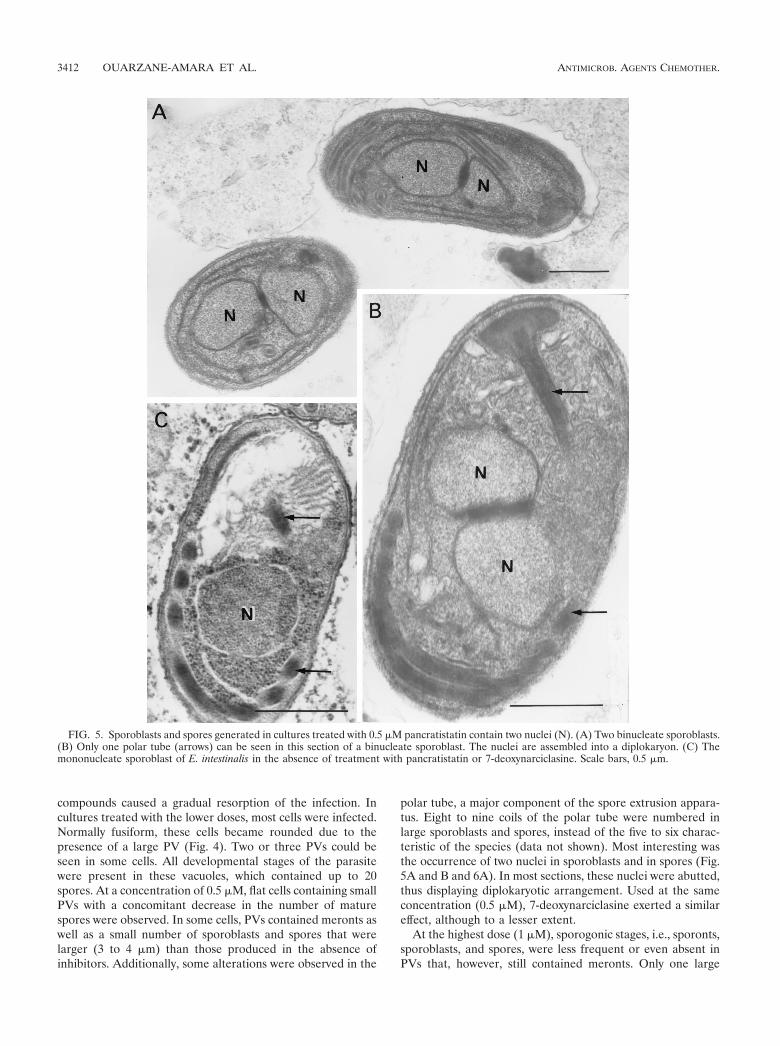
polar tube, a major component of the spore extrusion appara-tus. Eight to nine coils of the polar tube were numbered inlarge sporoblasts and spores, instead of the five to six charac-teristic of the species (data not shown). Most interesting wasthe occurrence of two nuclei in sporoblasts and in spores (Fig.5A and B and 6A). In most sections, these nuclei were abutted,thus displaying diplokaryotic arrangement. Used at the sameconcentration (0.5 �M), 7-deoxynarciclasine exerted a similareffect, although to a lesser extent.
At the highest dose (1 �M), sporogonic stages, i.e., sporonts,sporoblasts, and spores, were less frequent or even absent inPVs that, however, still contained meronts. Only one large
FIG. 5. Sporoblasts and spores generated in cultures treated with 0.5 �M pancratistatin contain two nuclei (N). (A) Two binucleate sporoblasts.(B) Only one polar tube (arrows) can be seen in this section of a binucleate sporoblast. The nuclei are assembled into a diplokaryon. (C) Themononucleate sporoblast of E. intestinalis in the absence of treatment with pancratistatin or 7-deoxynarciclasine. Scale bars, 0.5 �m.
3412 OUARZANE-AMARA ET AL. ANTIMICROB. AGENTS CHEMOTHER.
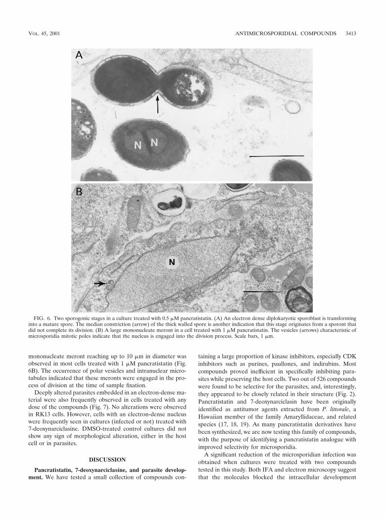
mononucleate meront reaching up to 10 �m in diameter wasobserved in most cells treated with 1 �M pancratistatin (Fig.6B). The occurrence of polar vesicles and intranuclear micro-tubules indicated that these meronts were engaged in the pro-cess of division at the time of sample fixation.
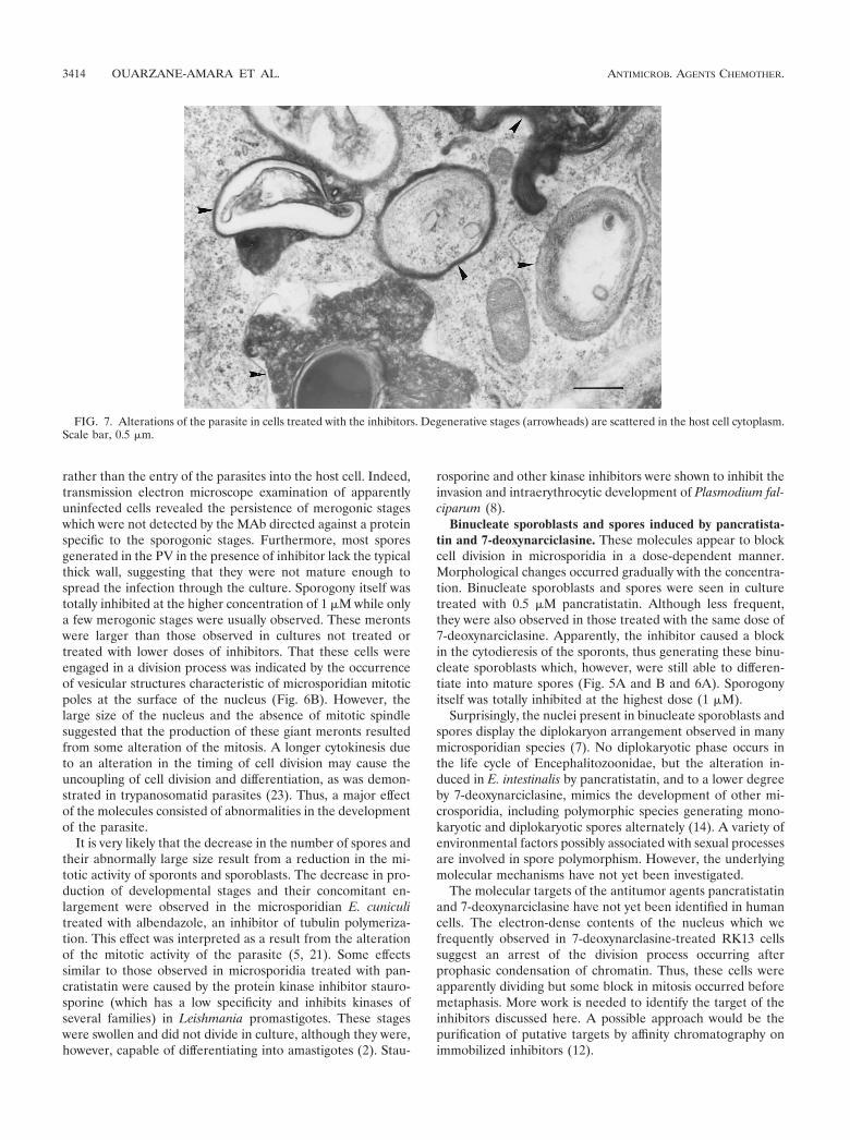
Deeply altered parasites embedded in an electron-dense ma-terial were also frequently observed in cells treated with anydose of the compounds (Fig. 7). No alterations were observedin RK13 cells. However, cells with an electron-dense nucleuswere frequently seen in cultures (infected or not) treated with7-deoxynarciclasine. DMSO-treated control cultures did notshow any sign of morphological alteration, either in the hostcell or in parasites.
DISCUSSION
Pancratistatin, 7-deoxynarciclasine, and parasite develop-ment. We have tested a small collection of compounds con-
taining a large proportion of kinase inhibitors, especially CDKinhibitors such as purines, paullones, and indirubins. Mostcompounds proved inefficient in specifically inhibiting para-sites while preserving the host cells. Two out of 526 compoundswere found to be selective for the parasites, and, interestingly,they appeared to be closely related in their structure (Fig. 2).Pancratistatin and 7-deoxynarciclasin have been originallyidentified as antitumor agents extracted from P. littorale, aHawaiian member of the family Amaryllidaceae, and relatedspecies (17, 18, 19). As many pancratistatin derivatives havebeen synthesized, we are now testing this family of compounds,with the purpose of identifying a pancratistatin analogue withimproved selectivity for microsporidia.
A significant reduction of the microsporidian infection wasobtained when cultures were treated with two compoundstested in this study. Both IFA and electron microscopy suggestthat the molecules blocked the intracellular development
FIG. 6. Two sporogonic stages in a culture treated with 0.5 �M pancratistatin. (A) An electron dense diplokaryotic sporoblast is transforminginto a mature spore. The median constriction (arrow) of the thick walled spore is another indication that this stage originates from a sporont thatdid not complete its division. (B) A large mononucleate meront in a cell treated with 1 �M pancratistatin. The vesicles (arrows) characteristic ofmicrosporidia mitotic poles indicate that the nucleus is engaged into the division process. Scale bars, 1 �m.
VOL. 45, 2001 ANTIMICROSPORIDIAL COMPOUNDS 3413
rather than the entry of the parasites into the host cell. Indeed,transmission electron microscope examination of apparentlyuninfected cells revealed the persistence of merogonic stageswhich were not detected by the MAb directed against a proteinspecific to the sporogonic stages. Furthermore, most sporesgenerated in the PV in the presence of inhibitor lack the typicalthick wall, suggesting that they were not mature enough tospread the infection through the culture. Sporogony itself wastotally inhibited at the higher concentration of 1 �M while onlya few merogonic stages were usually observed. These merontswere larger than those observed in cultures not treated ortreated with lower doses of inhibitors. That these cells wereengaged in a division process was indicated by the occurrenceof vesicular structures characteristic of microsporidian mitoticpoles at the surface of the nucleus (Fig. 6B). However, thelarge size of the nucleus and the absence of mitotic spindlesuggested that the production of these giant meronts resultedfrom some alteration of the mitosis. A longer cytokinesis dueto an alteration in the timing of cell division may cause theuncoupling of cell division and differentiation, as was demon-strated in trypanosomatid parasites (23). Thus, a major effectof the molecules consisted of abnormalities in the developmentof the parasite.
It is very likely that the decrease in the number of spores andtheir abnormally large size result from a reduction in the mi-totic activity of sporonts and sporoblasts. The decrease in pro-duction of developmental stages and their concomitant en-largement were observed in the microsporidian E. cuniculitreated with albendazole, an inhibitor of tubulin polymeriza-tion. This effect was interpreted as a result from the alterationof the mitotic activity of the parasite (5, 21). Some effectssimilar to those observed in microsporidia treated with pan-cratistatin were caused by the protein kinase inhibitor stauro-sporine (which has a low specificity and inhibits kinases ofseveral families) in Leishmania promastigotes. These stageswere swollen and did not divide in culture, although they were,however, capable of differentiating into amastigotes (2). Stau-
rosporine and other kinase inhibitors were shown to inhibit theinvasion and intraerythrocytic development of Plasmodium fal-ciparum (8).
Binucleate sporoblasts and spores induced by pancratista-tin and 7-deoxynarciclasine. These molecules appear to blockcell division in microsporidia in a dose-dependent manner.Morphological changes occurred gradually with the concentra-tion. Binucleate sporoblasts and spores were seen in culturetreated with 0.5 �M pancratistatin. Although less frequent,they were also observed in those treated with the same dose of7-deoxynarciclasine. Apparently, the inhibitor caused a blockin the cytodieresis of the sporonts, thus generating these binu-cleate sporoblasts which, however, were still able to differen-tiate into mature spores (Fig. 5A and B and 6A). Sporogonyitself was totally inhibited at the highest dose (1 �M).
Surprisingly, the nuclei present in binucleate sporoblasts andspores display the diplokaryon arrangement observed in manymicrosporidian species (7). No diplokaryotic phase occurs inthe life cycle of Encephalitozoonidae, but the alteration in-duced in E. intestinalis by pancratistatin, and to a lower degreeby 7-deoxynarciclasine, mimics the development of other mi-crosporidia, including polymorphic species generating mono-karyotic and diplokaryotic spores alternately (14). A variety ofenvironmental factors possibly associated with sexual processesare involved in spore polymorphism. However, the underlyingmolecular mechanisms have not yet been investigated.
The molecular targets of the antitumor agents pancratistatinand 7-deoxynarciclasine have not yet been identified in humancells. The electron-dense contents of the nucleus which wefrequently observed in 7-deoxynarclasine-treated RK13 cellssuggest an arrest of the division process occurring afterprophasic condensation of chromatin. Thus, these cells wereapparently dividing but some block in mitosis occurred beforemetaphasis. More work is needed to identify the target of theinhibitors discussed here. A possible approach would be thepurification of putative targets by affinity chromatography onimmobilized inhibitors (12).
FIG. 7. Alterations of the parasite in cells treated with the inhibitors. Degenerative stages (arrowheads) are scattered in the host cell cytoplasm.Scale bar, 0.5 �m.
3414 OUARZANE-AMARA ET AL. ANTIMICROB. AGENTS CHEMOTHER.
The recent classification of microsporidia with the fungi (25)suggests that the different phases of their cell cycle may becontrolled by a single CDK, as is the case in yeasts (13).However, some of the results reported here tend to indicatethat a combination of different factors and effectors ensuresthe regulation of the life cycle in microsporidia. Thus, theinformation provided by this study is of dual interest: on onehand it points to new potential therapeutic tools which cer-tainly deserve further characterization (notably, in terms oftheir effect during infection of animals), and on the other handthese molecules represent promising tools for investigating thediversity of the microsporidian life cycles.
ACKNOWLEDGMENTS
This study was supported by a grant from the Fondation pour laRecherche Medicale (SIDACTION). Meryem Ouarzane-Amara is therecipient of a fellowship from the Fondation pour la Recherche Medi-cale. Work in the laboratories of C.D. and L.M. is supported by theFrench Ministry of Research (PRFMMIP program) and the Ministryof Defence (Delegation Generale pour L’Armement).
We thank Jean-Jaques Hauw (Laboratoire Escourolle, Hopital de laPitie-Salpetriere) for providing electron microscopy facilities andElaine Giboyau for technical assistance.
REFERENCES
1. Achbarou, A., M. Thellier, I. Accoceberry, O. Prigneau, N. Bouladoux, A.Datry, D. Mazier, and I. Desportes-Livage. 1999. Production of immunolog-ical probes raised against Enterocytozoon bieneusi and Encephalitozoon in-testinalis, two microsporidian species causing intestinal infections in mice.J. Eukaryot. Microbiol. 46:32S–33S.
2. Becker, S., and C. L. Jaffe. 1997. Effect of protein kinase inhibitors on thegrowth, morphology, and infectivity of Leishmania promastigotes. Parasitol.Res. 83:273–280.
3. Cali, A., D. P. Kotler, and J. M. Orenstein. 1993. Septata intestinalis N.G., N.Sp, an intestinal microsporidian associated with chronic diarrhea and dis-semination in AIDS patients. J. Eukaryot. Microbiol. 40:101–112.
4. Canning, E. U., and J. Lom. 1986. The microsporidia of vertebrates. Aca-demic Press, London, United Kingdom.
5. Colbourn, N. I., W. S. Hollister, A. Curry, and E. U. Canning. 1994. Activityof albendazole against Encephalitozoon cuniculi in vitro. Eur. J. Protistol.30:211–220.
6. Coyle, C. M., M. Wittner, D. P. Kotler, C. Noyer, J. M. Orenstein, H. B.Tanowitz, and L. M. Weiss. 1996. Prevalence of microsporidiosis due toEnterocytozoon bieneusi and Encephalitozoon (Septata) intestinalis amongpatients with AIDS-related diarrhea: determination by polymerase chainreaction to the microsporidian small-subunit rRNA gene. Clin. Infect. Dis.23:1002–1006.
7. Desportes-Livage, I. 2000. Biology of microsporidia. Contrib. Microbiol.6:140–165.
8. Dluzewski, A. R., and C. R. Garcia. 1996. Inhibition of invasion and intra-erythrocytic development of Plasmodium falciparum by kinase inhibitors.Experientia 52:621–623.
9. Doerig, C., C. Doerig, P. Horrocks, J. Coyle, J. Carlton, A. Sultan, D. Arnot,and R. Carter. 1995. Pfcrk-1, a developmentally regulated cdc2-related pro-tein kinase of Plasmodium falciparum. Mol. Biochem. Parasitol. 70:167–174.
10. Graeser, R., B. Wernli, R. M. Franklin, and B. Kappes. 1996. Plasmodiumfalciparum protein kinase 5 and the malarial nuclear division cycles. Mol.Biochem. Parasitol. 82:37–49.
11. Hartskeerl, R. A., T. Van Gool, A. R. J. Schuitema, E. S. Didier, and W. J.Terpstra. 1995. Genetic and immunological characterization of the micro-sporidian Septata intestinalis Cali, Kotler and Orenstein, 1993: reclassifica-tion to Encephalitozoon intestinalis. Parasitology 110:277–285.
12. Knockaert, M., N. Gray, E. Damiens, Y.-T. Chang, P. Grant, D. Fergusson,J. Mottram, M. Soete, J.-F. Dubremetz, K. Le Roch, C. Doerig, P. G. Schultz,and L. Meijer. 2000. Intracellular targets of cyclin-dependent kinase inhib-itors: identification by affinity chromatography using immobilised inhibitors.Chem. Biol. 7:411–422.
13. Lee, M. G., and P. Nurse. 1987. Complementation used to clone a humanhomologue of the fission yeast control gene cdc2. Nature 327:31–35.
14. Micieli, M. V., J. J. Garcia, and J. J. Becnel. 2000. Life cycle and descriptionof Amblyospora camposi n. sp. (Microsporidia: Amblyosporidae) in the mos-quito Culex renatoi (Diptera, Culicidae) and the copepod Paracyclops fim-briatus fimbriatus (Copepoda, Cyclopidae). J. Eukaryot. Microbiol. 47:575–580.
15. Mottram, J. C. 1994. cdc2-related protein kinases and cell cycle control intrypanosomatids. Parasitol. Today 10:253–257.
16. Orenstein, J. M., H. P. Gaetz, A. T. Yachnis, S. S. Frankel, R. B. Mertens,and E. S. Didier. 1997. Disseminated microsporidiosis in AIDS: are anyorgans spared? AIDS 11:385–386.
17. Pettit, G. R., V. Gaddamidi, D. L. Herald, S. B. Singh, G. M. Cragg, J. M.Schmidt, F. E. Boettner, M. Williams, and Y. Sagawa. 1986. Antineoplasticagents, 120. Pancratium littorale. J. Nat. Prod. 49:995–1002.
18. Pettit, G. R., R. A. Backhaus, M. R. Boyd, and A. W. Meerow. 1993. Anti-neoplastic agents, 256. Cell growth inhibitory isocarbostyrils from Hymeno-callis. J. Nat. Prod. 56:1682–1687.
19. Pettit, G. R., G. Groszek, R. A. Backhaus, D. L. Doubek, R. J. Barr, andA. W. Meerow. 1995. Antineoplastic agents, 301. An investigation of theAmaryllidaceae genus Hymenocallis. J. Nat. Prod. 58:756–759.
20. Peyreteillade, E., P. Peyret, G. Metenier, and C. P. Vivares. 1999. Isolationof a highly divergent gene encoding CDC2-related protein kinase fromEncephalitozoon cuniculi (Microspora). J. Eukaryot. Microbiol. 46:19S–20S.
21. Shadduck, J. A. 1980. Effect of fumagillin on in vitro multiplication ofEncephalitozoon cuniculi. J. Protozool. 27:202–208.
22. Van Gool, T., E. U. Canning, H. Gilis, M. A. Van den Bergh Weerman,J. K. M. Eeftinck Schattenkerk, and J. Dankert. 1994. Septata intestinalisfrequently isolated from stool of AIDS patients with a new cultivationmethod. Parasitology 109:281–289.
23. Wang, Y., K. Dimitrov, L. K. Garrity, S. Sazer, and S. M. Beverley. 1998.Stage-specific activity of the Leishmania major CRK3 kinase and functionalrescue of Schizosaccharomyces pombe cdc2 mutant. Mol. Biochem. Parasitol.96:139–150.
24. Weber, R., and R. T. Bryan. 1994. Microsporidial infections in immunode-ficient and immunocompetent patients. Clin. Infect. Dis. 19:517–521.
25. Weiss, L. M., T. D. Edlind, C. R. Vossbrinck, and T. Hashimoto. 1999.Microsporidian molecular phylogeny: the fungal connexion. J. Eukaryot.Microbiol. 46:17S–18S.
VOL. 45, 2001 ANTIMICROSPORIDIAL COMPOUNDS 3415