Embed Size (px)
Citation preview
Microsporidium rhabdophilum sp. n. (Microsporida : Pansporoblastina),
a parasite of the nematode, Rhabditis myriophila (Rhabditina : Rhabditidae)
George 0. POINAB, Jr. and Roberta HESS
Division of Entomology and Parasitology, University of Califomia, Berkeley, Ca 94720, USA.
Microsporidium rhabdophilum sp. n. (Microsporida : Pansporoblastina) is described from the microbotrophic nematode Rhabditis mny-iophila Poinar. This microsporidian normally infects the hypodermal and reproductive tissues of the nematode. The parasite possesses small uninucleate, bacilliform spores produced in pansporoblasts. The number of spores per pansporoblast is norrmally 8 or 16 but cari range from 4 to 25. The developmental stages of the parasite are examined ultrastructurally.
RESUME
Microsporidium rhabdophilum sp. n. (Microsporida : Pansporoblastinal parasite des nématodes Rbabditis myriophila (Rhabdita : Rhabditidael
Microsporidium rhabdophilwn sp. n. (Microsporida : Pansporoblastina), provenant des nématodes bacilliphages Rhabditis myriophila Poinar, est décrit. Cette microsporidie infecte normalement l’hypoderme et les tissus de l’appareil reproducteur du nématode. Le parasite posséde de petites spores bacilliformes, uninucléées, produites dans des pansporoblastes. Le nombre de spores par pansporoblaste est normalement de 8 ou 16, mais peut varier de 4 à 25. L’ultrastructure des différents stades de développement du parasite a été étudiée.
During routine studies on the development of Rhabditis myriophila Poinar, 1986, individuals suffering from a microsporidan infection were discovered. A detailed examination of the parasites showed them to differ from previous microsporidans and they are described as a new species. Since the systematics of the Microsporida are in a state of flux and the present parasites of R. myriophilu do not fa11 into an existing genus, placement in the collective group Micro- sporidium was considered appropriate at this time.
Materials and methods
The nematode, Rhabditis ntyriophila was originally recovered from the intestinal lumen and body cavity of the garden millipede, Oxidis grucilis collected in Southern California (Poinar, 1986). This protandric hermaphrodite was cultivated at 20’ on nutrient agar plates seeded with bacteria associated with the millipede. Light microscope observations of infections in living and squashed nematodes were made by placing specimens in 1 0’0 saline and examining them under
bright fïeld and differential interference contrast microscopy.
For electron microscope studies, nematodes which had their heads and tails removed were immersed in 2.5 9/0 phosphate-buffered (0.1 M, pH 7.2) glutaraldehyde for one hour, dehydrated and embedded in Araldite 6005. Sections were stained with saturated aqueous uranyl acetate followed by lead citrate and examined in a Philips EM 300 electron microscope.
Microspot-idium rhabdophilum sp. n. (Figs l-3)
Microspora Sprague, 1977; Microsporea Delphy, 1936; Microsporida Balbiani, 1882, Pansporoblastina
Tuzet et cd., 1971.
DESCRIPTION
Spores bacilliform, tubular, straight or slightly curved; 1.28-2.56 w long by 0.6-1.0 pm wide (F?g. 1 48. Polar filament uniform in diameter. Merogony not
Revue Nématol., 9 (4) : 369-375 (I986) 369
G. 0. Poinar, Jr. & R. Hess
Fig. 1. Spores of the microsporidian, Microsporidium rhabdophilutn n. sp. from the nematode, Rhabditis myriophila. CI : Region of the basa1 pharyngeal bulb showing pansporoblasts with manu-e spores in the hypodermis and spores in the pharyngeal gland ce11 (arrow) (Mag. same as 1 b); b : Dauer juvenile with spores in paired sacs adjacent to the basa1 bulb; c : Spores in pansporoblasts maturing in the wall of the uterus and oviduct (Mag. same as 1 b); d : Pansporoblasts and spores removed from the hypodermal tissue (Mag. same as 1 ./); e :Tip of ovotestis infected with spores (Mag. same as 1 b); f : Isolated spores showing typical bacilliform shape.
370 Revue Nématol., 9 (4) : 369-37.5 (1986)
Microsporidium rhabdophilum sp. n., parasite of Rhabditis myriophila
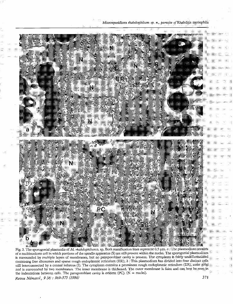
a^^ ,2./. Fig. 2. The sporogonial plasmodia of M. rhabdophilum n. sp. Both manification lines represent 0.5 km. a :The plasmodium consists of a multinucleate ce11 in which portions of the spindle apparatus (S) are still present within the nuclei. The sporogonial plasmodium is surrounded by multiple layers of membranes, but no pansporoblast cavity is present. The cytoplasm is fairly undifferentiated containing free ribosomes and sparse rough endoplasmic reticulum (ER); b : This plasmodium has divided into four distinct cells still interconnected by a central isthmus (1). The cytoplasm contains a prominent rough endoplasmic reticulum (ER), some golgi and is surrounded by two membranes. The inner membrane is thickened. The outer membrane is faint and cari best be seen in the indentations between cells. The pansporoblast cavity is evident (PC). (N = nuclei). Revue Nématol., 9 (4) : 369-375 (1986. 371
G. 0. Poinar, Jr. & R. Hess
observed. Sporoblasts uninucleate, formed within a semi-persistent to persistent pansporoblastic membrane. Spore number per pansporoblast variable, ranging from 4 to 25, but most frequently 8 or 16. Sporogonial plasmodia (the earliest stages observed) undergo multiple fission, during which a rosette stage occurs (Fig. 1 a, b).
TYPE HOST
Rhabditis myriophila Poinar, 1986 (Rhabditida : Rhabditidae).
TYPE LOCALITY
Infected nematodes were recovered from garden millipedes, Oxidis gracilis collected from Azusa, California.
LIGHTMICROSCOPE OBSERVATIONS
Infected hermaphrodites of R. myriophila were recovered from 12 day old nutrient cultures at a ratio of one diseased to twenty apparently healthy individuals. The most obvious sign of microsporidan infection was pansporoblasts containing mature spores in various nematode tissues. In hermaphrodites, developing spores were observed in the pharyngeal glands and hypodcrmis, especially the tissue surrounding the basa1 bulb (Fig. 1 a), the wall of the uterus, oviduct (Fig. 1 c) and ovotestis (Fig. 1 e). The hypodermis and ovotestis were the tissues most commonly infected. Occasionally, spores could be found in the intestinal ceIls.
Dauer juveniles found on exhausted nutrient agar plates sometimes contained spores in a pair of pouches on either side of the collapsed basa1 pharyngeal bulb (Fig. 1 b). Whether these pouches represent nervous tissue or expanded pharyngeal gland cells that have hypertrophied as a result of the infection is not known.
Heavy infections involving the entire reproductive system would result in sterility. In such individuals, eggs were lacking. Mortality of R. rhabdophila occurred when the hypodermis of hermaphrodites and third and fourth stage juveniles was infected throughout.
ELECTKONMICROSCOPEOBSERVATIONS
Meronts with a single limiting unit membrane were not observed. Uninucleate sporonts surrounded by two membranes were observed in the cytoplasm of infected cells. The cytoplasm was largely undifferentiated and only free ribosomes and some rough endoplasmic reticulum were observed. The host ccl1 membranes were closely appressed to the surface of the sporonts. Plasmodia were observed with two to five nuclei in the plane of sectioning (Fig. 2 a). The nuclei frequently had portions of the spindle apparatus connected to the outer nuclear membrane. Microtubules were present in the
312
nucleoplasm as wcll as dense patches of nuclear material considered to be chromosomal. The cytoplasm contained rough endoplasmic reticulum as well as somc membrane-bound packets of cytoplasm. Small vacuoles were occasionally observed. At this stage the host ce11 was still closely appresscd to the outer surface but additional membranes were present between the host and the plasmodium. In later stages of division, the inner membrane of the sporogonial plasmodium had increased in electron density and rosettes with individual cells were visible within a cavity (Fig. 2 b) bounded by the pansporoblast membrane. Division of the plasmodium seemed to be equal from a central isthmus and a maximum of four connected cells were noted in any one plane of sectioning. However, many pansporoblasts appeared to contain additional cells either previously separated or in some cases still connected to another ce11 in the process of dividing. Fully scparated sporoblasts wcre observed within a well-defined pansporoblastic cavity. The maximum number of sporoblasts found in the plane of sectioning was eight. These sporoblasts were in various stages of morphogenesis (Fig. 3 a, b). Newly derived sporoblasts were ovoid and contained more dilated rough endoplasmic reticulum. Nuclei were lobulatc. Patches of Golgi vesicles were present as well as vacuoles. The inner surface membrane was thickened, uniformly dense and in some instances the presence of two membranes was suggested. An outer fine membrane was present. In contrast to the complete separation of sporoblasts just described, in some sporoblasts maturation appcared to progress while the individual cells were still interconnected. In thesc cases it was diffïcult to accurately Count the number of sporoblasts, but more than eight portions of sporoblasts were frequently present within a pansporoblast cavity. The maturing cells, both thosc that finishcd division and those still interconnected contained profiles of developing polar filaments, polar caps, polaroplasts and posterior vacuoles. The polar filament was observed in clear cytoplasmic areas around the nucleus. Three to four cross-section profiles could be observed on either side of the nucleus (Fig. 3 b). Al1 cells were separated before elongation occurred. Elongation of thc sporoblast into a bacilliform shape appeared to be associated with the positioning of the polar cap (anchoring disc and polar aperature) and polaroplast at the anterior end of the ce11 (Fig. 3 a, b). The posterior vacuole was retained in the opposite end of the ce11 and the nucleus maintained a central position.
As spore maturation continued, the cytoplasm of the ce11 became dense. Ribosomes incrcased in numbers and became arranged in tight spirals around the outer portion of the ce11 in the arca below the polaroplast (Fig. 3 c). The polar cap was locatcd beneath the multiple layers of surface membranes and consisted of a dense outer layer, thc anchoring disc surrounding a cleared inner area and the polar aperture. The
Revue Nématol., 9 (4) : 369-375 (1986)
Microsporidium rhabdophilum sp. n., parasite ofRhabditis myriophila
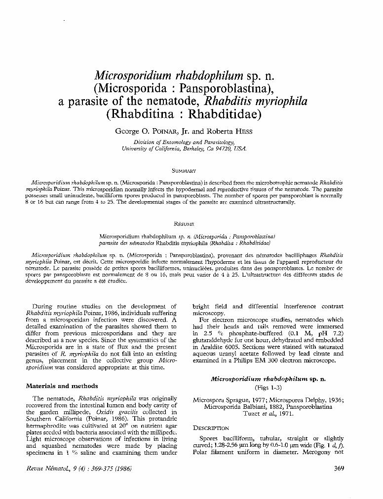
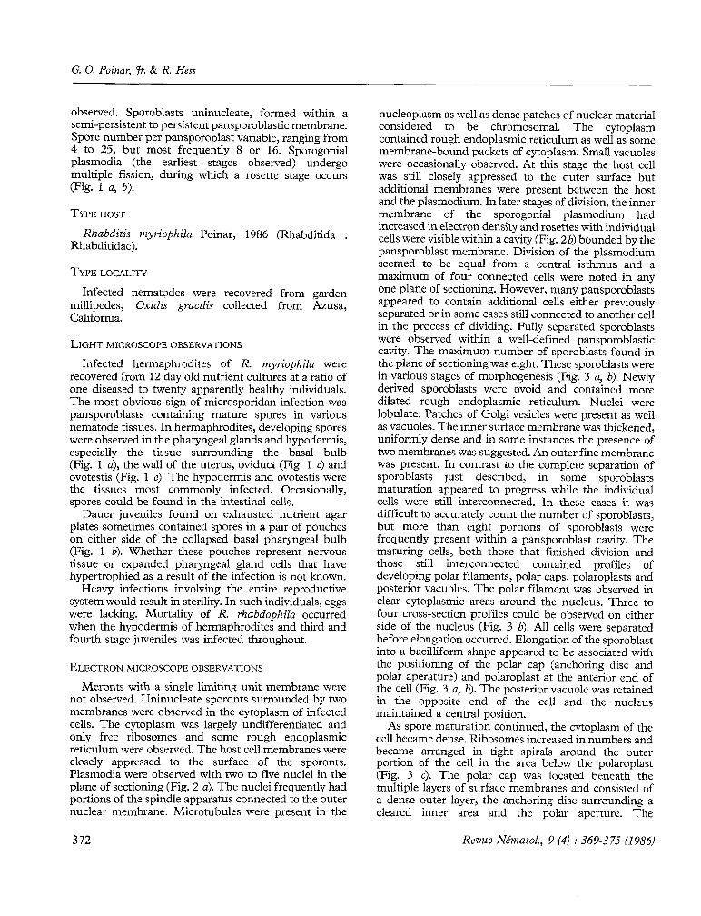
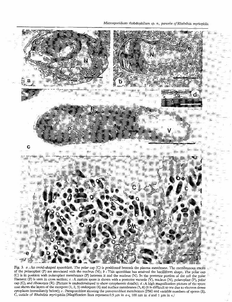
Fig. 3. a :An ovoid-shaped sporoblast. The polar cap (C) is positioned beneath the plasma membrane. The membranous stacks of the polaroplast (P) are associated with the nucleus (N); b : This sporoblast has attained the bacilliform shape. The polar cap (C) is in position with polaroplast membranes (P) between it and the nucleus (N). In the posterior portion of the ce11 the polar filament (F) is seen in cross section; c :A mature spore is shown with a posterior vacuole (V), nucleus (N), polaroplast (P), polar cap (C), and ribosomes (R). (Picture is underdeveloped to show cytoplasmic details); d :A high magnification picture of the spore coat shows the layers of the exospore (1,2,3) endospore (4) and surface membranes (5,6) (6 is diificult to see due to electron dense cytoplasm immediately below); e : Pansporoblast showing the pansporoblast membranes (PM) and variable numbers of spores (S). C, cuticle of Rhabditis myriophilu.(Magification lines represent0.5 prn in a-c, 100 mn in d and 1 prn in e.)
G. 0. Poinur, Jr. & R. Hess
manubroid portion of the polar filament, similar in diameter to the rest of the polar filament, terminated in the polar aperture. From the anterior portion of the ce11 the wide polar filament extended posteriorly, displaced toward the periphery of the cell. The polaroplast, consisting of stacked membranous lamellae, formed around thc manubroid portion of the polar filament in the anterior third of the ce11 (Fig. 3 c). Thcir appearance was uniformly lamellar. The nucleus was located in thc mid-portion of the ce11 with the posterior vacuole adjacent (Fig. 3 c). The wall of the mature spore was approximately 50 nm thick. The exospore (Fig. 3 d) was composed of a dense outer layer, a transparent middle layer and an inner dense laycr. The inner and outer layers sometimes structurally resembled unit membranes. The exospore surface was slightly wrinkled. In some areas small vesicles and larger tubular profiles were observed budding at the exospore. In cross section, the tubular profiles were observed to consist of an outer layer (a unit membrane?) followed by a clear zone, then an inner layer (membrane?) surrounding a dense innermost matrix. Total diameter of the tubules and vesicles varied due to the thickness of the inncr matrix. The endospore (Fig. 3 d) was approximately 25 nm thick and contained fine flocculent matcrial. There were apparently two membranes surrounding the spore cytoplasm. The inner membrane was difficult to resolve due to the dense underlying cytoplasm.
30
E 0 k 2 25 8
0
15
10
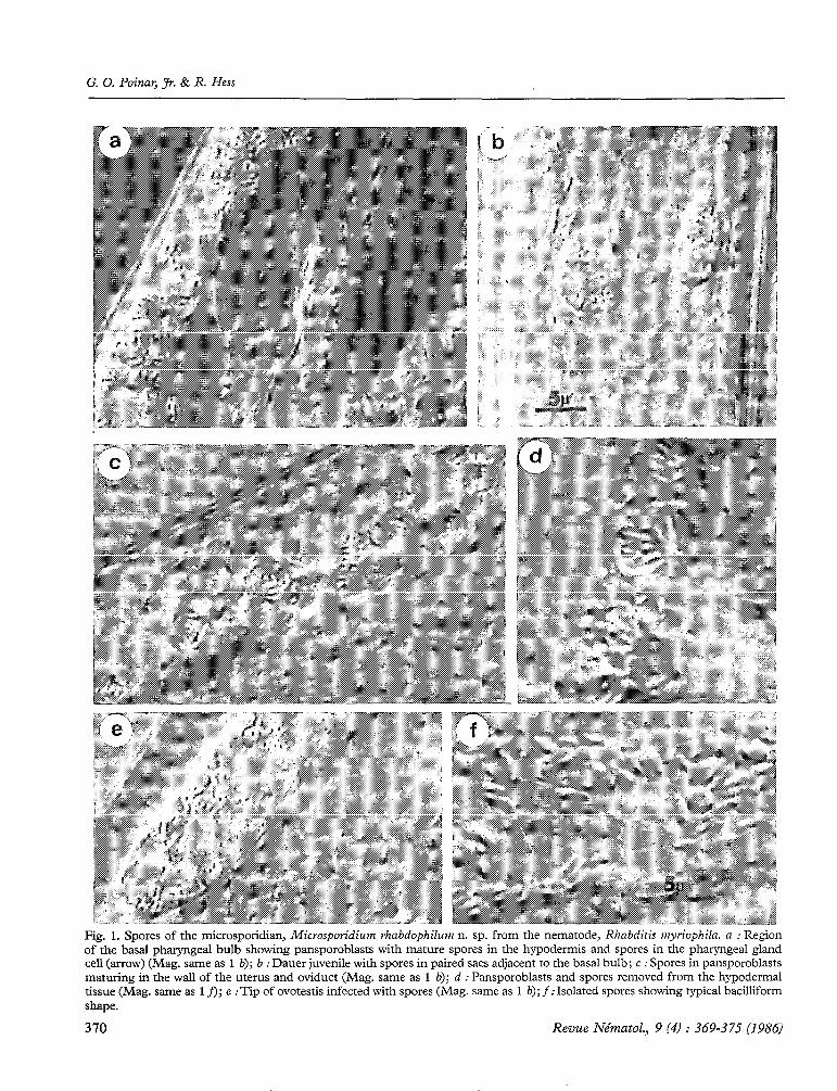
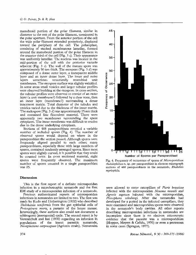
Sections of 468 pansporoblasts revealed a variable number of included spores (Fig. 4). The number of observcd spores would depend on where in thc pansporoblast the section passed. Although spores were frequently aligned parallel to each other; many pansporoblasts, especially those with large numbers of spores, contained randomly arranged spores. Since most spores were slightly curved, it is possible that they could be counted twice. In cross sectioned material, eight spores were frequently observed. The maximum number of spores counted in a single pansporoblast was 25.
5
0
Number of Spores per Pansporoblast
Fig. 4. Frequeney of occurrence of spores of Microsporidiurrz rhubdophilwrz n. sp. per pansporoblast in electron micrograph sections of 468 pansporoblasts in the nematode, Rhabditis myn’ophila.
Discussion
This is the first report of a definite microsporidan infection in a microbotrophic nematodc and the first EM study of a microsporidan infection of a nematode.
Previous authenticated reports of microsporidan infections in nematodes are limited to two. The fïrst was made by Kudo and Hetherington (1922) who described Thdohaniu renifomis from the gut epithelial cells of Protospirura ~nrrrîs, a parasite of thc house mouse. Interestingly, these authors also could not document a schizogonic (mcrogonial) cycle. The second report is by Veremtchuk and Issi (1970) regarding an infection in populations of the insect parasitic nematode, Neoaplectana ccqpocapsae (Agriotos strain). Nematodes
were allowed to enter caterpillars of Pieris brassicae infected with thc microsporidan Nosema mesnili and Agrotis segetum infected with the microsporidan, Plistophora schubergi. Aftcr the nematodes had developed for a period in the infected caterpillars, they were examined and microsporidan spores were observcd in thc nematode’s body cavities. Al1 other reports describing microsporidan infections in nematodes are incomplete since there is no electron microscopic evidence that the parasite was a microsporidium (Hopper, Meyers & Cefalu, 1970) or even a protozoan in some cases (Sprague, 1977).
374 Revue Nématol., 9 (4) : 369-375 (19861
Microsporidium rhabdophilum sp. n., parasite ofRhabditis myriophila
The report by Veremtchuk and Issi (1970) brings up an interesting point. In nature, R. myriophilu is associated with the millipede Oxidis grucilis (Poinar, 1986) and we considered the possibility that R. mytiophila had acquired the infection by feeding on dead millipedes. We examined tissues of living and dead specimens of 0. gracilis but could find no evidence of a microsporidan infection. Thus for the present, M. rhabdophilum cari be considered as a primary parasite of R. nzyriophila.
The presently described microsporidium possesses several unique features. First, the size of the spores makes it one of the smallest described microsporidans from an invertebrate host. Second, the apparent range in spore number per pansporoblast is unusual in comparison with other members of the phylum. Although we describe only a sporogonial cycle, it is possible that merogony does occur but was missed because by the time we could determine if a nematode was infected, spores were already forming and the merogonial cycle could have been terminated.
The significance of the spore filled sacs in the dauer stage of M. rhabdophilum is that it offers a method of transport and distribution for the microsporidan. In nature, dauer juveniles would leave the millipede cadaver, and enter the alimentary tract of another living host. After the host died and the nematodes initiated development on the cadaver, the spores would be
Accepté pour publication le 25 mars 1986.
released into the environment or even infect the dauer carrier. It is interesting that no other sign of infection was noted in dauer juveniles that carried the microsporidan spores.
ACKNOWLEGDMENTS
We would like to thank Victor Sprague for reviewing the manuscript and providing nomenclatorial advice.
HOPPER, B. E., MEYERS, S. P. & CEFALC, R. (1970). Microsporidian infection of a mürine nematode, hfletoncholaimus scissus. J. Invert. Pathol., 16 : 371-377.
KUDO, R. & HETHERISGTOS, D. C. (1922). Notes on a microsporidian parasite of a nematode. ‘J Parusitol., 8 : 129-132.
POINAR, Jr. G. 0. (1986). Rhabditis myriophilu sp. n. (Rhabditidae : Rhabditida), associated with the millipede, Oxidis gracilis (Polydesmida : Diplopoda). Proc. helminth. Soc. Wash.
SIX~GCE, V. (1977). The zoological distribution of microsporidia. In : Comparative Pathobiology, Vol. 2 Lon- don-New York. Plenum Press : 335386.
VEREMTCHUK, G. V. & ISSI, 1. V. (1970). [Development of insect microsporidians in the entomopathogenic nematode, Neoaplectana agriotos (Nematodea : Steinernematidae)]. Parasitologiya, 4 : 3-7.
Revue Nématol., 9 (41 : 369-375 (1986) 315