Embed Size (px)
Citation preview
Introduction to Anatomy and
Physiology:
Tissues and Integumentary
System
Biology 105
Lecture 7
Chapter 4

Copyright © 2009 Pearson Education, Inc.
Outline
I. Tissues
A. Epithelial
B. Connective
C. Muscle
D. Nervous tissues
II. Cell-to-cell contact
III. Body cavities
IV. Membranes
V. Homeostasis
VI. Integumentary System
Includes: skin, hair, nails
Copyright © 2009 Pearson Education, Inc.
Copyright © 2009 Pearson Education, Inc.
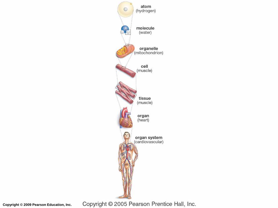
Organization of the Human Body
Multicellular organisms require specialized
cells to perform specific tasks.
These cells then organize into tissues, organs,
and organ systems.

Copyright © 2009 Pearson Education, Inc.
Tissues
A tissue is a group of cells that work together
to accomplish a common function.
There are four primary tissue types:
1. Epithelial tissue
2. Connective tissue
3. Muscle tissue
4. Nervous tissue

Copyright © 2009 Pearson Education, Inc.
Tissues
Epithelial tissue covers the body surfaces,
lines cavities and organs, and forms glands.
Connective tissue binds and supports the
body, provides protection for our organs,
serves as a storage site for fat, and
participates in immunity.
Muscle tissue is responsible for movement.
Nervous tissue receives stimuli and conducts
nerve impulses.
Copyright © 2009 Pearson Education, Inc.
Epithelial Tissue
Epithelial tissues (epithelium) cover surfaces
such as the outside of the body (our skin), as
well as line internal cavities and tubes and the
inside surface of the stomach and the lungs.
Serves for protection, secretion and absorption,
and may contain glands.
Cells are tightly packed together.
Copyright © 2009 Pearson Education, Inc.
Epithelial Tissue
All epithelial tissues share two characteristics:
1. A free surface that may be specialized for
protection, secretion, or absorption
2. A basement membrane, which binds the
epithelial cells to underlying connective tissue
Copyright © 2009 Pearson Education, Inc.
Epithelial Tissue - Shapes
The three basic shapes of epithelial cells:
1. Squamous epithelium
2. Cuboidal epithelium
3. Columnar epithelium

Copyright © 2009 Pearson Education, Inc.
Types of Epithelial Tissue
Simple epithelial – a single layer of cells
classified according to cell type.
Stratified epithelial – two or more layers of
cells, with one on top of the other.
Pseudostratified epithelial – looks like it has
more than one layer, but really does not.
Glandular epithelial – secretes products like
mucus, digestive enzymes, and hormones.
Copyright © 2009 Pearson Education, Inc.
Simple Squamous Epithelium
Simple squamous epithelium – one
layer of flattened cells
Forms the lining of blood vessels and
air sacs in lungs (= alveoli).
Functions: exchange of nutrients, waste
and gases, and protection

Copyright © 2009 Pearson Education, Inc.
Simple Squamous Epithelium
Figure 4.1 (1 of 6)
Simple Squamous
• One layer of flattened cells
• Located in air sacs of lungs, and forms the
lining of the heart and blood vessels • Allows exchange of nutrients, gases, and wastes

Copyright © 2009 Pearson Education, Inc.
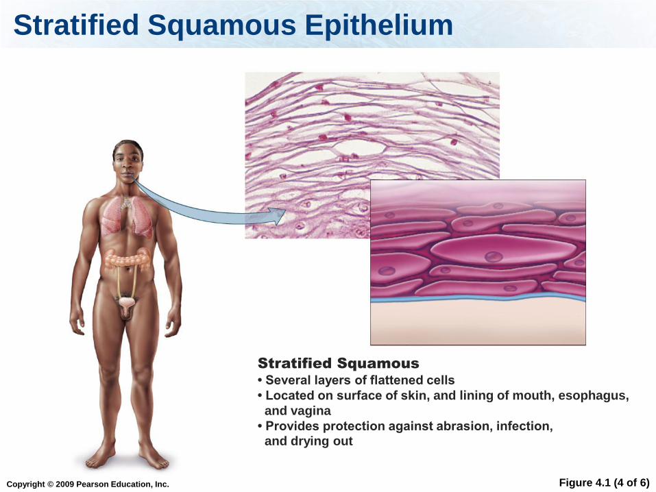
Stratified Squamous Epithelium
Several layers of flattened cells
Located on surface of skin, and lining of
mouth, esophagus, and vagina
Functions: provides protection against
abrasion, infection, and drying out
Copyright © 2009 Pearson Education, Inc.
Stratified Squamous Epithelium
Figure 4.1 (4 of 6)
Stratified Squamous
• Several layers of flattened cells
• Located on surface of skin, and lining of mouth, esophagus,
and vagina
• Provides protection against abrasion, infection, and drying out

Copyright © 2009 Pearson Education, Inc.
Simple Cuboidal Epithelium
Simple cuboidal epithelium – one layer
of cube-shaped cells
Lines the kidney tubules, ovaries, and
glands
Functions: secretion and absorption

Copyright © 2009 Pearson Education, Inc.
Simple Cuboidal Epithelium
Figure 4.1 (2 of 6)
Simple Cuboidal
• One layer of cube-shaped cells
• Located in linings of kidney tubules and glands
• Functions in absorption and secretion
Copyright © 2009 Pearson Education, Inc.
Stratified Cuboidal Epithelium
Stratified cuboidal epithelium – more
than one layer of cube-shaped cells
Located in ducts of mammary glands,
sweat glands, and salivary glands
Functions: protection

Copyright © 2009 Pearson Education, Inc.
Stratified Cuboidal Epithelium
Figure 4.1 (5 of 6)
STRATIFIED EPITHELIUM
Stratified Cuboidal
• Usually two layers of cube-shaped cells
• Located in ducts of mammary glands, sweat glands,
and salivary glands • Functions in protection

Copyright © 2009 Pearson Education, Inc.
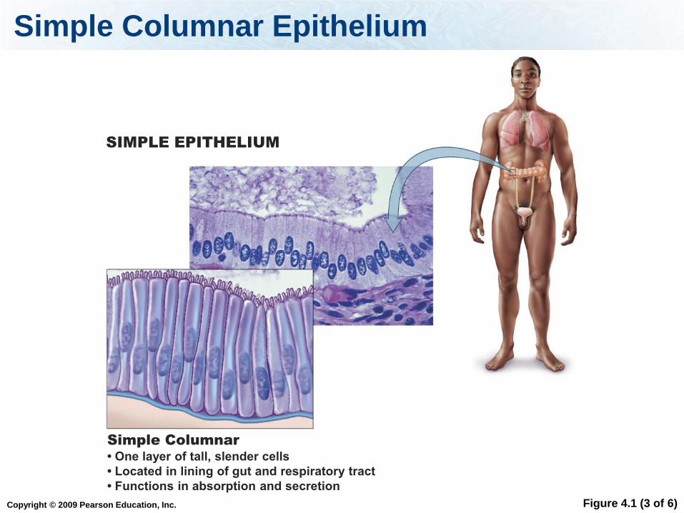
Simple Columnar Epithelium
Simple columnar epithelium – one layer
of rectangular cells
Lines the digestive tract, respiratory
tract, and the uterus
Functions: absorption and secretion
Copyright © 2009 Pearson Education, Inc.
Simple Columnar Epithelium
Figure 4.1 (3 of 6)
SIMPLE EPITHELIUM
Simple Columnar
• One layer of tall, slender cells
• Located in lining of gut and respiratory tract
• Functions in absorption and secretion

Copyright © 2009 Pearson Education, Inc.
Pseudostratified Ciliated Columnar Epithelium
Pseudostratified ciliated columnar
epithelium – looks like it has more than
one layer, but it does not
Lines respiratory tract.
Function: removes debris from the lungs
Copyright © 2009 Pearson Education, Inc.
Stratified Columnar Epithelium
Stratifed columnar epithelium – more
than one layer of rectangular cells
Location (rare!): urethra, and junction of
esophagus and stomach
Functions: protection and secretion

Copyright © 2009 Pearson Education, Inc.
Stratified Columnar Epithelium
Figure 4.1 (6 of 6)
STRATIFIED EPITHELIUM
Stratified Columnar
• Several layers of tall, slender cells
• Rare: located in urethra (tube through which urine
leaves the body)
• Functions in protection and secretion

Copyright © 2009 Pearson Education, Inc.
Tissue Specialization Location
Simple
squamous
Diffusion Alveoli and blood vessels
Simple
cuboidal
Absorption and
secretion
Kidney tubules, ovaries,
and glands
Simple
columnar
Absorption and
secretion
Digestive tract, respiratory
tract, and uterus
Pseudo-
stratified
Removing
debris
Respiratory tract

Copyright © 2009 Pearson Education, Inc.
Tissue Specialization Location
Stratified
squamous
Protection Skin, mouth, esophagus,
vagina
Stratified
cuboidal
Protection Ducts of mammary, sweat,
and salivary glands
Stratified
columnar
Protection and
secretion
Urethra, junction of
esophagus and stomach

Copyright © 2009 Pearson Education, Inc.
Table 4.1 Epithelial Tissues
Table 4.1
Copyright © 2009 Pearson Education, Inc.
Glands
Glands are composed of epithelial tissue.
Exocrine glands secrete their products into
ducts.
Endocrine glands secrete their products
directly into blood.
Copyright © 2009 Pearson Education, Inc.
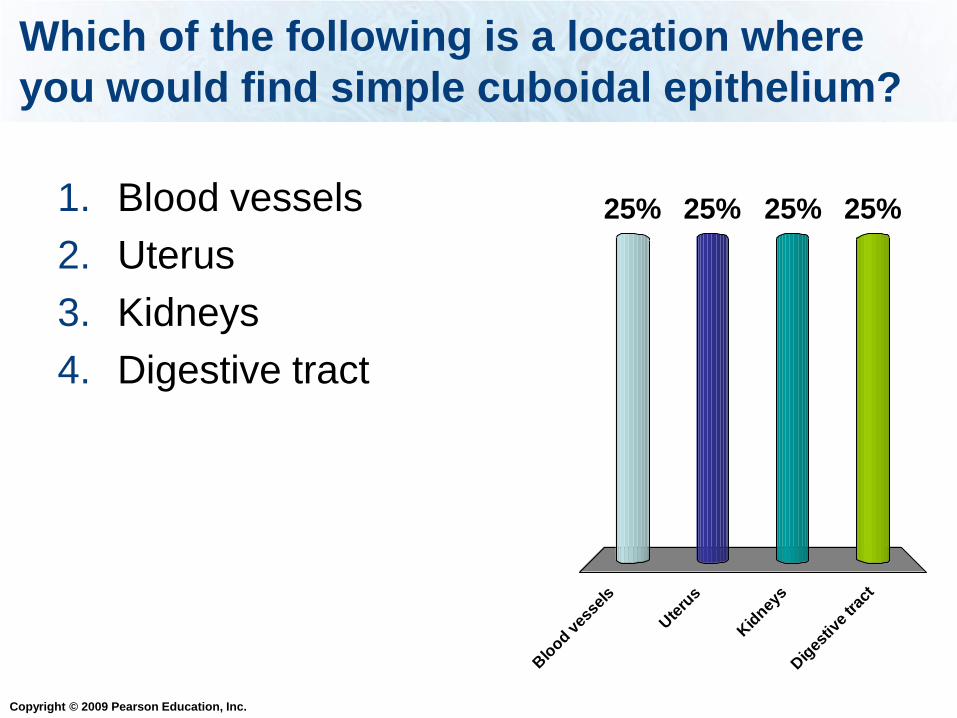
Which of the following is a location where
you would find simple cuboidal epithelium?
Blo
od ves
sels
Ute
rus
Kid
neys
Dig
estiv
e trac
t
25% 25%25%25%1. Blood vessels
2. Uterus
3. Kidneys
4. Digestive tract

Copyright © 2009 Pearson Education, Inc.
Connective Tissue
Connective tissues stabilize, bind, and support
other tissues.
Cells in connective tissue are usually
separated from each other by extracellular
material (examples: fibers, carbohydrates).
The connective tissue cells secrete this
extracellular material.

Copyright © 2009 Pearson Education, Inc.
Connective Tissue
Many different types of connective tissue:
1. Areolar
2. Adipose
3. Dense (tendons and ligaments)
4. Cartilage
5. Bone
6. Blood

Copyright © 2009 Pearson Education, Inc.
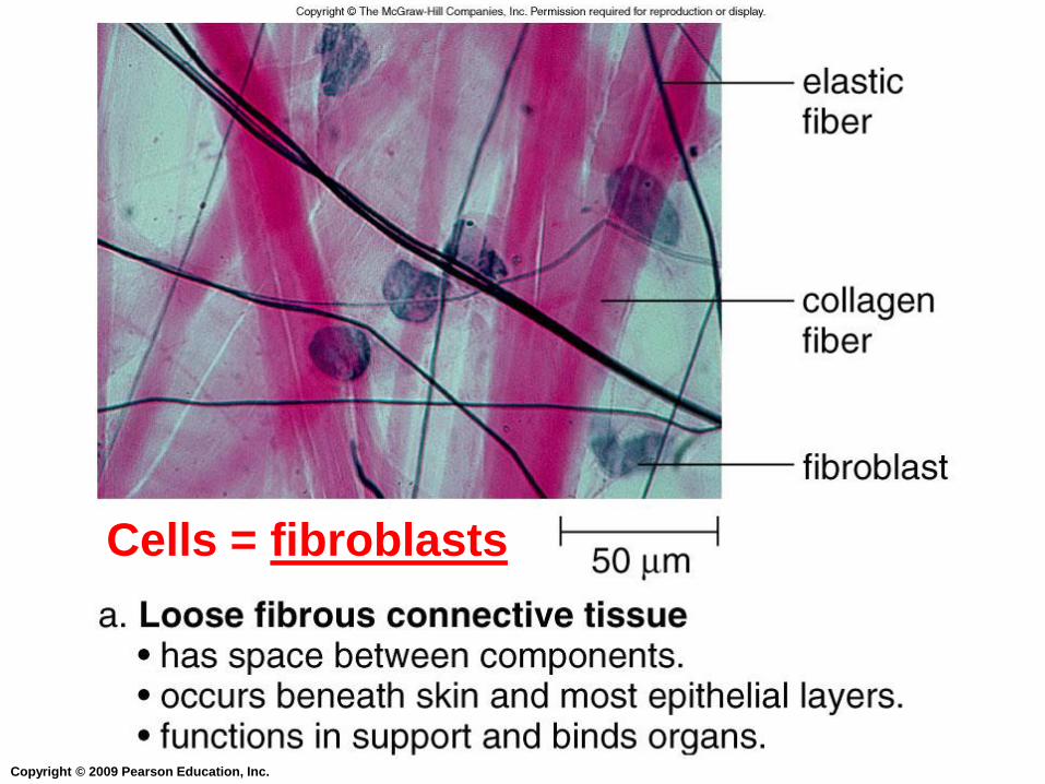
1. Loose Areolar Connective Tissue
Cells: fibroblasts
Fibroblasts secrete protein fibers
Functions – bind and support
Locations – under skin, around organs,
between muscles
Copyright © 2009 Pearson Education, Inc.
Cells = fibroblasts

Copyright © 2009 Pearson Education, Inc.
2. Loose Adipose Connective Tissue
Cells: adipose cells
Stores triglycerides
Functions – energy storage, insulation,
cushioning for organs
Locations – under skin, and around kidneys
and heart

Copyright © 2009 Pearson Education, Inc.
Loose Adipose Tissue
Figure 4.2 (2 of 6)
Adipose (Fat) Tissue
• Found under skin, around kidneys
and heart
• Functions in energy storage and insulation;
provides cushioning for organs

Copyright © 2009 Pearson Education, Inc.
3. Dense Connective Tissue
Cells: fibroblasts
Functions: attaches bone to bone (ligaments),
and attaches muscle to bone (tendons)
Locations – tendons and ligaments
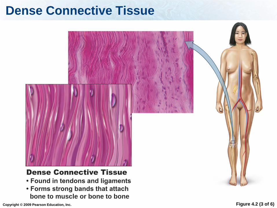
Copyright © 2009 Pearson Education, Inc.
Dense Connective Tissue
Figure 4.2 (3 of 6)
Dense Connective Tissue
• Found in tendons and ligaments
• Forms strong bands that attach bone to muscle or bone to bone
Copyright © 2009 Pearson Education, Inc.
4. Cartilage (Specialized Connective Tissue)
Cells: chondrocytes
Cells are located in chambers = lacunae
Lacunae are surrounded by a matrix:
This type of tissue is strong but flexible.
Functions: support and protection (cushioning)
Locations: nose, ends of long bones, ribs, in
joints, outer ear, and between the vertebrae in
the backbone
There is not a direct blood supply, so this type
of tissue heals slowly.

Copyright © 2009 Pearson Education, Inc.

Copyright © 2009 Pearson Education, Inc.
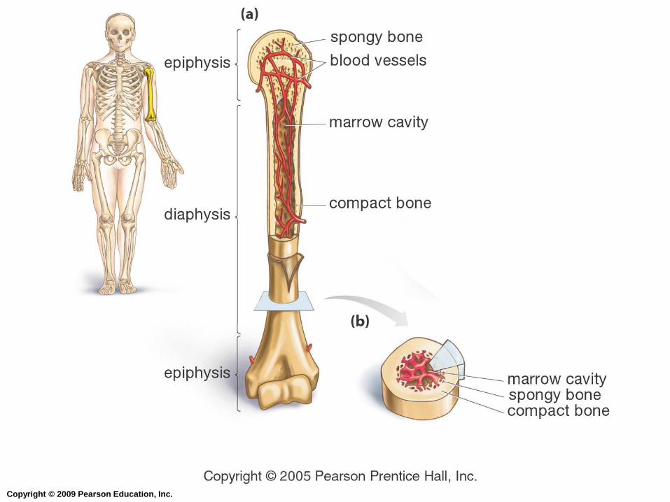
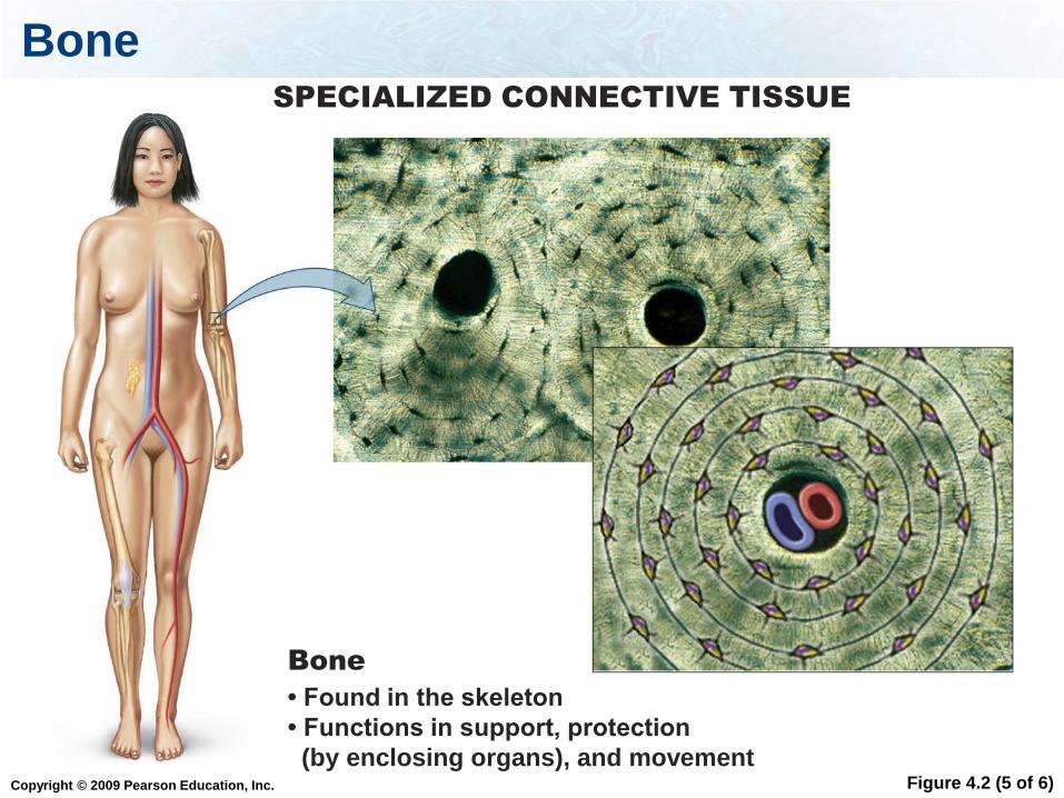
5. Bone (Specialized Connective Tissue)
Bone cells: osteocytes
Cells are found in lacunae.
Rigid connective tissue.
Made of hard matrix (provides strength), and
protein fibers including collagen (provide
strength and flexibility)
Functions:
1. Protects and supports internal structures
2. Facilitates movement along with the muscles
3. Stores lipids, calcium, and phosphorus
4. Produces blood cells
Copyright © 2009 Pearson Education, Inc.
Copyright © 2009 Pearson Education, Inc.
Bone
Figure 4.2 (5 of 6)
SPECIALIZED CONNECTIVE TISSUE
Bone
• Found in the skeleton
• Functions in support, protection
(by enclosing organs), and movement
Copyright © 2009 Pearson Education, Inc.
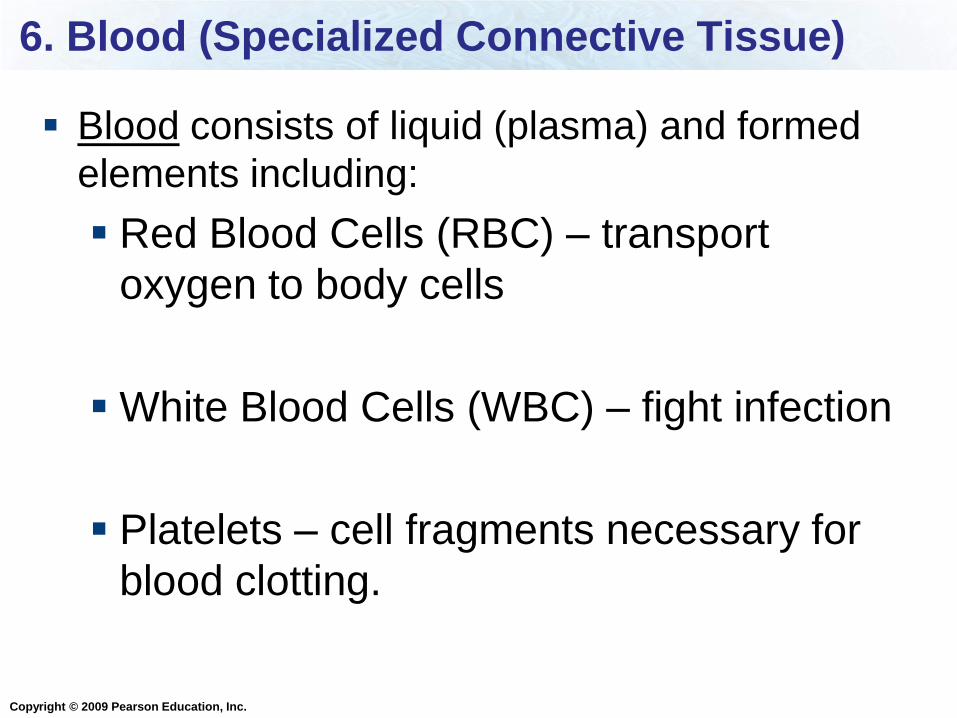
6. Blood (Specialized Connective Tissue)
Blood consists of liquid (plasma) and formed
elements including:
Red Blood Cells (RBC) – transport
oxygen to body cells
White Blood Cells (WBC) – fight infection
Platelets – cell fragments necessary for
blood clotting.

Copyright © 2009 Pearson Education, Inc.
Blood
Figure 4.2 (6 of 6)
Blood
• Found within blood vessels
• Transports nutrients, gases, hormones, wastes;
fights infections

Copyright © 2009 Pearson Education, Inc.
Connective Tissue
Table 4.2
Copyright © 2009 Pearson Education, Inc.
Which cells are found in dense connective tissue?
Chond
rocy
tes
Ost
eocy
tes
Fib
robla
sts
Ost
eobl
asts
25% 25%25%25%1. Chondrocytes
2. Osteocytes
3. Fibroblasts
4. Osteoblasts
Copyright © 2009 Pearson Education, Inc.
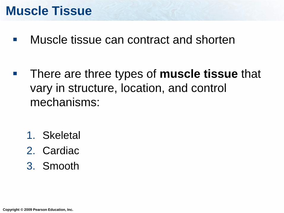
Muscle Tissue
Muscle tissue can contract and shorten
There are three types of muscle tissue that
vary in structure, location, and control
mechanisms:
1. Skeletal
2. Cardiac
3. Smooth
Copyright © 2009 Pearson Education, Inc.
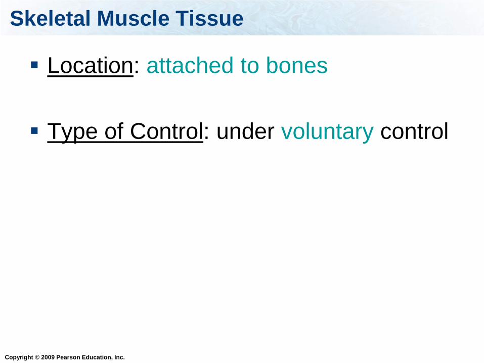
Skeletal Muscle Tissue
Location: attached to bones
Type of Control: under voluntary control
Copyright © 2009 Pearson Education, Inc.
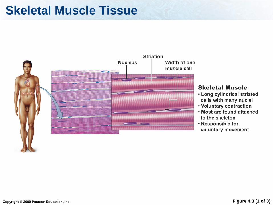
Skeletal Muscle Tissue
Figure 4.3 (1 of 3)
Skeletal Muscle
• Long cylindrical striated
cells with many nuclei
• Voluntary contraction
• Most are found attached
to the skeleton
• Responsible for
voluntary movement
Nucleus Width of one
muscle cell
Striation

Copyright © 2009 Pearson Education, Inc.
Cardiac Muscle Tissue
Location: walls of the heart
Type of Control: under involuntary
control
Copyright © 2009 Pearson Education, Inc.
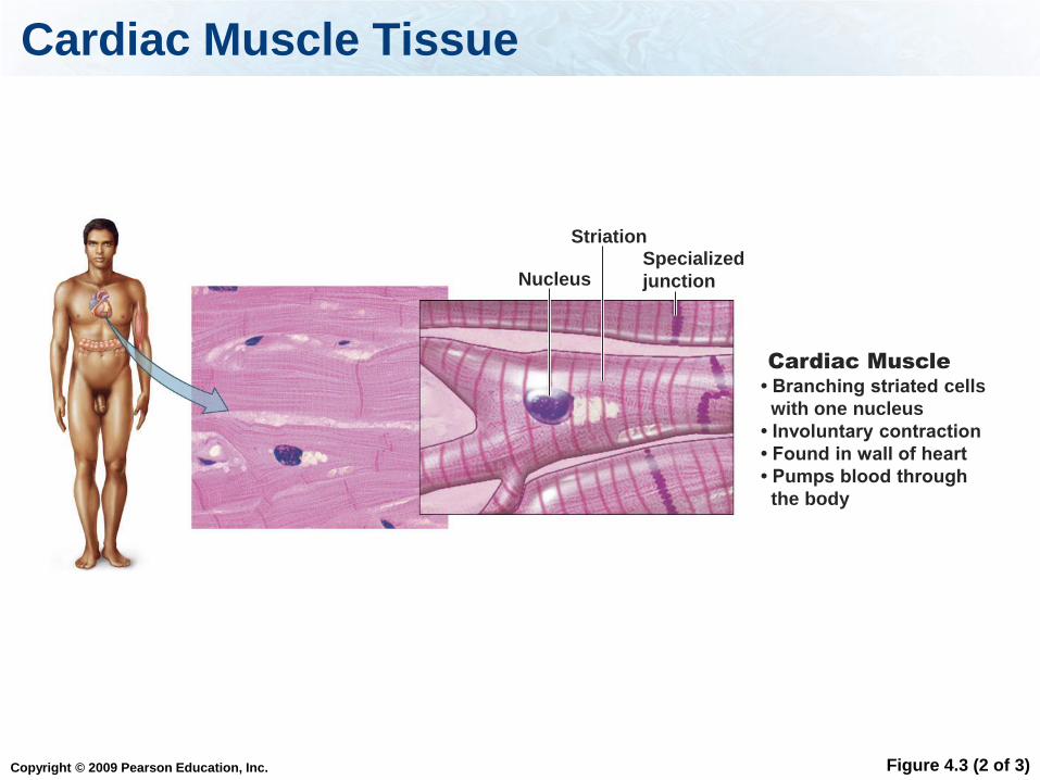
Cardiac Muscle Tissue
Figure 4.3 (2 of 3)
Cardiac Muscle
• Branching striated cells
with one nucleus
• Involuntary contraction
• Found in wall of heart
• Pumps blood through
the body
Specialized
junction Nucleus
Striation

Copyright © 2009 Pearson Education, Inc.
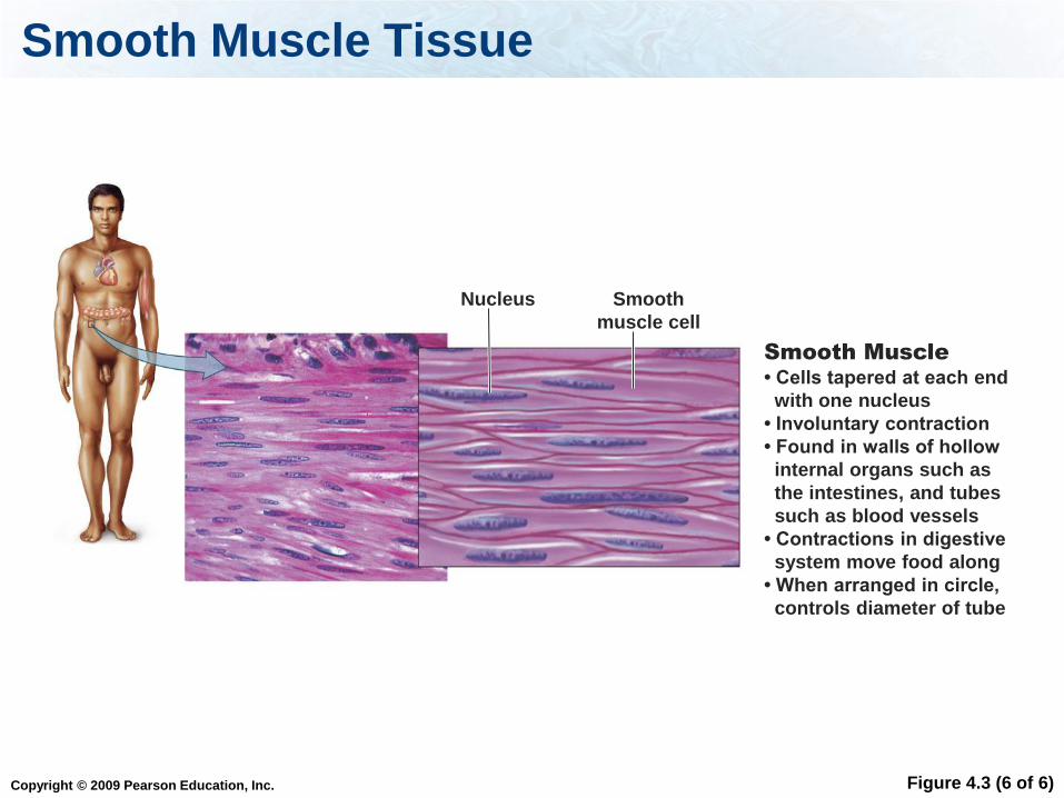
Smooth Muscle Tissue
Locations: surrounds other organs and
structures (examples: blood vessels,
digestive system, lungs)
Type of Control: under involuntary control
Copyright © 2009 Pearson Education, Inc.
Smooth Muscle Tissue
Figure 4.3 (6 of 6)
Smooth Muscle
• Cells tapered at each end
with one nucleus
• Involuntary contraction
• Found in walls of hollow
internal organs such as
the intestines, and tubes
such as blood vessels
• Contractions in digestive
system move food along
• When arranged in circle,
controls diameter of tube
Nucleus Smooth
muscle cell

Copyright © 2009 Pearson Education, Inc.
Muscle Tissue
Table 4.3
Copyright © 2009 Pearson Education, Inc.
This type of muscle is under voluntary control:
Ske
leta
l
Sm
ooth
Car
diac
33% 33%33%1. Skeletal
2. Smooth
3. Cardiac
Copyright © 2009 Pearson Education, Inc.
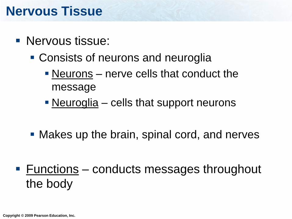
Nervous Tissue
Nervous tissue:
Consists of neurons and neuroglia
Neurons – nerve cells that conduct the
message
Neuroglia – cells that support neurons
Makes up the brain, spinal cord, and nerves
Functions – conducts messages throughout
the body

Copyright © 2009 Pearson Education, Inc.
Nervous Tissue
Figure 4.4
Dendrite
Cell body
Axon
Neuron
Neuroglia

Copyright © 2009 Pearson Education, Inc.
Cell Junctions
The cells that make up tissues are held
together by three types of junctions:
1. Tight junctions
2. Adhesion junctions
3. Gap junctions

Copyright © 2009 Pearson Education, Inc.
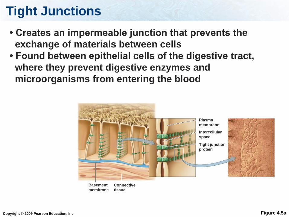
Tight Junctions
Function – prevent substances from
leaking across tissues
Locations – urinary and digestive tracts
Copyright © 2009 Pearson Education, Inc.
Tight Junctions
Figure 4.5a
• Creates an impermeable junction that prevents the
exchange of materials between cells
• Found between epithelial cells of the digestive tract,
where they prevent digestive enzymes and
microorganisms from entering the blood
Plasma
membrane
Intercellular
space
Tight junction
protein
Basement
membrane
Connective
tissue

Copyright © 2009 Pearson Education, Inc.
Adhesion Junctions
Function – holds adjacent cells together
and allows tissues to be flexible
Locations – skin, and opening of the
uterus

Copyright © 2009 Pearson Education, Inc.
Adhesion Junctions
Figure 4.5b
Intercellular
space
• Holds cells together despite stretching
• Found in tissues that are often stretched, such
as the skin and the opening of the uterus

Copyright © 2009 Pearson Education, Inc.
Gap Junctions
Function – open channels between cells
allowing rapid communication due to
quick transfer of ions and small
molecules between neighboring cells
Locations – heart and smooth muscle

Copyright © 2009 Pearson Education, Inc.
Gap Junctions
Figure 4.5c
• Allows cells to communicate by allowing small molecules
and ions to pass from cell to cell
• Found in epithelia where the movement of ions
coordinates functions, such as the beating of cilia
• Found in excitable tissue, such as heart and smooth
muscle
Protein
channels
Intercellular
space
Copyright © 2009 Pearson Education, Inc.
Which junction allows rapid communication
between neighboring cells?
Tig
ht
Adher
ing
Gap
33% 33%33%
1. Tight
2. Adhering
3. Gap

Copyright © 2009 Pearson Education, Inc.
Which junction prevent substances from leaking
across tissues?
Tig
ht
Adher
ing
Gap
33% 33%33%
1. Tight
2. Adhering
3. Gap
Copyright © 2009 Pearson Education, Inc.
Body Cavities
We have two main body cavities:
Dorsal cavity (posterior)
Ventral cavity (anterior)
Copyright © 2009 Pearson Education, Inc.
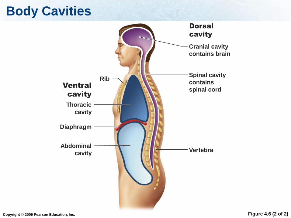
Body Cavities
Figure 4.6 (2 of 2)
Dorsal
cavity
Abdominal
cavity
Thoracic
cavity
Ventral
cavity
Cranial cavity
contains brain
Spinal cavity
contains
spinal cord
Diaphragm
Vertebra
Rib
Copyright © 2009 Pearson Education, Inc.
Ventral Body Cavity
The ventral cavity is divided into two cavities:
Thoracic cavity
The thoracic cavity is further subdivided into:
Pleural cavities – contains lungs
Pericardial cavity – contains heart
Abdominal cavity
The abdominal cavity contains the digestive
system, the urinary system, and the
reproductive system.
The diaphragm is a broad sheet of muscle
that divides the two cavities.

Copyright © 2009 Pearson Education, Inc.
Body Cavities
Figure 4.6 (1 of 6)
Pleural
cavity
contains
a lung
Abdominal
cavity
Thoracic
cavity
Thoracic cavity
Ventral
cavity
Pericardial
cavity
contains
heart
Diaphragm

Copyright © 2009 Pearson Education, Inc.
Dorsal Cavity
The dorsal cavity is divided into two cavities:
Cranial – contains brain
Spinal – contains spinal cord

Copyright © 2009 Pearson Education, Inc.
Body Cavities
Figure 4.6 (2 of 2)
Dorsal
cavity
Cranial cavity
contains brain
Spinal cavity
contains
spinal cord
Vertebra

Copyright © 2009 Pearson Education, Inc.
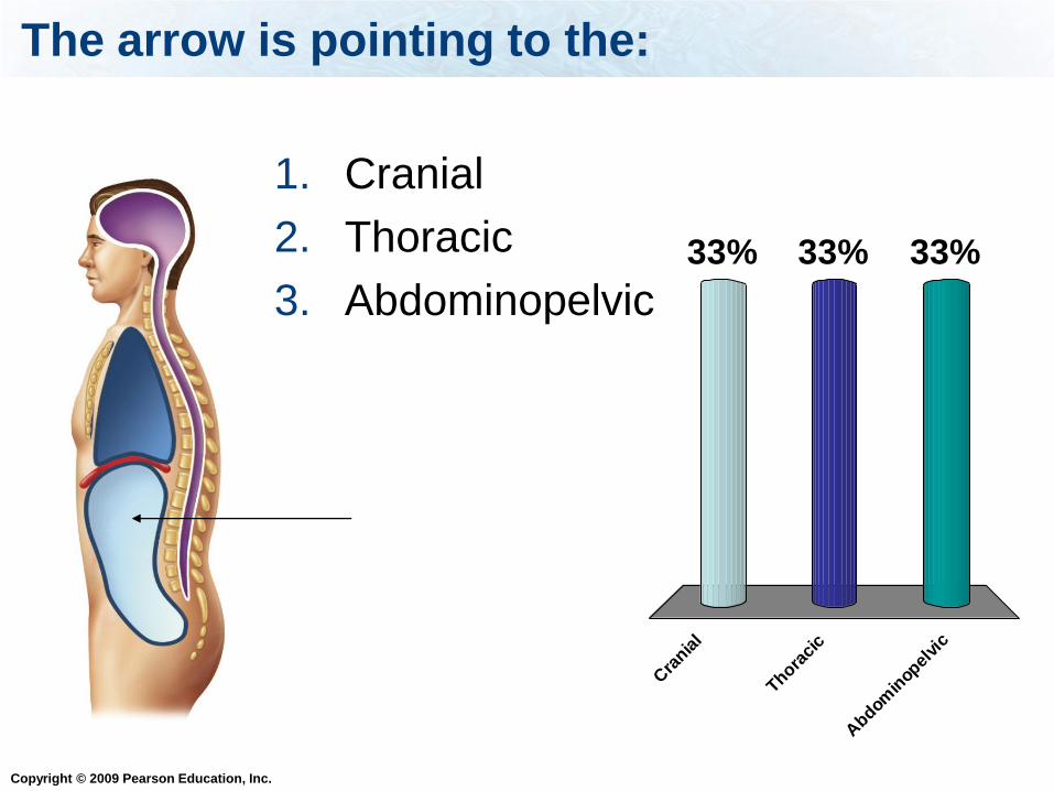
The arrow is pointing to the:
Cra
nial
Thora
cic
Abdom
inop
elvi
c
33% 33%33%
1. Cranial
2. Thoracic
3. Abdominopelvic
Copyright © 2009 Pearson Education, Inc.
The arrow is pointing to the:
Cra
nial
Thora
cic
Abdom
inop
elvi
c
33% 33%33%
1. Cranial
2. Thoracic
3. Abdominopelvic

Copyright © 2009 Pearson Education, Inc.
The arrow is pointing to the:
Ple
ural
Per
icar
dial
50%50%1. Pleural
2. Pericardial
Copyright © 2009 Pearson Education, Inc.
Membranes
Body cavities and surfaces of organs are
covered with membranes.
Membranes are sheets of epithelium
supported by connective tissues.
Membranes protect tissues and organs.

Copyright © 2009 Pearson Education, Inc.
Membranes
There are four types of membranes:
1. Mucous
2. Serous
3. Synovial
4. Cutaneous

Copyright © 2009 Pearson Education, Inc.
Mucous Membranes
Mucous – line passages to the exterior world, including those of the respiratory, digestive, reproductive, and urinary systems in the body
Secrete mucus
Copyright © 2009 Pearson Education, Inc.
Serous Membranes
Serous – line thoracic and
abdominopelvic cavities and the organs
contained in them
Secrete lubricating fluid

Copyright © 2009 Pearson Education, Inc.
Synovial Membranes
Synovial – line cavities of freely movable
joints
Secrete a lubricating fluid

Copyright © 2009 Pearson Education, Inc.
Cutaneous Membranes
Cutaneous – skin, lines the outside of the
body (thick and dry)

Copyright © 2009 Pearson Education, Inc.
Organs and Organ System
An organ is a group of tissues that work
together to perform a specific function.
In turn, organs work together to form an
organ system.

Copyright © 2009 Pearson Education, Inc.
Example Organ: Stomach
1. Epithelium lines the stomach and secretes
acid to digest the food.
2. Nervous tissue stimulates cells to release the
acid.
3. Muscles contract to push food through the
stomach.
4. Connective tissue supports these other
tissues.


Copyright © 2009 Pearson Education, Inc.
Remember Homeostasis?!?
Homeostasis – the ability to maintain a
relatively stable environment in the body
How does the body accomplish this daunting
task?!?
Copyright © 2009 Pearson Education, Inc.
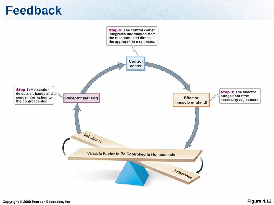
Feedback
The body uses the nervous system and the
endocrine systems to maintain homeostasis.
Controlled by negative or positive feedback
Copyright © 2009 Pearson Education, Inc.
Feedback
Figure 4.12
Copyright © 2009 Pearson Education, Inc.
Feedback Mechanism
A sensor/receptor detects a change (= stimulus)
in the internal or external environment.
A control center, such as a part of the brain,
integrates the information coming from all
receptors and sends out an appropriate
response.
The effector carries out the response, returning
the system to homeostasis again.
Copyright © 2009 Pearson Education, Inc.
Hormones
Hormone – a substance released into the
blood that carries a message to other parts of
the body.
When hormones are released from one part
of the body, they cause another part of the
body to react.

Copyright © 2009 Pearson Education, Inc.
Feedback
In general, Negative Feedback is used to
keep the body in balance, and it maintains
the “status quo”.
Positive Feedback is used to change the
situation.
Copyright © 2009 Pearson Education, Inc.
Example: calcium regulation
Calcium is stored in the bones and circulates in
the blood stream.
Cells in the bones, osteoclasts, release calcium
from bone.
Negative Feedback Example: Calcium
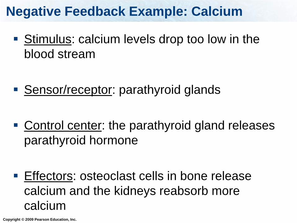
Copyright © 2009 Pearson Education, Inc.
Stimulus: calcium levels drop too low in the
blood stream
Sensor/receptor: parathyroid glands
Control center: the parathyroid gland releases
parathyroid hormone
Effectors: osteoclast cells in bone release
calcium and the kidneys reabsorb more
calcium
Negative Feedback Example: Calcium
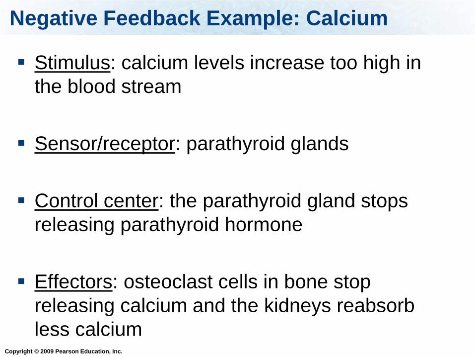
Copyright © 2009 Pearson Education, Inc.
Stimulus: calcium levels increase too high in
the blood stream
Sensor/receptor: parathyroid glands
Control center: the parathyroid gland stops
releasing parathyroid hormone
Effectors: osteoclast cells in bone stop
releasing calcium and the kidneys reabsorb
less calcium
Negative Feedback Example: Calcium

Copyright © 2009 Pearson Education, Inc.
Negative Feedback Example: Temperature
Read pages 79-81: temperature regulation in
the body
Hyperthermia: abnormally elevated body
temperature
Hypothermia: abnormally low body
temperature
The thermostat for the body is located in the
hypothalamus.
Copyright © 2009 Pearson Education, Inc.
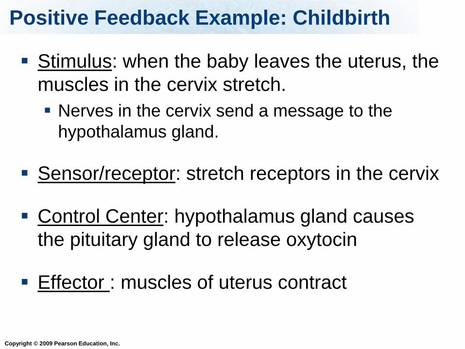
Positive Feedback Example: Childbirth
Stimulus: when the baby leaves the uterus, the
muscles in the cervix stretch.
Nerves in the cervix send a message to the
hypothalamus gland.
Sensor/receptor: stretch receptors in the cervix
Control Center: hypothalamus gland causes
the pituitary gland to release oxytocin
Effector : muscles of uterus contract
Copyright © 2009 Pearson Education, Inc.
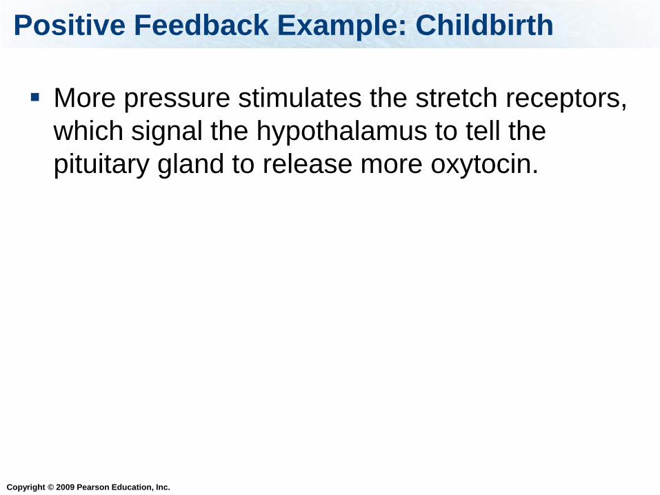
Positive Feedback Example: Childbirth
More pressure stimulates the stretch receptors,
which signal the hypothalamus to tell the
pituitary gland to release more oxytocin.

Copyright © 2009 Pearson Education, Inc.
Integumentary System
Components of the integumentary system:
Skin
Nails
Hair
Exocrine glands (sweat and oil glands)

Copyright © 2009 Pearson Education, Inc.
Integumentary System Functions
1. Provides protection from bacteria, UV
radiation, chemicals, physical injury
2. Reduces water loss
3. Temperature regulation
4. Vitamin D production
5. Contains sensors that detect pain,
temperature, and pressure

Copyright © 2009 Pearson Education, Inc.
Skin Layers
The skin has two layers:
1. Epidermis – thin outer layer of stratified
squamous epithelial tissue
2. Dermis – thick underlying layer of
mainly connective tissue
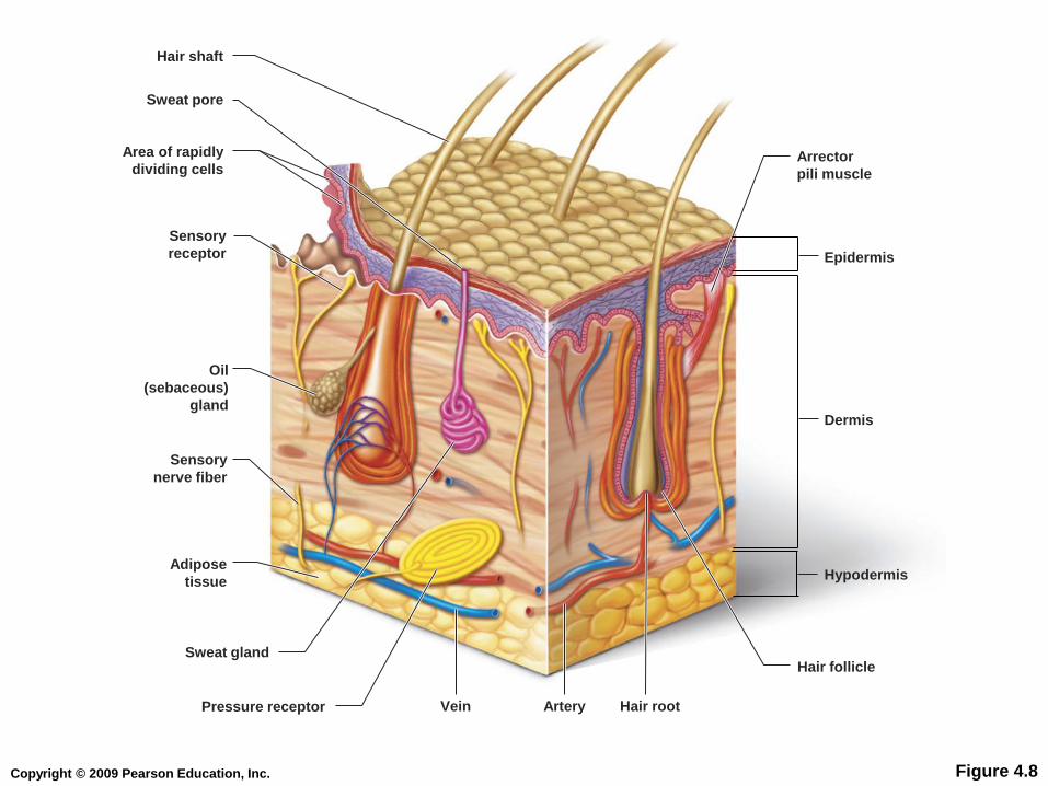
Copyright © 2009 Pearson Education, Inc. Figure 4.8
Hair shaft
Sweat pore
Area of rapidly
dividing cells
Sensory
receptor
Sensory
nerve fiber
Epidermis
Arrector
pili muscle
Dermis
Hypodermis
Hair follicle
Hair root Vein Pressure receptor
Sweat gland
Adipose
tissue
Artery
Oil
(sebaceous)
gland

Copyright © 2009 Pearson Education, Inc.
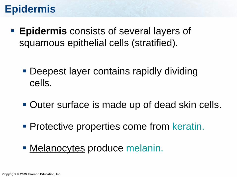
Epidermis
Epidermis consists of several layers of
squamous epithelial cells (stratified).
Deepest layer contains rapidly dividing
cells.
Outer surface is made up of dead skin cells.
Protective properties come from keratin.
Melanocytes produce melanin.
Copyright © 2009 Pearson Education, Inc.
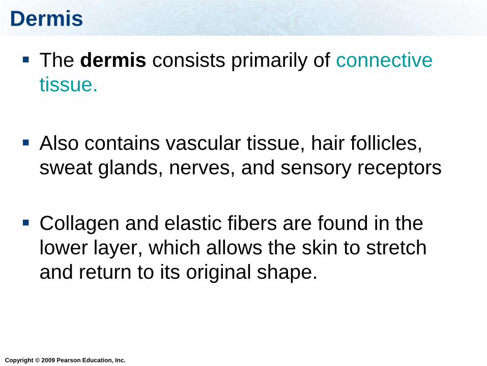
Dermis
The dermis consists primarily of connective
tissue.
Also contains vascular tissue, hair follicles,
sweat glands, nerves, and sensory receptors
Collagen and elastic fibers are found in the
lower layer, which allows the skin to stretch
and return to its original shape.
Copyright © 2009 Pearson Education, Inc.
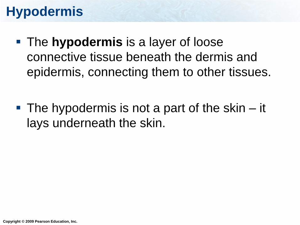
Hypodermis
The hypodermis is a layer of loose
connective tissue beneath the dermis and
epidermis, connecting them to other tissues.
The hypodermis is not a part of the skin – it
lays underneath the skin.
Copyright © 2009 Pearson Education, Inc.
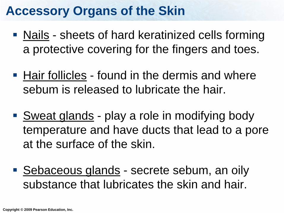
Accessory Organs of the Skin
Nails - sheets of hard keratinized cells forming
a protective covering for the fingers and toes.
Hair follicles - found in the dermis and where
sebum is released to lubricate the hair.
Sweat glands - play a role in modifying body
temperature and have ducts that lead to a pore
at the surface of the skin.
Sebaceous glands - secrete sebum, an oily
substance that lubricates the skin and hair.

Copyright © 2009 Pearson Education, Inc.
Skin Cancer
Melanin protects against UV radiation.
Three types of skin cancer:
1. Basal cell carcinoma – from rapidly dividing
cells deep in the epidermis
2. Squamous cell carcinoma – from newly formed
cells as they flatten
3. Melanoma – from melanocytes; far more
dangerous than other skin cancers and more
likely to spread to other body parts
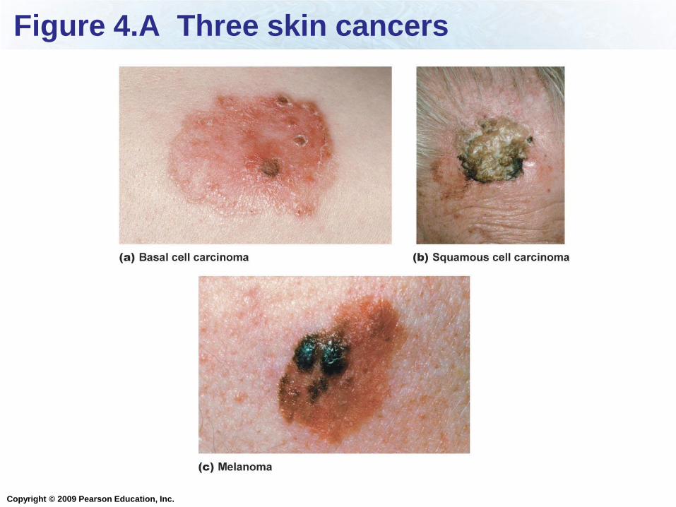
Copyright © 2009 Pearson Education, Inc.
Figure 4.A Three skin cancers
Copyright © 2009 Pearson Education, Inc.
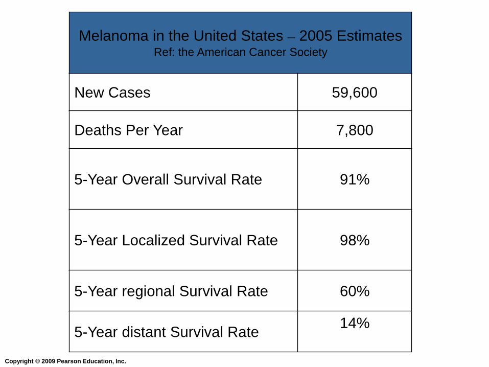
Melanoma in the United States – 2005 Estimates Ref: the American Cancer Society
New Cases 59,600
Deaths Per Year 7,800
5-Year Overall Survival Rate 91%
5-Year Localized Survival Rate 98%
5-Year regional Survival Rate 60%
5-Year distant Survival Rate 14%
Copyright © 2009 Pearson Education, Inc.
These cells found in skin produce pigments:
Chond
rocy
tes
Mel
anoc
ytes
Fib
robla
sts
Ost
eocy
tes
25% 25%25%25%1. Chondrocytes
2. Melanocytes
3. Fibroblasts
4. Osteocytes
Copyright © 2009 Pearson Education, Inc.
Important Concepts
Read chapter 4
What are the four tissue types, their functions,
and examples of each type?
What are examples, functions, and locations of
each of the types of connective tissue?
Why does it take longer for cartilage to heal?
What cell types are found in each type of
connective tissue?

Copyright © 2009 Pearson Education, Inc.
Important Concepts
What are the functions of red blood cells, white
blood cells, and platelets?
What are the three types of muscle? What are
their functions, and where they are found? Are
they under voluntary or involuntary control?
What are the two types of nervous tissue cells
and their functions?
What are the types of epithelial tissue, where
are they found, and what are their functions?

Copyright © 2009 Pearson Education, Inc.
Important Concepts
What are the two types of glands?
How do negative and positive feedback
mechanisms work?
Be able to describe the examples of negative and
positive feedback given in class and in the
textbook (e.g. identify the sensor, control center,
and effector for each example).
What are the three cell-to-cell junctions and
their functions?

Copyright © 2009 Pearson Education, Inc.
Important Concepts
Identify the body cavities: what are their
locations and what is contained in each of the
cavities?
What are the four types of membranes, their
functions and locations?
What are the functions of the integumentary
system?
Components of the integumentary system and
their functions

Copyright © 2009 Pearson Education, Inc.
Important Concepts
What are the two layers of the skin? Which
type of tissues comprise each layer, and where
are the layers located?
What layer is found underneath the skin, and
which tissue type comprises this layer?
What are the three types of skin cancer, where
do they originate, and which is more likely to
spread to other parts of the body?
What are melanocytes, and what is their
function?

Copyright © 2009 Pearson Education, Inc.
Definitions
Tissue, organ, organ system, tight junctions,
adhesion junctions, gap junctions, exocrine
glands, endocrine glands, homeostasis,
hormone, hypodermis, diaphragm, sebum,
sebaceous glands, keratin, basement
membrane, lacunae, voluntary control,
involuntary control, hyperthermia, hypothermia,
melanin