Embed Size (px)
Citation preview
JB ReviewRotation and structure of FoF1-ATP synthase
Received March 23, 2011; accepted April 18, 2011; published online April 26, 2011
Daichi Okuno, Ryota Iino and Hiroyuki Noji*
Department of Applied Chemistry, School of Engineering, TheUniversity of Tokyo, Bunkyo-ku, Tokyo 113-8656, Japan
*Hiroyuki Noji, Department of Applied Chemistry, School ofEngineering, The University of Tokyo, Bunkyo-ku, Tokyo 113-8656,Japan. Tel: þ81-3-5841-7252, Fax: þ81-3-5841-1872,email: [email protected]
FoF1-ATP synthase is one of the most ubiquitous en-zymes; it is found widely in the biological world, includ-ing the plasma membrane of bacteria, inner membraneof mitochondria and thylakoid membrane of chloro-plasts. However, this enzyme has a unique mechanismof action: it is composed of two mechanical rotarymotors, each driven by ATP hydrolysis or proton fluxdown the membrane potential of protons. The two mo-lecular motors interconvert the chemical energy of ATPhydrolysis and proton electrochemical potential via themechanical rotation of the rotary shaft. This uniqueenergy transmission mechanism is not found in otherbiological systems. Although there are other similarman-made systems like hydroelectric generators,FoF1-ATP synthase operates on the nanometre scaleand works with extremely high efficiency. Therefore,this enzyme has attracted significant attention in awide variety of fields from bioenergetics and biophysicsto chemistry, physics and nanoscience. This review sum-marizes the latest findings about the two motors ofFoF1-ATP synthase as well as a brief historicalbackground.
Keywords: ATP hydrolysis/FoF1-ATP synthase/highreversibility/rotary motor/stepping rotation.
Abbreviations: ADP, adenosine diphosphate;AMP-PNP, adenosine-50-(b,g-imino)-triphosphate;ATP, Adenosine-50-triphosphate; ATPgS, adenosine50-(g-thio)triphosphate; Pi, inorganic phosphate.
FoF1-ATP synthase
Adenosine-50-triphosphate (ATP) is the ubiquitous en-ergy currency of the cell. The human body containsabout 50 g of ATP that is sustained by strict dynamicequilibrium between hydrolysis and synthesis. The totalATP produced under basal metabolism in humans am-ounts to 50�75kg per day, and the same amount of ATPis consumed for the large variety of energy-requiringreactions such as muscle contraction, synthesis of bio-molecules and mass transfer across biomembranes.Under aerobic conditions, the major ATP synthesispathway is oxidative phosphorylation of which theterminal reaction is catalysed by FoF1-ATP synthase.
This enzyme is found widely in the biological world,including in thylakoid membranes, the mitochondrialinner membrane and the plasma membrane of bac-teria. This enzyme catalyses ATP synthesis from ad-enosine diphosphate (ADP) and inorganic phosphate(Pi) by using the electrochemical potential of protons(or sodium ions in some bacteria) across the mem-brane, i.e. it converts the electrochemical potentialinto its chemical form. This enzyme also functions inthe reverse direction when the electrochemical poten-tial becomes insufficient: it catalyses proton pumpingto form an electrochemical potential to hydrolyse ATPinto ADP and Pi. FoF1-ATP synthase is a super-complex enzyme with a molecular weight of4500 kDa and consists of two rotary motors. One isF1 (�380 kDa), which is the water-soluble part of ATPsynthase. When isolated from the membrane portion,it acts as an ATP-driven motor: it rotates its innersubunit to hydrolyse ATP and is therefore termed F1-ATPase. The other rotary motor of ATP synthase is Fo
(�120 kDa), which is embedded in the membrane andgenerates rotary torque upon proton translocation thatis driven by proton electrochemical potential (Fig. 1)(1). Bacterial F1 is composed of a3b3gde-subunits. Thethree a- and b-subunits form the hexameric stator ringin which the a- and b-subunits are alternatelyarranged. The rotor shaft is the g-subunit, which isaccommodated in the central cavity of the a3b3-ring.The e-subunit binds onto the protruding part of theg-subunit and provides a connection between the rotorparts of F1 and Fo. The e-subunit acts as the endogen-ous inhibitor of F1 (2�4), by transforming the con-formational state from the closed form to extendedform that blocks the g rotation due to steric hindrance(5�8). This inhibitory function is thought to be physio-logically important to avoid ATP consumption (9).The d-subunit acts as a connector between F1 and Fo
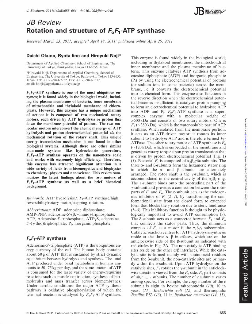
that connects the stator parts. Thus, the minimumcomplex of F1 as a motor is the a3b3g subcomplex.Catalytic reaction centres for ATP hydrolysis/synthesisreside at the three a�b interfaces, which are on theanticlockwise side of the b-subunit as indicated withred circles in Fig. 2A. The non-catalytic ATP-bindingsites reside on the other a/b interfaces. While the cata-lytic site is formed mainly with amino-acid residuesfrom the b-subunit, the non-catalytic sites are primar-ily within the a-subunit. Upon ATP hydrolysis on thecatalytic sites, F1 rotates the g-subunit in the anticlock-wise direction viewed from the Fo side. Fo part consistsof ab2c10�15 subunits. The number of c subunits variesamong species. For example, the copy number of the csubunit is eight in bovine mitochondria (10), 10 inyeast (11), Escherichia coli (12) and thermophilicBacillus PS3 (13), 11 in Ilyobactor tartaricus (14, 15),
Featu
red
Art
icle
J. Biochem. 2011;149(6):655–664 doi:10.1093/jb/mvr049
� The Authors 2011. Published by Oxford University Press on behalf of the Japanese Biochemical Society. All rights reserved 655
at Ekigaku-K
yoshitsu (UN
IV O
F T
OK
YO
) on May 31, 2011
jb.oxfordjournals.orgD
ownloaded from
Propionigenium modestum (16) and Clostridiumparadpxum (17), 13 in thermoalkaliphilic BacillusTA2.TA1 (18) and Bacillus pseudofirmus OF4 (19),14 in spinach chloroplast (20) and 15 in Spirulinaplatensis (21). The c subunits form a ring complex byaligning in a circle. It is widely thought that the c-ringand the a subunit form a proton pathway (for details,see the ‘Proton translocation pathway of Fo’ section).With the downhill proton flow through the protonchannel, the c-ring rotates against the ab2 subunits inthe opposite direction of the g-subunit of the F1 motor(22). Thus, in the FoF1 complex, Fo and F1 push eachother in the opposite direction. Under physiologicalcondition where the electrochemical potential of theprotons is large enough to surpass the free energy ofATP hydrolysis, Fo forcibly rotates the g-subunit in theclockwise direction and then F1 catalyses the reversereaction, i.e. ATP synthesis which is the principlephysiological function of ATP synthase. In contrast,when the electrochemical potential is small or de-creases, F1 forces Fo to rotate the c-ring in the reversedirection to pump protons against the electrochemicalpotential.
Binding change mechanism and structureof F1-ATPase
The three catalytic sites on the b-subunits work co-operatively during catalysis. The classic workingmodel for F1 is the ‘binding-change mechanism’ pro-posed by Paul Boyer (23). The early stage of this model
postulated alternating transition between two chemicalstates, assuming two catalytic sites residing on F1. Itwas later revised to propose the cyclic transition of thecatalytic states among three catalytic sites based on thebiochemical and electron microscopic experiments thatrevealed that F1 has the three catalytic sites (24�26).One important feature of this model is that the affinityfor nucleotide at each catalytic site is different fromeach other at any given time, and the status of thethree b-subunits cooperatively change in one directionaccompanying g rotation. This hypothesis is stronglysupported by X-ray crystallographic studies performedby Walker’s group (27). The first resolved crystal struc-ture of F1 (27) revealed many essential structuralfeatures of F1 at atomic resolution. Importantly, thecatalytic b-subunits differ from each other in conform-ation and catalytic state: one binds to an ATP analogue,adenosine-50-(b,g-imino)-triphosphate (AMP-PNP), thesecond binds to ADP and the third site is empty(Fig. 2A). Therefore, these sites are termed bTP, bDP
and bEmpty, respectively. While bTP and bDP have aclosed conformation wrapping bound nucleotides onthe catalytic sites, bEmpty has an open conformation
Top(view from membrane side)
Side
βDP
Empty
ATP
ADP
βEmptyβTP
αEmpty
αTP
αDP
βEmpty βTP βDP
βDPβEmpty βTP αEmpty αTPαDP
A
B
membrane
90o
cytoplasm
Fig. 2 Crystal structure of the a3b3c subcomplex of F1. The crystalstructure of F1 from bovine mitochondria (PDB code; 1BMF).The a-, b- and g-subunits are shown in yellow, green and red,respectively. (A) The left figure is viewed from the membrane side(Fo side), and is rotated 90� in anticlockwise direction to arrow (rightfigure). The protruding part of g is directed toward the membraneside (15). The catalytic sites are located at the a�b interface indicatedby red circles, which are primarily on the b-subunit. Each site carriesAMP-PNP, ADP, or is empty and is designated as bTP, bDP, orbEmpty, respectively. The other interfaces are non-catalytic sites (bluecircles), all of which bind with AMP-PNP. Each a-subunit forming acatalytic site with the b-subunit is designated as aTP, aDP and aEmpty,respectively. (B) Conformational states of the b-subunit and thecatalytic a�b interfaces. Three a�b pairs with the g-subunit areshown in yellow and green with the central g-subunit (red). Thea and b-subunits are composed of the N-terminal domain,nucleotide-binding domain and C-terminal domain (from bottomto top). bEmpty has an open conformation in which the a-helicalC-terminal domain rotates upwards to open the cleft of thenucleotide-binding pocket. Both bATP and bADP have a closedconformation entrapping the nucleotide within the closed pocket.
ADP + PiATP
FoF1-ATP synthase
H+
Membrane
ADP + Pi
ATP
F1 motor
Fo motor
H+
H+
H+
H+
α3β3
ε γ
c10-15
a
b2
δ
ΔΨΔpH
Fig. 1 Fo and F1 motors of ATP synthase. Schematic images ofFoF1-ATP synthase. The rotor and stator parts are shown in redand blue, respectively. The subunit composition of bacterial Fo isab2c10�15 (the number of c subunits varies from 10 to 15 in differentspecies). Fo is embedded in the cell membrane and rotates the c-ringagainst the ab2 stator, driven by passive proton translocation alongthe proton electrochemical potential that comprises the protonconcentration (�pH) and membrane voltage (��) across themembrane. Bacterial F1 is composed of a3b3gd" and is anATP-driven rotary motor in which the g-subunit rotates againstthe a3b3-cylinder. The "-subunit binds to the protruding partof the g-subunit. The d binds to the bottom of the a3b3-ring (notethat the rotational direction of Fo is opposite to that of F1). In thewhole complex of FoF1, Fo reverses the rotation of F1, leading toATP synthesis from ADP and Pi.
D. Okuno et al.
656
at Ekigaku-K
yoshitsu (UN
IV O
F T
OK
YO
) on May 31, 2011
jb.oxfordjournals.orgD
ownloaded from
swinging the C-terminal domain away from the bind-ing site to open the cleft of the catalytic site (Fig. 2B).These features are consistent with the binding-changemechanism. Another important feature found in thecrystal structure is that while the N-terminal domainsof the a and b-subunits form a symmetrical smoothcavity as the bearing for g rotation at the bottom ofthe a3b3-ring, the C-terminal domains of the b-subunitshow distinct asymmetric interactions with theg-subunit. Therefore, the most feasible inference isthat the open-to-closed transition of the b-subunitsupon ATP binding pushes g, and the sequential con-formational change among b-subunits leads the unidir-ectional g rotation, which was recently visualized insimultaneous imaging of the conformational changeof the b-subunit and the g rotation (28).
Verification of F1 rotation bysingle-molecule observation
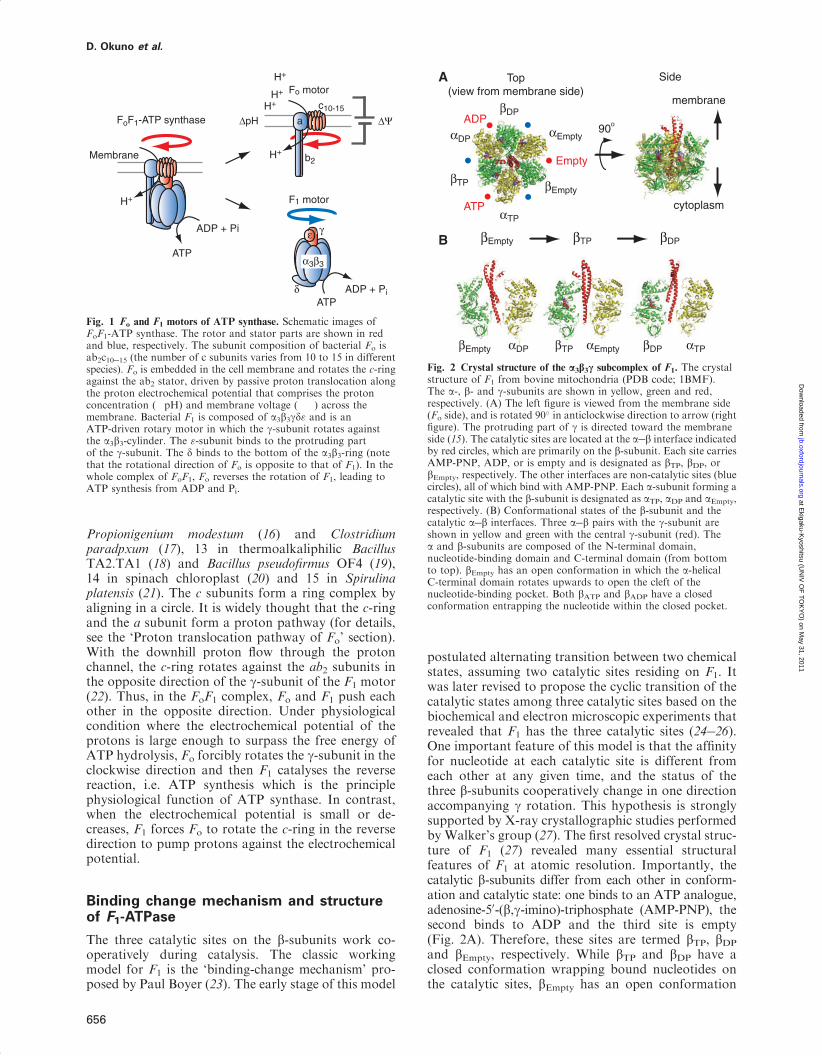
Since the publication of the crystal structure, manystudies have attempted to demonstrate the rotationof F1. Crosslink exchange experiment between theb- and g-subunits of F1 derived from E. coli (29) andthe polarized absorption relaxation of F1 from spinachchloroplasts (30) have proven the rotational motionof the g-subunit during catalysis. Unidirectional rota-tion of the g-subunit upon ATP hydrolysis was provedwith the direct observation of F1 rotation fromthermophilic Bacillus PS3 (TF1) under the microscope(31). In order to suppress rotary Brownian motionstudy, F1 was immobilized on a glass surface modifiedwith Ni-nitrilotriacetic acid (NTA) thorough the inter-action between Ni2þ and the His-tag, which was intro-duced into the N-terminus of the b-subunit. Inaddition, a fluorescently labelled actin filament withlength of 0.6�4 mm and diameter of 10 nm was at-tached to the g-subunit as the rotation marker to mag-nify the subtle motion of the g-subunit of which theradius is only 1 nm, which is much smaller than thespatial resolution (�200 nm) of a conventional micro-scope (Fig. 3A). Note that in recent studies, other typesof probes such as polystyrene beads, gold colloidalbeads, gold nanorods, and magnetic beads are fre-quently used instead of actin filaments because theimaging of fluorescently labelled actin filaments suffersfrom photobleaching. The rotational direction isalways anticlockwise when viewed from the Fo sideand, importantly, it was consistent with the expectedrotary direction from the crystal structure. Assumingthe b-subunit undergoes the conformational transitionfrom bEmpty, bTP and bDP, each catalytic state propa-gates in the anticlockwise direction, accompanyingthe anticlockwise g rotation. The rotational velocitywas far slower than the expected rate from bulkATPase measurements because of the large hydro-dynamic friction exerted on the rotating actin filament.However, this allows us to estimate the torque gener-ated by individual F1 molecules from the hydrodynam-ic friction that should be in equilibrium with F1’storque. The torque was determined to be around40 pNnm. Although this is a rough estimation without
consideration of the viscosity increment in the imme-diate vicinity of surface, the value was recently con-firmed to be valid using more precise torquemeasurements based on fluctuation theorem, which es-timates the entropy generation upon the rotation with-out assuming the friction coefficient (32). Taking intoaccount that the step size is 120�, each coupled withsingle ATP hydrolysis turnover as below, F1 workswith 80 pNnm, which corresponds to the free energyreleased from hydrolysis of a single ATP moleculeunder physiological conditions, suggesting high 100%energy conversion efficiency of F1.
Stepping rotation of F1
Many attempts have been made to resolve rotarymotion into discrete steps to clarify how the rotationis coupled with each elementary catalytic step of ATPhydrolysis: ATP-binding, hydrolysis and product re-lease. The stepping rotation was first observed in therotation assay with actin filaments under ATP-limitingconditions, where the ATP-binding process determinesthe net turnover rate of ATP hydrolysis and rotation.
F1 (10 nm)
Coverglass
Streptavidin
A
Actin filament
Rev
olu
tio
ns
0
1
2
3
4
5
0 2 4 6 8 10Time / sec
B Binding pauses
0 2 4 60
1
2
3
4
5
Time / sec
D Catalytic pauses
Rev
olu
tio
ns
C
0
1
2
3
4
5
0 1 2 3 4 5 6 7Time / sec
Binding + Catalyticpauses
Rev
olu
tio
ns
BindingCatalytic
Fig. 3 Single-molecule rotation assay of F1. (A) A schematic imageof the experimental setup. The a3b3-ring is fixed on the glass surfaceto suppress translational and rotational Brownian motion of theF1 molecule. A rotation probe (fluorescently-labelled actin filament)is attached to the g-subunit to visualize the rotary motion under anoptical microscope. (B) Rotation of F1-ATPase under ATP-limitingconditions (60 nM ATP). Inset shows the trajectory of the centroidof the probe. (C) Rotation of mutant F1-ATPase, b(E190D), at2 mM ATP. Under this condition, 120� step is divided into 0� and80� dwelling positions. Each pause corresponds to ATP bindingand ATP catalytic dwelling positions, respectively. Arrow headsand arrows indicate the positions of ATP binding and catalyticdwell, respectively. (D) Rotation of a mutant F1-ATPase, b(E190D),at saturating ATP (2mM). Hydrolysis rate is slowed by the muta-tion so that three pauses to wait for the hydrolysis reaction areobserved.
Rotation and structure of FoF1-ATP synthase
657
at Ekigaku-K
yoshitsu (UN
IV O
F T
OK
YO
) on May 31, 2011
jb.oxfordjournals.orgD
ownloaded from
When [ATP] is well below the Michaelis�Menten con-stant (KM) of the rotation (�1 mM), F1 showed discrete120� steps that were intervened with pauses, consistentwith the pseudo 3-fold symmetry of the a3b3-ring(Fig. 3B). The mean dwell time of the pause beforethe steps was inversely proportional to [ATP], suggest-ing that each step is triggered by a single event of ATPbinding. A histogram of the dwell time showed anexponential decay with the time constant in consistentwith the observed mean dwell time, implying that thesingle event triggers the 120� step (33). The couplingratio of a single 120� step per ATP was directly con-firmed in a later study (34). However, the steppingrotation was not detected at ATP-saturating condi-tions owing to damping by high viscous frictionagainst actin filaments. Therefore, a very small probewas employed to detect the intrinsic stepping motionof F1. A single gold colloid (40 nm) was attached tothe g-subunit so that viscous friction was negligible,and the maximum rotational velocity reached andexceeded the expected rate from bulk ATPase (35).The discrepancy from bulk ATPase is attributed tosome fraction of F1 being in an inactive state, theso-called ADP-inhibited form (36), in the ensemblemeasurement. In this rotation assay, the 120� steprotation was observed even under ATP-saturating con-ditions. Near the KM, where time constants for theATP-binding step and other catalytic steps are com-parable, the rotation showed two substeps of whichangular displacement were resolved into 90� and 30�
(35). In a following experiment, in order to facilitatethe analysis of the catalytic dwell, a mutation wasintroduced at the catalytic site, bE190D (thermophilicBacillus PS3) that significantly slows the rate constantof hydrolysis step (37). Around KM, the mutantF1 shows six pauses composed of 0� and 80� dwellingpositions during rotation, revising the substep sizesto be 80� and 40� (Fig. 3C). Kinetic analyses of thedwell time at 0� and 80� dwelling positions revealedthat these substeps are triggered by ATP bindingand two consecutive reactions with time constantsaround 1ms, respectively. Recent studies have revisedthe two time constants at 80� dwelling position to be1.3ms and �0.1�0.3ms (38, 39). One of the two reac-tions at 80� dwelling position was revealed to be thehydrolysis step in the experiment that employed theaforementioned mutant F1 with slow hydrolysisrate and a slowly hydrolysing ATP analogue adeno-sine 50-(g-thio)triphosphate (ATPgS) (37). The angulardwelling positions at 0� and 80� are, therefore, termedthe binding angle and catalytic angle (Fig. 3C and D),respectively. The angular positions of product releasewere investigated by adding an excess of ADP or Pi inthe solution (38, 40). In the presence of ADP, the ro-tation was slowed because of lengthening of the dwelltime at the binding angle, suggesting that the ADP-releasing angle is at a binding angle. Simultaneousimaging of fluorescently labelled nucleotide with theg rotation also verified this point: fluorescently labelledATP is released presumably as ADP after theg-subunit rotates 240� or more from the angle wherethe nucleotide is bound to F1. In contrast, in the pres-ence of Pi, F1 showed longer pauses at the catalytic
angle. Thus, it is thought that the release of ADPand Pi occurs at the binding and catalytic angles, re-spectively. Another intermediate of F1 at the bindingangle was unexpectedly found in the rotation assay atlow temperature, �4�C (40). This reaction showedan extremely high Q10 factor of 19, so this reaction istermed the temperature-sensitive reaction (TS). Adirect correlation between TS and the ATP-bindingor ADP-release step was not found although TStakes place at the binding. Considering the high Q10
factor, TS reaction might be some conformationalrearrangement before or after ATP binding (41).
Reaction scheme of F1-ATPase
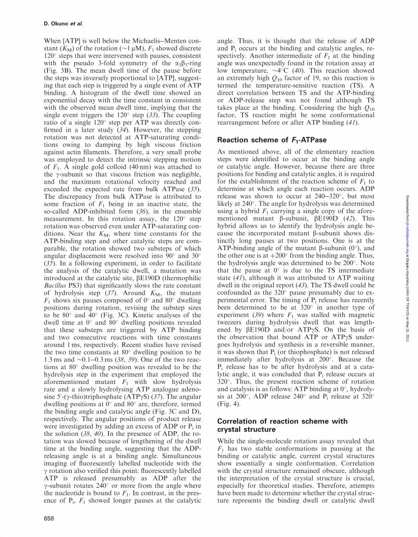
As mentioned above, all of the elementary reactionsteps were identified to occur at the binding angleor catalytic angle. However, because there are threepositions for binding and catalytic angles, it is requiredfor the establishment of the reaction scheme of F1 todetermine at which angle each reaction occurs. ADPrelease was shown to occur at 240�320�, but mostlikely at 240�. The angle for hydrolysis was determinedusing a hybrid F1 carrying a single copy of the afore-mentioned mutant b-subunit, bE190D (42). Thishybrid allows us to identify the hydrolysis angle be-cause the incorporated mutant b-subunit shows dis-tinctly long pauses at two positions. One is at theATP-binding angle of the mutant b-subunit (0�), andthe other one is at þ200� from the binding angle. Thus,the hydrolysis angle was determined to be 200�. Notethat the pause at 0� is due to the TS intermediatestate (41), although it was attributed to ATP waitingdwell in the original report (43). The TS dwell could beconfounded as the 320� pause presumably due to ex-perimental error. The timing of Pi release has recentlybeen determined to be at 320� in another type ofexperiment (39) where F1 was stalled with magnetictweezers during hydrolysis dwell that was length-ened by bE190D and/or ATPgS. On the basis ofthe observation that bound ATP or ATPgS under-goes hydrolysis and synthesis in a reversible manner,it was shown that Pi (or thiophosphate) is not releasedimmediately after hydrolysis at 200�. Because thePi release has to be after hydrolysis and at a cata-lytic angle, it was concluded that Pi release occurs at320�. Thus, the present reaction scheme of rotationand catalysis is as follows: ATP binding at 0�, hydroly-sis at 200�, ADP release 240� and Pi release at 320�
(Fig. 4).
Correlation of reaction scheme withcrystal structure
While the single-molecule rotation assay revealed thatF1 has two stable conformations in pausing at thebinding or catalytic angle, current crystal structuresshow essentially a single conformation. Correlationwith the crystal structure remained obscure, althoughthe interpretation of the crystal structure is crucial,especially for theoretical studies. Therefore, attemptshave been made to determine whether the crystal struc-ture represents the binding dwell or catalytic dwell
D. Okuno et al.
658
at Ekigaku-K
yoshitsu (UN
IV O
F T
OK
YO
) on May 31, 2011
jb.oxfordjournals.orgD
ownloaded from
(28, 44, 45). On the basis of the crystal structure, thecharacteristic interaction with the g-subunit was iden-tified in the bDP form. Cysteine residues were genetic-ally incorporated into the residues involved in thedirect b-g contact. The mutant F1 was analysed inthe rotation assay. During observation, incorporatedcysteine residues were cross-linked through disulphidebonds by infusing an oxidizing buffer to stall F1 inthe crystal structure form. The pausing angle corres-ponded to the catalytic angle. Thus, it was shown thatthe crystal structure represents the catalytic dwellingstate and that bTP, bDP and bEmpty correspond to the80�, 200� and 320� state, respectively (Fig. 4, greencircles). These data were supported by experimentsby Masaike et al. where the C-terminal domain ofthe b-subunit that undergoes the large swing motionupon ATP binding was labelled with a fluorescentdye (28). The observed angular positions in the cata-lytic dwell corresponded to those observed in the crys-tal structure. Interestingly, they found that at 240�, theb-subunit takes a new conformation, which theytermed ‘half-closed’, while at other binding angles,the b-subunit takes the same conformation as thecatalytic angles: open at 0� and closed at 120� (Fig. 4).
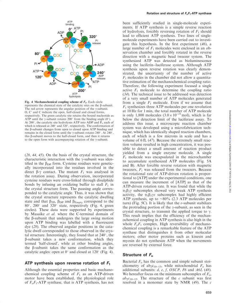
ATP synthesis upon reverse rotation of F1
Although the essential properties and basic mechano-chemical coupling scheme of F1 as an ATP-drivenmotor have been established, the physiological roleof FoF1-ATP synthase, that is ATP synthesis, has not
been sufficiently studied in single-molecule experi-ments. If ATP synthesis is a simple reverse reactionof hydrolysis, forcibly reversing rotation of F1 shouldlead to efficient ATP synthesis. Two lines of single-molecule experiments have been carried out to investi-gate this hypothesis. In the first experiment (46), alarge number of F1 molecules were enclosed in an ob-servation chamber and forcibly rotated in the reversedirection with a magnetic bead tweezer system. Thesynthesized ATP was detected as bioluminescenceusing the luciferin�luciferase system. Although ATPsynthesis upon reverse rotation was clearly demon-strated, the uncertainty of the number of activeF1 molecules in the chamber did not allow a quantita-tive estimation of the mechanochemical coupling ratio.Therefore, the following experiment focused a singleactive F1 molecule to determine the coupling ratio(34). The technical issue to be addressed was detectionof a very small number of ATP molecules generatedfrom a single F1 molecule. Even if we assume thatF1 synthesizes three ATP molecules per one revolutionat 10Hz for 1min, the total number of ATP moleculeis only 1,800 molecules (3.0�10�21mol), which is farbelow the detection limit of the luciferase assay. Toaddress this issue, a microscopic reaction chambersystem was developed using a microfabrication tech-nique, which has identically shaped reaction chambers,each of which is a few microns in scale and has avolume of 6 fL (47). Because the extremely small reac-tion volume resulted in high concentration, it was pos-sible to detect a small amount of reaction productyielded from a single enzyme molecule. A singleF1 molecule was encapsulated in the microchamberto accumulate synthesized ATP molecules (Fig. 5Aand B). After forcible reverse rotation with magnetictweezers, F1 was released from the tweezers. Becausethe rotational rate of ATP-driven rotation is propor-tional to [ATP] under the experimental conditions, onecan measure the increment of [ATP] as that of theATP-driven rotation rate. It was found that while thea3b3g subcomplex showed very weak ATP synthesisactivity, the a3b3g" subcomplex had highly efficientATP synthesis, up to �80% (2.3 ATP molecules perturn) (Fig. 5C). It is likely that the "-subunit stabilizesthe protruding portion of the g-subunit, as seen in thecrystal structure, to transmit the applied torque to g.This result implies that the efficiency of the mechan-ochemical coupling in ATP synthesis is also high in thewhole FoF1 complex. High reversibility of mechano-chemical coupling is a remarkable feature of the ATPsynthase that distinguishes it from other molecularmotors; other motor proteins such as kinesin andmyosin do not synthesize ATP when the movementsare reversed by external force.
Structure of Fo
Bacterial Fo has the common and simple subunit stoi-chiometry of ab2c10�15, while mitochondrial Fo hasadditional subunits: d, e, f, OSCP, F6 and A6L (48).We hereafter focus on the minimum subcomplex of Fo,ab2c10�15. The structure of the c subunit was firstresolved in a monomer state by NMR (49). The c
ADP
ATP
*ATP
PiATP
ADPPi
*ATP
ADPPi
*ATP
*ATP
ADPPi
Pi
0°
120°
200°
240°
Pi *ATP
ATP
Pi
*ATP ATP
80°
320°
ADP
ATP
ADPATP
Pi
Pi
ATPbinding
ATPhydrolysis
ADPrelease
Pirelease
COC
COC’
OC’C
CCO
C’OC
OCC
Fig. 4 Mechanochemical coupling scheme of F1. Each circlerepresents the chemical state of the catalytic sites on the b-subunit.The red arrow represents the angular position of the g-subunit.O, C0 and C indicate the open, half-closed and closed forms,respectively. The green catalytic site retains the bound nucleotide asATP until the g-subunit rotates 200� from the binding angle (0�).At 200�, the catalytic site hydrolyses ATP into ADP and Pi, each ofwhich is released at 240� and 320�, respectively. The conformation ofthe b-subunit changes from open to closed upon ATP binding andremains in the closed form until the g-subunit rotates 240�. At 240�,this b-subunit moves to the half-closed form, and then it returnsto the open form with accompanying rotation of the g-subunit.
Rotation and structure of FoF1-ATP synthase
659
at Ekigaku-K
yoshitsu (UN
IV O
F T
OK
YO
) on May 31, 2011
jb.oxfordjournals.orgD
ownloaded from
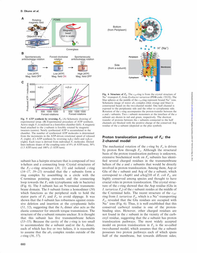
subunit has a hairpin structure that is composed of twoa-helices and a connecting loop. Crystal structures ofthe F1�c-ring structure (10, 11) and isolated c-ring(14�17, 19�21) revealed that the c subunits form aring complex by assembling in a circle with theC-terminus pointing outwards and the connectingloop towards the F1 side (cytoplasmic side in bacteria)(Fig. 6). The b subunit has an N-terminal transmem-brane domain. The b subunit forms a homodimer (50)which functions as the peripheral stalk to hold thestator parts of F1 and Fo to avoid slippage. It wasshown that the b subunit has robustness against exten-sive deletion and insertion at the cytoplasmic helix(51, 52), suggesting that the b2 dimer also acts as theelastic connector for smooth torque transmission. Thestructure of the a subunit remains unclear. It is thoughtthat this subunit has five transmembrane helices(53�55). Because the cavity of the c-ring is too smallto accommodate the a subunit and/or the b2 dimer,each of which has five or two helices, it is reasonableto assume that the ab2 complex resides outside of thec-ring (56, 57).
Proton translocation pathway of Fo: the2-channel model
The mechanical rotation of the c-ring by Fo is drivenby proton flow through Fo. Although the structuralbasis of the proton translocation pathway is unknown,extensive biochemical work on Fo subunits has identi-fied several charged residues in the transmembranehelices of the a and c subunits that would be directlyinvolved in proton translocation. Among them, Asp orGlu of the c subunit and Arg of the a subunit, whichcorrespond to cAsp61 and aArg210 of E. coli Fo, arehighly conserved among species and thought to havecrucial roles in proton translocation. The crystal struc-ture of the c-ring showed that the Asp residue (Glu inI. tartaricus Fo) of the c subunit resides at the middle ofthe C-terminal helix. The recent structure of the c10-ring from I. tartaricus Fo, which is a Naþ-transportingFo, revealed that the Glu residues are occupied withNaþ ions (Fig. 6). Thus, it is well established that thisconserved carboxyl residue is one of the proton-binding sites. However, other charged residues arenot found in the c subunit in the vicinity of the carb-oxyl residue, suggesting that the a subunit has protontranslocation pathways. The most widely acceptedmodel on proton translocation in Fo is the so-calledtwo-channel model, which assumes that the a subunitpossesses two proton pathways each of which spanshalf of the membrane, but towards different sides;
PDMSCoverglass
Rotating magnetic field
Objective lens
High [ATP]Low [ATP]
Forced clockwiserotation
Low [ATP]Mag. field OFF Mag. field ON Mag. field OFF
0
2000
4000
6000
0 500 1000 1500Pro
duce
d A
TP
/ m
olec
ules
Forced rotations0 500 1000 1500
Forced rotations
3 ATP/tu
rn
1.5 ATP/turn
0 ATP/turn
α3β3γ α3β3γε
A
B
C
Magneticbead
(not to scale)
Coverglass
F1
PDMS
Spontaneouslyanticlockwise rotation
Spontaneouslyanticlockwise rotation
Fig. 5 ATP synthesis by reversing F1. (A) Schematic drawing ofexperimental setup. (B) Experimental procedure of ATP synthesis.Active single F1 is enclosed in a femtolitre chamber (left). A magneticbead attached to the g-subunit is forcibly rotated by magnetictweezers (centre). Newly synthesized ATP is accumulated in thechamber. The number of synthesized ATP molecules is determinedfrom the increments in the ATP-driven rotational speed of releasedF1 (right). (C) ATP synthesis by reversing a3b3g (left) and a3b3g"(right). Each trace is derived from individual F1 molecules. Dottedlines indicate slopes of the coupling ratio of 0% (0 ATP/turn), 50%(1.5 ATP/turn) and 100% (3 ATP/turn).
Bottom(from Periplasm)
Side(from a-subunit)
H+ (Na+)
Membrane
Periplasm
Cytoplasm
a
b2
+
a
b2
c11+a
Fig. 6 Structure of Fo. The c10-ring is from the crystal structure ofNaþ-transport Fo from Ilyobacter tartaricus (PDB code; 1YCE). Theblue spheres at the middle of the c10-ring represent bound Naþ ions.Schematic image of stator ab2 complex (thin orange and blue) isconstructed based on the two-channel model. One half channel isexposed to the periplasmic side and the other to cytoplasmic side.Rotation of the c-ring accompanies the proton-transfer between thea and c subunits. Two c subunit monomers at the interface of the asubunit are shown in red and green, respectively. The shortcuttransfer of protons between the c subunits connected to the halfchannels are blocked with the positive charge of the conserved Argresidue of the a subunit (depicted as the plus symbol).
D. Okuno et al.
660
at Ekigaku-K
yoshitsu (UN
IV O
F T
OK
YO
) on May 31, 2011
jb.oxfordjournals.orgD
ownloaded from
the channels connect the proton-binding site of thec subunit with the periplasmic or cytoplasmic space(58�60) (Fig. 6). Notice each channel has contactwith a different c subunit, which are adjacent to eachother. In other words, the a subunit interacts withtwo c subunits, each contacting via a different halfchannel. The proposed mechanism of proton transferin ATP synthesis mode is as follows (60�62): a protonenters the half channel exposed to the periplasmic side(or intermembrane space of mitochondria) and is thentransferred to the carboxy residue of the c subunit.This protonation neutralizes the negative charge ofthe residue, allowing the c subunit to rotate apartfrom the a subunit towards the surrounding lipidlayer. At the same time, the neighbouring c subunitat the anticlockwise side returns from the lipid layerto form contacts with the other half channel, whichhas a hydrophilic environment to promote deproto-nation of the carboxyl residue. The released protonthen enters into the cytoplasmic space. The role ofthe conserved Arg in the a subunit is likely to blockthe futile rotation of the c subunit without deproto-nation by attracting only the deprotonated c subunitwith its positive charge (62, 63). In the ATP-drivenproton-pumping mode, the sequence of events isreversed.
Rotation of c-ring in Fo
After the direct observation of F1, the verification ofthe c-ring rotation against the ab2 complex becamean important issue. Although around 10 years havepassed since the verification of the c-ring rotation,little progress has been made on the rotary dynamicsof Fo, compared with F1, owing to challenges in hand-ling the complicated membrane system and diffi-culty in stably charging the membrane potential highenough to reverse F1. Although detergent-solubilizedFoF1 was subjected to the rotation assay in ATP hy-drolysis conditions in early studies (64, 65), theobserved rotation was insensitive to the gold-standardinhibitor of Fo, dicyclohexyl-carbodiimide (DCCD),implying that the observed rotating is not coupledwith the proton translocation of Fo (64, 66). The sub-unit interactions of Fo are weakened in the presenceof detergent, which often causes subunit dissoci-ation in biochemical assays (67). Actually, it has beenlater reported that the rotation in this system is insensi-tive to mutation at the conserved Arg of the a sub-unit (68). Verification of the c-ring rotation camefrom biochemical experiments showing that crosslink-age of the c-ring with the rotor subunits of F1 (g ande subunits) does not diminish ATP synthesis activity(69), while the a�c crosslink abolishes ATPase activ-ity coupled with proton translocation (70). Furtherverification was made by detection of the exchangedcross-link product between the a and c subunits, whichwas probed with a 14C-labelled c subunit (71). Single-molecule imaging of rotation under ATP synthesisconditions has also been attempted. The rotation ofFoF1 reconstituted in liposome was detected fromthe dipole moment angle of the fluorescent markerdye incorporated into a rotor subunit (72) or Forster
resonance energy transfer (FRET) efficiency betweentwo fluorescent dyes introduced into the statorand rotor subunits (73). A drawback of these experi-ments is that the membrane potential is transient and,therefore, it is very difficult to correlate the observedrotational velocity with the membrane potential.However, one essential property of Fo rotation wasrevealed with the FRET experiment: multiple stepp-ing rotation was detected that was interpreted as 36�
steps based on the 10-fold symmetry of the rotor (74).The 36� stepping rotation was later proved in therotation assay under ATP hydrolysis conditionswhere a gold nanorod was used as the rotation probe(75). FoF1 was reconstituted into a nanodisc of lipidbilayer, and the rotation was monitored from theangle of polarized scattered light along the long axisof the nanorod. However, understanding the dynamicsof Fo rotation is still in its early stages. Experimentalsystems that allow stable charging of the membranepotential simultaneously with observation of F1 rota-tion with high spatiotemporal resolution are highlyawaited.
Acknowledgements
The authors thank all members of Noji Laboratory.
FundingGrant-in-Aid for Scientific Research No. 18074005 (to H.N.);21700168 (to R.I.); the Ministry of Education, Culture, Sports,Science and Technology, Japan, special education and researchexpenses.
Conflict of InterestNone declared.
References
1. Yoshida, M., Muneyuki, E., and Hisabori, T. (2001)ATP synthase�a marvellous rotary engine of the cell.Nat. Rev. Mol. Cell. Biol. 2, 669�677
2. Kato, Y., Matsui, T., Tanaka, N., Muneyuki, E.,Hisabori, T., and Yoshida, M. (1997) ThermophilicF1-ATPase is activated without dissociation of an en-dogenous inhibitor, epsilon subunit. J. Biol. Chem. 272,24906�24912
3. Smith, J.B. and Sternweis, P.C. (1977) Purification ofmembrane attachment and inhibitory subunits of theproton translocating adenosine triphosphatase fromEscherichia coli. Biochemistry 16, 306�311
4. Sternweis, P.C. and Smith, J.B. (1980) Characterizationof the inhibitory (epsilon) subunit of theproton-translocating adenosine triphosphatase fromEscherichia coli. Biochemistry 19, 526�531
5. Iino, R., Murakami, T., Iizuka, S., Kato-Yamada, Y.,Suzuki, T., and Yoshida, M. (2005) Real-time monitor-ing of conformational dynamics of the epsilon subunit inF1-ATPase. J. Biol. Chem. 280, 40130�40134
6. Saita, E., Iino, R., Suzuki, T., Feniouk, B.A., Kinosita,K. Jr., and Yoshida, M. (2010) Activation and stiffnessof the inhibited states of F1-ATPase probed bysingle-molecule manipulation. J. Biol. Chem. 285,11411�11417
7. Suzuki, T., Murakami, T., Iino, R., Suzuki, J., Ono, S.,Shirakihara, Y., and Yoshida, M. (2003) F0F1-ATPase/synthase is geared to the synthesis mode by
Rotation and structure of FoF1-ATP synthase
661
at Ekigaku-K
yoshitsu (UN
IV O
F T
OK
YO
) on May 31, 2011
jb.oxfordjournals.orgD
ownloaded from
conformational rearrangement of epsilon subunit in re-sponse to proton motive force and ADP/ATP balance. J.Biol. Chem. 278, 46840�46846
8. Iino, R., Hasegawa, R., Tabata, K.V., and Noji, H.(2009) Mechanism of inhibition by C-terminalalpha-helices of the epsilon subunit of Escherichia coliFoF1-ATP synthase. J. Biol. Chem. 284, 17457�17464
9. Feniouk, B.A., Suzuki, T., and Yoshida, M. (2006) Therole of subunit epsilon in the catalysis and regulation ofFOF1-ATP synthase. Biochim. Biophys. Acta 1757,326�338
10. Watt, I.N., Montgomery, M.G., Runswick, M.J., Leslie,A.G., and Walker, J.E. (2010) Bioenergetic cost ofmaking an adenosine triphosphate molecule in animalmitochondria. Proc. Natl Acad. Sci. USA 107,16823�16827
11. Stock, D., Leslie, A.G., and Walker, J.E. (1999)Molecular architecture of the rotary motor in ATP syn-thase. Science. 286, 1700�1705
12. Jiang, W., Hermolin, J., and Fillingame, R.H. (2001)The preferred stoichiometry of c subunits in the rotarymotor sector of Escherichia coli ATP synthase is 10.Proc. Natl Acad. Sci. USA 98, 4966�4971
13. Mitome, N., Suzuki, T., Hayashi, S., and Yoshida, M.(2004) Thermophilic ATP synthase has a decamer c-ring:indication of noninteger 10:3 Hþ/ATP ratio and permis-sive elastic coupling. Proc. Natl Acad. Sci. USA 101,12159�12164
14. Meier, T., Polzer, P., Diederichs, K., Welte, W., andDimroth, P. (2005) Structure of the rotor ring ofF-Type Naþ-ATPase from Ilyobacter tartaricus.Science 308, 659�662
15. Stahlberg, H., Muller, D.J., Suda, K., Fotiadis, D.,Engel, A., Meier, T., Matthey, U., and Dimroth, P.(2001) Bacterial Na(þ)-ATP synthase has an undeca-meric rotor. EMBO Rep. 2, 229�233
16. Meier, T., Matthey, U., von Ballmoos, C., Vonck, J.,Krug von Nidda, T., Kuhlbrandt, W., and Dimroth, P.(2003) Evidence for structural integrity in the undeca-meric c-rings isolated from sodium ATP synthases.J. Mol. Biol. 325, 389�397
17. Meier, T., Ferguson, S.A., Cook, G.M., Dimroth, P.,and Vonck, J. (2006) Structural investigations of themembrane-embedded rotor ring of the F-ATPase fromClostridium paradoxum. J. Bacteriol. 188, 7759�7764
18. Meier, T., Morgner, N., Matthies, D., Pogoryelov, D.,Keis, S., Cook, G.M., Dimroth, P., and Brutschy, B.(2007) A tridecameric c ring of the adenosine triphos-phate (ATP) synthase from the thermoalkaliphilicBacillus sp. strain TA2.A1 facilitates ATP synthesis atlow electrochemical proton potential. Mol. Microbiol.65, 1181�1192
19. Preiss, L., Yildiz, O., Hicks, D.B., Krulwich, T.A., andMeier, T. (2010) A new type of proton coordination in anF(1)F(o)-ATP synthase rotor ring. PLoS Biol. 8,e1000443
20. Seelert, H., Poetsch, A., Dencher, N.A., Engel, A.,Stahlberg, H., and Muller, D.J. (2000) Structural biol-ogy. Proton-powered turbine of a plant motor. Nature405, 418�419
21. Pogoryelov, D., Yu, J., Meier, T., Vonck, J., Dimroth,P., and Muller, D.J. (2005) The c15 ring of the Spirulinaplatensis F-ATP synthase: F1/F0 symmetry mismatch isnot obligatory. EMBO Rep. 6, 1040�1044
22. Diez, M., Zimmermann, B., Borsch, M., Konig, M.,Schweinberger, E., Steigmiller, S., Reuter, R., Felekyan,S., Kudryavtsev, V., Seidel, C.A., and Graber, P. (2004)Proton-powered subunit rotation in single
membrane-bound F0F1-ATP synthase. Nat. Struct.Mol. Biol. 11, 135�141
23. Boyer, P.D. (1997) The ATP synthase�a splendidmolecular machine. Annu Rev Biochem. 66, 717�749
24. Wakabayashi, T., Kubota, M., Yoshida, M., andKagawa, Y. (1977) Structure of ATPase (couplingfactor TF1) from a thermophilic bacterium. J. Mol.Biol. 117, 515�519
25. Kagawa, Y., Sone, N., Yoshida, M., Hirata, H., andOkamoto, H. (1976) Proton translocating ATPase of athermophilic bacterium. Morphology, subunits, andchemical composition. J. Biochem 80, 141�151
26. Yoshida, M., Sone, N., Hirata, H., and Kagawa, Y.(1975) A highly stable adenosine triphosphatase from athermophillie bacterium. Purification, properties, and re-constitution. J. Biol. Chem. 250, 7910�7916
27. Abrahams, J.P., Leslie, A.G., Lutter, R., and Walker, J.E.(1994) Structure at 2.8 A resolution of F1-ATPase frombovine heart mitochondria. Nature 370, 621�628
28. Masaike, T., Koyama-Horibe, F., Oiwa, K., Yoshida,M., and Nishizaka, T. (2008) Cooperative three-stepmotions in catalytic subunits of F(1)-ATPase correlatewith 80 degrees and 40 degrees substep rotations.Nat. Struct. Mol. Biol. 15, 1326�1333
29. Duncan, T.M., Bulygin, V.V., Zhou, Y., Hutcheon,M.L., and Cross, R.L. (1995) Rotation of subunitsduring catalysis by Escherichia coli F1-ATPase.Proc. Natl Acad. Sci. USA 92, 10964�10968
30. Sabbert, D., Engelbrecht, S., and Junge, W. (1996)Intersubunit rotation in active F-ATPase. Nature 381,623�625
31. Noji, H., Yasuda, R., Yoshida, M., and Kinosita, K. Jr.(1997) Direct observation of the rotation of F1-ATPase.Nature 386, 299�302
32. Hayashi, K., Ueno, H., Iino, R., and Noji, H. (2010)Fluctuation theorem applied to F1-ATPase. Phys. Rev.Lett. 104, 218103
33. Yasuda, R., Noji, H., Kinosita, K. Jr., and Yoshida, M.(1998) F1-ATPase is a highly efficient molecular motorthat rotates with discrete 120 degree steps. Cell 93,1117�1124
34. Rondelez, Y., Tresset, G., Nakashima, T., Kato-Yamada, Y., Fujita, H., Takeuchi, S., and Noji, H.(2005) Highly coupled ATP synthesis by F1-ATPasesingle molecules. Nature 433, 773�777
35. Yasuda, R., Noji, H., Yoshida, M., Kinosita, K. Jr., andItoh, H. (2001) Resolution of distinct rotational substepsby submillisecond kinetic analysis of F1-ATPase. Nature410, 898�904
36. Hirono-Hara, Y., Noji, H., Nishiura, M., Muneyuki, E.,Hara, K.Y., Yasuda, R., Kinosita, K. Jr., and Yoshida, M.(2001) Pause and rotation of F(1)-ATPase during catalysis.Proc. Natl Acad. Sci. USA 98, 13649�13654
37. Shimabukuro, K., Yasuda, R., Muneyuki, E., Hara,K.Y., Kinosita, K. Jr., and Yoshida, M. (2003)Catalysis and rotation of F1 motor: cleavage of ATPat the catalytic site occurs in 1 ms before 40 degree sub-step rotation. Proc. Natl Acad. Sci. USA 100,14731�14736
38. Adachi, K., Oiwa, K., Nishizaka, T., Furuike, S., Noji,H., Itoh, H., Yoshida, M., and Kinosita, K. Jr. (2007)Coupling of rotation and catalysis in F(1)-ATPase re-vealed by single-molecule imaging and manipulation.Cell 130, 309�321
39. Watanabe, R., Iino, R., and Noji, H. (2010) Phosphaterelease in F1-ATPase catalytic cycle follows ADP release.Nat. Chem. Biol. 6, 814�820
D. Okuno et al.
662
at Ekigaku-K
yoshitsu (UN
IV O
F T
OK
YO
) on May 31, 2011
jb.oxfordjournals.orgD
ownloaded from
40. Watanabe, R., Iino, R., Shimabukuro, K., Yoshida, M.,and Noji, H. (2008) Temperature-sensitive reaction inter-mediate of F1-ATPase. EMBO Rep. 9, 84�90
41. Enoki, S., Watanabe, R., Iino, R., and Noji, H. (2009)Single-molecule study on the temperature-sensitive reac-tion of F1-ATPase with a hybrid F1 carrying a singlebeta(E190D). J. Biol. Chem. 284, 23169�23176
42. Ariga, T. (2008) The concerted nature between threecatalytic subunits driving the F1 rotary motor.Biosystems 93, 68�77
43. Ariga, T., Muneyuki, E., and Yoshida, M. (2007)F1-ATPase rotates by an asymmetric, sequential mech-anism using all three catalytic subunits. Nat. Struct. Mol.Biol. 14, 841�846
44. Okuno, D., Ikeguchi, M., and Noji, H. (2010)Measurement of the conformational state of F(1)-ATPase by single-molecule rotation. Methods Enzymol.475, 279�296
45. Sielaff, H., Rennekamp, H., Engelbrecht, S., and Junge,W. (2008) Functional halt positions of rotaryFOF1-ATPase correlated with crystal structures.Biophys. J. 95, 4979�4987
46. Itoh, H., Takahashi, A., Adachi, K., Noji, H., Yasuda,R., Yoshida, M., and Kinosita, K. (2004) Mechanicallydriven ATP synthesis by F1-ATPase. Nature 427,465�468
47. Rondelez, Y., Tresset, G., Tabata, K.V., Arata, H.,Fujita, H., Takeuchi, S., and Noji, H. (2005)Microfabricated arrays of femtoliter chambers allowsingle molecule enzymology. Nat. Biotechnol. 23,361�365
48. Collinson, I.R., van Raaij, M.J., Runswick, M.J.,Fearnley, I.M., Skehel, J.M., Orriss, G.L., Miroux, B.,and Walker, J.E. (1994) ATP synthase from bovine heartmitochondria. In vitro assembly of a stalk complex in thepresence of F1-ATPase and in its absence. J. Mol. Biol.242, 408�421
49. Girvin, M.E., Rastogi, V.K., Abildgaard, F., Markley,J.L., and Fillingame, R.H. (1998) Solution structureof the transmembrane Hþ-transporting subunitc of the F1F0 ATP synthase. Biochemistry 37, 8817�8824
50. Dunn, S.D., McLachlin, D.T., and Revington, M. (2000)The second stalk of Escherichia coli ATP synthase.Biochim. Biophys. Acta 1458, 356�363
51. Sorgen, P.L., Bubb, M.R., and Cain, B.D. (1999)Lengthening the second stalk of F(1)F(0) ATP synthasein Escherichia coli. J. Biol. Chem. 274, 36261�36266
52. Sorgen, P.L., Caviston, T.L., Perry, R.C., and Cain, B.D.(1998) Deletions in the second stalk of F1F0-ATP syn-thase in Escherichia coli. J. Biol. Chem. 273,27873�27878
53. Long, J.C., Wang, S., and Vik, S.B. (1998) Membranetopology of subunit a of the F1F0 ATP synthase asdetermined by labeling of unique cysteine residues.J. Biol. Chem. 273, 16235�16240
54. Valiyaveetil, F.I. and Fillingame, R.H. (1998)Transmembrane topography of subunit a in theEscherichia coli F1F0 ATP synthase. J. Biol. Chem.273, 16241�16247
55. Wada, T., Long, J.C., Zhang, D., and Vik, S.B. (1999)A novel labeling approach supports thefive-transmembrane model of subunit a of theEscherichia coli ATP synthase. J. Biol. Chem. 274,17353�17357
56. Rubinstein, J.L., Walker, J.E., and Henderson, R. (2003)Structure of the mitochondrial ATP synthase by electroncryomicroscopy. EMBO J. 22, 6182�6192
57. Takeyasu, K., Omote, H., Nettikadan, S., Tokumasu, F.,Iwamoto-Kihara, A., and Futai, M. (1996) Molecularimaging of Escherichia coli F0F1-ATPase in recons-tituted membranes using atomic force microscopy.FEBS Lett. 392, 110�113
58. Elston, T., Wang, H., and Oster, G. (1998) Energy trans-duction in ATP synthase. Nature 391, 510�513
59. Junge, W., Lill, H., and Engelbrecht, S. (1997) ATPsynthase: an electrochemical transducer with rotatorymechanics. Trends Biochem. Sci. 22, 420�423
60. Vik, S.B. and Antonio, B.J. (1994) A mechanism ofproton translocation by F1F0 ATP synthases suggestedby double mutants of the a subunit. J. Biol. Chem. 269,30364�30369
61. Dimroth, P., von Ballmoos, C., and Meier, T. (2006)Catalytic and mechanical cycles in F-ATP synthases.Fourth in the Cycles Review Series. EMBO Rep. 7,276�282
62. Oster, G. and Wang, H. (2003) Rotary protein motors.Trends Cell Biol. 13, 114�121
63. Mitome, N., Ono, S., Sato, H., Suzuki, T., Sone, N., andYoshida, M. (2010) Essential arginine residue ofthe F(o)-a subunit in F(o)F(1)-ATP synthase has arole to prevent the proton shortcut without c-ringrotation in the F(o) proton channel. Biochem J. 430,171�177
64. Panke, O., Gumbiowski, K., Junge, W., andEngelbrecht, S. (2000) F-ATPase: specific observationof the rotating c subunit oligomer of EF(o)EF(1).FEBS Lett. 472, 34�38
65. Sambongi, Y., Iko, Y., Tanabe, M., Omote, H.,Iwamoto-Kihara, A., Ueda, I., Yanagida, T., Wada,Y., and Futai, M. (1999) Mechanical rotation of the csubunit oligomer in ATP synthase (F0F1): direct obser-vation. Science 286, 1722�1724
66. Tanabe, M., Nishio, K., Iko, Y., Sambongi, Y.,Iwamoto-Kihara, A., Wada, Y., and Futai, M. (2001)Rotation of a complex of the gamma subunit and cring of Escherichia coli ATP synthase. The rotor andstator are interchangeable. J. Biol. Chem. 276,15269�15274
67. Tsunoda, S.P., Aggeler, R., Noji, H., Kinosita, K. Jr.,Yoshida, M., and Capaldi, R.A. (2000) Observations ofrotation within the F(o)F(1)-ATP synthase: deciding be-tween rotation of the F(o)c subunit ring and artifact.FEBS Lett. 470, 244�248
68. Hosokawa, H., Nakanishi-Matsui, M., Kashiwagi, S.,Fujii-Taira, I., Hayashi, K., Iwamoto-Kihara, A.,Wada, Y., and Futai, M. (2005) ATP-dependent rotationof mutant ATP synthases defective in proton transport.J. Biol. Chem. 280, 23797�23801
69. Tsunoda, S.P., Aggeler, R., Yoshida, M., and Capaldi,R.A. (2001) Rotation of the c subunit oligomer in fullyfunctional F1Fo ATP synthase. Proc. Natl Acad. Sci.USA 98, 898�902
70. Suzuki, T., Ueno, H., Mitome, N., Suzuki, J., andYoshida, M. (2002) F(0) of ATP synthase is a rotaryproton channel. Obligatory coupling of proton trans-location with rotation of c-subunit ring. J. Biol. Chem.277, 13281�13285
71. Hutcheon, M.L., Duncan, T.M., Ngai, H., and Cross,R.L. (2001) Energy-driven subunit rotation at the inter-face between subunit a and the c oligomer in the F(O)sector of Escherichia coli ATP synthase. Proc. Natl Acad.Sci. USA 98, 8519�8524
72. Kaim, G., Prummer, M., Sick, B., Zumofen, G., Renn,A., Wild, U.P., and Dimroth, P. (2002) Coupled rotation
Rotation and structure of FoF1-ATP synthase
663
at Ekigaku-K
yoshitsu (UN
IV O
F T
OK
YO
) on May 31, 2011
jb.oxfordjournals.orgD
ownloaded from
within single F0F1 enzyme complexes during ATP syn-thesis or hydrolysis. FEBS Lett. 525, 156�163
73. Borsch, M., Diez, M., Zimmermann, B., Reuter, R.,and Graber, P. (2002) Stepwise rotation of thegamma-subunit of EF(0)F(1)-ATP synthase obser-ved by intramolecular single-molecule fluores-cence resonance energy transfer. FEBS Lett. 527,147�152
74. Duser, M.G., Zarrabi, N., Cipriano, D.J., Ernst, S.,Glick, G.D., Dunn, S.D., and Borsch, M. (2009) 36 de-grees step size of proton-driven c-ring rotation inFoF1-ATP synthase. EMBO J. 28, 2689�2696
75. Ishmukhametov, R., Hornung, T., Spetzler, D., andFrasch, W.D. (2010) Direct observation of stepped pro-teolipid ring rotation in E. coli F(o)F(1)-ATP synthase.EMBO J. 29, 3911�3923
D. Okuno et al.
664
at Ekigaku-K
yoshitsu (UN
IV O
F T
OK
YO
) on May 31, 2011
jb.oxfordjournals.orgD
ownloaded from


![ATP Synthase Subunit a Supports Permeability Transition in ...Mitochondrial ATP synthase, an enzyme that provides cellular energy in the form of ATP, is composed of 17 subunits [1]](https://img.pdfslide.net/doc/110x75/5f101bf57e708231d4477d9e/atp-synthase-subunit-a-supports-permeability-transition-in-mitochondrial-atp.jpg)