Embed Size (px)
Citation preview
L O C A L I Z A T I O N OF A D E N O S I N E
T R I P H O S P H A T A S E A C T I V I T Y I N T H E
R A T S P E R M T A I L AS R E V E A L E D BY
E L E C T R O N M I C R O S C O P Y
T O S H I O N A G A N O , M.D.
From the Department of Anatomy, School of Medicine, Chiba University, Chiba, Japan
A B S T R A C T
The epididymides of rat testis were fixed in glutaraldehyde and cut as frozen sections. The sections were incubated in lead nitrate solution containing as a substrate either ATP, AMP, creatinine phosphate, beta glycerophosphate, or phenyl phosphate. Then they were post- fixed in osmium tetroxide, embedded, sectioned, and examined with the electron micro- scope. In the sperm tail, when A T P is used as a substrate the reaction product (lead phos- phate) is observed both in the tail filament complex and on the surface membrane of the mitochondrial helix of the middle piece. In the tail filament complex, this product is seen near the nine paired peripheral and two central filaments, and in the matrix between the outer coarse fibers. But the product is not observed within these filaments and fibers. In longitudinal sections, no periodicity of the deposits in the complex is observed. When the other phosphate compounds are used as substrates the reaction products appear on the surface membrane of the mitochondrial helix, and are not found in the tail filament com- plex. No distinctly different localization of the reaction products is observed when sub- strates other than A T P are used. Possible relationships between the structure and the func- tion of the sperm tail are discussed in the light of these findings.
I N T R O D U C T I O N
Since the introduction of aldehyde fixatives for electron microscopy, it has been possible to demon- strate the localization of some enzymatic activities in cells and tissues at the electron microscope level (8, 9, 14, 26, 28).
Nelson, in 1958, published the first report on the electron microscopic localization of adenosine triphosphatase (ATPase) activity in the rat spermatozoon (23). He used a freeze-drying pro- cedure and a calcium method. He has also re- ported on the localization of other enzymes in the sperm tail (24). Daems and co-workers have noted ATPase activity in the Drosophila sperm tail prefixed with osmium tetroxide (6). Recently,
Tice and Barrnett have reported on the localiza- tion of rat testicular phosphatases, but these enzymes were not observed in the flagellum and surrounding mitochondria of the developing spermatid (33). From biochemical studies it is known that the motile sperm possesses not only A T P and ATPase but also succinic dehydrogenase, cytochrome oxidase, and other enzymes (19, 31). These enzymes are chiefly concentrated in the middle piece of the sperm tail (25). Morphological analyses of sperm tail movement have been per formed in considerable detail by electron micros- copy (1, 10, 11, 29, 30). It has been suggested that the sperm tail filaments might be contractile,
101
on December 26, 2018jcb.rupress.org Downloaded from http://doi.org/10.1083/jcb.25.2.101Published Online: 1 May, 1965 | Supp Info:
with A T P - A T P a s e serving as an energ iz ing m e c h -
anism, as in muscle con t r ac t ion (4, 10). This pape r will descr ibe some e lec t ron micro-
scope observat ions on the ra t s p e r m tail in the ep id idymis i n c u b a t e d so as to reveal ATP-sp l i t t i ng a n d o the r enzymes after g lu t a ra ldehyde fixation.
M A T E R I A L S A N D M E T H O D S
Young adult malc rats o f thc Wistar strain wcrc used in this study. Unde r ether anesthesia, both the caput cpididymis and the cauda cpididymis of the testes were removed and cut into blocks about 3 m m in diameter. The blocks wcrc fixed for 1 to 2 hours in cold 6 per cent glutaraldchydc buffered to p H 7.2 with 0.1 M cacodylatc, as recommended by Sabatini et al. (28). No sucrose was added to the fixative. Aftcr fixation, the blocks were washcd, frozen, and sectioned at about 50 D with a s tandard freezing microtomc. The sections were incubatcd at room temperature for 5 to 60 minutes in Wachstcin and Mcisel's mixture (35) at p H 7.2. ATP, adenosinc- 5 ' -phosphatc (AMP), crcatinine phosphate, and beta glycerophosphatc werc used as substratcs. M61bert's med ium (21) containing phenyl phosphatc at p H 7.6 for demonstrat ing alkaline phosphatasc was also employed. Aftcr incubation, the sections wcrc washcd, postfixcd for 1 hour in cold 2 per ccnt os- mium tctroxidc buffcrcd with s-collidine (3), dehy- dra ted with ethanol, and embedded in Epon 812 (18).
Some sections ware incubated in substrata-free media as controls. Other sections were immersed in
2 per cent osmium tetroxide for 1 hour before incuba- tion. In some cases, 5 X 10 -4 M p-chloromercuri- benzoate for sulfhydryl group inhibition was added to Wachstein and Meiscl's mixture containing ATP. This produced no observable effect in the present study.
Embedded blocks were oriented in a convenient direction and cut with a Porter-Blum microtome. Most of the thin sections were immersed in 2 per cent uranyl acetate (36) or lead solution (20), or in both, for counterstaining. Sections ,~ere examined with a Hitachi 11A or J E M 4C electron microscope. Some frozen sections were treated with ammonium sulfide after incubation for light microscope examination.
O B S E R V A T I O N S
T h e reac t ion p roduc t ( lead phospha te ) is found as granules a b o u t 30 m g in d iamete r . T h c p roduc t
does not a p p e a r in the contro l p repa ra t ions (Fig.
1). A l t h o u g h morpho log ica l and func t iona l dif- fe ren t ia t ion of spc rm cclls pass ing t h r o u g h the cp id idymis has bccn repor ted (5, 12), cy tochemica l dif ferences in the spe rm tails could no t bc dc- tec tcd in thc c a p u t cp id idymis and c a u d a cp id idy- mis in the p rcsen t study. Fo r convenicnce , thc
n o m c n c l a t u r c in this p a p e r is based on Fawce t t ' s review (10).
A T P as a Substrate
W h e n A T P is used as a substratc , thc reac t ion
p roduc t is obscrved in the fol lowing threc por t ions
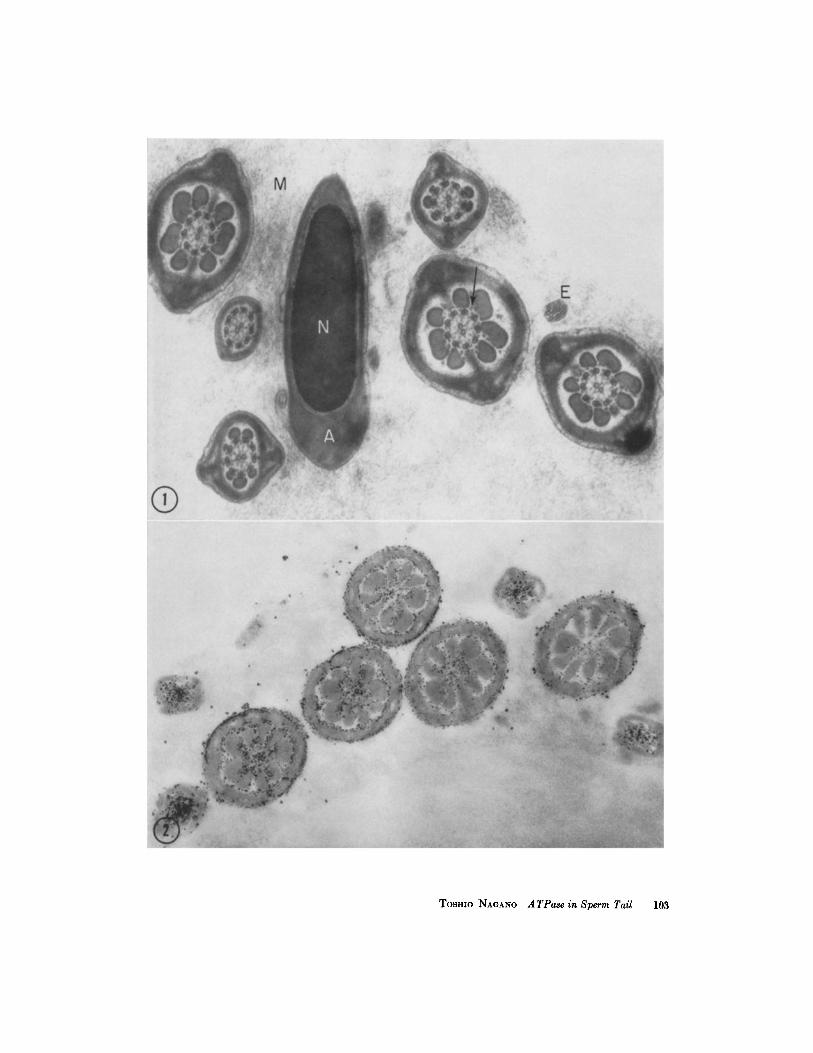
All electron micrographs are of sections of rat epididymis. The specimens were fixed in glutaraldehyde and frozen-sectioned, and the sections were incubated, postfixed in osmium tetroxide, and embedded in Epon 81~. Thin sections were stained with uranyl aaatate followed by the lead solution of Millonig (~0), or otherwise as indicated.
FIGURE 1 A control section incubated for 80 minutes in Wachstein and Meisel's mixture without a substrate, showing cross-sections of the main pieces of sperm tails and a section through one sperm head. No reaction product is seen in the sperm tail or in the nucleus (N) with the acrosome (A). The axial filaments, consisting of two central and nine paired peripheral filaments, are illustrated. Each peripheral pair is subdivided into a dense and a light subfiber. Surrounding the axial filaments, the outer coarse fibers and the fibrous sheath are seen. The spokelike structures between the central and peripheral filaments, and a gran- ular component (arrow) between the outer coarse fibers are visible. The tip of an end piece is indicated by E. Outside the sperm cells, a fine filamentous component in the lumen of the epididymis is interpreted as representing a mucous substance (M).)< 40,000.
FIGURE ~ Incubated for 80 minutes with ATP as substrate. This section was not stained. I t shows cross-sections of the middle and main pieces of sperm tails. The reaction product (lead phosphate) is deposited as granules about 30 mg in diameter in the central area of the sperm tail and along the surface of the mitochondrial sheath of the middle piece. )< 30,000.
102 THE JOURNAL OF CELL BIOLOGY - VOLUME ~5, 1965
Tosmo NAGANO A TPase in Sperm Tail 103
of the sperm tail: in the axial f i lament complex, between the outer coarse fibers, and on the surface m e m b r a n e of mi tochondr ia in the middle piece (Figs. 2 to 9). In cross-sections, the react ion de- posits are found near the central and per ipheral filaments and in the mat r ix sur rounding the outer coarse fibers (Figs. 5 and 6). No deposit appears within the axial filaments themselves, bu t one can see the react ion product in the vicinity of the fila- ments. Relat ively little react ion product is found among the outer coarse fibers of the middle and main pieces. I t seems tha t the deposits are fewer in the per ipheral area between these fibers t han in the centra l area (Figs. 5 and 6). A granular componen t between the outer coarse fibers is visible even when specimens are incuba ted wi thout a substrate (Fig. 1). This has been recognized after OsO4 fixation (30). In the middle piece, m a n y deposits can be seen on bo th the outer and inner surfaces of the mi tochondr ia l helix sur rounding the axial f i lament complex and the outer coarse fibers. However, reaction product granules are not found within the mi tochondr ia themselves (Figs. 2 to 7). In the ma in piece, the deposits are not seen in the fibrous sheath nor on the plasma m e m b r a n e (Figs. 4 to 6, 8, and 9). In the end piece, a react ion product is not clearly observed. In the head region of the spermatozoon, a few deposits are observed on the acrosomal m e m b r a n e enclosing the acrosome, bu t not inside the nucleus (Fig. 5).
W h e n epithelial cells of the epididymis are ex- amined, the deposits are observed within the microvilli. However, the n u m b e r of deposits is far less t han in the sperm tail in the same section (Fig. 4).
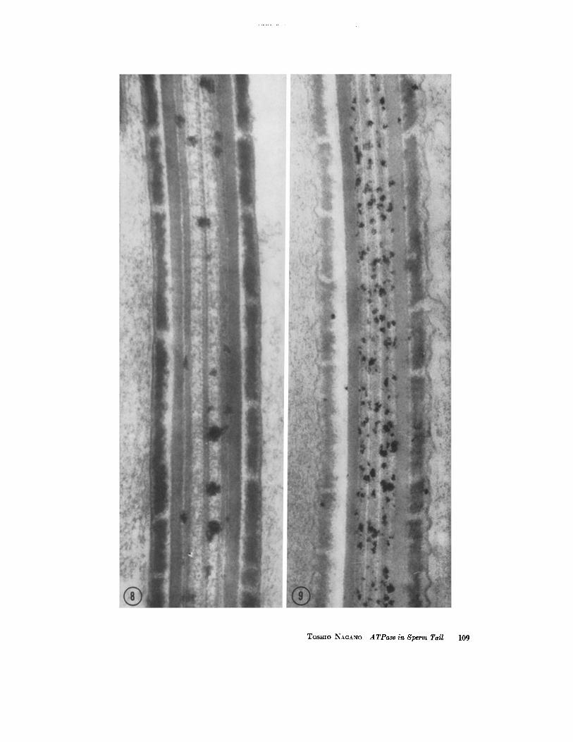
In longi tudinal sections of sperm tails, the reac- t ion deposits seem to be dis tr ibuted longi tudinal ly at r a n d o m in the axial f i lament complex and a round the mitochondria . No periodicity of the
deposits can be observed (Figs. 8 and 9). The reac-
t ion product is not seen in the segmented fibrous sheath or on the plasma membrane .
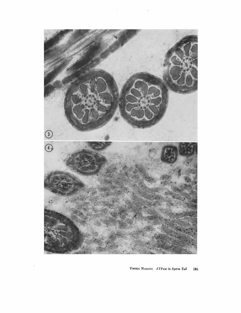
W h e n the dura t ion of incubat ion is short, it seems tha t the product appears first on the mito- chondr ia l surface, and not in the tail f i lament complex (Fig. 3).
Other Phosphate Compounds as Substrates
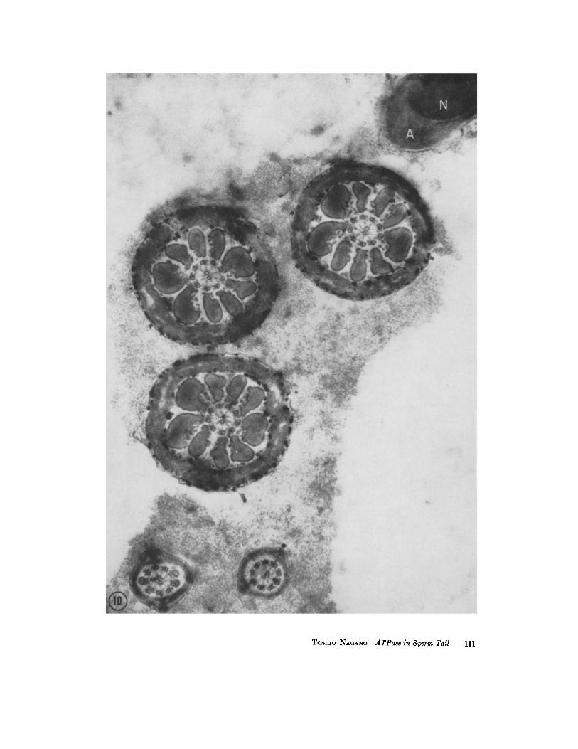
The react ion product appears similarly lo- calized in the sperm tail after AMP, creat inine phosphate , be ta glycerophosphate, or phenyl phosphate is used as a substrate. W h e n these sub- strates are used, however, the react ion deposits are dis tr ibuted on the mi tochondr ia l surface in the middle piece of the sperm tail, bu t are seen very sparingly if at all in the tail f i lament complex. This is the ma in difference in the localization of deposits when these phosphates are used as sub- strates instead of ATP. Fig. 10 shows cross-sec- tions of the middle and main pieces and the head after phenyl phosphate has been used as a sub- strate. In this figure, a few deposits are seen near the axial filaments of the ma in piece.
D I S C U S S I O N
T h e results of electron microscopic observations on the localization of ATPase activity in cells seem to differ among the various investigators. Con- flicting results have been obtained even in the same kind of tissue, muscular tissue for example. Tice and Barrnet t have described the localization of ATPase in the A band of ra t cardiac muscle prefixed or not prefixed with hydroxyadipalde- hyde (32). In contrast, de Beyer et al. have re- ported tha t ATPase localization is in the Z band of mouse hear t muscle briefly prefixed in osmium tetroxide (7). Fur ther , Hor i and Takahash i (15) have demonst ra ted ATPase activity in the A band and in the sarcoplasmic re t iculum as well as in the
FIGURE 3 Incubated for 5 minutes with ATP as substrate. A few granules of the reaction product are seen on the surface of the mitochondrial sheath in the middle pieces of sperm tails sectioned both transversely and obliquely. No deposits are seen in the axial filament complex or among the outer coarse fibers in this section. X 45,000.
FIGIYRE 4 Incubation for 30 minutes with ATP as substrate. The section shows sperm tails and the microvilli of the epithelium of the epididymis. The reaction product is seen in the sperm tall and on its mitochondrial surface. The product is also seen within the micro- villi, where the number of granules is much less than in the sperm tails. X SS,000.
104 THE JOURNAL OF CELL BIOLOGY • VOLUME ~5, 1965
TOSHIO NAGANO A TPase in Sperm Tail 105
mitochondria of rat skeletal muscle treated by freezing-substitution. In rat colonic epithelium prefixed in formalin, Otero-Vilardeb6 et al., using the same technique, have shown that ATPase is localized on the cristae of mitochondria as well as on the microvilli and the cell membrane, al- though in the distal tubule of rat kidney the mitochondria show no reaction product (27). Lansing and Lamy have reported that in rotifer cilia prefixed in OsO4 for 6 minutes the reaction granules of ATPase appear in the vicinity of cer- tain peripheral filaments (16). In the Drosophila sperm tail briefly prefixed in OsO4, Daems et al. have observed that the ATPase activity is located in the "intersatellite space," outside the peripheral filaments (6). These diverse results may very well reflect differences in tissue treatment and fixation and the use of different tissues from various species of experimental animals.
In our results with rat sperm tails, when A T P is used as a substrate significant deposits of lead phosphate appear in the axial filament complex, in the matrix between the outer coarse fibers, and on the surface of the mitochondrial sheath. The surface membrane of the mitochondria also shows the deposits when other phosphates are used as substrates. The axial filament complex and ttie matrix between the coarse fibers do not show the deposits when phosphates other than A T P are used as substrates.
The deposits are also found within the micro- villi of the epididymis epithelium when A T P is used as a substrate, a result similar to that re- ported in the proximal tubule cells of rat kidney by Wachstein and Besen (34). Since the number of deposits is much greater in the sperm tail than in the epithelial microvilli in the same section (Fig. 4), the activity of the ATPase is considered to be higher in the sperm tail than in the microvilli.
With respect to the rat spermatozoon, Nelson (23) has stated that "ATPase is confined to the nine longitudinal fibers of the outer axial fiber
bundle. In none of these is there any darkening of the cortical helix. However, when glycerophos- phate is substituted for ATP, regions of the helix do show signs of deposition of Ca-phosphate where- as the fibers do not show increase in density." He suggested that the phosphatase activity is in the mitochondrial helix. I t is worth noting that his nine longitudinal fibers probably would not corre- spond to the nine peripheral pairs of filaments described in the present paper, but rather are to be identified with the outer coarse fibers, to judge from his micrographs (23, 25). Although inhibi- tion tests have not been completed in this study, the observation that the axial filament complex shows reaction product only when A T P is used as a substrate suggests that ATPase is located in this complex, as well as in the matrix between the outer coarse fibers, but probably not within the filaments and fibers themselves. Although ATPase and other phosphatases have not been observed in the spermatid flagellum in the rat testis after glutaraldehyde fixation (33), our results might be explained on the assumption that the sperm tail in the epididymis has greater enzymatic develop- ment than the spermatid flagellum in the testis.
Mitochondrial ATPase activity has been demon- strated in some tissues at the fine structural level, whether the tissues have been treated with alde- hyde fixatives (2, 17, 27) or prepared without chemical fixatives (15, 17, 29). However, none of these reports has demonstrated that the activity is on the surface membrane of the mitochondria rather than on the cristae. Moreover, when other substrates are substituted for ATP, the reaction product appears also on the surface membrane of the mitochondria. On this surface there may be phosphatase with the capacity to cleave the link- ages in several different kinds of phosphates, including ATP. Similar phosphatase activity has been reported in the Golgi complex of the rat spermatid (33). As shown in the sperm tails in Fig. 3, the reaction products appear first on the
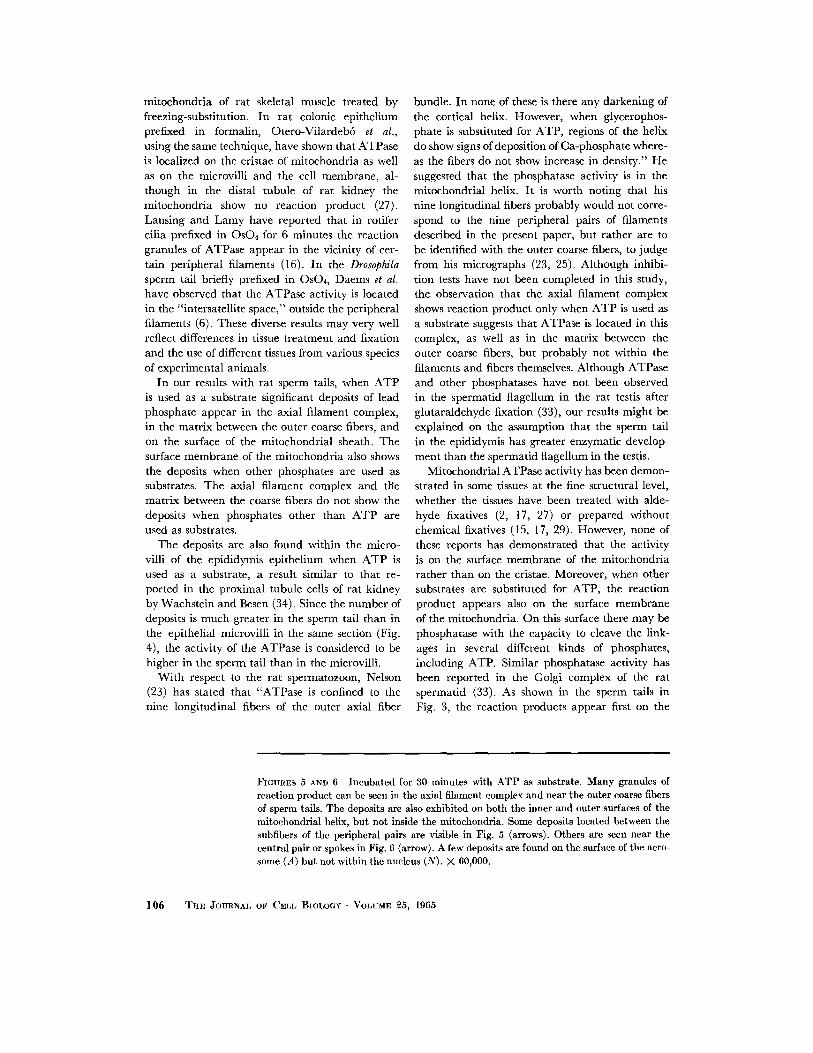
FIGURES 5 AND 6 Incubated for 30 minutes with ATP as substrate. Many granules of reaction product can be seen in the axial filament complex and near the outer coarse fibers of sperm tails. The deposits are also exhibited on both the inner and outer surfaces of the mitochondrial helix, bat not inside the mitochondria. Some deposits located between the subfibers of the peripheral pairs are visible in Fig. 5 (arrows). Others are seen near the central pair or spokes in Fig. 6 (arrow). A few deposits are found on the surface of the acro- some (A) but not within the nucleus (N). X 60,000.
106 THE JOURNAL OF CELL BIOLOGY - VOLUME ~5, 1965
Tosaio NAGANO A TPase in Sperm Tail 107
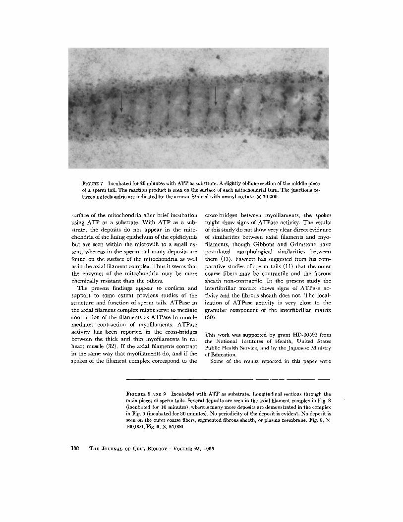
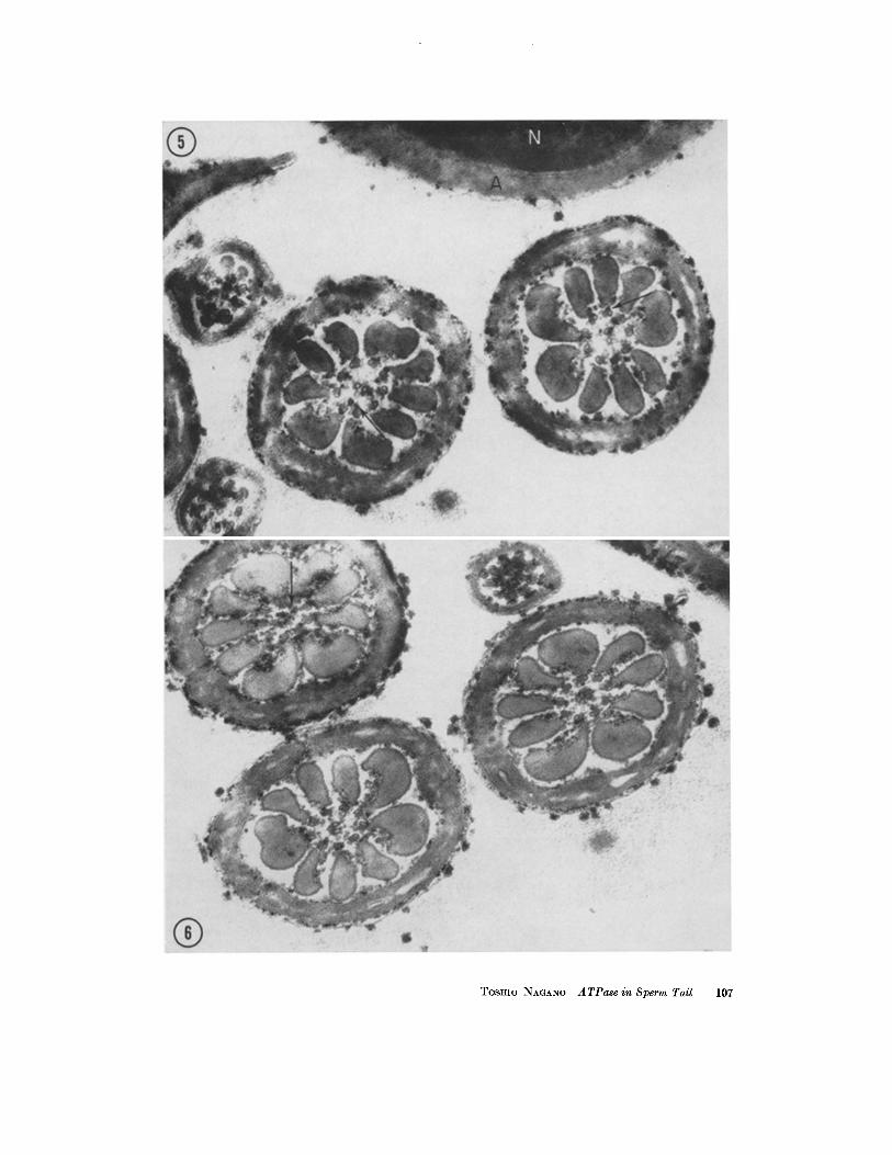
FIGUaE 7 Incubated for ~0 minutes with ATP as substrate. A slightly oblique section of the middle piece of a sperm tail. The reaction product is seen on the surface of each mitochondrial turn. The junctions be- tween mitochondria are indicated by the arrows. Stained with uranyl acetate. X 70,000.
surface of the mi tochondr ia after brief incubat ion using A T P as a substrate. Wi th A T P as a sub- strate, the deposits do not appear in the mito- chondr ia of the l ining epi thel ium of the epididymis but are seen wi th in the microvill i to a small ex- tent, whereas in the sperm tail m a n y deposits are found on the surface of the mi tochondr ia as well as in the axial f i lament complex. Thus it seems tha t the enzymes of the mi tochondr ia may be more chemically resistant than the others.
The present findings appear to confirm and suppor t to some extent previous studies of the structure and function of sperm tails. ATPase in the axial f i lament complex might serve to mediate contrac t ion of the filaments as ATPase in muscle mediates contract ion of myofilaments. ATPase activity has been reported in the cross-bridges between the thick and th in myofi laments in ra t hear t muscle (32). If the axial fi laments contract in the same way tha t myofi laments do, and if the spokes of the f i lament complex correspond to the
cross-bridges between myofilaments, the spokes might show signs of ATPase activity. The results of this s t u d y d o not show very clear direct evidence of similarities between axial filaments and myo- filaments, though Gibbons and Grimstone have postulated morphological similarities between them (13). Fawcet t has suggested from his com- parat ive studies of sperm tails (11) tha t the outer coarse fibers may be contract i le and the fibrous sheath non-contract i le . In the present study the interf ibri l lar mat r ix shows signs of ATPase ac- tivity an d the fibrous sheath does not, The local- ization of ATPase activity is very close to the granular componen t of the interfibri l lar mat r ix (30).
This work was supported by grant HD-00593 from the National Institutes of Health, United States Public Health Service, and by the Japanese Ministry of Education.
Some of the results reported in this paper were
FIGURES 8 AND 9 Incubated with ATP as substrate. Longitudinal sections through the main pieces of sperm tails. Several deposits are seen in the axial filament complex in Fig. 8 (incubated for 10 minutes), whereas many more deposits are demonstrated in the complex in Fig. 9 (incubated for 30 minutes). No periodicity of the deposit is evident. No deposit is seen on the outer coarse fibers, segmented fibrous sheath, or plasma membrane. Fig. 8, X 100,000; Fig. 9, X 85,000.
108 TH~ JOURNAL OF CELL BIOLOGY • VOLUME ~5, 1965
TOSHIO NAaANO ATPase in 8perm Tail 109
presented at the 10th Symposium of the Society of Electron Microscopy, Japan, 1963, and at the 14th Symposium of the Society for Cellular Chemistry, Japan, 1963.
R E F E R E N C E S
1. AFZELIUS, B. A., Electron microscopy of the sperm tail. Results obtained with a new fixa- tive, J. Biophysic. and Biochem. Cytol., 1959, 5, 269.
2. ASHWORTH, C. T., LUIBEL, F. J., and STEWART, S. C., The fine structural localization of adenosine triphosphatase in the small in- testine, kidney, and liver of the rat, J. Cell Biol., 1963, 17, I.
3. BENNETT, H. S., and LUFT, J. H., s-Collidine as a basis for buffering fixatives, J. Biophysic. and Biochem. Cytol., 1959, 6, 113.
4. BISHOP, D. W., Reactivation of extracted sperm cell models in relation to the mechanism of motility, in Spermatozoan Motility, (D. W. Bishop, editor), Washington, D, C., American Association for the Advancement of Science, 1962, p. 251.
5. BLANDAU, R. J., and RUMERY, R. E., Fertilizing capacity of rat spermatozoa recovered from various segments of the epididymis, Anat. Record, 1961, 139, 209.
6. DAEMS, W. T., PERSIJN, J. P., and TATES, A. D., Fine-structural localization of ATPase ac- tivity in mature sperm of Drosophila melano- gaster, Exp. Cell Research, 1963, 32, 163.
7. DE BEYER, J. M., DE MAN, J. C. H., and PERSIJN, J. P., ATPase activity on the intercalated disc and C, bands of mouse heart muscle, J. Cell Biol., 1962, 13,452.
8. EPSTEIN, M. A., and HOLT, S. J. , The localiza- tion by electron microscopy of HeLa cell surface enzymes splitting adenosine triphos- pate, J. Cell Biol., 1963, 19, 325.
9. ESSNER, E., and NOVIKOFF, A. B., Localization of acid phosphatase activity in hepatic lyso- somes by means of electron microscopy, J. Biophysic. and Biochem. Cytol., 1961, 9, 773.
10. FAWCETT, D. M., Cilia and flagella, in The Cell,
The author wishes to thank Dr. H. Stanley Bennett for suggestions during the course of this study and re- vision of this manuscript.
Received for publication, June 15, 1964.
(j . Brachet and A. E. Mirsky, editors), New York, Academic Press, Inc., 1961, 2, 217.
11. FAWCETT, D. W., Sperm tail structure in relation to the mechanism of movement, in Spermato- zoan Motility, (D. W, Bishop, editor), Wash- ington, D. C., American Association for the Advancement of Science, 1962, p. 147.
12. FAWCETT, D. W., and HOLLENBERG, R. D., Changes in the acrosome of guinea pig sperma- tozoa during passage through the epididymis, Z. Zellforseh., 1963, 60, 276.
13. GIBBONS, I. R., and GRIMSTONE, A. V., On flagellar structure in certain flagellates, or. Biophysic. and Biochem. Cytol., 1960, 7, 697.
14. HOLT, S. J., and HicKs, R. M., The localization of acid phosphatase in rat liver cells as re- vealed by combined cytochemical staining and electron microscopy, J. Biophysic. and Biochem. Cytol., 1961, 11, 47.
15. HORI, S. H., and TAKAHASHI, M., An electron microscopic study of adenosine triphosphate splitting enzyme in rat skeletal muscle by means of the section freeze substitution tech- nique, Cytologia, 1963, 28, 331.
16. LANSING, A. I., and LAMY, F., Localization of ATPase in rotifer cilia, J. Biophysic. and Bio- chem. Cytol., 1961, 44,498.
17. LAZARUS, S. S., and BARDEN, H., Histochemistry and electron microscopy of mitochondrial adenosinetriphosphatase, J. Histochem. and Cytochem., 1962, 10, 285.
18. LUFT, J. H., Improvements in epoxy resin em- bedding methods, J. Biophysic. and Biochem. Cytol., 1961, 9, 409.
19. MANN, T., Studies of the metabolism of semen. I. General aspects. Occurrence and distribution of cytochrome, certain enzymes and coen- zymes, Biochem. J., 1945, 39, 451.
20. MILLONIG, G., A modified procedure for lead
FIGURE 10 Incubated in M(ilbert's mixture for 30 minutes with phenyl phosphate as substrate. The section shows cross-sections of the middle and main pieces of sperm tails and a section through one sperm head with an acrosome (A). The reaction product is found on the surface of the mitochondrial helix. No deposits are observable in the axial filament complex, between the outer coarse fibers, or in the nucleus (N). However, a few deposits appear in the axial filament complex of the main piece. X 48,000.
l l 0 THE JOURNAL OF CELL BIOLOGY • VOLUME ~5, 1965
TOSHI0 NAGAN0 A TPase in Sperm Tail 111
staining of thin sections, J. Biophysic. and Bio- chem. Cytol., 1961, 11,736.
21. M6LEERT, E. R. G., DTJSPIVA, F., and voN DEI~LmO, O. H., The demonstration of alkaline phosphatase in the electron micro- scope. J. Biophysic. and Biochem. Cytol., 1960, 7,387.
22. NAOANO, T., Fine structural changes in the flagellum of the spermatid in experimental cryptorchidism of the rat, J. Cell. Biol., 1963, 18, 337.
23. NELSON, L., Cytochemical studies with the elec- tron microscope. I. Adenosinetriphosphatase in rat spermatozoa, Biochim. et Biophysica Acta, 1958, 27, 634.
24. NELSON, L., Cytochemical studies with the elec- tron microscope. I1. Succinic dehydrogenase in rat spermatozoa, Exp. Cell Research, 1959, 16,403.
25. NELSON, L., Cytochemical aspects of spermato- zoan motility, in Spermatozoan Motility, (D. W. Bishop, editor), Washington, D. C., American Association for the Advancement of Science, 1962, p. 171.
26. NOVIKOFF, A. B., and GOLDFISCHER, S., Nucleo- sidephosphatase activity in the Golgi appara- tus and its usefulness for cytological studies, Proc, Nat. Acad. So., 1961, 47, 802.
27. OTERO-VILARDEB6, L. R., LANE, N., and GOEMAN, G. C., Demonstration of mito- chondrial ATPase activity in formalin-fixed colonic epithelial cells, J. Cell Biol., 1963, 19, 647.
28. SABATINI, D. D., BENSCH, K., and BARRNETT, R. J., Cytochemistry and electron microscopy: The preservation of cellular ultrastructure and
enzymatic activity by aldehyde fixation, J. Cell Biol., 1963, 17, 19.
29. SCARPELU, D. G., and CRAIG, E. L., The fine localization of nucleoside triphosphatase ac- tivity in the retina of the frog, J. Cell Biol., 1963, 17,279.
30. TELKKA, A., FAWCETT, D. W., and CHRISTENSEN, K. A., Further observations on the structure of the mammalian sperm tail, Anat. Record, 1961, 141,231.
31. Tress, J., Adenosine triphosphatase and acetyl- cholinesterase in relation to sperm motility, in Spermatozoan Motility, (D. W. Bishop, editor), Washington, D. C., American Asso- ciation for the Advancement of Science, 1962, p. 233.
32. TICE, L. W., and BARRNETT, R. J., Fine struc- tural localization of adenosinetriphosphatase activity in heart muscle myofibrils, J. Cell Biol., 1962, 15,401.
33. TICE, L. W., and BARRNETT, R. J., The fine structural localization of some testicular phos- phatases, Anat. Record, 1963, 147, 43.
34. WACnSTEm, M,, and BESEN, M., Electron micro- scopic localization of phosphatase activity in the brush border of the rat kidney, J. Histo- chem. and Cytochem., 1963, 11, 447.
35. WACHSTEIN: M., and MEISEL, E. M., Histo- chemistry of hepatic phosphatases at a physi- ological pH with special reference to the dem- onstration of bile canaliculi, Am. J. Clin. Pathol., 1957, 27, 13.
36. WATSON, M. L., Staining of tissue sections for electron microscopy with heavy metals, J. Biophysic. and Biochem. Cytol., 1958, 4, 475.
112 THE JOURNAL OF CELL BIOLOGY • VOLUME ~5, 1965