Embed Size (px)
Citation preview

1
Educational material created within the Erasmus+ Strategic
Partnerships for Higher Education Programme
Online courses with videos for the field of veterinary communication dealing with
prevention, diagnosis and treatment of diseases transferable from animals to humans
Ref. no. 2016-1-RO01-KA203-024732
MALARIA
GUIDE OF MAIN INFECTIOUS DISEASES TRANSMITTED FROM NON-HUMAN
ANIMALS TO HUMANS – MALARIA IN HUMANS AND ANIMALS
This project has been funded with support from the European Commission.
This publication reflects the views only of the author, and the Commission cannot be held
responsible for any use which may be made of the information contained therein.

2
Main author: Liviu Miron
Co-authors:
Romania: Dumitru Acatrinei, Olimpia Iacob, Larisa Ivanescu, Lavinia Ciuca , Constantin Roman, Raluca Mindru,
Andrei Lupu, Andrei Cimpan, Gabriela Martinescu, Elena Velescu, Mioara Calipsoana Matei, Doina Carmen
Manciuc, Alina Manole, Doina Azoicai, Florentina-Manuela Miron, Gianina-Ana Massari, Anca Colibaba, Cintia
Colibaba, Stefan Colibaba, Elza Gheorghiu, Andreea Ionel, Irina Gheorghiu, Carmen Antonita, Anais Colibaba
Croatia: Nenad Turk, Zoran Milas, Zeljana Kljecanin Franic
Lithuania: Tomas Karalis, Rūta Karalienė, Virginija Jarulė, Leonora Norviliene, Donata Katinaite, Daiva
Malinauskiene
Italy: Ilaria Pascucci, Ombretta Pediconi, Antonio Giordano
Copyright© 2016-2019 University of Veterinary Medicine Ion Ionescu de la Brad, Iasi (Romania). All rights
reserved.
University of Veterinary Medicine Ion Ionescu de la Brad, Iasi (Romania) is the beneficiary of the Erasmus+
project Online courses with videos for the field of veterinary communication dealing with
prevention, diagnosis and treatment of diseases transferable from animals to humans
2016-1-RO01-KA203-024732
No part of this volume may be copied or transmitted by any means, electronic or mechanical, including
photocopying, without the prior written permission of the 2016-1-RO01-KA203-024732 project partnership.
GUIDE OF MAIN INFECTIOUS DISEASES TRANSMITTED FROM NON-HUMAN ANIMALS TO HUMANS – MALARIA
IN HUMANS AND ANIMALS
Online courses with videos for the field of veterinary communication dealing with prevention, diagnosis and
treatment of diseases transferable from animals to humans
2016-1-RO01-KA203-024732
www.zoeproject.eu
Erasmus+ Strategic Partnerships for higher education
Project partnership:

3
DEFINITION AND HISTORY
DEFINITION Malaria is an important disease caused by parasites from the Plasmodium genus that are
transmitted to humans through the bites of the infected female of Anopheles mosquitoes,
mostly in tropical and subtropical regions. It is the disease with the largest prevalence on
the globe, millions of people being infected annually in Africa, India, South-East Asia, the
Middle East, Central and South America, which means that almost 50% of the world
population is at risk of infestation with this disease.
HISTORY
Since the end of the Pleistocene, people migrating to the north have brought the parasite of
malaria with them. Since the Neolithic Age and the Bronze Age, the skeletal remains
indicate the existence of chronic anemia caused by Plasmodium falciparum infection. The
widespread presence of thalassemia (Mediterranean anemia) - a genetic disease that
provides some protection against malaria, and which indicates a long history of contact with
the malaria pathogen has been reported since ancient Greek and ancient Rome. The
introduction of agriculture around 7000 B.C. resulted in increased population stability and,
consequently, the creation of favorable conditions for the transmission of malaria. The
deforestation that began around this time contributed to the preservation of the necessary
habitat for the development of Anopheles mosquitoes. Around 400 AD (Jesus Christ's birth),
a series of ecological changes occurred, with a gradual warming and drought in the
Mediterranean region and a gradual increase of the sea level.
The first literary mention of the existence of autumn fever was registered in the Iliad by
Homer (800 or 900 BC), when Achilles was fighting with Hector, and king Priam "sees a fatal
sign emblazoned on the heavens, which brings such killing fever down on wretched men".
We cannot know precisely if he refers to malaria, but it is obvious that the devastating fever
is associated with harvesting time, which from an ecological point of view is ideal for the
development of the mosquito’s biological cycle. It is certain that the subsequent texts
confirm that the disease had become significant in ancient Greece. Hippocrates (460-377
BC) was the first to describe in detail the seriousness of tertiary infections associated with
humid areas, noticing even splenomegaly (chronic malaria symptom) as a manifestation
present in people living in swampy areas. The physicians Praxagoras and Heraclites offered
similar descriptions of a disease in Greece. Horace, Lucretius, Martial and Tacitus were
among the Latin authors who mentioned the disease in ancient Rome (Reiter, 2001). The 2nd
century A.D. occasioned a series of Galen’s and Celsus’s writings, which, beside symptoms
and treatment, also mention the existence of three species of parasites - P. falciparum, P.
ovale, and P. vivax, which are frequently present. In the Middle Ages there were mentions
of a period of coldness, so in 763-764 A.D. ice presence is mentioned on the Adriatic Sea.

4
Nevertheless, the Visigoths, Ostrogoths, and other barbarian armies were destroyed by
malaria and several popes died from malaria on their journeys to Rome. The decline of
malaria started in the second part of the 19th century. Denmark suffered from devastating
epidemics until the 1860s, especially in the rural areas, and then the transmission lowered,
and even stopped at the beginning of the 20th century. The situation was similar in Sweden,
though isolated cases were reported until 1939; also, in England there was a gradual
decrease until 1880, after which it became rare, except for a short period after World War I.
In Germany the transmission also lowered after the 1880s; the last outburst took place in
Paris in 1865, during the construction of the Grand Boulevards, while in the rest of France
the disease had stopped, as in Switzerland. The decline of malaria in all these countries
cannot be attributed to climatic changes, because it occurred during a phase of temperature
increase. Other factors that have been involved in the decline of malaria include: the
improvement of drainage and the adoption of new agricultural methods which helped
eliminate the mosquito habitats. The demographic changes and people’s living conditions
had a significant role, the populations decreased in the rural area as manual work was
replaced by machines, thus reducing the availability of humans (compared to animals) as
hosts for mosquitoes and of humans as hosts for the parasite. The improvement of medical
services and the use of quinine on a larger scale lowered the rate of survival of the malaria
parasite in the human host.
Fig.1. Malaria endemic areas
(Source: Hay and Snow, 2006)
Malaria is endemic stable in regions where anophelines are anthropophilic and have a
higher rate of survival, the temperature and the humidity are generally higher, and there is

5
a relatively small seasonal variation. The disease is hard to control because the transmission
is extremely efficient – most of the people being infected several times a year. The severe
disease and death generally occur in children and immigrants. The temperature plays an
important role in the transmission of the disease because the extrinsic incubation of the
parasite inside the mosquito depends on it. Thus, the vector Anopheles gambiae can be
found both at sea level and at 3000 m above sea level, but endemic malaria disappears at
over 1,800 - 2,000 m (Reiter, 2001).
ETIOLOGY, ETIOLOGIC AGENT, TAXONOMY, MORPHOLOGICAL
DESCRIPTION AND BIOLOGICAL CYCLE
ETIOLOGY Malaria is a disease produced by a protozoan of the genus Plasmodium, which infects the
red blood cells (RBC) through innoculation in humans by the female mosquito of the genus
Anopheles during blood feed (Fig. 2). There are five species of mosquitoes parasitizing
humans, and which can be transmitted from one person to the other: P. falciparum, P.
vivax, P. ovale (two species), and P. malariae. Lately, an increasing number of human
infections has been reported about species P. knowlesi parasitizing the monkey in the forest
regions of South-East Asia, and especially in Borneo Island.
Fig. 2. Intraerythocytic (RBC) infestation
(Source: Boddey and Cowman, 2013)
6
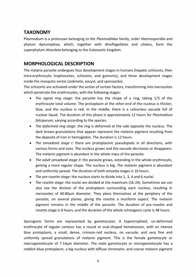
TAXONOMY Plasmodium is a protozoan belonging to the Plasmodiidae family, order Haemosporidia and
phylum Apicomplexa, which, together with dinoflagellates and ciliates, form the
superphylum Alveolata belonging to the Eukaryotic kingdom.
MORPHOLOGICAL DESCRIPTION The malaria parasite undergoes four development stages in humans (hepatic schizonts, then
intra-erythrocytic trophozoites, schizonts, and gamonts), and three development stages
inside the mosquito vector (ookinete, oocyst, and sporozoite).
The schizonts are activated under the action of certain factors, transforming into merozoites
which penetrate the erythrocytes, with the following stages:
The signet ring stage: the parasite has the shape of a ring, taking 1/3 of the
erythrocyte total volume. The protoplasm at the other end of the nucleus is thicker,
blue, and the nucleus is red. In the middle, there is a colourless vacuole full of
nuclear liquid. The duration of this phase is approximately 12 hours for Plasmodium
falciparum, varying according to the species.
The deformed ring stage: the ring is deformed at the side opposite the nucleus. The
dark brown granulations that appear represent the melanin pigment resulting from
the deposits of iron in hemoglobin. The duration is 12 hours.
The amoeboid stage I: there are protoplasmic pseudopods in all directions, with
various forms and sizes. The nucleus grows and the vacuole decreases or disappears.
The melanin pigment is abundant in the whole mass of the parasite.
The adult amoeboid stage II: the parasite grows, extending in the whole erythrocyte,
getting a more regular shape. The nucleus is big. The melanin pigment is abundant
and uniformly spread. The duration of both amoeba stages is 16 hours.
The pre-rosette stage: the nucleus starts to divide into 2, 3, 4 and 6 nuclei.
The rosette stage: the nuclei are divided at the maximum (16-24). Sometimes we can
also see the division of the protoplasm surrounding each nucleus, resulting in
merozoites of 40-80μm diameter. They place themselves at the periphery of the
parasite, on several planes, giving the rosette a muriform aspect. The melanin
pigment remains in the middle of the parasite. The duration of pre-rosette and
rosette stage is 8 hours, and the duration of the whole schizogonic cycle is 48 hours.
Sporogonic forms are represented by gametocytes. A hypertrophied, un-deformed
erythrocyte of regular contour has a round or oval-shaped hematozoon, with an intense
blue protoplasm, a small, dense, crimson-red nucleus, no vacuole, and very fine and
uniformly spread granulations of melanin pigment. This is the female gametocyte or
macrogametocyte of 7-14μm diameter. The male gametocyte or microgametocyte has a
reddish-blue protoplasm, a big nucleus with diffuse chromatin, and coarse melanin pigment

7
placed in heaps around the nucleus. The microgametocytes of the mosquito are long (15-
25μm length), and they are produced by exflagellation of fertilised round
macrogametocytes to form ookinetes (15-20 x 2-5μm), which migrate through the intestinal
wall to form ovoid oocytes (up to 50μm diameter) on the exterior surface. The oocyst
produces thousands of thin long sporozoites (~ 15μm length) that eventually infect the
salivary glands.
BIOLOGICAL CYCLE The malaria biological cycle is one of the most fascinating and complicated of all organisms,
presenting a special interest, especially in molecular biology, cellular biology, and
immunology. The malaria vector transmission by mosquitoes of the Anopheles genus was
established by Grassi at the end of last century. The mosquitoes involved in the transmission
of malaria are: A. hyrcanus, A. labranchiae, A. maculipennis maculipennis, A. maculipennis
atroparvus, A. maculipennis messae, A. plumbeus, A. claviger, A. sacharovi. In our country,
the following species were signalled: A. maculipennis, A. messae, A. atroparvus, A.
sacharovi, and Anopheles labranchiae.
Malaria infection in humans involves an asymptomatic stage in the liver and a symptomatic
erythrocytic stage. Drug research has focused solely on the erythrocytic stage, which has a
limited potential for transmission blocking and prophylaxis. A new technique may be used
for the study of drugs with regard to their efficacy on the hepatic stage using the parasites
expressing luciferase, i.e., bioluminescence. Most of these studies concern the activity of
the drugs during the erythrocytic stages, since there are few known drugs to actually
destroy the hepatic phases. The best known and efficient drug is primaquine, the only drug
used also for the treatment of the hypnozoite forms of Plasmodium vivax, which may lead
to relapses after months, even years from the infection. The problem is that primaquine
produces haemolytic anaemia in individuals with glucozo-6-phosphate (G6PD) deficit, such
enzyme deficit being specific mostly in regions affected by malaria from Africa, South
America and Asia (Adoubryn et al, 2004; Derbyshire, 2012).
THE SPOROGONIC CYCLE OF THE MALARIA PARASITE Changing the habitat causes significant losses for the malaria parasite, leading to an
unbalance in population density. This unbalance is compensated by the growth and
reproduction in cellular niches protected by the host’s immune responses (Frevert, 2004). In
the stage of ookinete and sporozoite, the parasite undergoes the greatest losses, these
stages taking place in the mosquito vector (Silvie et al, 2008). The ookinete develops from
the zygote in the lumen of the host’s middle intestine, being the result of fertilisation
between a female macrogametocyte and a male microgametocyte. The ookinete crosses the
layer of intestine epithelial cells on the apical side, reaching eventually the basal lamina. As
a response to the invasion, the body activates its protection mechanisms, leading to a

8
decrease in the parasite population density. The ookinetes are transformed into oocysts,
which constitute an extracellular development stage, leading to the formation of
sporozoites which are released in the cavity of the mosquito body, thus invading afterwards
the salivary glands (Al-Olayan et al, 2002; Vlachou et al, 2006) (Fig.3). During this migration,
there is a decrease in parasite population density, the sporozoites innoculated to the
mammal host reaching the liver in a very short time. In 1948, Shortt and Garnham
discovered that the sporozoites infect the hepatocytes, developing a stage there before
infecting the erythrocytes. The hepatic stage is asymptomatic, certainly interesting from a
scientific point of view as regards vaccine production. Here is the amplification up to 10,000
times of the number of parasites, culminating with the release of exo-erythrocytic
merozoites. The merozoites invade the erythrocytes, consequently initiating the
erythrocytic cycle, which is pathogenic (Vaughan et al, 2008). Today we know the expression
of genes and proteins involved in the evolution of the stages inside the mosquito and the
pre-erythrocytic stage (Greenwood et al, 2005; Kappe et al, 2001; Lasonder, 2008). The
discovery of the gene responsible for the hepatic stage led to the formation of attenuated
sporozoites, obtaining an attenuated live vaccine.
The parasite sexually reproduces only once in the mosquito body. The factors initiating the
formation of gametocytes are not very well known. The ingestion of gametocytes during the
blood feed activates the formation of gametes (gametogenesis) in the mosquito’s intestinal
lumen.
A derived molecule of xanthurenic acid, together with the change in temperature and pH,
may start the gametogenesis of the male gender under the form of exflagellation (Billker et
al, 1997; Billker et al, 1998). The protein P48\45 is essential in enabling the male gamete to
fertilise the female gametes (which have the protein P47expressed on the surface).
Fertilisation takes place to form the zygote, which after meiosis is transformed into an
ookinete that crosses several epithelial cells of the intestine (Mueller et al, 2005). The
ookinetes cross the epithelial cells up to the basal lamina and transform into oocysts, which
grow extensively, and in 10-14 days lead to the production of thousands of sporozoites.
After migrating through the haemolymph, the sporozoites resulted from oocysts attach to
the basal side of the acinar cells of the salivary gland. The sporozoites cross the acinar cells
and enter the ducts of the salivary glands where they are transmitted to the mammal host
during the blood intake (Pimenta et al, 1994; Robert and Boudin, 2003).

9
Fig. 3. The sporogonic cycle of the malaria parasite
(Source: Personal collection)
THE ERYTHROCYTIC SCHIZOGONIC CYCLE OF THE MALARIA
PARASITE The mammal host is infected with sporozoites from the salivary glands of mosquitoes.
Approximately 100 sporozoites are injected by a single mosquito (Medica and Sinnis, 2005).
They remain in the derma for a period of time, displaying an apparently random movement
until they find a blood vessel, and then migrate to the liver (Mota et al, 2001; Amino, 2007).
After injection, the sporozoites remain in the derma for 1-3 hours and then migrate to the
liver (Yamauchi, 2007). Not all the sporozoites get into the circulation and into the liver,
some of them are stuck at the injection place to be probably be eliminated by phagocytosis.
Some of them reach the lymphatic system, that is, the lymphatic ganglions, where the
response of T and CD8 cells is activated. These cells are capable of eliminating the
developing parasites from the liver (Chakravarty et al, 2007; Amino et al, 2008). In the skin,
the sporozoites actively migrate through the cells, leading to perturbation of the plasmatic
membrane of the host cell. For the cellular crossing, at least three proteins are involved:
SPECT-1, SPECT-2, and phospholipase. The sporozoites lacking SPECT-1, SPECT-2 are
immobilised in the derma as a result of the impairment of the cell crossing capacity (Amino
et al, 2006; Sturm et al, 2006; Ménard, 2007; Sturm et al, 2006; Aly et al, 2009). After
sporozoite innoculation by Anopheles vector, the exo-erythrocytic cycle starts, with a

10
duration, according to the species, as follows: Plasmodium falciparum – 6 days; Plasmodium
vivax – 8 days; Plasmodium ovale – 9 days; Plasmodium malariae – 15 days. Some
sporozoites of Plasmodium vivax and Plasmodium ovale, when penetrating the hepatocytes,
remain in a latent stage for a long period of time (a few years), being called hyposonts
(Cogswell, 1992).
Fig.4. Asexual replication cycle of the parasite inside the red blood cells
(Source: Silvie et al, 2008)
(1) The extracellular merozoites are randomly attached to the surface of the erythrocytes.
(2) Then, the apical end of merozoites is directed towards the surface of the erythrocytes.
(3) Formation of Tight junction and the active merozoites enter the erythrocytes. (4)
Invasion of merozoites occurs by simultaneous formation of a parasite phorous vacuole.
After the invasion, the merozoites become ring-shaped. (5) This stage is characterised by a
big digestive vacuole, where digestion of hemoglobin results in the formation of the malaria
pigment known as hemozoin. (6) The parasite grows intraerythrocytic, and it is called
trophozoite. The hemozoin pigment accumulates in the digestive vacuole. (7) DNA
replication precedes the future cell, and the process is called schizogony. (8) The merozoites
secrete exoneme, which initiates the exit from the parasite phorous vacuole and red blood
cells (RBC) of the host. (9) The exiting merozoites adhere to the adjacent erythrocytes in a
few seconds. (1) Initiation of a new erythrocytic cycle (Fig. 4, 5).

11
Fig.5. Various erythrocyte stages (1-2 ring form, the mature schizont 3, 4-gametocytes) of Plasmodium vivax,
Giemsa stained smear x 1000 (Source: Personal collection)
Sporogonic forms: gametocytes (Fig. 6). A hypertrophied un-deformed erythrocyte of
regular contour has a round or oval-shaped hematozoon, with an intense blue protoplasm, a
small, dense, crimson-red nucleus, no vacuole, and very fine and uniformly spread
granulations of melanin pigment. This is the female gametocyte or macrogametocyte of 7-
14μm diameter. The male gametocyte or microgametocyte has a reddish-blue protoplasm, a
big nucleus with diffuse chromatin, and coarse melanin pigment placed in heaps around the
nucleus (Field and Shute, 1956; Gratzer and Dluzewski, 1993; Moore et al, 2008).
Innoculated in humans, the parasite undergoes a series of transformations, first in the liver
(this constitutes the exo-erythrocytic or tissue cycle), then it penetrates the RBC, where it
develops until it leads to the destruction of RBC (the erythrocytic or asexual cycle). The
parasite released from the destroyed cells parasitizes other cells again (Tolle, 2009; Sturm et
al, 2006) (Fig. 7).
a b
Fig.6. a. Mature schizonts and merozoites
Col. Giemsa x100, immersion objective,
(Source: Personal collection)
b. Stage of gametocytes (macrogametocit pink,
blue microgametocit)
Col. Giemsa x100
(Source: Personal collection)

12
The mosquito innoculates the parasite under the form of sporozoites (infecting
forms for humans) which form, upon reaching the liver, a big plasmodial mass from which
merozoites come off, reach the blood and penetrate the RBC. Inside the RBC, the parasite
goes through the amoeboid stage, then the rosette stage, and then it destroys the RBC
releasing the merozoites which will parasitize other RBC where the cycle starts over again
(Hafalla et al, 2011). Gametocytes (males, females) also result from the erythrocytic cycle,
joining in the body of the mosquito and forming zygotes. In the intestinal lumen, the zygotes
transform into ookinetes, from which the sporozoites are produced after a series of
transformations. Located in the salivary glands, they are innoculated in humans (Vlachou,
2006).
Fig.7. Life-cycle of Plasmodium-vivax
(Source: http://www.microbiologynotes.com/life-cycle-of-plasmodium-vivax/)
The Anopheles females biting a sick person whose blood contains gametocytes and
schizonts ingest with the blood a variable number of parasites under these two forms. The
schizonts are digested in the stomach of the mosquitoes, and the gametocytes ensure the
continuation of the sexual or sporogonic cycle, which leads to the sporozoites. It has been
demonstrated that some mosquitoes contain two genes which encode the
carboxypeptidaze from the middle intestine, which are involved in the parasite
development with Anopheles gambiae (Lavazec and Bourgouin, 2008; Aly et al, 2009).

13
EPIDEMIOLOGY
MALARIA TRANSMISSION By vectors – the vector transmitted to humans, and at the same time the compulsory hosts
for the sexual stage of the parasite, is represented by the mosquitoes of the Anopheles
genus (of the approximately 400 species on the globe, only 30-40 can be malaria vectors in
natural conditions). The period of formation of sporozoites in the salivary glands varies
between 7 and 30 days, according to the parasite species and environmental temperature.
By blood transfusion - malaria post-transfusion known from 1911 appears from latent
asymptomatic malaria carriers. In most cases, post-transfusion transmission was done by P.
malariae (which gives persistent forms with long time blood carriers), and more rarely by P.
vivax and P. falciparum.
The period of human contagiousness is the time that the infesting gametocytes of the
malaria parasite persist in the blood of the sick person, and it varies with Plasmodium
species. Consequently, the gametocytes of P. malariae can persist in the blood even up to
53 years, those of P. vivax for 1-3 years, and P. falciparum up to 1 year. The Anopheles
female remains an infesting agent its entire life.
There are three possible modalities of malaria transmission from one sick person or
plasmodia carrier to healthy people: in natural conditions via Anopheles mosquito bites;
intrauterine, from a sick mother to the foetus through placenta or during childbirth; by
transfusion - by blood containing the pathogen during medical manipulation or injections in
aseptic conditions with contaminated needles.
Malaria transmission occurs in the mosquito activity season, depending on the transmission
intensity and duration, and on the influence of natural and social factors with long term
effect.
MALARIA VECTORS In the majority of cases, malaria is transmitted through the bite of the mosquito female of
the Anopheles genus. There are more than 400 different species of mosquitoes of the
Anopheles genus; around 30 species represent vectors of major importance for malaria (Fig.
8). The intensity of the transmission depends on the factors relating to the parasite, vector,
human host and the environment. Each mosquito species of the Anopheles genus has its
own aquatic habitat; for example, some prefer small fresh water sources, such as ponds and
the water accumulated in the traces of hooves, which are abundant in tropical countries
during the rainy season.
Understanding the biology and behaviour of the Anopheles mosquitoes may help us
understand the way in which malaria is transmitted and also elaborate appropriate control

14
strategies. The factors that affect the capacity of a mosquito to transmit malaria include its
innate susceptibility with regard to Plasmodium, choosing the host and its longevity. The
factors that should be taken into account when elaborating a control program include the
malaria vectors’ susceptibility to insecticides and the preferred feeding and resting place of
adult mosquitoes. Transmission is more intense in places where the lifetime of mosquitoes
is longer (the parasite has enough time to finish its development within the mosquitoes) and
mosquitoes prefer feeding off humans, not animals. The long lifetime and the
anthropophilic character of the African mosquito species represents the main reason for
which almost 90% of malaria cases in the world are in Africa. Transmission depends as well
on the climatic conditions that may affect the number and survival of mosquitoes: rainfalls,
temperature and humidity. In many places, transmission is seasonal, reaching its peak
during and after the rainy season. Malaria epidemics may break out when the climate and
other conditions favour a sudden transmission in areas where people have low or no
immunity against malaria. They may also appear when people with low immunity travel to
areas with intense malaria transmission, for example, to find a workplace as refugees.
Fig.8 Global distribution of dominant or
potentially important malaria vectors (Robinson Projection)
(Source: Kiszewksi et al, 2004)
Mosquitoes are spread all over the world with the exception of the arctic area and from the
approximately 3500 existing species, three quarters come from tropical and subtropical

15
areas. The medical importance of these insects is represented by their need to feed on
blood in order to obtain the necessary protein for egg maturation. The feeding is done with
the help of a complex salivary secretion that facilitates the transmission of some pathogenic
agents to the mammal host while sucking the blood. The ecology, behaviour, survival of
mosquitoes and the dynamics of the transmission of diseases are strongly influenced by
climatic factors: temperature, rainfalls, humidity, wind, and sunshine duration. Also, the
development time of the pathogenic agent in the salivary glands of mosquitoes is directly
influenced by temperature (Reiter, 2001).
Malaria is transmitted through the bite of the female mosquitoes of the Anophleles genus.
In Europe, the mosquitoes of the Anopheles maculipennis complex have been incriminated
as vectors for the disease. The first person who noted as early as 1926 the existence of a
complex of the maculipennis species was Falleroni, who described the morphological
differences regarding the chorion of these species. There are approximately 430 species
registered as belonging to the Anopheles genus, out of which approximately 60 species are
considered vectors of malaria (Reiter, 2001).
The complexes of species are very closely related groups of species that are hard to
differentiate from a morphological point of view. Anophles maculipennis Meigen, the
historical vector of malaria in Europe and the Middle East, was the first complex with twin
species discovered in mosquitoes (Falleroni, 1926; van Thiel, 1927). The search included
morphological studies based on eggs (Falleroni, 1926; Falleroni, 1932; Corradetti, 1934; de
Buck and Swellengrebel, 1934; Hackett and Lewis, 1935; Korvenkontio et al, 1979), on
chromosomes (Frizzi, 1952; Frizzi, 1953; Kitzmille, 1967; Stegnii, 1976; Stegnii and Kabanova,
1976), sequences/DNJ (Marinucci et al, 1999; Proft et al, 1999; Linton et al, 2001; Linton et
al, 2002; Linton et al, 2003; Sedaghat, 2003; Porter, 1991), ecology (van Thiel, 1927; de
Buck and Swellengrebel, 1934; Hackett and Missiroli, 1935), hybridization (de Buck and
Swellengrebel, 1934; Kitzmiller et al, 1967), larvae (Bates, 1939; Buonomini, 1940; Suzzoni-
Blatger and Sevin, 1981; Boccolini et al, 1986; Suzzoni-Blatger et al, 1990; Romi, 2000), and
on wing characteristics (de Buck et al, 1933; Ungureanu and Shute, 1947; Sedaghat et al,
2003). As a result of all these studies the presence of eight species was established within
the Palearctic maculipennis complex:
A. atroparvus van Thiel, A. beklemishevi Stegnii and Kabanova, A. labranchiae Falleroni, A.
maculipennis, A. martinius Hingarev, A. melanoon Hackett, A. messeae Falleroniand, A.
sacharovi Favre (White, 1978; de Zulueta et al, 1983; Cianchi et al, 1987; Ribeiro et al, 1988;
Linton et al, 2002; Sedaghat et al, 2003; Knight and Stone, 1977; Kettle, 1995) (Fig. 9).

16
Fig.9. Species of the Palearctic maculipennis complex
(Source: https://ecdc.europa.eu/en/disease-vectors/facts/mosquito-factsheets/anopheles-sacharovi;
http://mosquito-taxonomic-inventory.info/file/5212)
All the members of this complex are considered vectors of malaria in Europe. Taking into
account the medical importance of the maculipennis complex, the differences regarding the
vectorial capacity and species distribution that are thought to be influenced worldwide by
global warming, what becomes necessary is a sure method of identifying the species. Once
the Anopheles female becomes infected with Plasmodium, it will have sporozoites in its
salivary glands for the duration of its entire life, therefore infecting a new host every time it
feeds on blood. The vectorial capacity is an index of the transmission capacity of the malaria
vector which is influenced by the density of the vector (by climatic conditions: temperature
and humidity; by geographic conditions: seasonal variations, and by zoophilic or
anthropophilic preference (Mullen and Durden, 2002; Marquardt, 2004; Toole, 2009). In the
absence of the preferred host, feeding behaviour may be modified (Boete, 2006; Pages et al,
2007; Nitzulescu and Gherman, 1990). A. atroparvus, A. labranchiae and A. sacharovi have
been incriminated as main vectors of malaria in the region of Europe (Vicente et al, 2011).
The duration of the gonotrophic cycle (the distance between egg laying) is of approximately
2 days, and the duration of the evolution of sporozoites is approximately 12 days, so that at
least 6 gonotrophic cycles are needed before the female becomes infected. The duration of
the sporogonic stage varies depending on the Plasmodium species involved; as such, for P.
falciparum, between the ingestion of gametocides and the presence of sporozoites in the
salivary glands, 12 days are needed at a temperature of 25°C, and 23 days, respectively, at
20°C. Below 18°C and over 33°C the cycle stops (Bruce-Chwatt and Zulueta, 1980). The
global distribution of malaria depends a lot on the intrinsic characteristics of the vector, the
vector’s competence (the capacity of an Anopheles species to ensure the complete
development of the parasite) and its vectorial capacity (Pratt and Moore, 1993). The
A. atroparvus (van Thiel,1927)
A. beklemishevi (Stegnii and Kabanova, 1976)
A. labranchiae (Falleroni,1926)
A. maculipennis (Meigen, 1818)
A. messae (Falleroni, 1926)
A. martinus Hingarev
A.sacharovi (Favre, 1903)
Anopheles melanoon Hackett, 1934

17
incapacity of the malaria parasite to develop in some mosquito species is tied to the
absence of some metabolic factors that are essential for the development of the parasite or
the presence of some toxins that inhibit the development of the parasite. The presence of
the vectors largely depends on the local conditions (Mouchet et al, 1999). The climatic
changes represented by an increase in temperature by as little as 0.5°C may lead to an
increase in the mosquito population by 30-100% (Patz and Olson, 2006). This highlights the
necessity of permanently reporting the Anopheles species that are present and the density
of the populations in order to be able to quantify the control measures for malaria. In the
mountainous areas of Africa, the incidence of malaria has increased since 1970 in direct
correlation with the change in climatic factors, more precisely the fact that an increase in
temparature has been recorded (Pascual et al, 2006). Molecular identification of the
Maculipennis complex species is important if we consider its medical signifocance, the
vectorial capacity and the distribution of the species that seems to be directly influenced by
global warming (Sedaghat et al, 2003). Anopheles claviger has a large distribution, all the
way from the sub-Carpathian areas to the lowlands. The larvae nests are usually found in
copses, orchards, in places shaded from direct sunshine and with moderate temperatures.
The larvae live even during winter, under the ice. It is a fairly aggressive mosquito which
attacks humans even during the day.
Anopheles hyrcanus is a widely spread species in the Danube Delta, but also in the
countryside, near brackish water. Its spreading epicenter is in the Middle East, where it is a
malaria vector, and it steadily decreases as we move towards the West.
Anopheles maculipennis, Anopheles messeae, Anopheles sacharovi are part of the
maculipennis group, which comprises anopheles members with stained wings, these dark
stains originating in the agglomeration of scales on some areas of the wing veins.
The species of the Maculipennis group is characterized by a high plasticity of the larvae,
which adapt to fairly varied larvae nests. The safest criterion for differentiating the three
anopheles species from the Maculipennis group is the egg. For each species, the ventral part
of the egg, directed upwards while floating on water, is characteristically ornated.
Anopheles maculipennis is a widely spread species in the country, near forests and on river
valleys. The well oxygenated freshwaters provide the egg-laying place, with a lower average
temperature, spared from direct sunlight. The female is zoophile and occasionally
anthropofile. During the winter hibernation is complete, but with a gonotrophic
concordance, in shelters away from humans, in open stables or hollows exposed to the cold.
Anopheles messeae is spread all over the low plains in the country and in places with
stagnant waters (ponds, lakes, the lower third part of rivers that are usually floodable, the
floodable area of the Danube).

18
The larvae nests are represented by stagnant, open freshwaters, with a shallow bottom and
abundant vegetation, warm and well exposed to the sunlight. The larvae develop well
especially on the banks of these waters and at the periphery of the islands of vegetation.
The female is predominantly zoophile but it also feeds off humans. It has a complete
hibernation, with gonotrophic concordance, in shelters fairly exposed to the cold.
Anopheles atroparvus is spread in the entire country, in areas with more or less salty waters.
In this regard, the larvae have a great plasticity of adaptability, from larvae nests in
freshwater to saltwaters (4-5%). They are not very sensitive to lower temperatures
(Ungureanu and Shute, 1947).
The female is over wintering in intradomestic and peridomestic shelters, free from the cold,
where it can find food. It does not hibernate completely and it awakens from time to time to
bite humans or cows, but it is predominantly anthropophile.
The Anopheles female in general mates once and keeps the sperm in the spermathecal,
from where it is used during the entire life of the female for fertilizing the eggs. For the egg
maturation an intake of blood is needed, the female being able to travel up to 3 km in
search of a host, generally attracted by the carbon dioxide emanated by the host
(Smallegange RC et al, 2005). It seems that the development of the gametocytes (the
infected form for the human) is more prosperous within the Anopheles gambiae species,
which is the main malaria vector in Africa (Lacroix R et al, 2005). Following mating, 48 hours
are needed for egg maturation, then they are laid on water, and their properties differ
depending on the Anopheles species (size, sunlight exposure, type of natural or artificial
water source, unpolluted). Adult females lay 50-200 eggs per oviposition. Eggs are laid
directly on water and may float on both sides. They are not resistant to drying and hatching
in 2-3 days, even though the sampling may take up to 2-3 weeks under lower temperature
conditions. These characteristics are valid for the Anopheles species from the rural areas
and the periphery of the urban area, the risk of malaria transmission being higher in the
rural area (Pages F et al, 2007). The preference for feeding inside (endophage) or outside
(exophage), for an ambiental environment – endophile or exophile, varies from one species
to the next or within the same species, depending on the geographic area. These
characteristics play an important role in the development of fighting strategies against the
anopheles (Pages F et al, 2007). The females feed from dusk till dawn, the peak of attack
differs depending on the species, A. cruzii, A. bellator from South America being active
during daytime. The flight of the species of the Anopheles genus is silent and the bite is not
painful.
The Anopheles genus comprises species of high medical importance, some 500 species
overall, of which 50 are malaria vectors (Harbach, 1994).The adults are characterized
morphologically by palps equally long as the trunk in both sexes, simple scutellum and

19
abdomen without scales. The hypopygium is made up of forcipules without lobes, claspers
are present, paraprocts do not chitinize, and the phalosome is often ornated with apical
growths. The larvae are missing the siphon. The eggs are laid isolatedly on the surface of the
water and have a pair of lateral floats, more developed in the species growing in freshwater
areas and smaller in the species from saltwater areas. The development cycle from egg to
adult takes from 8 days (at a temperature of 31°C) to 20 days (Foster and Walker, 2009).The
development period of the malaria agent inside the mosquito (extrinsic incubation) varies
between 10 and 21 days, depending on the Plasmodium species. The life span of a mosquito
in nature may be calculated exactly by estimating that over 20% of the mosquitoes would
survive more than a period of extrinsic incubation of 14 days. Thus, it can be concluded that
it is not the density of the mosquito population that is important, but rather their longevity,
therefore the purpose is to shorten the life of mosquitoes by using insecticides.
ANOPHELES MACULIPENNIS
FEMALES
The colour is dark or medium brown, even though colour and size variety is wide. The
characteristic that differentiates all the other European Anopheles species are the wings
with dark scales that form characteristic stains. The trunk has a dark brown colour and the
maxillary palps are the same colour and length as the trunk. The vertex has a long whitish
scale-patch directed towards the front. The scutum has a grey band that narrows towards
the front. The lateral parts of the scutum are brown in the front and dark brown on the rear
side. The scutum is brown with narrow golden scales. The postnotum is light brown and the
pleura are dark brown. The femur is dark brown on the dorsal part and light brown on the
ventral side, the tibia is brown and of a lighter colour towards the tip, the tarsus is dark
brown. The wings have dark, unevenly distributed scales that form black stains close to RS
and R2 3 and M. In A. sacharovi there are dark stains on the wing veins and on the rear
edge. The abdomen is brown or dark brown with a golden brown stripe (Capinera, 2002).
MALES
In the culicidae species of the Anopheles genus, or at least in the ones present in our
country, the coxit has missing lobes. The base of the coxit lengthens laterally, with three
apophyses or apodemes. The external and dorsal internal apodeme serve for attachment on
segment 9. The basal apodeme or basal plate, on which the penial and anal muscles are
inserted, reaches segments 9 and 8, and on the other side it is articulated with the
parameres of segment 10.
LARVAE
Quite variable in pigmentation and size, the larvae from the northern part of Europe are
usually bigger, with a closed head capsule, the antenna is almost straight, slightly spiculated,
almost 2/3 of the head size. The front needle (5-C to 7-C) is long. The needle trowns on the

20
abdominal segments I and II are rudimentary, but well developed on segments III-VII. In the
anopheles species there are bristles that ramify, the same as on the head and thorax, as
opposed to the culicinae whose bristles ramify from the base.
For the Anopheles species, a few other characteristic structures may be also observed on
each abdominal segment:
a chitinous tergal plate whose size varies depending on the species;
accessory plates, posterior to the median plate, one, two or three on each
segment;
needles grown on each segment, representing a surface-active floating system;
these bristles may be used to differentiate the species of the Maculipennis group
from the Anopheles hyrcanus species.
The 8th abdominal segment, having the same form as the other 7, constitutes the support
for some structures of taxonomic importance. On the lateral walls of segment 8 are situated
the abdominal comb or abdominal spines, one on each side. In the anopheles species, the
siphon is missing and the spiracles (stigmatic orifices) are held by a spiracular plate
(Cosoroaba, 1992; Cosoroaba, 2000).
ANOPHELES LABRANCHIAE (FELLERONI, 1926) The larvae develop in stagnant water, fresh or saltwater, in the coastal areas of Europe,
preferring the heat. Hibernation is done in the dark by the adult female, in stables or natural
cavities. The diapause may be complete or incomplete, with occasional blood feasts.
Females are mainly anthropophage, occasionally feeding off domestic animals.
Anopheles labranchiae is an endophilic species, living in households, stables, animal shelters
and only occasionally outdoors, in natural shelters (tree hollows). The presence of larvae has
been recorded from April to October. The flight of this species is limited to 2-5 km (Senevet
and Andarelli, 1956). Anopheles labranchiae has a limited distribution in the South and
South-East of Europe. It was reported in the South-East of Spain, Corsica, the coastal areas
of Italy, Sardinia, Sicily. In Northern Africa, the species is found in Morocco, Algeria and
Tunisia (Zahar, 1990). Anopheles labranchiae has registered an increase in numbers in
Sardinia in the last 35 years. Between 1940 and 1953, a period in which malaria was
eradicated, Anopheles labranchiae numbers dropped significantly, the eradication campaign
consisting of insecticides (DDT). The expansion of urban areas and changes in agricultural
practices have led to a new increase in Anopheles labranchiae populations.Sardinia presents
a biological interest due to its central geographic position (lat. 38-41 N, long. 8-9E) to the
West of the Mediterranean, halfway between Africa and Europe. Sardinia is the second big
island in the Mediterranean (250 x 120 km). The reproduction places for Anopheles
lambrachiae are varied: swamps, rivers covered with vegetation, holes, channels, basins,
sunny areas preferably. Anopheles labranchiae and Anopheles sacharovi are considered to
be the most efficient vectors in the transmission of malaria in the Palearctic region (Marchi,

21
1987). The main reproduction sites for Anopheles labranchiae in this region remain the rice
fields, a fact that continues to be a main concern for agriculture.
In Corsica, Anopheles labranchiae continues to be the main malaria vector, together with
Anophles sacharovi. Other species are considered possible vectors elsewhere in Europe,
namely Anopheles algeriensis, Anopheles atroparvus, Anopheles claviger, Anopheles
hyrcanus, Anopheles maculipennis SS, Anopheles marteri, Anopheles melanoon, Anopheles
messae, Anopheles petragnani, Anopheles plumbeus, Anopheles superpictus, Anopheles
subalpinus; because of their physiologic characteristics, especially zoophilism, their part is
minimal in the transmission of malaria (Toty C et al, 2010).
The center of Italy used to be a hyperendemic area for malaria up until the mid-20th century,
when the malaria eradication campaign drastically reduced the number of vectors
incriminated for malaria transmission, Anophele labranchiae.The introduction of rice crops
led to a substantial increase of this species, causing concerns for the re-emergence of
malaria in this area.
The changes in the use of land, the climatic variations, and the seasonal demographic
variations have all contributed to the growth of the Anopheles labranchiae populations, thus
increasing the risk of local malaria (Boccolini et al, 2012).
In Morocco, Anopheles labranchiae is considered the main vector of malaria, the last case of
local malaria being recorded in 2004. Malaria control in this region includes the use of the
biological control method, namely, the mosquitofish (Gambusia holbrooki). Insecticides are
used as a last resort, but they still occupy an important place in the National Plan for
Malaria Control (NMCP). The insecticide used for destroying the Anopheles labranchiae
larvae, also recommended by the WHO, is an organophosphorus insecticide called
temephos, 0.125mg / L being the smallest concentration with a 100% mortality rate (Faraj,
2010). Temephos is the insecticide used to destroy the larvae and DDT is used in the control
of adult mosquitoes. Large scale use of insecticides in agriculture may constitute a problem
since Anopheles labranchiae is frequently found on agricultural land. Anopheles labranchiae
is the only species of the maculipennis complex found in North Africa. Anopheles
labranchiae is the main vector in the transmission of malaria in Italy as well.
In the region of Tuscany there was a case of local malaria registered in 1997 and, based on
an in-depth study, it was noted that Anopheles labranchiae was the only Anopheles species
identified in the region. A further study of the same species proved that the probability of
malaria transmission is high in August, when the period needed for the development of the
parasite inside the mosquito is reached, namely, 11 days for Plasmodium falciparum and 10
days for Plasmodium vivax, at a temperature of 25°C, the daily average temperature (Romi,
1999). An article published in 1998 noted that Anopheles labranchiae had disappeared in

22
Spain (Eritja et al, 1998). The reason behind the lack of similarity between the distribution of
Anophles atroparvus and Anophles labranchiae seems to be the temperature, the latter
preferring warm waters. Both species prefer brackish waters and lagoons, but Anopheles
labranchiae prefers to lay eggs in freshwaters. In Sardinia, larvae have been found in almost
all habitats, with the exception of shaded ones (Sinka et al, 2010). The A. labranchiae
females attack humans readily; Romi et al. have signalled the presence of human blood in
86% - 90.7% fed females (Romi et al, 1997). Anopheles labranchiae may be both endo and
exophile, depending on the available habitats (D’Alessandro et al, 1971). Similar to A.
atroparvus, A. labranchiae proved to be refractory to infections with African strains of P.
falciparum, even though the Center for Production and Infection of Anopheles (Centre de
Production et d'Infection des Anophèles - CEPIA) from Paris has positively demonstfrated
the infection of A. labranchiae with an African strain of P.falciparum NF54 (Toty C et al,
2010).
ANOPHELES ATROPARVUS (VAN THIEL, 1927) Anopheles atroparvus becomes active at temperatures starting from 15°C, when it may
constitute a risk for the transmission of malaria (Knottnerus, 2002). For reproduction, it
prefers brackish waters or freshwaters, well-lighted areas (Potugalia, Spania). It prefers
relatively cold waters, and the larvae have been found in marshes, ditches, flooded basins in
the ground, pools formed in riverbeds, tanks for transporting cement, rice fields, fountains
and basins, as well as in water collected in used tyres. It prefers sunny sites with filamentous
green algae and other aquatic plants. Anopheles atroparvus is a predominantly zoophile
species, preferring in order: rabbits, horses, cows, swine and sheep, as such being the main
vector of myxomatosis in rabbits. However, it has been described as anthropophile as well,
since it can be found in households and has been recorded feeding off humans. Hibernation
is done in stables or households, the adult female feeding periodically, without laying eggs.
This has led to the spread of malaria in the Netherlands, Great Britain and other parts of
Europe at the beginning of the 20th century. The duration of the diapause depends on the
length of the day and the temperature; therefore, it varies with altitude and may vary from
September to October in Northern Europe, or from November to February in Southern
Europe. It is generally a littoral species which may be found in the South-East of Portugal,
along the coasts of the Atlantic Ocean, Great Britain and the Mediterranean Sea. In South
and South-East Europe it has an uneven distribution. In Macedonia it is largely spread in the
lowland areas, visibily dominant only in the areas with saline/alkaline soils from the
Pannonian Plain. In Portugal it is the most common and widely distributed species (Becker
et al, 2003).
In a 2010 study which aimed to identify the most frequent vectors from 49 countries in
Europe and the Middle East, comprising 2891 locations, Anopheles atroparvus was signalled
in the highest number of locations - 1044 (Sinka et al, 2010). Anopheles atroparvus was the
main incriminated vector in the transmission of malaria in the British Isles, being the main

23
source of P. vivax. In the case of P. falciparum it has not been established yet if the parasite
is capable to reach the end of its lifecycle and transmit the infection. Most research notes
that Anopheles atroparvus is capable of transmitting tropical strains of Plasmodium
falciparum, although it may transmit European strains as well (Cambournac, 1994). A study
by Sousa mentions the existence of an Anopheles atroparvus in Portugal which is capable of
transmitting tropical strains of P. falciparum (Sousa, 2008). Anopheles atroparvus is largely
spread in England, Scotland, Wales, and Ireland, together with Anopheles messeae, from
which it cannot be differentiated morphologically, unless it is in its egg phase (Snow, 1998).
Anopheles atroparvus is largely spread in the countries from the Mediterranean basin, but
because of its zoophilic character, it does not present a major risk in malaria transmission,
unless the populations show high density (Eritja et al, 1998). Its distribution varies in Europe,
it is absent in some Mediterranean regions, such as the South of Italy, Greece and Turkey
(Vicente et al, 2011). Anopheles atroparvus, together with Anopheles messae, are
considered the main vectors of malaria in the UK. The genetic identification based on the
ITS2 sequence was confirmed by Linton et al. in 2002. A study carried out in 2001 on the
river banks of the Rhine and Meuse, in the south-western part of the Netherlands,
demonstrated that the populations of Anopheles atroparvus, which is considered the main
and only vector of malaria in the area, had decreased a lot, with only 4 out of 150 larvae
examined representing A. atroparvus, and the rest being identified by PCR as A. messeae
(Takken, 2002).
ANOPHELES SACHAROVI (FAVRE 1903)
This species is the easiest to distinguish of the entire Maculipennis complex by the light
colour of the mesonot and the median band on the scutum, which is absent in the other
members of the complex. The lateral parts of the scutum are brown-yellow, the same as the
middle part. The stains on the wings are not very obvious, actually barely perceptible in
older individuals of the species, compared with the other members of the complex. A.
sacharovi eggs are distinguished by the missing floats and the larvae are smaller than those
of other members of the complex (Becker et al, 2003).
Anopheles sacharovi is considered to be the most efficient malaria vector, a main vector in
Turkey, Siria, the Middle East, as well as in Southern Europe (Yaghoobi-Ershadi et al, 2001;
Romi et al, 2002; Sedaghat et al, 2003).
Anopheles sacharovi is very flexible in choosing its habitats both as an adult and as a larva,
and it is found in small water sources with vegetation. It prefers habitats with freshawater,
but it has been described as the most tolerant species in the entire maculipennis group, up
to a salinity of 20%. It may survive in waters of up to 38-40°C, in stagnant waters, also being
able to withstand a weak current (Pener and Kitron, 1985). This species prefers sunny sites,
marshes being a perfect habitat, but it has also been found on the borders of rivers,
streams, springs, pools and ditches.

24
As a result of eradication campaigns and of using DDT in the past, a zoophilic characteristic
was initially recorded, but an anthropophilic behaviour is currently noted (Sinka et al, 2010).
Unlike the other members of the Maculipennis complex, A. sacharovi attacks throughout
the day, with maximum intensity in the early evening between 20:00-22:00 (Alten et al,
2003).
Hibernation is incomplete, the female feeds multiple times without laying eggs (Manguin et
al, 2008; Kasap, 1995). Anopheles sacharovi is an important vector in Turkey, Siria, Israel,
Jordan and the Middle East (Zahar, 1990; Sedaghat et al, 2003). For reproduction, it prefers
small water sources with aquatic vegetation. Anopheles sacharovi is considered the main
vector in the Balkans, but in Romania it was declared extinct after the eradication of malaria
in this area (Zahar, 1990), and it is very rarely found in Italy (because of environmental
changes and changes in agricultural practices). The species is still abundant in Turkey and
North-East Greece (Romi, 1999). Anopheles sacharovi is a species that quickly gains
resistance to insecticides, as was shown by a study carried out in 2012. The levels of
irritability described by the WHO are DDT 4%, dieldrin 0,4%, malathion 5%, fenitrothion 1%,
permethrin 0,75%, and 0,05% for deltamethrin. The analysis results of insecticide doses for
this species signalled resistance to DDT, tolerance to dieldrin and some sensitivity to
fenitrothion, malathion, permethrin, and deltamethrin. DDT had the most irritating effect,
while deltamethrin was the least irritating (Vatandoost and Abai, 2012).
In the laboratory, under varied ecological conditions, at altitudes ranging from 353 m to
1126 m in the Sanliurfa Turkish province, Anopheles sacharovi did not present any
significant difference from egg stage to adult stage, or any developmental differences.
Anopheles sacharovi is the main vector of malaria in Turkey and it is, together with
Anopheles superpictus and Anopheles pulcherrimus, the most important vector of malaria in
the former Soviet Union, even though A. messeae was directly involved in the re-emergence
of malaria in Russia and Ukraine. Anopheles maculipennis was considered the main malaria
vector on the Caspian Sea coast and Anopheles sacharovi is considered the main malaria
vector in the central plateau of the country. The members of this complex of species are
active in Northern Iran from May to September, with a peak in July, developing well in rice
fields and around clean waters, with adults found in animal shelters in proportion of 95%.
Blood feeding usually starts at 19.00, with a peak between 20.00-21.00, and the ELISA
immunoenzymatic test shows a preference for cattle, followed, to a smaller degree, by
sheep and poultry. The anthropophilic index for this species in the Northern Iran is 1.7-4.9%
(Djadid et al, 2007). Azerbaijan and Armenia have been free from malaria for more than 30
years, up until 1994, when it re-emerged with A. sacharovi and A. superpictus as vectors.
ANOPHELES MESSEAE (FALLERONI, 1926) Anopheles messeae is the most widely spread member of the maculipennis complex,
distribution ranging from Ireland in Europe to Asia, China and Rusia. For A. messeae, a series

25
of genetic polymorphisms has been identified, and five different haplotypes have been
defined relative to the different geographic areas of its distribution; it has not been possible
to demonstrate if these polymorphisms are indicative of an altered behaviour in these areas
(Di Luca et al, 2004). The larvae are found in shaded waters, with a very smooth flow or that
are stagnant, such as the borders of lakes and marshes. The larvae have been found in sites
with reed, floating aquatic weeds, algae, such as small lakes and dunes. It is a species found
predominantly in the floodable areas of great rivers, such as the Danube, the Rhone, the
Sava or the Rhine. It is very rarely or almost never found along the coast and in
mountainous areas (Becker et al, 2003). Anopheles messeae has been found in human
households, in indoor spaces, staircases, basements, stables, outdoor spaces (hollows,
underground spaces, ponds), but very few have actually been collected (Sinka et al, 2010).
In the Netherlands, A. messeae is not considered a risk for the transmission of malaria due
to its zoophilic characteristics, that is, feeding off humans only if populations are too dense
and animals too few (Takken et al, 2002).
Unlike A. labranchiae and A. atroparvus, A. messeae enters diapause and hibernates, the
adult female prefers abandoned buildings without animals, not needing to feed on blood
until spring and getting its energy from the fat reserves (Jaenson et al, 1991).
Anopheles messeae may transmit malaria at low temperatures, as low as 4 °C, in which case
44 days are needed for the mosquito to become infectious. This is how the cases in Scotland
and Norway can be explained. In Great Britain, Anopheles messeae and Anopheles
atroparvus are responsible for the malaria cases signalled there (Knottnerus, 2002).
Anopheles messae is rarely found in waters that contain more than 1.5g NaCl/l. The species
prefers shaded areas, being similar to A. typicus and A. melanoon (Zahar, 1990).
Anopheles messeae is an important malaria vector in Western Asia, and it was recently
identified as a main vector for the re-emergence of malaria in Ukraine and Russia (Sedaghat
et al, 2003).
ANOPHELES MELANOON (HACKETT 1934) Anopheles melanoon is found in freswaters and in rice fields from Northern Italy. Also, it is
found in marshlands, in large surface stagnant waters, on the borders of rivers and lakes,
ponds and pools. In Sardinia the larvae may be found in freshwaters, sunny places in spring
and shaded areas in summer. They were also reported in rice fields (Becker et al, 2003).
Hibernation is done with full diapause, and the species is mainly zoophile, feeding off
humans occasionally, both outdoors and indoors. It was identified in the Anatolian region in
Turkey, where malaria is endemic, together with A. sacharovi and A. maculipennis (Akiner
and Cağlar, 2010). A. melanoon is a rare species with limited distribution in South-West and
southern Europe.

26
ANOPHELES MACULIPENNIS (MEIGEN, 1818) Anopheles maculipennis has its reproduction habitat in cold waters, in mountainous areas,
but it has also been found close to the sea, in association with Anopheles messeae in
running waters. Also, it was found in warm waters, for example in Naples, Italy (Zahar,
1990).
Anopheles maculipennis has been identified as a major malaria vector on the Caspian
seacoast in Iran. The characteristic reproduction sites are the undisturbed areas of river
courses and banks, rice fields and artificial basins. In the mountainous regions from Central
Europe the species may be found at altitudes of 1000 m and more, being the only member
of the complex found under this type of conditions. Altitudes of 2190 m and up to 2,300 m
are reported by Bulgaria and Turkey. Hibernation is complete but may be short in areas with
a warm climate. Females are generally zoophile, feeding mainly off bovines but also off
swine and poultry, and when these are in small numbers, they may feed off humans as well,
both outdoors and indoors. The species is predominantly endophile, using stables and
houses as resting sites.
Distribution: A. maculipennis S.S. is largely spread in the entire Europe. Except for the
southern tip of the Iberian Peninsula, it is recorded in almost every European country. It
extends towards the East and the South-West of Asia and the Persian Gulf. It is considered
to be a more continental species, with humidity needs considerably smaller than those of A.
messae and A. atroparvus (Becker et al, 2003).
ANOPHELES DACIAE It was signalled by Nicolescu et al. in 2004 as a new species of the Maculipennis complex.
Captured on the Black Sea coast and in the plains adjacent to the Danube, in the south of
Romania (Nicolescu et al, 2004), this species is similar to Anopheles messae (the difference
was established based on the ITS2 sequence, noting a difference of 1.03% bases). Anopheles
daciae was signalled in the UK as well, proving for two years consecutively that it is a stable
population, with no susceptibility of artefact (Linton et al, 2005).
ANOPHELES SUBALPINUS (HACKETT ET LEWIS 1935) Anopheles subalpinus reproduces in freshwaters or slightly saline waters, such as sunny
swamps and ponds with vegetation, avoiding shadowed places for reproduction. It is also
present in the rice fields, preferring stagnant waters with horizontal vegetation in spring. In
summer the eggs are easier to recognize, as with A. typicus and A. messae, while during
spring and winter they are black (Zahar, 1990). The regular reproduction sites can be found
up to 1,200 m high. Hibernation in adult females is complete, usually in caves and
abandoned buildings. A. subalpinus is considered a strongly zoophilic species that rarely
feeds off humans. Females are endophile and may be found in large numbers during the day

27
in stables and barns, and more rarely in households or buildings. It is a South-European
species with an interval between the Iberian Peninsula, up north to the Mediterranean
countries, and the lowlands area around the Caspian Sea (Becker et al, 2003).
ANOPHELES GAMBIAE It is considered the most important malaria vector on the continent of Africa (Fig. 10). In
1960, the existence of an Anopheles gambiae complex or Anopheles gambiae sensu lato was
recognised, which comprises the following species: Anopheles arabiensis, Anopheles
bwambae, Anopheles melas, Anopheles merus, Anopheles quadriannulatus, Anopheles
gambiae sensu stricto, Anopheles coluzzii, Anopheles amharicus. Taken individually, the
species of the complex are very difficult to differentiate morphologically, this being possible
to some extent based on the larva or the adult female.
Fig.10. Anopheles gambiae
(Source: https://cameronwebb.wordpress.com/tag/anopheles-gambiae/)
However, the behavioural traits differ; for example, Anopheles quadriannulatus is a
saltwater and mineral water species, A. melas and A. merus are saltwater species, while the
rest of the species are freshwater species. Anopheles quadriannulatus is zoophile by
preference, feeding off animals, while Anopheles gambiae sensu stricto generally feeds off
humans, being therefore considered anthropofile. Identification through molecular methods
remains the most certain method, and the suggestions put forth by Fanello’s team carry
enough weight and have had important implications for the subsequent control measures
(Fanello et al, 2002). The members of the Anopheles gambiae complex are spread in tropical
Africa, the South of the Sahara Desert, with Anopheles arabiens presence extending to the

28
South of Arabia. Anopheles gambiae s.s. is distributed in Sub-Saharan Africa, including the
Madagascar. In open tree-less habitats, higher temperatures are recorded at noon
compared to wooded habitats, so that the anthropic effect shortens the gonotrophic cycle
of Anopheles gambiae female by 2.6 days (52%) and by 2.9 days (21%) during the dry
seasons and the rainy seasons respectively, as compared to the wooded areas (Patz and
Olson, 2006).
Adult females may be distinguished from a morphological point of view, the muscles around
the mouth lengthening, actrually as long as the trunk, and, while in static position, the
abdomen is kept in the air (Foster and Walker, 2009). The colour varies from light brown to
grey, with pale stains of yellow, white or cream colour and dark stains on the wings. The size
of adults is relatively small, varying between 2.8 and 4.4 mm (Gillies and de Meillon, 1968).
The eggs measure approximately 0.47 to 0.48 mm, are convex on the dorsal side and
concave on the ventral side, also presenting a polygonal drawing. They are laid individually
on water, each egg having floats on both sides and a low resistance to drought (Foster and
Walker, 2009).
The Anopheles gambiae larvae usually live in freshwater areas, with fresh, deep and
temporary sources of water, such as soil depressions, puddles and pools. This characteristic
may help avoid predators because the larvae can develop very quickly (six days from egg to
adult under optimal conditions); the vegetation is reduced as a result of the temporary
nature of the habitats; floating algae, rice fileds and sites lacking vegetation may constitute
appropriate habitats as well. Anopheles gambiae usually reproduces in places populated by
humans; after feeding, the female prefers to rest on the walls near or inside houses. Such
behaviour may offer an opportunity to eradicate the species from African villages and
houses by using residual insecticides. Females find their hosts using a variety of sensory
receptors which react to movement, carbon dioxide gradients and sweating (Konate et al
1999; Meijerink et al, 2000; Roberts and Janovy, 2000).
THE EPIDEMIOLOGY OF MALARIA IN HUMANS
THE SOURCE OF THE PATHOGENIC AGENT
Humans are the most important source of pathogens, with the exception of Plasmodium
malariae, which is common to humans, African monkeys and some species of South
American monkeys. Non-human primates are naturally infected with different species of the
parasite, some of them related to human species (P. cynomolgi, P. simium, P. brasilianum, P.
knowlesi). These parasites should be closely monitored because there have been cases of

29
laboratory infestations (experimental) with P. cynomolgi and natural infestations
(transmitted via mosquito from animal to human host) with P. knowlesi and P. brasilianum,
a fact that made possible documenting these species as a source of pathogens for human
malaria (Heymann, 2012; Bocsan et al, 1999).
Humans can be sources of pathogens in three situations: when they are ill, pre-infected
carriers (the future ill people during the incubation period) or chronic postinfected carriers.
The incubation period differs depending on the species (10-12 days for malaria induced by
P. falciparum, 13-15 days when P. vivax and P. ovale are involved, and 27-42 days for
malaria cases induced by P. malariae). The periods may be prolonged (8-10 months) if
chemoprophylaxis schedules are not correctly used. Also, the contagious period (during
which gametocytes are in the blood) varies depending on Plasmodium species and
treatment response: the chronic carrier status can be prolonged to 1-2 years in untreated P.
falciparum infections, up to 4 years for the infection with P. vivax and P. ovale, and up to 30
years or the entire life for the P. malariae infection (Heymann, 2012; Bocsan et al, 1999).
Another important source of pathogenic agents is represented by Anopheles mosquitoes.
MODES AND WAYS OF TRANSMISSION
Of the over 400 species of Anopheles, about 80-85 can transmit malaria and only 45 are
considered vectors of major importance (Bocsan et al, 1999; Dondorp et al, 2017; Al-Eryani
et al, 2016). In the mosquito's body, after going through the sporogonic cycle, gametocytes
are converted to sporozoites which will be innoculated through the bite from the
mosquito's salivary glands directly to the receptive human host. The sporogonic cycle has a
variable duration depending on the Plasmodium species (for P. vivax - 10-14 days, for P.
falciparum - 16-23 days and for P. malariae - 15-30 days) and the ambient temperature
(Bocsan et al, 1999).
Malaria can be transmitted either directly or indirectly. The direct mode can be effected
through the mosquito bite, by contaminated blood transfusion or medullary transplant.
Also, malaria can be transmitted from mother to foetus in case of placental abnormalities
(congenital malaria). The indirect mode involves the use of contaminated transmission
routes, generally contaminated needles and syringes for intravenous drug users.
Dissemination of the pathogen occurs as long as the asexual forms can be found in the
circulating blood of the source. In the body of the receptive subject the merozoites continue
to develop in the peripheral blood with their own periodicity, leading to malarial access. For
this category there is no relapse because there are no hypnozoites, but they become
infective to the mosquito due to the existence of gametocytes. The most common pathogen
involved in transfusion malaria is P. malariae (which persists asymptomatically for a long
period of time), and the most severe clinical form of the disease is produced by P.
falciparum (Bocsan et al, 1999; Luca, 2002).

30
Malaria transmission does not occur at an ambient temperature below 16°C or above 33°C,
nor at an altitude of more than 2000 meters above sea level because the development of
the sporozoite cannot take place (Dondorp et al, 2017).
As a result, the behaviour of the vector and the way the hematophagy lunch is performed
have an important role in the transmission of the disease, most often through a bite done in
the evening, during the night or in the early morning. Another major factor in the
transmission of malaria is that a gametocyte population occurs in the human host after a
series of asexual cycles for P. falciparum and somewhat earlier for P. vivax. The density of
gametocytes is higher among patients with high parasite density, among
immunosuppressed individuals (children), and when antimalarials to which the parasite is
resistant are used. Some genetic hemoglobinopathies (examples: siklemia, G6PD deficiency
or thalassemia) confer protection against malaria produced by P. falciparum (Dondorp et al,
2017; Abdul-Ghani et al, 2016).
RECEPTIVITY/ SUSCEPTIBILITY
Receptivity is general, with some peculiarities related to resistance to malaria produced by
some types of Plasmodium (Bocsan et al, 1999).
The immune response developed after the natural infection is of the humoral and cellular
type, humoral immunity (antibodies against the asexual red blood cells) being the most
important element of protection. The antibodies appear early during the parasitemia and
reach a maximum level with the decrease in the number of circulating parasites.
In malaria endemic areas, passive natural immunity (IgG of maternal origin) provides
protection for the newborn until around the age of 3 months, after which the level of these
antibodies gradually decreases until total disappearance. In the first year of life of these
children, the immune system is continuously exposed to Plasmodium and the body begins
its own production of antibodies (IgM and IgG) in the presence of parasitemia.
Epidemiological studies show that this immune response reduces the risk of severe
complications and premature death in children who have lived their first year in endemic
areas compared to those outside endemic areas (Bocsan et al, 1999; Luca, 2002).
As concerns the adult population in endemic areas, the appearance of some easier clinical
forms has been observed due to occult immunization through repeated contact with the
mosquito (source of pathogen) over the years (Heymann, 2012).
Studies have shown that a significant percentage of indigenous populations in West Africa
has a natural resistance to P. vivax infection due to the absence of the erythrocyte Duffy
antigen. Heterozygous individuals who sufferer from siklemia are relatively protected
against P. falciparum severe forms of infection, with parasitemia being low, but the

31
homozygous are at increased risk of developing serious forms of the disease, often
progressing to death (Heymann, 2012).
In non-immunized individuals (visitors from malaria-free areas), the disease is always
manifest and most of the time takes on serious forms, and immunity after experiencing the
disease is transient and non-specific (Bocsan et al, 1999).
A special category of receptors is represented by HIV-positive people in endemic areas who
develop the disease more frequently, but often have a low response to anti-malarial therapy
(Heymann, 2012). Receptivity to the disease is also influenced by the normal functioning of
the spleen, which has a role in defending the body (in immunity), but also acts as a filter for
infected erythrocytes (spherical and less deformable). People with functional or anatomic
asplenia develop forms that are rapidly progressive, with much of the circulating cells in the
peripheral blood being infected. Relapses are due to sleeping forms for malaria with P. vivax
and P. ovale, while for the infection with P. malariae persistence of the parasite in the blood
for long periods of time is involved (Bocsan et al, 1999).
MANIFESTATION FORMS OF MALARIA IN THE POPULATION
Malaria can be continuous, seasonal, and intermittent. Seasonal malaria is characterised by
a wave of infection in spring, and a wave of higher amplitude in autumn. Malaria is seasonal
in regions where climatic conditions differ from one season to the other, allowing for the
periodic development of the malaria parasite and vector because the indigenous population
does not have the time to develop sufficient immunity, and consequently the groups of
people at risk of contracting the disease are difficult to define, and mortality can be higher.
In most cases, the epidemics are related to the absence, decrease, or loss of immunity,
which should be alarming for our country, where the population lacks immunity completely.
The parasites develop in the mosquito at a speed that varies with the environmental
temperature (at the Tropics, the parasite gets into the salivary glands and is ready to infect
in approximately 12 days). On the other hand, in the case of stable malaria, transmission is
continuous or regulated per season, and all are either infected or re-infected. According to
the World Health Organisation there are five types of malaria outbreaks (Table I).
Table I. Types of malaria outbreaks according to WHO classification (Source: Colofiţchi, 2007)
1. Inactive residual
natural outbreak
Outbreak where transmission of the pathogen was stopped by
maintaining the morbidity from previous infections
2. Active residual natural
outbreak
Outbreak where antimalaria measures led to a considerable
decrease in the transmission of the pathogen, but transmission
is still performed.
3. Potential new natural
outbreak
Drained outbreak with the presence of cases of malaria
4. Active new natural
outbreak
Outbreak where transmission of the pathogen was performed
from an imported case (it may become endemic)

32
5. Drained outbreak Outbreak where transmission of the pathogen was stopped in
a time equal to or higher than three epidemic seasons
GEOGRAPHICAL DISTRIBUTION The report regarding global malaria presented by the WHO shows that for the year 2017 the
situation is alarming since less than half of the countries with ongoing malaria transmission
have managed to achieve the objectives regarding the reduction of the mortality rate
caused by the infection with the Plasmodium parasite. In 2016 there were 216 million
malaria cases at global level, 5 million more compared to 2015. Even though there was a
decrease of malaria cases starting with 2010, the rates stagnated or the trend was
ascending beginning with 2014 in some regions where an increase in malaria cases was
recorded (WHO World Malaria Report, 2017).
It is estimated that in sub-Saharan Africa the proportion of those infected with different
Plasmodium species decreased from 17% in 2010 to 13% in 2015 (CI = 11-15%). The number
of people infected with the parasite dropped from 131 million (CI = 126-136 million) in 2010
to 114 million in 2015 (CI = 99-130 million) (WHO World Malaria Report, 2016). For an
important part of sub-Saharan Africa the transmission is moderate to increased, with a
prevalence of malaria produced by P. falciparum of over 50% (corresponding to a number of
infectious mosquito bites = 10-1000 / year, also called entomological innoculation rate.
Outside Africa, malaria with P. falciparum has a low endemicity with a prevalence rate of
less than 10% (corresponding to an entomological innoculation rate = less than 1 / year). P.
vivax is very common in Asia, where it is responsible for about 50% of malaria cases, as well
as in Central and South America, North Africa and the Middle East. This species of the
parasite is very rare in West Africa, where cases of the P. ovale disease (circulating more
frequently in Asia) prevail. The prevalence of malaria with P. malaria and P. ovale generally
has a lower frequency when compared to other species (Dondorp et al, 2017).
In 2015, 212 million new cases of malaria were estimated worldwide (CI = 148-304 million).
Most of the cases were recorded in the WHO African Region (90%), followed by the WHO
Southeast Asia Region (7%) and the WHO Mediterranean Region (2%). The incidence rate of
malaria declined by 41% globally between 2000 and 2015, and by 21% between 2010 and
2015.
Most of the deaths produced by malaria occurred in the WHO African Region (92%),
followed by the WHO Southeast Asia Region (6%), and the WHO Mediterranean Region
(2%). Most deaths occurred as a result of infection with P. falciparum (99%). It was
estimated that P. vivax was responsible for 3,100 deaths in 2015 (CI = 1800-4900), while
86% were identified outside the African continent (WHO World Malaria Report, 2016).
International travellers could be at risk of malaria infection in 91 countries around the
world, mainly in Africa, Asia and the Americas (Fig. 11).

33
But the data is not 100% accurate, exact statistics of the actual situation concerning malaria
in Africa are hard to obtain from the health system, while many treatments and deaths are
not registered.
Fig.11. Malaria Endemic countries
(Source: http://wwwnc.cdc.gov/travel/yellowbook/2012/chapter-3-infectious-diseases-related-to-
travel/malaria.htm)
In Europe, malaria has been eradicated, with the exception of Azerbaijan, Georgia,
Kyrgyzstan, Tajikistan and Turkey. It is estimated that 25-30 million people travel every year
from Europe in areas with ongoing malaria transmission. For the year 2010, WHO reported
6,244 malaria cases in Europe, a smaller number compared to 2000, when 14,703 cases
were reported; in fact, the real figures were estimated to be six times higher. Recent data
from Great Britain and Europe show an important increase in malaria cases, the same
picture being present in USA, where an increase of 14% was registered compared to 2010
(Askling et al, 2012).
In 2011, 40 cases of infection with Plasmodium vivax were reported in Greece, in patients
with no history of travel in a malaria-endemic area, people living in five different districts,
namely Lakonia (n = 34), Attiki (n = 2), Evoia (n = 2), Viotia (n = 1), and Larissa (n = 1). A
group of cases was identified in the Evrotas area, Lakonia, where 27 cases were reported in
Greek citizens who lived in the area and in 7 immigrants from non-endemic countries. Also,
23 cases of P. vivax from Lakonia were reported among immigrant agricultural workers
coming from malaria-endemic countries.

34
On 22nd June 2012 the Hellenic Center for Disease Prevention and Control (KEELPNO)
reported the first indigenous case of malaria caused by Plasmodium vivax in Marathon,
Attica region, Greece. This was a man aged 78 with symptoms since 7th June, with no history
of recent travel in malaria-endemic countries. The Marathon area in Attica was identified as
the possible place of infection. A second case of local malaria caused by Plasmodium vivax
was reportred by KEELPNO on the 17th July. That case was a woman aged 48, a resident of
Evrotas municipality in Lakonia. The patient had reported the beginning of symptoms on the
29th June. She patient had not travelled to a malaria-endemic place in the previous 5 years.
The two indigenous cases of malaria caused by Plasmodium vivax reported in 2012 were
from regions that had been affected in 2011. Currently, there are seasonal (spring-autumn)
measures set in place for preventing malaria, and the risk of spreading the disease to other
countries does not exist because the affected areas are agricultural, not touristic (ECDC,
2012).
Local malaria cases were reported in Germany as well (Krüger et al 2001).
More than 5 million migrants from Africa live in Europe, one third coming from Sub-Saharan
Africa. As a result of the 2011malaria outbreak in Greece, the European Center for Diseases
Prevention and Control (ECDC) organized a conference in January 2012 in order to establish
the risk of transmission of the Plasmodium vivax parasite in Europe. In some member states
of the EU, malaria had been eradicated since 1975 even though the vectors, mosquitoes
from the Anopheles genus, are still present. In the last 10 years sporadic transmission of
malaria was signalled in many countries from Europe: Bulgaria, France, Germany, Greece,
Italy and Spain. Between May 21st and December 5th 2011, 63 cases of malaria caused by
Plasmodium vivax were reported in Greece. Based on the available information, it seems
that the transmission of malaria in Greece could be a result of the reintroduction of the
parasite annually by immigrants. The increase in the number of indigenous cases reported in
2011 indicates that conditions may be favourable for locally sustained transmission in the
affected areas.
The majority of malaria cases in Europe are represented by migrants living in Europe who
visited their friends and relatives (VFR) in their countries of origin, and who were more
vulnerable than tourists. In Paris, a study carried out in a hospital located in a
neighbourhood with an African community presented 239 cases of imported malaria. 81.2%
were cases of VFR that were not treated with prophylactic medication before going to their
native country (mainly from Sub-Saharan Africa, Africa and Comoros) (Develoux et al, 2012).
The data presented by TropNetEurop until 1999 showed that the majority of malaria cases
with Pl. falciparum were imported from West Africa and, as a result, Europeans developed
many complications (Jelinek et al, 2002).

35
Malaria represented a major health problem in France, especially in Camargue, where cases
kept being reported until the beginning of the 20th century. Malaria was eradiacted in
France in 1943 as a result of marshland drainage, animal breeding, construction
development, appropriate use of quinine. Recent entomological studies have signalled
significant mosquito populations from the Anopheles genus, the main potential vector for
the Anopheles hyrcanus malaria based on anthropophilism and their abundance.
With increased international travel, the number of imported malaria cases has increased a
lot starting from 1970 to an average of approximately 6,400 cases per year over the last ten
years in France (Poncon et al, 2008). The number of imported malaria cases has increased in
Europe too, a fact associated with the increase in the number of immigrants relocated in
non-endemic countries who are frequently visiting relatives in their countries of origin. In
the case of immigrants, the clinical symptoms are mild, asymptomatic or delayed, due to the
semi-immunity built after long periods of exposure to stable malaria.
The cases of malaria among immigrants, as well as the asymptomatic cases with microscopic
parasitaemia, lead to an increased risk of transmission and re-emergence of malaria in some
areas in which the vectors are present and the climate conditions are favourable. Malaria
could re-emerge through immigrants and through methods that do not involve the presence
of the vector: blood transfusions, organ transplant, and congenital transmission. Malaria
screening must be carried out among the immigrants who recently arrived from malaria-
endemic countries with the purpose of reducing the risk of clinical malaria, as well as to
prevent local transmission of malaria in the areas in which it had been eradicated.
In 2010 there were 47.300.000 people of foreign origin present in the European Union (EU)
corresponding to 9.4% of the total population. The majority of them, 31.4 million, were born
in countries outside the EU, while 16 million were born in another member state of the EU.
The data regarding those originating from malaria-endemic countries is insufficient.
Estimates indicate the fact that more than 5 million African immigrants could be living in
Europe. Among these, approximately two thirds are from North Africa (Algeria, Morocco
and Tunisia), and the rest are from Sub-Saharan Africa (SSA), with the majority from West
Africa (Ghana, Nigeria and Senegal). Approximately 4 million originate in South-East Asia
and almost 2.2 million in Latin America. The majority of cases were imported in Western
Europe, France, the United Kingdom (UK), Germany and Italy, representing more than 70%
of all the cases (Monge-Maillo and López-Vélez, 2012). The persons infected with the
malaria agent present fever and chills, similar symptoms to those given by the flu.
Untreated, the disease may lead to severe complications and in some cases even death. The
symptoms of malaria appear after a period of incubation of 7 days or longer, depending on
the species of Plasmodium. The fever that occurs with travellers within three months from
exposure represents a medical emergency that must be immediately investigated. Human
immunity is another important factor, especially among adults, in areas with moderate or

36
intense transmission conditions. Partial immunity develops during the years of repeated
exposure to the malaria parasite and even though it never ensures complete protection, it
reduces the risk of a severe form of infection. For this reason, the majority of deaths caused
by malaria in Africa happen with small children, while all age groups are at risk in the areas
with more reduced transmission and low immunity.
In Romania, malaria was eradicated in 1965, and in 1967 the WHO proclaimed Romania a
malaria-free country. The risk of malaria re-emergence in Romania is represented by the
presence of the Anopheles vector, which is part of the Anopheles maculipennis complex and
the pathogenic agent in nature, and by the modification of climate factors (Ivănescu et al,
2016).
According to the European Center for Disease Prevention and Control (ECDC) 2015 report,
the situation of malaria cases in Romania shows an upward trend in recent years. For the
period 2004-2015 "Victor Babes" Hospital for Tropical Diseases reported 150 cases of
malaria in Romania. Of these, 99% come from the African continent and only one case from
Europe – Greece, reported in 2011. Plasmodium falciparum was registered in 75 cases,
followed by P. vivax, P. malariae and P. ovale with the lowest ratio. In 2010 there were 19
cases, in 2011 - 40 cases, in 2012 - 32 diseases, in 2013 - 43, and in 2014 - 57 cases of illness.
In 2015, 30 confirmed cases of malaria were reported (representing an incidence of 0.15 per
100,000 inhabitants, 37% less than in 2014), of which 3 deaths. All cases had the source of
pathogenic agent on the African continent, especially in Equatorial Guinea (6 cases) and
Nigeria (6 cases), and Plasmodium falciparum was predominant (27 cases). Out of the total
number of cases diagnosed, 88% occurred on the continent of Africa, Asia following closely
behind with 8% of the cases (ECDC, 2016; CNSCBT, 2016) (Fig. 12).
Fig.12. Number of malaria cases in Romania
(Source: National Institute of Public Health.
National Center for Communicable Disease Surveillance and Control, 2016)

37
The malaria cases recorded in Romania were all imported as a result of increased travel (for
professional or tourist purposes) to malaria-endemic areas, but also because preventive
measures (nonspecific - mechanical protection or specific - chemoprophylaxis) are not
applied, are unknown or simply ignored by travellers (CNSCBT, 2016).
ELIMINATING AND ERADICATING MALARIA After two failed attempts to eliminate and subsequently eradicate malaria worldwide (1950
and 1960), there is now a new wave of enthusiasm about these goals (Dondorp et al, 2017).
Elimination is defined as the interruption of local transmission of the parasite in a particular
geographic region as a result of sustained efforts by specialists. This stage requires
continued implementation of the necessary measures to prevent re-transmission (WHO -
Malaria, 2018; Bocsan et al, 1999; Velarde-Rodriguez, 2015).
Eradication of malaria represents the disappearance of all cases of the disease (typical or
atypical, manifest or subclinical) caused by Plasmodium, but also the end of the pathogen
circulation as a result of the sustinuted efforts of specialists. Once this goal is achieved,
intervention measures are no longer needed (WHO - Malaria, 2018; Bocsan et al, 1999).
All the countries declaring zero disease cases for three consecutive years are eligible to
apply to the World Health Organization (WHO) and obtain malaria elimination certification.
In the last years, 7 countries have achieved the disease elimination target: United Arab
Emirates (2007), Morocco (2010), Turkmenistan (2010), Armenia (2011), Maldives (2015),
Sri Lanka (2016) and Kyrgyzstan). Between 2000 and 2015, 17 countries eliminated malaria
(WHO - Malaria, 2018; Velarde-Rodriguez et al, 2015).
The WHO “Global Technical Strategy for Malaria 2016-2030” – adopted by the World Health
Assembly in May 2015 – offers the technical framework for all malaria-endemic countries.
This was designed to guide and support regional and national programs that aim to control
and eliminate malaria. The strategy establishes ambitious but achievable global objectives,
among which: reducing the incidence of malaria by at least 90% until 2030; reducing the
rates of mortality caused by malaria by at least 90% until 2030; eliminating malaria in at
least 35 countries until 2030; preventing the re-emergence of malaria in all countries that
are malaria-free.
This strategy was the result of an extensive consulting process carried out for 2 years and
which involved the participation of over 400 technical experts from 70 member states. The
strategy is based on three key pillars: to ensure universal access to the prevention, diagnosis
and treatment of malaria; to accelerate the efforts towards elimination and obtaining the
status of malaria-free country; to transform malaria surveillance into a basic intervention
(WHO - Global Technical Strategy for Malaria 2016-2030, 2015).

38
During the 1950s and 1960s, the campaigns for malaria eradication and control were a
success in countries with temperate climate or with reduced malaria cases. The emergence
of resistance to insecticides in mosquitoes and the resistance of the malaria parasite to
chloroquine have put an end to the period of malaria eradication, not to mention the weak
infrastructure in the areas of ongoing malaria as well as the decline in resources for research
on malaria (Lines, 1996; Carter and Mendis, 2002; Korenromp et al, 2003; ter Kuile et al,
2004). The international conference of 1992 re-established malaria control as a health
priority at global level. WHO declared in 1998 that poverty would be the main risk factor
concerning malaria (Lucas and McMichael, 2005). The funding planned for the 2-year
project (1997-1998) represented 12 times more than the contribution made by the WHO
during the previous decade. In 1998, the campaign Roll Back Malaria had launched
partnerships for basic techniques in malaria control strategies. In 2000 the United Nations
declared 2001-2010 to be the decade for Roll Back Malaria in the developing countries,
especially in Africa. In 2001 the resources for malaria control were amplified through the
creation of the Global Fund for fighting HIV, tuberculosis and malaria.
Malaria was eliminated in 5 countries from North Africa, namely Tunisia, Libya, Egypt,
Morocco, Algeria, where malaria was caused most frequently by P. vivax and transmitted by
mosquito species that were easier to control; but the risk of reintroducing malaria in these
countries is continuous. In the East, malaria is endemic in Madagascar, the Comoro Islands
(the Federal Islamic Republic of the Comoro Islands) and the French collectivity Mayotte and
Zanzibar. In Mauritius, malaria has been controlled well since 1950, but occasional malaria
outbreaks caused by the P. vivax species still occur, the last one after a cyclone in 1982.
Since then there has been a constant decrease of cases and the risk is now extremely low.
Since 1930 the Seychelles is free of malaria and it is thought that malaria vectors do not
exist there anymore (WHO/UNICEF - The Africa Malaria Report, 2003). A study carried out in
Africa, in West Kenya, showed that the main vector of malaria is Anopheles gambiae
(Fillinger et al, 2004) and the decrease in child mortality in this area is mainly achieved
through the use of insecticide-treated mosquito nets (Nevill et al, 1996).
In Asia, malaria represents a major problem in the Eastern Mediterranean sub-region,
especially in areas where in the last 30 years there have been emergency cases following
the deterioration of the health systems. Starting from 1998-1999, regional funds for malaria
control have increased. A study carried out in 2007 showed the effectiveness of neem oil (a
tree originating in West Asia) as a larvicide in malaria control (Okumu et al, 2007).
Considering resistance to synthetic insecticides built over the last years and their negative
effect on the environment, the use of neem oil may represent a cheap option for larvae
control. Used in various concentrations, it has proved to be a good larvicide, and the
average lethal concentration [LC (50)] of the composition against Anopheles stephensi, Culex
quinquefasciatus and Aedes aegypti was found to be 1.6, 1.8 and 1.7 ppm (Dua et al, 2009).

39
The main control strategies are access to quick and efficient treatment and the use of nets
treated with insecticide and biolarvicides (Mittal, 2003; WHO, 2006). Malaria with P.vivax is
endemic in Central Asia and Transcaucasia, and malaria with P. falciparum re-emerged in
Tajikistan in 1990, so starting from 2002 vector control strategies have been organized. A
degree of control of malaria epidemics has been achieved, although the report from 2003
showed an incidence of malaria 10 times higher than in 1990. South-East Asia is the region
with the highest resistance to medication in the world, contributing to the re-emergence of
malaria in many areas, especially along international borders. All the countries in South-East
Asia have adopted control strategies by using mosquito nets treated with insecticides and
larvicides. In Indonesia and Sri Lanka some very good results have been recorded, reducing
the incidence of malaria to its lowest level after 1967. In the West Pacific sub-region, where
malaria had re-emerged as a result of economic decline, large-scale population movement
and faulty sanitary systems, the control strategies implemented have yielded good results.
Malaria transmission has been reported in eight countries from Central America and the
Caribbean, as well as in the tropical forest area from the Amazon due to population
movement, mining and deforestation. All these areas have adopted control strategies based
on the use of mosquito nets treated with insecticides and larvicides, and therefore managed
to keep malaria epidemics under control. The use of mosquito nets treated with insecticides
came as a necessity after the resistance shown by Pl. falciparum to chloroquine (Pietra et al,
1991). Chloroquine has maintained its efficiency in the treatment and prophylaxis of malaria
with P. falciparum in Central America, north of the Panama Canal, the Dominican Republic
and Haiti, as well as in the treatment of malaria with P. vivax in the entire region. A new
strategy for creating new medication is the use of hybrid molecules that allow the
production of antipaludic molecules, such as trioxaquines, which are active against some
strains of infectious agents resistant to classic medication (Meunier and Robert, 2010).
Primaquine is the only medication that has an effect on the hepatic forms (hypnozoites), but
its use is controversial because it produces hemolysis in people with glucose-6-phosphate
dehydrogenase deficiency (Oliver et al, 2008; Bray et al, 2005). Primaquine may be used as
prophylaxis during travels and upon return from travels, as well as prophylaxis for persons
at a very high risk of being infected with P. vivax. Also, primaquine is administered to
persons suffering from P. vivax or P. ovale malaria in order to avoid the risk of a relapse,
more or less delayed because of the hypnozoites (Baird and Rieckmann, 2003; Baird et al,
2003; Baird and Hoffman, 2004; Ajdukiewicz, Ong, 2007; Carmona-Fonseca and Maestre,
2009).
For example, in Chimoio, Mozambique, there was a descending trend of malaria mortality
rates between 2010 and 2014, partially explained by the use of insecticide-treated mosquito
nets (Fig. 13) (Ferrão et al. 2017).

40
Fig.13. Malaria mortality trend in Chimoio, Mozambique, per month and year
(Source: Ferrão et al. 2017)
International funding for malaria control has increased in the last eight years, being
estimated at around 1.66 billion dollars in 2011 and 1.84 billion dollars in 2012. Funding
from the national government increased in 2011 to 625 million dollars. In spite of this, the
funds currently available for malaria prevention and control are noticeably below the level
of resources necessary to reach the global objectives regarding malaria. An estimated
amount of 5.1 billion dollars would be necessary annually between 2011 and 2020 so as to
allow universal access to interventions in case of malaria. In 2011, only 2.3 billion dollars
were available, less than half of what was necessary.
Malaria remains closely tied to poverty and the highest mortality rate is reported in the
poorest countries from Africa. Therefore, the global objectives for the control of malaria
cases and deaths will not be reached unless progress is made in the 17 countries targeted
where 80% of malaria cases are reported. Nigeria, the Democratic Republic of Congo, the
United Republic of Tanzania, Uganda, Mozambique and the Ivory Coast are the countries
with an estimated number of 103 million (47%) of malaria cases. In South-East Asia, the
second most affected region, India ranks first with 24 million malaria cases per year,
followed by Indonesia and Myanmar.
Malaria surveillance systems can detect arround 10% of the total number of malaria cases
worldwide in 41 countries in the entire world, but no reliable estimates can be provided
because of incomplete reports.
Malaria resistance to medication is of global interest in the fight to control P. falciparum
malaria (Girod et al, 2006). In four countries from South-East Asia resistance to artemisinin
was reported, and resistance to insecticides in 64 countries.
The funds allocated for malaria control had increased between 2004 and 2009 with very
good results, but they declined in the 2010-2011 period, a fact that could lead to reversing

41
the victories and giving malaria a new momentum instead. Thus, the number of mosquito
nets treated with insecticides in Sub-Saharan Africa decreased from 145 million in 2010 to
66 million in 2012. The malaria-control steps taken in the last eight years helped to avoid 1
million deaths. Nigeria and the Democratic Republic of Congo are the most affected
countries in Sub-Saharan Africa, while India is the most affected country in South-East Asia.
The 2012 World Malaria Report indicates that international funds for malaria seem to have
reached a level far below what is needed to reach the established objectives. An estimated
5.1 billion dollars are necessary every year between 2011 and 2020 in order to carry out all
interventions planned for the 99 countries with ongoing malaria transmission. While many
countries have increased internal funding for malaria control, the total available funds
worldwide remained at the level of 2.3 billion euro in 2011 – less than half of what is
necessary (WHO World Malaria Report, 2012).
Fig.14. Estimated number of deaths caused by malaria, 2002-2012
(Source: https://www.statista.com/chart/1758/estimated-number-of-deaths-caused-by-malaria-worldwide/)
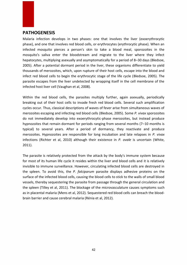
42
PATHOGENESIS
Malaria infection develops in two phases: one that involves the liver (exoerythrocytic
phase), and one that involves red blood cells, or erythrocytes (erythrocytic phase). When an
infected mosquito pierces a person's skin to take a blood meal, sporozoites in the
mosquito's saliva enter the bloodstream and migrate to the liver where they infect
hepatocytes, multiplying asexually and asymptomatically for a period of 8–30 days (Bledsoe,
2005). After a potential dormant period in the liver, these organisms differentiate to yield
thousands of merozoites, which, upon rupture of their host cells, escape into the blood and
infect red blood cells to begin the erythrocytic stage of the life cycle (Bledsoe, 2005). The
parasite escapes from the liver undetected by wrapping itself in the cell membrane of the
infected host liver cell (Vaughan et al, 2008).
Within the red blood cells, the parasites multiply further, again asexually, periodically
breaking out of their host cells to invade fresh red blood cells. Several such amplification
cycles occur. Thus, classical descriptions of waves of fever arise from simultaneous waves of
merozoites escaping and infecting red blood cells (Bledsoe, 2005). Some P. vivax sporozoites
do not immediately develop into exoerythrocytic-phase merozoites, but instead produce
hypnozoites that remain dormant for periods ranging from several months (7–10 months is
typical) to several years. After a period of dormancy, they reactivate and produce
merozoites. Hypnozoites are responsible for long incubation and late relapses in P. vivax
infections (Richter et al, 2010) although their existence in P. ovale is uncertain (White,
2011).
The parasite is relatively protected from the attack by the body's immune system because
for most of its human life cycle it resides within the liver and blood cells and it is relatively
invisible to immune surveillance. However, circulating infected blood cells are destroyed in
the spleen. To avoid this, the P. falciparum parasite displays adhesive proteins on the
surface of the infected blood cells, causing the blood cells to stick to the walls of small blood
vessels, thereby sequestering the parasite from passage through the general circulation and
the spleen (Tilley et al, 2011). The blockage of the microvasculature causes symptoms such
as in placental malaria (Mens et al, 2012). Sequestered red blood cells can breach the blood-
brain barrier and cause cerebral malaria (Rénia et al, 2012).

43
CLINICAL MANIFESTATIONS Usually, patients with malaria become symptomatic a few weeks after the infection, but the
host’s previous exposure or host immunity against malaria could influence symptomatology
and the incubation period, which is typical for each Plasmodium species.
One important symptom is the typical recurrent fever (which may be periodic at 48 or 72
hours, depending on the species) (Fig. 15).
Fig.15. Malaria symptom - typical recurrent fever
(Source: https://www.dailystar.co.uk/health/592904/Malaria-symptoms-tablets-treatment-prevention
The classic paroxysm begins with a period of shivering and chills lasting for around 1-2 hours
and is followed by high fever. The excessive sweats experienced by the patient are followed
by the decrease of body temperature to normal or below normal. Virtually, all patients with
malaria experience headache. Other symptoms include tiredness, malaise, nausea and
vomiting, diarrhea, cough, shortness of breath, arthralgia and myalgia (Dondorp et al, 2017;
Nadjm et al, 2012; Bartoloni and Zammarchi, 2012) (Fig. 16, 17).
Fig.16. Malaria symptoms
(Source: https://www.lybrate.com/topic/symptoms-of-malaria-and-
chikunguniya/dd771f92ae9155433ef28c1ecc43adc7)

44
Many patients, particularly during the early infection, do not present the classic paroxysm
but may have several small fever spikes a day. Fever periodicity is specific for each species
(i.e., 48 h for P. falciparum, P. vivax, and P. ovale – so called tertian fever; 72 h for P.
malariae – also called quartan fever), but is not apparent during the initial infection
because of multiple broods emerging in the bloodstream, and is often not observed in P.
falciparum infections. Patients with long-standing, synchronous infections are more likely to
present classic fever patterns.
In severe cases, usually caused by P. falciparum, the following symptoms could be present:
jaundice, anemia, renal failure, shock, adult respiratory distress syndrome, confusion, coma,
encephalopathy, and acidosis (Dondorp et al, 2017; Nadjm et al, 2012; Bartoloni and
Zammarchi, 2012) (Fig. 17).
Fig.17. Malaria symptoms
(Source: http://zeenews.india.com/health/how-to-fight-dengue-malaria-flu-more-symptoms-prevention-
2046312
The World Health Organization (WHO) recommends that pregnant women should avoid
travelling to areas where there is a risk of malaria (WHO Information for travellers, 2018). If
women get malaria while pregnant, the mother and the baby are at increased risk of
developing serious complications such as: premature birth (birth before 37 weeks of
pregnancy), low birth weight, restricted growth of the baby in the womb, stillbirth,
miscarriage, and even mother death.
45
DIAGNOSTIC Malaria must be promptly recognized in order to treat the patient effectively and in time,
and in order to control the spreading of the infection in the community through the local
mosquitoes. Delaying the diagnosis and the beginning of the treatment represents the main
cause of death in patients with malaria.
Malaria may be suspected on the basis of the patient’s history of recent travel or living in
malaria-endemic areas, of their symptoms and of the findings from clinical examination.
However, in order to confirme the diagnosis, there should be done laboratory tests that
would demonstrate the presence of the malaria parasites.
There could be a problem in countries were malaria is no longer endemic because doctors
may not be familiar with the disease. Clinicians who see a patient with malaria may not take
into account malaria as one of the possible diagnoses and therefore not recommend the
necessary diagnostic tests. The laboratory may not have the required experience with
malaria and may not be able to detect the parasites during examination of blood smears
under the microscope.
CLINICAL DIAGNOSTIC The patient’s history should include inquiries into the following: recent or remote travel to
an endemic area; immune status, age, and pregnancy status; allergies or other medical
conditions; medications currently being taken.
Most of the patients with malaria have no specific physical findings, but splenomegaly may
be present. Symptoms of malarial infection are nonspecific and may manifest as a flu-like
illness with fever, headache, malaise, fatigue, and muscle aches. Some patients with malaria
present diarrhoea and other gastrointestinal symptoms. Immune individuals may be
completely asymptomatic or may present mild anemia. Nonimmune patients may quickly
become very ill. In general, however, the occurrence of fever periodicity is not a reliable clue
for the diagnosis of malaria.
Severe malaria primarily involves the P. falciparum infection, although death due to splenic
rupture has been reported in patients with non-P. falciparum malaria. Severe malaria
includes cerebral malaria (sometimes with coma); severe anemia; respiratory abnormalities
(with metabolic acidosis, associated respiratory distress, and pulmonary edema); signs of
malarial hyperpneic syndrome (alar flaring, chest retraction, use of accessory muscles for
respiration, and abnormally deep breathing); renal failure (usually reversible) (Dondorp et
al, 2017; Bartoloni and Zammarchi, 2012).

46
In children, malaria has a shorter evolution, often rapidly progressing to severe malaria.
Children are more likely to present hypoglycemia, seizures, severe anemia, and sudden
death, but they are much less likely to develop renal failure, pulmonary edema, or jaundice
(Bartoloni and Zammarchi, 2012).
In some malaria-endemic areas, transmission is so intense that a large part of the
population is infected, but does not become ill. These types of carriers develop sufficient
immunity to protect them from malaria.
Malaria is clinically suspected on the basis of fever or a history of fever, but antimalarial
treatment should be limited to cases with positive tests. Patients with negative results
should be reassessed for other common causes of fever and treated appropriately. In
patients with suspected severe malaria and in other high-risk groups, such as patients living
with HIV/AIDS, absence or delay of parasitological diagnosis should not delay the immediate
start of antimalarial treatment.
LABORATORY DIAGNOSTIC The following blood investigations should be performed:
blood culture, hemoglobin concentration, platelet count, investigation of liver and
renal function, electrolyte concentrations (especially sodium), monitoring of
parameters suggestive of hemolysis (haptoglobin, lactic dehydrogenase,
reticulocyte count);
in selected cases, rapid HIV testing is recommended;
white blood cell count: leukocytosis is present in less than 5% of malaria patients
(in this case the differential diagnosis should be broadened);
G6PD level has to be checked if the patient is to be treated with primaquine;
the glucose level has to be evaluated to rule out hypoglycemia if the patient has
cerebral malaria.
The following imaging investigations may be considered:
chest radiography if respiratory symptoms are present;
Computed Tomography of the head if central nervous system symptoms are
present.
Specific tests for malaria infection should be carried out:
microhematocrit centrifugation (sensitive but incapable of speciation);
fluorescent dyes/ultraviolet indicator tests (may not yield speciation information);
the standard tests: thin (qualitative) or thick (quantitative) blood smears (1
negative smear does not exclude malaria as a diagnosis and several more smears
should be examined over a 36-hour period);

47
alternatives to blood smear testing (used if the blood smear expertise is
insufficient) are rapid diagnostic tests, polymerase chain reaction assay, nucleic
acid sequence-based amplification, and quantitative buffy coat (Bartoloni and
Zammarchi, 2012; Greenwood et al, 2005).
All malaria suspect patients should be treated on the basis of a confirmed diagnosis through
microscopic examination or by a rapid diagnostic test (RDT) (immunochromatography) that
aim to detect the antigens in the blood (Fig. 18 ). However, before using these rapid tests on
a large scale, their accuracy should be improved. It is recommended that all RDTs should be
followed by microscopy in order to confirm the diagnosis and, if positive, in order to
quantify the proportion of the red cells from the infected blood. These rapid tests may
reduce the necessary time for malaria diagnosis.
Fig.18. Rapid Diagnostic Test for Malaria
(Source: http://adulldayatwork.blogspot.com/2011/10/25-october-2011-tuesday-rapid-malaria.html
Molecular identification of the malaria parasite represents the certain diagnostic method,
but the Polymerase Chain Reaction (PCR) results are not always available fast enough in
order to be of great value in establishing the diagnosis of infection with Plasmodium (Fig.
19). The PCR is most useful for the confirmation of the parasite species after the diagnosis
was done either by microscopy of thick drops or by RDT.

48
Fig.19. PCR test for malaria identification
(Source: Ivănescu, 2015)
Serology detects antibodies against malaria, using either the indirect immunofluorescence
(IFA) or the ELISA test. Serology does not detect the current infection, but measures all past
exposure. Malaria parasites can be detected using microscopic hematological examination
(thick peripheral blood drops) (Fig. 20).
Fig.20. Thick peripheral blood drops examination
(Source: https://labmedicineblog.com/2018/06/19/hematology-case-study-a-51-year-old-woman-with-fever-
and-chills/
Before the examination, the specimen is often coloured with Giemsa coloration (Fig. 21).
This technique remains the golden standard for laboratory malaria confirmation. However,
it depends on the quality of the reagents, the microscope, and the laboratory’s experience.

49
Fig.21. Hematologic diagnosis of malaria
(Source: Personal collection)
The malaria suspected cases from Africa are increasingly verified by using a rapid test before
prescribing antimalarial treatment, thus ensuring an efficient use of resources available for
the elimination of the parasites, although the main cause of patient fever remains unknown
because of the potential of a co-infection with other febrile illness. The large-scale use of
the rapid tests does not necessarily preclude the over evaluation of clinical malaria cases or
the management of the sub-optimal cases of febrile patients. In a recent publication from
the journal eLife, a new approach was presented that allows the deduction of the spatio-
temporal prevalence of the malarial fever associated with Plasmodium falciparum, as well
as of the non-malarial fever presented by children from Sub-Saharan Africa between 2006
and 2014. It is estimated that 35.7% of the febrile cases reported by the author were
accompanied by malaria infection in 2014, but only 28.0% of these (10.0% of all febrile
cases) were caused by malaria. The majority of the febrile cases among children with
positive malaria are therefore caused by non-malarial diseases (Fig. 22).
Also, anemia or the decrease of blood hemoglobin concentration levels is frequently
associated with malaria. Anemia is very prevalent in Sub-Saharan Africa, especially among
children and women at the reproductive age, but so far, it has not been quantified in order
to establish the contribution of malaria, nor the contribution of other causes to the onset of
anemia (for example, the iron deficiency diet, helminth infection) (Dalrymple et al, 2017).

50
In collaboration with the ROAD-MAP team, data have been collected from the survey done
in households where respondents are asked about their fever and treatment history and
receive a diagnosis based on malaria parasites and the measurement of their hemoglobin
concentration in the blood. Also, the data from the survey are associated with the distance-
sensitive variables collected by ROAD-MAP in order to help with the spatial prediction of the
measurements of interest.
Fig.22. The proportion of malaria positive fever from all causes of fever
with children under 5
(Source: https://elifesciences.org/articles/29198)
It has therefore been noted that prior to using the rapid tests on a large scale, antimalarial
treatment used to be administered in high proportions, and an inaccurate diagnosis of this
disease was documented.
Moreover, not all the people that develop malaria request treatment in public health
institutions. Some of them receive treatment in units that do not report malaria cases, such
as private medical institutions, while others do not get treated. This contributes to under-
reported malaria cases. Before the administration of any treatment it is recommended that
patients have done all the tests for evaluating resistance to medication. These tests take
place in specialized laboratories which assess the susceptibility of the parasites found in a
certain patient to antimalarial compounds. There are two main laboratory methods:
in vitro tests: the parasites are grown in culture in the presence of an
increasing concentration of medication; the concentration of the medication
that inhibits the growth of the parasite is used as a final step;
molecular characterization: the molecular markers evaluated through PCR or
genetic sequencing also allow to predict, to a certain degree, resistance to
some medication.

51
PROPHYLAXIS In 2016 the governments of the malaria-endemic countries and their international partners
invested over 2.7 billion dollars in malaria control measures and eradication. The African
region takes up 74% of the budget, the percentages decreasing for the regions in South-East
Asia (7%), Eastern Mediterranean and America (each 6%) and West Pacific (4%). The
governments of the endemic countries contributed only 800 million USD, which represents
just 31% of the invested sum, a fact that shows that in the absence of involvement of
partners from non-endemic countries, the fight to eradicate malaria would have been lost
from the start. The United States of America (USA) has offered one billion dollars (38%),
followed by an important contribution from the United Kingdom of Great Britain and
Northern Ireland (Great Britain), as well as France, Germany and Japan (WHO - World
Malaria Report, 2017).
At the 2010 annual meeting, "Global Technical Strategy for Malaria 2016–2030", an annual
budget was established until the year 2020 in order to decrease the incidence of malaria
cases and the rate of mortality by at least 40%, a budget that reaches 6.5 billion dollars per
year. The deficit causes concern since it is not even sufficient for maintaining the level
reached until now.
First line prevention consists in protection against mosquito bites of the Anopheles genus.
Prophylactic measures involve sleeping under nets treated with long-lasting insecticide and
the use of protective clothing and insect repellents (Fig. 23).
In 2016, the percentage of households that used mosquito nets treated with insecticide had
increased up to 80% compared to the 50% level registered in 2010; however, there is a
periodic decline when countries change insecticides or take them by rotation, using more
expensive ones. The residual spraying of indoor spaces with insecticide is a strong means of
rapidly reducing malaria transmission. Its potential is achieved when at least 80% of the
houses from the targeted areas are sprayed. Indoor spraying is efficient for 3-6 months,
depending on the insecticide and the type of surface sprayed. In some situations, more
rounds of spraying are needed in order to protect the population for the entire malaria
season.

52
Fig.23. Long-lasting insecticide-treated mosquito nets
(Source: http://www.who.int/immunization/programmes_systems/interventions/malaria_llins/en/)
Depending on the risk of malaria transmission in the area to be visited and the species of
Plasmodium that exist, travellers may also take prophylactic medication (chemoprophylaxis)
before, during and upon returning from the journey.
Some categories of travellers, especially small children, pregnant women and persons with a
weak immune system, present an increased risk of developing serious forms of the disease,
if they were to contract it. In pregnant women, malaria increases the risk of maternal death,
spontaneous miscarriage, the birth of a dead baby or a baby with a low weight at birth, as
well as the associated risk of neonatal death. Because of this, pregnant women should avoid
travelling to malaria-endemic countries, and parents are advised to not take infants or
yuong children in areas where there is the risk of infection with the P. falciparum strain.
When travel cannot be avoided it is important that travellers take effective preventive
measures against malaria, even when they travel to areas with transmission of malaria
caused by P. vivax. In 2012, WHO recommended seasonal chemoprevention of malaria as an
additional measure of preventing malaria for the areas from the African sub-region of Sahel.
The strategy involves monthly administration of amodiaquine and sulfadoxine
pyrimethamine to all children under the age of 5 during the high-transmission season. It is
recommended that travellers consult all national centers for fighting against the disease or
other institutions that can offer information regarding preventive measures that must be
taken before going to malaria-endemic countries or regions.
Prevention of drug resistance or delaying its occurrence is essential for the success of both
strategies – national and global – for malaria control and eradication. In order to contribute
to the protection of current and future antimalarial medication, all malaria episodes should

53
be treated with combined therapy and antimalarial medication shoud be administered in
optimal doses. All cases of suspected malaria should be confirmed through RDT or
parasitological tests.
VECTOR CONTROL A big part of the success in fighting malaria is due to vector control. Vector control largely
depends on the use of pyrethroids, which are the only class of insecticides recommended at
present.
In the last years, resistance of mosquitoes to pyrethroids has grown in many countries. In
some areas, a resistance to all 4 classes of insecticides used for public health has been
detected. Fortunately, this resistance was only rarely associated with decreased efficiency of
mosquito nets treated with insecticides with prolongued action, which continue to offer a
substantial level of protection in the majority of cases. The use, in turn, of different classes
of insecticides for indoor spraying is recommended as an approach for the management of
resistance to insecticides.
However, the malaria-endemic areas from Sub-Saharan Africa and India raise significant
concerns because of high levels of malaria transmission and recent reports regarding the
spread of areas with resistance to insecticides. Using two different insecticides on a
mosquito net is an opportunity to reduce the risk of development and spread of resistance
to insecticides; the further development of these new nets is a priority.
Detection of resistance to insecticides should be an essential component of all national
efforts for malaria control in order to ensure that the most efficient methods of vector
control are used. Choosing the insecticide for indoor residual spraying (IRS) should always
depend on recent and local data about the susceptibility of target vectors.
The mosquitoes of the Anopheles genus are found all over the world with the exception of
the arctic area, while the anopheles species is found not only in malaria-endemic regions,
but also in areas where malaria had been eradicated. In order to avoid the risk of spread of
a number of vector-borne diseases, the transport of vectors must be controlled; therefore,
the transfer of mosquitoes as a result of trade on tyres or in containers must be prevented.
Also, infected people (tourists and immigrants) with pathogenic agents transmitted through
vectors must be diagnosed and treated as quickly as possible, since the diseases transmitted
by mosquitoes have no borders, and their spread is also a consequence of people mobility
and globalization. Therefore, increased mobility of people and international commerce play
a more important role in the spread of vectors and their pathogenic agents / parasites than
the increase in temperature (Yi et al, 2014).

54
The data collected from 76 malaria-endemic countries for the period 2010-2016 showed
that resistance to at least one insecticide was reported in 61 countries. Resistance to two or
more classes of insecticides was reported in 50 countries. Resistance to pyrethroides – the
only class of insecticides currently used in treating the mosquito nets, is largely spread in all
areas, and the percentage of countries that reported resistance to insecticides in 2016
increased to 81%, as compared to 71% in 2010 (WHO - World Malaria Report, 2017).
Despite the resistance evidenced, mosquito bed nets treated with insecticide remain the
most efficient instrument used in malaria prevention, even in areas where resistance to
pyrethroides has been reported.
PREVENTION OF MALARIA IN HUMANS At this time prevention and control of malaria includes three directions:
reducing the contact between the vector and the human host,
special prevention with antimalarials, and
early diagnosis and appropriate treatment of malaria episodes to limit
transmission (Heymann, 2012; Dondorp, 2017).
GENERAL PREVENTION - MINIMIZING THE CONTACT BETWEEN THE HUMAN HOST AND
THE MOSQUITO
A very important method of general prevention for malaria in endemic areas is the
use of mosquito bed nets treated with insecticide (ITN). They have to be retreated
after 3 washes or at least once a year. An improved variant are long-lasting
insecticide-treated mosquito nets (LLINs) incorporating pyrethroids (Heymann,
2012; Ouattara et al, 2014). There are new hopes regarding the use of mosquito
nets with two insecticides to reduce the risk of resistance (Heymann, 2012).
Another general prevention method is indoor residual insecticide spraying (IRS),
which targets the adult mosquito population and has a high rate of success when
more than 80% of the houses in the area are sprayed (WHO - Malaria, 2018;
Heymann, 2012). This method, which is a public health intervention, has
maximum efficacy when mosquitoes remain indoors on surfaces that can be
sprayed, when people are exposed in the house or in the vicinity of the house,
when the method is applied before the start of the transmission season or before
the peak transmission period (Heymann, 2012; Salam et al, 2014). At the moment,
there are 12 WHO recommended insecticides for this measure, and the choice of
these substances is dependent on the susceptibility of the insecticide, the
peculiarities of the vector, the degree of safety for humans and the environment,
the efficacy and the cost-effectiveness ratio. The DDT is still used to control the
Anopheles species, in line with the Stockholm Convention on Persistent Organic
Pollutants which prohibits the use of DDT, except for the purpose of public health
insurance (Heymann, 2012).

55
Controlling larval stages by eliminating mosquito reproduction sites has limited
value in most areas where malaria transmission persists. Such a measure is
represented by filling and draining or increasing the flow of water in natural or
artificial channels. Chemical or biological control methods, such as bacterial
larvicides or larvivorus fish, can be difficult to implement in rural areas, but some
African urban areas have been successful. These methods are useful adjuvants in
arid, coastal and urban areas, but methods for maintaining a low level of
responsiveness may be necessary in areas where malaria elimination has been
achieved (Heymann, 2012; Bocsan et al, 1999).
Intermittent preventive use of a full dose of antimalarial at predetermined
intervals in trimesters 2 and 3 of pregnancy is an effective measure for reducing
malaria in pregnant women in areas with a P. falciparum stable transmission, with
moderate to intensive intensity such as Africa. The method has limited use in
other parts of the world, where transmission is unstable and of low intensity.
Also, in endemic areas, using ITN and LLIN for this category of people diminishes
the effects of malaria during pregnancy (Heymann, 2012; Mpogoro et al, 2014).
Another general prevention measure is the health education of people travelling
to endemic areas so that they are aware what regions may pose a risk, what are
the protective measures to be taken and how to manage feverish syndromes if
they occur after coming back (Luca, 2002).
SPECIAL PREVENTION USING ANTIMALARIAL DRUGS
Before travel chemoprophylaxis should include recommendations for the use of the
appropriate antimalarial medication (if any) for the chosen destination, as follows
(Heymann, 2012; Bocsan, 1999; Luca, 2002):
In areas with a very low risk of malaria transmission, chemoprophylaxis may not be
recommended, taking into account that the risk of side effects associated with
antimalarials may outweigh the potential benefits. But the travellers should be aware
of the possibility of malaria if they develop a febrile illness.
Chloroquine chemoprophylaxis can be used in areas where only P. vivax malaria
occurs and those areas where P. falciparum is susceptible to chloroquine. 5 mg base /
kg / week is recommended at single dose or 10 mg base / kg / week, divided into 6
doses / day (Heymann, 2012; Luca, 2002).
In areas where there is a risk of transmission for P. vivax and P. falciparum and there
is resistance to chloroquine, chloroquine chemoprophylaxis should be combined with
proguanil (3 mg / kg / day).
In areas of high risk for P. falciparum malaria and resistance to antimalarial drugs,
elective chemoprophylaxis is performed with atovaquone-proguanil (250 mg
atovaquone and 100 mg proguanil / day), doxycycline (1.5 mg / kg / day) or
mefloquine (5 mg / kg / week). These recommendations also apply to areas with

56
moderate or low risk of P. falciparum malaria, but with high levels of resistance to
antimalarial medication (Heymann, 2012; Luca, 2002).
The use of daily antimalarials (atovaquone-proguanil, chloroquine, doxycycline,
proguanil) should be started one day before arrival in the risk area. With regard to
chloroquine, administration should be started one week before arrival and for
mefloquine with 2-3 weeks before departure to achieve high, effective antibody
levels and to detect possible side effects before departure.
Preventive drug administration should be strictly used throughout the journey in
areas at risk of malaria and should be continued for 4 weeks after the last possible
exposure (Heymann, 2012; Luca, 2002).
SPECIFIC PREVENTION - VACCINE DEVELOPMENT
Although there are promises of a vaccine against malaria, there is currently no licensed
population-based vaccine. he most advanced is the research for the development of a
candidate vaccine against P. falciparum, a preparation known as RTS, S/AS01 (a
recombinant vaccine using pre-erythrocyte stage proteins) - Mosquirix. This vaccine was
evaluated in an extended clinical trial conducted in 7 African countries and was positively
evaluated by the European Medicines Agency in July 2015 (WHO - Malaria, 2018; Dondorp,
2017; Umeh et al, 2014). The vaccine efficacy against malaria with clinical manifestations in
children aged 5 to 17 months was 53% (CI95% = 28-69%), evaluated over a period of 8
months. For the older child the efficacy of this vaccine was lower. Other candidate vaccines
include the attenuated whole-parasitic vaccine, which provides a high level of protection,
but raises problems of production and administration. Other types of candidate vaccines
target parasite stages which can be found in the bloodstream or asexual stages of
Plasmodium (Dondorp, 2017). WHO strongly supports these pilot studies as an important
step in obtaining the world's first antimalarial vaccine, so in October 2015 it recommended
the implementation of the pilot studies for RTS, S/AS01 vaccine in a limited number of
African countries. In November 2016, WHO announced that the safety and efficacy of the
RTS, S/AS01 vaccine would be evaluated in two pilot studies (Phase III clinical trials)
conducted in 3 Saharan African countries (Ghana, Kenya and Malawi) that were scheduled
to start at the begining of 2018 (WHO - Malaria, 2018). This pilot project could open the way
for a larger development of the vaccine if its safety and efficacy are considered acceptable.
THE PREVENTION OF P.VIVAX OR P.OVALE INFECTION RELAPSES In order to prevent the relapses, primaquine for 14 days (0.25-0.5mg / kg body weight) in all
transmission environments is the recommendation for malaria caused by P.vivax or P.ovale
in children and adults (with the exception of pregnant women and infants before the age of
6 months). In patients with G6PD deficit, the prevention of relapses is made with
primaquine in a dose of 0.75mg / kg body weight once a week, for 8 weeks, under strict
57
medical surveillance because of the hemolysing potential induced by primaquine (WHO -
Guidelines for the Treatment of Malaria, 2015).
Artemisinin and its derivatives must not be used as oral monotherapy because they could
produce artemisinin resistance. The WHO recommends regular monitorization of
antimalarial medication efficiency in order to ensure that the selected treatments remain
efficient. These actions have to be implemented by the national programs for malaria
control. Resistance to antimalarial medication represents a recurring problem. The
resistance of P. falciparum to previous drugs generations, such as chloroquine and
sulfadoxine pyrimethamine (SP), had become largely spread in the 1950s and 1960s,
undermining malaria control efforts and decreasing the chances of survival in children. The
WHO recommends routine monitoring of resistance to antimalarial medication and supports
countries in consolidating their efforts in this regard. In the past years, resistance of
parasites to artemisinin was detected in 5 countries from the Mekong subregion: Cambodia,
Lao People's Democratic Republic, Myanmar, Thailand and Vietnam. Studies have confirmed
that resistance to artemisinin has appeared independently in many areas of these
subregions.
In 2013, the WHO launched an emergency answer to resistance to artemisinin (ERAR) from
the Mekong subregion, a high-level action plan in order to limit the spread of parasites that
are resistant to medication and offer instruments that can help the population exposed to
the risk of malaria infection. In parallel, there were reported some cases of high resistance
levels to medication that was associated with ACT. A new approach was necessary in order
to keep up with the changes in the malaria landscape. As a consequence, the Advisory
Committee for policies in the domain of malaria of the WHO recommended in September
2014 the adoption of the objective to eliminate the malaria caused by P. falciparum in this
subregion until 2030. The WHO launched the Strategy for the elimination of malaria in the
Mekong subregion (2015-2030) in 2015, which was approved by all the countries from the
subregion. With technical guidance from the WHO, all GMS countries have elaborated
national plans for the elimination of malaria. Together with partners, the WHO offers
permanent support to all countries in their efforts to eliminate malaria through the
dedicated program of elimination of malaria in Mekong, a new initiative that evolved from
ERAR (WHO - Strategy for malaria elimination in the Greater Mekong Subregion: 2015-2030,
2015).

58
TREATMENT
Malaria is an entirely preventable and treatable disease. The primary objective of the
treatment is to ensure the rapid and complete elimination of the Plasmodium parasite from
the patient’s blood in order to prevent the progression from uncomplicated malaria to
severe disease and to prevent the chronic infection that leads to severe anemia. From a
public health perspective, the goal of the treatment is to reduce transmission of the
infection to other people by reducing the infectious reservoir, and to prevent the spread of
the antimalarial drug resistance.
In patients with suspected malaria, the parasitological diagnosis must be confirmed before
actually beginning the treatment for malaria, either by microscopy or by a rapid diagnostic
test. The treatment based only on clinical manifestations has to be used solely for the cases
in which the diagnostic tests are not accessible upon 2 hours from the arrival of patients for
treatment. In order to prevent any life threatening complications the treatment with an
efficient and sure antimalarial drug has to be used within 24 hours from the onset of the
fever.
Malaria is treated with antimalarial medications, but the selected drugs depend on the type
and severity of the disease. Also, medications against fever are commonly used in malaria,
but their effects on outcomes are not very clear (Meremikwu et al, 2012; Amadi et al, 2015).
Simple or uncomplicated malaria may be treated with oral medications. Artemisinins in
combination with other additional antimalarials (including: amodiaquine, lumefantrine,
mefloquine or sulfadoxine/pyrimethamine), also known as the artemisinin-combination
therapy (ACT), is the most effective treatment for the P. falciparum infection that decreases
the resistance to any single drug component (WHO - Guidelines for the Treatment of
Malaria, 2015; Amadi et al, 2015; Kokwaro, 2009). Another recommended combination is
dihydroartemisinin and piperaquine. ACT is about 90% effective when used to treat
uncomplicated malaria (WHO - Guidelines for the Treatment of Malaria, 2015; Amadi et al,
2015; Howitt et al, 2012). To treat uncomplicated malaria during pregnancy, WHO
recommends the use of quinine plus clindamycin early in the pregnancy (1st trimester), and
ACT in later stages (2nd and 3rd trimesters) (WHO - Guidelines for the Treatment of Malaria,
2015; Piola et al, 2010; McGready et al, 2012). Infections with P. vivax, P. ovale or P.
malariae usually do not require hospitalization. Treatment of P. vivax should include both
treatment of blood stages (with chloroquine or ACT) and clearance of liver forms with
primaquine (WHO - Guidelines for the Treatment of Malaria, 2015; Meremikwu et al, 2012).
Treatment with tafenoquine prevents relapses after confirmed P. vivax malaria (Kokwaro,
2009; Rajapakse et al, 2015).
In areas with low transmission, one single small dose of primaquine should be used in the
antimalarial treatment with the purpose of reducing transmission of the infection. The G6PD

59
testing is not necessary because one single small dose of primaquine is efficient in blocking
transmission and it is less likely to cause severe toxicity for subjects with G6PD deficit.
The infection with P. falciparum almost always causes severe and complicated malaria, but
the other species usually cause only febrile disease (White, 2011). Severe and complicated
malaria are medical emergencies that produce high mortality rates (between 10% and 50%)
(Howitt et al, 2012). Cerebral malaria is the form of severe and complicated malaria with the
worst neurological symptoms (Manyando et al, 2011). Recommended treatment for severe
malaria is the intravenous use of antimalarial drugs. For severe malaria, parenteral
artesunate is superior to quinine in both children and adults (WHO - Guidelines for the
Treatment of Malaria, 2015; Sinclair et al, 2012). Other studies show that artemisinin
derivatives (artemether and arteether) have been as efficacious as quinine in the treatment
of cerebral malaria in children (Rajapakse et al, 2015).
It is essential that full doses of effective parenteral (or rectal for children under the age of 6
years) antimalarial treatment be given promptly in the initial treatment of severe malaria (at
least for 24 hours). This should be followed by a full dose (3 days administration) of effective
oral ACT if the patient can tolerate the drug. Two classes of medicines are available for
parenteral treatment of severe malaria: artemisinin derivatives (artesunate or artemether)
and the Cinchona alkaloids (quinine and quinidine). Parenteral artesunate is the treatment
of choice for all severe malaria. A large randomized clinical trial regarding the treatment of
severe falciparum malaria showed an important reduction in the mortality rate with
parenteral (intravenous or intramuscular) artesunate as compared with parenteral quinine.
The artesunate was simpler and safer to use, with no increase in neurological sequelae in
artesunate-treated survivors (WHO - Guidelines for the Treatment of Malaria, 2015).
The most used drugs in malaria treatment are active against the parasite forms in the blood
(the form responsible for the occurrence of the disease) and include: chloroquine;
atovaquone-proguanil (Malarone®); artemether-lumefantrine (Coartem®); mefloquine
(Lariam®); quinine; quinidine; doxycycline (used in combination with quinine); clindamycin
(used in combination with quinine), and artesunate (Bartoloni and Zammarchi, 2011;
Greenwood et al, 2005).

60
REFERENCES
1. Abdul-Ghani R, Mahdy MAK, Saif-Ali R, Alkubati SA, Alqubaty AR, Al-Mikhlafy AA, Al-
Eryani SM et al. Glucose-6-phosphate dehydrogenase deficiency among Yemeni children
residing in malaria-endemic areas of Hodeidah governorate and evaluation of a rapid
diagnostic test for its detection. Malari J 2016; 15:237. DOI 10.1186/s12936-016-1372-9.
2. Adoubryn KD, Ouhon J, Yapo CG, N’guettia KS, Kouakou KAE, Adou SJ et al. Efficacitѐ
thѐrapeutique de la chloroquine dans le traitement du paludisme simple á Plasmodium
falciparum de lꞌenfant dans lꞌest de la Côte-dꞌIvoire (1999-2002). Med Mal Infect 2004;
34 (3) : 127-131. https://doi.org/10.1016/j.medmal.2003.11.004.
3. Ajdukiewicz KMB, Ong ELC. Management of vivax malaria with low sensitivity to
primaquine. J Infection 2007; 54 (3) : 209–211.DOI: https://doi.org/10.1016/j.jinf. 2006.
05.008.
4. Al-Olayan EM, Beetsma AL, Butcher GA, Sinden RE, Hurd H. Complete development of
mosquito phases of the malaria parasite in vitro. Science 2002; 295 (5555) : 677-679.
DOI: 10.1126/science.1067159.
5. Al-Eryani SMA, Kelly-Hope L, Harbach RE, Briscoe AG, Barnish G, Azazy A, McCall PJ.
Entomological aspects and the role of human behaviour in malaria transmission in a
highland region of the Republic of Yemen. Malari J 2016; 15 : 130. DOI 10.1186/s12936-
016-1179-8.
6. D’Alessandro G, Bruno Smiraglia C, Lavagnino A. Further studies on the biology of
Anopheles labranchiae labranchiae Falleroni in Sicily. World Health Organization, 1971,
pp 1-11.
7. Alten B, Caglar SS, Simsek FM, Kaynas S. Effect of insecticide-treated bednets for malaria
control in Southeast Anatolia-Turkey. J Vector Ecol 2003; 28 : 97-107.
8. Aly AS, Vaughan AM, Kappe SH. Malaria parasite development in the mosquito and
infection of the mammalian host. Annu Rev Microbiol 2009; 63 : 195-221. doi:
10.1146/annurev.micro.091208.073403.
9. Amadi OK, Otuokere IE, Bartholomew CF. Synthesis, Characterization, in vivo
Antimalarial Studies and Geometry. Optimization of Lumefantrine/Artemether Mixed
Ligand Complexes. Research Journal of Pharmaceutical Dosage Forms and Technology
2015; 7 (1): 59-68.
10. Amino R, Giovannini D, Thiberge S, Gueirard P, Boisson B, Dubremetz JF et al. Host cell
traversal is important for progression of the malaria parasite through the dermis to the
liver. Cell Host Microbe 2008; 3 (2) : 88-96. doi: 10.1016/j.chom.2007.
11. Amino R, Thiberge S, Blazquez S, Baldacci P, Renaud O, Shorte S, Ménard R. Imaging
malaria sporozoites in the dermis of the mammalian host. Nat Protoc 2007; 2 (7) : 1705-
1712. DOI: 10.1038/nprot.2007.120.
12. Amino R, Thiberge S, Martin B, Celli S, Shorte S, Frischknecht F, Ménard R. Quantitative
imaging of Plasmodium transmission from mosquito to mammal. Nat Med 2006; 12 (2) :
220-224. DOI: 10.1038/nm1350.
13. Askling H, Bruneel F, Burchard G, Castelli F, Chiodini PL, Grobusch M et al. Management
of imported malaria in Europe. Malaria J 2012; 11: 328. https://doi.org/10.1186/1475-
2875-11-328.
14. Akiner MM, Cağlar SS. Identification of Anopheles maculipennis group species using
polymerase chain reaction (PCR) in the regions of Birecik. Beyşehir and Cankiri. Turkiye
Parazitol Derg 2010; 34 (1) : 50-54.

61
15. Baird JK, Fryauff DJ, Hoffman SL. Primaquine for prevention of malaria in travelers. Clin
Infect Dis 2003; 37 (12) : 1659-1667. DOI: 10.1086/379714
16. Baird JK, Rieckmann KH. Can primaquine therapy for vivax malaria be improved? Trends
Parasitol 2003;19 (3) : 115-20.
17. Baird JK, Hoffman SL. Primaquine therapy for malaria. Clin Infect Dis 2004; 39 (9) : 1336-
1345. DOI: 10.1086/424663.
18. Bartoloni A, Zammarchi L. Clinical aspects of uncomplicated and severe malaria.
Mediterr J Hematol Infect Dis 2012; 4 (1): e2012026. doi: 10.4084/MJHID.2012.026.
22. Bates M. Variations in the antepalmate hairs of larvae ofthe Anopheles maculipennis
group. Rivista di Malariologia 1939; 18 :299-312.
23. Becker N, Petric D, Boase C, Lane J, Zgomba M, Dahl C, Kaiser A. Mosquitoes and their
control. Kluwer Academic / Plenum Publishers, New York, 2003.
24. Billker O, Lindo V, Panico M, Etienne AE, Paxton T, Dell A et al. Identification of
xanthurenic acid as the putative inducer of malaria development in the mosquito.
Nature 1998; 392 (6673) : 289-292. DOI: 10.1038/32667.
25. Billker O, Shaw MK, Margos G, Sinden RE. The roles of temperature, pH and mosquito
factors as triggers of male and female gametogenesis of Plasmodium berghei in vitro.
Parasitology 1997; 115 ( Pt 1) : 1-7.
26. Bledsoe GH. Malaria primer for clinicians in the United States. South Med J 2005; 98 (12):
1197–1204.
27. Boccolini D, Sabatini A, Coluzzi M. Valore diagnostico del numero dei rami delle setole
antepalmate per l'identificazione del complesso Anopheles maculipennis. Ann Ist Super
Sanita 1986; 22 : 201-204.
28. Boccolini D, Toma L, Di Luca M, Severini F, Cocchi M, Bella A et al. Impact of
environmental changes and human-related factors on the potential malaria vector,
Anopheles labranchiae (Diptera: Culicidae), in Maremma, Central Italy. J Med Entomol
2012; 49 (4) : 833-842. https://doi.org/10.1603/ME11252.
29. Bocşan IS, Rădulescu A, Brumboiu I, Şuteu O, Achimaş A. Epidemiologie practică pentru
medici de familie. Editura Medicală Universitară Iuliu Haţieganu, Cluj Napoca, 1999, pg.
393-405. [Bocsan IS, Radulescu A, Brumboiu I, Suteu O, Achimas A. Practical
epidemyology for general practitioners. Iuliu Hatieganu Medical University Publishing
House, Cluj Napoca, 1999, pg. 393-405.]
30. Boddey JA and Cowman AF. Plasmodium nesting: remaking the erythrocyte from the
inside out. Annu Rev Microbiol 2013; 67 : 243-269. doi: 10.1146/annurev-micro-092412-
155730.
31. Boëte C (ed). Genetically Modified Mosquitoes for Malaria Control. Eurekah/Landes
Bioscience, Georgetown, 2006.
32. Bogdan O, Marinica I. Hazardele climatice din zona temperată. Geneză și vulnerabilitate
cu aplicații la România. Edit. Universitară "L. Blaga", Sibiu, 2007. [Climate risks in the
temperate zone. Genesis and application vulnerability in Romania. “L. Blaga” University
Publishing House, sibiu, 2007].
33. Bray PG, Martin RE, Tilley L, Ward SA, Kirk K, Fidock DA. Defining the role of PfCRT in
Plasmodium falciparum chloroquine resistance. Mol Microbiol 2005; 56 (2) : 323-333.
DOI: 10.1111/j.1365-2958.2005.04556.x.
34. Bruce-Chwatt LJ, de Zulueta J. The rise and fall of malaria in Europe. Oxford University
Press, London, 1980, 240pp.
35. Buonomini G. L'esame delle setole antepalmate per lostudio della popolazione larvale
dei focolai naturali di Anopheles maculipennis. Rivista di Parassitologia 1940; 6 : 163-
174.
36. Cambournac FJ. Contribution to the history of malaria epidemiology and control in
Portugal and some other places. Parassitologia 1994; 36 (1-2) : 215-222.

62
37. Capinera JL. Encyclopedia of Entomology. 2nd edition, Springer, New York, 2002, 4 vol,
4411pp.
38. Carmona-Fonseca J, Maestre A. Prevention of Plasmodium vivax malaria recurrence:
efficacy of the standard total dose of primaquine administered over 3 days. Acta Trop
2009; 112 (2) : 188-192. doi: 10.1016/j.actatropica.2009.07.024.
39. Carter R, Mendis KN. Evolutionary and Historical Aspects of the Burden of Malaria. Clin
Microbiol Rev 2002; 15 (4) : 564–594. doi: [10.1128/CMR.15.4.564-594.2002].
40. Chakravarty S, Cockburn IA, Kuk S, Overstreet MG, Sacci JB, Zavala F. CD8+ T
lymphocytes protective against malaria liver stages are primed in skin-draining lymph
nodes. Nat Med 2007; 13 (9) : 1035-41. DOI: 10.1038/nm1628.
41. Cianchi R, Sabatini A, Boccolini D, Bullini L,Coluzzi M. Electrophoretic evidence of
reproductive isolation between sympatric populations of Anopheles melanoon and An.
subalpinus. Third International Congress on Malaria and Babesiosis, International
Laveran Foundation, 7–11 September, 1987, Annecy, France.
42. Cogswell FB. The hypnozoite and relapse in primate malaria. Clin Microbiol Rev 1992; 5
(1) : 26-35.
43. Colofiţchi A, Nicolescu G, Purcărea-Ciulacu V, Vladimirescu A, Coipan EC, Steriu D. Is
malaria a re-emerging disease in Romania? 14th European Conference of the Society of
vector Ecology, Bellinzona, Elvetia, 3 – 6 Septembrie 2003.
44. Corradetti A. Richerche sugli incroci tra le varietà di Anopheles maculipennis. Rivista di
Malariologia 1934; 13 : 707–720.
45. Cosoroaba I. Entomologie veterinară. Editura Ceres, București, 1992 [Veterinary
entomology. Ceres Publishing House, Bucharest, 1992].
46. Cosoroaba I. Parazitologie veterinară. Editura Mirton, Timisoara, 2000 [Veterinary
parasitology. Mirton Publishing House, Bucharest, 2000].
47. Dalrymple U, Cameron E, Bhatt S, Weiss DJ, Gupta S, Gething PW. Quantifying the
contribution of Plasmodium falciparum malaria to febrile illness amongst African
children. eLlife 2017; 6: e29198. doi: 10.7554/eLife.29198.
48. de Buck A, Swellengrebel NH. Behaviour of Dutch Anopheles atroparvus and messeae in
winter under artificial conditions. Rivista di Malariologia 1934; 8 : 404.
49. de Buck A, van der Torren G, Swellengrebel NH. Report for the year 1932 on
investigation into the racial composition of Anopheles maculipennis in Holland. Rivista di
Malariologia 1933; 12 : 265.
50. Derbyshire ER, Mazitschek R, Clardy J. Characterization of Plasmodium liver stage
inhibition by halofuginone. Chem Med Chem 2012; 7 (5) : 844-849. doi:
10.1002/cmdc.201200045.
51. Develoux M, Le Loup G, Dautheville S, Belkadi G, Magne D, Lassel L et al. Malaria among
immigrants, experience of a Parisian hospital (2006-2010). Bull Soc Pathol Exot 2012; 105
(2) : 95-102. doi: 10.1007/s13149-012-0217-1.
52. de Zulueta J, Ramsdale CD, Cianchi R, Bullini L, Coluzzi M. Observations on the taxonomic
status of Anopheles sicaulti. Parassitologia 1983; 23 :73-92.
53. Di Luca M, Boccolini D, Marinucci M, Romi R. Intrapopulation polymorphism in
Anopheles messeae (An. maculipennis complex) inferred by molecular analysis. J Med
Entomol 2004; 41 : 582-586.
54. Djadid ND, Gholizadeh S, Tafsiri E, Romi R, Gordeev M, Zakeri S. Molecular identification
of Palearctic members of Anopheles maculipennis in northern Iran. Malaria J 2007; 6 : 6.
https://doi.org/10.1186/1475-2875-6-6.
55. Dondorp AM, von Seidlein L. Malaria. In: Cohen J, Powderly WG, Opal SM. (eds).
Infectious Diseases. 4th edition, Vol I. Elsevier, 2017, p 1014-1025.

63
56. Dua VK, Pandey AC, Raghavendra K, Gupta A, Sharma T, Dash AP. Larvicidal activity of
neem oil (Azadirachta indica) formulation against mosquitoes. Malar J 2009; 8 : 124. doi:
10.1186/1475-2875-8-124.
57. Eritja R, Aranda C, Padros J, GouIa M. Revised checklist of the Spanish mosquitoes. Acta
Virologica Portuguesa 1998; 5 : 25.
58. Falleroni D. Zooprofilassi e sua applicazione in Ardea. (Argo Romano). Diverse razze di "A.
maculipennis". Tipografia Cuggiani, Roma, 1932, pp. 48+ x.
59. Falleroni D. Fauna anofelica italiana e suo 'habitat' (paludi, risaie, canali). Metodi di lotta
contro la malaria. Rivista di Malariologia 1926; 5 : 553-559.
60. Fanello C, Santolamazza F, della Torre A. Simultaneous identification of species and
molecular forms of the Anopheles gambiae complex by PCR-RFLP. Med Vet Entomol
2002; 16 (4) : 461-464.
61. Faraj C, Adlaoui E, Elkohli M, Herrak T, Ameur B, Chandre F. Review of Temephos
Discriminating Concentration for Monitoring the Susceptibility of Anopheles labranchiae
(Falleroni, 1926), Malaria Vector in Morocco. Malar Res Treat 2010; 2010: 126085. doi:
10.4061/2010/126085.
62. Ferrão JL, Mendes JM, Painho M, Zacarias S. Malaria mortality characterization and the
relationship between malaria mortality and climate in Chimoio, Mozambique. Malaria J
2017; 16 : 212. https://doi.org/10.1186/s12936-017-1866-0.
63. Field JW and Shute PG. The microscopic diagnosis of human malaria. II. A morphological
study of the erythrocyticparasite. Government Press, Kuala Lumpur, Malaysia, 1956.
64. Fillinger U, Sonye G, Killeen GF, Knols BG, Becker N. The practical importance of
permanent and semipermanent habitats for controlling aquatic stages of Anopheles
gambiae sensu lato mosquitoes: operational observations from a rural town in western
Kenya. Trop Med Int Health 2004; 9 (12) : 1274-1289. DOI: 10.1111/j.1365-
3156.2004.01335.x.
65. Foster WA, Walker ED. Mosquitoes (Culicidae). In Mullen, G, Durden, L. (Eds.) Medical
and veterinary entomology. 2nd edition. (pp. 207-259). Academic Press, Burlington, MA,
2009. 637 pp.
66. Frevert U. Sneaking in through the back entrance: the biology of malaria liver stages.
Trends Parasitol 2004; 20 (9) : 417-424. doi:10.1016/j.pt.2004.07.007.
67. Frizzi G. Etude cytogénétique d'Anopheles maculipennis en Italie. Bull World Health
Organ 1953; 9 : pp. 335-344.
68. Frizzi G. Nuovi contributi e prospetti di ricerca nel gruppo Anopheles maculipennis in
base allo studio del dimorfismo cromosomico. Symposia Genetica 1952; 3 : pp. 231-265.
69. Gillies MT, de Meillon B. The Anophelini of Africa south of the Sahara (Ethiopian
zoogeographical region). 2nd edition. Publ S Afr Inst Med Res 1968; 54 : l-343.
70. Girod R, Orlandi-Pradines E, Rogier C, Pagès F. Malaria transmission and insecticide
resistance of Anopheles gambiae (Diptera: Culicidae) in the French Military Camp of
Port-Bouët, Abidjan (Côte d’Ivoire): implications for vector control. J Med Entomol 2006;
43 (5) : 1082–1087.
71. Gratzer WB, Dluzewski AR. The red blood cell and malaria parasite invasion. Semin
Hematol 1993; 30 (3) : 232-247.
72. Greenwood BM, Bojang K, Whitty CJ, Targett GA. Malaria. Lancet 2005; 365 (9469) :
1487–1498. doi: 10.1016/S0140-6736(05)66420-3.
73. Hackett LW, Lewis DJ. A new variety of Anopheles maculipennis in southern Europe.
Rivista di Malariologia 1935; 14 : 377-383.
74. Hackett LW, Missiroli A. The varieties of Anopheles maculipennis and their relation to the
distribution of malaria in Europe. Rivista di Malariologia 1935; 14 : 45-109.
75. Hafalla JC, Silvie O, Matuschewski K. Cell biology and immunology of malaria. Immunol
Rev 2011; 240 (1) : 297-316. doi: 10.1111/j.1600-065X.2010.00988.x.

64
76. Hay S, Snow RW. The Malaria Atlas Project: Developing Global Maps of Malaria Risk.
Plos Medicine 2006; 3 (12) : 2204-2208. https://doi.org/10.1371/journal.pmed.0030473.
77. Harbach RE. Review of the internal classification of the genus Anopheles (Diptera:
Culicidae): the foundation for comparative systematics and phylogenetic research. Bull
a. Entomol Res 1994; 84: 331-342.
78. Heymann DL. Manual de Management al Bolilor Transmisibile. Un raport oficial al
American Public Health Association. Ediţia a 19-a . Editura Medicală Amaltea, Bucureşti,
2012, pag. 251-267. [Control Of Communicable Diseases Manual. An official report of the
American Public Health Association. 19th Edition. Amaltea Medical Publishing House,
Bucharest, 2012, pg. 251-267].
79. Howitt P, Darzi A, Yang GZ, Ashrafian H, Atun R, Barlow J et al. Technologies for global
health. Lancet 2012; 380 (9840) : 507–535. doi: 10.1016/S0140-6736(12)61127-1.
80. Ivanescu Maria Larisa – Bioecologia si patogenitatea unor culicide .
Teza de doctorat USAMV Iasi –Ro, 246 p., 2014
81. Ivanescu ML, Acatrinei D, Pavel I, Sulesco T, Miron L. PCR identification of five species
from the Anopheles maculipennis complex (Diptera: Culicidae) in North-Eastern
Romania. Acta Parasitologica 2015; 60 (2) : 283-289. doi: 10.1515/ap-2015-0040.
82. Ivănescu ML, Bodale I, Florescu SA, Roman C, Acatrinei D, Miron L. Climate Change Is
Increasing the Risk of the Reemergence of Malaria in Romania. Bio Med Res Internat
2016; http://dx.doi.org/ 10.1155/ 2016/8560519.
83. Jaenson TG, Ameneshewa B. Prehibernation diet and reproductive condition of female
Anopheles messeae in Sweden. Med Vet Entomol 1991; 5 (2) : 243-252.
84. Jelinek T, Schulte C, Behrens R, Grobusch MP, Coulaud JP et al. Imported Falciparum
malaria in Europe: sentinel surveillance data from the European network on surveillance
of imported infectious diseases. Clin Infect Dis 2002; 34 (5) : 572-576. DOI:
10.1086/338235.
85. Kappe SH, Gardner MJ, Brown SM, Ross J, Matuschewski K, Ribeiro JM et al. Exploring
the transcriptome of the malaria sporozoite stage. Proc Natl Acad Sci USA 2001; 98 (17) :
9895-9900. DOI: 10.1073/pnas.171185198.
86. Kasap H, Akbaba M, Kasap M, Lüleyap Ü, Alpaslan N, Alptekin D. A study of malaria
andAnopheles sacharovi in Tuzla, Adana, Turkey.Acta Parasit Turcica 1995; 19 : 535-540.
87. Kettle DS (ed). Medical and veterinary entomology. 2nd ed. CAB International Publishing
House, Wallingford, UK, 1995, 725 pp.
88. Kiszewski A, Mellinger A, Spielman A, Malaney P, Sachs SE, Sachs J. A global index
representing the stability of malaria transmission. Am J Trop Med Hyg 2004; 70 (5) : 486-
498.
89. Kitzmiller JB, Frizzi G, Baker R. Evolution and speciation within the maculipennis complex
of the genus Anopheles. In Wright JW, Pal R (eds): Genetics of Insect Vectors of Disease.
Elsevier Publishing, London, 1967, pp. 151-210.
90. Knight KL, Stone A. A catalog of the mosquitoes of the world. 2nd edition, Thomas say
Foundation, 1977; 611 pp.
91. Knottnerus O.S. Malaria around the North Sea: a survey. In: Wefer G., Berger W.H.,
Behre KE., Jansen E. (eds) Climate Development and History of the North Atlantic Realm.
Springer, Berlin, Heidelberg, 2002.
92. Kokwaro G. Ongoing challenges in the management of malaria. Malaria J 2009; 8 (Suppl.
1) : S2. doi:10.1186/1475-2875-8-S1-S2.
93. Konaté L, Zwetyenga J, Rogier C, Bischoff E, Fontenille D, Tall A et al. Variation of Plasmodium falciparum msp1 block 2 and msp2 allele prevalence and of infection complexity in two neighbouring Senegalese villages with different transmission conditions. Trans R Soc Trop Med Hyg 1999; 93 (Suppl 1) : 21-28.

65
94. Korenromp EL, Miller J, Cibulskis RE, Kabir Cham M, Alnwick D et al. Monitoring mosquito net coverage for malaria control in Africa: possession vs. use by children under 5 years. Trop Med Int Health 2003; 8 (8) : 693-703.
95. Korvenkontio P, Lokki J, Saura A, Ulmanen I. Anopheles maculipennis complex (Diptera: Culicidae) in northern Europe: species diagnosis by egg structure and enzyme polymorphism. J Med Entomol 1979; 16 : 169-170.
96. Krüger A, Rech A, Su XZ, Tannich E. Two cases of autochthonous Plasmodium falciparum
malaria in Germany with evidence for local transmission by indigenous Anopheles
plumbeus. Trop Med Int Health 2001; 6 (12) : 983-985.
97. Lacroix R, Mukabana WR, Gouagna LC, Koella JC. Malaria infection increases
attractiveness of humans to mosquitoes. PLoS Biol 2005; 3 : 1590-1593. DOI:
10.1371/journal.pbio.0030298.
98. Lasonder E, Janse CJ, van Gemert GJ, Mair GR, Vermunt AM, Douradinha BG et al.
Proteomic profiling of Plasmodium sporozoite maturation identifies new proteins
essential for parasite development and infectivity. PLoS Pathog 2008; 4 (10) : e1000195.
doi: 10.1371/journal.ppat.1000195.
99. Lavazec C, Bourgouin C. Mosquito-based transmission blocking vaccines for interrupting
Plasmodium development. Microbes Infect 2008; 10 (8) : 845-849. doi:
10.1016/j.micinf.2008.05.004.
100. Lines J. The technical issues. In: Lengeler C, de Savigny D, Catani J (eds.). Net Gain: a new
method of preventing malaria deaths. International Development Research Center,
Ottawa, Canada and World Health Organization, Geneva, Switzerland, 1996.
101. Linton YM, Leel AS, Curtis C. Discovery of a third member of the Maculipennis Group in
SW England. Europ Mosq Bull 2005; 19 : 5-9.
102. Linton YM, Harbach RE, Chang MS, Anthony TG, Matusop A. Morphological and
molecular identity of Anopheles (Cellia) sundaicus (Diptera: Culicidae), the nominotypical
member of a malaria vector species complex in Southeast Asia. Syst Entomol 2001; 26
:357-366.
103. Linton YM, Samanidou-Voyadjoglou A, Harbach RE. Ribosomal ITS2 sequence data for
Anopheles maculipennis and An. messeae in northern Greece, with a critical assessment
of previously published sequences. Insect Mol Biol 2002; 11 : 379-383.
104. Linton YM, Smith L, Koliopoulos G, Samanidou-Voyadjoglou A, Zounos AK, Harbach RE.
Morphological and molecular characterization of Anopheles (Anopheles) maculipennis
Meigen, type species of the genus and nominotypical member of the Maculipennis
Complex. Syst Entomol 2003; 28 : 36-56.
105. Luca M. Malaria. În: Ivan A (ed.): Tratat de epidemiologie a bolilor transmisibile. Editura
Polirom, Iaşi, 2002, pag. 676-681. [Malaria. In: Ivan A (ed.): Communicable diseases
epidemiology handbook. Polirom Publishing House, Iasi, 2002, pg. 676-681].
106. Lucas RM, McMichael AJ. Association or causation: evaluating links between
"environment and disease". Bull World Health Organ 2005; 83 (10) : 792–795.
107. Manguin S, Garros C, Dusfour I, Harbach RE, Coosemans M. Bionomics, taxonomy, and
distribution of the major malaria vector taxa of Anopheles subgenus Cellia in Southeast
Asia: an updated review. Infect Genet Evol 2008; 8 (4) : 489-503. doi:
10.1016/j.meegid.2007.11.004.
108. Manyando C, Kayentao K, D'Alessandro U, Okafor HU, Juma E, Hamed K. A systematic
review of the safety and efficacy of artemether-lumefantrine against uncomplicated
Plasmodium falciparum malaria during pregnancy. Malaria J 2011; 1 (11) : 141. doi:
10.1186/1475-2875-11-141.
109. Marchi A, Munstermann LE. The mosquitoes of Sardinia: species records 35 years after
the malaria eradication campaign. Med Vet Entomol 1987; 1 (1) : 89-96.

66
110. Marinucci M, Romi R, Mancini P, Di Luca M, Severini C. Phylogenetic relationships of
seven Palearctic members of the maculipennis complex inferred from ITS2 sequence
analysis. Insect Mol Biol 1999; 8 (4) : 469–480. 10.1046/j.1365-2583.1999.00140.x.
111. Marquardt WC, Kondratieff BC. Biology of disease vectors. 2nd edition. Elsevier
Academic Press, 2004. 816pp.
112. McGready R, Lee SJ, Wiladphaingern J, Ashley EA, Rijken MJ, Boel M et al. Adverse
effects of falciparum and vivax malaria and the safety of antimalarial treatment in early
pregnancy: a population-based study. Lancet Infect Dis 2012; 12 (5) : 388-396. doi:
10.1016/S1473-3099(11)70339-5.
113. Medica DL, Sinnis P. Quantitative dynamics of Plasmodium yoelii sporozoite transmission
by infected anopheline mosquitoes. Infect Immun 2005; 73 (7) : 4363-4369. DOI:
10.1128/IAI.73.7.4363-4369.2005.
114. Meijerink J, Braks MAH, Brack AA, Adam W, Dekker T, Posthumus MA et al. Identification
of Olfactory Stimulants for Anopheles gambiae from Human Sweat Samples. J Chem Ecol
2000; 26 : 1367–1382.
115. Ménard D, Andrianina NNH, Ramiandrasoa Z, Randriamanantena A, Rasoarilalao N,
Jahevitra M. Randomized clinical trial of artemisinin versus non-artemisinin combination
therapy for uncomplicated falciparum malaria in Madagascar. Malaria J 2007; 6 : 65.
doi:10.1186/1475-2875-6-65.
116. Mens PF, Bojtor EC, Schallig HDFH. Molecular interactions in the placenta during malaria
infection. Eur J Obstet Gynecol Reprod Biol 2012; 152 (2): 126–132. doi:
10.1016/j.ejogrb.2010.05.013.
117. Meremikwu MM, Odigwe CC, Akudo Nwagbara B, Udoh EE. Antipyretic measures for
treating fever in malaria. Cochrane Database Syst Rev 2012; 9 : CD002151. DOI:
10.1002/14651858.CD002151.pub2.
118. Meunier B, Robert A. Heme as Trigger and Target for Trioxane-Containing Antimalarial
Drugs. Acc Chem Res 2010; 43 (11) : 1444-1451. DOI: 10.1021/ar100070k
119. Mittal PK. Biolarvicides in vector control: challenges and prospects. J Vector Borne Dis
2003;40 (1-2) : 20-32.
120. Monge-Maillo B, López-Vélez R. Migration and Malaria in Europe. Mediterr J Hematol
Infect Dis 2012; 4 (1) : e2012014. doi: [10.4084/MJHID.2012.014].
121. Moore RB, Oborník M, Janouskovec J, Chrudimský T, Vancová M, Green DH et al. A
photosynthetic alveolate closely related to apicomplexan parasites. Nature 2008; 451
(7181) : 959-963. doi: 10.1038/nature06635.
122. Mota MM, Pradel G, Vanderberg JP, Hafalla JC, Frevert U, Nussenzweig RS et al.
Migration of Plasmodium sporozoites through cells before infection. Science 2001; 291
(5501) : 141-414. DOI: 10.1126/science.291.5501.141.
123. Mouchet J, Carnevale P, Coosemans M, Fontenille D, Ravaonjanahary C, Richard A et al.
Typologie du paludisme en Afrique. Cahiers Santé 1999; 3 : 220–238.
124. Mpogoro FJ, Matovelo D, Dosani A, Ngallaba S, Mugono M, Mazigo HD. Uptake of
intermittent preventive treatment with sulphadoxine-pyrimethamine for malaria during
pregnancy and pregnancy outcomes: a cross-sectional study in Geita district, North-
Western Tanzania. Malaria J 2014; 13:455. doi.10.1186/1475-2875-13-455.
125. Mueller AK, Labaied M, Kappe SH, Matuschewski K. Genetically modified Plasmodium
parasites as a protective experimental malaria vaccine. Nature 2005; 433 (7022) : 164-
167. DOI: 10.1038/nature03188.
126. Mullen G, Durden L. Medical and Veterinary Entomology. 1st edition, Academic Press,
2002. 597pp.
127. Nadjm B, Behrens RH. Malaria: An update for physicians. Infect Dis Clin N Am 2012; 26
(2) : 243–259. doi: 10.1016/j.idc.2012.03.010.

67
128. Nevill CG, Some ES, Mung'ala VO, Mutemi W, New L, Marsh K et al. Insecticide-treated
bednets reduce mortality and severe morbidity from malaria among children on the
Kenyan coast. Trop Med Int Health 1996; 1 (2) : 139-146.
129. Nicolescu G, Linton YM, Vladimirescu A, Howard TM, Harbach RE. Mosquitoes of the
Anopheles maculipennis group (Diptera; Culicidae) in Romania, with the discovery and
formal recognition of a new species based on molecular and morphological evidence.
Bull Entomol Res 2004; 94: 525–535. G. doi.org/10.1079/BER2004330.
130. Nitzulescu V, Gherman I. Entomologie medicală. Editura Academiei Romane, 1990, 117-
129. [Medical entomology. Romanian Academy Publishing House, 1990, 117-129.]
131. Okumu FO, Knols BGJ, Fillinger U. Larvicidal effects of a neem (Azadirachta indica) oil
formulation on the malaria vector Anopheles gambiae. Malaria J 2007; 6 : 63.
doi:10.1186/1475-2875-6-63.
132. Oliver M, Simon F, de Monbrison F, Beavogui AH, Pradines B, Ragot C et al. New use of
primaquine for malaria. Med Mal Infect 2008; 38 (4) : 169-179. doi:
10.1016/j.medmal.2008.01.011.
133. Ouattara AF, Dagnogo M, Constant EA, Kone M, Raso G, Tanner M, et al. Transmission of
malaria in relation to distribution and coverage of long-lasting insecticidal nets in central
Cote d'Ivoire. Malar J 2014; 13 : 109. doi: 10.1186/1475-2875-13-109.
134. Pages F, Orlandi-Pradines E, Corbel V. Vecteurs du paludisme: biologie, diversité,
contrôle et protection individuelle Vectors of malaria: biology, diversity, prevention, and
individual protection. Med Mal Infect 2007; 37 : 153–161.
135. Pascual M, Ahumada JA, Chaves LF, Rodó X, Bouma M. Malaria resurgence in the East
African highlands: temperature trends revisited. Proc Natl Acad Sci USA 2006; 103 (15) :
5829-5834. DOI: 10.1073/pnas.0508929103.
136. Patz JA, McGeehin MA, Bernard SM, Ebi KL, Epstein PR, Grambsch A et al. The potential
health impacts of climate variability and change for the United States: executive
summary of the report of the health sector of the US National Assessment. Environ
Health Perspect 2000; 108 (4) : 367–376.
137. Patz JA, Olson SH. Malaria risk and temperature: Influences from global climate change
and local land use practices. Proc Natl Acad Sci USA 2006; 103 (15) : 5635–5636.
doi: 10.1073/pnas.0601493103.
138. Patz JA, Frumkin H, Holloway T, Vimont DJ, Hains A. Climate change: challenges and
opportunities for global health. JAMA 2014; 312 (15) : 1565-1580. doi:
10.1001/jama.2014.13186.
139. Pener H and Kitron U. Distribution of mosquitoes (Diptera: Culicidae) in northern Israel: a
historical perspective. I. Anopheline mosquitoes. J Med Entomol 1985; 22 (5) : 536-543.
140. Pietra Y, Procacci PG, Sabatinelli G, Kumlien S, Lamizana L, Rotigliano G. Impact de
l'utilisation des rideaux imprégnés de perméthrine sur le paludisme dans une zone rurale
de haute transmission au Burkina Faso. Bull Soc Pathol Exot 1991; 84 : 375–385.
141. Pimenta PFP, Orfano AS, Bahia AC, Duarte APM, Ríos-Velásquez CM, Melo FF et al. An
overview of malaria transmission from the perspective of Amazon Anopheles vectors.
Mem Inst Oswaldo Cruz 2015; 110 (1) : 23–47. doi:10.1590/0074-02760140266.
142. Piola P, Nabasumba C, Turyakira E, Dhorda M, Lindegardh N, Nyehangane D et al.
Efficacy and safety of artemether-lumefantrine compared with quinine in pregnant
women with uncomplicated Plasmodium falciparum malaria: an open-label, randomised,
non-inferiority trial. Lancet Infect Dis 2010; 10 (11) : 762-779. doi: 10.1016/S1473-
3099(10)70202-4.
143. Ponçon N1, Tran A, Toty C, Luty AJ, Fontenille D. A quantitative risk assessment
approach for mosquito-borne diseases: malaria re-emergence in southern France. Malar
J 2008; 7 : 147. doi: 10.1186/1475-2875-7-147.

68
144. Porter CH, Collins FH. Species-diagnostic differencesin a ribosomal internal transcribed
spacer from the sibling species Anopheles freeborni and Anopheles hermsi
(Diptera:Culicidae). Am J Trop Med Hyg 1991; 45 : 271-279.
145. Pratt H, Moore C. Mosquitoes of public health importance and their control. CDC
Department of Health and Human Services. Public Health Service, Centers for Diseases
Control and Prevention, Public Health Practice Program Office, Atlanta, Georgia, 1993.
146. Proft J, Maier WA, Kampen H. Identi®cation of six sibling species of the Anopheles
maculipennis complex (Diptera: Culicidae) by a polymerase chain reaction assay.
Parasitol Res 1999; 85: 837-843.
147. Rajapakse S, Rodrigo C, Fernando SD. Tafenoquine for preventing relapse in people with
Plasmodium vivax malaria. Cochrane Database Syst Rev 2015; (4): 1–55. doi:
10.1002/14651858.CD010458.pub2.
148. Reiter P. Climate change and mosquito-borne disease. Environ Health Perspect 2001; 109
(Suppl 1) : 141-161. DOI: 10.1289/ehp.01109s1141.
149. Rénia L, Howland SW, Claser C, Charlotte Gruner A, Suwanarusk R, Hui Teo T, Russell B,
Ng LF. Cerebral malaria: mysteries at the blood-brain barrier. Virulence 2012; 3 (2) : 193–
201. doi: 10.4161/viru.19013.
150. Ribeiro H, Ramos HC, Pires CA, Capela RA. An annotated checklist of the mosquitoes of
continental Portugal (Diptera, Culicidae). Actas III Congreso Ibérico de Entomologia 1988;
3 : 233-254.
151. Richter J, Franken G, Mehlhorn H, Labisch A, Häussinger D. What is the evidence for the
existence of Plasmodium ovale hypnozoites? Parasitol Res 2010; 107 (6): 1285–1290.
https://doi.org/10.1007/s00436-010-2071-z.
152. Robert V, Boudin C. Biology of man-mosquito plasmodium transmission. Bull Soc Pathol
Exot 2003; 96 (1) : 6-20.
153. Roberts LS, Janovy J Jr. Foundations of Parasitology - Phylum Apicomplexa: Gregarines,
Coccidia, and Related Organisms. 6th edition. McGraw-Hill, Boston, MA, 2000; 117–139.
154. Romi R, Pierdominici G, Severini C, Tamburro A, Cocchi M, Menichetti D, et al. Status of
malaria vectors in Italy. J Med Entomol 1997; 34 : 263-271.
155. Romi R. Anopheles labranchiae, an important malaria vector in Italy, and other potential
malaria vectors in Southern Europe. European Mosquito Bulletin 1999; 4 : 8-10.
156. Romi R, Boccolini D, Di Luca M, La Rosa G, Marinucci M. Identification of the sibling
species of the Anopheles maculipennis complex by heteroduplex analysis. Insect Mol Biol
2000; 9 : 509-513.
157. Romi R, Boccolini D, Hovanesyan I, Grigoryan G, Di Luca M, Sabatinelli G. Anopheles
sacharovi (Diptera: Culicidae): A reemerging malaria vector in the Ararat Valley of
Armenia. J Med Entomol 2002; 39 (3) : 446-450.
158. Salam RA, Das JK, Lassi ZS, Bhutta ZA. Impact of community-based interventions for the
prevention and control of malaria on intervention coverage and health outcomes for the
prevention and control of malaria. Infect Dis Poverty 2014; 3 : 25. doi: 10.1186/2049-
9957-3-25.
159. Sarneckis K. Mosquitoes in Constructed Wetlands. Government of South Australia, 2002.
Available from: https://epdf.tips/mosquitoes-in-constructed-wetlands.html.
160. Sedaghat MM, Linton Y-M, Nicolescu G, Smith L, Koliopoulos G, Zounos AK et al.
Morphological and molecular characterization of Anopheles (Anopheles) sacharovi Favre,
a primary vector of malaria in the Middle East. Syst Entomol 2003; 28 : 241-256.
161. Senevet G, Andarelli L. Les Anophèles d’Afrique du Nord et du Bassin méditérranéen.
Encyclopédie Entomologique XXXIII. 1st edition. Paul Lechevalier Publishing House, Paris,
1956.

69
162. Silvie O, Mota MM, Matuschewski K, Prudencio M. Interactions of the malaria parasite
and its mammalian host. Curr Opin Microbiol 2008; 11 (4) : 352-359. doi:
10.1016/j.mib.2008.06.005.
163. Sinka ME, Bangs MJ, Manguin S, Coetzee M, Mbogo CM, Hemingway J et al. The
dominant Anopheles vectors of human malaria in Africa, Europe and the Middle East:
occurrence data, distribution maps and bionomic précis. Paras Vect 2010; 3 : 117.
doi:10.1186/1756-3305-3-117.
164. Sinclair D, Donegan S, Isba R, Lalloo DG. Artesunate versus quinine for treating severe
malaria. Cochrane Database Syst Rev 2012; (6) : CD005967. doi:
10.1002/14651858.CD005967.pub4.
165. Smallegange RC, Qiu YT, van Loon JJ, Takken W. Synergism between ammonia, lactic acid
and carboxylic acids as kairomones in the host-seeking behaviour of the malaria
mosquito Anopheles gambiae sensu stricto (Diptera: Culicidae). Chem Senses 2005; 30
(2) : 145-152. DOI: 10.1093/chemse/bji010.
166. Snow K. Distribution of Anopheles mosquitoes in the British Isles. Europ Mosq Bull 1998;
1 : 9-13.
167. Sousa CA. Malaria vectorial capacity and competence of Anopheles atroparvus Van Thiel,
1927 (Diptera: Culicidae): implications for the potential re-emergence of malaria in
Portugal. PhD Thesis, New University of Lisbon, Portugal, 2008, 187 pp.
168. Stegnii VN. Revealing of chromosome races in malarialmosquitoes Anopheles sacharovi
(Diptera: Culicidae). Tsitologiya 1976; 18 : 1039-1041 (in Russian).
169. Stegnii VN, Kabanova VM. Cytological study of indigenous populations of the malaria
mosquito in the territory of the USSR. I. Identification of a new species of Anopheles in
the maculipennis complex by the cytodiagnostic method. Meditsinskaya Parazitologiya I
Partazitarnye Bolezni 1976; 45: 192-198 (in Russian). English translation: Mosquito
Systematics 1978; 10 : pp. 1-12).
170. Sturm A, Amino R, van de Sand C, Regen T, Retzlaff S, Rennenberg A et al. Manipulation
of host hepatocytes by the malaria parasite for delivery into liver sinusoids. Science
2006; 313 (5791) : 1287-1290. DOI: 10.1126/science.1129720.
189. Suzzoni-Blatger J, Sevin A. Étude de la chétotaxie larvaire du complexe maculipennis
(Diptera: Culicidae) dans la région Toulousaine. Ann Parasitol Hum Comp 1981; 57 : 640-
654.
190. Suzzoni-Blatger J, Cianchi R, Bullini L, Coluzzi M. Le complexe maculipennis: critéres
morphologiques et enzym-atiques de détermination. Ann Parasitol Hum Comp 1990; 65 :
37-40.
191. Takken W, Geene R, Adam W, Jetten TH, van der Velden JA. Distribution and dynamics of
larval populations of Anopheles messeae and A. atroparvus in the delta of the rivers
Rhine and Meuse, The Netherlands. Ambio 2002; 31 (3) : 212-218.
192. ter Kuile FO, Parise ME, Verhoeff FH, Udhayakumar V, Newman RD, van Eijk AM et al.
The burden of co-infection with human immunodeficiency virus type 1 and malaria in
pregnant women in sub-saharan Africa. Am J Trop Med Hyg 2004; 71 (2 Suppl) : 41-54.
193. Tilley L, Dixon MW, Kirk K. The Plasmodium falciparum-infected red blood cell. Int J
Biochem Cell B 2011; 43 (6): 839–842. doi: 10.1016/j.biocel.2011.03.012.
194. Tolle MA. Mosquito-borne diseases. Curr Probl Pediatr Adolesc Health Care 2009; 39 (4) :
97-140. doi: 10.1016/j.cppeds.2009.01.001.
195. Toty C, Barré H, Le Goff G, Larget-Thiéry I, Rahola N, Couret D et al. Malaria risk in
Corsica, former hot spot of malaria in France. Malaria J 2010; 9 : 231 doi:10.1186/1475-
2875-9-231.
196. Umeh R, Oguche S, Oguonu T, Pitmang S, Shu E, Onyia JT, et al. Immunogenicity and
safety of the candidate RTS,S/AS01 vaccine in young Nigerian children: A randomized,

70
double-blind, lot-to-lot consistency trial. Vaccine 2014; 32 (48) : 6556-6562. doi:
10.1016/j.vaccine.2014.07.067.
201. Ungureanu E, Shute PG. The value of the wing scales asan aid to the taxonomy of adult
Anopheles maculipennis. Proceedings of the Royal Entomological Society of
London,Series B, Taxonomy, 1947; 16 : 79-85.
202. Vatandoost H; Abai MR. Irritability of malaria vector, Anopheles sacharovi to different
insecticides in a malaria-prone area. Asian Pac J Trop Med 2012; 5 (2) : 113-116.
https://doi.org/10.1016/S1995-7645(12)60007-8.
203. Vaughan AM, Aly ASI, Kappe SHI. Malaria parasite pre-erythrocytic stage infection:
Gliding and Hiding. Cell Host Microbe 2008; 4 (3) : 209–218. doi:
10.1016/j.chom.2008.08.010.
204. van Thiel PH. Sur l'origine des variations de taille de l'Anopheles maculipennis dans les
Pays-Bas. Bull Soc Pathol Exot 1927; 20 : 66-390.
205. Velarde-Rodriguez M, Van den Bergh R, Fergus C, Casellas A, Sanz S, Cibulskis R, et al.
Origin of malaria cases: a 7-year audit of global trends in indigenous and imported cases
in relation to malaria elimination. Global Health Action 2015; 8: 29133.
doi.org/10.3402/gha.v8.29133.
206. Vicente JL, Sousa CA, Alten B, Caglar SS, Falcutá E, Latorre JM et al. Genetic and
phenotypic variation of the malaria vector Anopheles atroparvus in southern Europe.
Malaria J 2011; 10: 5. https://doi.org/10.1186/1475-2875-10-5.
207. Vlachou D, Schlegelmilch T, Runn E, Mendes A, Kafatos FC. The developmental migration
of Plasmodium in mosquitoes. Curr Opin Genet Dev 2006; 16 (4) : 384-391. DOI:
10.1016/j.gde.2006.06.012.
208. White NJ. Determinants of relapse periodicity in Plasmodium vivax malaria. Malaria J
2011; 10 : 297. Available from:
a. https://malariajournal.biomedcentral.com/track/pdf/10.1186/1475-2875-10-297.
209. White GB. Systematic reappraisal of the Anopheles maculipennis complex. Mosquito
Systematics 1978; 10 : 3-44.
210. Zahar AR. Vector bionomics in the epidemiology and control of malaria. Part II. The WHO
European Region and the WHO Eastern Mediterranean Region. Vol. II. Applied field
studies. 1st edition. World Health Organisation, Geneva; 1990.
211. Yaghoobi-Ershadi MR, Namazi J, Piazak N. Bionomics of Anopheles sacharovi in Ardebil
province, northwestern Iran during a larval control program. Acta Tropica 2001; 78 :
207-215. DOI: 10.1016/S0001-706X(01)00080-8.
212. Yamauchi LM, Coppi A, Snounou G, Sinnis P. Plasmodium sporozoites trickle out of the
injection site. Cell Microbiol 2007; 9 (5) : 1215-1222. DOI: 10.1111/j.1462-
5822.2006.00861.x.
213. Yi H. Effects of global warming on mosquitoes & mosquito‐borne diseases and the new
strategies for mosquito control. Entomol Res 2014; 44 (6) : 215-235 . DOI: 10.1111/1748-
5967.12084.
214. *** World Health Organisation. Information for travellers. 2018. [Online]. Available
from: http://www.who.int/malaria/travellers/en/.
215. *** World Health Organization. Malaria. [Online]. Available from:
http://www.who.int/mediacentre/factsheets/fs094/en/.
216. *** European Centre for Diseases Prevention and Control. Annual Epidemiological
Report 2015. Malaria Stocholm, ECDC, 2016.
217. *** European Centre for Diseases Prevention and Control. Epidemiological update:
Malaria in Greece, 20 July 2012. In: Communicable Disease Threats Report. Week 31, 29
July-4 August 2012. Available from:
a. https://ecdc.europa.eu/sites/portal/files/media/en/publications/Publications/CDTR_ECDC_2
0120803.pdf.

71
218. *** Institutul Naţional de Sănătate Publică. Centrul Naţional de Supraveghere şi Control
al Bolilor Transmisibile. Analiza evoluţiei bolilor transmisibile aflate în supraveghere.
Raport pentru anul 2015. Bucureşti, 2016, pg. 48-50. [National Institute of Public Health.
National Center for Communicable Disease Control and Control. Analysis of the evolution
of communicable diseases under surveillance. Report for the year 2015. Bucharest, 2016,
pp. 48-50].
219. *** World Malaria Report 2017. World Health Organization, Geneva; 2017. Licence: CC
BY-NC-SA 3.0 IGO. Available from:
a. http://apps.who.int/iris/bitstream/handle/10665/259492/9789241565523-
eng.pdf?sequence=1.
220. *** World Malaria Report 2016. World Health Organization, Geneva; 2016. Licence: CC
BY-NC-SA 3.0 IGO. Available from:
a. http://apps.who.int/iris/bitstream/handle/10665/252038/9789241511711-
eng.pdf?sequence=1.
221. *** World Health Organization . World Malaria Report 2012. World Health Organization,
WHO Press, Geneva; 2012. Available from:
222. http://www.who.int/malaria/publications/world_malaria_report_2012/en/
223. *** World Health Organisation. Global technical strategy for malaria 2016-2030. WHO
Press, Geneva, Switzerland, 2015. Available from:
a. https://www.who.int/malaria/publications/atoz/9789241564991/en/
224. ***World Health Organization. Guidelines for the Treatment of Malaria. 3rd edition.
WHO Press, Geneva, Switzerland, 2015. Available from:
a. http://apps.who.int/iris/bitstream/handle/10665/162441/9789241549127_eng.pdf?sequenc
e=1.
225. *** World Health Organisation. Strategy for malaria elimination in the Greater Mekong
Subregion: 2015-2030. WHO Press, Geneva, Switzerland, 2015. Available from:
226. http://iris.wpro.who.int/bitstream/handle/10665.1/10945/9789290617181_eng.pdf.
227. *** World Health Organization. Pesticides and their application : for the control of
vectors and pests of public health importance. 6th edition. WHO Press, Geneva, 2006.
Available from: http://www.who.int/iris/handle/10665/69223.
228. *** World Health Organization. The Africa Malaria Report 2003. World Health
Organization/UNICEF, 2003. Available from:
a. http://apps.who.int/iris/bitstream/handle/10665/67869/WHO_CDS_MAL_2003.1093.pdf?se
quence=1.

72
SOURCES FOR FIGURES AND TABLES - ADDITIONAL REFERENCES: 1. http://www.microbiologynotes.com/life-cycle-of-plasmodium-vivax/
2. https://ecdc.europa.eu/en/disease-vectors/facts/mosquito-
factsheets/anopheles-sacharovi
3. http://mosquito-taxonomic-inventory.info/file/5212
4. https://cameronwebb.wordpress.com/tag/anopheles-gambiae/
5. http://wwwnc.cdc.gov/travel/yellowbook/2012/chapter-3-infectious-
diseases-related-to-travel/malaria.htm
6. https://www.statista.com/chart/1758/estimated-number-of-deaths-
caused-by-malaria-worldwide/
7. https://www.dailystar.co.uk/health/592904/Malaria-symptoms-tablets-
treatment-prevention
8. https://www.lybrate.com/topic/symptoms-of-malaria-and-
chikunguniya/dd771f92ae9155433ef28c1ecc43adc7
9. http://zeenews.india.com/health/how-to-fight-dengue-malaria-flu-
more-symptoms-prevention-2046312.
10. http://adulldayatwork.blogspot.com/2011/10/25-october-2011-
tuesday-rapid-malaria.html
11. https://labmedicineblog.com/2018/06/19/hematology-case-study-a-51-
year-old-woman-with-fever-and-chills/
12. https://elifesciences.org/articles/29198
13. http://www.who.int/immunization/programmes_systems/interventions
/malaria_llins/en/


![Melanin Translation[1]](https://img.pdfslide.net/doc/110x75/577d22411a28ab4e1e96f1ae/melanin-translation1.jpg)