Embed Size (px)
Citation preview
Monoclonal Gammopathy of Undetermined
Significance (MGUS)
and
Smoldering Multiple Myeloma (SMM)
BHS training
08/05/2015
Jo Caers
CHU Liège
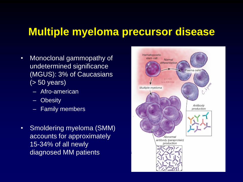
Multiple myeloma precursor disease
• Monoclonal gammopathy of
undetermined significance
(MGUS): 3% of Caucasians
(> 50 years)
– Afro-american
– Obesity
– Family members
• Smoldering myeloma (SMM)
accounts for approximately
15-34% of all newly
diagnosed MM patients

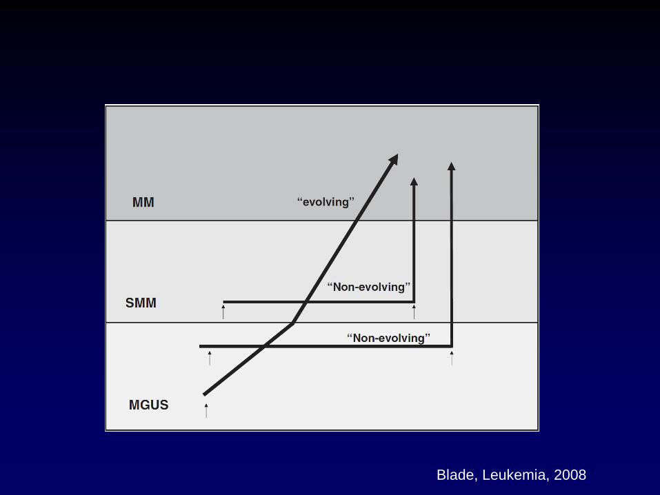
BM microenvironment changes Osteoclast activation increased angiogenesis Osteoblast inhibition altered expression of cytokines, growth factors and adhesion molecules
Initiation Progression
Germinal centre Bone marrow Peripheral blood
Normal pre- germinal B cell MGUS
Smoldering myeloma
Myeloma EM-MM / PCL
Primary genetic events: •IgH@ translocations •Hyperdiploidy
Secondary genetic events: •Copy number abnormalities
•DNA hypomethylation •Acquired mutations
Tumor cell diversity
Genetic lesions
Clonal advantage Competition selection for BM niche Migration & founder effect
Myeloma progenitor cell
Progression and clonal evolution in Myeloma
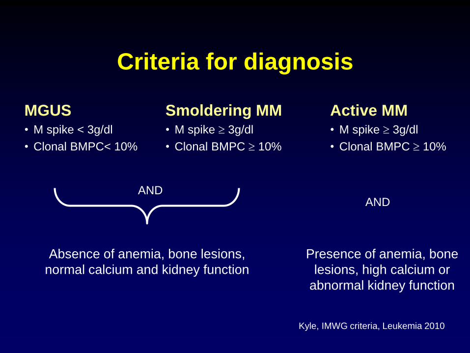
Criteria for diagnosis
MGUS • M spike < 3g/dl
• Clonal BMPC< 10%
Smoldering MM • M spike 3g/dl
• Clonal BMPC 10%
Active MM • M spike 3g/dl
• Clonal BMPC 10%
Absence of anemia, bone lesions,
normal calcium and kidney function
AND AND
Presence of anemia, bone
lesions, high calcium or
abnormal kidney function
Kyle, IMWG criteria, Leukemia 2010
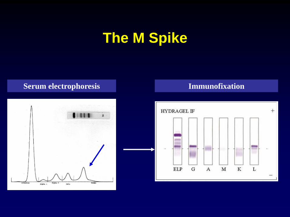
The M Spike
Serum electrophoresis Immunofixation


Causes of monoclonal gammopathies
Plasma cell disorders – MGUS
– Multiple myeloma
– Amyloid light chain amyloidosis
– Solitary plasmacytoma
– POEMS syndrome
– Castleman’s disease
B-cell lymphoproliferative disorders
– Non-Hodgkin's lymphoma
– Chronic lymphocytic leukemia
– Waldenström’s macroglobulinemia
– Post-transplant monoclonal gammopathies
Infections – Bacterial
– Viral (hepatitis, EBV, CMV, HIV)
Autoimmune disorders – Systemic lupus erythematosus
– Rheumatoid arthritis
– Sjögren syndrome
– Scleroderma
– Psoriatic arthritis
Skin disorders
Liver disorders
Glomerular nephropathies
Epithelial cancers (paraneoplastic syndromes)
Other hematological disorders
– Cryoglobulinaemia
– Myelodysplastic or myeloproliferative disorders
– Coagulation disorders
Caers J et al Ann Med 2013
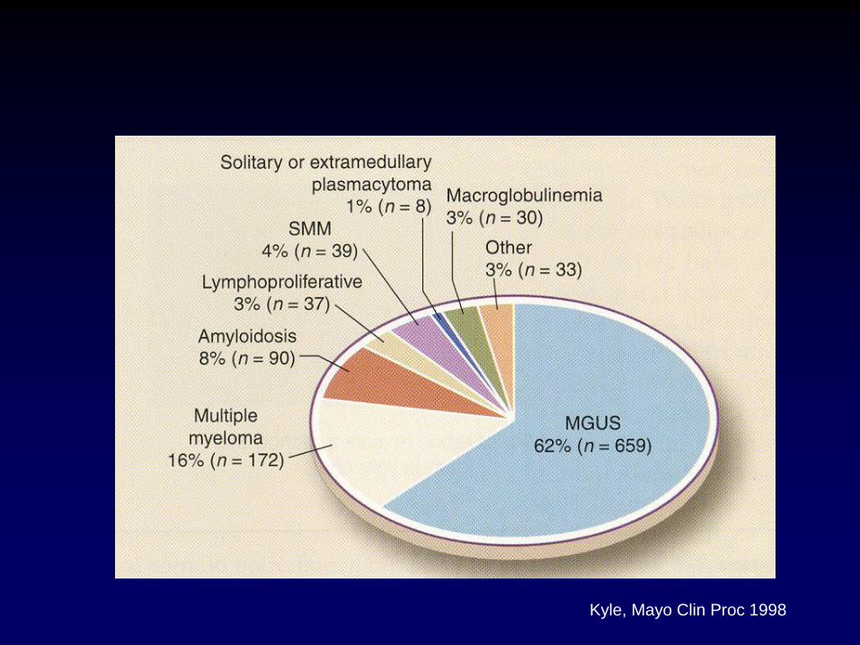
Kyle, Mayo Clin Proc 1998
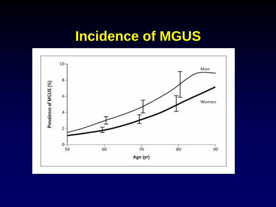
Incidence of MGUS
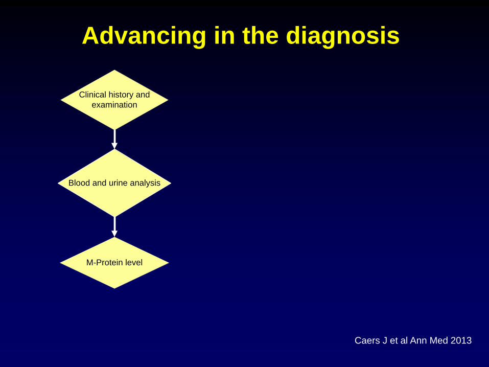
Clinical history and
examination
Blood and urine analysis
M-Protein level
Advancing in the diagnosis
Caers J et al Ann Med 2013

Alerting symptoms
Bianchi G, Hematol Oncol Clin North Am. 2012

Clinical history and
examination
Blood and urine analysis
M-Protein level
Monoclonal Gammapathy
of unknown significanceFurther exploration for myeloma
or lymphoproliferative disorder
Symptomatic
Abnormal: presence of
renal failure, anemia,
hypercalcemia
> 15 g/l
Asymptomatic
Normal
< 15 g/l
Caers J et al Ann Med 2013
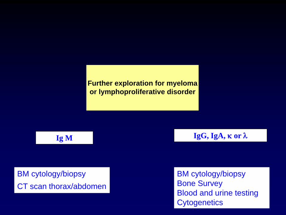
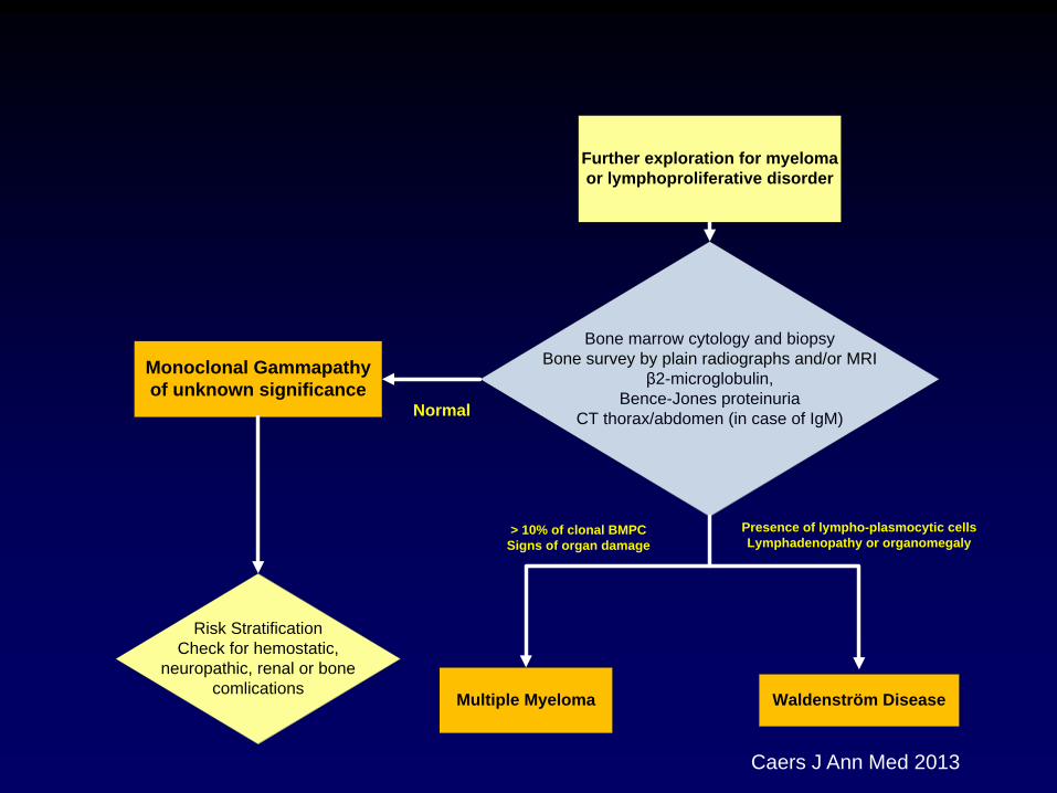
Further exploration for myeloma
or lymphoproliferative disorder
Ig M IgG, IgA, or
BM cytology/biopsy
CT scan thorax/abdomen BM cytology/biopsy
Bone Survey
Blood and urine testing
Cytogenetics
Waldenström Disease
Monoclonal Gammapathy
of unknown significance
Risk Stratification
Check for hemostatic,
neuropathic, renal or bone
comlications
Further exploration for myeloma
or lymphoproliferative disorder
Bone marrow cytology and biopsy
Bone survey by plain radiographs and/or MRI
β2-microglobulin,
Bence-Jones proteinuria
CT thorax/abdomen (in case of IgM)
Multiple Myeloma
Normal
> 10% of clonal BMPC
Signs of organ damage
Presence of lympho-plasmocytic cells
Lymphadenopathy or organomegaly
Caers J Ann Med 2013

Risk stratification
• Level of M-protein 1.5 g/dl
• Isotype IgG vs IgA, IgM
• BM plasmocytosis 5%
• Reduced Ig levels
• Serum Free Light Chain ratio
Kyle, NEJM, 2002
Rosinol, Mayo Clin Proc, 2007
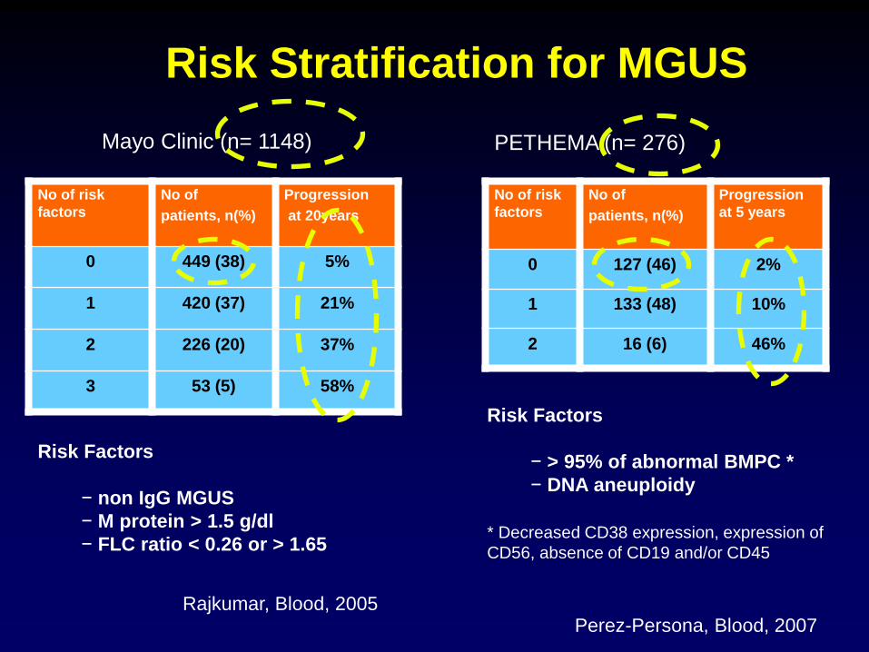
Risk Stratification for MGUS
No of risk
factors
No of
patients, n(%)
Progression
at 20years
0 449 (38) 5%
1 420 (37) 21%
2 226 (20) 37%
3 53 (5) 58%
No of risk
factors
No of
patients, n(%)
Progression
at 5 years
0 127 (46) 2%
1 133 (48) 10%
2 16 (6) 46%
Mayo Clinic (n= 1148) PETHEMA (n= 276)
Risk Factors
− non IgG MGUS
− M protein > 1.5 g/dl
− FLC ratio < 0.26 or > 1.65
Rajkumar, Blood, 2005
Risk Factors
− > 95% of abnormal BMPC *
− DNA aneuploidy
* Decreased CD38 expression, expression of
CD56, absence of CD19 and/or CD45
Perez-Persona, Blood, 2007
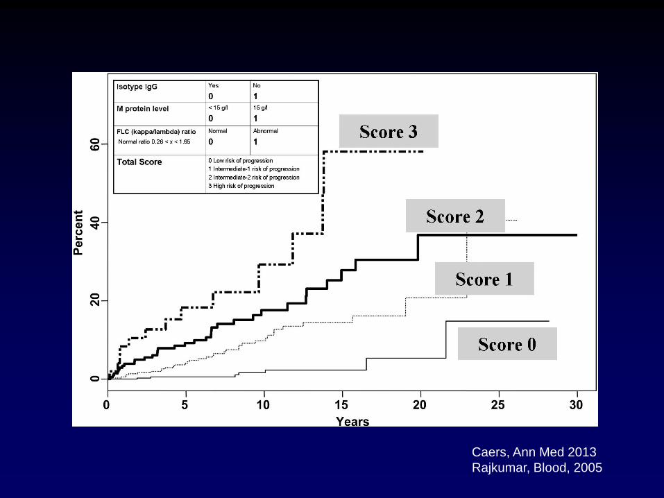
Caers, Ann Med 2013
Rajkumar, Blood, 2005

Current IMWG recommendation
• Low-risk MGUS
– Baseline BM cytology and skeletal survey not routinely
indicated
– Serum electrophoresis in 6 months and if stable, follow
either every 2 years or if symptoms arise
• Intermediate and high-risk MGUS
– Baseline BM cytology/biopsy and skeletal survey
– Blood analysis (including serum electrophoresis) repeated in
6 months and than annually
Kyle, IMWG criteria, Leukemia 2010
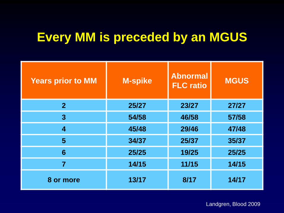
Every MM is preceded by an MGUS
Years prior to MM M-spike Abnormal
FLC ratio MGUS
2 25/27 23/27 27/27
3 54/58 46/58 57/58
4 45/48 29/46 47/48
5 34/37 25/37 35/37
6 25/25 19/25 25/25
7 14/15 11/15 14/15
8 or more 13/17 8/17 14/17
Landgren, Blood 2009
Blade, Leukemia, 2008

MGUS, not that benign
• Increased risk of fractures
• Decreased bone densities
• Increased risk for venous and arterial thrombosis
• Neuropathy
– IgM anti-MAG neuropathie
– IgA, IgA CIPD
• Increased risk of infections
Smoldering Multiple Myeloma

Smoldering MM
Kyle, NEJM, 2007
•276 SMM patients diagnosed
1970-1995
•163 (59%) progressed
•158 MM
•5 amyloidosis
•Overall risk of progression (per
year)
• 10% in the first 5 years
• 3% in the next 5 years
• 1% in the next 5 years
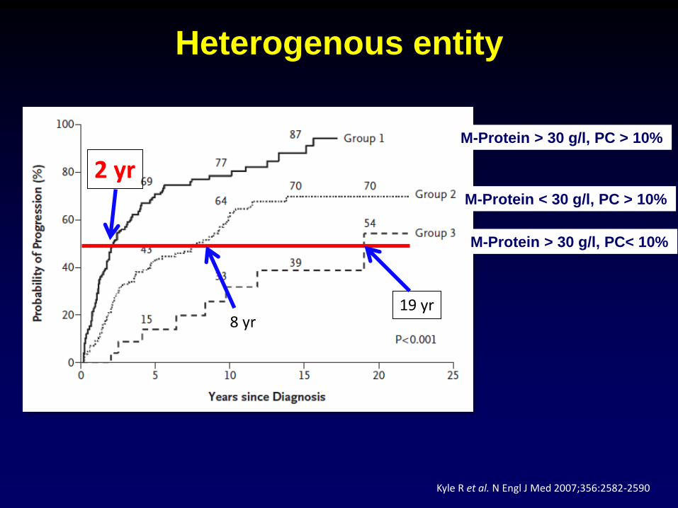
Heterogenous entity
Kyle R et al. N Engl J Med 2007;356:2582-2590
8 yr
2 yr
19 yr
M-Protein > 30 g/l, PC > 10%
M-Protein < 30 g/l, PC > 10%
M-Protein > 30 g/l, PC< 10%

Delay progression?
Select an agressive
clone ? Cure ?
Limit complications ? (bone disease, renal
failure)
Smoldering myeloma
Is it possible to identify high-risk patients?
Has an early treatment an additive value?
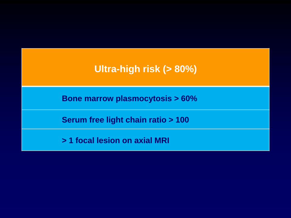
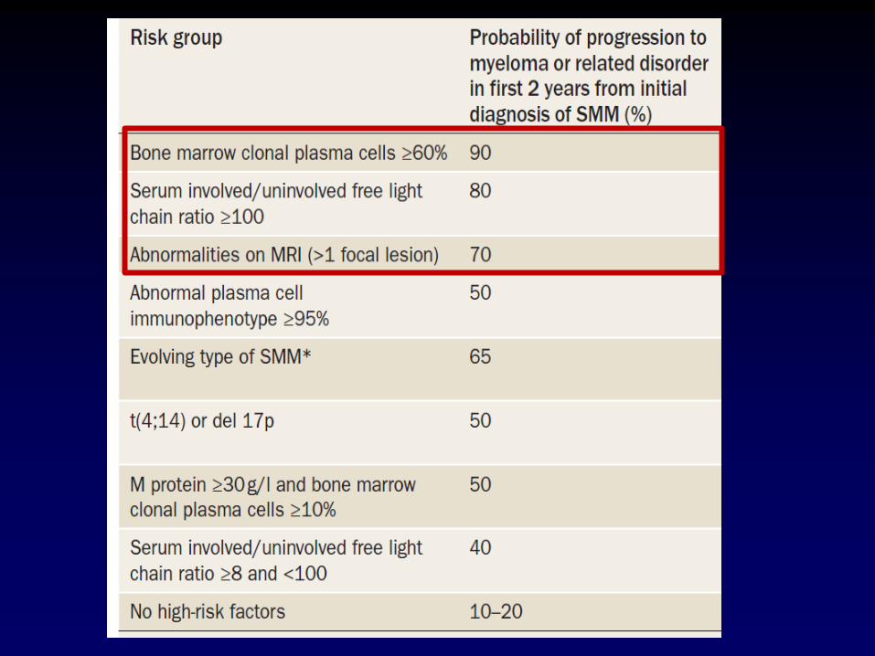
Ultra-high risk (> 80%)
Bone marrow plasmocytosis > 60%
Serum free light chain ratio > 100
> 1 focal lesion on axial MRI

Bone Marrow: plasmocytosis
Rajkumar, NEJM, 2011 Rago, Cancer, 2012
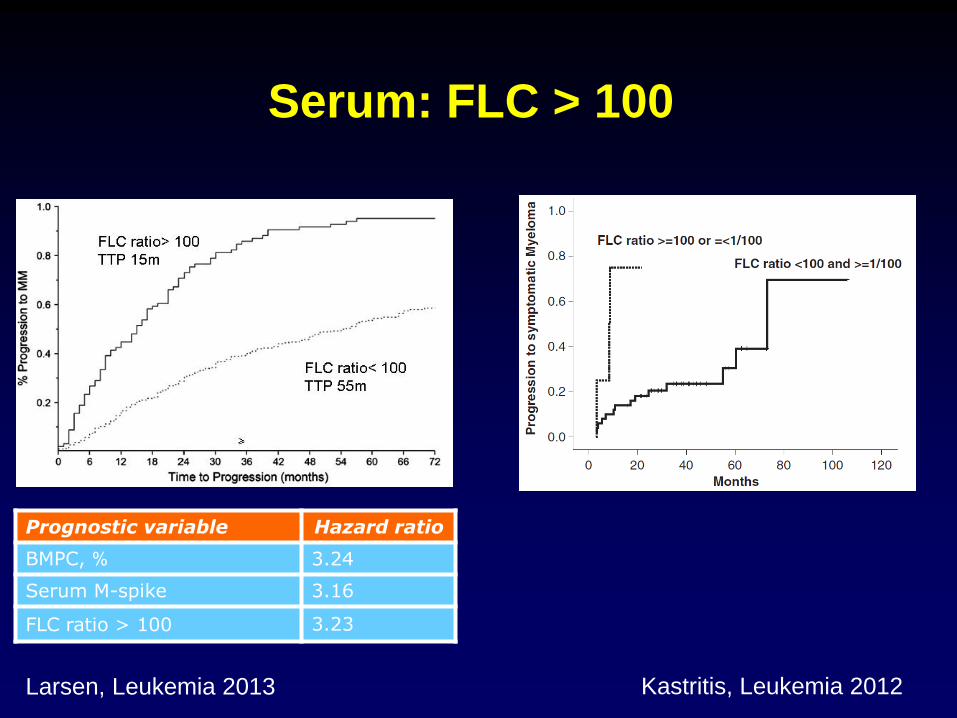
Serum: FLC > 100
Prognostic variable Hazard ratio
BMPC, % 3.24
Serum M-spike 3.16
FLC ratio > 100 3.23
Larsen, Leukemia 2013 Kastritis, Leukemia 2012
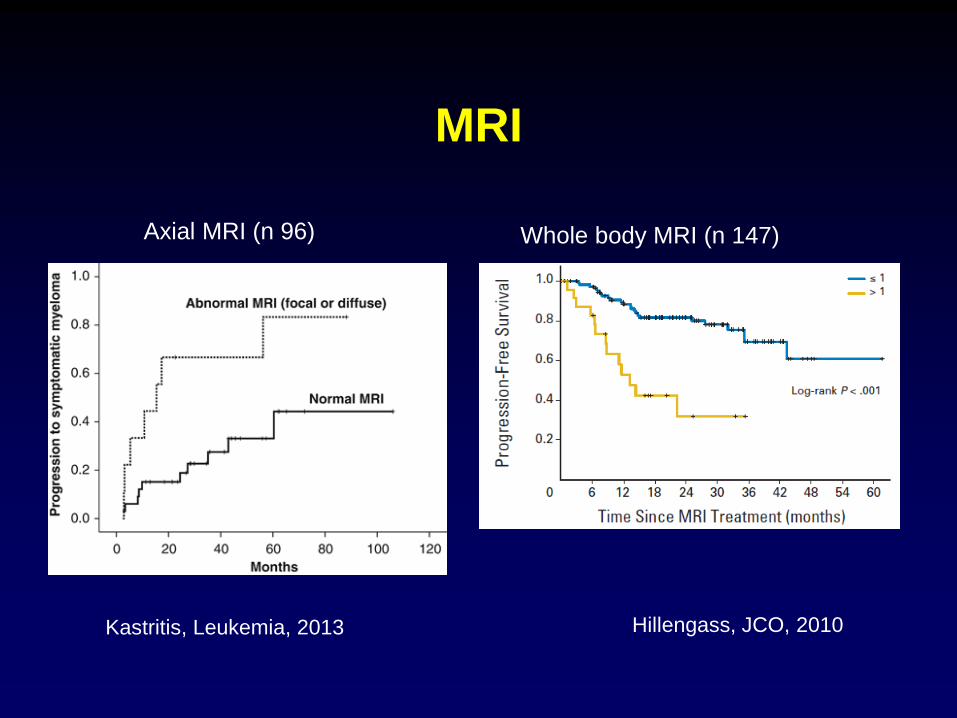
MRI
Kastritis, Leukemia, 2013 Hillengass, JCO, 2010
Axial MRI (n 96) Whole body MRI (n 147)
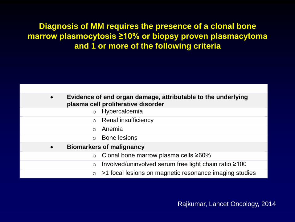
Diagnosis of MM requires the presence of a clonal bone
marrow plasmocytosis ≥10% or biopsy proven plasmacytoma
and 1 or more of the following criteria
Evidence of end organ damage, attributable to the underlying plasma cell proliferative disorder
o Hypercalcemia
o Renal insufficiency
o Anemia
o Bone lesions
Biomarkers of malignancy
o Clonal bone marrow plasma cells ≥60%
o Involved/uninvolved serum free light chain ratio ≥100
o >1 focal lesions on magnetic resonance imaging studies
Rajkumar, Lancet Oncology, 2014
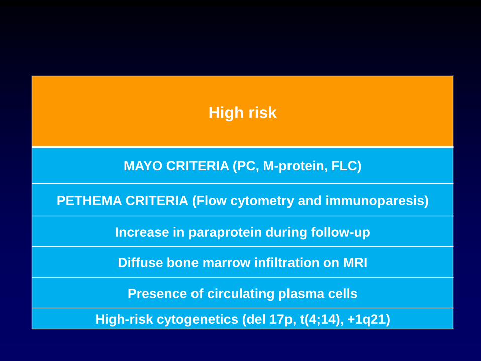
High risk
MAYO CRITERIA (PC, M-protein, FLC)
PETHEMA CRITERIA (Flow cytometry and immunoparesis)
Increase in paraprotein during follow-up
Diffuse bone marrow infiltration on MRI
Presence of circulating plasma cells
High-risk cytogenetics (del 17p, t(4;14), +1q21)

Risk Stratification for SMM
No of risk
factors
No of
patients, n(%)
Progression
at 5 years
1 76 (25) 25%
2 115 (42) 51%
3 82 (30) 76%
No of risk
factors
No of
patients, n(%)
Progression
at 5 years
0 28 (31) 4%
1 22 (25) 46%
2 39 (44) 72%
Mayo Clinic (n= 273) PETHAMA (n= 89)
Risk Factors
− BMPC > 10%
− M protein > 3 g/dl
− FLC ratio < 0.126 or > 8
Dispenzieri, Blood, 2008
Risk Factors
− > 95% of abnormal BMPC *
− Immunoparesis
* Decreased CD38 expression, expression of
CD56, absence of CD19 and/or CD45
Perez-Persona, Blood, 2007

Perez-Persona, Blood, 2007
- > 95% of abnormal BMPC *
- Immunoparesis
PETHEMA
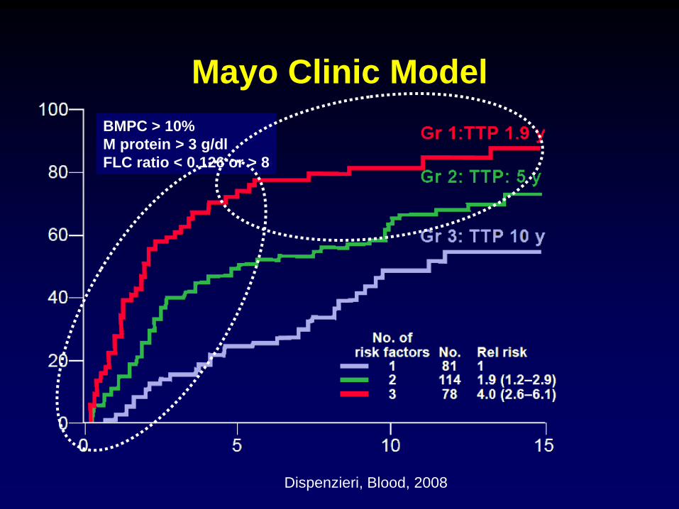
Mayo Clinic Model
Dispenzieri, Blood, 2008
BMPC > 10%
M protein > 3 g/dl
FLC ratio < 0.126 or > 8
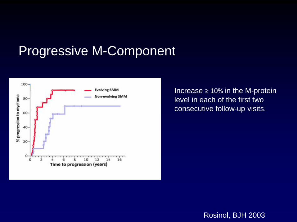
Progressive M-Component
Increase ≥ 10% in the M-protein
level in each of the first two
consecutive follow-up visits.
Rosinol, BJH 2003
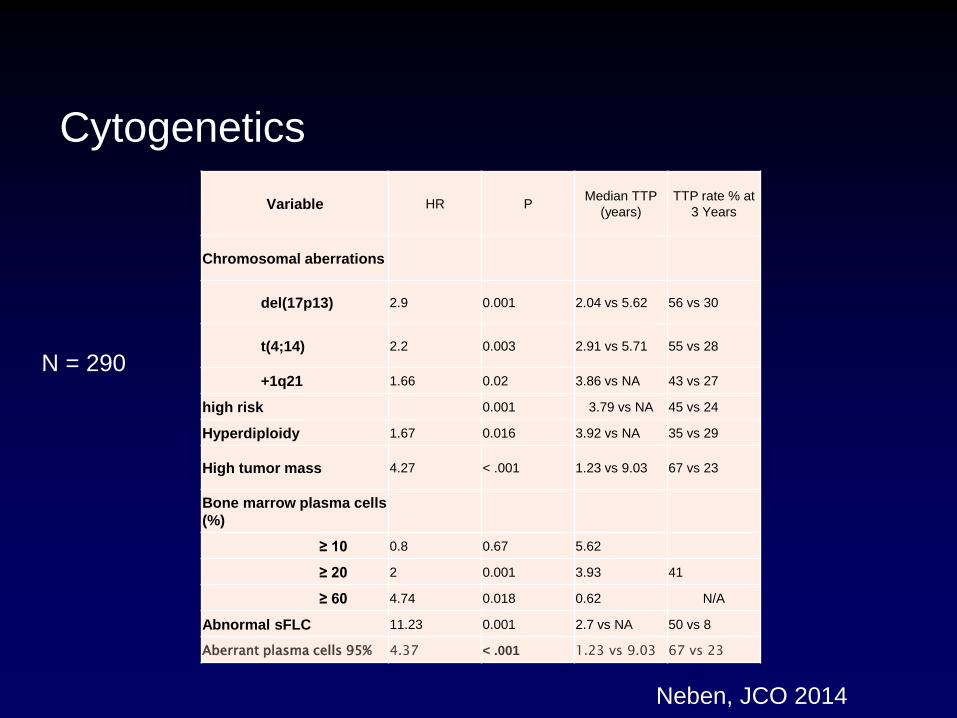
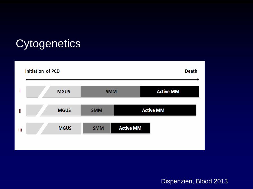
Cytogenetics
Neben, JCO 2014
Variable HR P Median TTP
(years) TTP rate % at
3 Years
Chromosomal aberrations
del(17p13) 2.9 0.001 2.04 vs 5.62 56 vs 30
t(4;14) 2.2 0.003 2.91 vs 5.71 55 vs 28
+1q21 1.66 0.02 3.86 vs NA 43 vs 27
high risk 0.001 3.79 vs NA 45 vs 24
Hyperdiploidy 1.67 0.016 3.92 vs NA 35 vs 29
High tumor mass 4.27 < .001 1.23 vs 9.03 67 vs 23
Bone marrow plasma cells
(%)
≥ 10 0.8 0.67 5.62
≥ 20 2 0.001 3.93 41
≥ 60 4.74 0.018 0.62 N/A
Abnormal sFLC 11.23 0.001 2.7 vs NA 50 vs 8
Aberrant plasma cells 95% 4.37 < .001 1.23 vs 9.03 67 vs 23
N = 290
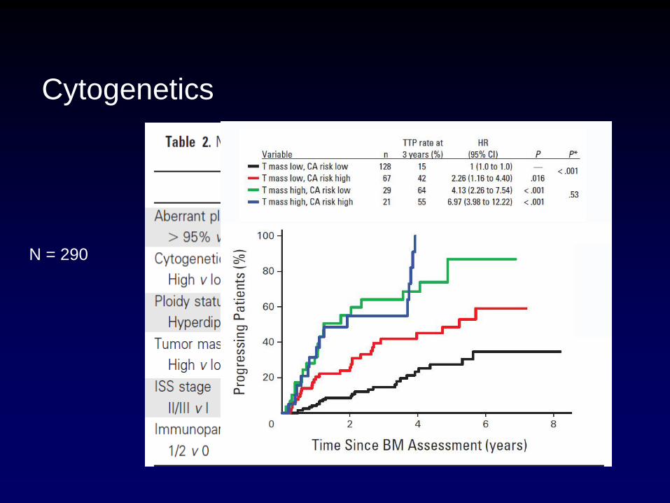
Cytogenetics
N = 290
Dispenzieri, Blood 2013
Cytogenetics

Diffuse MRI pattern
Kastritis, Leukemia, 2013
Axial MRI (n 96) Whole body MRI (n 96)
Hillengass, JCO, 2010
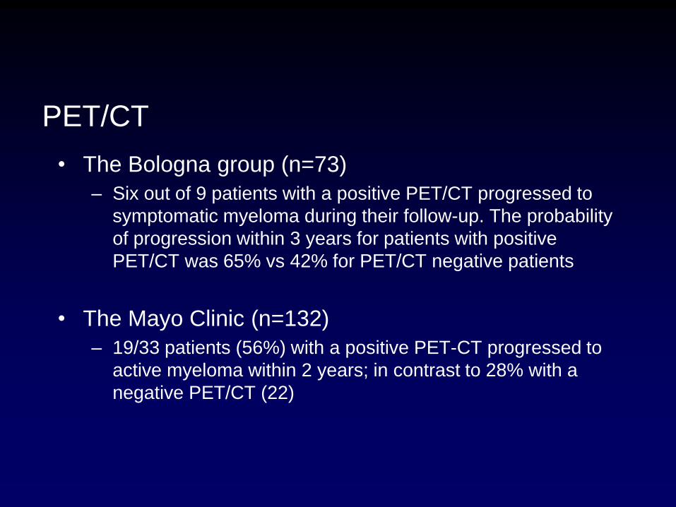
• The Bologna group (n=73)
– Six out of 9 patients with a positive PET/CT progressed to
symptomatic myeloma during their follow-up. The probability
of progression within 3 years for patients with positive
PET/CT was 65% vs 42% for PET/CT negative patients
• The Mayo Clinic (n=132)
– 19/33 patients (56%) with a positive PET-CT progressed to
active myeloma within 2 years; in contrast to 28% with a
negative PET/CT (22)
PET/CT
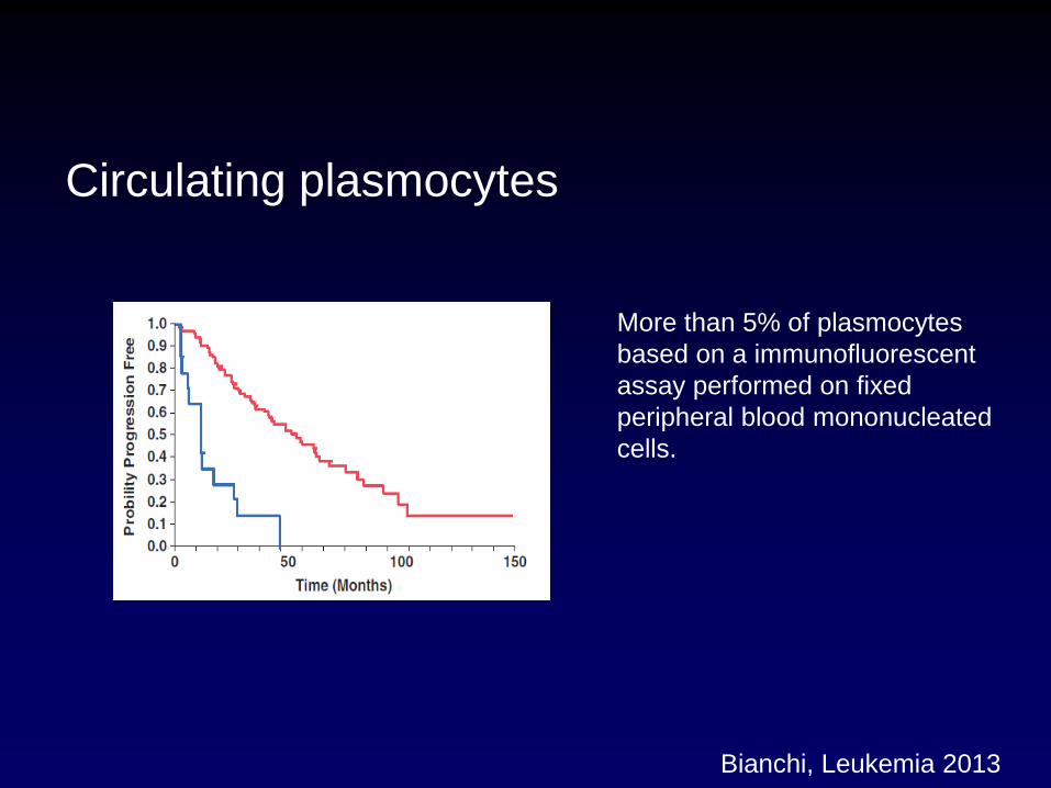
Circulating plasmocytes
More than 5% of plasmocytes
based on a immunofluorescent
assay performed on fixed
peripheral blood mononucleated
cells.
Bianchi, Leukemia 2013
IMWG considers that a prognostic factors
that is able to identify
SMM cases with ~80% risk of
progression at 2 years (median time of
transformation 12 months)
justifies an early intervention

Diagnosis of MM requires the presence of a clonal bone
marrow plasmocytosis ≥10% or biopsy proven plasmacytoma
and 1 or more of the following criteria
Evidence of end organ damage, attributable to the underlying plasma cell proliferative disorder
o Hypercalcemia
o Renal insufficiency
o Anemia
o Bone lesions
Biomarkers of malignancy
o Clonal bone marrow plasma cells ≥60%
o Involved/uninvolved serum free light chain ratio ≥100
o >1 focal lesions on magnetic resonance imaging studies
Rajkumar, Lancet Oncology, 2014
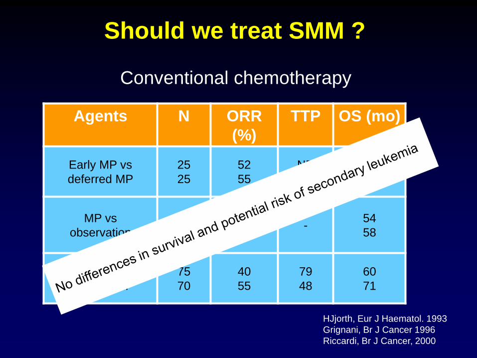
Should we treat SMM ?
Agents N ORR
(%)
TTP OS (mo)
Early MP vs
deferred MP
25
25
52
55
NR
12 m
52
53
MP vs
observation
22
22 - -
54
58
Early MP vs
deferred MP
75
70
40
55
79
48
60
71
HJjorth, Eur J Haematol. 1993
Grignani, Br J Cancer 1996
Riccardi, Br J Cancer, 2000
Conventional chemotherapy

Should we treat SMM ?
Agents N ORR
(%)
TTP OS (mo)
Pamidronate 12 8 - -
Pamidronate vs
observation
89
88 -
46
48 -
Zolendronate vs
Observation
81
82 -
67
59 -
Martin, Br J Haematol, 2002
D’arena, Leuk Lymphoma, 2011
Musto, Cancer, 2008
Biphosphonates
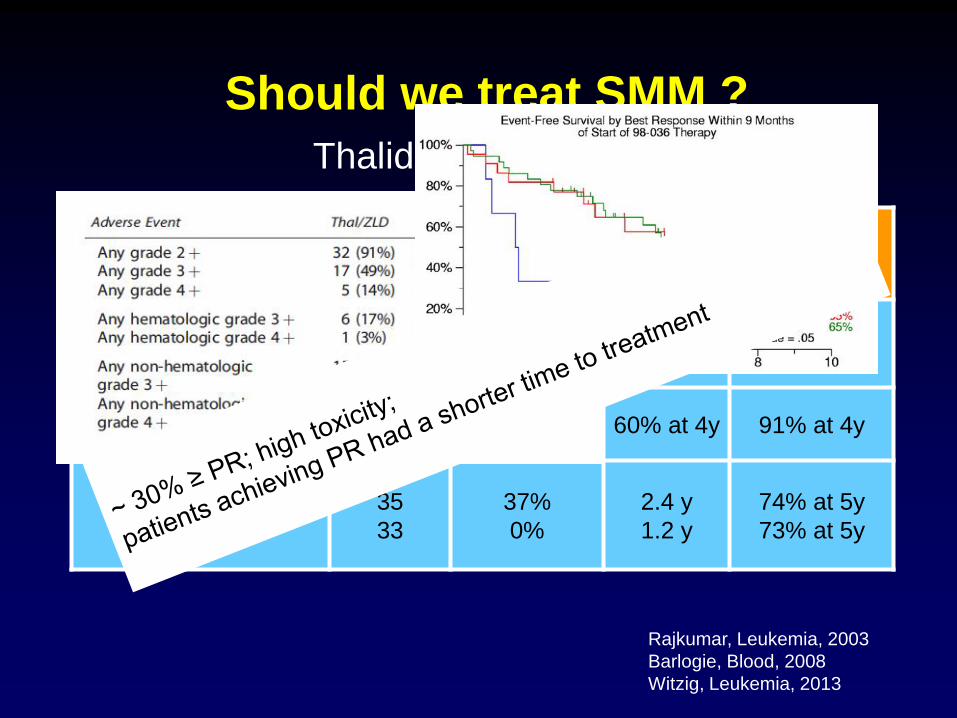
Should we treat SMM ?
Agents N ORR (%) TTP OS (mo)
Thalidomide 29 34 63 at 2y 96% at 2y
Thalidomide plus
pamidronate 76 25 60% at 4y 91% at 4y
Thal –Zol vs
Zol
35
33
37%
0%
2.4 y
1.2 y
74% at 5y
73% at 5y
Rajkumar, Leukemia, 2003
Barlogie, Blood, 2008
Witzig, Leukemia, 2013
Thalidomide
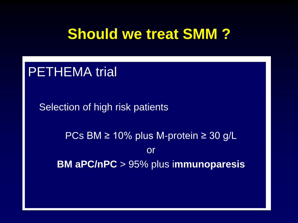
PETHEMA trial
Selection of high risk patients
PCs BM ≥ 10% plus M-protein ≥ 30 g/L
or
BM aPC/nPC > 95% plus immunoparesis
Should we treat SMM ?
Should we treat SMM ?
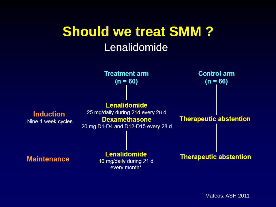
Mateos, ASH 2011
Lenalidomide
Should we treat SMM ? Lenalidomide
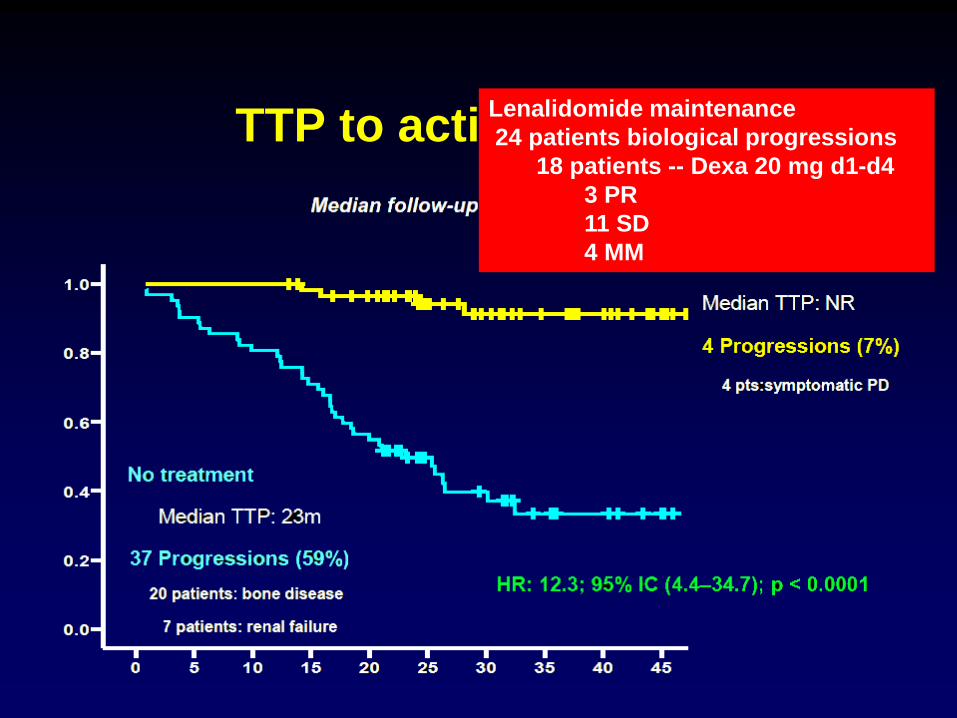
TTP to active disease Lenalidomide maintenance
24 patients biological progressions
18 patients -- Dexa 20 mg d1-d4
3 PR
11 SD
4 MM
Mateos, ASH 2011
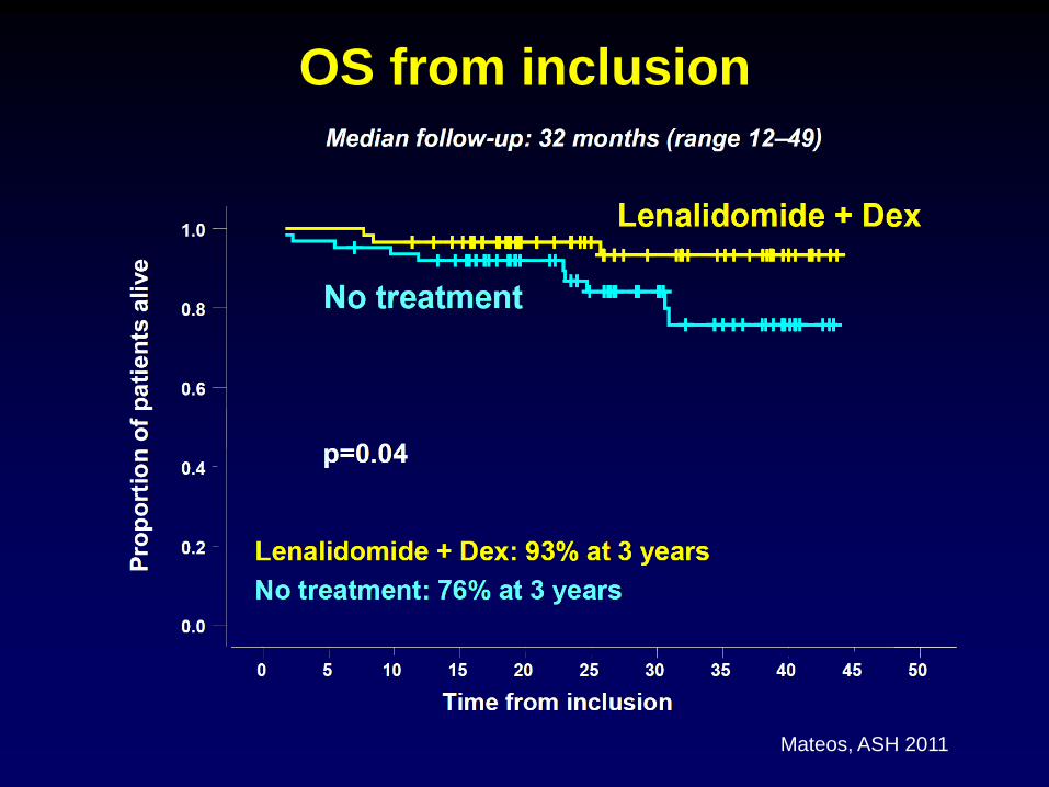
OS from inclusion

IMWG recommendations
• Ultra-risk patients are recommended to be treated
– Potential harmful organ complications with significant long-term
morbidity need to be avoided
– Based patients' health status and patients’choice
• High risk patients should be followed regularly and
might be candidates for early intervention clinical
studies.
• Low risk patients: follow-up.
Conclusions
• MGUS and sMM are the most prevalent premalignant conditions in worldwide population
• Active myeloma for nearly all patients is preceded by MGUS/sMM.
• Prognostic categorization of MGUS and sMM is crucial to tailor their follow-up