Embed Size (px)
Citation preview
MURDOCH RESEARCH REPOSITORY
This is the author’s final version of the work, as accepted for publication following peer review but without the publisher’s layout or pagination.
The definitive version is available at http://dx.doi.org/10.1016/j.vetpar.2010.10.003
FitzGerald, L., Bennett, M.D., Ng, J., Nicholls, P.K., James, F.E.,
Elliot, A., Slaven, M. and Ryan, U. (2011) Morphological and molecular characterisation of a mixed Cryptosporidium
muris/Cryptosporidium felis infection in a cat. Veterinary Parasitology, 175 (1-2). pp. 160-164.
http://researchrepository.murdoch.edu.au/3565/
Copyright: © 2010 Elsevier B.V.
It is posted here for your personal use. No further distribution is permitted.
Accepted Manuscript
Title: Morphological and molecular characterisation of amixed Cryptosporidium muris/Cryptosporidium felis infectionin a cat
Authors: Louise FitzGerald, Mark Bennett, Josephine Ng,Philip Nicholls, Fleur James, Aileen Elliot, Mike Slaven, UnaRyan
PII: S0304-4017(10)00550-9DOI: doi:10.1016/j.vetpar.2010.10.003Reference: VETPAR 5491
To appear in: Veterinary Parasitology
Received date: 1-7-2010Revised date: 24-9-2010Accepted date: 4-10-2010
Please cite this article as: FitzGerald, L., Bennett, M., Ng, J., Nicholls, P., James,F., Elliot, A., Slaven, M., Ryan, U., Morphological and molecular characterisation ofa mixed Cryptosporidium muris/Cryptosporidium felis infection in a cat, VeterinaryParasitology (2010), doi:10.1016/j.vetpar.2010.10.003
This is a PDF file of an unedited manuscript that has been accepted for publication.As a service to our customers we are providing this early version of the manuscript.The manuscript will undergo copyediting, typesetting, and review of the resulting proofbefore it is published in its final form. Please note that during the production processerrors may be discovered which could affect the content, and all legal disclaimers thatapply to the journal pertain.
Page 1 of 21
Accep
ted
Man
uscr
ipt
Morphological and molecular characterisation of a mixed Cryptosporidium 1
muris/Cryptosporidium felis infection in a cat. 2
3
Louise FitzGeralda, Mark Bennett
a, Josephine Ng
a, Philip Nicholls
a, Fleur James
b, Aileen 4
Elliota, Mike Slaven
a and Una Ryan
a* 5
aSchool of Veterinary and Biomedical Sciences, Murdoch University, Murdoch, WA, 6
6150, Australia. 7
bSchool of Veterinary Clinical Sciences, Murdoch University, Murdoch, WA, 6150, 8
Australia. 9
10
11
Short Communication 12
13
*Corresponding author. Phone: +61 8 9360 2482 14
E-mail: [email protected] 15
16
17
18
*Manuscript
Page 2 of 21
Accep
ted
Man
uscr
ipt
Abstract 19
To date Cryptosporidium muris has been identified by microscopy and genotyping in 20
cats in two studies. We report morphological and genetic evidence of a mixed C. muris 21
and C. felis infection in a cat and provide the first histological, immunohistochemical, in 22
situ hybridisation and genetic confirmation of a C. muris infection in the stomach of a cat. 23
The cat suffered persistent diarrhoea after the initial consultation, which remained 24
unresolved, despite several medical interventions. Further studies are required to 25
determine the range, prevalence and clinical impact of Cryptosporidium species infecting 26
cats. 27
28
Keywords: Cryptosporidium muris; Cryptosporidium felis; cat, mixed infection; 29
morphology; genotyping. 30
31
32
33
34
35
Page 3 of 21
Accep
ted
Man
uscr
ipt
36
1. Introduction 37
38
Cryptosporidium is a genus of protozoan parasites whose members can cause 39
diarrhoea in many hosts including humans and domestic animals. Currently 23 species of 40
Cryptosporidium are accepted as valid including C. muris, which infects rodents as its 41
primary host and C. felis in cats (Xiao, 2010; Fayer et al., 2010). 42
Cryptosporidium spp. infection is relatively common in cats and epidemiological 43
surveys conducted worldwide have reported that the prevalence in cats ranges from 0 to 44
29% (Lucio-Forster et al., 2010). This apparent variation in the rate of infection might be 45
due, in part, to the method of detection (e.g. concentration of oocysts and direct light 46
microscopy versus microscopy of stained smears or PCR), as well as the population being 47
sampled (animal age differences, owned animals, stray populations, shelter animals) and 48
symptomatic versus asymptomatic animals (Lucio-Forster et al., 2010). 49
Genetic characterisation of oocysts recovered from faecal samples of cats have 50
identified C. felis (Ballweber et al., 2009; Palmer et al., 2008; Huber et al., 2007; Thomaz 51
et al., 2007; Fayer et al., 2006; Santin et al., 2006; Morgan et al., 1998; Sargent et al., 52
1998; Gasser et al., 2001; Ryan et al., 2003; Hajdusek et al., 2004) and C. muris in two 53
studies (Santin et al., 2006; Pavlasek and Ryan, 2007). The identification of C. muris in 54
cats in the latter two studies was based on genotyping of oocysts recovered from faeces. 55
No histological studies were conducted and therefore it was not possible to determine if 56
the cats were actually infected with C. muris or were merely acting as mechanical 57
Page 4 of 21
Accep
ted
Man
uscr
ipt
vectors. In the present study, we report on genetic, morphological and histological 58
characterisation a mixed C. muris/C. felis infection in a cat. 59
60
2. Materials, Methods and Results 61
62
In 2008, a 2 year old male neutered domestic long haired cat presented for 63
investigation of chronic diarrhoea. The clinical signs were characteristic of small bowel 64
diarrhoea with an increased frequency of defecation. Appetite was normal and weight 65
loss and vomiting were not features of his initial clinical presentation. Physical 66
examination at the time of initial presentation was unremarkable. Screening haematology, 67
biochemistry and urinalysis identified a mild increase in creatine kinase activity (413 68
U/L; reference range 50 – 100 U/L). Fasting feline trypsin-like immunoreactivity was 69
normal (30 ug/L; control reference 12 – 82 ug/L). The cat tested negative for feline 70
leukaemia virus and feline immunodeficiency virus (Simplify, AGEN Biomedical; 71
Brisbane, Australia). Initial symptomatic therapy consisted of cobalamin (Vitamin B12, 72
Troy, Australia) at 200 mg/kg by subcutaneous injection weekly for 6 treatments and 73
dietary modification to increase the content of soluble fibre, however there was little 74
response to these interventions. Further symptomatic therapy was trialled, including 75
metronidazole (Flagyl, Sanofi Aventis, Spain) at 9.4 mg/kg every 12 hours for 10 days 76
and fenbendazole (Panacur 100, Virbac Animal Health, Australia) at 50 mg/kg once daily 77
per os for 5 days. 78
The cat re-presented 13 months later with continuing diarrhoea and he had also 79
begun to vomit most days. An abdominal ultrasound was performed and identified mild 80
Page 5 of 21
Accep
ted
Man
uscr
ipt
mesenteric lymphadenomegaly, mildly irregular splenomegaly and normal 81
gastrointestinal wall thickness and layering. Fine needle aspirate cytology of the 82
mesenteric lymph nodes and spleen identified mild reactivity in both locations. 83
Gastroduodenoscopy showed that there were areas of marked gastric mucosal oedema, 84
however the duodenal mucosa was unremarkable. Mucosal pinch biopsies were collected 85
from the stomach and duodenum. The cat was prescribed empirical amoxycillin-86
clavulanate (Clavulox; Pfizer, Australia) at 13.9 mg/kg every 12h per os and a novel 87
protein diet trial whilst results were pending. 88
Faecal samples were collected and examined using malachite green staining as 89
previously described (Eliott et al., 1999). Parasites were examined with the aid of an 90
ocular micrometer in a Zeiss Axioskop microscope at 1000 × magnification and this 91
revealed the presence of two different sized Cryptosporidium sp. oocysts; large oocysts 92
which resembled C. muris in size and shape (8.0 × 5.8 µm, mean width/length ratio 1.4, 93
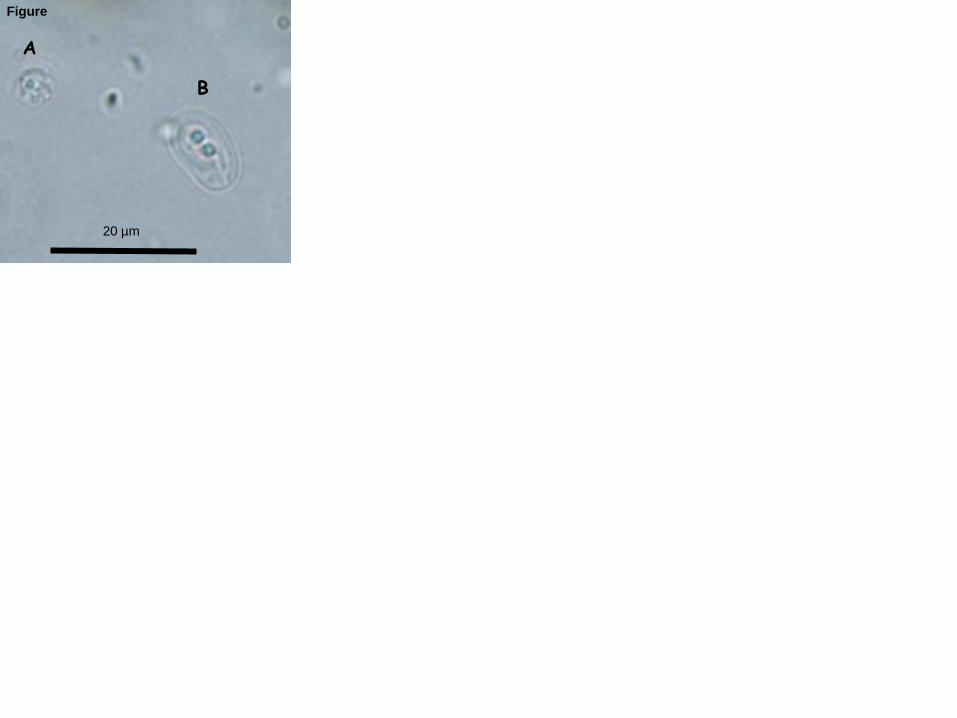
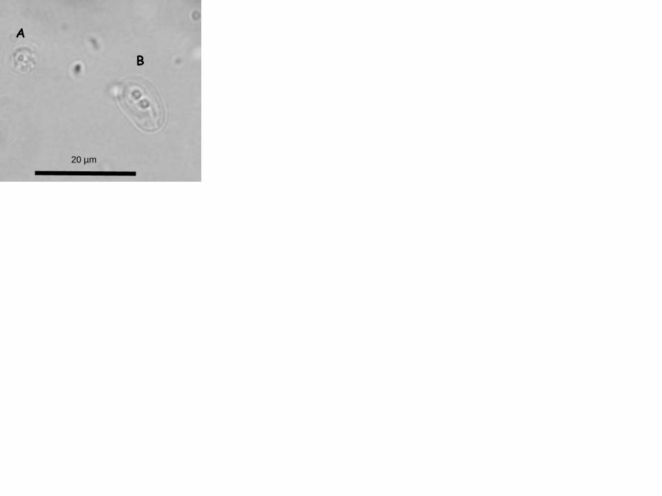
n=30) and smaller oocysts which resembled C. felis in size and shape (4.6 × 4.0 µm 94
width/length ratio 1.15, n=20) (Fig. 3). 95
Endoscopic biopsy specimens from the stomach and duodenum were fixed in 10% 96
neutral buffered formalin for 24 hours, then processed routinely and embedded in 97
paraffin. Histological sections were cut at 5 µm and stained with hematoxylin and eosin. 98
Microscopic examination of the stomach biopsies revealed the presence of abundant 99
Cryptosporidium sp. organisms within the gastric pits and within the lumina of fundic 100
glands. The affected glands were frequently dilated and filled with numerous 101
Cryptosporidium spp. organisms (Figure 1, A-B). There was a mild increase in fibrous 102
tissue within some areas of the lamina propria, leading to mild separation of glands 103
Page 6 of 21
Accep
ted
Man
uscr
ipt
accompanied by a mild, multifocal lymphoplasmacytic and neutrophilic inflammatory 104
cell infiltrate. In the duodenum, the Cryptosporidium sp. stages were closely associated 105
with the apical surface of enterocytes (Figure 1, C). There was a mild, patchy increase in 106
lymphocytes and plasma cells in the lamina propria along with low numbers of scattered 107
neutrophils and a mild, multifocal increase in intraepithelial lymphocytes. 108
Approximately 1 µg of purified PCR product DNA (~500 bp) from the C. muris 109
18S rRNA gene, from a rodent-derived C. muris isolate, was labelled with digoxigenin to 110
produce DNA probes for in situ hybridisation using the DIG-Nick Translation Mix, 111
according to the manufacturer’s instructions (Roche Diagnostics). The digoxigenin-112
labelled DNA was added to a probe cocktail mixture consisting of 50% formamide, 10% 113
dextran sulfate and 2× SSC buffer. Sections were deparaffinised, rehydrated, probed, 114
washed, developed, counter-stained and mounted as previously described (Bennett et al., 115
2008). An irrelevant DNA probe for bandicoot papillomatosis carcinomatosis virus type 1 116
was used as a negative control. 117
For immunohistochemistry, histological sections were dewaxed in xylene and 118
rehydrated through graded ethanols to water. Endogenous peroxidase activity was then 119
blocked using 3% hydrogen peroxide. The primary antibody, mouse anti-120
Cryptosporidium (Serotec MCA-2571), was diluted 1:200 with antibody diluent 121
(DakoCytomation) and applied to tissue sections for 30 minutes. Following thorough 122
rinsing with phosphate buffered saline (PBS), primary antibody binding was detected 123
using a horseradish peroxidase-labeled streptavidin biotin system (LSAB – Dako) 124
according to the manufacturer’s instructions. Slides were rinsed in tap water and the slide 125
Page 7 of 21
Accep
ted
Man
uscr
ipt
was counterstained lightly with Harris’ hematoxylin. Omission of the primary antibody 126
was used as a negative control. 127
In situ hybridisation and immunohistochemical experiments confirmed that the 128
organisms deep within the lumina of scattered gastric glands (Fig. 2a – 2c) were indeed 129
members of the genus Cryptosporidium. Immunohistochemistry also confirmed the 130
presence of Cryptosporidium organisms in the duodenal biopsies, whilst in situ 131
hybridisation was unsuccessful at this anatomical site. 132
DNA was extracted from intestine and stomach paraffin embedded biopsies using 133
a Qiagen DNeasy tissue kit (Qiagen, Germany). DNA was eluted in 50 µL of AE buffer 134
to concentrate the DNA. DNA was amplified at the 18S and actin loci using a nested PCR 135
as previously described (Ryan et al., 2003; Ng et al., 2006). The amplified DNA 136
fragments from the secondary PCR product were separated by gel electrophoresis and 137
purified using the freeze-squeeze method (Ng et al., 2006). Purified PCR products were 138
sequenced using an ABI PrismTM
Dye Terminator cycle sequencing kit (Applied 139
Biosystems, Foster City, California) according to the manufacturer’s instructions with the 140
exception that the annealing temperature was raised to 58ºC for the 18S and 55°C for the 141
actin sequencing reaction. Nucleotide sequences were analyzed using Chromas lite 142
version 2.0 (http://www.technelysium.com.au) and aligned with reference genotypes 143
from GenBank (http://www.ncbi.nlm.nih.gov/Genbank/) using ClustalW 144
(http://www.clustalw.genome.jp). Partial sequence analysis of a ~580 and ~818 base pair 145
section of the 18S rRNA and actin gene loci, respectively, identified the Cryptosporidium 146
species in the intestine as C. felis and the species in the stomach as C. muris (100% 147
identities). 148
Page 8 of 21
Accep
ted
Man
uscr
ipt
The cat was subsequently treated with 5.3 mg/kg of azithromycin every 12h per os for 149
2 weeks. Tylosin, a commonly recommended treatment for cryptosporidiosis, was 150
temporarily unavailable at time of diagnosis. The vomiting resolved and the diarrhoea 151
improved but persisted. Rechallenge with the previous diet led to reoccurrence of severe 152
vomiting and the novel protein diet was recommenced. The cat continued to maintain 153
body weight. Follow up faecal analysis conducted 12 months after initial faecal analysis 154
and collection of endoscopic biopsies demonstrated oocysts resembling C. muris in 155
morphology, while C. felis oocysts were not identified at this time. 156
157
4. Discussion 158
159
In the present study, morphological and genetic characterisation has confirmed the 160
presence of a mixed C. muris infection in the stomach and a C. felis infection in the 161
intestine of a cat. This is the first histological, immunohistochemical, in situ hybridisation 162
and genetic confirmation of a natural C. muris infection in a cat and only the third report 163
of C. muris in cats. Cryptosporidium muris has been found in many rodents (mice, wood 164
mice, rats, bank voles, Syrian hamsters, desert hamsters, squirrels, and Siberian 165
chipmunks), a marsupial (bilbies), other mammals (Bactrian camels, mountain goats, 166
reticulated giraffe, ringed seals, cats, rock hyraxes, cynomolgus monkeys, dogs, and pigs) 167
(Warren et al., 2003; Santin et al., 2006; Pavlasek and Ryan, 2007; Lupo et al., 2008; 168
Kodadkova et al., 2009, Kvac et al., 2009; Feng, 2010), and birds (tawny frogmouth) (Ng 169
et al., 2006). It has also been identified in a few humans in developing countries (Palmer 170
Page 9 of 21
Accep
ted
Man
uscr
ipt
et al., 2003; Gatei et al., 2006; Muthusamy et al., 2006). Experimental C. muris infections 171
have been reported in dogs, rabbits, lambs and cats (Iseki et al., 1989). 172
Cryptosporidium felis has a much more restricted host range and has been confirmed 173
using molecular techniques in cats, immunocompetent and immunocompromised humans 174
and a cow (Bornay-Llinares et al., 1999; Lucio-Forster et al., 2010). In children in 175
developing countries, C. felis is responsible for as much as 3.3% of overall 176
cryptosporidiosis cases (Lucio-Forster et al., 2010). However, most human cases of 177
cryptosporidiosis, worldwide, are associated with C. hominis and C. parvum (Xiao et al., 178
2010) and therefore C. muris and C. felis in cats are likely to be of low zoonotic risk to 179
humans. It has also been suggested that some C. felis infections in humans were 180
anthroponotically transmitted (Cama et al., 2006). In the present study, the source of 181
infection in the cat is unknown as the cat was acquired as a stray and there were several 182
other pets in the household. There was no clinical evidence of diarrhoea in any other 183
household members. 184
As the cat was infected with both C. muris and C. felis, it is difficult to attribute the 185
clinical presentations to either species. The presence of mild inflammation accompanied 186
by mild fibrosis in the stomach and inflammation within the duodenum in association 187
with the Cryptosporidium spp. is suggestive of an ongoing host response secondary to the 188
presence of the organisms, however contribution from other factors (such as concurrent 189
food hypersensitivity) cannot be ruled out especially given the partial response to a novel 190
protein diet. 191
In the present study, azithromycin was unsuccessful in resolving the diarrhoea. 192
Tylosin, which was temporarily unavailable at time of diagnosis, has been used 193
Page 10 of 21
Accep
ted
Man
uscr
ipt
successfully in cats but requires a long course of treatment (Barr and Bowman, 2006). 194
Nitazoxanide has also been shown to reduce oocyst shedding in cats (Barr and Bowman, 195
2006). There was no overt evidence of immunosuppression in this cat as it was feline 196
leukemia virus and feline immunodeficiency virus negative, yet 9 months after the initial 197
faecal analysis, the cat was still shedding C. muris but apparently not C. felis indicating a 198
persistent infection, however the possibility of reinfection cannot be discounted. 199
The present study has confirmed that C. muris naturally infects the stomach of cats 200
and therefore cats are not merely acting as mechanical vectors. Further studies are 201
required to determine the range, prevalence and clinical impact of Cryptosporidium 202
species infecting cats, and the status of the host immune system in persistent or recurrent 203
Cryptosporidium spp. infections. 204
205
Acknowledgements 206
We are grateful to staff at the Murdoch University Veterinary Hospital’s Internal 207
Medicine, Clinical Pathology, Histology and Diagnostic Imaging sections for 208
professional services and to the owner of the cat for provision of samples. 209
210
References 211
Ballweber, L.R., Panuska, C., Huston, C.L., Vasilopulos, R., Pharr, G.T., Mackin, A., 212
2009. Prevalence of and risk factors associated with shedding of Cryptosporidium felis 213
in domestic cats of Mississippi and Alabama. Vet. Parasitol. 160, 306-10. 214
Page 11 of 21
Accep
ted
Man
uscr
ipt
Barr, S.C. and Bowman, D.D., 2006. Cryptosporidiosis. In The 5-Minute Veterinary 215
Consult Clinical Companion: Canine and Feline Infectious Diseases and Parasitology 216
(Barr, S.C. and Bowman, D.D., eds), pp. 157–161, Blackwell Publishing. 217
Bennett, M.D., Woolford, L., O’Hara, A.J., Warren, K.S., Nicholls, P.K., 2008. In situ 218
hybridization to detect bandicoot papillomatosis carcinomatosis virus type 1 in 219
biopsies from endangered western barred bandicoots (Perameles bougainville). J 220
Gen. Virol. 89, 419-23. 221
Bornay-Llinares, F.J., da Silva, A.J., Mourna, I.N., Myjap, P., Pietkiewicz, H., Kruminis-222
Lozowska, W., Graczak, T.K. and Pieniazek, N.J., 1999. Identification of 223
Cryptosporidium felis in a cow by morphologic and molecular methods. Appl. 224
Environ. Microbiol. 65, 1455-8. 225
Cama, V., Gilman, R.H., Vivar, A., Ticona, E., Ortega, Y., Bern, C., Xiao, L. 2006. 226
Mixed Cryptosporidium infections and HIV, Emerg. Infect. Dis. 12, 1025–1028. 227
Elliot, A., Morgan, U.M. and R.C.A., Thompson. 1999. Improved staining method for 228
detecting Cryptosporidium oocysts in stools using Malachite Green. J. Gen. Appl. 229
Microbiol. 45, 139-142. 230
Fayer, R., Santín, M., Trout, J.M., Dubey, J.P., 2006. Detection of Cryptosporidium felis 231
and Giardia duodenalis Assemblage F in a cat colony. Vet Parasitol. 140, 44-53. 232
Fayer, R., Santín, M., Macarisin, D., 2010. Cryptosporidium ubiquitum n. sp. in animals 233
and humans. Vet Parasitol. In press. 234
Feng, Y., 2010. Cryptosporidium in wild placental mammals. Exp Parasitol. 124, 128-37. 235
Gasser, R.B., Zhu, X., Caccio, S., Chalmers, R., Widmer, G., Morgan, U.M., Thompson, 236
R.C., Pozio, E., Browning, G.F., 2001. Genotyping Cryptosporidium parvum by 237
Page 12 of 21
Accep
ted
Man
uscr
ipt
single-strand conformation polymorphism analysis of ribosomal and heat shock gene 238
regions. Electrophor. 22, 433-7. 239
Gatei, W., Wamae, C.N., Mbae, C., Waruru, A., Mulinge, E., Waithera, T., Gatika, S.M., 240
Kamwati, S.K., Revathi, G., Hart, C.A., 2006. Cryptosporidiosis: prevalence, genotype 241
analysis, and symptoms associated with infections in children in Kenya. Am. J. Trop. 242
Med. Hyg. 75, 78-82. 243
Hajdusek, O., Ditrich, O., Slapeta, J., 2004. Molecular identification of Cryptosporidium 244
spp. in animal and human hosts from the Czech Republic. Vet. Parasitol. 122, 183-92. 245
Huber, F., da Silva, S., Bomfim, T.C., Teixeira, K.R., Bello, A.R., 2007. Genotypic 246
characterization and phylogenetic analysis of Cryptosporidium sp. from domestic 247
animals in Brazil. Vet. Parasitol. 150, 65-74. 248
Iseki, M., Maekawa, T., Moriya, K., Uni, S., Takada, S., 1989. Infectivity of 249
Cryptosporidium muris (strain RN 66) in various laboratory animals. Parasitol. Res. 250
75, 218-22. 251
Kodadkova, A., Kvac, M., Ditrich, O., Sak, B., Xiao, L., 2009. Cryptosporidium muris in 252
a reticulated giraffe (Giraffa camelopardalis reticulata). J. Parasitol. 96, 211-2. 253
Kvac, M., Hanzlikova, D., Sak, B., Kvetonova, D., 2009. Prevalence and age-related 254
infection of Cryptosporidium suis, C. muris and Cryptosporidium pig genotype II in 255
pigs on a farm complex in the Czech Republic. Vet. Parasitol. 160, 319-322. 256
Lucio-Forster, A., Griffiths, J.K., Cama, V.A., Xiao, L., Bowman, D.D., 2010. Minimal 257
zoonotic risk of cryptosporidiosis from pet dogs and cats. Trends Parasitol. in press. 258
Page 13 of 21
Accep
ted
Man
uscr
ipt
Lupo, P.J., Langer-Curry, R.C.. Robinson, M., Okhuysen, P.C., Chappell, C.L., 2008. 259
Cryptosporidium muris in a Texas canine population. Am. J. Trop. Med. Hyg. 78, 917-260
921. 261
Morgan, U.M., Sargent, K.D., Elliot, A., Thompson, R.C., 1998. Cryptosporidium in 262
cats--additional evidence for C. felis. Vet. J. 156, 159-61. 263
Muthusamy, D., Rao, S.S., Ramani, S., Monica, B, Banerjee, I., Abraham, O.C., Mathai, 264
D.C., Primrose, B., Muliyil, J., Wanke, C.A., Ward, H.D., Kang, G. 2006. Multilocus 265
genotyping of Cryptosporidium sp. isolates from human immunodeficiency virus-266
infected individuals in South India. J. Clin. Microbiol. 44, 632-634. 267
Ng, J., Pavlasek, I., Ryan, U., 2006. Identification of novel Cryptosporidium genotypes 268
from avian hosts. Appl. Environ. Microbiol. 72, 7548-7553. 269
Palmer, C.J., Xiao, L., Terashima, A., Guerra, H., Gotuzzo, E., Saldías, G., Bonilla, J.A., 270
Zhou, L., Lindquist, A., Upton, S.J., 2003. Cryptosporidium muris, a rodent pathogen, 271
recovered from a human in Peru. Emerg. Infect. Dis. 9, 1174-1176. 272
Palmer, C.S., Traub, R.J., Robertson, I.D., Devlin, G., Rees, R., Thompson, R.C., 2008. 273
Determining the zoonotic significance of Giardia and Cryptosporidium in Australian 274
dogs and cats. Vet. Parasitol. 154, 142-7. 275
Pavlasek, I., Ryan, U., 2007. The first finding of a natural infection of Cryptosporidium 276
muris in a cat. Vet. Parasitol. 144, 349-52. 277
Ryan, U., Xiao, L., Read, C., Zhou, L., Lal, A.A., Pavlasek, I., 2003. Identification of 278
novel Cryptosporidium genotypes from the Czech Republic. Appl. Environ. 279
Microbiol. 69, 4302-7. 280
Page 14 of 21
Accep
ted
Man
uscr
ipt
Santín M, Trout JM, Vecino JA, Dubey JP, Fayer R. 2006. Cryptosporidium, Giardia and 281
Enterocytozoon bieneusi in cats from Bogota (Colombia) and genotyping of isolates. 282
Vet. Parasitol. 141, 334-9. 283
Sargent, K.D., Morgan, U.M., Elliot, A., Thompson, R.C., 1998. Morphological and 284
genetic characterisation of Cryptosporidium oocysts from domestic cats. Vet. 285
Parasitol. 77, 221-7. 286
Thomaz, A., Meireles, M.V., Soares, R.M., Pena, H.F., Gennari, S.M., 2007. Molecular 287
identification of Cryptosporidium spp. from fecal samples of felines, canines and 288
bovines in the state of São Paulo, Brazil. Vet. Parasitol. 150; 291-6. 289
Warren, K.S., Swan, R.A. Morgan-Ryan, U.M. Friend, J.A., Elliot A. 2003. 290
Cryptosporidium muris infection in bilbies (Macrotis lagotis). Aust. Vet. J. 81, 739-291
741. 292
Xiao, L., 2010. Molecular epidemiology of cryptosporidiosis: an update. Exp. Parasitol. 293
124, 80-9. 294
Page 15 of 21
Accep
ted
Man
uscr
ipt
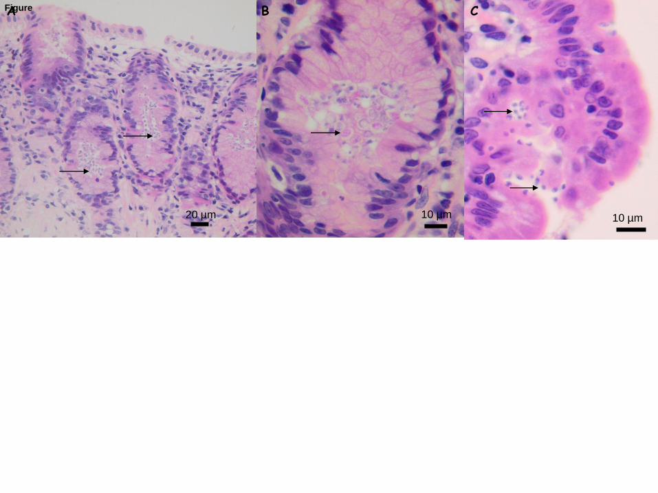
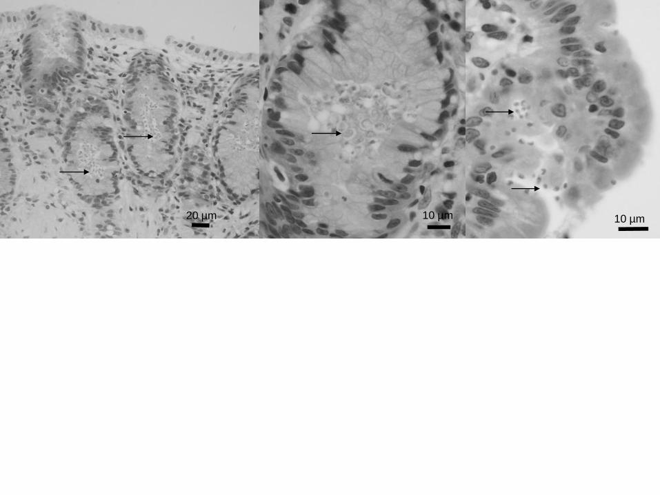
Figure 1. Hematoxylin & eosin stained sections of biopsies from the cat showing (A-B) 295
clusters of C. muris stages located within the glands of the gastric mucosa; and (C) C. 296
felis organisms along the enterocyte lining of the duodenum. 297
298
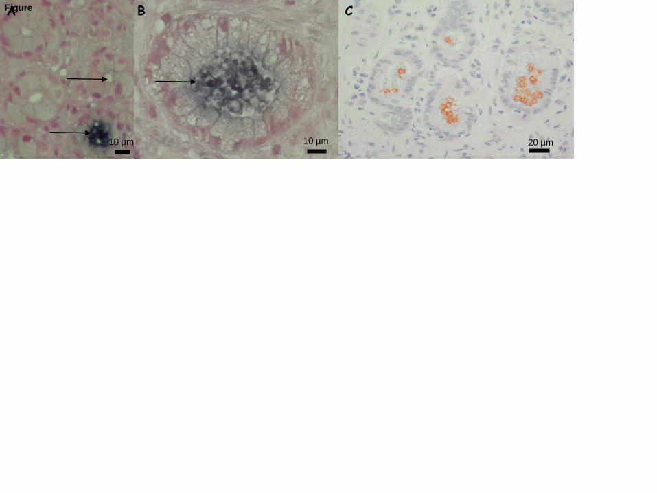
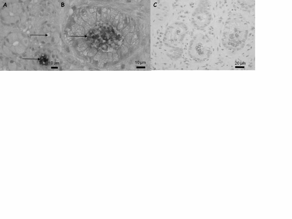
Figure 2. In situ hybridisation of digoxigenin-labelled C. muris 18S rRNA DNA probe 299
on tissue sections from the cat stomach showing (A) parasitised and non-parasitised 300
glands and (B) parasite stages deep within the glands. 2C. Immunohistochemistry on 301
tissue sections from the cat stomach showing parasite stages deep within the glands. 302
303
Figure 3. Malachite green stained wet mount of cat faecal sample showing (A) C. felis-304
like oocysts and (B) C. muris-like oocysts. 305
306
Page 16 of 21
Accep
ted
Man
uscr
ipt
A B C
20 µm 10 µm 10 µm
Figure
Page 17 of 21
Accep
ted
Man
uscr
ipt
20 µm 10 µm 10 µm
Page 18 of 21
Accep
ted
Man
uscr
ipt
A B
10 µm10 µm 20 µm
CFigure
Page 19 of 21
Accep
ted
Man
uscr
ipt
Page 20 of 21
Accep
ted
Man
uscr
ipt
A
B
20 µm
Figure
Page 21 of 21
Accep
ted
Man
uscr
ipt
20 µm
A
B