Embed Size (px)
Citation preview
ACR Appropriateness Criteria® Non-Invasive Clinical Staging of Bronchogenic Carcinoma
EVIDENCE TABLE
* See Last Page for Key 2013 Review Ravenel Page 1
Reference Study Type Patients/ Events
Study Objective (Purpose of Study) Study Results Study
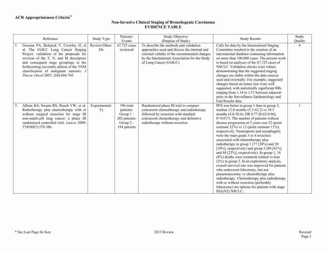
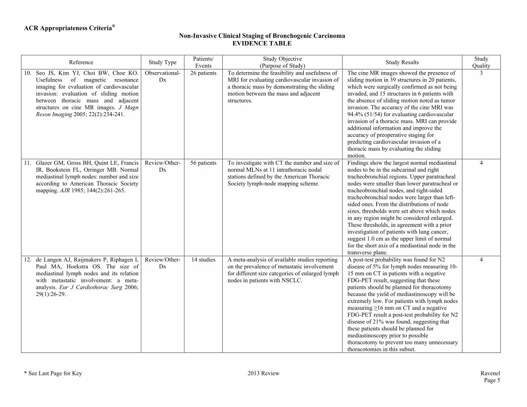
Quality 1. Groome PA, Bolejack V, Crowley JJ, et
al. The IASLC Lung Cancer Staging Project: validation of the proposals for revision of the T, N, and M descriptors and consequent stage groupings in the forthcoming (seventh) edition of the TNM classification of malignant tumours. J Thorac Oncol 2007; 2(8):694-705.
Review/Other-Dx
67,725 cases reviewed
To describe the methods and validation approaches used and discuss the internal and external validity of the recommended changes by the International Association for the Study of Lung Cancer (IASLC).
Calls for data by the International Staging Committee resulted in the creation of an international database containing information on more than 100,000 cases. The present work is based on analyses of the 67,725 cases of NSCLC. Validation checks were robust, demonstrating that the suggested staging changes are stable within the data sources used and externally. For example, suggested changes based on tumor size were well supported, with statistically significant HRs ranging from 1.14 to 1.51 between adjacent pairs in the Surveillance Epidemiology and End Results data.
4
2. Albain KS, Swann RS, Rusch VW, et al. Radiotherapy plus chemotherapy with or without surgical resection for stage III non-small-cell lung cancer: a phase III randomised controlled trial. Lancet 2009; 374(9687):379-386.
Experimental-Tx
396 total patients:
Group 1 - 202 patients;
Group 2 - 194 patients
Randomized phase III trial to compare concurrent chemotherapy and radiotherapy followed by resection with standard concurrent chemotherapy and definitive radiotherapy without resection.
PFS was better in group 1 than in group 2, median 12.8 months (5.3-42.2) vs 10.5 months (4.8-20.6), HR 0.77 [0.62-0.96]; P=0.017). The number of patients without disease progression at 5 years was 32 (point estimate 22%) vs 13 (point estimate 11%), respectively. Neutropenia and oesophagitis were the main grade 3 or 4 toxicities associated with chemotherapy plus radiotherapy in group 1 (77 [38%] and 20 [10%], respectively) and group 2 (80 [41%] and 44 [23%], respectively). In group 1, 16 (8%) deaths were treatment related vs four (2%) in group 2. In an exploratory analysis, overall survival rate was improved for patients who underwent lobectomy, but not pneumonectomy vs chemotherapy plus radiotherapy. Chemotherapy plus radiotherapy with or without resection (preferably lobectomy) are options for patients with stage IIIA(N2) NSCLC.
1
ACR Appropriateness Criteria® Non-Invasive Clinical Staging of Bronchogenic Carcinoma
EVIDENCE TABLE
* See Last Page for Key 2013 Review Ravenel Page 2
Reference Study Type Patients/ Events
Study Objective (Purpose of Study) Study Results Study
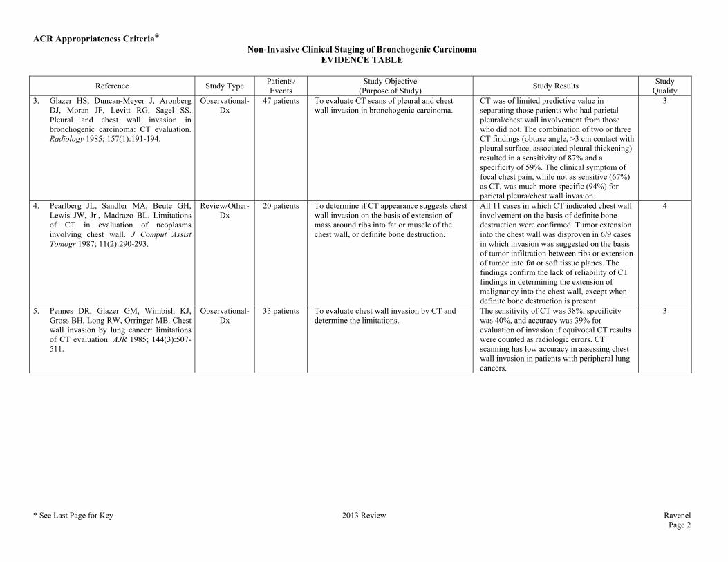
Quality 3. Glazer HS, Duncan-Meyer J, Aronberg
DJ, Moran JF, Levitt RG, Sagel SS. Pleural and chest wall invasion in bronchogenic carcinoma: CT evaluation. Radiology 1985; 157(1):191-194.
Observational-Dx
47 patients To evaluate CT scans of pleural and chest wall invasion in bronchogenic carcinoma.
CT was of limited predictive value in separating those patients who had parietal pleural/chest wall involvement from those who did not. The combination of two or three CT findings (obtuse angle, >3 cm contact with pleural surface, associated pleural thickening) resulted in a sensitivity of 87% and a specificity of 59%. The clinical symptom of focal chest pain, while not as sensitive (67%) as CT, was much more specific (94%) for parietal pleura/chest wall invasion.
3
4. Pearlberg JL, Sandler MA, Beute GH, Lewis JW, Jr., Madrazo BL. Limitations of CT in evaluation of neoplasms involving chest wall. J Comput Assist Tomogr 1987; 11(2):290-293.
Review/Other-Dx
20 patients To determine if CT appearance suggests chest wall invasion on the basis of extension of mass around ribs into fat or muscle of the chest wall, or definite bone destruction.
All 11 cases in which CT indicated chest wall involvement on the basis of definite bone destruction were confirmed. Tumor extension into the chest wall was disproven in 6/9 cases in which invasion was suggested on the basis of tumor infiltration between ribs or extension of tumor into fat or soft tissue planes. The findings confirm the lack of reliability of CT findings in determining the extension of malignancy into the chest wall, except when definite bone destruction is present.
4
5. Pennes DR, Glazer GM, Wimbish KJ, Gross BH, Long RW, Orringer MB. Chest wall invasion by lung cancer: limitations of CT evaluation. AJR 1985; 144(3):507-511.
Observational-Dx
33 patients To evaluate chest wall invasion by CT and determine the limitations.
The sensitivity of CT was 38%, specificity was 40%, and accuracy was 39% for evaluation of invasion if equivocal CT results were counted as radiologic errors. CT scanning has low accuracy in assessing chest wall invasion in patients with peripheral lung cancers.
3
ACR Appropriateness Criteria® Non-Invasive Clinical Staging of Bronchogenic Carcinoma
EVIDENCE TABLE
* See Last Page for Key 2013 Review Ravenel Page 3
Reference Study Type Patients/ Events
Study Objective (Purpose of Study) Study Results Study
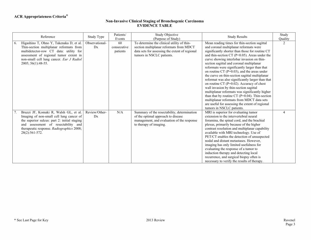
Quality 6. Higashino T, Ohno Y, Takenaka D, et al.
Thin-section multiplanar reformats from multidetector-row CT data: utility for assessment of regional tumor extent in non-small cell lung cancer. Eur J Radiol 2005; 56(1):48-55.
Observational-Dx
60 consecutive
patients
To determine the clinical utility of thin-section multiplanar reformats from MDCT data sets for assessing the extent of regional tumors in NSCLC patients.
Mean reading times for thin-section sagittal and coronal multiplanar reformats were significantly shorter than those for routine CT and thin-section CT (P<0.05). Areas under the curve showing interlobar invasion on thin-section sagittal and coronal multiplanar reformats were significantly larger than that on routine CT (P=0.03), and the areas under the curve on thin-section sagittal multiplanar reformat was also significantly larger than that on routine CT (P=0.02). Accuracy of chest wall invasion by thin-section sagittal multiplanar reformats was significantly higher than that by routine CT (P=0.04). Thin-section multiplanar reformats from MDCT data sets are useful for assessing the extent of regional tumors in NSCLC patients.
2
7. Bruzzi JF, Komaki R, Walsh GL, et al. Imaging of non-small cell lung cancer of the superior sulcus: part 2: initial staging and assessment of resectability and therapeutic response. Radiographics 2008; 28(2):561-572.
Review/Other-Dx
N/A Summary of the resectability, determination of the optimal approach to disease management, and evaluation of the response to therapy of imaging.
MRI is superior for evaluating tumor extension to the intervertebral neural foramina, the spinal cord, and the brachial plexus, primarily because of the higher contrast resolution and multiplanar capability available with MRI technology. Use of PET/CT enables the detection of unsuspected nodal and distant metastases. However, imaging has only limited usefulness for evaluating the response of a tumor to induction therapy and detecting local recurrence, and surgical biopsy often is necessary to verify the results of therapy.
4
ACR Appropriateness Criteria® Non-Invasive Clinical Staging of Bronchogenic Carcinoma
EVIDENCE TABLE
* See Last Page for Key 2013 Review Ravenel Page 4
Reference Study Type Patients/ Events
Study Objective (Purpose of Study) Study Results Study
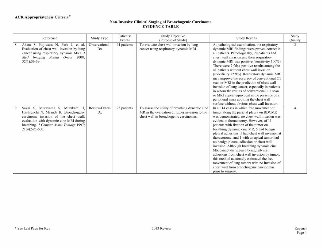
Quality 8. Akata S, Kajiwara N, Park J, et al.
Evaluation of chest wall invasion by lung cancer using respiratory dynamic MRI. J Med Imaging Radiat Oncol 2008; 52(1):36-39.
Observational-Dx
61 patients To evaluate chest wall invasion by lung cancer using respiratory dynamic MRI.
At pathological examination, the respiratory dynamic MRI findings were proved correct in all patients. Pathologically, 20 patients had chest wall invasion and their respiratory dynamic MRI was positive (sensitivity 100%). There were 7 false-positive results among the 41 patients without chest wall invasion (specificity 82.9%). Respiratory dynamic MRI may improve the accuracy of conventional CT scan or MRI in the prediction of chest wall invasion of lung cancer, especially in patients in whom the results of conventional CT scan or MRI appear equivocal in the presence of a peripheral mass abutting the chest wall surface without obvious chest wall invasion.
3
9. Sakai S, Murayama S, Murakami J, Hashiguchi N, Masuda K. Bronchogenic carcinoma invasion of the chest wall: evaluation with dynamic cine MRI during breathing. J Comput Assist Tomogr 1997; 21(4):595-600.
Review/Other-Dx
25 patients To assess the utility of breathing dynamic cine MR in the evaluation of tumor invasion to the chest wall in bronchogenic carcinomas.
In all 14 cases in which free movement of tumor along the parietal pleura on BDCMR was demonstrated, no chest wall invasion was evident at thoracotomy. However, of 11 patients with fixation of the tumor on breathing dynamic cine MR, 5 had benign pleural adhesions, 5 had chest wall invasion at thoracotomy, and 1 with an apical tumor had no benign pleural adhesion or chest wall invasion. Although breathing dynamic cine MR cannot distinguish benign pleural adhesions from chest wall invasion by tumor, this method accurately estimated the free movement of lung tumors with no invasion of chest wall from bronchogenic carcinomas prior to surgery.
4
ACR Appropriateness Criteria® Non-Invasive Clinical Staging of Bronchogenic Carcinoma
EVIDENCE TABLE
* See Last Page for Key 2013 Review Ravenel Page 5
Reference Study Type Patients/ Events
Study Objective (Purpose of Study) Study Results Study
Quality 10. Seo JS, Kim YJ, Choi BW, Choe KO.
Usefulness of magnetic resonance imaging for evaluation of cardiovascular invasion: evaluation of sliding motion between thoracic mass and adjacent structures on cine MR images. J Magn Reson Imaging 2005; 22(2):234-241.
Observational-Dx
26 patients To determine the feasibility and usefulness of MRI for evaluating cardiovascular invasion of a thoracic mass by demonstrating the sliding motion between the mass and adjacent structures.
The cine MR images showed the presence of sliding motion in 39 structures in 20 patients, which were surgically confirmed as not being invaded, and 15 structures in 6 patients with the absence of sliding motion noted as tumor invasion. The accuracy of the cine MRI was 94.4% (51/54) for evaluating cardiovascular invasion of a thoracic mass. MRI can provide additional information and improve the accuracy of preoperative staging for predicting cardiovascular invasion of a thoracic mass by evaluating the sliding motion.
3
11. Glazer GM, Gross BH, Quint LE, Francis IR, Bookstein FL, Orringer MB. Normal mediastinal lymph nodes: number and size according to American Thoracic Society mapping. AJR 1985; 144(2):261-265.
Review/Other-Dx
56 patients To investigate with CT the number and size of normal MLNs at 11 intrathoracic nodal stations defined by the American Thoracic Society lymph-node mapping scheme.
Findings show the largest normal mediastinal nodes to be in the subcarinal and right tracheobronchial regions. Upper paratracheal nodes were smaller than lower paratracheal or tracheobronchial nodes, and right-sided tracheobronchial nodes were larger than left-sided ones. From the distributions of node sizes, thresholds were set above which nodes in any region might be considered enlarged. These thresholds, in agreement with a prior investigation of patients with lung cancer, suggest 1.0 cm as the upper limit of normal for the short axis of a mediastinal node in the transverse plane.
4
12. de Langen AJ, Raijmakers P, Riphagen I, Paul MA, Hoekstra OS. The size of mediastinal lymph nodes and its relation with metastatic involvement: a meta-analysis. Eur J Cardiothorac Surg 2006; 29(1):26-29.
Review/Other-Dx
14 studies A meta-analysis of available studies reporting on the prevalence of metastatic involvement for different size categories of enlarged lymph nodes in patients with NSCLC.
A post-test probability was found for N2 disease of 5% for lymph nodes measuring 10-15 mm on CT in patients with a negative FDG-PET result, suggesting that these patients should be planned for thoracotomy because the yield of mediastinoscopy will be extremely low. For patients with lymph nodes measuring ≥16 mm on CT and a negative FDG-PET result a post-test probability for N2 disease of 21% was found, suggesting that these patients should be planned for mediastinoscopy prior to possible thoracotomy to prevent too many unnecessary thoracotomies in this subset.
4
ACR Appropriateness Criteria® Non-Invasive Clinical Staging of Bronchogenic Carcinoma
EVIDENCE TABLE
* See Last Page for Key 2013 Review Ravenel Page 6
Reference Study Type Patients/ Events
Study Objective (Purpose of Study) Study Results Study
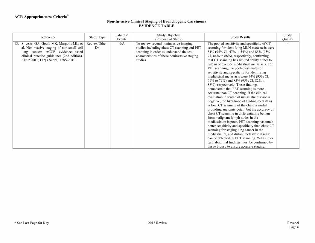
Quality 13. Silvestri GA, Gould MK, Margolis ML, et
al. Noninvasive staging of non-small cell lung cancer: ACCP evidenced-based clinical practice guidelines (2nd edition). Chest 2007; 132(3 Suppl):178S-201S.
Review/Other-Dx
N/A To review several noninvasive imaging studies including chest CT scanning and PET scanning in order to understand the test characteristics of these noninvasive staging studies.
The pooled sensitivity and specificity of CT scanning for identifying MLN metastasis were 51% (95% CI, 47% to 54%) and 85% (95% CI, 84% to 88%), respectively, confirming that CT scanning has limited ability either to rule in or exclude mediastinal metastasis. For PET scanning, the pooled estimates of sensitivity and specificity for identifying mediastinal metastasis were 74% (95% CI, 69% to 79%) and 85% (95% CI, 82% to 88%), respectively. These findings demonstrate that PET scanning is more accurate than CT scanning. If the clinical evaluation in search of metastatic disease is negative, the likelihood of finding metastasis is low. CT scanning of the chest is useful in providing anatomic detail, but the accuracy of chest CT scanning in differentiating benign from malignant lymph nodes in the mediastinum is poor. PET scanning has much better sensitivity and specificity than chest CT scanning for staging lung cancer in the mediastinum, and distant metastatic disease can be detected by PET scanning. With either test, abnormal findings must be confirmed by tissue biopsy to ensure accurate staging.
4
ACR Appropriateness Criteria® Non-Invasive Clinical Staging of Bronchogenic Carcinoma
EVIDENCE TABLE
* See Last Page for Key 2013 Review Ravenel Page 7
Reference Study Type Patients/ Events
Study Objective (Purpose of Study) Study Results Study
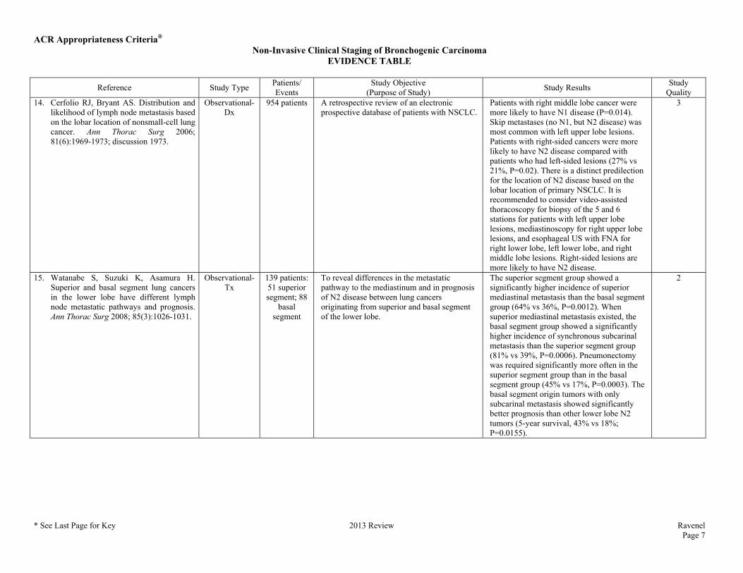
Quality 14. Cerfolio RJ, Bryant AS. Distribution and
likelihood of lymph node metastasis based on the lobar location of nonsmall-cell lung cancer. Ann Thorac Surg 2006; 81(6):1969-1973; discussion 1973.
Observational-Dx
954 patients A retrospective review of an electronic prospective database of patients with NSCLC.
Patients with right middle lobe cancer were more likely to have N1 disease (P=0.014). Skip metastases (no N1, but N2 disease) was most common with left upper lobe lesions. Patients with right-sided cancers were more likely to have N2 disease compared with patients who had left-sided lesions (27% vs 21%, P=0.02). There is a distinct predilection for the location of N2 disease based on the lobar location of primary NSCLC. It is recommended to consider video-assisted thoracoscopy for biopsy of the 5 and 6 stations for patients with left upper lobe lesions, mediastinoscopy for right upper lobe lesions, and esophageal US with FNA for right lower lobe, left lower lobe, and right middle lobe lesions. Right-sided lesions are more likely to have N2 disease.
3
15. Watanabe S, Suzuki K, Asamura H. Superior and basal segment lung cancers in the lower lobe have different lymph node metastatic pathways and prognosis. Ann Thorac Surg 2008; 85(3):1026-1031.
Observational-Tx
139 patients: 51 superior segment; 88
basal segment
To reveal differences in the metastatic pathway to the mediastinum and in prognosis of N2 disease between lung cancers originating from superior and basal segment of the lower lobe.
The superior segment group showed a significantly higher incidence of superior mediastinal metastasis than the basal segment group (64% vs 36%, P=0.0012). When superior mediastinal metastasis existed, the basal segment group showed a significantly higher incidence of synchronous subcarinal metastasis than the superior segment group (81% vs 39%, P=0.0006). Pneumonectomy was required significantly more often in the superior segment group than in the basal segment group (45% vs 17%, P=0.0003). The basal segment origin tumors with only subcarinal metastasis showed significantly better prognosis than other lower lobe N2 tumors (5-year survival, 43% vs 18%; P=0.0155).
2
ACR Appropriateness Criteria® Non-Invasive Clinical Staging of Bronchogenic Carcinoma
EVIDENCE TABLE
* See Last Page for Key 2013 Review Ravenel Page 8
Reference Study Type Patients/ Events
Study Objective (Purpose of Study) Study Results Study
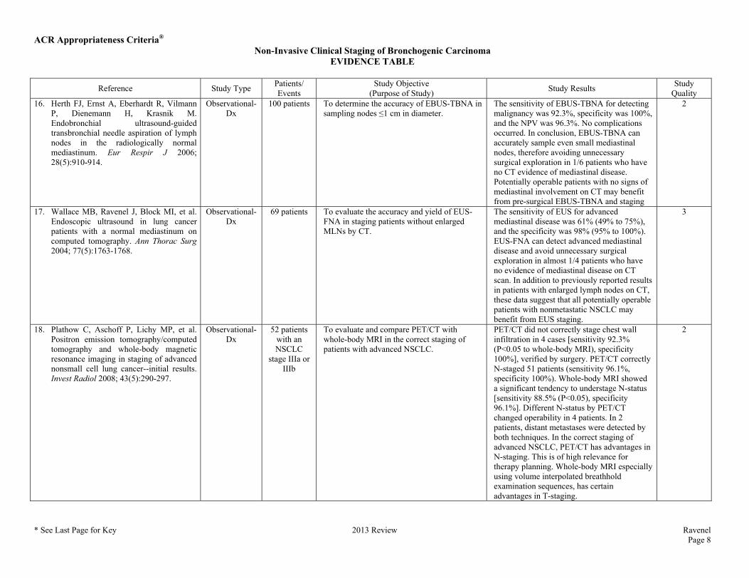
Quality 16. Herth FJ, Ernst A, Eberhardt R, Vilmann
P, Dienemann H, Krasnik M. Endobronchial ultrasound-guided transbronchial needle aspiration of lymph nodes in the radiologically normal mediastinum. Eur Respir J 2006; 28(5):910-914.
Observational-Dx
100 patients To determine the accuracy of EBUS-TBNA in sampling nodes ≤1 cm in diameter.
The sensitivity of EBUS-TBNA for detecting malignancy was 92.3%, specificity was 100%, and the NPV was 96.3%. No complications occurred. In conclusion, EBUS-TBNA can accurately sample even small mediastinal nodes, therefore avoiding unnecessary surgical exploration in 1/6 patients who have no CT evidence of mediastinal disease. Potentially operable patients with no signs of mediastinal involvement on CT may benefit from pre-surgical EBUS-TBNA and staging
2
17. Wallace MB, Ravenel J, Block MI, et al. Endoscopic ultrasound in lung cancer patients with a normal mediastinum on computed tomography. Ann Thorac Surg 2004; 77(5):1763-1768.
Observational-Dx
69 patients To evaluate the accuracy and yield of EUS-FNA in staging patients without enlarged MLNs by CT.
The sensitivity of EUS for advanced mediastinal disease was 61% (49% to 75%), and the specificity was 98% (95% to 100%). EUS-FNA can detect advanced mediastinal disease and avoid unnecessary surgical exploration in almost 1/4 patients who have no evidence of mediastinal disease on CT scan. In addition to previously reported results in patients with enlarged lymph nodes on CT, these data suggest that all potentially operable patients with nonmetastatic NSCLC may benefit from EUS staging.
3
18. Plathow C, Aschoff P, Lichy MP, et al. Positron emission tomography/computed tomography and whole-body magnetic resonance imaging in staging of advanced nonsmall cell lung cancer--initial results. Invest Radiol 2008; 43(5):290-297.
Observational-Dx
52 patients with an NSCLC
stage IIIa or IIIb
To evaluate and compare PET/CT with whole-body MRI in the correct staging of patients with advanced NSCLC.
PET/CT did not correctly stage chest wall infiltration in 4 cases [sensitivity 92.3% (P<0.05 to whole-body MRI), specificity 100%], verified by surgery. PET/CT correctly N-staged 51 patients (sensitivity 96.1%, specificity 100%). Whole-body MRI showed a significant tendency to understage N-status [sensitivity 88.5% (P<0.05), specificity 96.1%]. Different N-status by PET/CT changed operability in 4 patients. In 2 patients, distant metastases were detected by both techniques. In the correct staging of advanced NSCLC, PET/CT has advantages in N-staging. This is of high relevance for therapy planning. Whole-body MRI especially using volume interpolated breathhold examination sequences, has certain advantages in T-staging.
2
ACR Appropriateness Criteria® Non-Invasive Clinical Staging of Bronchogenic Carcinoma
EVIDENCE TABLE
* See Last Page for Key 2013 Review Ravenel Page 9
Reference Study Type Patients/ Events
Study Objective (Purpose of Study) Study Results Study
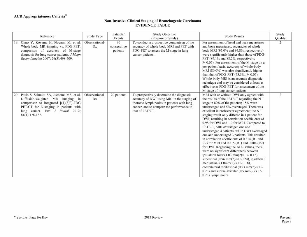
Quality 19. Ohno Y, Koyama H, Nogami M, et al.
Whole-body MR imaging vs. FDG-PET: comparison of accuracy of M-stage diagnosis for lung cancer patients. J Magn Reson Imaging 2007; 26(3):498-509.
Observational-Dx
90 consecutive
patients
To conduct a prospective comparison of the accuracy of whole-body MRI and PET with FDG-PET to assess the M-stage in lung cancer patients.
For assessment of head and neck metastases and bone metastases, accuracies of whole-body MRI (95.0% and 94.8%, respectively) were significantly higher than those of FDG-PET (89.1% and 88.2%, respectively; P<0.05). For assessment of the M-stage on a per-patient basis, accuracy of whole-body MRI (80.0%) was also significantly higher than that of FDG-PET (73.3%; P<0.05). Whole-body MRI is an accurate diagnostic technique and may be considered at least as effective as FDG-PET for assessment of the M-stage of lung cancer patients.
2
20. Pauls S, Schmidt SA, Juchems MS, et al. Diffusion-weighted MR imaging in comparison to integrated [(1)(8)F]-FDG PET/CT for N-staging in patients with lung cancer. Eur J Radiol 2012; 81(1):178-182.
Observational-Dx
20 patients To prospectively determine the diagnostic accuracy of DWI using MRI in the staging of thoracic lymph nodes in patients with lung cancer, and to compare the performance to that of PET/CT.
MRI with or without DWI only agreed with the results of the PET/CT regarding the N stage in 80% of the patients; 15% were understaged and 5% overstaged. There was excellent interobserver agreement; the N-staging result only differed in 1 patient for DWI, resulting in correlation coefficients of 0.98 for DWI and 1.0 for MRI. Compared to PET/CT, MRI overstaged one and understaged 4 patients, while DWI overstaged one and understaged 3 patients. This resulted in correlation coefficients of 0.814 (R1 and R2) for MRI and 0.815 (R1) and 0.804 (R2) for DWI. Regarding the ADC values, there were no significant differences between ipsilateral hilar (1.03 mm(2)/s +/- 0.13), subcarinal (0.96 mm(2)/s+/-0.24), ipsilateral mediastinal (1.0mm(2)/s +/- 0.18), contralateral mediastinal (0.93 mm(2)/s +/- 0.23) and supraclavicular (0.9 mm(2)/s +/- 0.23) lymph nodes.
2
ACR Appropriateness Criteria® Non-Invasive Clinical Staging of Bronchogenic Carcinoma
EVIDENCE TABLE
* See Last Page for Key 2013 Review Ravenel Page 10
Reference Study Type Patients/ Events
Study Objective (Purpose of Study) Study Results Study
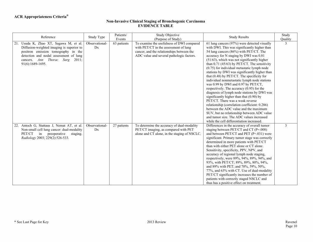
Quality 21. Usuda K, Zhao XT, Sagawa M, et al.
Diffusion-weighted imaging is superior to positron emission tomography in the detection and nodal assessment of lung cancers. Ann Thorac Surg 2011; 91(6):1689-1695.
Observational-Dx
63 patients To examine the usefulness of DWI compared with PET/CT in the assessment of lung cancer, and the relationships between the ADC value and several pathologic factors.
61 lung cancers (97%) were detected visually with DWI. This was significantly higher than 54 lung cancers (86%) with PET/CT. The accuracy for N staging by DWI was 0.81 (51/63), which was not significantly higher than 0.71 (45/63) by PET/CT. The sensitivity (0.75) for individual metastatic lymph node stations by DWI was significantly higher than that (0.48) by PET/CT. The specificity for individual nonmetastatic lymph node stations was 0.99 by DWI and 0.97 by PET/CT, respectively. The accuracy (0.95) for the diagnosis of lymph node stations by DWI was significantly higher than that (0.90) by PET/CT. There was a weak reverse relationship (correlation coefficient: 0.286) between the ADC value and the maximum SUV, but no relationship between ADC value and tumor size. The ADC values increased while the cell differentiation increased.
3
22. Antoch G, Stattaus J, Nemat AT, et al. Non-small cell lung cancer: dual-modality PET/CT in preoperative staging. Radiology 2003; 229(2):526-533.
Observational-Dx
27 patients To determine the accuracy of dual-modality PET/CT imaging, as compared with PET alone and CT alone, in the staging of NSCLC.
Differences in the accuracy of overall tumor staging between PET/CT and CT (P=.008) and between PET/CT and PET (P=.031) were significant. Primary tumor stage was correctly determined in more patients with PET/CT than with either PET alone or CT alone. Sensitivity, specificity, PPV, NPV, and accuracy of regional lymph node staging, respectively, were 89%, 94%, 89%, 94%, and 93%, with PET/CT; 89%, 89%, 80%, 94%, and 89% with PET; and 70%, 59%, 50%, 77%, and 63% with CT. Use of dual-modality PET/CT significantly increases the number of patients with correctly staged NSCLC and thus has a positive effect on treatment.
3
ACR Appropriateness Criteria® Non-Invasive Clinical Staging of Bronchogenic Carcinoma
EVIDENCE TABLE
* See Last Page for Key 2013 Review Ravenel Page 11
Reference Study Type Patients/ Events
Study Objective (Purpose of Study) Study Results Study
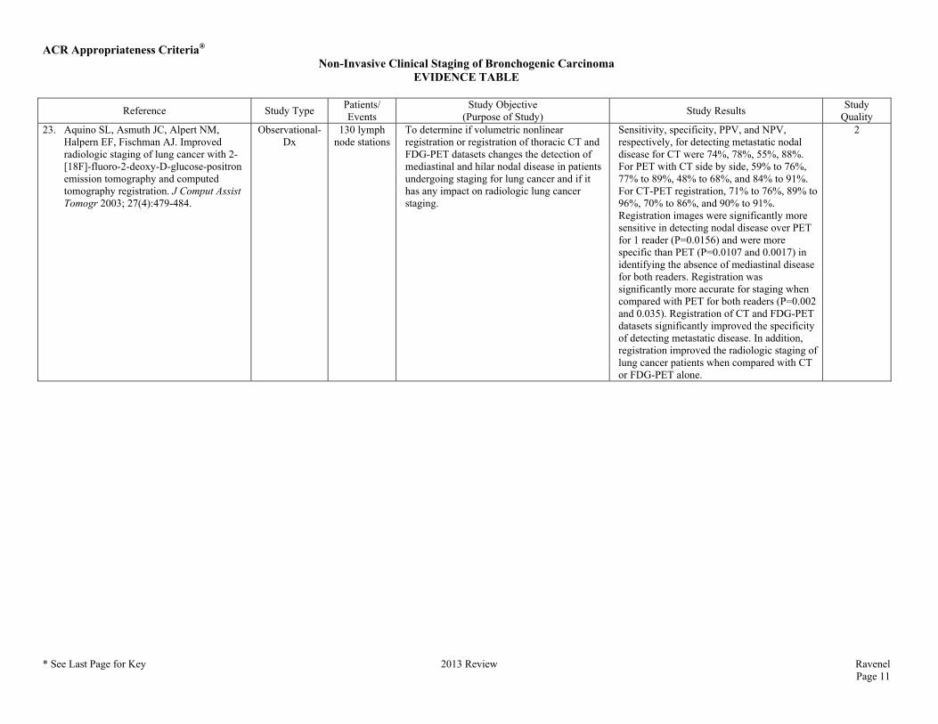
Quality 23. Aquino SL, Asmuth JC, Alpert NM,
Halpern EF, Fischman AJ. Improved radiologic staging of lung cancer with 2-[18F]-fluoro-2-deoxy-D-glucose-positron emission tomography and computed tomography registration. J Comput Assist Tomogr 2003; 27(4):479-484.
Observational-Dx
130 lymph node stations
To determine if volumetric nonlinear registration or registration of thoracic CT and FDG-PET datasets changes the detection of mediastinal and hilar nodal disease in patients undergoing staging for lung cancer and if it has any impact on radiologic lung cancer staging.
Sensitivity, specificity, PPV, and NPV, respectively, for detecting metastatic nodal disease for CT were 74%, 78%, 55%, 88%. For PET with CT side by side, 59% to 76%, 77% to 89%, 48% to 68%, and 84% to 91%. For CT-PET registration, 71% to 76%, 89% to 96%, 70% to 86%, and 90% to 91%. Registration images were significantly more sensitive in detecting nodal disease over PET for 1 reader (P=0.0156) and were more specific than PET (P=0.0107 and 0.0017) in identifying the absence of mediastinal disease for both readers. Registration was significantly more accurate for staging when compared with PET for both readers (P=0.002 and 0.035). Registration of CT and FDG-PET datasets significantly improved the specificity of detecting metastatic disease. In addition, registration improved the radiologic staging of lung cancer patients when compared with CT or FDG-PET alone.
2
ACR Appropriateness Criteria® Non-Invasive Clinical Staging of Bronchogenic Carcinoma
EVIDENCE TABLE
* See Last Page for Key 2013 Review Ravenel Page 12
Reference Study Type Patients/ Events
Study Objective (Purpose of Study) Study Results Study
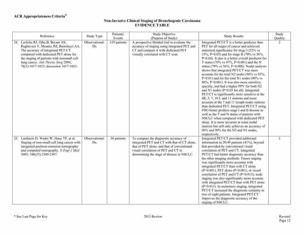
Quality 24. Cerfolio RJ, Ojha B, Bryant AS,
Raghuveer V, Mountz JM, Bartolucci AA. The accuracy of integrated PET-CT compared with dedicated PET alone for the staging of patients with nonsmall cell lung cancer. Ann Thorac Surg 2004; 78(3):1017-1023; discussion 1017-1023.
Observational-Dx
129 patients A prospective blinded trial to evaluate the accuracy of staging using integrated PET and CT and compare it with dedicated PET visually correlated with CT scan.
Integrated PET/CT is a better predictor than PET for all stages of cancer and achieved statistical significance for stage I (52% vs 33%, P=0.03) and for stage II (70% vs 36%, P=0.04). It also is a better overall predictor for T status (70% vs 47%, P=0.001) and the N status (78% vs 56%, P=0.008). Nodal analysis shows that integrated PET/CT was more accurate for the total N2 nodes (96% vs 93%, P=0.01) and for the total N1 nodes (90% vs 80%, P=0.001). It was also more sensitive, specific, and had a higher PPV for both N2 and N1 nodes (P<0.05 for all). Integrated PET/CT is significantly more sensitive at the 4R, 5, 7, 10 L and 11 stations and more accurate at the 7 and 11 lymph nodes stations than dedicated PET. Integrated PET/CT using FDG better predicts stage I and II disease as well as the T and N status of patients with NSCLC when compared with dedicated PET alone. It is more accurate at some nodal stations but still only achieves an accuracy of 96% and 90% for the N2 and N1 nodes, respectively.
2
25. Lardinois D, Weder W, Hany TF, et al. Staging of non-small-cell lung cancer with integrated positron-emission tomography and computed tomography. N Engl J Med 2003; 348(25):2500-2507.
Observational-Dx
50 patients To compare the diagnostic accuracy of integrated PET and CT with that of CT alone, that of PET alone, and that of conventional visual correlation of PET and CT in determining the stage of disease in NSCLC.
Integrated PET/CT provided additional information in 20/49 patients (41%), beyond that provided by conventional visual correlation of PET and CT. Integrated PET/CT had better diagnostic accuracy than the other imaging methods. Tumor staging was significantly more accurate with integrated PET/CT than with CT alone (P=0.001), PET alone (P<0.001), or visual correlation of PET and CT (P=0.013); node staging was also significantly more accurate with integrated PET/CT than with PET alone (P=0.013). In metastasis staging, integrated PET/CT increased the diagnostic certainty in two of eight patients. Integrated PET/CT improves the diagnostic accuracy of the staging of NSCLC.
2
ACR Appropriateness Criteria® Non-Invasive Clinical Staging of Bronchogenic Carcinoma
EVIDENCE TABLE
* See Last Page for Key 2013 Review Ravenel Page 13
Reference Study Type Patients/ Events
Study Objective (Purpose of Study) Study Results Study
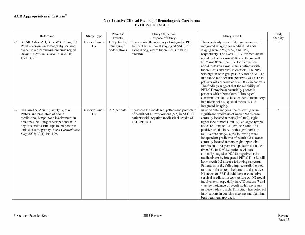
Quality 26. Sit AK, Sihoe AD, Suen WS, Cheng LC.
Positron-emission tomography for lung cancer in a tuberculosis-endemic region. Asian Cardiovasc Thorac Ann 2010; 18(1):33-38.
Observational-Dx
107 patients; 249 lymph
node stations
To examine the accuracy of integrated PET for mediastinal nodal staging of NSCLC in Hong Kong, where tuberculosis remains endemic.
The sensitivity, specificity, and accuracy of integrated imaging for mediastinal nodal staging were 52%, 86%, and 80%, respectively. The overall PPV for mediastinal nodal metastasis was 46%, and the overall NPV was 89%. The PPV for mediastinal nodal metastasis was 39% in patients with tuberculosis and 50% in controls. The NPV was high in both groups (92% and 87%). The likelihood ratio for true positives was 6.47 in patients with tuberculosis vs 10.97 in controls. The findings suggest that the reliability of PET/CT may be substantially poorer in patients with tuberculosis. Histological confirmation should be considered mandatory in patients with suspected metastasis on integrated imaging.
3
27. Al-Sarraf N, Aziz R, Gately K, et al. Pattern and predictors of occult mediastinal lymph node involvement in non-small cell lung cancer patients with negative mediastinal uptake on positron emission tomography. Eur J Cardiothorac Surg 2008; 33(1):104-109.
Observational-Dx
215 patients To assess the incidence, pattern and predictors of occult MLN involvement (N2) in NSCLC patients with negative mediastinal uptake of FDG-PET/CT.
In univariate analysis, the following were significant predictors of occult N2 disease: centrally located tumors (P=0.049), right upper lobe tumors (P=0.04), enlarged lymph nodes (>1 cm) on CT (P=0.048) and PET positive uptake in N1 nodes (P=0.006). In multivariate analysis, the following were independent predictors of occult N2 disease: centrally located tumors, right upper lobe tumors and PET positive uptake in N1 nodes (P<0.05). In NSCLC patients who are clinically staged as N2/N3 negative in the mediastinum by integrated PET/CT, 16% will have occult N2 disease following resection. Patients with the following: centrally located tumors, right upper lobe tumors and positive N1 nodes on PET should have preoperative cervical mediastinoscopy to rule out N2 nodal involvement, especially in ATS stations 7 and 4 as the incidence of occult nodal metastasis in these nodes is high. This study has potential implications in decision-making and planning best treatment approach.
4
ACR Appropriateness Criteria® Non-Invasive Clinical Staging of Bronchogenic Carcinoma
EVIDENCE TABLE
* See Last Page for Key 2013 Review Ravenel Page 14
Reference Study Type Patients/ Events
Study Objective (Purpose of Study) Study Results Study
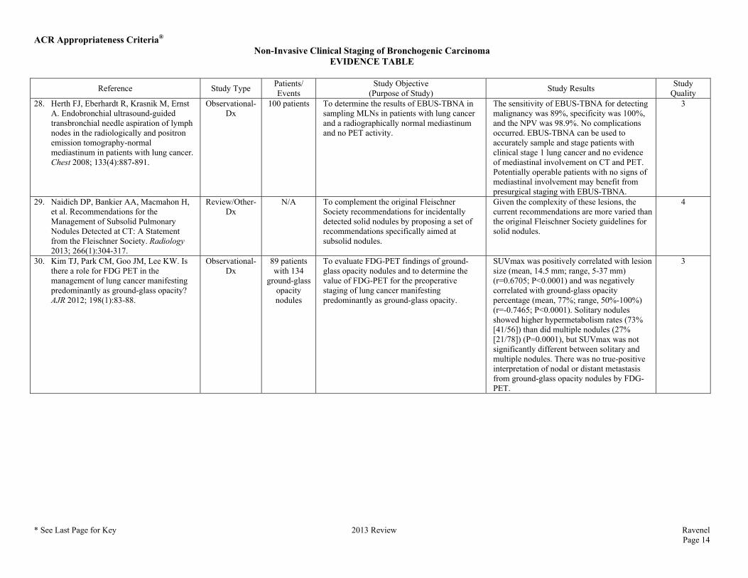
Quality 28. Herth FJ, Eberhardt R, Krasnik M, Ernst
A. Endobronchial ultrasound-guided transbronchial needle aspiration of lymph nodes in the radiologically and positron emission tomography-normal mediastinum in patients with lung cancer. Chest 2008; 133(4):887-891.
Observational-Dx
100 patients To determine the results of EBUS-TBNA in sampling MLNs in patients with lung cancer and a radiographically normal mediastinum and no PET activity.
The sensitivity of EBUS-TBNA for detecting malignancy was 89%, specificity was 100%, and the NPV was 98.9%. No complications occurred. EBUS-TBNA can be used to accurately sample and stage patients with clinical stage 1 lung cancer and no evidence of mediastinal involvement on CT and PET. Potentially operable patients with no signs of mediastinal involvement may benefit from presurgical staging with EBUS-TBNA.
3
29. Naidich DP, Bankier AA, Macmahon H, et al. Recommendations for the Management of Subsolid Pulmonary Nodules Detected at CT: A Statement from the Fleischner Society. Radiology 2013; 266(1):304-317.
Review/Other-Dx
N/A To complement the original Fleischner Society recommendations for incidentally detected solid nodules by proposing a set of recommendations specifically aimed at subsolid nodules.
Given the complexity of these lesions, the current recommendations are more varied than the original Fleischner Society guidelines for solid nodules.
4
30. Kim TJ, Park CM, Goo JM, Lee KW. Is there a role for FDG PET in the management of lung cancer manifesting predominantly as ground-glass opacity? AJR 2012; 198(1):83-88.
Observational-Dx
89 patients with 134
ground-glass opacity nodules
To evaluate FDG-PET findings of ground-glass opacity nodules and to determine the value of FDG-PET for the preoperative staging of lung cancer manifesting predominantly as ground-glass opacity.
SUVmax was positively correlated with lesion size (mean, 14.5 mm; range, 5-37 mm) (r=0.6705; P<0.0001) and was negatively correlated with ground-glass opacity percentage (mean, 77%; range, 50%-100%) (r=-0.7465; P<0.0001). Solitary nodules showed higher hypermetabolism rates (73% [41/56]) than did multiple nodules (27% [21/78]) (P=0.0001), but SUVmax was not significantly different between solitary and multiple nodules. There was no true-positive interpretation of nodal or distant metastasis from ground-glass opacity nodules by FDG-PET.
3
ACR Appropriateness Criteria® Non-Invasive Clinical Staging of Bronchogenic Carcinoma
EVIDENCE TABLE
* See Last Page for Key 2013 Review Ravenel Page 15
Reference Study Type Patients/ Events
Study Objective (Purpose of Study) Study Results Study
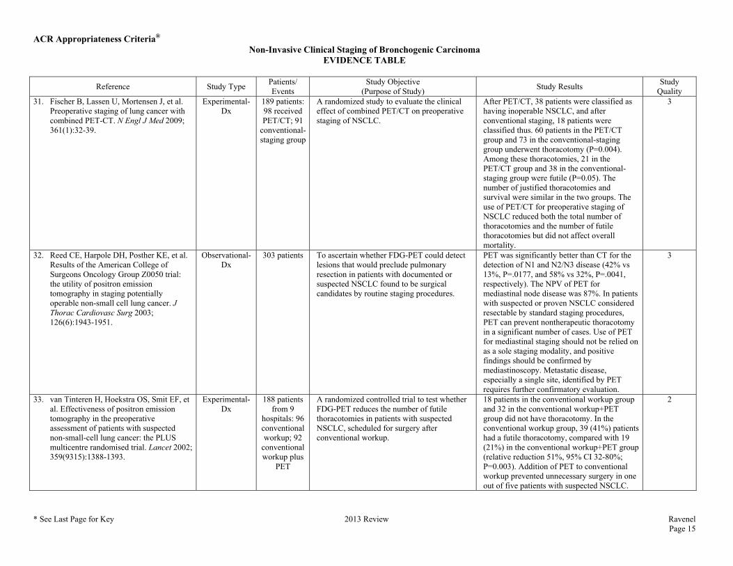
Quality 31. Fischer B, Lassen U, Mortensen J, et al.
Preoperative staging of lung cancer with combined PET-CT. N Engl J Med 2009; 361(1):32-39.
Experimental-Dx
189 patients: 98 received PET/CT; 91
conventional-staging group
A randomized study to evaluate the clinical effect of combined PET/CT on preoperative staging of NSCLC.
After PET/CT, 38 patients were classified as having inoperable NSCLC, and after conventional staging, 18 patients were classified thus. 60 patients in the PET/CT group and 73 in the conventional-staging group underwent thoracotomy (P=0.004). Among these thoracotomies, 21 in the PET/CT group and 38 in the conventional-staging group were futile (P=0.05). The number of justified thoracotomies and survival were similar in the two groups. The use of PET/CT for preoperative staging of NSCLC reduced both the total number of thoracotomies and the number of futile thoracotomies but did not affect overall mortality.
3
32. Reed CE, Harpole DH, Posther KE, et al. Results of the American College of Surgeons Oncology Group Z0050 trial: the utility of positron emission tomography in staging potentially operable non-small cell lung cancer. J Thorac Cardiovasc Surg 2003; 126(6):1943-1951.
Observational-Dx
303 patients To ascertain whether FDG-PET could detect lesions that would preclude pulmonary resection in patients with documented or suspected NSCLC found to be surgical candidates by routine staging procedures.
PET was significantly better than CT for the detection of N1 and N2/N3 disease (42% vs 13%, P=.0177, and 58% vs 32%, P=.0041, respectively). The NPV of PET for mediastinal node disease was 87%. In patients with suspected or proven NSCLC considered resectable by standard staging procedures, PET can prevent nontherapeutic thoracotomy in a significant number of cases. Use of PET for mediastinal staging should not be relied on as a sole staging modality, and positive findings should be confirmed by mediastinoscopy. Metastatic disease, especially a single site, identified by PET requires further confirmatory evaluation.
3
33. van Tinteren H, Hoekstra OS, Smit EF, et al. Effectiveness of positron emission tomography in the preoperative assessment of patients with suspected non-small-cell lung cancer: the PLUS multicentre randomised trial. Lancet 2002; 359(9315):1388-1393.
Experimental-Dx
188 patients from 9
hospitals: 96 conventional workup; 92
conventional workup plus
PET
A randomized controlled trial to test whether FDG-PET reduces the number of futile thoracotomies in patients with suspected NSCLC, scheduled for surgery after conventional workup.
18 patients in the conventional workup group and 32 in the conventional workup+PET group did not have thoracotomy. In the conventional workup group, 39 (41%) patients had a futile thoracotomy, compared with 19 (21%) in the conventional workup+PET group (relative reduction 51%, 95% CI 32-80%; P=0.003). Addition of PET to conventional workup prevented unnecessary surgery in one out of five patients with suspected NSCLC.
2
ACR Appropriateness Criteria® Non-Invasive Clinical Staging of Bronchogenic Carcinoma
EVIDENCE TABLE
* See Last Page for Key 2013 Review Ravenel Page 16
Reference Study Type Patients/ Events
Study Objective (Purpose of Study) Study Results Study
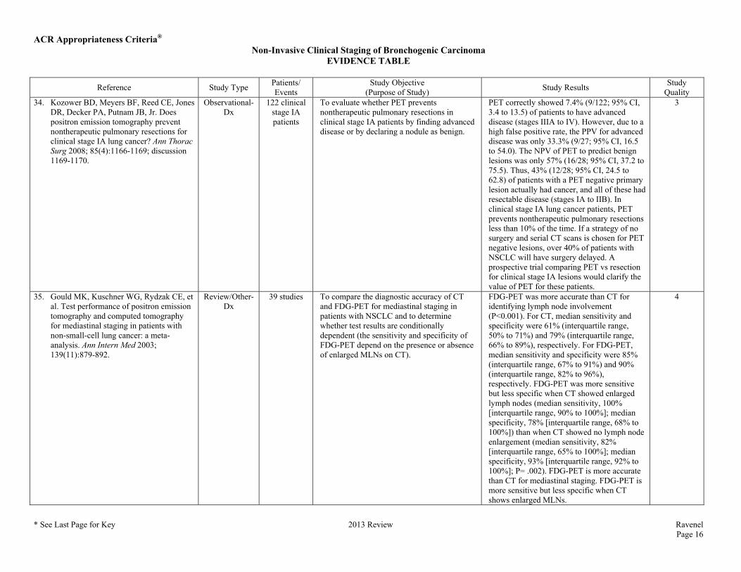
Quality 34. Kozower BD, Meyers BF, Reed CE, Jones
DR, Decker PA, Putnam JB, Jr. Does positron emission tomography prevent nontherapeutic pulmonary resections for clinical stage IA lung cancer? Ann Thorac Surg 2008; 85(4):1166-1169; discussion 1169-1170.
Observational-Dx
122 clinical stage IA patients
To evaluate whether PET prevents nontherapeutic pulmonary resections in clinical stage IA patients by finding advanced disease or by declaring a nodule as benign.
PET correctly showed 7.4% (9/122; 95% CI, 3.4 to 13.5) of patients to have advanced disease (stages IIIA to IV). However, due to a high false positive rate, the PPV for advanced disease was only 33.3% (9/27; 95% CI, 16.5 to 54.0). The NPV of PET to predict benign lesions was only 57% (16/28; 95% CI, 37.2 to 75.5). Thus, 43% (12/28; 95% CI, 24.5 to 62.8) of patients with a PET negative primary lesion actually had cancer, and all of these had resectable disease (stages IA to IIB). In clinical stage IA lung cancer patients, PET prevents nontherapeutic pulmonary resections less than 10% of the time. If a strategy of no surgery and serial CT scans is chosen for PET negative lesions, over 40% of patients with NSCLC will have surgery delayed. A prospective trial comparing PET vs resection for clinical stage IA lesions would clarify the value of PET for these patients.
3
35. Gould MK, Kuschner WG, Rydzak CE, et al. Test performance of positron emission tomography and computed tomography for mediastinal staging in patients with non-small-cell lung cancer: a meta-analysis. Ann Intern Med 2003; 139(11):879-892.
Review/Other-Dx
39 studies To compare the diagnostic accuracy of CT and FDG-PET for mediastinal staging in patients with NSCLC and to determine whether test results are conditionally dependent (the sensitivity and specificity of FDG-PET depend on the presence or absence of enlarged MLNs on CT).
FDG-PET was more accurate than CT for identifying lymph node involvement (P<0.001). For CT, median sensitivity and specificity were 61% (interquartile range, 50% to 71%) and 79% (interquartile range, 66% to 89%), respectively. For FDG-PET, median sensitivity and specificity were 85% (interquartile range, 67% to 91%) and 90% (interquartile range, 82% to 96%), respectively. FDG-PET was more sensitive but less specific when CT showed enlarged lymph nodes (median sensitivity, 100% [interquartile range, 90% to 100%]; median specificity, 78% [interquartile range, 68% to 100%]) than when CT showed no lymph node enlargement (median sensitivity, 82% [interquartile range, 65% to 100%]; median specificity, 93% [interquartile range, 92% to 100%]; P= .002). FDG-PET is more accurate than CT for mediastinal staging. FDG-PET is more sensitive but less specific when CT shows enlarged MLNs.
4
ACR Appropriateness Criteria® Non-Invasive Clinical Staging of Bronchogenic Carcinoma
EVIDENCE TABLE
* See Last Page for Key 2013 Review Ravenel Page 17
Reference Study Type Patients/ Events
Study Objective (Purpose of Study) Study Results Study
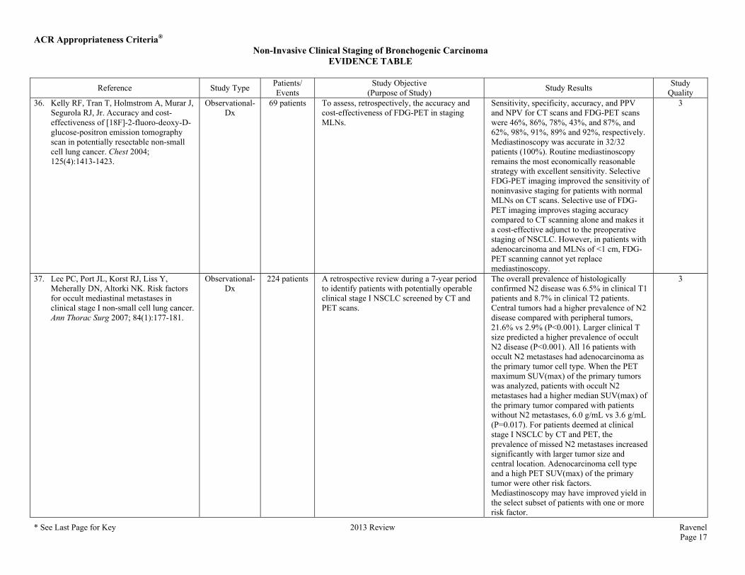
Quality 36. Kelly RF, Tran T, Holmstrom A, Murar J,
Segurola RJ, Jr. Accuracy and cost-effectiveness of [18F]-2-fluoro-deoxy-D-glucose-positron emission tomography scan in potentially resectable non-small cell lung cancer. Chest 2004; 125(4):1413-1423.
Observational-Dx
69 patients To assess, retrospectively, the accuracy and cost-effectiveness of FDG-PET in staging MLNs.
Sensitivity, specificity, accuracy, and PPV and NPV for CT scans and FDG-PET scans were 46%, 86%, 78%, 43%, and 87%, and 62%, 98%, 91%, 89% and 92%, respectively. Mediastinoscopy was accurate in 32/32 patients (100%). Routine mediastinoscopy remains the most economically reasonable strategy with excellent sensitivity. Selective FDG-PET imaging improved the sensitivity of noninvasive staging for patients with normal MLNs on CT scans. Selective use of FDG-PET imaging improves staging accuracy compared to CT scanning alone and makes it a cost-effective adjunct to the preoperative staging of NSCLC. However, in patients with adenocarcinoma and MLNs of <1 cm, FDG-PET scanning cannot yet replace mediastinoscopy.
3
37. Lee PC, Port JL, Korst RJ, Liss Y, Meherally DN, Altorki NK. Risk factors for occult mediastinal metastases in clinical stage I non-small cell lung cancer. Ann Thorac Surg 2007; 84(1):177-181.
Observational-Dx
224 patients A retrospective review during a 7-year period to identify patients with potentially operable clinical stage I NSCLC screened by CT and PET scans.
The overall prevalence of histologically confirmed N2 disease was 6.5% in clinical T1 patients and 8.7% in clinical T2 patients. Central tumors had a higher prevalence of N2 disease compared with peripheral tumors, 21.6% vs 2.9% (P<0.001). Larger clinical T size predicted a higher prevalence of occult N2 disease (P<0.001). All 16 patients with occult N2 metastases had adenocarcinoma as the primary tumor cell type. When the PET maximum SUV(max) of the primary tumors was analyzed, patients with occult N2 metastases had a higher median SUV(max) of the primary tumor compared with patients without N2 metastases, 6.0 g/mL vs 3.6 g/mL (P=0.017). For patients deemed at clinical stage I NSCLC by CT and PET, the prevalence of missed N2 metastases increased significantly with larger tumor size and central location. Adenocarcinoma cell type and a high PET SUV(max) of the primary tumor were other risk factors. Mediastinoscopy may have improved yield in the select subset of patients with one or more risk factor.
3
ACR Appropriateness Criteria® Non-Invasive Clinical Staging of Bronchogenic Carcinoma
EVIDENCE TABLE
* See Last Page for Key 2013 Review Ravenel Page 18
Reference Study Type Patients/ Events
Study Objective (Purpose of Study) Study Results Study
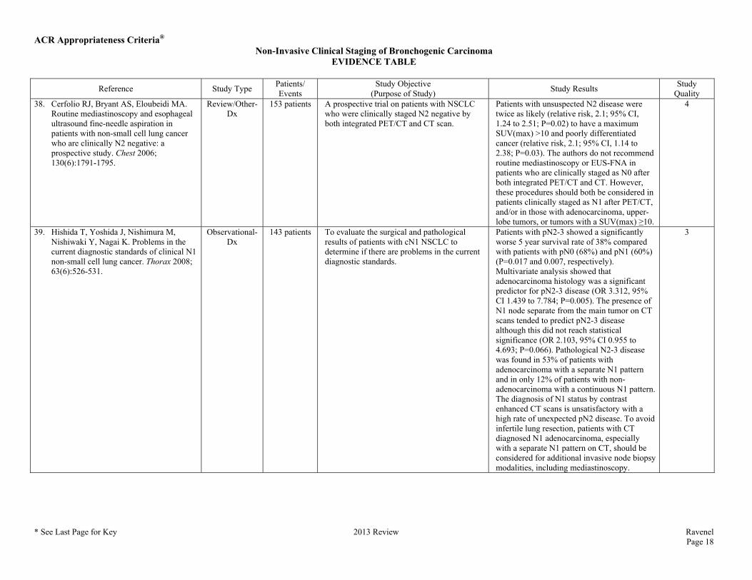
Quality 38. Cerfolio RJ, Bryant AS, Eloubeidi MA.
Routine mediastinoscopy and esophageal ultrasound fine-needle aspiration in patients with non-small cell lung cancer who are clinically N2 negative: a prospective study. Chest 2006; 130(6):1791-1795.
Review/Other-Dx
153 patients A prospective trial on patients with NSCLC who were clinically staged N2 negative by both integrated PET/CT and CT scan.
Patients with unsuspected N2 disease were twice as likely (relative risk, 2.1; 95% CI, 1.24 to 2.51; P=0.02) to have a maximum SUV(max) >10 and poorly differentiated cancer (relative risk, 2.1; 95% CI, 1.14 to 2.38; P=0.03). The authors do not recommend routine mediastinoscopy or EUS-FNA in patients who are clinically staged as N0 after both integrated PET/CT and CT. However, these procedures should both be considered in patients clinically staged as N1 after PET/CT, and/or in those with adenocarcinoma, upper-lobe tumors, or tumors with a SUV(max) ≥10.
4
39. Hishida T, Yoshida J, Nishimura M, Nishiwaki Y, Nagai K. Problems in the current diagnostic standards of clinical N1 non-small cell lung cancer. Thorax 2008; 63(6):526-531.
Observational-Dx
143 patients To evaluate the surgical and pathological results of patients with cN1 NSCLC to determine if there are problems in the current diagnostic standards.
Patients with pN2-3 showed a significantly worse 5 year survival rate of 38% compared with patients with pN0 (68%) and pN1 (60%) (P=0.017 and 0.007, respectively). Multivariate analysis showed that adenocarcinoma histology was a significant predictor for pN2-3 disease (OR 3.312, 95% CI 1.439 to 7.784; P=0.005). The presence of N1 node separate from the main tumor on CT scans tended to predict pN2-3 disease although this did not reach statistical significance (OR 2.103, 95% CI 0.955 to 4.693; P=0.066). Pathological N2-3 disease was found in 53% of patients with adenocarcinoma with a separate N1 pattern and in only 12% of patients with non-adenocarcinoma with a continuous N1 pattern. The diagnosis of N1 status by contrast enhanced CT scans is unsatisfactory with a high rate of unexpected pN2 disease. To avoid infertile lung resection, patients with CT diagnosed N1 adenocarcinoma, especially with a separate N1 pattern on CT, should be considered for additional invasive node biopsy modalities, including mediastinoscopy.
3
ACR Appropriateness Criteria® Non-Invasive Clinical Staging of Bronchogenic Carcinoma
EVIDENCE TABLE
* See Last Page for Key 2013 Review Ravenel Page 19
Reference Study Type Patients/ Events
Study Objective (Purpose of Study) Study Results Study
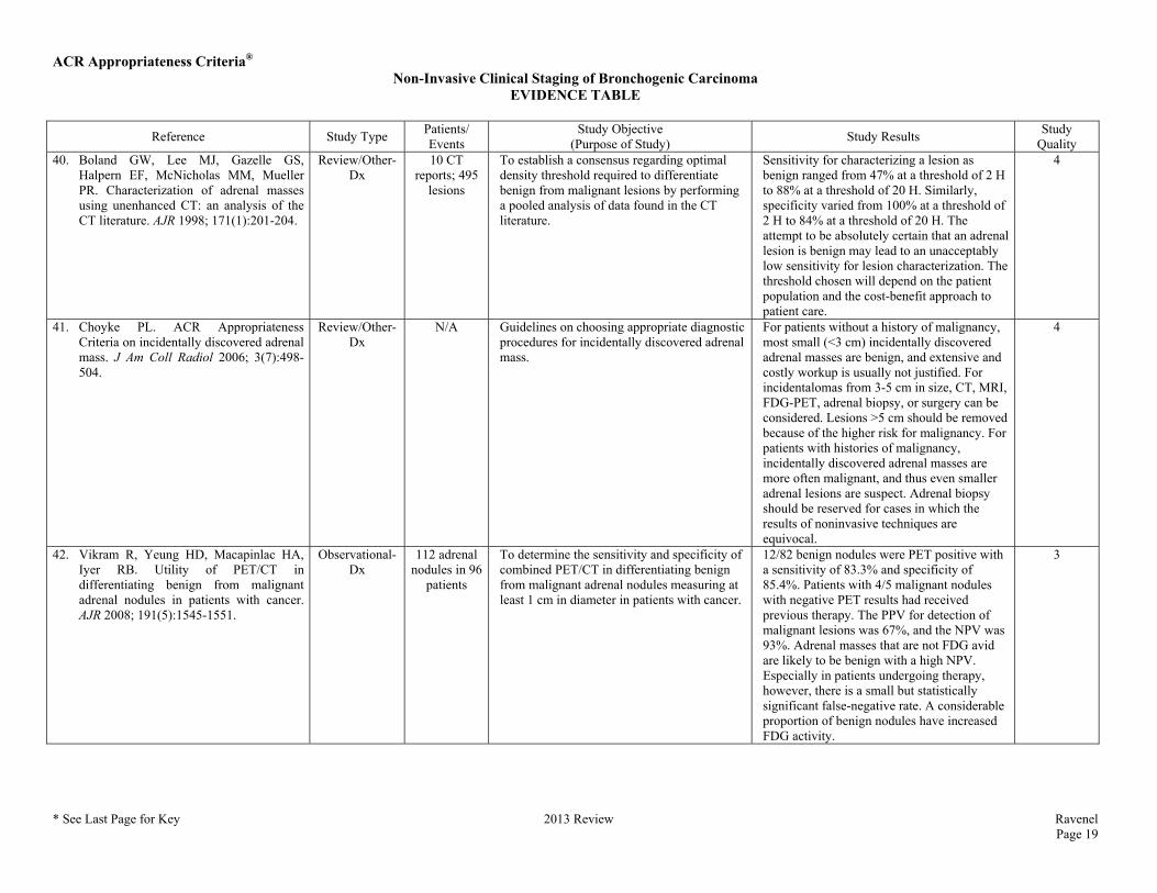
Quality 40. Boland GW, Lee MJ, Gazelle GS,
Halpern EF, McNicholas MM, Mueller PR. Characterization of adrenal masses using unenhanced CT: an analysis of the CT literature. AJR 1998; 171(1):201-204.
Review/Other-Dx
10 CT reports; 495
lesions
To establish a consensus regarding optimal density threshold required to differentiate benign from malignant lesions by performing a pooled analysis of data found in the CT literature.
Sensitivity for characterizing a lesion as benign ranged from 47% at a threshold of 2 H to 88% at a threshold of 20 H. Similarly, specificity varied from 100% at a threshold of 2 H to 84% at a threshold of 20 H. The attempt to be absolutely certain that an adrenal lesion is benign may lead to an unacceptably low sensitivity for lesion characterization. The threshold chosen will depend on the patient population and the cost-benefit approach to patient care.
4
41. Choyke PL. ACR Appropriateness Criteria on incidentally discovered adrenal mass. J Am Coll Radiol 2006; 3(7):498-504.
Review/Other-Dx
N/A Guidelines on choosing appropriate diagnostic procedures for incidentally discovered adrenal mass.
For patients without a history of malignancy, most small (<3 cm) incidentally discovered adrenal masses are benign, and extensive and costly workup is usually not justified. For incidentalomas from 3-5 cm in size, CT, MRI, FDG-PET, adrenal biopsy, or surgery can be considered. Lesions >5 cm should be removed because of the higher risk for malignancy. For patients with histories of malignancy, incidentally discovered adrenal masses are more often malignant, and thus even smaller adrenal lesions are suspect. Adrenal biopsy should be reserved for cases in which the results of noninvasive techniques are equivocal.
4
42. Vikram R, Yeung HD, Macapinlac HA, Iyer RB. Utility of PET/CT in differentiating benign from malignant adrenal nodules in patients with cancer. AJR 2008; 191(5):1545-1551.
Observational-Dx
112 adrenal nodules in 96
patients
To determine the sensitivity and specificity of combined PET/CT in differentiating benign from malignant adrenal nodules measuring at least 1 cm in diameter in patients with cancer.
12/82 benign nodules were PET positive with a sensitivity of 83.3% and specificity of 85.4%. Patients with 4/5 malignant nodules with negative PET results had received previous therapy. The PPV for detection of malignant lesions was 67%, and the NPV was 93%. Adrenal masses that are not FDG avid are likely to be benign with a high NPV. Especially in patients undergoing therapy, however, there is a small but statistically significant false-negative rate. A considerable proportion of benign nodules have increased FDG activity.
3
ACR Appropriateness Criteria® Non-Invasive Clinical Staging of Bronchogenic Carcinoma
EVIDENCE TABLE
* See Last Page for Key 2013 Review Ravenel Page 20
Reference Study Type Patients/ Events
Study Objective (Purpose of Study) Study Results Study
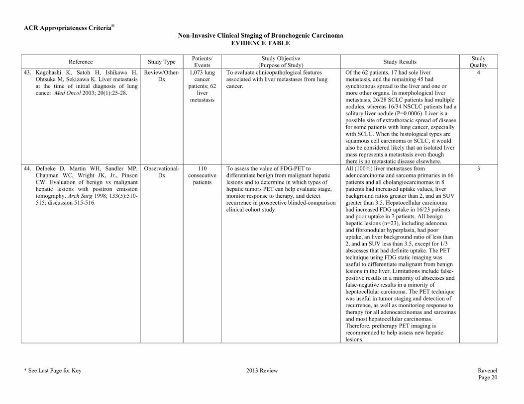
Quality 43. Kagohashi K, Satoh H, Ishikawa H,
Ohtsuka M, Sekizawa K. Liver metastasis at the time of initial diagnosis of lung cancer. Med Oncol 2003; 20(1):25-28.
Review/Other-Dx
1,073 lung cancer
patients; 62 liver
metastasis
To evaluate clinicopathological features associated with liver metastases from lung cancer.
Of the 62 patients, 17 had sole liver metastasis, and the remaining 45 had synchronous spread to the liver and one or more other organs. In morphological liver metastasis, 26/28 SCLC patients had multiple nodules, whereas 16/34 NSCLC patients had a solitary liver nodule (P=0.0006). Liver is a possible site of extrathoracic spread of disease for some patients with lung cancer, especially with SCLC. When the histological types are squamous cell carcinoma or SCLC, it would also be considered likely that an isolated liver mass represents a metastasis even though there is no metastatic disease elsewhere.
4
44. Delbeke D, Martin WH, Sandler MP, Chapman WC, Wright JK, Jr., Pinson CW. Evaluation of benign vs malignant hepatic lesions with positron emission tomography. Arch Surg 1998; 133(5):510-515; discussion 515-516.
Observational-Dx
110 consecutive
patients
To assess the value of FDG-PET to differentiate benign from malignant hepatic lesions and to determine in which types of hepatic tumors PET can help evaluate stage, monitor response to therapy, and detect recurrence in prospective blinded-comparison clinical cohort study.
All (100%) liver metastases from adenocarcinoma and sarcoma primaries in 66 patients and all cholangiocarcinomas in 8 patients had increased uptake values, liver background ratios greater than 2, and an SUV greater than 3.5. Hepatocellular carcinoma had increased FDG uptake in 16/23 patients and poor uptake in 7 patients. All benign hepatic lesions (n=23), including adenoma and fibronodular hyperplasia, had poor uptake, an liver background ratio of less than 2, and an SUV less than 3.5, except for 1/3 abscesses that had definite uptake. The PET technique using FDG static imaging was useful to differentiate malignant from benign lesions in the liver. Limitations include false-positive results in a minority of abscesses and false-negative results in a minority of hepatocellular carcinoma. The PET technique was useful in tumor staging and detection of recurrence, as well as monitoring response to therapy for all adenocarcinomas and sarcomas and most hepatocellular carcinomas. Therefore, pretherapy PET imaging is recommended to help assess new hepatic lesions.
3
ACR Appropriateness Criteria® Non-Invasive Clinical Staging of Bronchogenic Carcinoma
EVIDENCE TABLE
* See Last Page for Key 2013 Review Ravenel Page 21
Reference Study Type Patients/ Events
Study Objective (Purpose of Study) Study Results Study
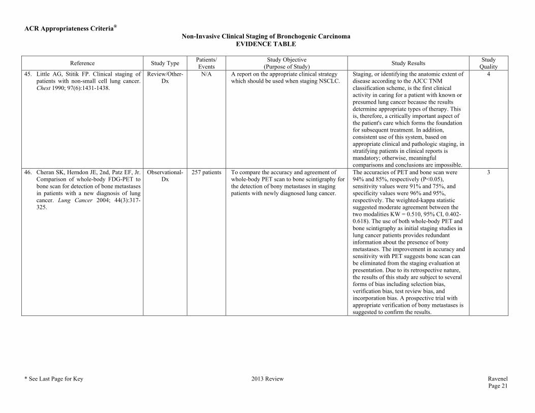
Quality 45. Little AG, Stitik FP. Clinical staging of
patients with non-small cell lung cancer. Chest 1990; 97(6):1431-1438.
Review/Other-Dx
N/A A report on the appropriate clinical strategy which should be used when staging NSCLC.
Staging, or identifying the anatomic extent of disease according to the AJCC TNM classification scheme, is the first clinical activity in caring for a patient with known or presumed lung cancer because the results determine appropriate types of therapy. This is, therefore, a critically important aspect of the patient's care which forms the foundation for subsequent treatment. In addition, consistent use of this system, based on appropriate clinical and pathologic staging, in stratifying patients in clinical reports is mandatory; otherwise, meaningful comparisons and conclusions are impossible.
4
46. Cheran SK, Herndon JE, 2nd, Patz EF, Jr. Comparison of whole-body FDG-PET to bone scan for detection of bone metastases in patients with a new diagnosis of lung cancer. Lung Cancer 2004; 44(3):317-325.
Observational-Dx
257 patients To compare the accuracy and agreement of whole-body PET scan to bone scintigraphy for the detection of bony metastases in staging patients with newly diagnosed lung cancer.
The accuracies of PET and bone scan were 94% and 85%, respectively (P<0.05), sensitivity values were 91% and 75%, and specificity values were 96% and 95%, respectively. The weighted-kappa statistic suggested moderate agreement between the two modalities KW = 0.510, 95% CI, 0.402-0.618). The use of both whole-body PET and bone scintigraphy as initial staging studies in lung cancer patients provides redundant information about the presence of bony metastases. The improvement in accuracy and sensitivity with PET suggests bone scan can be eliminated from the staging evaluation at presentation. Due to its retrospective nature, the results of this study are subject to several forms of bias including selection bias, verification bias, test review bias, and incorporation bias. A prospective trial with appropriate verification of bony metastases is suggested to confirm the results.
3
ACR Appropriateness Criteria® Non-Invasive Clinical Staging of Bronchogenic Carcinoma
EVIDENCE TABLE
* See Last Page for Key 2013 Review Ravenel Page 22
Reference Study Type Patients/ Events
Study Objective (Purpose of Study) Study Results Study
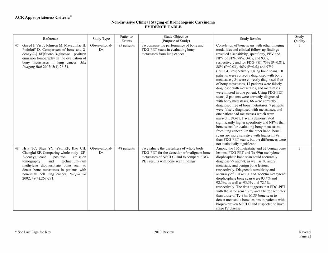
Quality 47. Gayed I, Vu T, Johnson M, Macapinlac H,
Podoloff D. Comparison of bone and 2-deoxy-2-[18F]fluoro-D-glucose positron emission tomography in the evaluation of bony metastases in lung cancer. Mol Imaging Biol 2003; 5(1):26-31.
Observational-Dx
85 patients To compare the performance of bone and FDG-PET scans in evaluating bony metastases from lung cancer.
Correlation of bone scans with other imaging modalities and clinical follow-up findings revealed a sensitivity, specificity, PPV and NPV of 81%, 78%, 34%, and 93%, respectively and for FDG-PET 73% (P=0.81), 88% (P=0.03), 46% (P=0.5,) and 97% (P=0.04), respectively. Using bone scans, 10 patients were correctly diagnosed with bony metastases, 54 were correctly diagnosed free of bony metastases, 17 patients were falsely diagnosed with metastases, and metastases were missed in one patient. Using FDG-PET scans, 8 patients were correctly diagnosed with bony metastases, 66 were correctly diagnosed free of bony metastases, 7 patients were falsely diagnosed with metastases, and one patient had metastases which were missed. FDG-PET scans demonstrated significantly higher specificity and NPVs than bone scans for evaluating bony metastases from lung cancer. On the other hand, bone scans are more sensitive with higher PPVs than FDG-PET scans, but the differences were not statistically significant.
3
48. Hsia TC, Shen YY, Yen RF, Kao CH, Changlai SP. Comparing whole body 18F-2-deoxyglucose positron emission tomography and technetium-99m methylene diophosphate bone scan to detect bone metastases in patients with non-small cell lung cancer. Neoplasma 2002; 49(4):267-271.
Observational-Dx
48 patients To evaluate the usefulness of whole body FDG-PET for the detection of malignant bone metastases of NSCLC, and to compare FDG-PET results with bone scan findings.
Among the 106 metastatic and 32 benign bone lesions, FDG-PET and Tc-99m methylene diophosphate bone scan could accurately diagnose 99 and 98, as well as 30 and 2 metastatic and benign bone lesions, respectively. Diagnostic sensitivity and accuracy of FDG-PET and Tc-99m methylene diophosphate bone scan were 93.4% and 92.5%, as well as 93.5% and 72.5%, respectively. The data suggests that FDG-PET with the same sensitivity and a better accuracy than those of Tc-99m MDP bone scan to detect metastatic bone lesions in patients with biopsy-proven NSCLC and suspected to have stage IV disease.
3
ACR Appropriateness Criteria® Non-Invasive Clinical Staging of Bronchogenic Carcinoma
EVIDENCE TABLE
* See Last Page for Key 2013 Review Ravenel Page 23
Reference Study Type Patients/ Events
Study Objective (Purpose of Study) Study Results Study
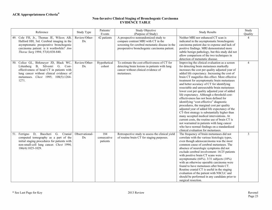
Quality 49. Cole FH, Jr., Thomas JE, Wilcox AB,
Halford HH, 3rd. Cerebral imaging in the asymptomatic preoperative bronchogenic carcinoma patient: is it worthwhile? Ann Thorac Surg 1994; 57(4):838-840.
Review/Other-Dx
42 patients A prospective nonrandomized study to compare contrast MRI with CT in the screening for cerebral metastatic disease in the preoperative bronchogenic carcinoma patient.
Neither MRI nor enhanced CT scan is indicated in the asymptomatic bronchogenic carcinoma patient due to expense and lack of positive findings. MRI demonstrated more subtle benign pathology, but this study did not allow comparison of the two techniques in detection of metastatic disease.
4
50. Colice GL, Birkmeyer JD, Black WC, Littenberg B, Silvestri G. Cost-effectiveness of head CT in patients with lung cancer without clinical evidence of metastases. Chest 1995; 108(5):1264-1271.
Review/Other-Dx
Hypothetical cohort
To estimate the cost-effectiveness of CT for detecting brain lesions in patients with lung cancer without clinical evidence of metastases.
Improving the clinical evaluation as a screen for detecting brain metastases markedly increases the cost per quality adjusted year of added life expectancy. Increasing the cost of brain CT magnifies this effect. More effective treatment for asymptomatic brain metastases and better accuracy of CT for identifying resectable and unresectable brain metastases lower cost per quality adjusted year of added life expectancy. Although a threshold cost-effectiveness has not been defined for identifying “cost-effective” diagnostic procedures, the marginal cost per quality adjusted year of added life expectancy of the CT-first strategy is substantially higher than many accepted medical interventions. At current costs, the routine use of brain CT is not warranted in patients with lung cancer who have normal findings on a standardized clinical evaluation for metastases.
4
51. Ferrigno D, Buccheri G. Cranial computed tomography as a part of the initial staging procedures for patients with non-small-cell lung cancer. Chest 1994; 106(4):1025-1029.
Observational-Dx
184 consecutive
patients
Retrospective study to assess the clinical yield of routine brain CT for staging purposes.
The frequency of brain metastases did not correlate with the various histologic types, even though adenocarcinoma was the most common cause of cerebral metastases. The absence of neurologic symptoms did not exclude cerebral involvement: 16/25 patients with positive brain CT scans were asymptomatic (64%). 3/31 subjects (10%) with an otherwise operable carcinoma were found to have metastases after brain CT. Routine cranial CT is useful in the staging evaluation of the patient with NSCLC and should be performed in any candidate prior to surgical resection.
3
ACR Appropriateness Criteria® Non-Invasive Clinical Staging of Bronchogenic Carcinoma
EVIDENCE TABLE
* See Last Page for Key 2013 Review Ravenel Page 24
Reference Study Type Patients/ Events
Study Objective (Purpose of Study) Study Results Study
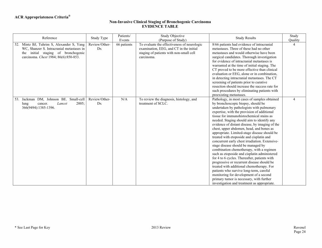
Quality 52. Mintz BJ, Tuhrim S, Alexander S, Yang
WC, Shanzer S. Intracranial metastases in the initial staging of bronchogenic carcinoma. Chest 1984; 86(6):850-853.
Review/Other-Dx
66 patients To evaluate the effectiveness of neurologic examination, EEG, and CT in the initial staging of patients with non-small cell carcinoma.
8/66 patients had evidence of intracranial metastases. Three of these had no other metastases and would otherwise have been surgical candidates. Thorough investigation for evidence of intracranial metastases is warranted at the time of initial staging. The CT proved to be more effective than clinical evaluation or EEG, alone or in combination, in detecting intracranial metastases. The CT screening of patients prior to curative resection should increase the success rate for such procedures by eliminating patients with preexisting metastases.
4
53. Jackman DM, Johnson BE. Small-cell lung cancer. Lancet 2005; 366(9494):1385-1396.
Review/Other-Dx
N/A To review the diagnosis, histology, and treatment of SCLC.
Pathology, in most cases of samples obtained by bronchoscopic biopsy, should be undertaken by pathologists with pulmonary expertise, with the provision of additional tissue for immunohistochemical stains as needed. Staging should aim to identify any evidence of distant disease, by imaging of the chest, upper abdomen, head, and bones as appropriate. Limited-stage disease should be treated with etoposide and cisplatin and concurrent early chest irradiation. Extensive-stage disease should be managed by combination chemotherapy, with a regimen such as etoposide and cisplatin administered for 4 to 6 cycles. Thereafter, patients with progressive or recurrent disease should be treated with additional chemotherapy. For patients who survive long-term, careful monitoring for development of a second primary tumor is necessary, with further investigation and treatment as appropriate.
4
ACR Appropriateness Criteria® Non-Invasive Clinical Staging of Bronchogenic Carcinoma
EVIDENCE TABLE
* See Last Page for Key 2013 Review Ravenel Page 25
Reference Study Type Patients/ Events
Study Objective (Purpose of Study) Study Results Study
Quality 54. Sher T, Dy GK, Adjei AA. Small cell lung
cancer. Mayo Clin Proc 2008; 83(3):355-367.
Review/Other-Tx
N/A To review treatment of SCLS. Because of the high propensity of SCLS to metastasize early, surgery has a limited role as primary therapy. The combination of platinum and etoposide is the accepted standard chemotherapeutic regimen. It is also the accepted standard therapy in combination with thoracic radiotherapy for limited-stage disease. Thoracic radiotherapy administered concurrently with chemotherapy is more efficacious than sequential therapy. Furthermore, the survival benefit is greater if thoracic radiotherapy is given early rather than late in the course of chemotherapy. Regardless of disease stage, no relevant survival benefit results from increased chemotherapy dose intensity or dose density, altered mode of administration (eg, alternating or sequential administration) of various chemotherapeutic agents, or maintenance chemotherapy.
4
55. Stahel RA, Ginsberg R, Havemann K, et al. Staging and prognostic factors in small cell lung cancer: a consensus report. Lung Cancer 1989; 5(4-6):119-126.
Review/Other-Dx
N/A Outlines the consensus about staging in SCLS formed at the 3rd International Workshop on Small Cell Lung Cancer and updates the first consensus report published in 1983.
In clinical practice outside a trial setting patient history, physical examination, histological or cytological confirmation of SCLS, chest radiograph, hematology with complete blood count, biochemistry including electrolytes, liver function tests, alkaline phosphatase and creatinine are often sufficient for staging workup. For clinical trials a continuation of the anatomical staging with examination of the major extrathoracic systems is recommended.
4
56. Vallieres E, Shepherd FA, Crowley J, et al. The IASLC Lung Cancer Staging Project: proposals regarding the relevance of TNM in the pathologic staging of small cell lung cancer in the forthcoming (seventh) edition of the TNM classification for lung cancer. J Thorac Oncol 2009; 4(9):1049-1059.
Review/Other-Tx
349 cases of resected SCLC
To examine the impact of the TNM system on the pathologic staging of SCLC and to assess the new IASLC proposals in resected SCLC.
Survival after resection correlated with both T and N category with nodal status having a stronger influence on survival. Stage groupings using the 6th edition of TNM clearly identify patient subgroups with different prognoses. The IASLC proposals for the 7th edition of TNM classification also apply to this population and to the SEER database. This analysis further strengthens the previous recommendation to use TNM staging for all SCLC cases.
4
ACR Appropriateness Criteria® Non-Invasive Clinical Staging of Bronchogenic Carcinoma
EVIDENCE TABLE
* See Last Page for Key 2013 Review Ravenel Page 26
Reference Study Type Patients/ Events
Study Objective (Purpose of Study) Study Results Study
Quality 57. Mirvis SE, Whitley NO, Aisner J, Moody
M, Whitacre M, Whitley JE. Abdominal CT in the staging of small-cell carcinoma of the lung: incidence of metastases and effect on prognosis. AJR 1987; 148(5):845-847.
Observational-Dx
72 patients Retrospective study to assess the role of abdominal CT in staging.
Statistical analysis of patients with extensive disease revealed a significant increase in complete therapeutic response (P=.0054) and in the length of survival (P=.001) among those who had extensive disease without abdominal metastases as compared with those who had abdominal metastases on their initial abdominal CT examination. The development of new or recurrent abdominal metastases in general or in specific organs on follow-up scans obtained in 35 patients did not significantly decrease their survival time as compared with that of patients without such metastases. The findings suggest that CT of the abdomen is beneficial in the initial staging of patients with SCLC and provides prognostic information concerning response to therapy and length of survival.
3
58. Whitley NO, Mirvis SE. Abdominal CT in the staging of small cell carcinoma of the lung. Crit Rev Diagn Imaging 1989; 29(2):103-116.
Review/Other-Dx
N/A Review of abdominal CT in staging SCLC. CT scanning has made a valuable contribution to the delineation of intrathoracic and metastatic disease and is now included in the staging workup of patients with SCLC since metastatic involvement of the liver, bone, bone marrow, central nervous system, and adrenal glands is common.
4
59. Simon GR, Turrisi A. Management of small cell lung cancer: ACCP evidence-based clinical practice guidelines (2nd edition). Chest 2007; 132(3 Suppl):324S-339S.
Review/Other-Tx
N/A Guideline for the management of patients with SCLS.
Limited-stage disease is treated with curative intent with chemotherapy and radiation therapy, with approximately 20% of patients achieving a cure. For all patients with limited-stage disease, median survival is 16-22 months. Extensive-stage disease is primarily treated with chemotherapy with a high initial response rate of 60%-70% but with a median survival of 10 months. All patients achieving a complete remission should be offered prophylactic cranial irradiation. Relapsed or refractory SCLC has a uniformly poor prognosis.
4
ACR Appropriateness Criteria® Non-Invasive Clinical Staging of Bronchogenic Carcinoma
EVIDENCE TABLE
* See Last Page for Key 2013 Review Ravenel Page 27
Reference Study Type Patients/ Events
Study Objective (Purpose of Study) Study Results Study
Quality 60. Fischer BM, Mortensen J, Langer SW, et
al. A prospective study of PET/CT in initial staging of small-cell lung cancer: comparison with CT, bone scintigraphy and bone marrow analysis. Ann Oncol 2007; 18(2):338-345.
Observational-Dx
34 consecutive
patients
A prospective study to examine the role of combined PET/CT compared with standard staging (CT, bone scintigraphy and immunocytochemical assessment of bone marrow biopsy) of patients with SCLC.
The sensitivity for accurate staging of patients with extensive disease was the following: for standard staging 79%, PET 93% and PET/CT 93%. Specificity was 100%, 83% and 100%, respectively. The results from this first study on PET/CT in SCLC indicate that PET/CT can simplify and perhaps even improve the accuracy of the current staging procedure in SCLC. A larger clinical trial, preferably with consequent histological confirmation in case of discordance, however, is warranted.
3
61. Bradley JD, Dehdashti F, Mintun MA, Govindan R, Trinkaus K, Siegel BA. Positron emission tomography in limited-stage small-cell lung cancer: a prospective study. J Clin Oncol 2004; 22(16):3248-3254.
Observational-Dx
24 patients To determine how often FDG-PET detects extensive-stage SCLC in patients considered to have limited-stage disease based on conventional staging procedures, and to determine the impact of PET on treatment planning for presumed limited-stage SCLC.
FDG-PET demonstrated findings consistent with extensive-stage SCLC in 3/24 patients. FDG-PET correctly upstaged two (8.3%) of 24 patients to extensive-stage disease (95% CI, 1.03% to 27.0%). PET correctly identified tumor in each SCLC mass (primary or nodal) that was suspected on CT, thus giving a lesion-based sensitivity relative to CT of 100%. PET identified unsuspected regional nodal metastasis in 6 (25%) of 24 patients, and the radiation therapy plan was significantly altered to include the PET-positive/CT-negative nodes within the high-dose region in each of these patients. FDG-PET has high sensitivity for SCLC and appears to be of value for initial staging and treatment planning of patients with presumed limited-stage disease.
2
62. Brink I, Schumacher T, Mix M, et al. Impact of [18F]FDG-PET on the primary staging of small-cell lung cancer. Eur J Nucl Med Mol Imaging 2004; 31(12):1614-1620.
Observational-Dx
120 consecutive
patients
To evaluate the impact of FDG-PET on the primary staging of patients with SCLC.
Sensitivity of FDG-PET was significantly superior to that of CT in the detection of extrathoracic lymph node involvement (100% vs 70%, specificity 98% vs 94%) and distant metastases except to the brain (98% vs 83%, specificity 92% vs 79%). However, FDG-PET was significantly less sensitive than cranial MRI/CT in the detection of brain metastases (46% vs 100%, specificity 97% vs 100%). The introduction of FDG-PET in the diagnostic evaluation of SCLC will improve the staging results and affect patient management, and may reduce the number of tests and invasive procedures.
2
ACR Appropriateness Criteria® Non-Invasive Clinical Staging of Bronchogenic Carcinoma
EVIDENCE TABLE
* See Last Page for Key 2013 Review Ravenel Page 28
Reference Study Type Patients/ Events
Study Objective (Purpose of Study) Study Results Study
Quality 63. Bradley J, Thorstad WL, Mutic S, et al.
Impact of FDG-PET on radiation therapy volume delineation in non-small-cell lung cancer. Int J Radiat Oncol Biol Phys 2004; 59(1):78-86.
Review/Other-Dx
26 patients To determine the impact of treatment simulation with FDG-PET and CT on radiation therapy target volume definition and toxicity profiles by comparison to simulation with CT scanning alone.
The FDG-PET findings altered the AJCC TNM stage in 8/26 (31%) patients; 2 patients were diagnosed with metastatic disease based on FDG-PET and received palliative radiation therapy. Of the 24 patients who were planned with 3D conformal radiation therapy, PET clearly altered the radiation therapy volume in 14 (58%), as follows. PET helped to distinguish tumor from atelectasis in all 3 patients with atelectasis. Unsuspected nodal disease was detected by PET in 10 patients, and 1 patient had a separate tumor focus detected within the same lobe of the lung. Increases in the target volumes led to increases in the mean lung dose, V20, and mean esophageal dose. Decreases in the target volumes in the patients with atelectasis led to decreases in these normal-tissue toxicity parameters.
4
64. van Loon J, Offermann C, Bosmans G, et al. 18FDG-PET based radiation planning of mediastinal lymph nodes in limited disease small cell lung cancer changes radiotherapy fields: a planning study. Radiother Oncol 2008; 87(1):49-54.
Observational-Dx
21 patients To investigate the influence of selective irradiation of FDG-PET positive mediastinal nodes on radiation fields and normal tissue exposure in limited disease SCLC.
There were no significant differences in gross tumor volume, lung, and esophageal parameters between CT- and PET-based plans. Incorporating FDG-PET information in radiotherapy planning for patients with limited disease SCLC changed the treatment plan in 24% of patients compared to CT. Both increases and decreases of the gross tumor volume were observed, theoretically leading to the avoidance of geographical miss or a decrease of radiation exposure of normal tissues, respectively. Based on these findings, a phase II trial, evaluating PET-scan based selective nodal irradiation, is ongoing.
3
ACR Appropriateness Criteria® Non-Invasive Clinical Staging of Bronchogenic Carcinoma
EVIDENCE TABLE
* See Last Page for Key 2013 Review Ravenel Page 29
Reference Study Type Patients/ Events
Study Objective (Purpose of Study) Study Results Study
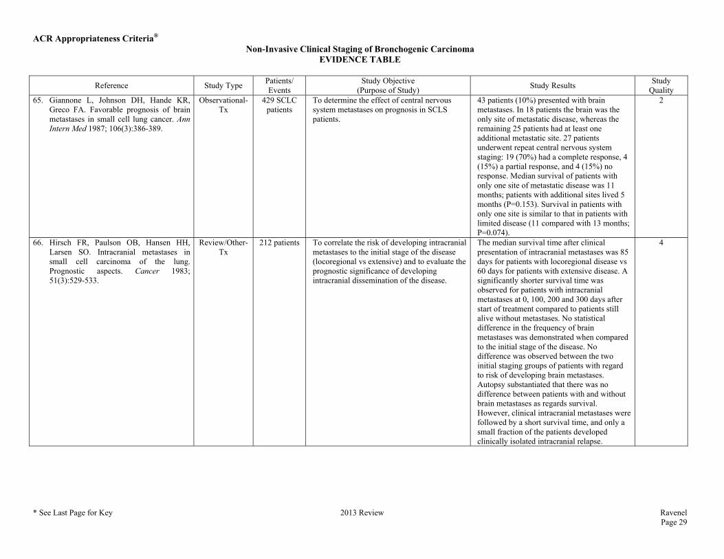
Quality 65. Giannone L, Johnson DH, Hande KR,
Greco FA. Favorable prognosis of brain metastases in small cell lung cancer. Ann Intern Med 1987; 106(3):386-389.
Observational-Tx
429 SCLC patients
To determine the effect of central nervous system metastases on prognosis in SCLS patients.
43 patients (10%) presented with brain metastases. In 18 patients the brain was the only site of metastatic disease, whereas the remaining 25 patients had at least one additional metastatic site. 27 patients underwent repeat central nervous system staging: 19 (70%) had a complete response, 4 (15%) a partial response, and 4 (15%) no response. Median survival of patients with only one site of metastatic disease was 11 months; patients with additional sites lived 5 months (P=0.153). Survival in patients with only one site is similar to that in patients with limited disease (11 compared with 13 months; P=0.074).
2
66. Hirsch FR, Paulson OB, Hansen HH, Larsen SO. Intracranial metastases in small cell carcinoma of the lung. Prognostic aspects. Cancer 1983; 51(3):529-533.
Review/Other-Tx
212 patients To correlate the risk of developing intracranial metastases to the initial stage of the disease (locoregional vs extensive) and to evaluate the prognostic significance of developing intracranial dissemination of the disease.
The median survival time after clinical presentation of intracranial metastases was 85 days for patients with locoregional disease vs 60 days for patients with extensive disease. A significantly shorter survival time was observed for patients with intracranial metastases at 0, 100, 200 and 300 days after start of treatment compared to patients still alive without metastases. No statistical difference in the frequency of brain metastases was demonstrated when compared to the initial stage of the disease. No difference was observed between the two initial staging groups of patients with regard to risk of developing brain metastases. Autopsy substantiated that there was no difference between patients with and without brain metastases as regards survival. However, clinical intracranial metastases were followed by a short survival time, and only a small fraction of the patients developed clinically isolated intracranial relapse.
4
ACR Appropriateness Criteria® Non-Invasive Clinical Staging of Bronchogenic Carcinoma
EVIDENCE TABLE
* See Last Page for Key 2013 Review Ravenel Page 30
Reference Study Type Patients/ Events
Study Objective (Purpose of Study) Study Results Study
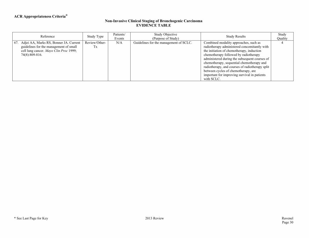
Quality 67. Adjei AA, Marks RS, Bonner JA. Current
guidelines for the management of small cell lung cancer. Mayo Clin Proc 1999; 74(8):809-816.
Review/Other-Tx
N/A Guidelines for the management of SCLC. Combined modality approaches, such as radiotherapy administered concomitantly with the initiation of chemotherapy, induction chemotherapy followed by radiotherapy administered during the subsequent courses of chemotherapy, sequential chemotherapy and radiotherapy, and courses of radiotherapy split between cycles of chemotherapy, are important for improving survival in patients with SCLC.
4
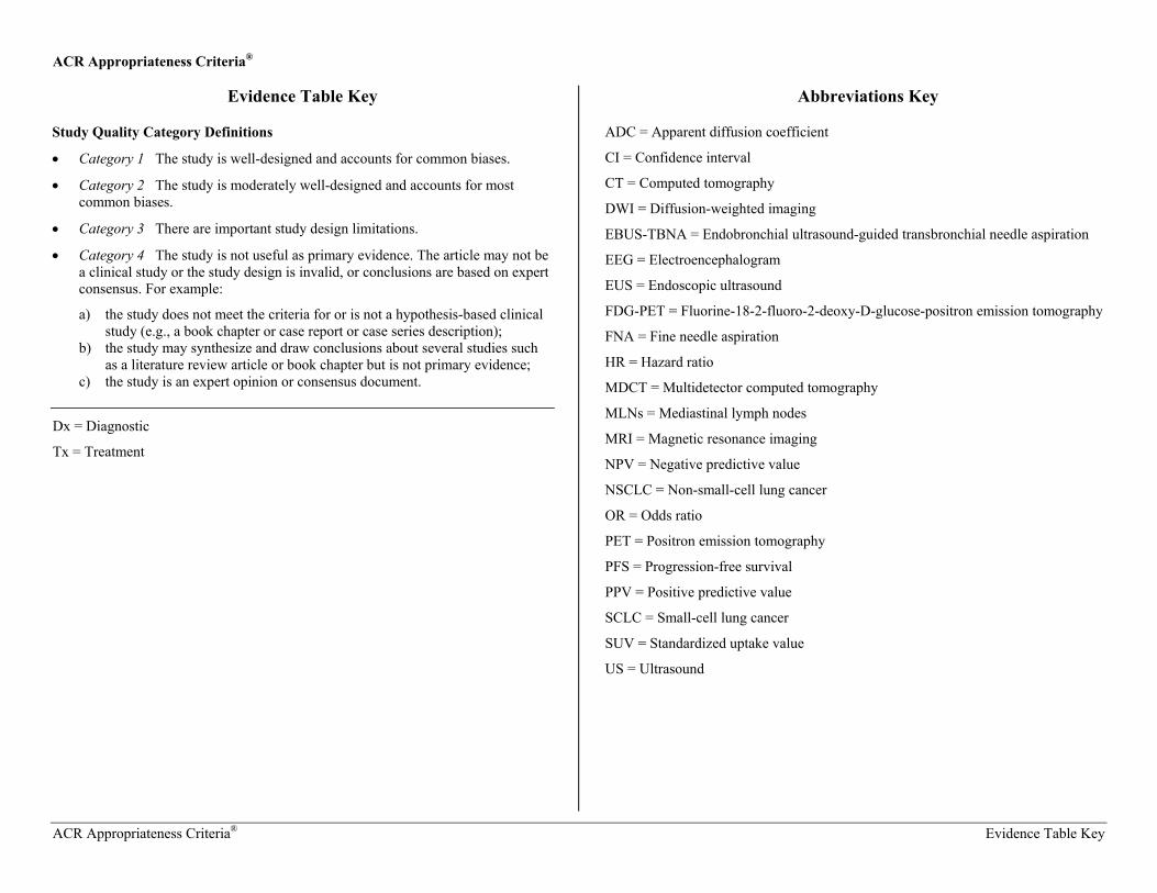
ACR Appropriateness Criteria®
ACR Appropriateness Criteria® Evidence Table Key
Evidence Table Key
Study Quality Category Definitions
Category 1 The study is well-designed and accounts for common biases.
Category 2 The study is moderately well-designed and accounts for most common biases.
Category 3 There are important study design limitations.
Category 4 The study is not useful as primary evidence. The article may not be a clinical study or the study design is invalid, or conclusions are based on expert consensus. For example:
a) the study does not meet the criteria for or is not a hypothesis-based clinical study (e.g., a book chapter or case report or case series description);
b) the study may synthesize and draw conclusions about several studies such as a literature review article or book chapter but is not primary evidence;
c) the study is an expert opinion or consensus document.
Dx = Diagnostic
Tx = Treatment
Abbreviations Key
ADC = Apparent diffusion coefficient
CI = Confidence interval
CT = Computed tomography
DWI = Diffusion-weighted imaging
EBUS-TBNA = Endobronchial ultrasound-guided transbronchial needle aspiration
EEG = Electroencephalogram
EUS = Endoscopic ultrasound
FDG-PET = Fluorine-18-2-fluoro-2-deoxy-D-glucose-positron emission tomography
FNA = Fine needle aspiration
HR = Hazard ratio
MDCT = Multidetector computed tomography
MLNs = Mediastinal lymph nodes
MRI = Magnetic resonance imaging
NPV = Negative predictive value
NSCLC = Non-small-cell lung cancer
OR = Odds ratio
PET = Positron emission tomography
PFS = Progression-free survival
PPV = Positive predictive value
SCLC = Small-cell lung cancer
SUV = Standardized uptake value
US = Ultrasound