Embed Size (px)
Citation preview

International Journal of
Molecular Sciences
Review
Notch Signaling in Endothelial Cells: Is It theTherapeutic Target for VascularNeointimal Hyperplasia?
Ding-Yuan Tian 1,2, Xu-Rui Jin 1,2, Xi Zeng 2 and Yun Wang 2,*1 Trainee Brigade, Third Military Medical University, Chongqing 400038, China;
[email protected] (D.-Y.T.); [email protected] (X.-R.J.)2 Department of Cell Biology, Third Military Medical University, Chongqing 400038, China; [email protected]* Correspondence: [email protected]; Tel.: +86-23-6875-2252
Received: 19 June 2017; Accepted: 21 July 2017; Published: 25 July 2017
Abstract: Blood vessels respond to injury through a healing process that includes neointimalhyperplasia. The vascular endothelium is a monolayer of cells that separates the outer vascularwall from the inner circulating blood. The disruption and exposure of endothelial cells (ECs) tosubintimal components initiate the neointimal formation. ECs not only act as a highly selectivebarrier to prevent early pathological changes of neointimal hyperplasia, but also synthesize andrelease molecules to maintain vascular homeostasis. After vascular injury, ECs exhibit variedresponses, including proliferation, regeneration, apoptosis, phenotypic switching, interacting withother cells by direct contact or secreted molecules and the change of barrier function. This briefreview presents the functional role of the evolutionarily-conserved Notch pathway in neointimalhyperplasia, notably by regulating endothelial cell functions (proliferation, regeneration, apoptosis,differentiation, cell-cell interaction). Understanding endothelial cell biology should help us definemethods to prompt cell proliferation, prevent cell apoptosis and dysfunction, block neointimalhyperplasia and vessel narrowing.
Keywords: neointimal hyperplasia; vascular injury; endothelial cell; Notch signaling;biological function
1. Introduction
Neointimal hyperplasia is an exaggerated wound-healing process that occurs in the vessel wallafter injury. As a major morphological feature of many cardiovascular diseases (CVD), such asatherosclerosis and hypertension, neointimal hyperplasia is also responsible for the stenosis of vascularsurgery, including bypass grafting, angioplasty and arteriovenous fistula [1,2]. The development ofneointimal hyperplasia is a complex process initiated by the damage of endothelial cells (ECs) andexposure of vascular smooth muscle cells (VSMCs) to circulating blood elements. The process isfurther characterized by proliferative and inflammatory responses including VSMC proliferation andmigration, platelet aggregation, leukocyte recruitment and extracellular matrix (ECM) deposition.Finally, EC proliferation or regeneration occurs at the lesion [3].
One of the important candidates for triggering neointimal formation is the dysfunction ofendothelium. In the cardiovascular system, the endothelium is not only a barrier between thecirculating blood and VSMCs, but also, it releases factors that regulate vascular tone, vessel growth,platelet function and coagulation [4]. For the underlying VSMCs, ECs could harmonize their growthand regression, through direct contact with VSMCs or secreted mediators that affect their proliferation,migration and death. In addition, ECs can regulate the thickness of intimal ECM through secreting
Int. J. Mol. Sci. 2017, 18, 1615; doi:10.3390/ijms18081615 www.mdpi.com/journal/ijms

Int. J. Mol. Sci. 2017, 18, 1615 2 of 18
enzymes, or inhibitors of these enzymes, which are able to degrade its components. The balance ofthese endothelial-derived activities regulates vessel development and vascular remodeling [5].
Recent advances in the understanding of the biology of neointimal formation indicate thatECs play a central role in the development of intimal hyperplasia during the process of vascularreconstruction. However, the mechanism of vascular neointimal hyperplasia is complicated, anda number of different intercellular signaling pathways has been implicated in this process. Thesepathways include the vascular endothelial growth factor (VEGF) pathway, the transforming growthfactor-β (TGF-β) pathway, the Notch pathway, the Wnt pathway and many other pathways [6–8].Among these pathways, the evolutionarily-conserved Notch signaling pathway controls cell fatethrough local cell-cell interactions. It plays a key role in the development of the cardiovascular system,as well as in the stability and remodeling of the vessel wall [9,10]. The purpose of this review isto summarize certain aspects of Notch signaling in endothelial cell biology and suggest how thisknowledge might be used to reduce neointimal hyperplasia in cardiovascular disease and vascularsurgical procedures.
2. The Notch Signaling Pathway
Notch signaling is significant in determining cell fate and regulating cell proliferation, apoptosisand differentiation [11,12]. It was originally identified in Drosophila, in which a mutant allele gives riseto a notched wing [13]. Mammals express four Notch transmembrane receptors (Notch-1, Notch-2,Notch-3 and Notch-4) and five typical transmembrane ligands (Delta-like 1 (Dll-1), Delta-like 3 (Dll-3)and Delta-like 4 (Dll-4), Jagged-1 and Jagged-2). Notch receptors are synthesized as single-chainprecursors and cleaved into an extracellular and a transmembrane subunit by furin like convertase inthe Golgi apparatus (Figure 1). These two subunits are held together on cell membrane by non-covalentbonds. Interaction of Notch receptors with their ligands leads to the transmembrane Notch receptorcleaved by a disintegrin and metalloproteinases (ADAM) proteases to remove the extracellularsubunit. After that, a multisubunit membrane protease γ-secretase is responsible for the secondproteolytic event that gives rise to the translocation of the Notch intracellular domain (NICD) intothe nucleus. In the nucleus, NICD binds with a transcription factor, RBP-Jκ (also known as CSL forCBF1/Su(H)/Lag-1), and forms an activated transcriptional complex. Then, the activated complexupregulates the expression of target genes, such as hairy and enhancer of split (HES)-1, -5, -7 andHES-related repressor protein (HERP)-1 to -3 [14].
The fact that Notch signaling plays a crucial role in vascular biology has been clearly demonstrated.Abnormalities in vascular system caused by mutations of Notch receptors (Notch-1, -2, -4), ligands(Dll-1, -3, Jagged-1, -2) and effectors (HES-1, -5, -7, HERP-1) in mice have been reviewed in detail [15].The disruption of Dll-4 or RBP-Jκ in mice also results in lethality due to defects in vascular remodelingor angiogenesis [16]. Human hereditary vascular disorders, such as cerebral autosomal-dominantarteriopathy with subcortical infarcts and leukoencephalopathy (CADASIL) and Alagille syndrome(AGS), which manifest abnormalities in the cardiovascular system, are caused by mutations of Notch-3and Jagged-1, respectively [17,18].
Recent studies support the emerging concept that Notch signaling is also involved in thedevelopment of neointimal hyperplasia. The increased Notch-1 signaling mediates neointimalformation in integrin β3(−/−)-induced arteriovenous graft occlusion through impairing ECregeneration [19]. Dll-4-mediated Notch activation promotes VSMC proliferation and migrationin vein graft lesions and leads to vein graft failure [20,21]. Moreover, blocking the Notch pathway byusing soluble Jagged-1 or by genetic deletion of the RBP-Jκ gene can inhibit neointimal formation aftervessel injury [22,23].
As mentioned above, the triggering event in neointimal hyperplasia is EC damage. The ligands,receptors and other components of Notch signaling are expressed in ECs of different vascularorigins [15,24]. During vascular injury, the Notch signal is definitely modulated, resulting in EC

Int. J. Mol. Sci. 2017, 18, 1615 3 of 18
proliferation, apoptosis and differentiation. How these physiological alterations and barrier functionimpairments of ECs contribute to neointimal formation will be discussed later.
Int. J. Mol. Sci. 2017, 18, 1615 3 of 17
Figure 1. The canonical Notch signaling pathway. Mammal Notch family members are composed of four Notch transmembrane receptors (Notch-1, Notch-2, Notch-3 and Notch-4) and five typical transmembrane ligands (Delta-like 1, Delta-like 3 and Delta-like 4, Jagged-1 and Jagged-2). Notch receptors are synthesized as single-chain precursors and transported to the Golgi apparatus (Black arrow points to the Golgi apparatus). In the Golgi apparatus, the precursors are cleaved into an extracellular and a transmembrane subunit by furin and modified by glycosyltransferases Fringe. Then the matured proteins are transported and inserted into cell membrane (Black arrow points to the cell membrane). Interaction between Notch receptors and their ligands triggers the canonical Notch signaling pathway. The transmembrane Notch receptor is cleaved by a disintegrin and metalloproteinases (ADAM) to remove the extracellular subunit, and then, a multisubunit membrane protease γ-secretase catalyzes the second proteolytic cleavage that gives rise to the translocation of the Notch intracellular domain (NICD) into the nucleus (Black arrow points to the nucleus). In the nucleus, NICD binds with a transcription factor (Black arrow in the nucleus), RBP-Jκ (also known as CSL for CBF1/Su(H)/Lag-1), coactivator Mastermind-like (MAML) proteins, and forms an activated transcriptional complex. Then, the activated complex upregulates the expression of target genes (red arrow), such as hairy and enhancer of split (HES)-1, -5, -7 and HES-related repressor protein (HERP)-1 to -3. The deep blue pentagon represents coactivator MAML protein and the light blue hexagon represents transcriptional factor CSL. The orange arrows indicate cleavage sites; the arrow with dotted line in Golgi aparatus indicates protein glycosylation by Fringe and the arrow with dotted line between Notch ligand and receptor indicates the interaction of the two proteins.
3. Endothelial Cell Proliferation and Regeneration
Notch signaling is an important regulator of EC proliferation and regeneration. During angiogenesis, Notch signaling suppresses EC proliferation and acts as an angiogenic “off” switch by making ECs unresponsive to VEGF [25,26]. It is estimated that only 0.01% of cells are actively proliferating in the vasculature of the adult [27,28]. Notch activation seems absent in vessels when ECs are proliferating at the early stages of angiogenesis; however, Notch is reactivated when ECs stop proliferating and vessels begin to stabilize [29,30]. Activation of Notch-1 and Notch-4 by Jagged-1 or Dll-4 reduces EC proliferation and contributes to contact inhibition of ECs [24,31]. Consistently, an extensive literature also reported that genetic or shRNA-mediated Dll-4 blockade in ECs leads to increased proliferation [32].
Compared with its role in angiogenesis, the role of Notch signaling during neointimal formation is more complicated. As shown in Figure 2, Notch activation suppresses EC proliferation, regeneration and promotes neointimal formation (Figure 2A). In integrin β3 knockout mice, the increased Notch-1 signaling inhibits circulating angiogenic cells’ (CACs) homing and differentiation, delays endothelial regeneration and promotes neointimal formation at the sites of arteriovenous grafts [19]. It is also reported that endothelial progenitor cells’ (EPCs) activity is greater in Notch-
Figure 1. The canonical Notch signaling pathway. Mammal Notch family members are composedof four Notch transmembrane receptors (Notch-1, Notch-2, Notch-3 and Notch-4) and five typicaltransmembrane ligands (Delta-like 1, Delta-like 3 and Delta-like 4, Jagged-1 and Jagged-2). Notchreceptors are synthesized as single-chain precursors and transported to the Golgi apparatus (Blackarrow points to the Golgi apparatus). In the Golgi apparatus, the precursors are cleaved into anextracellular and a transmembrane subunit by furin and modified by glycosyltransferases Fringe.Then the matured proteins are transported and inserted into cell membrane (Black arrow points tothe cell membrane). Interaction between Notch receptors and their ligands triggers the canonicalNotch signaling pathway. The transmembrane Notch receptor is cleaved by a disintegrin andmetalloproteinases (ADAM) to remove the extracellular subunit, and then, a multisubunit membraneprotease γ-secretase catalyzes the second proteolytic cleavage that gives rise to the translocation ofthe Notch intracellular domain (NICD) into the nucleus (Black arrow points to the nucleus). In thenucleus, NICD binds with a transcription factor (Black arrow in the nucleus), RBP-Jκ (also known asCSL for CBF1/Su(H)/Lag-1), coactivator Mastermind-like (MAML) proteins, and forms an activatedtranscriptional complex. Then, the activated complex upregulates the expression of target genes (redarrow), such as hairy and enhancer of split (HES)-1, -5, -7 and HES-related repressor protein (HERP)-1to -3. The deep blue pentagon represents coactivator MAML protein and the light blue hexagonrepresents transcriptional factor CSL. The orange arrows indicate cleavage sites; the arrow with dottedline in Golgi aparatus indicates protein glycosylation by Fringe and the arrow with dotted line betweenNotch ligand and receptor indicates the interaction of the two proteins.
3. Endothelial Cell Proliferation and Regeneration
Notch signaling is an important regulator of EC proliferation and regeneration. Duringangiogenesis, Notch signaling suppresses EC proliferation and acts as an angiogenic “off” switchby making ECs unresponsive to VEGF [25,26]. It is estimated that only 0.01% of cells are activelyproliferating in the vasculature of the adult [27,28]. Notch activation seems absent in vessels whenECs are proliferating at the early stages of angiogenesis; however, Notch is reactivated when ECs stopproliferating and vessels begin to stabilize [29,30]. Activation of Notch-1 and Notch-4 by Jagged-1or Dll-4 reduces EC proliferation and contributes to contact inhibition of ECs [24,31]. Consistently,an extensive literature also reported that genetic or shRNA-mediated Dll-4 blockade in ECs leads toincreased proliferation [32].
Compared with its role in angiogenesis, the role of Notch signaling during neointimal formationis more complicated. As shown in Figure 2, Notch activation suppresses EC proliferation, regenerationand promotes neointimal formation (Figure 2A). In integrin β3 knockout mice, the increased Notch-1signaling inhibits circulating angiogenic cells’ (CACs) homing and differentiation, delays endothelial
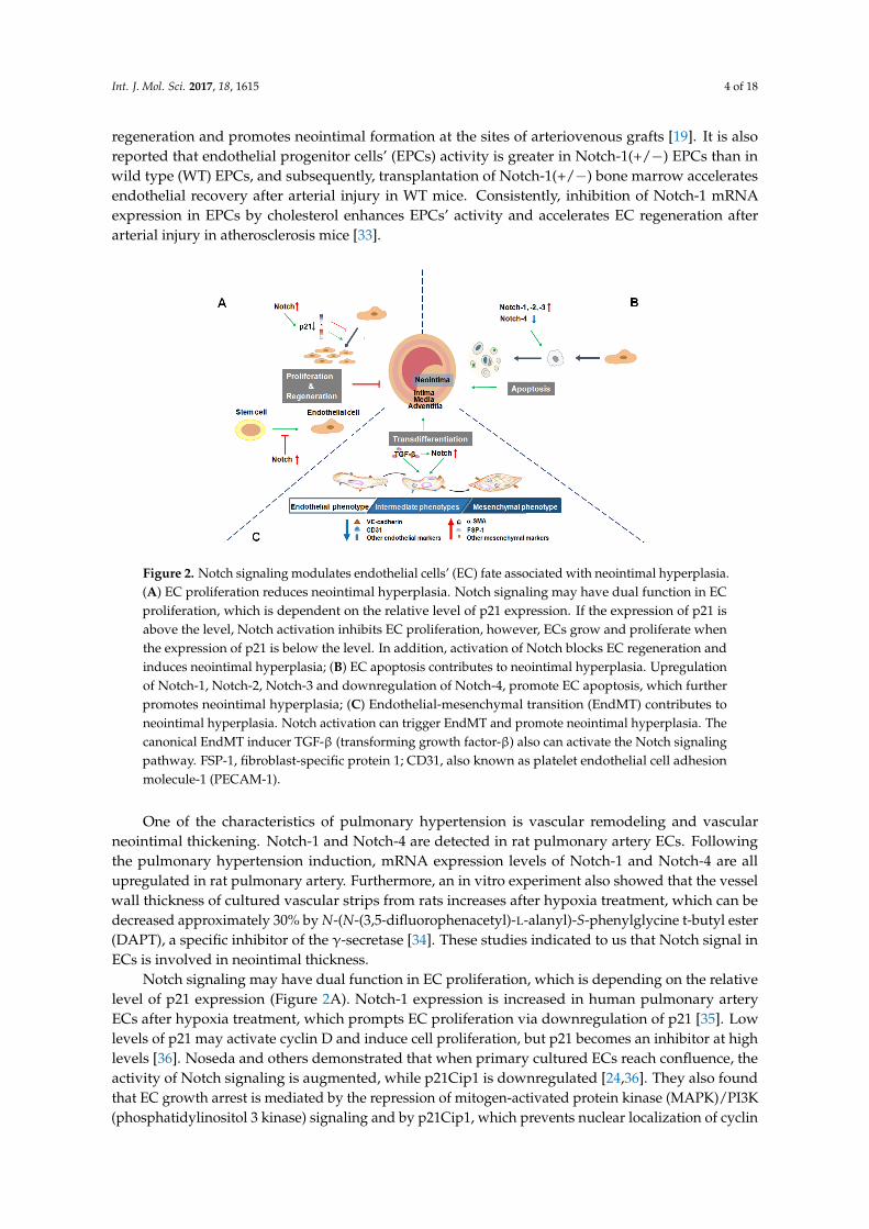
Int. J. Mol. Sci. 2017, 18, 1615 4 of 18
regeneration and promotes neointimal formation at the sites of arteriovenous grafts [19]. It is alsoreported that endothelial progenitor cells’ (EPCs) activity is greater in Notch-1(+/−) EPCs than inwild type (WT) EPCs, and subsequently, transplantation of Notch-1(+/−) bone marrow acceleratesendothelial recovery after arterial injury in WT mice. Consistently, inhibition of Notch-1 mRNAexpression in EPCs by cholesterol enhances EPCs’ activity and accelerates EC regeneration afterarterial injury in atherosclerosis mice [33].
Int. J. Mol. Sci. 2017, 18, 1615 4 of 17
1(+/−) EPCs than in wild type (WT) EPCs, and subsequently, transplantation of Notch-1(+/−) bone marrow accelerates endothelial recovery after arterial injury in WT mice. Consistently, inhibition of Notch-1 mRNA expression in EPCs by cholesterol enhances EPCs’ activity and accelerates EC regeneration after arterial injury in atherosclerosis mice [33].
One of the characteristics of pulmonary hypertension is vascular remodeling and vascular neointimal thickening. Notch-1 and Notch-4 are detected in rat pulmonary artery ECs. Following the pulmonary hypertension induction, mRNA expression levels of Notch-1 and Notch-4 are all upregulated in rat pulmonary artery. Furthermore, an in vitro experiment also showed that the vessel wall thickness of cultured vascular strips from rats increases after hypoxia treatment, which can be decreased approximately 30% by N-(N-(3,5-difluorophenacetyl)-L-alanyl)-S-phenylglycine t-butyl ester (DAPT), a specific inhibitor of the γ-secretase [34]. These studies indicated to us that Notch signal in ECs is involved in neointimal thickness.
Notch signaling may have dual function in EC proliferation, which is depending on the relative level of p21 expression (Figure 2A). Notch-1 expression is increased in human pulmonary artery ECs after hypoxia treatment, which prompts EC proliferation via downregulation of p21 [35]. Low levels of p21 may activate cyclin D and induce cell proliferation, but p21 becomes an inhibitor at high levels [36]. Noseda and others demonstrated that when primary cultured ECs reach confluence, the activity of Notch signaling is augmented, while p21Cip1 is downregulated [24,36]. They also found that EC growth arrest is mediated by the repression of mitogen-activated protein kinase (MAPK)/PI3K (phosphatidylinositol 3 kinase) signaling and by p21Cip1, which prevents nuclear localization of cyclin D/cdk4 (cyclin-dependent kinase 4) required for Rb (retinoblastoma gene product) phosphorylation and S-phase entry [24].
Furthermore, EC behavior is at least partially dependent on reactive oxygen species (ROS) level downregulated by the Notch pathway. Blockade of Notch signaling can increase ROS in human umbilical vein endothelial cells (HUVECs), in contrast, suppression of ROS generation abolishes Notch blockade-induced HUVEC proliferation [37].
Figure 2. Notch signaling modulates endothelial cells’ (EC) fate associated with neointimal hyperplasia. (A) EC proliferation reduces neointimal hyperplasia. Notch signaling may have dual function in EC proliferation, which is dependent on the relative level of p21 expression. If the expression of p21 is above the level, Notch activation inhibits EC proliferation, however, ECs grow and proliferate when the expression of p21 is below the level. In addition, activation of Notch blocks EC regeneration and induces neointimal hyperplasia; (B) EC apoptosis contributes to neointimal hyperplasia. Upregulation of Notch-1, Notch-2, Notch-3 and downregulation of Notch-4, promote EC apoptosis, which further promotes neointimal hyperplasia; (C) Endothelial-mesenchymal transition (EndMT) contributes to neointimal hyperplasia. Notch activation can trigger EndMT and promote neointimal hyperplasia. The canonical EndMT inducer TGF-β (transforming growth factor-β) also can activate the Notch signaling pathway. FSP-1, fibroblast-specific protein 1; CD31, also known as platelet endothelial cell adhesion molecule-1 (PECAM-1).
Figure 2. Notch signaling modulates endothelial cells’ (EC) fate associated with neointimal hyperplasia.(A) EC proliferation reduces neointimal hyperplasia. Notch signaling may have dual function in ECproliferation, which is dependent on the relative level of p21 expression. If the expression of p21 isabove the level, Notch activation inhibits EC proliferation, however, ECs grow and proliferate whenthe expression of p21 is below the level. In addition, activation of Notch blocks EC regeneration andinduces neointimal hyperplasia; (B) EC apoptosis contributes to neointimal hyperplasia. Upregulationof Notch-1, Notch-2, Notch-3 and downregulation of Notch-4, promote EC apoptosis, which furtherpromotes neointimal hyperplasia; (C) Endothelial-mesenchymal transition (EndMT) contributes toneointimal hyperplasia. Notch activation can trigger EndMT and promote neointimal hyperplasia. Thecanonical EndMT inducer TGF-β (transforming growth factor-β) also can activate the Notch signalingpathway. FSP-1, fibroblast-specific protein 1; CD31, also known as platelet endothelial cell adhesionmolecule-1 (PECAM-1).
One of the characteristics of pulmonary hypertension is vascular remodeling and vascularneointimal thickening. Notch-1 and Notch-4 are detected in rat pulmonary artery ECs. Followingthe pulmonary hypertension induction, mRNA expression levels of Notch-1 and Notch-4 are allupregulated in rat pulmonary artery. Furthermore, an in vitro experiment also showed that the vesselwall thickness of cultured vascular strips from rats increases after hypoxia treatment, which can bedecreased approximately 30% by N-(N-(3,5-difluorophenacetyl)-L-alanyl)-S-phenylglycine t-butyl ester(DAPT), a specific inhibitor of the γ-secretase [34]. These studies indicated to us that Notch signal inECs is involved in neointimal thickness.
Notch signaling may have dual function in EC proliferation, which is depending on the relativelevel of p21 expression (Figure 2A). Notch-1 expression is increased in human pulmonary arteryECs after hypoxia treatment, which prompts EC proliferation via downregulation of p21 [35]. Lowlevels of p21 may activate cyclin D and induce cell proliferation, but p21 becomes an inhibitor at highlevels [36]. Noseda and others demonstrated that when primary cultured ECs reach confluence, theactivity of Notch signaling is augmented, while p21Cip1 is downregulated [24,36]. They also foundthat EC growth arrest is mediated by the repression of mitogen-activated protein kinase (MAPK)/PI3K(phosphatidylinositol 3 kinase) signaling and by p21Cip1, which prevents nuclear localization of cyclin

Int. J. Mol. Sci. 2017, 18, 1615 5 of 18
D/cdk4 (cyclin-dependent kinase 4) required for Rb (retinoblastoma gene product) phosphorylationand S-phase entry [24].
Furthermore, EC behavior is at least partially dependent on reactive oxygen species (ROS) leveldownregulated by the Notch pathway. Blockade of Notch signaling can increase ROS in humanumbilical vein endothelial cells (HUVECs), in contrast, suppression of ROS generation abolishes Notchblockade-induced HUVEC proliferation [37].
4. Endothelial Cell Apoptosis
Except for regulating cell proliferation and regeneration, Notch also affects EC apoptosis andsurvival, key cellular behaviors associated with vascular remodeling processes (Figure 2B). Vascularinjury activates Notch signaling promptly, which further destroys the balance between EC proliferationand apoptosis, eventually influencing neointimal hyperplasia.
During the development of transplant arteriosclerosis (TA), EC apoptosis leads to neointimalhyperplasia in aortic allografts and allograft dysfunction. EC apoptosis induces the production ofTGF-β1 in both apoptotic and neighboring viable cells, resulting in increased TGF-β1 in the culturemedia. In transgenic rat aorta transplantation models, inhibition of EC apoptosis in B-cell lymphoma(Bcl)-xL(+/+) knock-in rat aortic allografts significantly reduces TGF-β1 production in both allograftendothelia and blood plasma, which in turn decreases accumulation of SM22α+ cells from transgenicrecipient ECs in neointima and alleviated TA [38].
It has been reported that the transcript levels of Notch-2, -3, and -4 are markedly downregulatedin TA. In Quillard et al.’s research, TA correlates with high levels of tumor necrosis factor (TNF),TGF-β and interleukin (IL)-10. They also found that Notch-4 expression is decreased in transplantsand cultured ECs. Further knockdown of Notch-4 and HES-1 by small interfering RNA (siRNA)promotes ECs apoptosis. As expected, silencing Notch-4 or HES-1 drastically inhibits repair ofendothelial injury [39]. Mackenzie et al.’s studies demonstrated that Notch-4 provides endothelialprotection in two ways: inhibition of the c-Jun N-terminal kinase (JNK)-dependent pro-apoptoticpathway in an RBP-Jκ-dependent manner and induction of an anti-apoptotic pathway through anRBP-Jκ-independent upregulation of Bcl-2 [40].
However, after Notch-2 ICD transduction in cultured human arterial endothelial cells (HAEC)and HUVECs or induced Notch-2 expression in HUVECs, EC apoptosis is promoted, notably throughinhibiting the expression of survivin [41,42]. In addition, using a mouse model of pulmonary arterialhypertension, Li et al. showed that activation of nuclear factor kappa B (NF-κB) upregulates theexpression levels of Notch-3, proapoptotic gene caspase 3 and Bax, downregulates antiapoptoticgene Bcl-2 expression in lung microvascular endothelial cells, which leads to EC apoptosis andendothelial-mesenchymal transition (EndMT) occurring in the lung [43]. Moreover, through activatingNotch-1, HES-1 and caspase-3, accelerated cell apoptosis has been observed in human Eahy926 cellstreated with high glucose [44].
All of this evidence supports that Notch activation is involved in neointimal formation throughregulating EC apoptosis and survival. It is possible that the four different Notch receptors havedifferent roles in EC apoptosis.
5. Endothelial-Mesenchymal Transition
EndMT is a specific form of epithelial-mesenchymal transition (EMT), which is an importantbiologic transdifferentiation process that participates in embryogenesis, organ development, tissueregeneration, organ fibrosis and cancer metastasis [45]. Similar to the process of EMT, when undergoingEndMT, ECs lose their endothelial specific markers, such as CD31, also known as platelet endothelialcell adhesion molecule-1 (PECAM-1) or VE-cadherin, gain mesenchymal markers, such as α-smoothmuscle actin (α-SMA) or fibroblast-specific protein 1 (FSP-1, also known as S100A4), lose cell-celljunctions and acquire invasive and migratory properties [46,47].

Int. J. Mol. Sci. 2017, 18, 1615 6 of 18
EndMT has emerged as a player in the pathogenesis of vascular neointimal hyperplasia.Notintimal cells may arise through migration and proliferation of VSMCs; recent studies showedthat the endothelium is also a source of smooth muscle-like cells [48]. It has been reported thatEndMT occurs in myoendothelial cells in human atherosclerotic plaques and porcine aortic tissues.In vitro and in vivo experiments all showed that ECs exposed to disturbed flow undergo EndMT,which contributes to neointimal hyperplasia and induces atherogenic differentiation of ECs [49].Through introducing endothelial-specific deletion of fibroblast growth factor receptor substrate 2α (Frs2α) in atherosclerotic ApoE(−/−) mice, Chen et al. reported that these double-knockoutmice exhibit extensive development of EndMT and increased neointimal formation. Furthermore,patients with coronary atherosclerosis showed that the loss of endothelial fibroblast growth factorreceptor1 (FGFR1) expression leads to activation of endothelial TGF-β signaling and the developmentof EndMT in atherosclerotic plaques [50]. In vivo murine cell lineage-tracing models also presentedthat endothelial-derived cells contribute to neointimal formation through EndMT, which is dependenton the activation of the TGF-β mediated Smad2/3-Slug signaling pathway [51].
As a major regulator of cell phenotype, Notch is involved in the process of EndMT. Noseda et al.provided the first evidence that Jagged-1/Notch interactions induce endothelial-to-mesenchymaltransformation. Notch activation in ECs results in morphological, phenotypic and functionalchanges, which is consistent with mesenchymal transformation [52]. Notch and TGF-β/smad3signaling synergistically induce Snail expression in ECs and promote EndMT in cardiac cushionmorphogenesis [53]. Blocking the Notch signaling pathway by using DAPT, EndMT in rat cornealECs induced by TGF β1, -β2 or -β3 is prevented and the transformed ECs are reversed to a normalphenotype [54].
Neointimal hyperplasia occurs seriously in arteriovenous fistulas (AVFs) of chronic kidney disease(CKD) mice or patients. ECs of AVFs in CKD mice or patients express mesenchymal markers (FSP-1and/or α-SMA) and exhibit increased expression and nuclear localization of the Notch intracellulardomain. Uremic mice also show a decreased expression of VE-cadherin, whereas the expressionsof Notch-1, -4, RBP-Jκ and Notch target genes are increased in ECs of AVFs. Blockade of the Notchpathway by DAPT or by RBP-Jκ knockout suppresses neointimal formation in mice [23]. Thus, datain the literature suggest that the Notch pathway is correlated with EndMT and contributes to theneointimal hyperplasia in vascular remodeling (Figure 2C).
6. The Contact Interaction between Endothelial Cells and Smooth Muscle Cells
Vessel wall is mainly composed of ECs and parietal cells (VSMCs and pericytes). The directcommunication between ECs and VSMCs through myoendothelial gap junctions and microprojectionshas been widely known [55,56]. In addition, Notch ligand-receptor interaction is another directcommunication means between ECs and VSMCs. Contact-mediated activation of Notch signalingplays important roles in cell and vessel maturation, survival and homeostasis. In these processes,different Notch receptors may have specific roles [57].
In vertebrates, ECs can express three Notch ligands (Dll-4, Jagged-1 and -2) and all of the knownNotch receptors (Notch-1, -2, -3 and -4), while VSMCs express ligand Jagged-1 and three receptors(Notch-1, -2 and -3) [15]. We summarized the effects of ECs-VSMCs interaction mediated by differentNotch receptor-ligand on neointimal formation in Table 1A. During vascular development, VSMCsrecognize Notch ligand Jagged-1 on ECs and induce the expression of integrin αvβ3 in VSMCs, whichfacilitates VSMCs adhering to endothelial basement membrane and promotes vessel maturation [58].In post-development vessels, the endothelial protein kinase B (PKB, also known as Akt) deletionreduces the expression of endothelial Jagged-1 and leads to the gradual loss of VSMCs due todiminished Jagged-1/Notch signaling. It sustains that contact-mediated activation of Notch signalingis critical in maintaining vascular stability and homeostasis [59]. Among the Notch signaling molecules,EC membrane ligand Jagged-1 is required for the induction of Notch-3 in VSMCs and inducing VSMCdifferentiation [60,61]. Neither the addition of soluble Jagged-1 nor EC-conditioned medium induces

Int. J. Mol. Sci. 2017, 18, 1615 7 of 18
VSMC differentiation, while co-cultured ECs with VSMCs induce VSMC differentiation; furthermore,knockdown Jagged-1 expression in ECs can abrogate the co-cultured VSMC phenotype change. All ofthis evidence strongly supports that the direct heterocellular cell-cell contact is necessary for regulatingVSMC differentiation via Jagged-1/Notch-3 signaling [62].
Table 1. The Notch pathway-mediated interaction between endothelial cells (ECs) and vascular smoothmuscle cells (VSMCs).
A. The Direct Contact between ECs and VSMCs Mediated by Notch
Ways ofInteraction Ligand Receptor Function Possible Effect on
Neointimal Hyperplasia
Direct Contact
ND VSMC Notch-1 VSMC migration [64]↑ Promotion
EC Jagged-1 VSMC Notch-2VSMC differentiation [63]↑
NDVSMC proliferation [63]↓
EC Jagged-1 VSMC Notch-3VSMC differentiation [60–63]↑
PromotionVSMC secretion [63]↑VSMC migration [64]↑
VSMC Jagged-1 EC Notch-1 EC proliferation [65]↑ InhibitionND EC Notch-2, -3, -4 [15] ND ND
B. The Indirect Communication between ECs and VSMCs Mediated by Notch Activation
Ways ofInteraction Notch Signaling Molecules Secreted
from EC Function Possible Effect onNeointimal Hyperplasia
IndirectCommunication
Activation
NOEC proliferation [66,67]↑
InhibitionVSMC proliferation [68]↓VSMC migration [68]↓
VEGFEC proliferation↑
InhibitionEC regeneration↑VSMC proliferation↓
PDGFVSMC proliferation [69]↑
PromotionVSMC migration [70]↑VSMC differentiation [71]↑
ND indicates not described; NO: nitric oxide; PDGF: platelet-derived growth factor; VEGF: vascular endothelialgrowth factor; ↑ indicates promotion; ↓ indicates inhibition.
Except for Notch-3, Notch-2 is also activated in VSMCs co-cultured with ECs. Both Notch-2 andNotch-3 in VSMCs are mediators of EC-induced differentiated phenotype and contribute to increasedcontractile protein expression. However, the two receptors have separate and distinct functions.Notch-2 is specifically required for the suppression of cell proliferation, while Notch-3 is mainlyresponsible for VSMC secretory function [63].
VSMC proliferation, phenotype change and ECM secretion are hallmarks of neointimal formationin cardiovascular diseases and vascular surgery. Balloon injury induced Notch-1, Notch-3 and Jagged-1expression in rat carotid arteries. However, soluble Jagged-1 inhibits neointimal formation after ballooninjury or vein graft by decreasing VSMC proliferation and migration through interference with theNotch signaling pathway [64]. This evidence suggested that inhibition of neointimal formation maybe due to inactivation of the Notch signaling pathway through soluble Jagged-1 competing with ECJagged-1 to bind with the Notch receptor.
Reciprocally, VSMCs’ Jagged-1 activate Notch pathway in ECs through the Notch-1 receptorand induce EC proliferation. Notch signaling-deficient primary VSMCs have reduced proliferationand migration capacities and a diminished expression of Jagged-1 ligand. After being co-culturedwith such VSMCs, ECs exhibit reduced growth rates and lower levels of activated Notch-1 receptor(Notch-1ICD) [65]. As discussed previously, EC proliferation and regeneration are critical eventsin neointimal formation. Through regulating the contact between ECs and VSMCs, activation ofNotch could be manipulated, which may represent a unique therapeutic target to improve neointimalformation after vascular injury.
Notch signaling between cells can also be transmitted by exosomes from a distance. Dll-4 isincorporated into endothelial exosomes; the Dll-4 containing exosomes can freely travel through the 3Dcollagen matrix, transfer Dll-4 protein to distant tip cells and induce tip cell retraction [72]. However,

Int. J. Mol. Sci. 2017, 18, 1615 8 of 18
whether there exists exosome-mediated Notch signaling between ECs and VSMCs in neointimalformation has not yet been reported.
Taken together, Jagged-1 seems to be the only ligand in the Notch pathway that plays an importantrole in both ECs and VSMCs during neointimal formation development. Notch-1 may be the onlyreceptor in the EC lineage that recognizes the Notch ligand in VSMCs, whereas several receptors(Notch-2, -3) are involved in the VSMC lineage to recognize the Notch ligand in ECs (Figure 3).
Int. J. Mol. Sci. 2017, 18, 1615 8 of 17
the only receptor in the EC lineage that recognizes the Notch ligand in VSMCs, whereas several receptors (Notch-2, -3) are involved in the VSMC lineage to recognize the Notch ligand in ECs (Figure 3).
Figure 3. Notch signaling in cell-cell communication between endothelial cells (ECs) and vascular smooth muscle cells (VSMCs). In the process of direct cell-cell interaction between ECs and VSMCs, EC membrane ligand Jagged-1 is recognized by Notch-2, -3 in VSMCs; reciprocally, VSMCs Jagged-1 binds with EC Notch-1 receptor and activates the Notch downstream pathway in ECs (also see Table 1A). In addition, activation of Notch signaling induces nitric oxide (NO), platelet-derived growth factor (PDGF), vascular endothelial growth factor (VEGF) and other factors’ production from ECs via autocrine and paracrine process; these factors can regulate EC and VSMC proliferation, migration, differentiation and play different roles in neointimal hyperplasia (see Table 1B).
7. Endothelial Cell Secretory Function
Although the vascular endothelium is made up from only a single layer of ECs, it works as an “endocrine organ” via an autocrine and/or paracrine process and contributes to vascular homeostasis, such as angiogenesis, inflammation, platelet aggregation and vascular remodeling [73,74]. The regulatory molecules derived from ECs include non-growth factors and growth factors, such as nitric oxide (NO), VEGF, platelet-derived growth factor (PDGF), basic fibroblast growth factor (bFGF) and insulin-like growth factor-1, etc. [75,76] (Figure 3). There are many reports about crosstalk between Notch signaling and the regulatory molecules mentioned above; we refer the readers to the three main kinds of factors derived from ECs regulated by Notch signaling and their effects on neointimal hyperplasia (Table 1B).
Mainly produced by endothelial NO synthase (eNOS), NO derived from EC is an important mediator of normal and pathologic vascular remodeling [77]. NO not only confers anti-platelet and anti-inflammatory properties to the vessels, but also promotes EC proliferation [66], inhibits VSMC proliferation and migration [68], controls M1 macrophage polarization [78], regulates redox balance [79], keeps the stability and function of blood vessels and suppresses neointimal hyperplasia [80]. A recent study reported that Notch induces Activin A expression, thereby activating the PI3K/Akt pathway to phosphorylate eNOS and promoting NO production [67]. Inhibition of Notch decreases endothelial NO production by reduced eNOS expression [81]. In vein grafts of aged rats, the reduced expression of Dll-4 and Notch-4 has been found, which is associated with the decreased eNOS protein expression, reduced eNOS membrane targeting and colocalization with caveolin-1, as well as significantly thicker neointima [82].
The contribution of VEGF in neointimal formation has been widely evaluated. Recent studies demonstrated that VEGF can block neointimal formation through inducing EC growth and reducing VSMC growth after vascular injury [83]. In addition, bone marrow-derived mesenchymal stem cells
Figure 3. Notch signaling in cell-cell communication between endothelial cells (ECs) and vascularsmooth muscle cells (VSMCs). In the process of direct cell-cell interaction between ECs and VSMCs, ECmembrane ligand Jagged-1 is recognized by Notch-2, -3 in VSMCs; reciprocally, VSMCs Jagged-1 bindswith EC Notch-1 receptor and activates the Notch downstream pathway in ECs (also see Table 1A). Inaddition, activation of Notch signaling induces nitric oxide (NO), platelet-derived growth factor (PDGF),vascular endothelial growth factor (VEGF) and other factors’ production from ECs via autocrine andparacrine process; these factors can regulate EC and VSMC proliferation, migration, differentiation andplay different roles in neointimal hyperplasia (see Table 1B).
7. Endothelial Cell Secretory Function
Although the vascular endothelium is made up from only a single layer of ECs, it works as an“endocrine organ” via an autocrine and/or paracrine process and contributes to vascular homeostasis,such as angiogenesis, inflammation, platelet aggregation and vascular remodeling [73,74]. Theregulatory molecules derived from ECs include non-growth factors and growth factors, such asnitric oxide (NO), VEGF, platelet-derived growth factor (PDGF), basic fibroblast growth factor (bFGF)and insulin-like growth factor-1, etc. [75,76] (Figure 3). There are many reports about crosstalk betweenNotch signaling and the regulatory molecules mentioned above; we refer the readers to the threemain kinds of factors derived from ECs regulated by Notch signaling and their effects on neointimalhyperplasia (Table 1B).
Mainly produced by endothelial NO synthase (eNOS), NO derived from EC is an importantmediator of normal and pathologic vascular remodeling [77]. NO not only confers anti-plateletand anti-inflammatory properties to the vessels, but also promotes EC proliferation [66], inhibitsVSMC proliferation and migration [68], controls M1 macrophage polarization [78], regulatesredox balance [79], keeps the stability and function of blood vessels and suppresses neointimalhyperplasia [80]. A recent study reported that Notch induces Activin A expression, thereby activatingthe PI3K/Akt pathway to phosphorylate eNOS and promoting NO production [67]. Inhibition ofNotch decreases endothelial NO production by reduced eNOS expression [81]. In vein grafts of

Int. J. Mol. Sci. 2017, 18, 1615 9 of 18
aged rats, the reduced expression of Dll-4 and Notch-4 has been found, which is associated withthe decreased eNOS protein expression, reduced eNOS membrane targeting and colocalization withcaveolin-1, as well as significantly thicker neointima [82].
The contribution of VEGF in neointimal formation has been widely evaluated. Recent studiesdemonstrated that VEGF can block neointimal formation through inducing EC growth and reducingVSMC growth after vascular injury [83]. In addition, bone marrow-derived mesenchymal stem cellstreated with VEGF differentiate into endothelial-like cells and significantly attenuate neointimalthickness [84]. However, lentivirus-mediated VEGF-A inhibition can decrease the venous neointimalhyperplasia of AVFs [85]. This evidence suggested that the effect of VEGF on neointimal formationis complicated. Although there is no direct evidence to support that Notch signaling mediatesneointimal hyperplasia through autocrine or paracrine of VEGF in ECs, inhibition of Notch-1 orNotch-4 can block thymosin β 4-induced VEGF expression in HUVECs [86]. In contrast, many studiesuncovered that VEGF can activate Notch signaling in ECs and act as an upstream mediator of theNotch pathway [87,88].
As a smooth muscle cell growth and survival factor, PDGF also plays a prominent role in VSMCsmigrating into the neointima following acute injury or in atherosclerosis. High shear stress inhibitsarterial wall thickening in vivo, which may be related to enhanced activation of PDGF-R alpha inVSMCs by PDGF isoforms secreted from the endothelium. The neutralizing antibody against PDGF-AAenhances VSMC migration; in contrast, antibodies against PDGF-BB abolish VSMC migration [70].VSMC-rich neointimal formation is accelerated in the ligated carotid artery of mice treated witherythropoietin delta, by which the expression and release of PDGF-B is induced in HUVECs [69].Findings also showed that PDGF-BB and PDGF-DD are all VSMC phenotypic modulators. PDGF-DDexpression is increased in neointimal lesions in the aortic arch region of apolipoprotein C-deficientApoE(−/−) mice. In addition, human ECs exposed to an atherosclerosis-prone flow pattern, as invascular regions susceptible to the development of atherosclerosis, exhibit a significant increase inPDGF-DD expression [71].
Several recent studies reported that Notch signaling is involved in regulating PDGF productionfrom ECs. Exposure of human brain microvascular endothelial cells to DAPT or silencing ofNotch-1 results in abrogation of cocaine-mediated induction of PDGF-B. The study provided the firstevidence of the involvement of Notch-1 activation in PDGF-B expression [89]. β-catenin, a key signalmolecule of the Wnt/β-catenin-Dll4/Notch signaling cascade in endothelia, its transcriptional activitydirectly regulates the endothelial expression of PDGF-B [90]. Conditional medium from matricellularprotein, secreted protein acidic and rich in cysteine (SPARC) overexpressed neuroblastoma cells showsuppressed expression of VEGF, PDGF, FGF and matrix metalloprotein 9 in ECs, which is mediated bythe inhibition of the Notch signaling pathway [91]. Taken together, these findings suggest that PDGFsecretion induced by Notch may be involved in neointimal hyperplasia.
There are also many other EC-derived molecules that have been proven or speculated to influenceneointimal hyperplasia. For instance, apoptotic ECs actively release paracrine mediators’ C-terminalfragment of perlecan and epidermal growth factor, which inhibit apoptosis of mesenchymal stem cells(MSC), which are pivotal to vascular repair and neointimal formation [92]. As a secreted glycoproteinthat has been implicated in regulating VSMC proliferation and migration, the downregulatedexpression of apolipoprotein D (APOD) is partly caused by paracrine secretion of ECs. In addition,Notch-3 on mural cells also promotes the downregulation of APOD, possibly through interaction withthe Jagged-1 ligands on ECs [93].
8. Endothelial Cell Barrier Dysfunction
The development of functional blood vessel requires an integrated layer of endothelial cells.Based on the EC junction, the structural and functional integrity of the endothelium is fundamentalfor maintaining vascular homeostasis [94]. Destruction of the protective endothelial barrier willsubsequently lead to vascular injury and neointimal formation. Changes in shear and/or hoop stress,

Int. J. Mol. Sci. 2017, 18, 1615 10 of 18
direct drug-induced cytotoxicity, mechanical device implant-induced injury or inflammatory responsein vascular surgery or cardiovascular surgery induce vascular injury and cause EC dysfunction [95].As discussed above, vascular injury is a complex cascade of events involving endothelial denudation,the release of growth factors and cytokines which triggers platelet degranulation and aggregation,subsequent inflammatory cells or mediators invading the injured locations, smooth muscle cellproliferation and migration to form neointimal hyperplasia at the subendothelial space. Reciprocally,platelet activation and inflammation response lead to delayed re-endothelialization and endothelialdysfunction [96].
Endothelial dysfunction is characterized by EC phenotype change with impairedendothelium-dependent barrier and imbalance between re-endothelialization and apoptosisor growth inhibiting and growth-promoting substances. Notch signaling cascades are involvedin and partially responsible for EC dysfunction events (Figure 4). For example, apoptotic orsenescent phenotype ECs lose their barrier function. Activation of Notch increases myosin light chainphosphorylation by activating Rho kinase, which further triggers EC acquiring senescence phenotypeand leads to hyperpermeability of the endothelium [97]. In atherosclerosis, Notch activation inducesEC senescence and prompts the expression and secretion of pro-inflammatory cytokines such asIL-6, IL-8, IL-1α. Among these factors, the upregulated IL-6 may mediate leukocyte transendothelialmigration [98]. In addition, Notch activation also causes EC to acquire VSMC phenotype, whichleads to ECs losing their barrier function and induces neointimal hyperplasia [23]. Consequently,attenuation of Notch signaling in ECs might provide a treatment strategy for neointimal formation.
Int. J. Mol. Sci. 2017, 18, 1615 10 of 17
Endothelial dysfunction is characterized by EC phenotype change with impaired endothelium-dependent barrier and imbalance between re-endothelialization and apoptosis or growth inhibiting and growth-promoting substances. Notch signaling cascades are involved in and partially responsible for EC dysfunction events (Figure 4). For example, apoptotic or senescent phenotype ECs lose their barrier function. Activation of Notch increases myosin light chain phosphorylation by activating Rho kinase, which further triggers EC acquiring senescence phenotype and leads to hyperpermeability of the endothelium [97]. In atherosclerosis, Notch activation induces EC senescence and prompts the expression and secretion of pro-inflammatory cytokines such as IL-6, IL-8, IL-1α. Among these factors, the upregulated IL-6 may mediate leukocyte transendothelial migration [98]. In addition, Notch activation also causes EC to acquire VSMC phenotype, which leads to ECs losing their barrier function and induces neointimal hyperplasia [23]. Consequently, attenuation of Notch signaling in ECs might provide a treatment strategy for neointimal formation.
As mentioned above, activation of Notch-2 induces EC apoptosis while Notch-4 plays a protective role [39,43]. In arterial ECs, pro-inflammatory cytokine TNF-α elicits a switch in Notch expression, which is characterized by Notch-2 predominance over Notch-4. The events lead to a reduced Notch activity, then promoting caspase-dependent EC apoptosis and vascular dysfunction [99]. In most cases, the upregulated expressions of Jagged-1, Dll-4, Notch-1 and Notch-4 are associated with inhibition of EC proliferation and vascular dysfunction [100,101]. This evidence proved that loss of ECs could lead to increased platelet reactivity and VSMC proliferation, thereby facilitating neointimal formation.
EC dysfunction may also be related to the change of the secretory function of ECs. Among the substances released from ECs, NO is involved in the impairment of endothelium-dependent vasodilatation. Endovascular interventions are associated with diminished bioavailability of NO and increased local inflammatory response, which triggers EC apoptosis [102–104]. Reducing eNOS expression or promoting inducible nitric oxide synthase (iNOS) expression induces EC apoptosis and dysfunction, which can be mediated through the Jagged-1/Notch pathway [105]. To conclude, enhanced degradation of NO and decreased eNOS expression and/or activity leads to EC dysfunction and contributes to neointimal formation. As shown in Figure 4, we summarized the relationship between Notch signaling and EC dysfunction during neointimal formation development.
Figure 4. Endothelial cell (EC) dysfunction caused by Notch signaling leads to neointimal hyperplasia. When vascular injury occurs, Notch signaling is activated, which further triggers EC phenotype change and causes EC dysfunction. In these processes, ECs acquire the senescent phenotype, the apoptotic phenotype or the mesenchymal phenotype and lose their barrier function, which leads to endothelial hyperpermeability, leakage and inflammatory responses. Furthermore, vascular smooth muscle cell -like cells can transmigrate into the media and proliferate, resulting in neointimal hyperplasia.
Figure 4. Endothelial cell (EC) dysfunction caused by Notch signaling leads to neointimal hyperplasia.When vascular injury occurs, Notch signaling is activated, which further triggers EC phenotype changeand causes EC dysfunction. In these processes, ECs acquire the senescent phenotype, the apoptoticphenotype or the mesenchymal phenotype and lose their barrier function, which leads to endothelialhyperpermeability, leakage and inflammatory responses. Furthermore, vascular smooth muscle cell-like cells can transmigrate into the media and proliferate, resulting in neointimal hyperplasia.
As mentioned above, activation of Notch-2 induces EC apoptosis while Notch-4 plays a protectiverole [39,43]. In arterial ECs, pro-inflammatory cytokine TNF-α elicits a switch in Notch expression,which is characterized by Notch-2 predominance over Notch-4. The events lead to a reduced Notchactivity, then promoting caspase-dependent EC apoptosis and vascular dysfunction [99]. In most cases,the upregulated expressions of Jagged-1, Dll-4, Notch-1 and Notch-4 are associated with inhibition of

Int. J. Mol. Sci. 2017, 18, 1615 11 of 18
EC proliferation and vascular dysfunction [100,101]. This evidence proved that loss of ECs could leadto increased platelet reactivity and VSMC proliferation, thereby facilitating neointimal formation.
EC dysfunction may also be related to the change of the secretory function of ECs. Amongthe substances released from ECs, NO is involved in the impairment of endothelium-dependentvasodilatation. Endovascular interventions are associated with diminished bioavailability of NOand increased local inflammatory response, which triggers EC apoptosis [102–104]. Reducing eNOSexpression or promoting inducible nitric oxide synthase (iNOS) expression induces EC apoptosisand dysfunction, which can be mediated through the Jagged-1/Notch pathway [105]. To conclude,enhanced degradation of NO and decreased eNOS expression and/or activity leads to EC dysfunctionand contributes to neointimal formation. As shown in Figure 4, we summarized the relationshipbetween Notch signaling and EC dysfunction during neointimal formation development.
9. Limits and Perspectives
We have reviewed here the accumulated evidence that the Notch pathway is involved in multipleaspects of EC key functions (proliferation, regeneration, apoptosis, differentiation, cell-cell interaction)and contributes to neointimal formation. However, our review also found a number of apparentinconsistencies and problems in these studies. Although the relative level of p21 expression maydecide EC growth after Notch activation, no data show the exact expression level or range of p21. Thepatterns of Notch-1 to -4 expression can play different roles in EC apoptosis, but which componentsfunction at what apoptosis steps in ECs? Notch signaling seems to induce endothelial mesenchymalphenotype switch, so does there exist a specific relationship among ligands, receptors, and targetgenes in the process of transdifferentiation? How does Notch play a role in EC regeneration anddifferentiation? Except NO, how does the Notch pathway play any role in the regulation of othercytokines secreted from ECs to cause EC dysfunction? How about the crosstalk between Notchand other signaling pathways? Given that the Notch has a significant role in vascular development,further understanding of the Notch signaling pathway in the context of vascular biology will likelyprovide novel insights into the mechanisms of neointimal formation and new opportunities for rationaltherapeutic intervention.
Acknowledgments: This work was supported by grants from National Nature Science Foundation of China81470963, the Natural Science Foundation Project of Chongqing cstc2014jcyjA10098 and the Student’s Platform forInnovation and Entrepreneurship Training Program 201690031045.
Author Contributions: Yun Wang conceptualized this review. Ding-Yuan Tian wrote the manuscript and preparedthe table. Xu-Rui Jin drew the figures. Xi Zeng edited the manuscript.
Conflicts of Interest: The authors declare no conflict of interest.
Abbreviations
ADAM a disintegrin and metalloproteinasesAGS alagille syndromeAPOD apolipoprotein DAVF arteriovenous fistulaAVG arteriovenous graftbFGF basic fibroblast growth factorCACs circulating angiogenic cellsCADASIL cerebral autosomal-dominant arteriopathy with subcortical infarcts and leukoencephalopathyCKD chronic kidney diseaseCVD cardiovascular diseasesDAPT N-[N-(3,5-Difluorophenacetyl)-L-alanyl]-S-phenylglycine t-butyl esterDll delta-likeEC endothelial cellECM extracellular matrix

Int. J. Mol. Sci. 2017, 18, 1615 12 of 18
EMT Epithelial-mesenchymal transitionEndMT Endothelial-mesenchymal transitioneNOS endothelial NO synthaseEPC endothelial progenitor cellFGFR1 Fibroblast growth factor receptor1Frs2α Fibroblast growth factor receptor substrate 2 α
FSP-1 fibroblast-specific protein 1HAEC human arterial endothelial cellHERP HES-related repressor proteinHES hairy and enhancer of splitHUVEC human umbilical vein endothelial cellIFNγ Interferon γ
IL interleukiniNOS inducible nitric oxide synthaseMAML Mastermind-likeMAPK mitogen-activated protein kinaseMSC mesenchymal stem cellNF-κB nuclear factor κBNICD Notch intracellular domainNO Nitric oxidePDGF platelet-derived growth factorPECAM-1 platelet endothelial cell adhesion molecule-1Rb retinoblastomaROS reactive oxygen speciesα-SMA α-smooth muscle actinSPARC secreted protein acidic and rich in cysteineTA transplant arteriosclerosisTGF-β transforming growth factor-βTNF tumor necrosis factorVEGF vascular endothelial growth factorVSMC vascular smooth muscle cellWT wild type
References
1. Brahmbhatt, A.; Remuzzi, A.; Franzoni, M.; Misra, S. The molecular mechanisms of hemodialysis vascularaccess failure. Kidney Int. 2016, 89, 303–316. [CrossRef] [PubMed]
2. Wang, X.W.; Zhao, X.J.; Xiang, X.Y. Gene therapy for vein graft failure. J. Card. Surg. 2013, 28, 144–147.[CrossRef] [PubMed]
3. Ahanchi, S.S.; Tsihlis, N.D.; Kibbe, M.R. The role of nitric oxide in the pathophysiology of intimal hyperplasia.J. Vasc. Surg. 2007, 45, 64–73. [CrossRef] [PubMed]
4. Butt, M.; Dwivedi, G.; Blann, A.; Khair, O.; Lip, G.Y. Endothelial dysfunction: Methods of assessment &implications for cardiovascular diseases. Curr. Pharm. Des. 2010, 16, 3442–3454. [PubMed]
5. Allaire, E.; Clowes, A.W. Endothelial cell injury in cardiovascular surgery: The intimal hyperplastic response.Ann. Thorac. Surg. 1997, 63, 582–591. [PubMed]
6. Bhardwaj, S.; Roy, H.; Heikura, T.; Yla-Herttuala, S. VEGF-A, VEGF-D and VEGF-D (DeltaNDeltaC) inducedintimal hyperplasia in carotid arteries. Eur. J. Clin. Investig. 2005, 35, 669–676. [CrossRef] [PubMed]
7. DiRenzo, D.M.; Chaudhary, M.A.; Shi, X.; Franco, S.R.; Zent, J.; Wang, K.; Guo, L.W.; Kent, K.C. A crosstalkbetween TGF-β/Smad3 and Wnt/β-catenin pathways promotes vascular smooth muscle cell proliferation.Cell Signal. 2016, 28, 498–505. [CrossRef] [PubMed]
8. Zhang, J.; Chen, J.; Xu, C.; Yang, J.; Guo, Q.; Hu, Q.; Jiang, H. Resveratrol inhibits phenotypic switchingof neointimal vascular smooth muscle cells after balloon injury through blockade of Notch pathway.J. Cardiovasc. Pharmacol. 2014, 63, 233–239. [CrossRef] [PubMed]

Int. J. Mol. Sci. 2017, 18, 1615 13 of 18
9. Benedito, R.; Hellstrom, M. Notch as a hub for signaling in angiogenesis. Exp. Cell. Res. 2013, 319, 1281–1288.[CrossRef] [PubMed]
10. Krebs, L.T.; Xue, Y.; Norton, C.R.; Shutter, J.R.; Maguire, M.; Sundberg, J.P.; Gallahan, D.; Closson, V.;Kitajewski, J.; Callahan, R.; et al. Notch signaling is essential for vascular morphogenesis in mice. Genes Dev.2000, 14, 1343–1352. [PubMed]
11. Artavanis-Tsakonas, S.; Rand, M.D.; Lake, R.J. Notch signaling: Cell fate control and signal integration indevelopment. Science 1999, 284, 770–776. [CrossRef] [PubMed]
12. Gama-Norton, L.; Ferrando, E.; Ruiz-Herguido, C.; Liu, Z.; Guiu, J.; Islam, A.B.; Lee, S.U.; Yan, M.;Guidos, C.J.; Lopez-Bigas, N.; et al. Notch signal strength controls cell fate in the haemogenic endothelium.Nat. Commun. 2015, 6, 8510. [CrossRef] [PubMed]
13. De Celis, J.F.; Garcia-Bellido, A. Roles of the Notch gene in Drosophila wing morphogenesis. Mech. Dev.1994, 46, 109–122. [CrossRef]
14. Hofmann, J.J.; Iruela-Arispe, M.L. Notch signaling in blood vessels: Who is talking to whom about what?Circ. Res. 2007, 100, 1556–1568. [CrossRef] [PubMed]
15. Iso, T.; Hamamori, Y.; Kedes, L. Notch signaling in vascular development. Arterioscler. Thromb. Vasc. Biol.2003, 23, 543–553. [CrossRef] [PubMed]
16. Krebs, L.T.; Shutter, J.R.; Tanigaki, K.; Honjo, T.; Stark, K.L.; Gridley, T. Haploinsufficient lethality andformation of arteriovenous malformations in Notch pathway mutants. Genes Dev. 2004, 18, 2469–2473.[CrossRef] [PubMed]
17. Louvi, A.; Arboleda-Velasquez, J.F.; Artavanis-Tsakonas, S. CADASIL: A critical look at a Notch disease.Dev. Neurosci. 2006, 28, 5–12. [CrossRef] [PubMed]
18. Gridley, T. Notch signaling and inherited disease syndromes. Hum. Mol. Genet. 2003, 12, 9–13. [CrossRef]19. Liang, M.; Wang, Y.; Liang, A.; Dong, J.F.; Du, J.; Cheng, J. Impaired integrin β3 delays endothelial cell
regeneration and contributes to arteriovenous graft failure in mice. Arterioscler. Thromb. Vasc. Biol. 2015, 35,607–615. [CrossRef] [PubMed]
20. Koga, J.; Nakano, T.; Dahlman, J.E.; Figueiredo, J.L.; Zhang, H.; Decano, J.; Khan, O.F.; Niida, T.; Iwata, H.;Aster, J.C.; et al. Macrophage notch ligand delta-like 4 promotes vein graft lesion development: Implicationsfor the treatment of vein graft failure. Arterioscler. Thromb. Vasc. Biol. 2015, 35, 2343–2353. [CrossRef][PubMed]
21. Liang, M.; Wang, Y.; Liang, A.; Mitch, W.E.; Roy-Chaudhury, P.; Han, G.; Cheng, J. Migration of smoothmuscle cells from the arterial anastomosis of arteriovenous fistulas requires Notch activation to formneointima. Kidney Int. 2015, 88, 490–502. [CrossRef] [PubMed]
22. Caolo, V.; Schulten, H.M.; Zhuang, Z.W.; Murakami, M.; Wagenaar, A.; Verbruggen, S.; Molin, D.G.; Post, M.J.Soluble Jagged-1 inhibits neointima formation by attenuating Notch-Herp2 signaling. Arterioscler. Thromb.Vasc. Biol. 2011, 31, 1059–1065. [CrossRef] [PubMed]
23. Wang, Y.; Liang, A.; Luo, J.; Liang, M.; Han, G.; Mitch, W.E.; Cheng, J. Blocking notch in endothelial cellsprevents arteriovenous fistula failure despite CKD. J. Am. Soc. Nephrol. 2014, 25, 773–783. [CrossRef][PubMed]
24. Noseda, M.; Chang, L.; McLean, G.; Grim, J.E.; Clurman, B.E.; Smith, L.L.; Karsan, A. Notch activationinduces endothelial cell cycle arrest and participates in contact inhibition: Role of p21Cip1 repression. Mol.Cell Biol. 2004, 24, 8813–8822. [CrossRef] [PubMed]
25. Leslie, J.D.; Ariza-McNaughton, L.; Bermange, A.L.; McAdow, R.; Johnson, S.L.; Lewis, J. Endothelialsignalling by the Notch ligand Delta-like 4 restricts angiogenesis. Development 2007, 134, 839–844. [CrossRef][PubMed]
26. Sainson, R.C.; Aoto, J.; Nakatsu, M.N.; Holderfield, M.; Conn, E.; Koller, E.; Hughes, C.C. Cell-autonomousnotch signaling regulates endothelial cell branching and proliferation during vascular tubulogenesis. FASEB J.2005, 19, 1027–1029. [CrossRef] [PubMed]
27. Hanahan, D.; Folkman, J. Patterns and emerging mechanisms of the angiogenic switch during tumorigenesis.Cell 1996, 86, 353–364. [CrossRef]
28. Hobson, B.; Denekamp, J. Endothelial proliferation in tumours and normal tissues: Continuous labellingstudies. Br. J. Cancer 1984, 49, 405–413. [CrossRef] [PubMed]

Int. J. Mol. Sci. 2017, 18, 1615 14 of 18
29. Henderson, A.M.; Wang, S.J.; Taylor, A.C.; Aitkenhead, M.; Hughes, C.C. The basic helix-loop-helixtranscription factor HESR1 regulates endothelial cell tube formation. J. Biol. Chem. 2001, 276, 6169–6176.[CrossRef] [PubMed]
30. Taylor, K.L.; Henderson, A.M.; Hughes, C.C. Notch activation during endothelial cell network formationin vitro targets the basic HLH transcription factor HESR-1 and downregulates VEGFR-2/KDR expression.Microvasc. Res. 2002, 64, 372–383. [CrossRef] [PubMed]
31. Liu, Z.J.; Xiao, M.; Balint, K.; Soma, A.; Pinnix, C.C.; Capobianco, A.J.; Velazquez, O.C.; Herlyn, M. Inhibitionof endothelial cell proliferation by Notch1 signaling is mediated by repressing MAPK and PI3K/Aktpathways and requires MAML1. FASEB J. 2006, 20, 1009–1011. [CrossRef] [PubMed]
32. Benedito, R.; Trindade, A.; Hirashima, M.; Henrique, D.; Da Costa, L.L.; Rossant, J.; Gill, P.S.; Duarte, A. Lossof Notch signalling induced by Dll4 causes arterial calibre reduction by increasing endothelial cell responseto angiogenic stimuli. BMC Dev. Biol. 2008, 8, 117. [CrossRef] [PubMed]
33. Ii, M.; Takeshita, K.; Ibusuki, K.; Luedemann, C.; Wecker, A.; Eaton, E.; Thorne, T.; Asahara, T.; Liao, J.K.;Losordo, D.W. Notch signaling regulates endothelial progenitor cell activity during recovery from arterialinjury in hypercholesterolemic mice. Circulation 2010, 121, 1104–1112. [CrossRef] [PubMed]
34. Qiao, L.; Xie, L.; Shi, K.; Zhou, T.; Hua, Y.; Liu, H. Notch signaling change in pulmonary vascular remodelingin rats with pulmonary hypertension and its implication for therapeutic intervention. PLoS ONE 2012, 7,e51514. [CrossRef] [PubMed]
35. Dabral, S.; Tian, X.; Kojonazarov, B.; Savai, R.; Ghofrani, H.A.; Weissmann, N.; Florio, M.; Sun, J.; Jonigk, D.;Maegel, L.; et al. Notch1 signalling regulates endothelial proliferation and apoptosis in pulmonary arterialhypertension. Eur. Respir. J. 2016, 48, 1137–1149. [CrossRef] [PubMed]
36. Emuss, V.; Lagos, D.; Pizzey, A.; Gratrix, F.; Henderson, S.R.; Boshoff, C. KSHV manipulates Notch signalingby DLL4 and JAG1 to alter cell cycle genes in lymphatic endothelia. PLoS Pathog. 2009, 5, e1000616.[CrossRef] [PubMed]
37. Cai, W.X.; Liang, L.; Wang, L.; Han, J.T.; Zhu, X.X.; Han, H.; Hu, D.H.; Zhang, P. Inhibition of Notch signalingleads to increased intracellular ROS by up-regulating Nox4 expression in primary HUVECs. Cell. Immunol.2014, 287, 129–135. [CrossRef] [PubMed]
38. Li, J.; Xiong, J.; Yang, B.; Zhou, Q.; Wu, Y.; Luo, H.; Zhou, H.; Liu, N.; Li, Y.; Song, Z.; et al. Endothelialcell apoptosis induces TGF-β signaling-dependent host endothelial-mesenchymal transition to promotetransplant arteriosclerosis. Am. J. Transplant. 2015, 15, 3095–3111. [CrossRef] [PubMed]
39. Quillard, T.; Coupel, S.; Coulon, F.; Fitau, J.; Chatelais, M.; Cuturi, M.C.; Chiffoleau, E.; Charreau, B. ImpairedNotch4 activity elicits endothelial cell activation and apoptosis: Implication for transplant arteriosclerosis.Arterioscler. Thromb. Vasc. Biol. 2008, 28, 2258–2265. [CrossRef] [PubMed]
40. MacKenzie, F.; Duriez, P.; Wong, F.; Noseda, M.; Karsan, A. Notch4 inhibits endothelial apoptosis viaRBP-Jkappa-dependent and -independent pathways. J. Biol. Chem. 2004, 279, 11657–11663. [CrossRef][PubMed]
41. Kiriakidis, S.; Henze, A.T.; Kruszynska-Ziaja, I.; Skobridis, K.; Theodorou, V.; Paleolog, E.M.; Mazzone, M.Factor-inhibiting HIF-1 (FIH-1) is required for human vascular endothelial cell survival. FASEB J. 2015, 29,2814–2827. [CrossRef] [PubMed]
42. Quillard, T.; Devalliere, J.; Chatelais, M.; Coulon, F.; Seveno, C.; Romagnoli, M.; Barille Nion, S.; Charreau, B.Notch2 signaling sensitizes endothelial cells to apoptosis by negatively regulating the key protective moleculesurvivin. PLoS ONE 2009, 4, e8244. [CrossRef] [PubMed]
43. Li, L.; Wei, C.; Kim, I.K.; Janssen-Heininger, Y.; Gupta, S. Inhibition of nuclear factor-kappaB in thelungs prevents monocrotaline-induced pulmonary hypertension in mice. Hypertension 2014, 63, 1260–1269.[CrossRef] [PubMed]
44. Qiu, Y.; Du, B.; Xie, F.; Cai, W.; Liu, Y.; Li, Y.; Feng, L.; Qiu, L. Vaccarin attenuates high glucose-inducedhuman EA*hy926 endothelial cell injury through inhibition of Notch signaling. Mol. Med. Rep. 2016, 13,2143–2150. [CrossRef] [PubMed]
45. Kalluri, R.; Weinberg, R.A. The basics of epithelial-mesenchymal transition. J. Clin. Investig. 2009, 119,1420–1428. [CrossRef] [PubMed]
46. Kovacic, J.C.; Mercader, N.; Torres, M.; Boehm, M.; Fuster, V. Epithelial-to-mesenchymal andendothelial-to-mesenchymal transition: From cardiovascular development to disease. Circulation 2012,125, 1795–1808. [CrossRef] [PubMed]

Int. J. Mol. Sci. 2017, 18, 1615 15 of 18
47. Lin, F.; Wang, N.; Zhang, T.C. The role of endothelial-mesenchymal transition in development andpathological process. IUBMB Life 2012, 64, 717–723. [CrossRef] [PubMed]
48. Yang, P.; Hong, M.S.; Fu, C.; Schmit, B.M.; Su, Y.; Berceli, S.A.; Jiang, Z. Preexisting smooth muscle cellscontribute to neointimal cell repopulation at an incidence varying widely among individual lesions. Surgery2016, 159, 602–612. [CrossRef] [PubMed]
49. Moonen, J.R.; Lee, E.S.; Schmidt, M.; Maleszewska, M.; Koerts, J.A.; Brouwer, L.A.; Van Kooten, T.G.;Van Luyn, M.J.; Zeebregts, C.J.; Krenning, G.; et al. Endothelial-to-mesenchymal transition contributes tofibro-proliferative vascular disease and is modulated by fluid shear stress. Cardiovasc. Res. 2015, 108, 377–386.[CrossRef] [PubMed]
50. Chen, P.Y.; Qin, L.; Baeyens, N.; Li, G.; Afolabi, T.; Budatha, M.; Tellides, G.; Schwartz, M.A.; Simons, M.Endothelial-to-mesenchymal transition drives atherosclerosis progression. J. Clin. Investig. 2015, 125,4514–4528. [CrossRef] [PubMed]
51. Cooley, B.C.; Nevado, J.; Mellad, J.; Yang, D.; St Hilaire, C.; Negro, A.; Fang, F.; Chen, G.; San, H.;Walts, A.D.; et al. TGF-β signaling mediates endothelial-to-mesenchymal transition (EndMT) duringvein graft remodeling. Sci. Transl. Med. 2014, 6, 227ra34. [CrossRef] [PubMed]
52. Noseda, M.; McLean, G.; Niessen, K.; Chang, L.; Pollet, I.; Montpetit, R.; Shahidi, R.; Dorovini-Zis, K.;Li, L.; Beckstead, B.; et al. Notch activation results in phenotypic and functional changes consistent withendothelial-to-mesenchymal transformation. Circ. Res. 2004, 94, 910–917. [CrossRef] [PubMed]
53. Fu, Y.; Chang, A.; Chang, L.; Niessen, K.; Eapen, S.; Setiadi, A.; Karsan, A. Differential regulation oftransforming growth factor β signaling pathways by Notch in human endothelial cells. J. Biol. Chem. 2009,284, 19452–19462. [CrossRef] [PubMed]
54. Li, C.; Dong, F.; Jia, Y.; Du, H.; Dong, N.; Xu, Y.; Wang, S.; Wu, H.; Liu, Z.; Li, W. Notch signal regulatescorneal endothelial-to-mesenchymal transition. Am. J. Pathol. 2013, 183, 786–795. [CrossRef] [PubMed]
55. Nagaraja, S.; Kapela, A.; Tran, C.H.; Welsh, D.G.; Tsoukias, N.M. Role of microprojections in myoendothelialfeedback—A theoretical study. J. Physiol. 2013, 591, 2795–2812. [CrossRef] [PubMed]
56. Sandow, S.L.; Haddock, R.E.; Hill, C.E.; Chadha, P.S.; Kerr, P.M.; Welsh, D.G.; Plane, F. What’s where andwhy at a vascular myoendothelial microdomain signalling complex. Clin. Exp. Pharmacol. Physiol. 2009, 36,67–76. [CrossRef] [PubMed]
57. Rostama, B.; Peterson, S.M.; Vary, C.P.; Liaw, L. Notch signal integration in the vasculature during remodeling.Vascul. Pharmacol. 2014, 63, 97–104. [CrossRef] [PubMed]
58. Scheppke, L.; Murphy, E.A.; Zarpellon, A.; Hofmann, J.J.; Merkulova, A.; Shields, D.J.; Weis, S.M.;Byzova, T.V.; Ruggeri, Z.M.; Iruela-Arispe, M.L.; et al. Notch promotes vascular maturation by inducingintegrin-mediated smooth muscle cell adhesion to the endothelial basement membrane. Blood 2012, 119,2149–2158. [CrossRef] [PubMed]
59. Kerr, B.A.; West, X.Z.; Kim, Y.W.; Zhao, Y.; Tischenko, M.; Cull, R.M.; Phares, T.W.; Peng, X.D.;Bernier-Latmani, J.; Petrova, T.V.; et al. Stability and function of adult vasculature is sustained byAkt/Jagged1 signalling axis in endothelium. Nat. Commun. 2016, 7, 10960. [CrossRef] [PubMed]
60. Bhattacharyya, A.; Lin, S.; Sandig, M.; Mequanint, K. Regulation of vascular smooth muscle cell phenotypein three-dimensional coculture system by Jagged1-selective Notch3 signaling. Tissue Eng. Part A 2014, 20,1175–1187. [CrossRef] [PubMed]
61. Liu, H.; Kennard, S.; Lilly, B. NOTCH3 expression is induced in mural cells through an autoregulatory loopthat requires endothelial-expressed JAGGED1. Circ. Res. 2009, 104, 466–475. [CrossRef] [PubMed]
62. Xia, Y.; Bhattacharyya, A.; Roszell, E.E.; Sandig, M.; Mequanint, K. The role of endothelial cell-boundJagged1 in Notch3-induced human coronary artery smooth muscle cell differentiation. Biomaterials 2012, 33,2462–2472. [CrossRef] [PubMed]
63. Lin, C.H.; Lilly, B. Notch signaling governs phenotypic modulation of smooth muscle cells. Vascul. Pharmacol.2014, 63, 88–96. [CrossRef] [PubMed]
64. Zhou, X.; Xiao, Y.; Mao, Z.; Huang, J.; Geng, Q.; Wang, W.; Dong, P. Soluble Jagged-1 inhibits restenosis ofvein graft by attenuating Notch signaling. Microvasc. Res. 2015, 100, 9–16. [CrossRef] [PubMed]
65. Yang, K.; Proweller, A. Vascular smooth muscle Notch signals regulate endothelial cell sensitivity toangiogenic stimulation. J. Biol. Chem. 2011, 286, 13741–13753. [CrossRef] [PubMed]

Int. J. Mol. Sci. 2017, 18, 1615 16 of 18
66. Cui, B.; Huang, L.; Fang, Y.; Guo, R.; Yin, Y.; Zhao, X. Transplantation of endothelial progenitor cellsoverexpressing endothelial nitric oxide synthase enhances inhibition of neointimal hyperplasia and restoresendothelium-dependent vasodilatation. Microvasc. Res. 2011, 81, 143–150. [CrossRef] [PubMed]
67. Chang, A.C.; Fu, Y.; Garside, V.C.; Niessen, K.; Chang, L.; Fuller, M.; Setiadi, A.; Smrz, J.; Kyle, A.;Minchinton, A.; et al. Notch initiates the endothelial-to-mesenchymal transition in the atrioventricularcanal through autocrine activation of soluble guanylyl cyclase. Dev. Cell 2011, 21, 288–300. [CrossRef][PubMed]
68. Tsihlis, N.D.; Oustwani, C.S.; Vavra, A.K.; Jiang, Q.; Keefer, L.K.; Kibbe, M.R. Nitric oxide inhibitsvascular smooth muscle cell proliferation and neointimal hyperplasia by increasing the ubiquitinationand degradation of UbcH10. Cell Biochem. Biophys. 2011, 60, 89–97. [CrossRef] [PubMed]
69. Janmaat, M.L.; Heerkens, J.L.; De Bruin, A.M.; Klous, A.; De Waard, V.; De Vries, C.J. Erythropoietinaccelerates smooth muscle cell-rich vascular lesion formation in mice through endothelial cell activationinvolving enhanced PDGF-BB release. Blood 2010, 115, 1453–1460. [CrossRef] [PubMed]
70. Palumbo, R.; Gaetano, C.; Antonini, A.; Pompilio, G.; Bracco, E.; Ronnstrand, L.; Heldin, C.H.;Capogrossi, M.C. Different effects of high and low shear stress on platelet-derived growth factor isoformrelease by endothelial cells: Consequences for smooth muscle cell migration. Arterioscler. Thromb. Vasc. Biol.2002, 22, 405–411. [CrossRef] [PubMed]
71. Thomas, J.A.; Deaton, R.A.; Hastings, N.E.; Shang, Y.; Moehle, C.W.; Eriksson, U.; Topouzis, S.; Wamhoff, B.R.;Blackman, B.R.; Owens, G.K. PDGF-DD, a novel mediator of smooth muscle cell phenotypic modulation,is upregulated in endothelial cells exposed to atherosclerosis-prone flow patterns. Am. J. Physiol. HeartCirc. Physiol. 2009, 296, 442–452. [CrossRef] [PubMed]
72. Sharghi-Namini, S.; Tan, E.; Ong, L.L.; Ge, R.; Asada, H.H. Dll4-containing exosomes induce capillary sproutretraction in a 3D microenvironment. Sci. Rep. 2014, 4, 4031. [CrossRef] [PubMed]
73. Feletou, M. The endothelium: Part 1: Multiple functions of the endothelial cells-focus onendothelium-derived vasoactive mediators. In Integrated Systems Physiology: From Molecule to Functionto Disease; Granger, D.N., Granger, J.P., Eds.; Morgan & Claypool Life Sciences: San Rafael, CA, USA, 2011.
74. Triggle, C.R.; Samuel, S.M.; Ravishankar, S.; Marei, I.; Arunachalam, G.; Ding, H. The endothelium:Influencing vascular smooth muscle in many ways. Can. J. Physiol. Pharmacol. 2012, 90, 713–738. [CrossRef][PubMed]
75. Peiro, C.; Redondo, J.; Rodriguez-Martinez, M.A.; Angulo, J.; Marin, J.; Sanchez-Ferrer, C.F. Influenceof endothelium on cultured vascular smooth muscle cell proliferation. Hypertension 1995, 25, 748–751.[CrossRef] [PubMed]
76. Ribatti, D.; Nico, B.; Crivellato, E. The role of pericytes in angiogenesis. Int. J. Dev. Biol. 2011, 55, 261–268.[CrossRef] [PubMed]
77. Miller, T.W.; Isenberg, J.S.; Roberts, D.D. Molecular regulation of tumor angiogenesis and perfusion via redoxsignaling. Chem. Rev. 2009, 109, 3099–3124. [CrossRef] [PubMed]
78. Lavin, B.; Gomez, M.; Pello, O.M.; Castejon, B.; Piedras, M.J.; Saura, M.; Zaragoza, C. Nitric oxide preventsaortic neointimal hyperplasia by controlling macrophage polarization. Arterioscler. Thromb. Vasc. Biol. 2014,34, 1739–1746. [CrossRef] [PubMed]
79. Bahnson, E.S.; Koo, N.; Cantu-Medellin, N.; Tsui, A.Y.; Havelka, G.E.; Vercammen, J.M.; Jiang, Q.;Kelley, E.E.; Kibbe, M.R. Nitric oxide inhibits neointimal hyperplasia following vascular injury via differential,cell-specific modulation of SOD-1 in the arterial wall. Nitric Oxide 2015, 44, 8–17. [CrossRef] [PubMed]
80. Pearce, C.G.; Najjar, S.F.; Kapadia, M.R.; Murar, J.; Eng, J.; Lyle, B.; Aalami, O.O.; Jiang, Q.; Hrabie, J.A.;Saavedra, J.E.; et al. Beneficial effect of a short-acting NO donor for the prevention of neointimal hyperplasia.Free Radic. Biol. Med. 2008, 44, 73–81. [CrossRef] [PubMed]
81. Patenaude, A.; Fuller, M.; Chang, L.; Wong, F.; Paliouras, G.; Shaw, R.; Kyle, A.H.; Umlandt, P.; Baker, J.H.;Diaz, E.; et al. Endothelial-specific Notch blockade inhibits vascular function and tumor growth through aneNOS-dependent mechanism. Cancer Res. 2014, 74, 2402–2411. [CrossRef] [PubMed]
82. Kondo, Y.; Muto, A.; Kudo, F.A.; Model, L.; Eghbalieh, S.; Chowdhary, P.; Dardik, A. Age-related Notch-4quiescence is associated with altered wall remodeling during vein graft adaptation. J. Surg. Res. 2011, 171,149–160. [CrossRef] [PubMed]

Int. J. Mol. Sci. 2017, 18, 1615 17 of 18
83. Li, Y.; McRobb, L.S.; Khachigian, L.M. Inhibition of intimal thickening after vascular injury with a cocktailof vascular endothelial growth factor and cyclic Arg-Gly-Asp peptide. Int. J. Cardiol. 2016, 220, 185–191.[CrossRef] [PubMed]
84. Xu, J.; Wu, D.; Yang, Y.; Ji, K.; Gao, P. Endotheliallike cells differentiated from mesenchymal stem cellsattenuate neointimal hyperplasia after vascular injury. Mol. Med. Rep. 2016, 14, 4830–4836. [PubMed]
85. Janardhanan, R.; Yang, B.; Kilari, S.; Leof, E.B.; Mukhopadhyay, D.; Misra, S. The Role of RepeatAdministration of Adventitial Delivery of Lentivirus-shRNA-Vegf-A in Arteriovenous Fistula to PreventVenous Stenosis Formation. J. Vasc. Interv. Radiol. 2016, 27, 576–583. [CrossRef] [PubMed]
86. Lv, S.; Cheng, G.; Zhou, Y.; Xu, G. Thymosin β4 induces angiogenesis through Notch signaling in endothelialcells. Mol. Cell. Biochem. 2013, 381, 283–290. [CrossRef] [PubMed]
87. Chiu, A.P.; Wan, A.; Lal, N.; Zhang, D.; Wang, F.; Vlodavsky, I.; Hussein, B.; Rodrigues, B. CardiomyocyteVEGF Regulates Endothelial Cell GPIHBP1 to relocate lipoprotein lipase to the coronary lumen duringdiabetes mellitus. Arterioscler. Thromb. Vasc. Biol. 2016, 36, 145–155. [CrossRef] [PubMed]
88. Chappell, J.C.; Mouillesseaux, K.P.; Bautch, V.L. Flt-1 (vascular endothelial growth factor receptor-1) isessential for the vascular endothelial growth factor-Notch feedback loop during angiogenesis. Arterioscler.Thromb. Vasc. Biol. 2013, 33, 1952–1959. [CrossRef] [PubMed]
89. Yao, H.; Duan, M.; Hu, G.; Buch, S. Platelet-derived growth factor B chain is a novel target gene ofcocaine-mediated Notch1 signaling: Implications for HIV-associated neurological disorders. J. Neurosci.2011, 31, 12449–12454. [CrossRef] [PubMed]
90. Reis, M.; Czupalla, C.J.; Ziegler, N.; Devraj, K.; Zinke, J.; Seidel, S.; Heck, R.; Thom, S.; Macas, J.;Bockamp, E.; et al. Endothelial Wnt/β-catenin signaling inhibits glioma angiogenesis and normalizestumor blood vessels by inducing PDGF-B expression. J. Exp. Med. 2012, 209, 1611–1627. [CrossRef][PubMed]
91. Gorantla, B.; Bhoopathi, P.; Chetty, C.; Gogineni, V.R.; Sailaja, G.S.; Gondi, C.S.; Rao, J.S. Notch signalingregulates tumor-induced angiogenesis in SPARC-overexpressed neuroblastoma. Angiogenesis 2013, 16,85–100. [CrossRef] [PubMed]
92. Soulez, M.; Sirois, I.; Brassard, N.; Raymond, M.A.; Nicodeme, F.; Noiseux, N.; Durocher, Y.; Pshezhetsky, A.V.;Hebert, M.J. Epidermal growth factor and perlecan fragments produced by apoptotic endothelial cellsco-ordinately activate ERK1/2-dependent antiapoptotic pathways in mesenchymal stem cells. Stem Cells2010, 28, 810–820. [CrossRef] [PubMed]
93. Pajaniappan, M.; Glober, N.K.; Kennard, S.; Liu, H.; Zhao, N.; Lilly, B. Endothelial cells downregulateapolipoprotein D expression in mural cells through paracrine secretion and Notch signaling. Am. J. Physiol.Heart Circ. Physiol. 2011, 301, 784–793. [CrossRef] [PubMed]
94. Park, K.H.; Park, W.J. Endothelial Dysfunction: Clinical Implications in Cardiovascular Disease andTherapeutic Approaches. J. Korean Med. Sci. 2015, 30, 1213–1225. [CrossRef] [PubMed]
95. Remuzzi, A.; Ene-Iordache, B. Novel paradigms for dialysis vascular access: Upstream hemodynamics andvascular remodeling in dialysis access stenosis. Clin. J. Am. Soc. Nephrol. 2013, 8, 2186–2193. [CrossRef][PubMed]
96. Tesfamariam, B. Endothelial Repair and Regeneration Following Intimal Injury. J. Cardiovasc. Transl. Res.2016, 9, 91–101. [CrossRef] [PubMed]
97. Venkatesh, D.; Fredette, N.; Rostama, B.; Tang, Y.; Vary, C.P.; Liaw, L.; Urs, S. RhoA-mediated signalingin Notch-induced senescence-like growth arrest and endothelial barrier dysfunction. Arterioscler. Thromb.Vasc. Biol. 2011, 31, 876–882. [CrossRef] [PubMed]
98. Liu, Z.J.; Tan, Y.; Beecham, G.W.; Seo, D.M.; Tian, R.; Li, Y.; Vazquez-Padron, R.I.; Pericak-Vance, M.;Vance, J.M.; Goldschmidt-Clermont, P.J.; et al. Notch activation induces endothelial cell senescence andpro-inflammatory response: Implication of Notch signaling in atherosclerosis. Atherosclerosis 2012, 225,296–303. [CrossRef] [PubMed]
99. Quillard, T.; Devalliere, J.; Coupel, S.; Charreau, B. Inflammation dysregulates Notch signaling in endothelialcells: Implication of Notch2 and Notch4 to endothelial dysfunction. Biochem. Pharmacol. 2010, 80, 2032–2041.[CrossRef] [PubMed]
100. Rooney, P.; Connolly, M.; Gao, W.; McCormick, J.; Biniecka, M.; Sullivan, O.; Kirby, B.; Sweeney, C.; Molloy, E.;Markham, T.; et al. Notch-1 mediates endothelial cell activation and invasion in psoriasis. Exp. Dermatol.2014, 23, 113–118. [CrossRef] [PubMed]

Int. J. Mol. Sci. 2017, 18, 1615 18 of 18
101. Ji, Y.; Chen, S.; Li, K.; Li, L.; Xu, C.; Xiang, B. Signaling pathways in the development of infantile hemangioma.J. Hematol. Oncol. 2014, 7, 13. [CrossRef] [PubMed]
102. Blum, A.; Schneider, D.J.; Sobel, B.E.; Dauerman, H.L. Endothelial dysfunction and inflammation afterpercutaneous coronary intervention. Am. J. Cardiol. 2004, 94, 1420–1423. [CrossRef] [PubMed]
103. Hofma, S.H.; Van der Giessen, W.J.; Van Dalen, B.M.; Lemos, P.A.; McFadden, E.P.; Sianos, G.; Ligthart, J.M.;Van Essen, D.; De Feyter, P.J.; Serruys, P.W. Indication of long-term endothelial dysfunction aftersirolimus-eluting stent implantation. Eur. Heart J. 2006, 27, 166–170. [CrossRef] [PubMed]
104. Togni, M.; Windecker, S.; Cocchia, R.; Wenaweser, P.; Cook, S.; Billinger, M.; Meier, B.; Hess, O.M.Sirolimus-eluting stents associated with paradoxic coronary vasoconstriction. J. Am. Coll. Cardiol. 2005, 46,231–236. [CrossRef] [PubMed]
105. Yin, J.; Huang, F.; Yi, Y.; Yin, L.; Peng, D. EGCG attenuates atherosclerosis through the Jagged-1/Notchpathway. Int. J. Mol. Med. 2016, 37, 398–406. [CrossRef] [PubMed]
© 2017 by the authors. Licensee MDPI, Basel, Switzerland. This article is an open accessarticle distributed under the terms and conditions of the Creative Commons Attribution(CC BY) license (http://creativecommons.org/licenses/by/4.0/).