Embed Size (px)
Citation preview
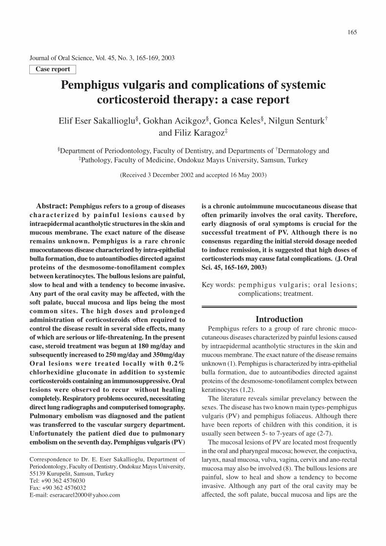
165
Abstract: Pemphigus refers to a group of diseasescharacter ized by painful les ions caused byintraepidermal acantholytic structures in the skin andmucous membrane. The exact nature of the diseaseremains unknown. Pemphigus is a rare chronicmucocutaneous disease characterized by intra-epithelialbulla formation, due to autoantibodies directed againstproteins of the desmosome-tonofilament complexbetween keratinocytes. The bullous lesions are painful,slow to heal and with a tendency to become invasive.Any part of the oral cavity may be affected, with thesoft palate, buccal mucosa and lips being the mostcommon sites. The high doses and prolongedadministration of corticosteroids often required tocontrol the disease result in several side effects, manyof which are serious or life-threatening. In the presentcase, steroid treatment was begun at 180 mg/day andsubsequently increased to 250 mg/day and 350mg/dayOral lesions were treated locally with 0.2%chlorhexidine gluconate in addition to systemiccorticosteroids containing an immunosuppressive. Orallesions were observed to recur without healingcompletely. Respiratory problems occured, necessitatingdirect lung radiographs and computerised tomography.Pulmonary embolism was diagnosed and the patientwas transferred to the vascular surgery department.Unfortunately the patient died due to pulmonaryembolism on the seventh day. Pemphigus vulgaris (PV)
is a chronic autoimmune mucocutaneous disease thatoften primarily involves the oral cavity. Therefore,early diagnosis of oral symptoms is crucial for thesuccessful treatment of PV. Although there is noconsensus regarding the initial steroid dosage neededto induce remission, it is suggested that high doses ofcorticosteriods may cause fatal complications. (J. OralSci. 45, 165-169, 2003)
Key words: pemphigus vulgaris; oral lesions;complications; treatment.
IntroductionPemphigus refers to a group of rare chronic muco-
cutaneous diseases characterized by painful lesions causedby intraepidermal acantholytic structures in the skin andmucous membrane. The exact nature of the disease remainsunknown (1). Pemphigus is characterized by intra-epithelialbulla formation, due to autoantibodies directed againstproteins of the desmosome-tonofilament complex betweenkeratinocytes (1,2).
The literature reveals similar prevelancy between thesexes. The disease has two known main types-pemphigusvulgaris (PV) and pemphigus foliaceus. Although therehave been reports of children with this condition, it isusually seen between 5- to 7-years of age (2-7).
The mucosal lesions of PV are located most frequentlyin the oral and pharyngeal mucosa; however, the conjuctiva,larynx, nasal mucosa, vulva, vagina, cervix and ano-rectalmucosa may also be involved (8). The bullous lesions arepainful, slow to heal and show a tendency to becomeinvasive. Although any part of the oral cavity may beaffected, the soft palate, buccal mucosa and lips are the
Journal of Oral Science, Vol. 45, No. 3, 165-169, 2003
Correspondence to Dr. E. Eser Sakallioglu, Department ofPeriodontology, Faculty of Dentistry, Ondokuz Mayıs University,55139 Kurupelit, Samsun, TurkeyTel: +90 362 4576030Fax: +90 362 4576032E-mail: [email protected]
Pemphigus vulgaris and complications of systemiccorticosteroid therapy: a case report
Elif Eser Sakallioglu§, Gokhan Acikgoz§, Gonca Keles§, Nilgun Senturk†
and Filiz Karagoz‡
§Department of Periodontology, Faculty of Dentistry, and Departments of †Dermatology and ‡Pathology, Faculty of Medicine, Ondokuz Mayıs University, Samsun, Turkey
(Received 3 December 2002 and accepted 16 May 2003)
Case report
166
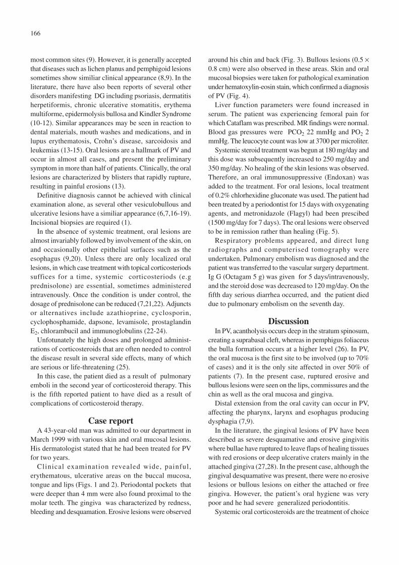
most common sites (9). However, it is generally acceptedthat diseases such as lichen planus and pemphigoid lesionssometimes show similiar clinical appearance (8,9). In theliterature, there have also been reports of several otherdisorders manifesting DG including psoriasis, dermatitisherpetiformis, chronic ulcerative stomatitis, erythemamultiforme, epidermolysis bullosa and Kindler Syndrome(10-12). Similar appearances may be seen in reaction todental materials, mouth washes and medications, and inlupus erythematosis, Crohn’s disease, sarcoidosis andleukemias (13-15). Oral lesions are a hallmark of PV andoccur in almost all cases, and present the preliminarysymptom in more than half of patients. Clinically, the orallesions are characterized by blisters that rapidly rupture,resulting in painful erosions (13).
Definitive diagnosis cannot be achieved with clinicalexamination alone, as several other vesiculobullous andulcerative lesions have a similiar appearance (6,7,16-19).Incisional biopsies are required (1).
In the absence of systemic treatment, oral lesions arealmost invariably followed by involvement of the skin, onand occasionally other epithelial surfaces such as theesophagus (9,20). Unless there are only localized orallesions, in which case treatment with topical corticosteriodssuffices for a time, systemic corticosteriods (e.gprednisolone) are essential, sometimes administeredintravenously. Once the condition is under control, thedosage of prednisolone can be reduced (7,21,22). Adjunctsor alternatives include azathioprine, cyclosporin,cyclophosphamide, dapsone, levamisole, prostaglandinE2, chlorambucil and immunoglobulins (22-24).
Unfotunately the high doses and prolonged administ-rations of corticosteroids that are often needed to controlthe disease result in several side effects, many of whichare serious or life-threatening (25).
In this case, the patient died as a result of pulmonaryemboli in the second year of corticosteroid therapy. Thisis the fifth reported patient to have died as a result ofcomplications of corticosteroid therapy.
Case reportA 43-year-old man was admitted to our department in
March 1999 with various skin and oral mucosal lesions.His dermatologist stated that he had been treated for PVfor two years.
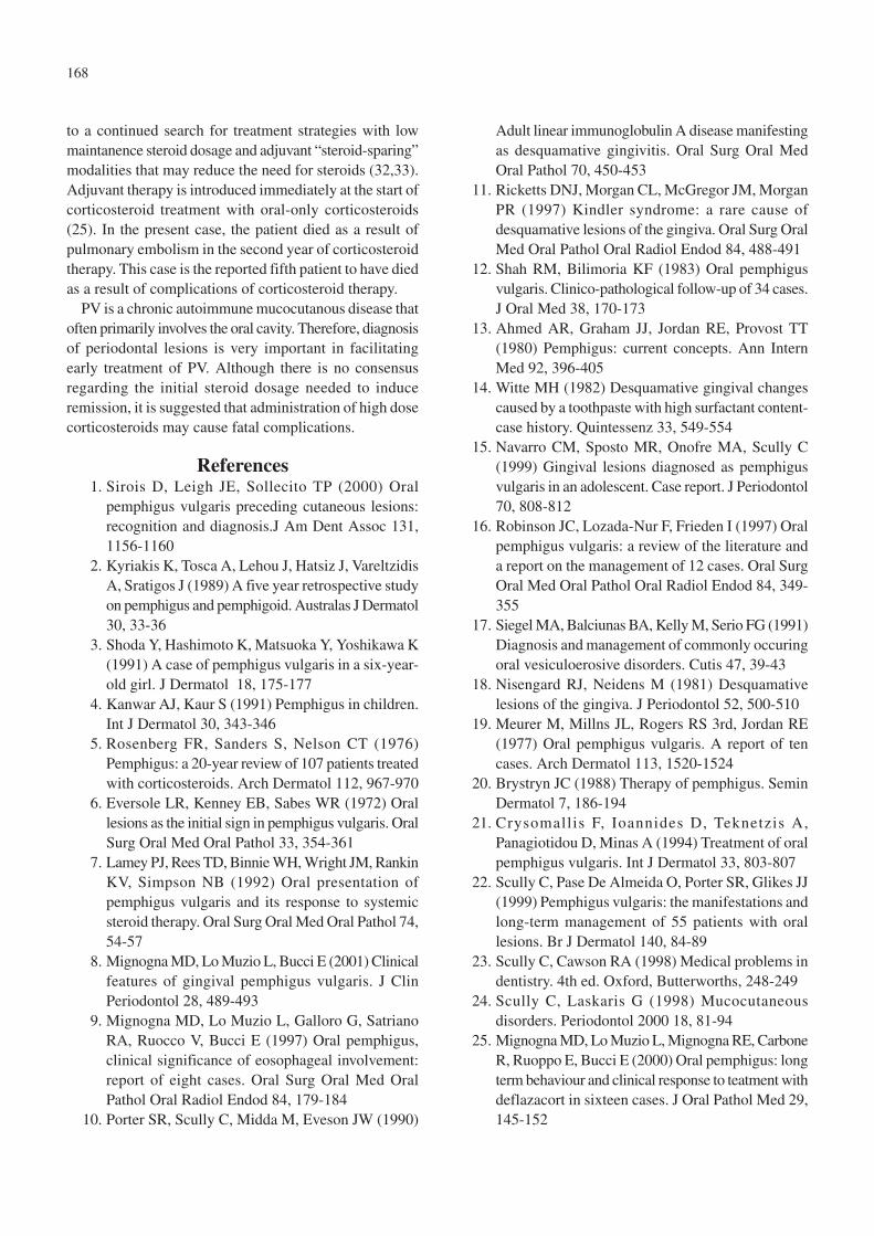
Clinical examination revealed wide, painful,erythematous, ulcerative areas on the buccal mucosa,tongue and lips (Figs. 1 and 2). Periodontal pockets thatwere deeper than 4 mm were also found proximal to themolar teeth. The gingiva was characterized by redness,bleeding and desquamation. Erosive lesions were observed
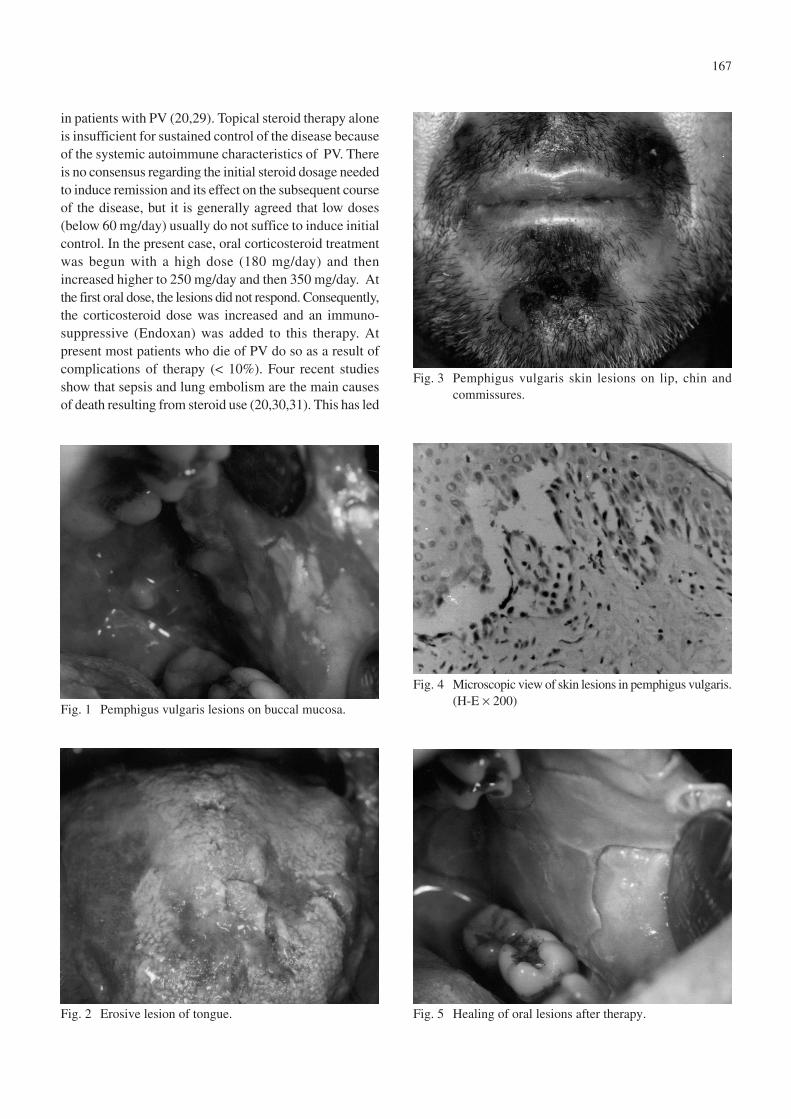
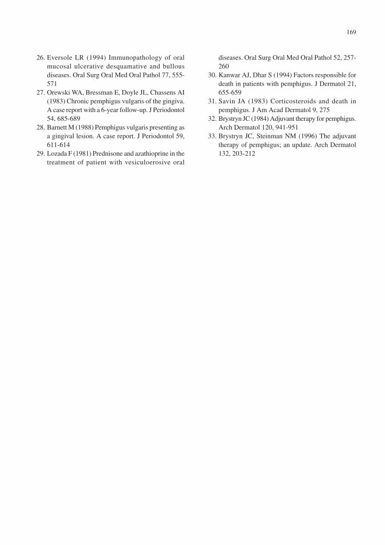
around his chin and back (Fig. 3). Bullous lesions (0.5 ×0.8 cm) were also observed in these areas. Skin and oralmucosal biopsies were taken for pathological examinationunder hematoxylin-eosin stain, which confirmed a diagnosisof PV (Fig. 4).
Liver function parameters were found increased inserum. The patient was experiencing femoral pain forwhich Cataflam was prescribed. MR findings were normal.Blood gas pressures were PCO2 22 mmHg and PO2 2mmHg. The leucocyte count was low at 3700 per microliter.
Systemic steroid treatment was begun at 180 mg/day andthis dose was subsequently increased to 250 mg/day and350 mg/day. No healing of the skin lesions was observed.Therefore, an oral immunosuppressive (Endoxan) wasadded to the treatment. For oral lesions, local treatmentof 0.2% chlorhexidine gluconate was used. The patient hadbeen treated by a periodontist for 15 days with oxygenatingagents, and metronidazole (Flagyl) had been prescibed(1500 mg/day for 7 days). The oral lesions were observedto be in remission rather than healing (Fig. 5).
Respiratory problems appeared, and direct lungradiographs and computerised tomography wereundertaken. Pulmonary embolism was diagnosed and thepatient was transferred to the vascular surgery department.Ig G (Octagam 5 g) was given for 5 days/intravenously,and the steroid dose was decreased to 120 mg/day. On thefifth day serious diarrhea occurred, and the patient dieddue to pulmonary embolism on the seventh day.
DiscussionIn PV, acantholysis occurs deep in the stratum spinosum,
creating a suprabasal cleft, whereas in pemphigus foliaceusthe bulla formation occurs at a higher level (26). In PV,the oral mucosa is the first site to be involved (up to 70%of cases) and it is the only site affected in over 50% ofpatients (7). In the present case, ruptured erosive andbullous lesions were seen on the lips, commissures and thechin as well as the oral mucosa and gingiva.
Distal extension from the oral cavity can occur in PV,affecting the pharynx, larynx and esophagus producingdysphagia (7,9).
In the literature, the gingival lesions of PV have beendescribed as severe desquamative and erosive gingivitiswhere bullae have ruptured to leave flaps of healing tissueswith red erosions or deep ulcerative craters mainly in theattached gingiva (27,28). In the present case, although thegingival desquamative was present, there were no erosivelesions or bullous lesions on either the attached or freegingiva. However, the patient’s oral hygiene was verypoor and he had severe generalized periodontitis.
Systemic oral corticosteroids are the treatment of choice
167
in patients with PV (20,29). Topical steroid therapy aloneis insufficient for sustained control of the disease becauseof the systemic autoimmune characteristics of PV. Thereis no consensus regarding the initial steroid dosage neededto induce remission and its effect on the subsequent courseof the disease, but it is generally agreed that low doses(below 60 mg/day) usually do not suffice to induce initialcontrol. In the present case, oral corticosteroid treatmentwas begun with a high dose (180 mg/day) and thenincreased higher to 250 mg/day and then 350 mg/day. Atthe first oral dose, the lesions did not respond. Consequently,the corticosteroid dose was increased and an immuno-suppressive (Endoxan) was added to this therapy. Atpresent most patients who die of PV do so as a result ofcomplications of therapy (< 10%). Four recent studiesshow that sepsis and lung embolism are the main causesof death resulting from steroid use (20,30,31). This has led
Fig. 3 Pemphigus vulgaris skin lesions on lip, chin andcommissures.
Fig. 4 Microscopic view of skin lesions in pemphigus vulgaris.(H-E × 200)
Fig. 2 Erosive lesion of tongue. Fig. 5 Healing of oral lesions after therapy.
Fig. 1 Pemphigus vulgaris lesions on buccal mucosa.
168
to a continued search for treatment strategies with lowmaintanence steroid dosage and adjuvant “steroid-sparing”modalities that may reduce the need for steroids (32,33).Adjuvant therapy is introduced immediately at the start ofcorticosteroid treatment with oral-only corticosteroids(25). In the present case, the patient died as a result ofpulmonary embolism in the second year of corticosteroidtherapy. This case is the reported fifth patient to have diedas a result of complications of corticosteroid therapy.
PV is a chronic autoimmune mucocutanous disease thatoften primarily involves the oral cavity. Therefore, diagnosisof periodontal lesions is very important in facilitatingearly treatment of PV. Although there is no consensusregarding the initial steroid dosage needed to induceremission, it is suggested that administration of high dosecorticosteroids may cause fatal complications.
References1. Sirois D, Leigh JE, Sollecito TP (2000) Oral
pemphigus vulgaris preceding cutaneous lesions:recognition and diagnosis.J Am Dent Assoc 131,1156-1160
2. Kyriakis K, Tosca A, Lehou J, Hatsiz J, VareltzidisA, Sratigos J (1989) A five year retrospective studyon pemphigus and pemphigoid. Australas J Dermatol30, 33-36
3. Shoda Y, Hashimoto K, Matsuoka Y, Yoshikawa K(1991) A case of pemphigus vulgaris in a six-year-old girl. J Dermatol 18, 175-177
4. Kanwar AJ, Kaur S (1991) Pemphigus in children.Int J Dermatol 30, 343-346
5. Rosenberg FR, Sanders S, Nelson CT (1976)Pemphigus: a 20-year review of 107 patients treatedwith corticosteroids. Arch Dermatol 112, 967-970
6. Eversole LR, Kenney EB, Sabes WR (1972) Orallesions as the initial sign in pemphigus vulgaris. OralSurg Oral Med Oral Pathol 33, 354-361
7. Lamey PJ, Rees TD, Binnie WH, Wright JM, RankinKV, Simpson NB (1992) Oral presentation ofpemphigus vulgaris and its response to systemicsteroid therapy. Oral Surg Oral Med Oral Pathol 74,54-57
8. Mignogna MD, Lo Muzio L, Bucci E (2001) Clinicalfeatures of gingival pemphigus vulgaris. J ClinPeriodontol 28, 489-493
9. Mignogna MD, Lo Muzio L, Galloro G, SatrianoRA, Ruocco V, Bucci E (1997) Oral pemphigus,clinical significance of eosophageal involvement:report of eight cases. Oral Surg Oral Med OralPathol Oral Radiol Endod 84, 179-184
10. Porter SR, Scully C, Midda M, Eveson JW (1990)
Adult linear immunoglobulin A disease manifestingas desquamative gingivitis. Oral Surg Oral MedOral Pathol 70, 450-453
11. Ricketts DNJ, Morgan CL, McGregor JM, MorganPR (1997) Kindler syndrome: a rare cause ofdesquamative lesions of the gingiva. Oral Surg OralMed Oral Pathol Oral Radiol Endod 84, 488-491
12. Shah RM, Bilimoria KF (1983) Oral pemphigusvulgaris. Clinico-pathological follow-up of 34 cases.J Oral Med 38, 170-173
13. Ahmed AR, Graham JJ, Jordan RE, Provost TT(1980) Pemphigus: current concepts. Ann InternMed 92, 396-405
14. Witte MH (1982) Desquamative gingival changescaused by a toothpaste with high surfactant content-case history. Quintessenz 33, 549-554
15. Navarro CM, Sposto MR, Onofre MA, Scully C(1999) Gingival lesions diagnosed as pemphigusvulgaris in an adolescent. Case report. J Periodontol70, 808-812
16. Robinson JC, Lozada-Nur F, Frieden I (1997) Oralpemphigus vulgaris: a review of the literature anda report on the management of 12 cases. Oral SurgOral Med Oral Pathol Oral Radiol Endod 84, 349-355
17. Siegel MA, Balciunas BA, Kelly M, Serio FG (1991)Diagnosis and management of commonly occuringoral vesiculoerosive disorders. Cutis 47, 39-43
18. Nisengard RJ, Neidens M (1981) Desquamativelesions of the gingiva. J Periodontol 52, 500-510
19. Meurer M, Millns JL, Rogers RS 3rd, Jordan RE(1977) Oral pemphigus vulgaris. A report of tencases. Arch Dermatol 113, 1520-1524
20. Brystryn JC (1988) Therapy of pemphigus. SeminDermatol 7, 186-194
21. Crysomallis F, Ioannides D, Teknetzis A,Panagiotidou D, Minas A (1994) Treatment of oralpemphigus vulgaris. Int J Dermatol 33, 803-807
22. Scully C, Pase De Almeida O, Porter SR, Glikes JJ(1999) Pemphigus vulgaris: the manifestations andlong-term management of 55 patients with orallesions. Br J Dermatol 140, 84-89
23. Scully C, Cawson RA (1998) Medical problems indentistry. 4th ed. Oxford, Butterworths, 248-249
24. Scully C, Laskaris G (1998) Mucocutaneousdisorders. Periodontol 2000 18, 81-94
25. Mignogna MD, Lo Muzio L, Mignogna RE, CarboneR, Ruoppo E, Bucci E (2000) Oral pemphigus: longterm behaviour and clinical response to teatment withdeflazacort in sixteen cases. J Oral Pathol Med 29,145-152
169
26. Eversole LR (1994) Immunopathology of oralmucosal ulcerative desquamative and bullousdiseases. Oral Surg Oral Med Oral Pathol 77, 555-571
27. Orewski WA, Bressman E, Doyle JL, Chassens AI(1983) Chronic pemphigus vulgaris of the gingiva.A case report with a 6-year follow-up. J Periodontol54, 685-689
28. Barnett M (1988) Pemphigus vulgaris presenting asa gingival lesion. A case report. J Periodontol 59,611-614
29. Lozada F (1981) Prednisone and azathioprine in thetreatment of patient with vesiculoerosive oral
diseases. Oral Surg Oral Med Oral Pathol 52, 257-260
30. Kanwar AJ, Dhar S (1994) Factors responsible fordeath in patients with pemphigus. J Dermatol 21,655-659
31. Savin JA (1983) Corticosteroids and death inpemphigus. J Am Acad Dermatol 9, 275
32. Brystryn JC (1984) Adjuvant therapy for pemphigus.Arch Dermatol 120, 941-951
33. Brystryn JC, Steinman NM (1996) The adjuvanttherapy of pemphigus; an update. Arch Dermatol132, 203-212

![Pemphigus Vulgaris [Print] - eMedicine Dermatology Vulgaris .pdf · emedicine.medscape.com eMedicine Specialties > Dermatology > Bullous Diseases Pemphigus Vulgaris Bassam Zeina,](https://img.pdfslide.net/doc/110x75/5c984ab609d3f21c3a8b874e/pemphigus-vulgaris-print-emedicine-vulgaris-pdf-emedicinemedscapecom.jpg)


![Oral Manifestations of Pemphigus Vulgaris: Clinical ... · bullous pemphigus, and paraneoplastic pemphigus [4]. The differential diagnosis includes other dermatological diseases with](https://img.pdfslide.net/doc/110x75/5cbb138688c9930c5f8bb27d/oral-manifestations-of-pemphigus-vulgaris-clinical-bullous-pemphigus-and.jpg)