Embed Size (px)
Citation preview
© 2016 Journal of Oral and Maxillofacial Pathology | Published by Wolters Kluwer - Medknow
Oral pemphigus vulgaris: A case report with direct immunofluorescence study
Sangeetha Jeevan Kumar, SP Nehru Anand1, Nandhini Gunasekaran, Rajkumar KrishnanDepartments of Oral Pathology and 1Oral Medicine, SRM Dental College, Chennai, Tamil Nadu, India
INTRODUCTION
Pemphigus refers to a group of autoimmune, mucocutaneous blistering diseases, in which the keratinocyte antigens are the target of the autoantibodies, leading to acantholysis and blister formation.[1] The word pemphigus originates from Greek pemphix, which translates as blister or bubble. Pemphigus can be classified into five major groups: Pemphigus vulgaris (PV), pemphigus foliaceus, paraneoplastic pemphigus (PNP), drug‑induced pemphigus and immunoglobulin A (IgA) pemphigus. Oral lesions have been associated with only PV and PNP.[2]
The cause of the disease remains obscure and many theories had been put forward. One commonly accepted theory is the loss of cell adhesion caused by the loss of desmoglein (Dsg);
one of the cell adhesion molecules being targeted by IgG autoantibodies, leading to acantholysis. Dsg‑1 is expressed in all layers of the epidermis, with a higher concentration in the more superficial layers, whereas Dsg‑3 is expressed in the parabasal and basal layers. The presence of a suprabasilar split reflects the expression of Dsg‑3 and relative lack of Dsg‑1 in the oral mucosa. The presence of antibodies to both, Dsg‑1 and Dsg‑3, is seen in patients who develop skin lesions. Another theory, known as the multiple hit hypothesis, suggests that pemphigus occurs as an effect of anti‑acetylcholine receptor antibodies, triggering acantholysis by weakening the cohesion of adjacent keratinocytes due to inhibition of the physiological control of their polygonal shape and intercellular attachment.[1]
Pemphigus vulgaris (PV) is a chronic, autoimmune, intraepidermal blistering disease of the skin and mucous membranes. The initial clinical manifestation is frequently the development of intraoral lesions, and later, the lesions involve the other mucous membranes and skin. The etiology of this disease still remains obscure although the presence of autoantibodies is consistent with an autoimmune disease. These antibodies are targeted against the adhesion proteins of keratinocytes, leading to acantholysis (disruption of spinous layer, leading to intraepidermal clefting) and blister formation. Because only oral lesions are present initially, the chances of misdiagnosing the disease as another condition are increased, leading to inappropriate therapy. In this article, we report a case of PV with only oral manifestations in a 36‑year‑old male.
Key Words: Autoantibodies, immunofluorescence, intra epithelial vesicle, pemphigus, ulceration
Abstract
Address for correspondence: Dr. Sangeetha Jeevan Kumar, Department of Oral Pathology, SRM Dental College, Ramapuram, Chennai ‑ 600 089, Tamil Nadu, India. E‑mail: [email protected]: 29.06.2015, Accepted: 26.08.2016
Access this article onlineQuick Response Code:
Website:www.jomfp.in
DOI:10.4103/0973-029X.190979
How to c i te th is a r t i c le : Kumar SJ , Neh ru Anand SP, Gunasekaran N, Krishnan R. Oral pemphigus vulgaris: A case report with direct immunofluorescence study. J Oral Maxillofac Pathol 2016;20:549.
This is an open access article distributed under the terms of the Creative Commons Attribution‑NonCommercial‑ShareAlike 3.0 License, which allows others to remix, tweak, and build upon the work non‑commercially, as long as the author is credited and the new creations are licensed under the identical terms.
For reprints contact: [email protected]
Case Report
Kumar, et al.: Oral pemphigus vulgaris
Journal of Oral and Maxillofacial Pathology | Sep - Dec 2016 | Vol 20 | Issue 3
CASE REPORT
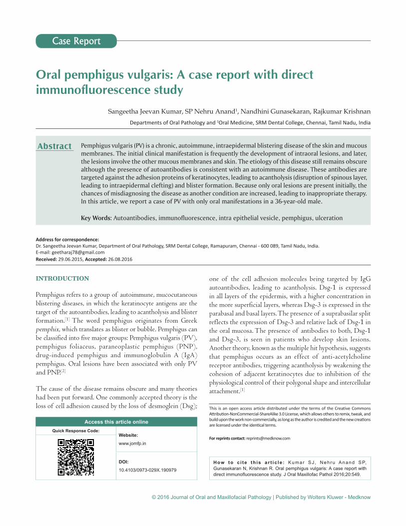
A 36‑year‑old male reported to the Department of Oral Medicine with a history of painful ulcerations involving the palate and gingiva for the past 2 weeks. History revealed that the ulcerations started initially as blisters and were associated with pain that was aggravated on chewing food. The ulcerations caused considerable discomfort, affecting his normal oral functions. Personal and family histories were uneventful.
Intraoral examination revealed areas of erosions on the palate and the lower vestibule [Figure 1a and b]. The gingivae appeared erythematous and showed numerous erosions on the marginal gingiva [Figure 1a]. The gingival mucosa exhibited a positive Nikolsky sign. Extraoral examination revealed no skin lesions.
Based on the history and clinical examination, a provisional diagnosis of PV was made although the differential diagnosis included mucous membrane pemphigoid and erosive lichen planus.
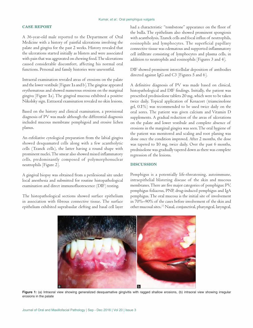
An exfoliative cytological preparation from the labial gingiva showed desquamated cells along with a few acantholytic cells (Tzanck cells), the latter having a round shape with prominent nuclei. The smear also showed mixed inflammatory cells, predominantly composed of polymorphonuclear neutrophils [Figure 2].
A gingival biopsy was obtained from a perilesional site under local anesthesia and submitted for routine histopathological examination and direct immunofluorescence (DIF) testing.
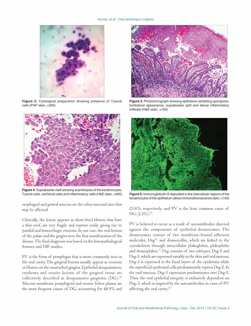
The histopathological sections showed surface epithelium in association with fibrous connective tissue. The surface epithelium exhibited suprabasilar clefting and basal cell layer
had a characteristic “tombstone” appearance on the floor of the bulla. The epithelium also showed prominent spongiosis with acantholysis, Tzanck cells and focal influx of neutrophils, eosinophils and lymphocytes. The superficial papillary connective tissue was edematous and supported inflammatory cell infiltrate consisting of lymphocytes and plasma cells, in addition to neutrophils and eosinophils [Figures 3 and 4].
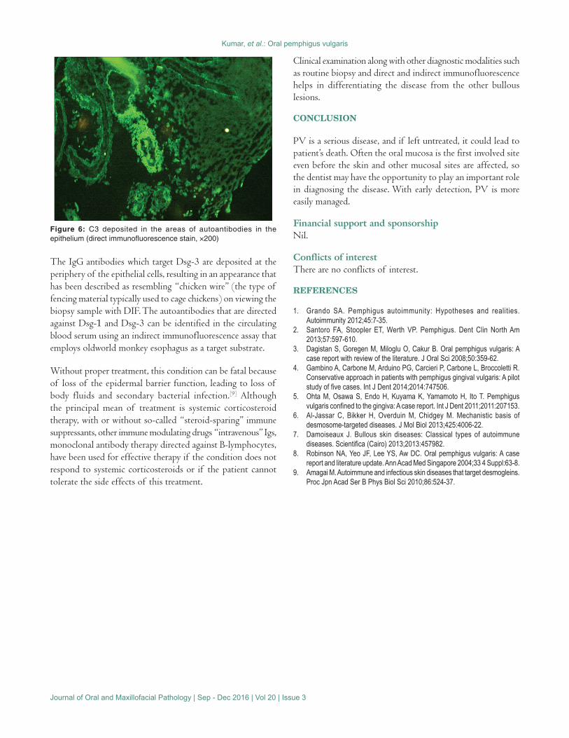
DIF showed prominent intercellular deposition of antibodies directed against IgG and C3 [Figures 5 and 6].
A definitive diagnosis of PV was made based on clinical, histopathological and DIF findings. Initially, the patient was prescribed prednisolone tablets 20 mg, which were to be taken twice daily. Topical application of Kenacort (triamcinolone gel, 0.1%) was recommended to be used twice daily on the oral sores. The patient was given calcium and Vitamin D supplements. A gradual reduction of the areas of ulcerations on the palate and lower vestibule and complete absence of erosions in the marginal gingiva was seen. The oral hygiene of the patient was monitored and scaling and root planing was done once the condition improved. After 2 months, the dose was tapered to 10 mg, twice daily. Over the past 6 months, prednisolone was gradually tapered down as there was complete regression of the lesions.
DISCUSSION
Pemphigus is a potentially life‑threatening, autoimmune, intraepithelial blistering disease of the skin and mucous membranes. There are five major categories of pemphigus: PV, pemphigus foliaceus, PNP, drug‑induced pemphigus and IgA pemphigus. The oral mucosa is the initial site of involvement in 70%–90% of the cases before involvement of the skin and other mucosal sites.[3] Nasal, conjunctival, pharyngeal, laryngeal,
Figure 1: (a) Intraoral view showing generalized desquamative gingivitis with ragged shallow erosions, (b) intraoral view showing irregular erosions in the palate
ba
Kumar, et al.: Oral pemphigus vulgaris
Journal of Oral and Maxillofacial Pathology | Sep - Dec 2016 | Vol 20 | Issue 3
esophageal and genital mucosa are the other mucosal sites that may be affected.
Clinically, the lesion appears as short‑lived blisters that have a thin roof, are very fragile and rupture easily, giving rise to painful and hemorrhagic erosions. In our case, the oral lesions of the palate and the gingiva were the first manifestation of the disease. The final diagnosis was based on the histopathological features and DIF studies.
PV is the form of pemphigus that is most commonly seen in the oral cavity. The gingival lesions usually appear as erosions or blisters on the unattached gingiva. Epithelial desquamation, erythema and erosive lesions of the gingival tissue are collectively described as desquamative gingivitis (DG).[4] Mucous membrane pemphigoid and erosive lichen planus are the most frequent causes of DG, accounting for 48.9% and
23.6%, respectively, and PV is the least common cause of DG (2.3%).[5]
PV is believed to occur as a result of autoantibodies directed against the components of epithelial desmosomes. The desmosomes consist of two membrane‑bound adhesion molecules, Dsg[6] and desmocollin, which are linked to the cytoskeleton through intracellular plakoglobin, plakophilin and desmoplakin.[7] Dsg consists of two subtypes, Dsg‑1 and Dsg‑3, which are expressed variably in the skin and oral mucosa. Dsg‑3 is expressed in the basal layers of the epidermis while the superficial epidermal cells predominantly express Dsg‑1. In the oral mucosa, Dsg‑3 expression predominates over Dsg‑1. Thus, the oral epithelial integrity is exclusively dependent on Dsg‑3, which is targeted by the autoantibodies in cases of PV affecting the oral cavity.[8]
Figure 2: Cytological preparation showing presence of Tzanck cells (PAP stain, ×200)
Figure 3: Photomicrograph showing epithelium exhibiting spongiosis, tombstone appearance, suprabasilar split and dense inflammatory infiltrate (H&E stain, ×100)
Figure 4: Suprabasilar cleft showing acantholysis of the keratinocytes, Tzanck cells, red blood cells and inflammatory cells (H&E stain, ×400) Figure 5: Immunoglobulin G deposited in the intercellular regions of the
keratinocytes of the epithelium (direct immunofluorescence stain, ×100)
Kumar, et al.: Oral pemphigus vulgaris
Journal of Oral and Maxillofacial Pathology | Sep - Dec 2016 | Vol 20 | Issue 3
The IgG antibodies which target Dsg‑3 are deposited at the periphery of the epithelial cells, resulting in an appearance that has been described as resembling “chicken wire” (the type of fencing material typically used to cage chickens) on viewing the biopsy sample with DIF. The autoantibodies that are directed against Dsg‑1 and Dsg‑3 can be identified in the circulating blood serum using an indirect immunofluorescence assay that employs oldworld monkey esophagus as a target substrate.
Without proper treatment, this condition can be fatal because of loss of the epidermal barrier function, leading to loss of body fluids and secondary bacterial infection.[9] Although the principal mean of treatment is systemic corticosteroid therapy, with or without so‑called “steroid‑sparing” immune suppressants, other immune modulating drugs “intravenous” Igs, monoclonal antibody therapy directed against B‑lymphocytes, have been used for effective therapy if the condition does not respond to systemic corticosteroids or if the patient cannot tolerate the side effects of this treatment.
Clinical examination along with other diagnostic modalities such as routine biopsy and direct and indirect immunofluorescence helps in differentiating the disease from the other bullous lesions.
CONCLUSION
PV is a serious disease, and if left untreated, it could lead to patient’s death. Often the oral mucosa is the first involved site even before the skin and other mucosal sites are affected, so the dentist may have the opportunity to play an important role in diagnosing the disease. With early detection, PV is more easily managed.
Financial support and sponsorshipNil.
Conflicts of interestThere are no conflicts of interest.
REFERENCES
1. Grando SA. Pemphigus autoimmunity: Hypotheses and realities. Autoimmunity 2012;45:7‑35.
2. Santoro FA, Stoopler ET, Werth VP. Pemphigus. Dent Clin North Am 2013;57:597‑610.
3. Dagistan S, Goregen M, Miloglu O, Cakur B. Oral pemphigus vulgaris: A case report with review of the literature. J Oral Sci 2008;50:359‑62.
4. Gambino A, Carbone M, Arduino PG, Carcieri P, Carbone L, Broccoletti R. Conservative approach in patients with pemphigus gingival vulgaris: A pilot study of five cases. Int J Dent 2014;2014:747506.
5. Ohta M, Osawa S, Endo H, Kuyama K, Yamamoto H, Ito T. Pemphigus vulgaris confined to the gingiva: A case report. Int J Dent 2011;2011:207153.
6. Al‑Jassar C, Bikker H, Overduin M, Chidgey M. Mechanistic basis of desmosome‑targeted diseases. J Mol Biol 2013;425:4006‑22.
7. Damoiseaux J. Bullous skin diseases: Classical types of autoimmune diseases. Scientifica (Cairo) 2013;2013:457982.
8. Robinson NA, Yeo JF, Lee YS, Aw DC. Oral pemphigus vulgaris: A case report and literature update. Ann Acad Med Singapore 2004;33 4 Suppl:63‑8.
9. Amagai M. Autoimmune and infectious skin diseases that target desmogleins. Proc Jpn Acad Ser B Phys Biol Sci 2010;86:524‑37.
Figure 6: C3 deposited in the areas of autoantibodies in the epithelium (direct immunofluorescence stain, ×200)


![Case Report AAtypical presentation of pemphigus vulgaris - A … · 2018-12-03 · involvement and pemphigus vulgaris presents as oral lesions in 50 to 70% patients [1-3]. These may](https://img.pdfslide.net/doc/110x75/5ccfc74d88c993cc718c625a/case-report-aatypical-presentation-of-pemphigus-vulgaris-a-2018-12-03.jpg)


![Oral Manifestations of Pemphigus Vulgaris: Clinical ... · bullous pemphigus, and paraneoplastic pemphigus [4]. The differential diagnosis includes other dermatological diseases with](https://img.pdfslide.net/doc/110x75/5cbb138688c9930c5f8bb27d/oral-manifestations-of-pemphigus-vulgaris-clinical-bullous-pemphigus-and.jpg)