Embed Size (px)
Citation preview

Presbyopia & Toric IOL Correction: Keys to Success with Multifocal & EDOF IOLs
SupplementDecember 2019/ January 2020

1
Trends in Presbyopia and Astigmatism Correction: 2018 ESCRS Clinical Survey ResultsBy Oliver Findl MD
19% of trifocal and 16% of EDOF IOL patients would have these visual
disturbances.
Toric IOL cost considerations play a significant role in IOL choice. Just
13% of respondents’ current cataract procedures involve toric IOLs,
but if cost were not an issue, they would choose toric IOLs for 61% of
cataract patients who have significant astigmatism.
ESCRS delegates were asked what their most common procedure to
manage astigmatism in monofocal cataract patients with 0.75D, 1.25D
and 1.75D of cylinder, and their responses indicate that for higher
astigmatism, toric lenses are the top choice, but for lower amounts of
astigmatism, on-axis incision seems to be an alternative.
The survey revealed that a third of delegates believe that 10 or
more degrees of rotational error is acceptable in patients who receive
a toric IOL, before visual quality and degradation of visual acuity are
significantly affected. Nearly 50% say 5-to-9 degrees of rotational error
is acceptable, and just 18% said less than 5 degrees is acceptable. Over
the past four years, the number of ESCRS delegates finding 10 degrees
or more of rotational error acceptable has significantly decreased, from
52% in 2014 to 34% in 2018 (Figure 2).
Oliver Findl MD is chief of the Department of Ophthalmology, Vienna Hanusch Hospital, and founder of the Vienna Institute for Research in Ocular Surgery, Austria.
E-mail: [email protected]
Financial disclosures: Dr Findl is a scientific adviser to Alcon, ZEISS and Johnson & Johnson Vision.
T he fourth annual ESCRS Clinical Survey revealed some
interesting trends about the use of multifocal, toric, and
extended depth of focus (EDOF) intraocular lenses (IOLs) for
the correction of presbyopia. For instance, while only 11% of current
cataract procedures among responding surgeons included presbyopia-
correcting IOLs, almost three times as many – 30% – use monovision or
mini-monovision as an alternative.
When asked how satisfied their patients are with monovision
vs presbyopia-correcting IOLs one year postoperatively, surgeons
reported that patient satisfaction for near, intermediate and distance
vision is higher in presbyopia-correcting IOL patients – especially for
intermediate, but even more so for near vision.
Participants were asked to identify the lowest amount of postoperative
residual cylinder error they consider to be visually significant in patients
implanted with bifocal/trifocal or EDOF IOLs. The responses suggest
that EDOF IOLs are believed to accept higher levels of cylinder error,
especially for higher amounts of astigmatism in the range of 0.75D to
more than 1.0D (Figure 1).
The cost to patients and concerns over contrast visual acuity and
nighttime quality of vision are the main reasons for not performing more
presbyopia-correcting IOL procedures. Trifocal and EDOF IOL patients
are believed to have over three times the chance of having significant
aberrations at night even if the patient has no residual refractive error
and a healthy ocular surface. Respondents said they believed just 5% of
monovision patients would have significant aberrations at night, while
Over time the number of ESCRS delegates finding 10 degrees or more of rotational error acceptable has significantly decreased
EDOF IOLs are believed to accept higher levels of cylinder error
In patients implanted with a bifocal/trifocal or an EDOF IOL, what is the lowest amount of postoperative residual CYLINDER error that is considered to be visually significant in DIOPTRES?
26%
0.5D or less
24%
41%
0.51D or 0.75D
Bifocal/Trifocal
31%
26%
0.76D or 1.0D
34%
7%
Over 1.0D
12%
EDOF
Figure 1: EDOF IOLs are believed to accept higher levels of cylinder error
Figure 2: Over time the number of ESCRS delegates finding 10 degrees or more of rotational error acceptable significantly decreased
After implanting a toric IOL, how many DEGREES of postoperative rotational error is acceptable before visual quality and degradation of visual acuity aresignificantly affected? ANSWER: 10 OR MORE DEGREES
52%
2014 2016 2017 2018
47%
40%
34%

2
Refractive IOLs: Importance of Patient Selection and Realistic Expectations By Béatrice Cochener-Lamard MD, PhD
in binocular vision, as well as any progressive systemic disease, such as
diabetes or auto-immune disease.
Other factors that should raise a red flag include a personal or family
history of keratoconus and age-related macular degeneration. It is also
recommended to exclude patients who are younger than 55 and have
a lens longer than 24mm, except in the case of traumatic or unilateral
congenital cataract, because their risk of retinal complications is higher.
KEY VALUATION PARAMETERSWhen evaluating a patient for RIOL surgery, consider their visual
function, near and far visual acuities, as well as intermediate vision
from 60-to-80cm. Also factor in refraction and binocular vision and
oculomotricity, especially in case of amblyopia and hyperopia.
It is particularly important to examine the ocular surface for potential
dry eye disease (DED), which is present in 50% of cataract cases (Figure
3). The prevalence of DED increases with age, so it is particularly critical
in this patient population to evaluate the lipid layer for blepharitis
and meibomian gland dysfunction. A successful surgical outcome is
dependent on identifying and treating DED prior to surgery.
Patients must understand that no RIOL will enable them to return to
the vision that they had in their 20s: There is a neuroadaptation process
that takes anywhere from two weeks up to a few months; there is an
impact on quality of vision, especially at night; and visual performance
will depend on light conditions.
With respect to RIOL choice, I recommend this decision tree (Figure
4). When the patient is below age 55, I lean toward a corneal correction
approach with presbyLASIK, and monovision is still efficient in myopia.
Above that age, there is a place for refractive lens exchange with
presbyopic IOLs, if inclusion criteria are respected. EDOF IOLs may best
serve true cataract patients, over 70 years of age, who are interested
in reduced spectacle dependence because they are more forgiving on
remaining refractive error and less demanding in vision quality.
T hanks to surgical progress and better
understanding and control of optics that
make toric, multifocal and extended depth
of focus (EDOF) intraocular lenses (IOLs) possible,
ammetropia and spectacle independence can be
targeted in select cataract patients. Careful patient
selection and clear communication regarding
realistic expectations are the keys to success with
refractive IOLs (RIOLs).
There are two scenarios in which RIOLs are
indicated. The first one is refractive lens exchange,
where presbyopia patients seek an intraocular
solution to spectacle independence. The benefit-
to-risk ratio is best in the case of patients who are
over 55 years old when LASIK and monovision are
inadequate solutions because of insufficient results for near vision and
early loss of crystalline transparency, which can be graded nowadays by
densitometry or scatter light diffusion changes evaluation.
The second scenario in which RIOLs are indicated is in the case of
true cataract patients. Traditional monofocal cataract surgery would
simply enable these patients to recover lost visual acuity. With RIOLs, we
can do better, especially with the use of toric IOLs that correct corneal
astigmatism as spectacles do. We can even achieve complete spectacle
independence, with the use of EDOF or multifocal IOLs, if there are no
ocular or systemic contraindications.
When patients are amenable to this option, it is vital to educate them
about the surgery and what they can expect from it, and it is critical to
obtain their informed consent. This process is time consuming: You must
explain what presbyopia is, manage expectations, perform the surgery,
including astigmatism correction, and ensure that they fully comprehend
the advantages and limitations of all steps throughout the process,1
including the risk of not being able to use the selected IOL in case of
surgical complication.
PATIENT SELECTIONWhen choosing candidates for RIOLs, it is important to rule out those
who have unreasonable expectations. These patients must understand
that they should not expect perfect vision at all distances in all conditions
because of the potential impact on quality of vision. It is also important to
rule out candidates whose profession entails specific vision requirements,
such as a nightshift ambulance driver.
Furthermore, we must eliminate any candidates who have ocular
progressive diseases, such as glaucoma, maculopathy, or corneal
disturbances. We must also take into consideration any disturbances
When choosing candidates for RIOLs, it is important to rule out those who have unreasonable expectations
Figure 3: Dry eye disease, such as meibomian gland dysfunction, seen here, is underestimated in the presbyopic IOL patient population
Figure 4: Refractive IOL decision tree helps identify which presbyopic IOL is best for each patient
<55 yearsearly cataract, high hyperopia
<55 yearsRLE
<70 years
• Corneal strategies - PresbyLASIK
• Monovision
• Multifocal IOL
• EDOF IOL
• If no contraindication (ocular, systemic, psychological)
• Multifocal IOL ± toric
• If special need for night vision - EDOF IOL
• If no contraindication (ocular, systemic, psychological and patient demand) - EDOF IOL
- Piggyback Multifocal IOL

3
Keys to Success with Toric IOLsBy Douglas D. Koch MD
REFERENCES1. Buznego C, Trattler WB. Presbyopia-correcting intraocular lenses.
Curr Opin Ophthalmol. 2009;20:13–8.
Béatrice Cochener-Lamard MD, PhD, is Head of the Ophthalmology Department at the University Hospital of Brest, Brest, France.
E-mail: [email protected]
Financial disclosures: Dr Cochener-Lamard is a clinical investigator and consultant for Alcon, Allergan, Cutting Edge, Dompe, Johnson & Johnson Vision, Physiol, Santen, Théa and ZEISS.
CONCLUSIONIncorrect candidate selection, imperfect measurements and
insufficient patient information can result in RIOL surgery failure.
Even if intraocular solutions for presbyopic correction appear to be
a more predictable, more stable, and faster way than any corneal
approach to correction, it is important not to offer this option too early.
It is vital to select patients who are motivated and have reasonable
expectations, who understand the compromises involved in RIOLs
and who aren’t looking for vision perfection. It is important to discuss
with them issues such as neuroadaptation and visual function issues
related to light conditions.
T oric IOLs are clearly underutilised, with only 13% of participants
in last year’s ESCRS Clinical Survey acknowledging use of them
for surgical presbyopia correction. If you keep the following tips
in mind, you can optimise outcomes with premium IOLs and help your
patients achieve the spectacle independence and clear vision they want.
First, keep in mind that the threshold for correcting astigmatism is
very low. My target is less than 0.5D of total corneal astigmatism, so
precision in all steps is critical. One study by Hayashi looked at subjects
implanted with a +3.0D add multifocal IOL and then introduced 0.5D of
astigmatism. Uncorrected distance vision dropped from 20/20 to 20/30,
which is significant.1
Next, let’s consider use of technology. I use biometers that have light-
emitting diodes for identifying power and meridian. Topography is also
essential. Refraction sometimes provides a clue about the magnitude
and meridian of astigmatism. If the refraction differs significantly from
your other measurements, it may indicate something about the posterior
cornea or lens tilt. Most importantly, you must validate your data.
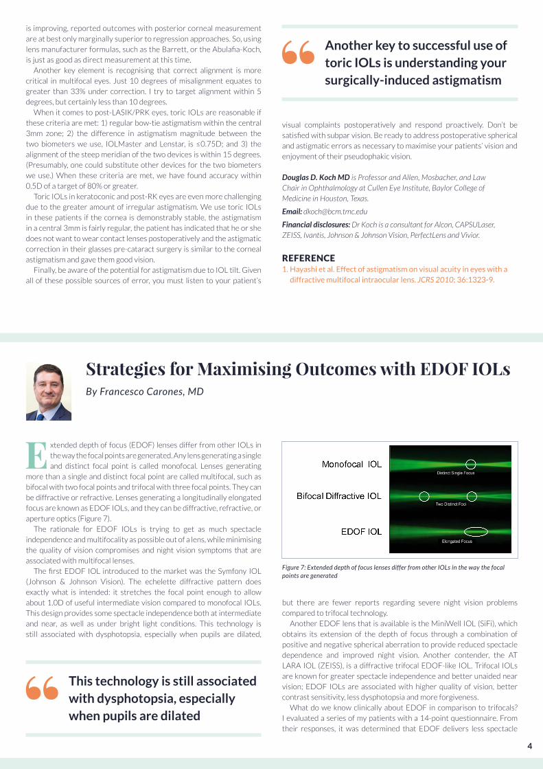
Another key to successful use of toric IOLs is understanding your
surgically induced astigmatism (SIA). It will likely be minimal with a temporal
2.2-to-2.4mm incision, but it is often higher with superior or larger incisions.
SIA with a temporal incision is low, but the scatter is large and that will play
into postoperative results. If you are uncertain about your SIA or use superior
incisions, I recommend that you determine it using a calculator, such as the
one Warren Hill MD has on his website: http://www.doctor-hill.com.It is important to rule out irregular astigmatism (Figures 5 and 6). This
is where topography becomes so important. I prefer Placido imaging; it
is reliable and given that irregular astigmatism causes poor outcomes, I
consider this technology a must-have in my practice. Placido mires are
a great way to validate surface quality and rule out conditions such as
anterior basement membrane disease, subepithelial scarring and visually
significant dry eye. All of these can be treated, but be sure to validate
surface quality post-treatment. I often find that, even with complete
removal of surface pathology such as Salzmann’s or anterior basement
membrane dystrophy, the cornea is no longer pristine, and these patients
may no longer qualify for a multifocal IOL. In addition, topography is
required to rule out ectatic disorders that would disqualify these corneas
for postoperative enhancement.
It is essential to validate your biometric data, especially corneal
measurements. I always take more than one measurement, either with two
different devices or the same device. You can validate your data qualitatively.
We look at the LED mires of every patient and remeasure and treat as
necessary to get perfect mires, which will give optimal measurements.
Another key to successful toric IOL use is factoring in the posterior
cornea. You can use regression formulas that are based on population
averages, or you can measure with one of the many technologies that are
available. The problem with regression approaches for selecting toric
IOLs is that individual variability will sometimes reduce the accuracy
of your outcomes. However, although direct measurement technology
Figure 5 and 6: Topography maps illustrate the importance of ruling out irregular astigmatism in toric IOL candidates
4
Strategies for Maximising Outcomes with EDOF IOLsBy Francesco Carones, MD
is improving, reported outcomes with posterior corneal measurement
are at best only marginally superior to regression approaches. So, using
lens manufacturer formulas, such as the Barrett, or the Abulafia-Koch,
is just as good as direct measurement at this time.
Another key element is recognising that correct alignment is more
critical in multifocal eyes. Just 10 degrees of misalignment equates to
greater than 33% under correction. I try to target alignment within 5
degrees, but certainly less than 10 degrees.
When it comes to post-LASIK/PRK eyes, toric IOLs are reasonable if
these criteria are met: 1) regular bow-tie astigmatism within the central
3mm zone; 2) the difference in astigmatism magnitude between the
two biometers we use, IOLMaster and Lenstar, is ≤0.75D; and 3) the
alignment of the steep meridian of the two devices is within 15 degrees.
(Presumably, one could substitute other devices for the two biometers
we use.) When these criteria are met, we have found accuracy within
0.5D of a target of 80% or greater.
Toric IOLs in keratoconic and post-RK eyes are even more challenging
due to the greater amount of irregular astigmatism. We use toric IOLs
in these patients if the cornea is demonstrably stable, the astigmatism
in a central 3mm is fairly regular, the patient has indicated that he or she
does not want to wear contact lenses postoperatively and the astigmatic
correction in their glasses pre-cataract surgery is similar to the corneal
astigmatism and gave them good vision.
Finally, be aware of the potential for astigmatism due to IOL tilt. Given
all of these possible sources of error, you must listen to your patient’s
visual complaints postoperatively and respond proactively. Don’t be
satisfied with subpar vision. Be ready to address postoperative spherical
and astigmatic errors as necessary to maximise your patients’ vision and
enjoyment of their pseudophakic vision.
Douglas D. Koch MD is Professor and Allen, Mosbacher, and Law Chair in Ophthalmology at Cullen Eye Institute, Baylor College of Medicine in Houston, Texas.
Email: [email protected]
Financial disclosures: Dr Koch is a consultant for Alcon, CAPSULaser, ZEISS, Ivantis, Johnson & Johnson Vision, PerfectLens and Vivior.
REFERENCE1. Hayashi et al. Effect of astigmatism on visual acuity in eyes with a
diffractive multifocal intraocular lens. JCRS 2010; 36:1323-9.
Another key to successful use of toric IOLs is understanding your surgically-induced astigmatism
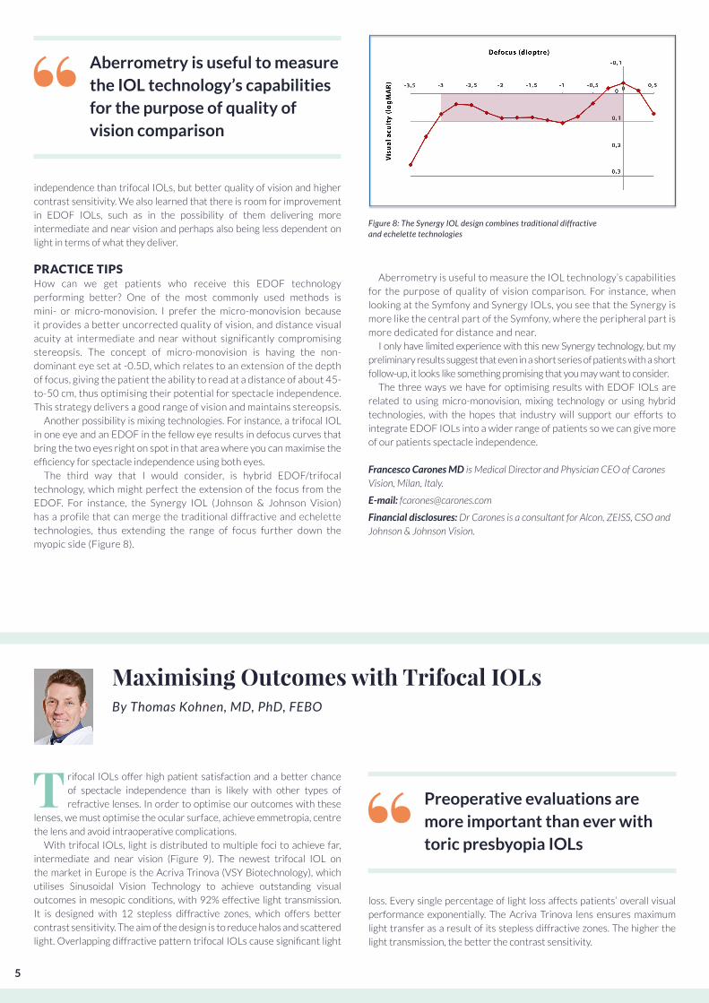
Figure 7: Extended depth of focus lenses differ from other IOLs in the way the focal points are generated
E xtended depth of focus (EDOF) lenses differ from other IOLs in
the way the focal points are generated. Any lens generating a single
and distinct focal point is called monofocal. Lenses generating
more than a single and distinct focal point are called multifocal, such as
bifocal with two focal points and trifocal with three focal points. They can
be diffractive or refractive. Lenses generating a longitudinally elongated
focus are known as EDOF IOLs, and they can be diffractive, refractive, or
aperture optics (Figure 7).
The rationale for EDOF IOLs is trying to get as much spectacle
independence and multifocality as possible out of a lens, while minimising
the quality of vision compromises and night vision symptoms that are
associated with multifocal lenses.
The first EDOF IOL introduced to the market was the Symfony IOL
(Johnson & Johnson Vision). The echelette diffractive pattern does
exactly what is intended: it stretches the focal point enough to allow
about 1.0D of useful intermediate vision compared to monofocal IOLs.
This design provides some spectacle independence both at intermediate
and near, as well as under bright light conditions. This technology is
still associated with dysphotopsia, especially when pupils are dilated,
but there are fewer reports regarding severe night vision problems
compared to trifocal technology.
Another EDOF lens that is available is the MiniWell IOL (SiFi), which
obtains its extension of the depth of focus through a combination of
positive and negative spherical aberration to provide reduced spectacle
dependence and improved night vision. Another contender, the AT
LARA IOL (ZEISS), is a diffractive trifocal EDOF-like IOL. Trifocal IOLs
are known for greater spectacle independence and better unaided near
vision; EDOF IOLs are associated with higher quality of vision, better
contrast sensitivity, less dysphotopsia and more forgiveness.
What do we know clinically about EDOF in comparison to trifocals?
I evaluated a series of my patients with a 14-point questionnaire. From
their responses, it was determined that EDOF delivers less spectacle
This technology is still associated with dysphotopsia, especially when pupils are dilated
5
Aberrometry is useful to measure the IOL technology’s capabilities
for the purpose of quality of vision comparison. For instance, when
looking at the Symfony and Synergy IOLs, you see that the Synergy is
more like the central part of the Symfony, where the peripheral part is
more dedicated for distance and near.
I only have limited experience with this new Synergy technology, but my
preliminary results suggest that even in a short series of patients with a short
follow-up, it looks like something promising that you may want to consider.
The three ways we have for optimising results with EDOF IOLs are
related to using micro-monovision, mixing technology or using hybrid
technologies, with the hopes that industry will support our efforts to
integrate EDOF IOLs into a wider range of patients so we can give more
of our patients spectacle independence.
Francesco Carones MD is Medical Director and Physician CEO of Carones Vision, Milan, Italy.
E-mail: [email protected]
Financial disclosures: Dr Carones is a consultant for Alcon, ZEISS, CSO and Johnson & Johnson Vision.
independence than trifocal IOLs, but better quality of vision and higher
contrast sensitivity. We also learned that there is room for improvement
in EDOF IOLs, such as in the possibility of them delivering more
intermediate and near vision and perhaps also being less dependent on
light in terms of what they deliver.
PRACTICE TIPS How can we get patients who receive this EDOF technology
performing better? One of the most commonly used methods is
mini- or micro-monovision. I prefer the micro-monovision because
it provides a better uncorrected quality of vision, and distance visual
acuity at intermediate and near without significantly compromising
stereopsis. The concept of micro-monovision is having the non-
dominant eye set at -0.5D, which relates to an extension of the depth
of focus, giving the patient the ability to read at a distance of about 45-
to-50 cm, thus optimising their potential for spectacle independence.
This strategy delivers a good range of vision and maintains stereopsis.
Another possibility is mixing technologies. For instance, a trifocal IOL
in one eye and an EDOF in the fellow eye results in defocus curves that
bring the two eyes right on spot in that area where you can maximise the
efficiency for spectacle independence using both eyes.
The third way that I would consider, is hybrid EDOF/trifocal
technology, which might perfect the extension of the focus from the
EDOF. For instance, the Synergy IOL (Johnson & Johnson Vision)
has a profile that can merge the traditional diffractive and echelette
technologies, thus extending the range of focus further down the
myopic side (Figure 8).
Figure 8: The Synergy IOL design combines traditional diffractive and echelette technologies
Aberrometry is useful to measure the IOL technology’s capabilities for the purpose of quality of vision comparison
Preoperative evaluations are more important than ever with toric presbyopia IOLs
Maximising Outcomes with Trifocal IOLsBy Thomas Kohnen, MD, PhD, FEBO
T rifocal IOLs offer high patient satisfaction and a better chance
of spectacle independence than is likely with other types of
refractive lenses. In order to optimise our outcomes with these
lenses, we must optimise the ocular surface, achieve emmetropia, centre
the lens and avoid intraoperative complications.
With trifocal IOLs, light is distributed to multiple foci to achieve far,
intermediate and near vision (Figure 9). The newest trifocal IOL on
the market in Europe is the Acriva Trinova (VSY Biotechnology), which
utilises Sinusoidal Vision Technology to achieve outstanding visual
outcomes in mesopic conditions, with 92% effective light transmission.
It is designed with 12 stepless diffractive zones, which offers better
contrast sensitivity. The aim of the design is to reduce halos and scattered
light. Overlapping diffractive pattern trifocal IOLs cause significant light
loss. Every single percentage of light loss affects patients’ overall visual
performance exponentially. The Acriva Trinova lens ensures maximum
light transfer as a result of its stepless diffractive zones. The higher the
light transmission, the better the contrast sensitivity.

6
those who have severe dry eye disease, pseudoexfoliation, glaucoma,
and/or severe retinal disease.
Preoperative evaluations are more important than ever with toric
presbyopia IOLs. Tomography, endothelial cell counts, macular function,
exclusion of keratitis sicca are all critical to successful outcomes and
satisfied patients. With respect to postoperative refraction, in most of these
eyes we need to achieve emmetropia. With so many new IOL calculation
formulas available, you must be sure that the one you use with these IOLs
is up to date. Equally important is the need for precise IOL centration, and
a carefully created capsulorhexis or capsulotomy. These lenses will not
function appropriately if they are decentered upon placement or if they
decenter over time. Some of my key pearls are summarised in Figure 10.
Preoperative optimisation of the ocular surface is also critical. An
expansive literature review of 16 papers confirms that an impaired ocular
surface affects preoperative planning for cataract surgery, including
IOL calculations, toric IOL axis and magnitude estimates, keratometry
and topography measurements; and also increases surgical difficulty.4
Surgeons should recognise and aggressively pre-treat cataract patients
who have pre-existing dry eye disease.
One of the final points that must be made is that the effect of
astigmatism is often underestimated. Make sure you don’t leave patients
with 1.5D of astigmatism – particularly multifocal IOL patients. We think
0.5D should be the highest amount of astigmatism that can remain and
still leave a patient enjoying the benefits of multifocality.
Thomas Kohnen MD, PhD, FEBO, is Professor and chairman, Department of Ophthalmology, at Goethe University, Frankfurt, Germany.
E-mail: [email protected]
Financial disclosures: Dr Kohnen is a consultant and does research for Alcon, Novartis and ZEISS.
REFERENCES:1. Kohnen T, Titke C, Böhm M. Trifocal intraocular lens implantation
to treat visual demands in various distances following lens removal. Am J Ophthalmol 2016; 161:71–77.
2. Kohnen T, Herzog M, Hemkeppler E, Schönbrunn S, De Lorenzo N, Petermann K, Böhm M. Visual performance of a quadrifocal (trifocal) intraocular lens following removal of the crystalline lens. Am J Ophthalmol 2017; 184:52–62.
3. Böhm M, Hemkeppler E, Herzog M, Schönbrunn S, de’Lorenzo N, Petermann K, Kohnen T. Comparison of a panfocal and trifocal diffractive intraocular lens after femtosecond laser-assisted lens surgery. J Cataract Refract Surg. 2018; 44:1454-1462.
4. Chuang J, Shih KC, Chan TC, Wan KH, Jhanji V, Tong L. Preoperative optimization of ocular surface disease before cataract surgery. J Cataract Refract Surg. 2017;43(12):1596-1607.
The trifocal IOLs that preceded the Acriva include the FineVision
(PhysIOL), AT LISA tri (ZEISS), PanOptix (Alcon) and RayOne Trifocal
(Rayner). The RayOne trifocal IOL, which is actually derived from bifocal
technology, has 16 diffractive steps and a 4.5mm diffractive zone. It was
developed to be less dependent on pupil size or lighting conditions, and it
improves distance vision in mesopic conditions. This can also be used as
an add-on to a monofocal lens in a procedure similar to the piggyback IOL
paradigm, where you put the trifocal lens into the sulcus where an IOL has
already been implanted. This way we can use trifocality – and maybe even
in the future EDOF technology – on top of our older monofocal lenses.
CLINICAL STUDIESWe have done several studies of the available trifocal IOLs. For instance,
a prospective, non-randomised, non-comparative case series of 27
patients undergoing implantation of AT LISA trifocal or toric, revealed
that patients were very happy with their outcomes overall1. Contrast
sensitivity was within normal range in photopic, mesopic and mesopic
with glare conditions. Despite some optical phenomena, patients had
high spectacle independence three months postoperatively; 92% of
patients said they would choose the same IOL again.
In a study of the PanOptix trifocal with a quadrifocal design, where 27
patients received bilateral implantation, we had similar results.2 Visual
performance of this IOL showed good visual acuity at all distances,
with best VA at 60cm, and high patient satisfaction and spectacle
independence three months postoperatively. We also found some optical
disturbances, but overall the patients were happy.
We compared the AT LISA trifocal and the PanOptix panfocal IOLs
in a recently published study.3 The prospective, non-randomised
comparative case series of 20 subjects undergoing bilateral implantation
of PanOptix panfocal or AT LISA trifocal revealed that with respect
to monocular visual performance, there was no significant difference
between the IOLs at far, intermediate or near distance. However, there
was better visual acuity at 50cm and 66cm with the panfocal IOL. The
main difference was in the defocus curve, with a bit of an advantage at
60cm for the panfocal lens.
IDEAL CANDIDATES AND PEARLSThe ideal trifocal IOL candidate is a patient who wants spectacle
independence and good vision at all distances; someone with no
pathology on the cornea, no pathology on the retina, no irregular
astigmatism. Patients who are contraindicated to this technology are
With trifocal IOLs, light is distributed to multiple foci to achieve far, intermediate and near vision
Figure 9: With trifocal IOLs, light is distributed to multiple foci to achieve far, intermediate, and near vision
40cm
near: +3.33 D intermediate: +1.66 D
80cm
far
Keys for Maximising Outcomes with Trifocal IOLs
• Preoperative optimisation of ocular surface• Achieving emmetropia• Correction of astigmatism• Correction of misaligned or dislocated toric IOL• Effect of manual capsulorhexis size and IOL position• Intraocular complications (posterior capsular rupture)
Figure 10: Essential considerations for maximizing patient outcomes with trifocal IOLs

Supported by an unrestricted medical education grant
GoldGold Bronze
SupplementDecember 2019/ January 2020