Embed Size (px)
Citation preview
Primary Bronchogenic CarcinomaPrimary Bronchogenic Carcinoma (LUNG CANCER)(LUNG CANCER)
SHEN JIN
The First Affiliated Hospital of Kunming Medical CollegeThe First Affiliated Hospital of Kunming Medical College
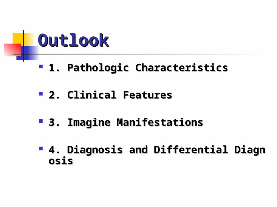
OutlookOutlook 1. Pathologic Characteristics1. Pathologic Characteristics
2. Clinical Features2. Clinical Features
3. Imagine Manifestations3. Imagine Manifestations
4. Diagnosis and Differential Diagnosis4. Diagnosis and Differential Diagnosis
Pathologic CharacteristicsPathologic Characteristics
1.Difinition1.Difinition Arise from the bronchial
epithelium, bronchial glands and epithelium of the alveolus.
2.Histologic Classification:2.Histologic Classification:
Squamous Carcinoma 40% Undifferentiated Carcinoma 25% Adenocarcinoma 30% Alvelolar Cell Carcinoma 5% *non-small cell and small cell
3. Location Classification:3. Location Classification:
Central Type Peripheral Type Diffuse Type

4. Growth Type:4. Growth Type: Inner Wall Outer Wall Across Wall Enlarge Infitrate

Growth in early central typeGrowth in early central type
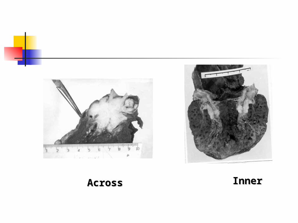
AcrossAcross InnerInner
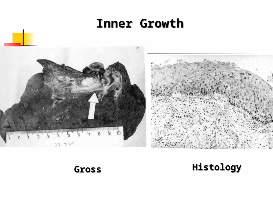
Inner GrowthInner Growth
GrossGross HistologyHistology
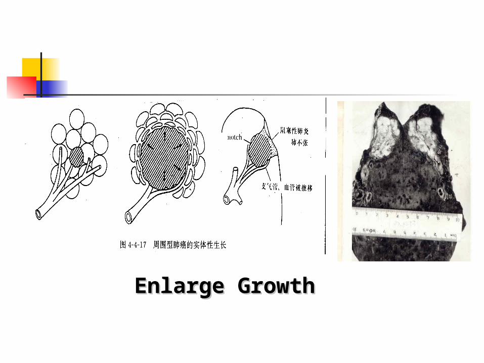
Enlarge GrowthEnlarge Growth
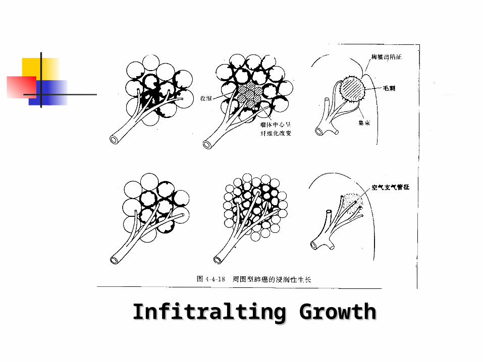
Infitralting GrowthInfitralting Growth
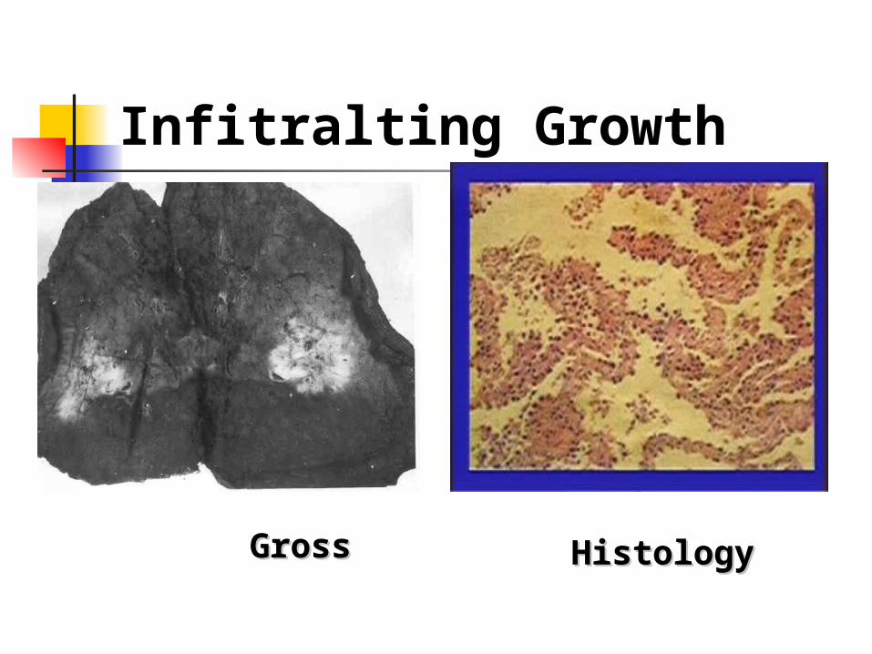
Infitralting Growth
GrossGross HistologyHistology
The clinical features are variable
correlated with the histological type,
site, and degree of development of
the carcinoma.
Clinical FeaturesClinical Features
Early Stage:Early Stage:
No signs! No symptoms!No signs! No symptoms!
Sign and Symptom 1.Manifestation Of Respiratory System:1.Manifestation Of Respiratory System: Cough Hemoptysis Sputum Breathlessness Stridor Chest pain
Sign and Symptom
2.Spread and Invasion:2.Spread and Invasion:
1).Pleura---effusion, pleuritic pain and chest wall pain.
Sign and Symptom
2).Mediastinum--- Left recurrent laryngeal nerve palsy Superior vena caval obstruction Dysphagia Phrenic nerve paralysis Pancoast’ tumors: Horner’s syndro
me
Sign and Symptom
3. Other abnormal:3. Other abnormal: Endocrine and metabolic
manifestations Neuromuscular manifestations Connective tissue and osseous
manifestations
Imaginal Manifestations Central TypeCentral Type
Peripheral TypePeripheral Type
Diffuse TypeDiffuse Type
Central Type of lung Central Type of lung CancerCancer
Early Stage :Early Stage :
No Abnormal X-ray Findings
Imaginal Manifestations

Central Type of lung Central Type of lung CancerCancer
Chest film + CT scansThe Direct Signs
1.Hilar mass: Unilateral hilar enlargement Increased density of hilum tumor mass and lymph nodes
Imaginal Manifestations
Central Type of lung Central Type of lung CancerCancer
2.Abnormality of bronchi (Encroachment )
Intralumen nodule Thickening Wall Irregular narrowing Completely obstructed
Imaginal Manifestations
Central Type of lung Central Type of lung CancerCancer
CT scans can demonstrate the ab
normality of bronchi and hilar mass clearly.
Imaginal Manifestations
Central Type of lung Central Type of lung CancerCancer
Indirect Signs:Indirect Signs: (Airway Obstruction)
1.Obstructive Emphysema (overinflation of lung) Rare 2%
2.Obstructive Atelectasis (collapse) Very common 3.Obstructive Pneumonia (consolidatio
n)
Imaginal Manifestations
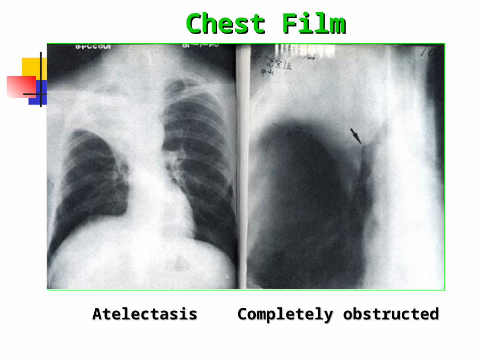
Chest FilmChest Film
Completely obstructedCompletely obstructedAtelectasisAtelectasis
Central Type of lung Central Type of lung CancerCancer
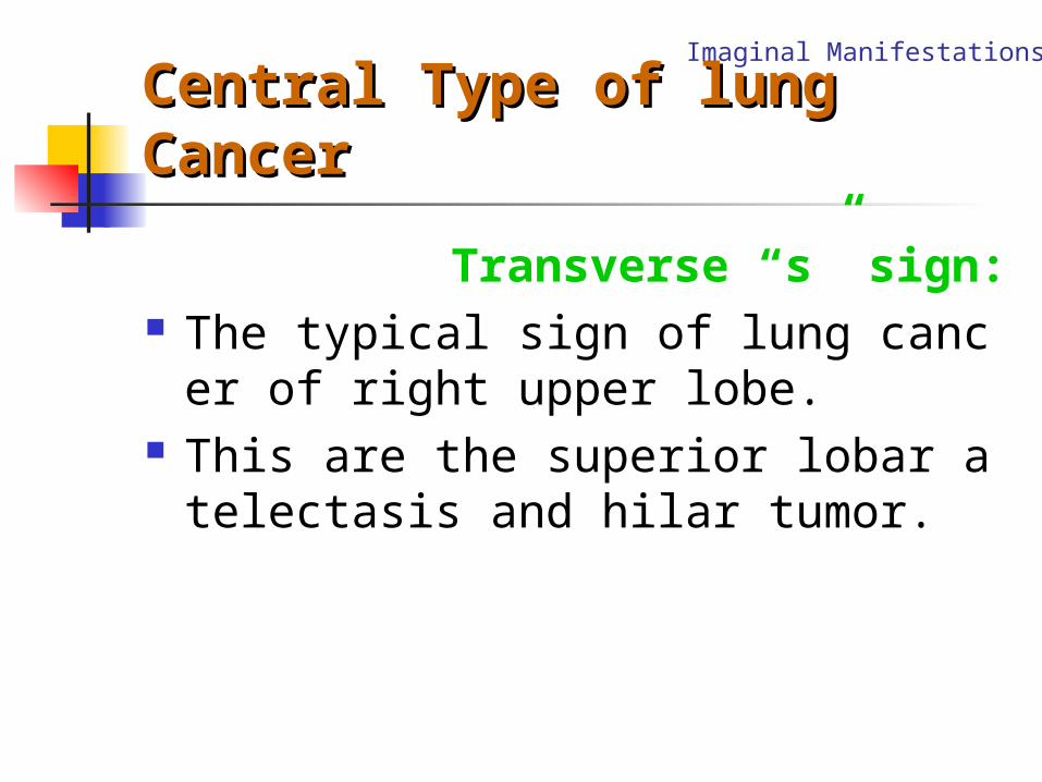
Transverse “s” sign: The typical sign of lung cancer of right
upper lobe. This are the superior lobar atelectasis
and hilar tumor.
Imaginal Manifestations
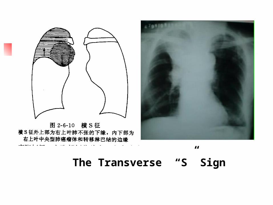
The Transverse “S” Sign
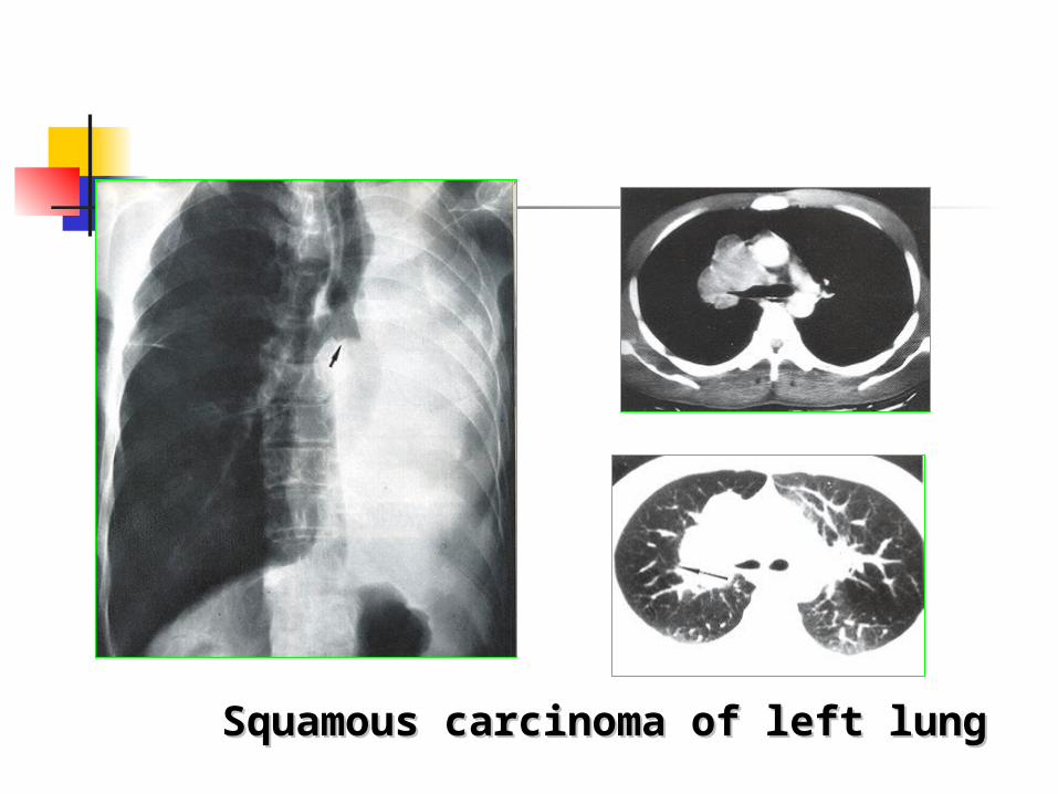
Squamous carcinoma of left lungSquamous carcinoma of left lung
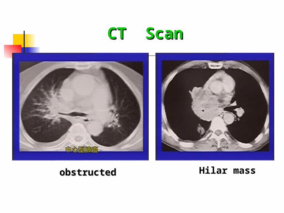
CT ScanCT Scan
obstructedobstructed Hilar mass
CT ScanCT Scan
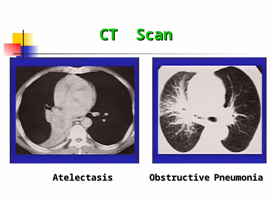
AtelectasisAtelectasis ObstructiveObstructive PneumoniaPneumonia
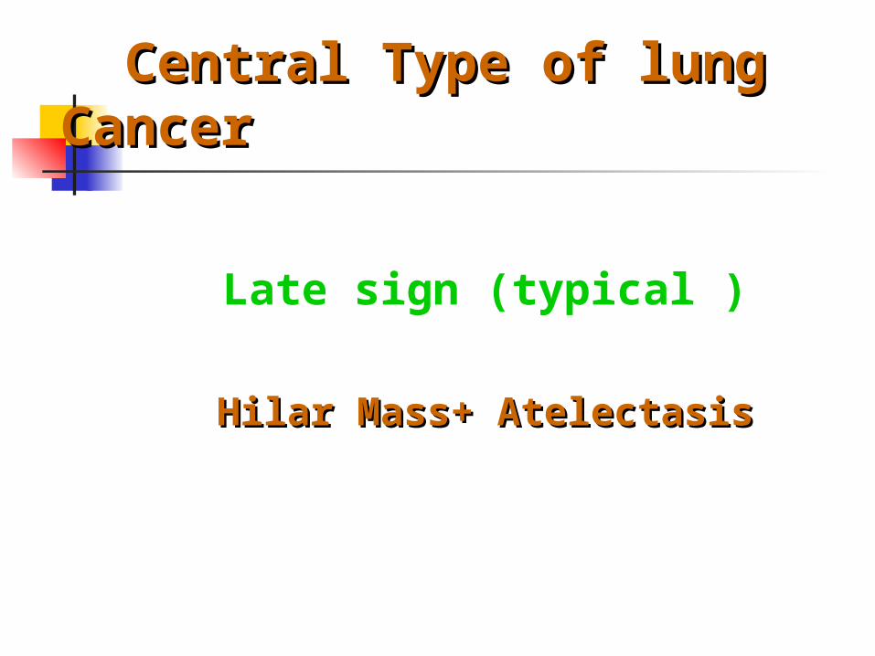
Central Type of lung Central Type of lung CancerCancer
Late sign (typical )
Hilar Mass+ AtelectasisHilar Mass+ Atelectasis
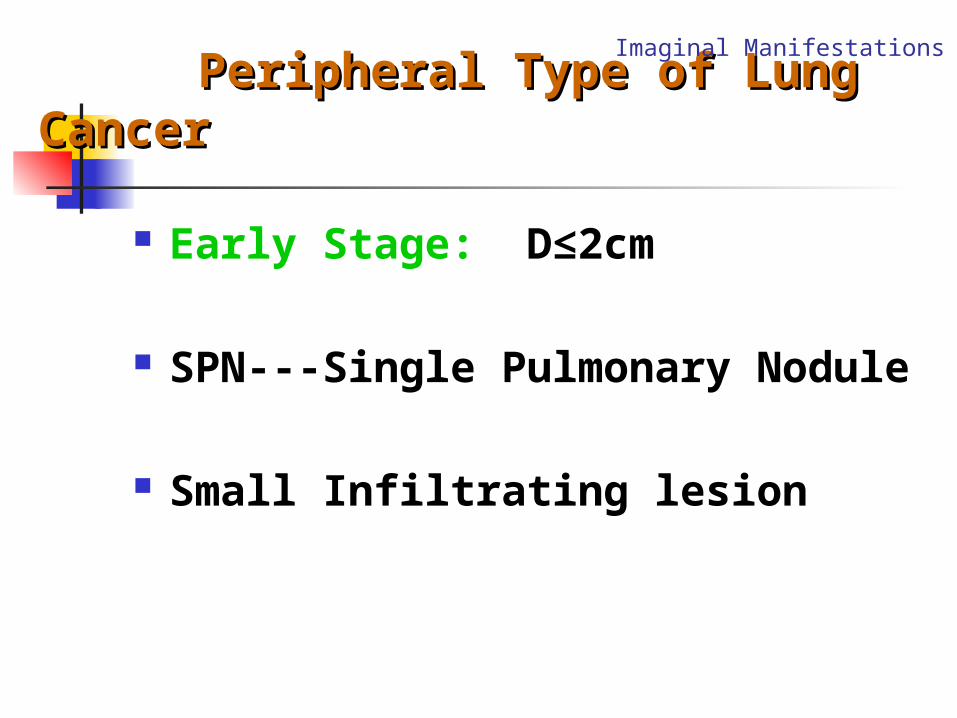
Peripheral Type of Lung Peripheral Type of Lung CancerCancer
Early Stage: D≤2cm
SPN---Single Pulmonary Nodule
Small Infiltrating lesion
Imaginal Manifestations
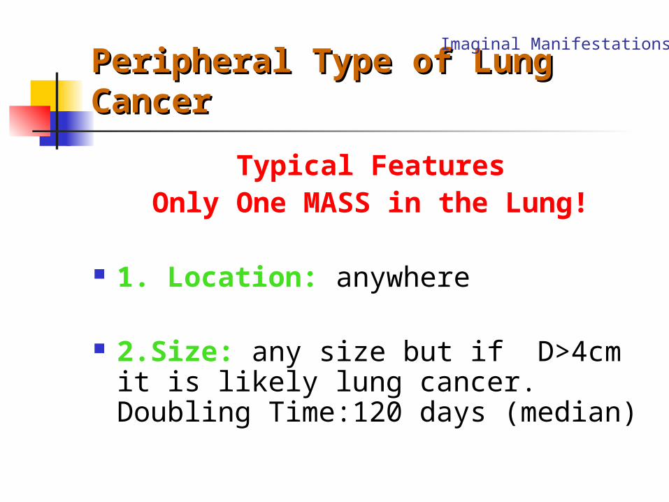
Peripheral Type of Lung Peripheral Type of Lung CancerCancer
Typical FeaturesOnly One MASS in the Lung!
1. Location: anywhere
2.Size: any size but if D>4cm it is likely lung cancer. Doubling Time:120 days (median)
Imaginal Manifestations
Peripheral Type of Lung Peripheral Type of Lung CancerCancer
3.Shape: spherical, oval, lobulated configuration. Notched (umbilicated) is very typical.
4.Border: clear (smooth), shaggy or cloudy( ill defined), spiculated infiltrating.
Imaginal Manifestations
Peripheral Type of Lung Peripheral Type of Lung CancerCancer
5.Densety: 1). Homogeneous 2).Calcification, very rare 3).Early, air bronchiologram or air bronc
hogram 4). Cavity, irregular inner wall, eccentric 5). Enhanced in CT scan.
Imaginal Manifestations
Peripheral Type of Lung Peripheral Type of Lung CancerCancer
6. Around Mass: Pleural Indrawn Sign: A tail appears as a peripheral line
shadow between a mass and the pleura.
7.Others:
Imaginal Manifestations
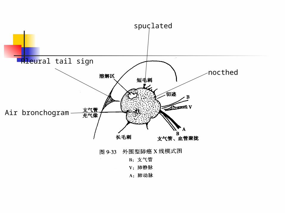
nocthed
Air bronchogram
spuclated
Pleural tail sign
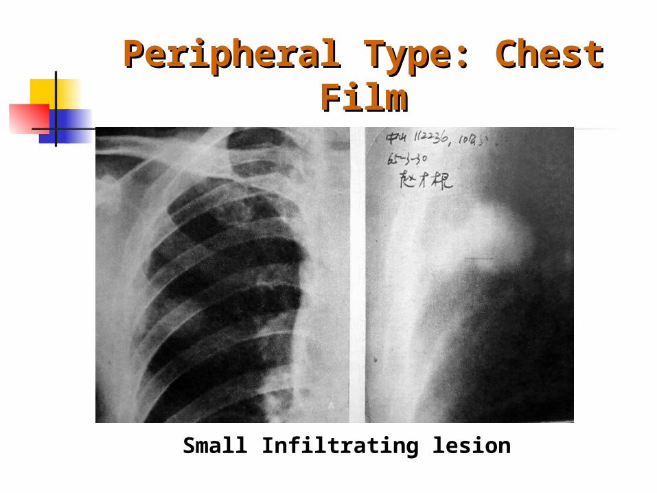
Peripheral Type: Chest Peripheral Type: Chest FilmFilm
Small Infiltrating lesion
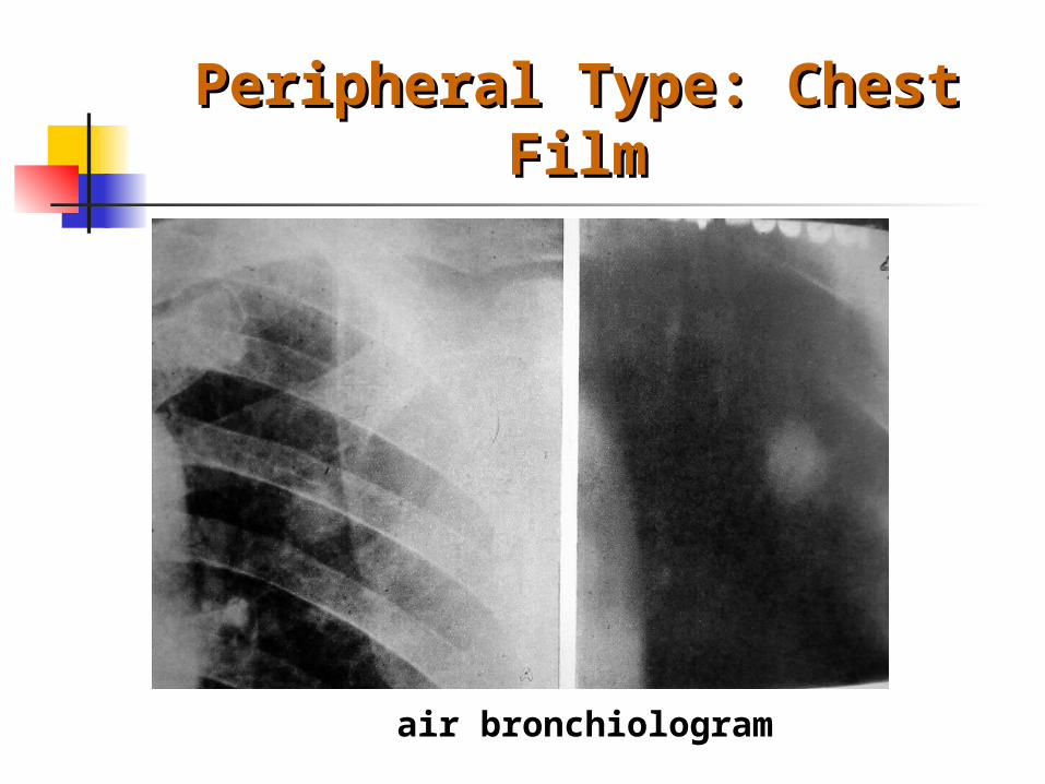
Peripheral Type: Chest Peripheral Type: Chest FilmFilm
air bronchiologram
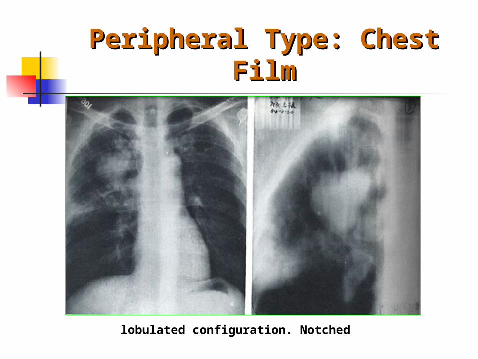
Peripheral Type: Chest Peripheral Type: Chest FilmFilm
lobulated configuration. Notched
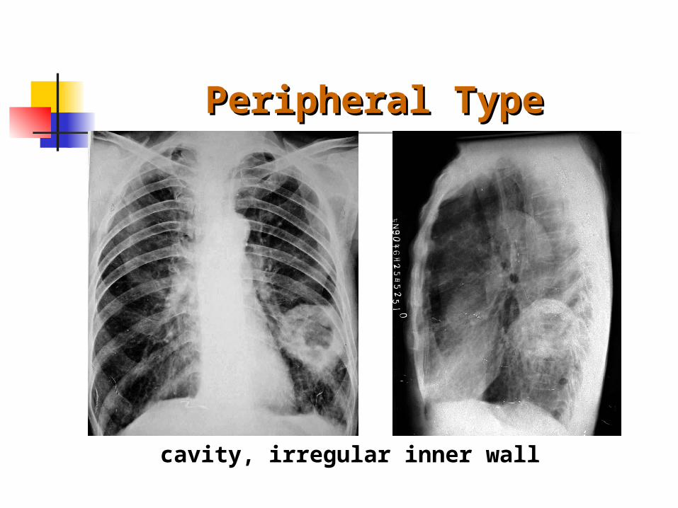
Peripheral TypePeripheral Type
cavity, irregular inner wall
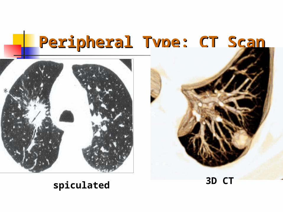
Peripheral Type: CT ScanPeripheral Type: CT Scan
spiculated 3D CT
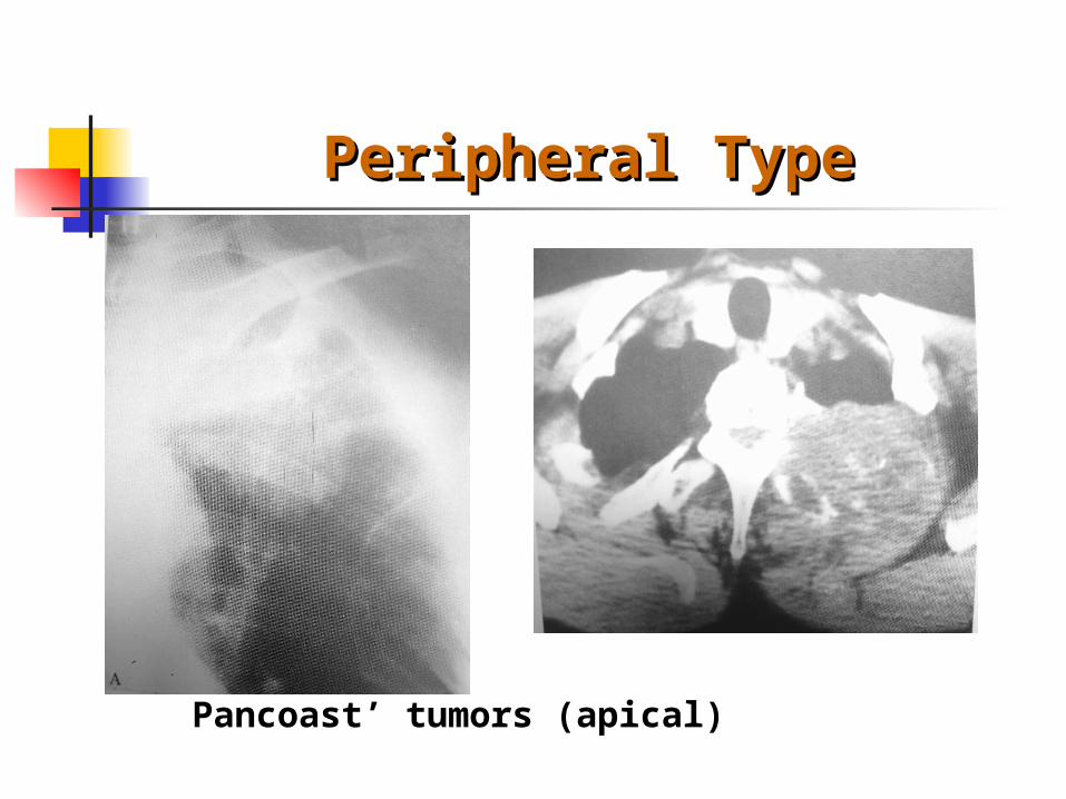
Peripheral TypePeripheral Type
Pancoast’ tumors (apical)
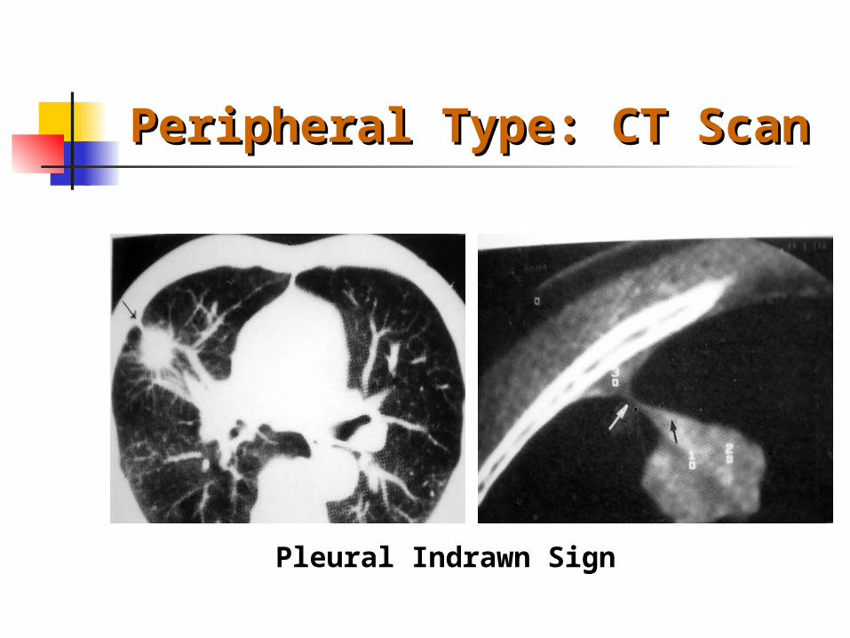
Peripheral Type: CT ScanPeripheral Type: CT Scan
Pleural Indrawn Sign

Peripheral Type: CT ScanPeripheral Type: CT Scan
cavitySmall Infiltrating lesion
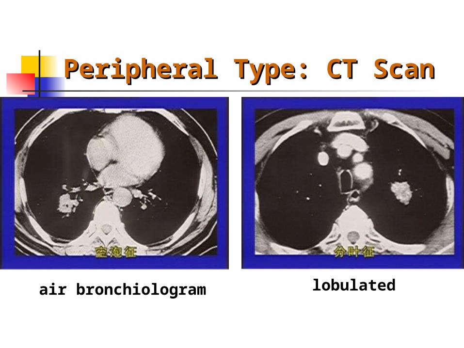
Peripheral Type: CT ScanPeripheral Type: CT Scan
air bronchiologram lobulated

Peripheral Type: CT ScanPeripheral Type: CT Scan
spiculated Pleural Indrawn Sign
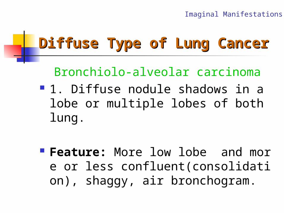
Diffuse Type of Lung CancerDiffuse Type of Lung Cancer
Bronchiolo-alveolar carcinoma 1. Diffuse nodule shadows in a lobe or
multiple lobes of both lung.
Feature: More low lobe and more or less confluent(consolidation), shaggy, air bronchogram.
Imaginal Manifestations
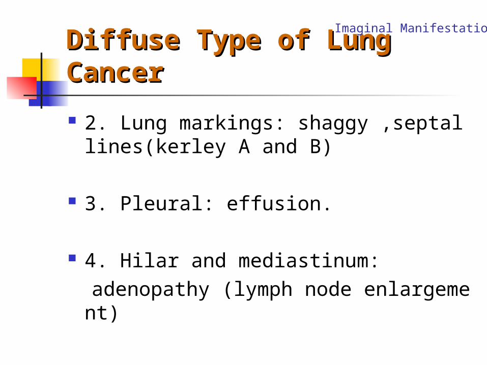
Diffuse Type of Lung Diffuse Type of Lung CancerCancer
2. Lung markings: shaggy ,septal lines(kerley A and B)
3. Pleural: effusion.
4. Hilar and mediastinum: adenopathy (lymph node enlargement)
Imaginal Manifestations
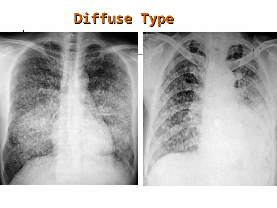
Diffuse TypeDiffuse Type
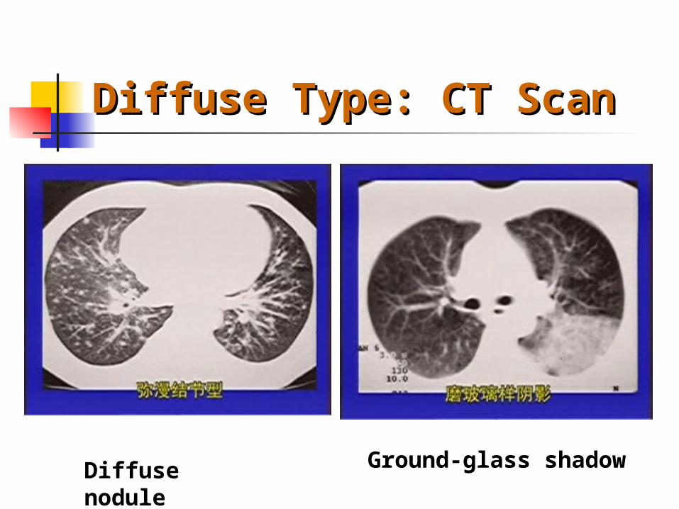
Diffuse Type: CT ScanDiffuse Type: CT Scan
Diffuse nodule
Ground-glass shadow

Diagnosis and Differential Diagnosis
1. Central type: Key point : Mass in hilar+ Atelectasis+ Encroachm
ent of bronchi DD: Bronchial Mucosa Tuberculosis
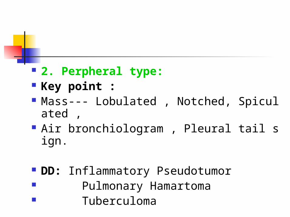
2. Perpheral type: Key point : Mass--- Lobulated , Notched, Spiculate
d , Air bronchiologram , Pleural tail sign.
DD: Inflammatory Pseudotumor Pulmonary Hamartoma Tuberculoma

Thank you!


![Transbronchial Needle Aspiration Staging of Bronchogenic ...downloads.hindawi.com/journals/dte/1996/237680.pdfChest, 80,48-50. [18] Transbronchialneedle bronchogenic carcinoma, In:](https://img.pdfslide.net/doc/110x75/5fef28f6c0cad34ae7313439/transbronchial-needle-aspiration-staging-of-bronchogenic-chest-8048-50-18.jpg)