Embed Size (px)
Citation preview
Prime Archives in Virology
1 www.videleaf.com
Book Chapter
Production of Highly and Broad-Range
Specific Monoclonal Antibodies against
Hemagglutinin of H5-Subtype Avian
Influenza Viruses and their
Differentiation by Mass Spectrometry
Violetta Sączyńska*†, Anna Bierczyńska-Krzysik*
†, Violetta
Cecuda-Adamczewska*†, Piotr Baran, Anna Porębska,
Katarzyna Florys, Marcin Zieliński and Grażyna Płucienniczak
Institute of Biotechnology and Antibiotics, Poland
†Equal contributors
*Corresponding Author: Violetta Sączyńska, Institute of
Biotechnology and Antibiotics, Starościńska 5 Street, 02-516
Warsaw, Poland
Anna Bierczyńska-Krzysik, Institute of Biotechnology and
Antibiotics, Starościńska 5 Street, 02-516 Warsaw, Poland
Cecuda-Adamczewska, Institute of Biotechnology and
Antibiotics, Starościńska 5 Street, 02-516 Warsaw, Poland
Published November 30, 2020
This Book Chapter is a republication of an article published by
Violetta Sączyńska, et al. at Virology Journal in January 2018.
(Sączyńska, V., Bierczyńska-Krzysik, A., Cecuda-
Adamczewska, V. et al. Production of highly and broad-range
specific monoclonal antibodies against hemagglutinin of H5-
subtype avian influenza viruses and their differentiation by mass
spectrometry. Virol J 15, 13 (2018).
https://doi.org/10.1186/s12985-017-0886-2)
Prime Archives in Virology
2 www.videleaf.com
How to cite this book chapter: Violetta Sączyńska, Anna
Bierczyńska-Krzysik, Violetta Cecuda-Adamczewska, Piotr
Baran, Anna Porębska, Katarzyna Florys, Marcin Zieliński,
Grażyna Płucienniczak. Production of Highly and Broad-Range
Specific Monoclonal Antibodies against Hemagglutinin of H5-
Subtype Avian Influenza Viruses and their Differentiation by
Mass Spectrometry. In: Prime Archives in Virology. Hyderabad,
India: Vide Leaf. 2020.
© The Author(s) 2020. This article is distributed under the terms
of the Creative Commons Attribution 4.0 International
License(http://creativecommons.org/licenses/by/4.0/), which
permits unrestricted use, distribution, and reproduction in any
medium, provided the original work is properly cited.
Funding: This work was supported by the Innovative Economy
Operational Program, Grant No. WND-POIG.01.01.02-00-
007/08-00, as a part of the project “Center of medicinal product
biotechnology. Package of innovative biopharmaceuticals for
human and animal therapy and prophylactics” and Grant No.
PBS2/A7/14/2014 (ID: 210,068): “Influenza vaccine -
innovative obtaining of subunit antigens.”
The funders had no role in the design of the study and collection,
analysis, and interpretation of data and in writing the manuscript.
Availability of Data and Materials: The results presented here
are contained in the Patent Applications P.418671 (2016–09-12),
PCT/PL2017/000084 (2017–09-11) - Monoclonal antibodies
against hemagglutinin of H5-serotype influenza viruses and their
uses, hybridomas producing said antibodies, compositions and
diagnostic kits. The datasets supporting the conclusions of this
article are included within the article and its additional files. The
newly established hybridoma cell lines, described in this paper,
were given the following Accession Numbers by the
International Depositary Authority: DSM ACC3292, DSM
ACC3293, DSM ACC3294, DSM ACC3295, DSM ACC3296
and DSM ACC3297.

Prime Archives in Virology
3 www.videleaf.com
Contributions: VS, AB-K conceived and designed the
experiments, analyzed the data and wrote the manuscript. VC-A,
AP, KF were involved in the production of monoclonal
antibodies and their characterization using ELISA. AB-K, PB,
MZ participated in peptide mapping of antibody fragments by
mass spectrometry. VS, AB-K, VC-A, PB prepared the tables.
GP provided analysis tools, supervised the research group and
contributed to funding acquisition. All of the authors have read
and approved the final manuscript.
Ethics Approval and Consent to Participate: All work with
mice was approved by the Second Local Ethical Committee for
Animal Experiments at the Medical University of Warsaw
(Poland), Permit Number 38/2008.
Competing Interests: All the authors (VS AB-K VC-A PB AP
KF & GP), except Marcin Zieliński, have filed the Patent
Applications P.418671 (2016–09-12), PCT/PL2017/000084
(2017-09-11): Monoclonal antibodies against hemagglutinin of
H5-serotype influenza viruses and their uses, hybridomas
producing said antibodies, compositions and diagnostic kits. The
Patent Application contains the findings reported in this paper.
Abstract
Background: The highly pathogenic avian influenza viruses of
the H5 subtype, such as the H5N1 viral strains or the novel
H5N8 and H5N2 reassortants, are of both veterinary and public
health concern worldwide. To combat these viruses, monoclonal
antibodies (mAbs) against H5 hemagglutinin (HA) play a
significant role. These mAbs are effective diagnostic and
therapeutic agents and powerful tools in vaccine development
and basic scientific research. The aim of this study was to obtain
diagnostically valuable mAbs with broad strain specificity
against H5-subtype AIVs.
Results: We applied the hybridoma method to produce anti-HA
mAbs. The cloning and screening procedures resulted in the
selection of 7 mouse hybridoma cell lines and their respective
antibody clones. Preliminary immunoreactivity studies showed

Prime Archives in Virology
4 www.videleaf.com
that these newly established mAbs, all of the IgG1 isotype, had
high specificity and broad-range activities against the H5 HAs.
However, these studies did not allow for a clear distinction
among the selected antibodies and mAb-secreting hybridoma
clones. To differentiate the analyzed mAbs and determine the
exact number of hybridoma clones, peptide mapping of the Fc
and Fab fragments was performed using a Matrix-Assisted Laser
Desorption Ionization Time of Flight (MALDI-TOF/TOF) mass
spectrometer. Detailed analyses of the acquired MS and MS/MS
spectra confirmed that the Fc fragments constituted highly
conserved species- and isotype-immunoglobulin components,
whereas the Fab fragments exhibited considerable variation in
the sequences that determine antibody specificity. This approach
enabled unambiguous characterization of the selected mAbs
according to their peptide composition. As a result, 6 different
clones were distinguished.
Conclusions: Our work provided a unique panel of anti-H5 HA
mAbs, which meets the demand for novel, high-specificity
analytical tools for use in serologic surveillance. Applications of
these mAbs in areas other than diagnostics are also possible.
Moreover, we demonstrated for the first time that peptide
mapping of antibody fragments with mass spectrometry is an
efficient method for the differentiation of antibody clones and
relevant antibody-producing cell lines. The method may be
successfully used to characterize mAbs at the protein level.
Abbreviations
AIV-Avian Influenza Virus; CDR-Complementarity-
Determining Region; ELISA-Enzyme-Linked Immunosorbent
Assay; Fab-Antigen-Binding Fragment; Fc-Crystallizable
Fragment; HA-Hemagglutinin; HI-Hemagglutination
Inhibition; HP-Highly Pathogenic; Ig-Immunoglobulin; IgG-
Immunoglobulin G Class; MAb-Monoclonal Antibody;
MALDI-TOF/TOF MS-Matrix-Assisted Laser Desorption
Ionization Time of Flight Mass Spectrometry; MS-Mass
Spectrometry; PMF-Peptide Mass Fingerprinting; m/z: Mass-
to-Charge Ratio; rHA-Ectodomain-based Recombinant
Prime Archives in Virology
5 www.videleaf.com
Hemagglutinin Protein; rHA1-HA1 Subunit-based
Recombinant Hemagglutinin Protein; VN-Virus Neutralization
Background
The highly pathogenic (HP) avian influenza viruses (AIVs) of
the H5 subtype pose a serious epidemiological problem. This
applies especially to H5N1 HPAIV, which was detected for the
first time among farmed geese in China in 1996 and in humans a
year later [1]. The spread of the H5N1 viruses to many regions
of the world has been accompanied by frequent avian flu
outbreaks in poultry that have resulted in mortality rates of up to
100% [2]. Moreover, as of January 2017, there have been a total
of 856 laboratory-confirmed human cases of H5N1 influenza,
452 of which were fatal [3]. From 2009 onward, the emergence
of the reassortant H5-subtype HPAIVs, such as H5N2, H5N5,
H5N6 and H5N8, has been noted [2]. In addition, the novel
H5N8 and H5N2 HPAIVs were observed to spread rapidly and
globally soon after their identification in 2014 [4,5]. Many
populations of domestic birds have been substantially affected by
these lethal viruses due to infection or mass culling [6,7].
In view of the threat to animal and human health and life, the
H5-subtype HPAIVs are under epidemiological surveillance.
Various strategies have been developed for prevention and
treatment of the infections caused by these viruses. The majority
of the strategies have focused on the H5 hemagglutinin (HA),
which determines the high pathogenicity of AIVs and is also the
main target for neutralizing antibodies. In the efforts to combat
H5-subtype HPAIVs, monoclonal antibodies (mAbs) against H5
HA play a significant role. When characterized by high
specificity and affinity and/or neutralizing activities, they
constitute effective diagnostic and therapeutic agents. Moreover,
mAbs are powerful as tools for vaccine development as well as
in basic research including studies of the antigenic architecture
of the HA of influenza H5N1 viruses [8].
Most therapeutic and diagnostically valuable mAbs are
immunoglobulins (Igs) of the G class (IgG). These are
homodimeric glycoproteins of ~150 kDa. Each IgG molecule

Prime Archives in Virology
6 www.videleaf.com
contains two identical heavy (H) chains and two identical (L)
light chains with molecular weights of ~50 kDa and ~25 kDa,
respectively [9]. The chains are connected by disulfide bonds to
form a Y-shaped structure. Both the H and L chains of IgG
antibodies consist of variable (V) and constant (C) domains
referred to as VL and CL and VH, CH1, CH2 and CH3,
respectively. The variable parts of the H and L chains, especially
the complementarity-determining regions (CDRs), comprise the
antigen-binding sites of the Ig molecules. They are responsible
for the target epitope recognition, antigen-antibody reaction and
the diversity of antibody specificity. Variations in mAbs may
also result from post-translational modifications such as
alternative disulfide pairings, deamidation, methionine oxidation
or pyroglutamate formation. The heterogeneity of the
glycosylation potently affects both pharmacokinetics and
stability of various isoforms, which alter the clinical efficacy and
safety of the therapeutic proteins [10].
Monoclonal antibodies can be generated using a hybridoma
technique based on spleen/myeloma fusion. The concept was
developed in the 1970s [11]. Another method involves the
transformation of human B lymphocytes with the Epstein-Barr
virus [12,13]. Irrespective of the production method, the mAbs
secreted by the immortalized cells need to be selected in terms of
the desired function and/or specificity. In the case of mAbs
against viral HAs, the hemagglutination inhibition (HI) and virus
neutralization (VN) tests are used to ascertain the ability of
established antibodies to confer protection against influenza.
Identification of antibodies with the desired specificity is
frequently accomplished by immunological techniques such as
the enzyme-linked immunosorbent assay (ELISA). Alternative
protocols that optimize the screening for antibody-producing
cells using flow cytometry are also available [14,15]. These
techniques do not, however, provide complete information
regarding the heterogeneity of the hybridoma clones.
In addition to the methods and techniques described above that
have been applied to mAb production, mass spectrometry (MS)
can be used. MALDI-TOF/TOF MS has been widely recognized
in the fields of the development of therapeutic antibodies,

Prime Archives in Virology
7 www.videleaf.com
determination of their structural features, glycan characterization
and profiling [16-18]. The technique enables evaluation of the
recombinant protein sequence and structure and provides
information on amino acid modifications and sequence
alterations. Although the resolution of the mass spectrometer is
insufficient to differentiate between large, intact proteins,
accurate measurements can be achieved at the peptide level [19].
To facilitate the structural analysis of antibodies, well known
methods may be applied to selectively cleave the Ig molecules
into fragments that have discrete characteristics and functions. If
the variable regions of IgG antibodies are of primary interest, it
is possible to generate their antigen-binding fragments, including
the F(ab’)2, Fab’, Fab and Fv. For example, the monovalent
fragment denoted Fab is composed of one constant and one
variable domain of each of the H and the L chain, i.e., CH1, VH
and CL, VL, respectively. The two variable domains, VH and
VL, specifically bind the epitope of their respective antigen [20].
Preparation of the Fab fragments of IgG antibodies is usually
accomplished by digestion of Igs with papain in the presence of
a reducing agent [21]. In the case of mouse IgG1 antibodies, the
enzyme of choice is ficin, applied with the optimized reductant
concentration [22]. The Fab fragment does not contain any part
of the crystallizable portion of the constant region of the Ig (Fc).
Formed entirely from the H chain constant domains, the Fc
fragment does not bind antigen and is responsible for the effector
functions of antibodies.
In this paper, we describe newly generated, highly specific mAbs
with a broad range of activities against the H5 HA of influenza
viruses. As obtained, the mAbs were indistinguishable on the
basis of the range of immunoreactivities determined by ELISA.
We therefore employed MALDI-TOF/TOF MS as a structural
tool for their differentiation. These mass measurements enabled
assessment of the heterogeneity of peptide maps, obtained for the
mAb-derived Fab and Fc fragments. Moreover, they provided
clear distinctions among the analyzed clones and the antibody-
secreting hybridoma cell lines. Thus, our results show for the
first time that peptide mapping of antibody fragments with MS is

Prime Archives in Virology
8 www.videleaf.com
an efficient alternative method for differentiation of antibody
clones and the relevant antibody-producing cell lines.
Methods Hemagglutinin Antigens
This work was performed with the use of recombinant H5 HA
proteins and inactivated AIVs of the H1-H16 subtypes, which
are listed in Additional file 1: Tables S2 and S4, respectively.
The ectodomain- or the HA1 subunit-based HA proteins (rHA,
rHA1, respectively) were produced in a mammalian expression
system (Immune Technology Corp., New York, NY, USA),
except for one rHA protein, which was of baculovirus-
expression system origin (Oxford Expression Technologies Ltd.,
Oxford, England, UK). Prior to use, the recombinant antigens
were characterized by MS, ELISA for antigenicity and
oligomerization and/or the hemagglutination test, performed as
described in Additional file 1. The proteins were used to
immunize mice, for plasma antibody titer determination, in
preliminary or further specificity testing of hybridoma culture
supernatants and/or reactivity studies of the finally selected,
purified mAbs. The applications of each antigen are shown in
Additional file 1: Table S2. Influenza viruses of the H5 (4
strains) and non-H5 subtypes (21 strains) were certified by
Istituto Zooprofilattico Sperimentale delle Venezie (Legnaro,
Padova, Italy) and originated from x-OvO Ltd. (Dunfermline,
Scotland, UK). The viral strains of the H5-subtype were used to
test the culture supernatants from the hybridoma clones, as
shown in Additional file 1: Table S4. Both H5 and non-H5 AIVs
were used in the studies of the finally selected, purified mAbs
(Additional file 1: Table S4). The H5 HA antigens intended for
antibody screening were chosen from those commercially
available to obtain the panel of antigens with diverse amino acid
sequences of the HA1 subunit. The antigenic diversity was
determined by homology searches against the immunogen’s
HA1 subunit using the BLAST program on NCBI. Complete
information on the HA antigens, including their abbreviated
names and relevant viral strains, is provided in Additional file 1.

Prime Archives in Virology
9 www.videleaf.com
Hybridoma Production and Screening
Female, 6-week-old BALB/c mice (Mossakowski Medical
Research Centre PAS, Warsaw, Poland) were first immunized
subcutaneously with 10 μg of rHA - A/H5N1/Qinghai,
emulsified with an equal volume of Complete Freund’s Adjuvant
(Sigma-Aldrich, St. Louis, MO, USA) using the two-syringe
method. Two weeks later, two subsequent 10 μg doses of the
same immunogen were given by intraperitoneal injection in the
absence of an adjuvant at 3-week interval. Thereafter, the mice
received an additional intravenous dose of 10 μg of rHA protein
in PBS and were euthanized 3 days later.
The mouse was chosen for fusion on the basis of the antibody
titers against rHA - A/H5N1/Qinghai and rHA - A/H5N1/Poland
using an ELISA of the plasma samples collected after the third
immunization. The splenocytes were fused with mouse myeloma
cells of SP2/0 line (ATCC, Rockville, MD, USA) in the presence
of 50% PEG 1500 and 5% DMSO. The fused hybrid cells were
cultured in RPMI-1640 medium containing FBS, L-glutamine,
sodium pyruvate, and antibiotics (streptomycin, penicillin), with
hypoxanthine, aminopterin and thymidine (HAT) as the selecting
agents. The hybridomas were subcloned by the limited dilution
method. To subclone, cells of each hybridoma were suspended in
5 mL of complete RPMI-1640 medium, counted and diluted to
10 or 5 cells per mL. The obtained suspension was transferred
into 96-well plates (100 μL per well equivalent to 1 or 0.5
cell per well). The resulting hybridoma cell lines were grown in
RPMI-1640 medium with the same supplements as the selection
culture medium except for HAT. The reagents used for fusion
and hybridoma culture were purchased from Sigma-Aldrich.
The hybridoma culture supernatants were screened for the
presence of anti-H5 HA antibodies using ELISA. Both before
and after subcloning, preliminary testing was performed using
the rHA - A/H5N1/Qinghai and the rHA - A/H5N1/Poland as
antigens. To select broadly reacting antibodies, the analyses were
completed using the various H5 HA antigens as shown in
Additional file 1: Tables S2 and S4. The recombinant H5 HA
proteins from mammalian and baculovirus expression systems

Prime Archives in Virology
10 www.videleaf.com
were coated on Ni-NTA strips (Qiagen, Hilden, Germany) and
MediSorp plates (Nunc, Roskilde, Denmark), respectively, and
the H5-subtype AIVs were coated on MaxiSorp plates (Nunc).
The hybridoma culture supernatants were analyzed in the
antigen-coated and also in the non-coated wells to control for
non-specific binding. Commercial antibodies against H5 HA
(mAb 8 in Additional file 1: Table S1) were used as a positive
control. The blank control was the culture medium. Anti-mouse
IgG (γ-chain specific) antibodies labeled with HRP (Sigma-
Aldrich) were applied to detect the antigen-antibody complexes.
At all stages of the procedure, the antibodies were isotyped using
a commercial kit: “Mouse Monoclonal Antibody Isotyping
Reagents” (ISO-2; Sigma-Aldrich).
The selected mAbs in the final set, a total of 7 clones, were
purified from the hybridoma culture supernatants using “HiTrap
Protein G HP” (GE Healthcare, Uppsala, Sweden) according to
the manufacturer’s instructions. The purified mAbs were stored
in PBS that contained sodium azide.
Determination of Monoclonal Antibody
Immunoreactivity by ELISA
The reactivity of the finally selected and purified mAbs was
studied using all of the HA antigens depicted in Additional
file 1: Tables S2 and S4. To perform the tests, Ni-NTA strips
(Qiagen) were coated with the rHA and rHA1 proteins (1 μg/mL
in 1% BSA/PBS) and MaxiSorp plates (Nunc) with AIVs of H1-
H16 subtypes (4000 hemagglutination units/mL in PBS), all by
overnight incubation at 2–8 °C. Because they were supplied pre-
blocked, the coated Ni-NTA strips were used without the
blocking step. The coated MaxiSorp plates were blocked with
2% BSA/PBS. Thereafter, the mAbs diluted in 2% BSA/PBS
were applied to the antigen-coated wells and also to the non-
coated wells to control for non-specific binding. The assay was
performed in the presence of other control samples. Commercial
antibodies against H5 HA (mAb 8 in Additional file 1: Table S1)
were used in antibody testing with H5 HA antigens and non-H5
subtype AIVs to serve as positive and negative controls,
respectively. The blank control was the dilution buffer. In the
Prime Archives in Virology
11 www.videleaf.com
assays for cross-reactivity, additional control was provided by
testing the mAbs with non-H5 subtype AIVs in parallel with
H5N3 and H5N9 viruses. The plates with tested and control
samples were incubated overnight at 2–8 °C.
Detection of signals was accomplished using HRP-labeled, anti-
mouse IgG (γ-chain specific) antibodies (Sigma-Aldrich). The
secondary antibodies, diluted 1:1000 in 2% BSA/PBS, were
incubated with the test plates for 1 h at 37 °C. The reactions were
developed with TMB (Sigma-Aldrich) at room temperature for
30 min and subsequently stopped by adding a solution of H2SO4.
The absorption was read at 450 nm using a μQuant microplate
spectrophotometer (BioTek Instr. Inc., Winooski, VT, USA). For
each antibody sample, the mean absorbance value for blank
control samples was subtracted.
Immobilized Ficin Digestion
The monoclonal antibodies were concentrated with the use of
VivaSpin6 MWCO 10000 units (Sartorius Stedim Biotech
GmbH, Goettingen, Germany) with simultaneous buffer
exchange to PBS. The digestion of the mAbs with Immobilized
Ficin (Pierce™ Mouse IgG1 Fab and F(ab’)2 Micro Preparation
Kit; Thermo Scientific, Waltham, MA, USA) was performed
according to the manufacturer’s protocol. For each mAb, a
sample containing 250 μg of the protein was applied to the spin
column tube containing the equilibrated enzyme. The digestion
was conducted for 5 h. After separation by affinity
chromatography, the two fractions, which contained the Fab and
the mixture of Fc and undigested IgG, were concentrated using
VivaSpin6 MWCO 5000 units (Sartorius Stedim Biotech GmbH)
with simultaneous buffer exchange to PBS.
Gel Electrophoresis, MS Measurements and Data
Analysis
Non-reducing and non-boiled SDS-PAGE was performed using
a 5% stacking gel, pH 6.8 and a 12.5% resolving gel, pH 8.8.
The wells were loaded with 40 μL of the concentrated fractions
mixed with an equal volume of sample buffer (62.5 mM Tris-

Prime Archives in Virology
12 www.videleaf.com
HCl, pH 6.8; 25% glycerol; 1% Bromophenol Blue). Coomassie
Brilliant Blue G-250 was used to visualize the proteins. The
protein bands corresponding to the Fab, Fc and undigested IgG
were excised from the gel, washed, reduced in the presence of
50 μL of 10 mM dithiothreitol at 60 °C for 45 min and alkylated
with 50 μL of 50 mM iodoacetamide at room temperature in the
dark for 60 min. The samples were further incubated with 30 μL
of trypsin buffer (10 ng/mL; Promega, Madison, WI, USA, Cat.
No. V5111) at 37 °C for 18 h. Prior to the MS analysis, the
samples were evaporated to dryness and dissolved in 10 μL of
0.1% trifluoroacetic acid (TFA). All other general-use reagents
were from Sigma-Aldrich.
The MALDI-TOF/TOF measurements were performed using
reflector mode with a 4800 Plus instrument (Applied
Biosystems, Waltham, MA, USA). α-Cyano-4-hydroxy-
cinnamic acid dissolved in 50:50 water/acetonitrile (J.T. Baker,
Deventer, The Netherlands) with 0.1% TFA (final concentration)
was the matrix used. External calibration was achieved with a
4700 proteomics analyzer calibration mixture provided by
Applied Biosystems. Each sample was spotted 5 times onto a
384 Opti-TOF MALDI plate and analyzed. Data Explorer
Software, Version 4.9 (Applied Biosystems) was applied to
process the acquired spectra. The peptide identification was
accomplished using the Mascot search engine (Matrix Science
Inc., Boston, MA, USA) against the Swiss-Prot and NCBInr
sequence databases. A mass tolerance of 25 ppm and one
missing cleavage site for the peptide mass fingerprinting (PMF)
and a tolerance of 0.6 Da and one missing cleavage site for the
MS/MS search were allowed. Carbamidomethyl was set as a
fixed modification, glycosylation was considered as variable
modification. The monoisotopic masses of the peptides
unambiguously identified by Mascot are given as [M + H]+. For
the peptides not assigned to a known protein, the masses of
represent the average experimental monoisotopic mass and are
given as [M + H]+. The data are presented as mass-to-charge
ratio (m/z) values.

Prime Archives in Virology
13 www.videleaf.com
Results Generation and Selection of Hybridoma Clones
Monoclonal antibodies against the influenza virus HA were
produced by a conventional hybridoma method that was
developed in a mouse model [11]. For mouse immunization, a
purified, recombinant, ectodomain-based HA protein (rHA) with
the sequence derived from A/Bar-headed
Goose/Qinghai/12/05(H5N1) strain of HPAIV was applied. The
immunogen is referred to as rHA - A/H5N1/Qinghai (Additional
file 1: Table S2). According to the manufacturer’s specifications,
the majority of the protein exists as trimer/oligomer forms. Prior
to immunization, the quality of the rHA protein was verified by
studies on its antigenicity, oligomerization status and the ability
to bind to sialic acid-containing receptors. Our studies proved
that rHA - A/H5N1/Qinghai comprises conformational epitopes
targeted by H5-subtype specific, HI and/or VN antibodies and
forms functional oligomers (Additional file 1: Figures S3 and S6,
Table S3). This led to the conclusion that the tested rHA protein
displays native-HA characteristics and is therefore a suitable
immunogen for mAb production with hybridoma technology.
According to the procedure, the splenocytes of the mouse with
the highest anti-HA plasma antibody titer were collected. The
spleen/myeloma fusion resulted in 440 hybridomas, which were
screened for the production of IgG antibodies against H5 HA by
ELISA. The preliminary hybridoma selection was performed
using two recombinant, ectodomain-based HA proteins as the
antigens for the antibody testing. The first of these proteins was
the rHA - A/H5N1/Qinghai that was described above as the
immunogen. The other, denoted rHA - A/H5N1/Poland
(Additional file 1: Table S2), was produced in a baculovirus
expression system with the HA sequence of the
A/swan/Poland/305-135 V08/2006(H5N1) strain of HPAIV. The
antigen was highly homologous with the immunogen (Table 1).
Conformational integrity of its HA1 subunit was confirmed
using ELISA and/or hemagglutination tests (Additional file 1:
Figure S4, Table S3).

Prime Archives in Virology
14 www.videleaf.com
Further positive selection of the hybridomas was performed
using ectodomain- or HA1 subunit-based recombinant H5 HA
proteins (rHA, rHA1, respectively) from a mammalian
expression system (Additional file 1: Table S2). Prior to use, the
rHA and rHA1 proteins were examined for antigenicity and
oligomerization using ELISA in procedures similar to those
applied to the antigens used for the preliminary hybridoma
screening. These studies enabled discrimination between the
properly folded and misfolded H5 HA proteins, which are further
described as conformational and non-conformational antigens,
respectively. According to the results presented in Additional
file 1, the vast majority of recombinant H5 HA antigens contain
well preserved conformational epitopes of the viral HA (Figure
S5). Moreover, all conformational rHA proteins were found to
form oligomeric structures (Additional file 1: Figure S6). This
mimics the trimeric HA in a viral envelope. To complete the
positive selection, certified, inactivated, H5-subtype AIVs were
used (Additional file 1: Table S4).
The H5 HA antigens used in the subsequent stages of hybridoma
screening are described in Additional file 1: Tables S2 and S4.
Before hybridoma subcloning, the tests were performed using
the rHA and rHA1 proteins with the HA sequences from the
H5N1 virus strains, A/Bar-headed Goose/Qinghai/12/05,
A/chicken/India/NIV33487/2006, A/swan/Poland/305-
135 V08/2006, A/Vietnam/1203/2004 and
A/goose/Guiyang/337/2006, and the H5N2 virus strain
A/American green-winged teal/California/HKWF609/2007.
After subcloning, the selection was performed using the rHA and
rHA1 proteins based on the H5 HA sequences of the H5N1 virus
strains, A/Bar-headed Goose/Qinghai/12/05,
A/chicken/India/NIV33487/2006, A/swan/Poland/305-
135 V08/2006, A/Vietnam/1203/2004, A/Hong Kong/156/97,
A/Hong Kong/483/97, A/goose/Guiyang/337/2006 and
A/chicken/Vietnam/NCVD-016/08 and the H5N2 virus strain
A/American green-winged teal/California/HKWF609/2007. In
addition, the H5N1, H5N2, H5N3 and H5N9 influenza viruses
were used for testing. The combination of the applied procedure
made it possible to efficiently select antibodies against H5 HAs
and subsequently to define their specificity range.

Prime Archives in Virology
15 www.videleaf.com
The preliminary screening resulted in the selection of 58
hybridomas that produced antibodies that recognized rHA -
A/H5N1/Qinghai from the initial 440 hybridomas (Additional
file 2: Table S5). The antibodies produced by most of these
hybridomas (50/58) were also capable of binding to rHA -
A/H5N1/Poland. Further examination with the panel of
recombinant proteins led to the selection of 25 hybridomas that
secreted antibodies reactive with all of the antigens used for
specificity testing, from which 6 were selected for subcloning
(Additional file 2: Tables S5 and S6). After the subcloning, a
total of 64 hybridoma cell lines were obtained (Additional file 2:
Table S7). All clones were shown to produce mAbs of the same
broad-range specificity against the conformation of the H5 HA
antigens. None of the tested antibodies bound to the non-
conformational antigen. For the final studies, 7 clones,
representing 6 groups of hybridoma cell lines, were chosen.
Thus, the clones denoted G-1-31-22, G-2-14-10, G-5-32-5, G-7-
24-17 and G-7-27-18 were derived from the subcloning of the
hybridomas designated G-1-31, G-2-14, G-5-32, G-7-24 and G-
7-27, respectively. The exception was that the clones denoted G-
6-42-42 and G-6-42-71 originated from the subcloning of the
same hybridoma (G-6-42). The antibodies produced by the
selected hybridoma cell lines were designated on the basis of the
names of the respective hybridoma clones. Details of the
hybridoma screening are provided in Additional file 2.
Immunoreactivity of Selected Monoclonal Antibodies
The selected G-1-31-22, G-2-14-10, G-5-32-5, G-6-42-42, G-6-
42-71, G-7-24-17 and G-7-27-18 mAbs in the hybridoma culture
supernatants were isotyped and then affinity purified and
examined for specificity against the H5-subtype influenza
viruses. The studies were performed using ELISAs against well-
characterized HA antigens with diverse properties. The panel of
antigens comprised recombinant proteins of various lengths, all
produced in mammalian cells except for one rHA protein that
was produced using a baculovirus expression system (Additional
file 1: Table S2). In particular, the rHA - A/H5N1/Qinghai, rHA
- A/H5N1/India, rHA - A/H5N1/Vietnam, rHA -
A/H5N1/Guiyang, rHA - A/H5N2/California, rHA -

Prime Archives in Virology
16 www.videleaf.com
A/H5N1/Ck/Vietnam, rHA - A/H5N1/Poland, rHA1 -
A/H5N1/Vietnam, rHA1 - A/H5N1/HK/156 and rHA1 -
A/H5N1/HK/483 proteins were used for testing. The majority of
these antigens (9/10), including 6 rHA and 3 rHA1 proteins
displayed native-HA characteristics (Additional file 1: Figures
S3-S5). The rHA1 proteins existed as monomers and the rHA
ones were at least partially present as oligomers (Additional
file 1: Figure S6, Table S3). The content of oligomeric forms
differed among the rHA antigens. For the antibody testing,
certified, inactivated AIVs of the H5 subtype, i.e., H5N1, H5N2,
H5N3 and H5N9, were also used (Additional file 1: Table S4).
The conformational antigens used in the specificity studies of
selected mAbs included the sequences of 12 strains of the H5-
subtype influenza viruses (Additional file 1: Tables S2 and S4).
Among these were the H5N3 (1 strain), H5N9 (1 strain), H5N2
(2 strains) viruses and primarily, the H5N1 viruses (8 strains),
which could be classified into 5 clades. Using the BLAST
algorithm, it was established that the HA1 subunit of these H5
HA antigens shared from 88% to 99% amino acid sequence
identity with immunogen’s HA1 subunit (Table 1). To ascertain
the cross-reactivity of the generated mAbs, 21 strains of AIVs,
which represented the H1-H4 and H6-H16 subtypes were used
for testing (Additional file 1: Table S4). Similar to the H5-
subtype influenza viruses, the AIVs of H1-H4 and H6-H16
subtypes were certified. The characteristics of the obtained
antibody clones are presented in Table 1.

Prime Archives in Virology
17 www.videleaf.com
Table 1: Results of preliminary immunoreactivity studies and isotyping of the
finally selected monoclonal antibodies.
The affinity-purified monoclonal antibodies (mAbs) were tested using ELISAs
that targeted the recombinant proteins based on the ectodomain (rHA) or HA1
subunit (rHA1) of the H5 hemagglutinins (HAs) and the avian influenza viruses
(AIVs) of H1-H16 subtypes as antigens. The HA antigens are listed in
Additional file 1: Tables S2 and S4. The characteristics of the rHA and rHA1
proteins are presented in Additional file 1: Figures S1-S6 and Table S3. On this
basis, the proteins were classified as conformational (properly folded) and non-
conformational (misfolded) antigens. The amino acid sequence identities for
the HA1 subunits of the H5 HAs were obtained using the BLAST program on
NCBI by alignments against the 17–338-aa sequence of HA from the A/Bar-
headed Goose/Qinghai/12/05(H5N1) HPAIV. The sequence homology with the
non-H5 HAs was not determined (n.d.). The immunoreactivity studies and
isotyping were performed in the presence of control samples as described in the
Methods. The mean absorbance values for blank control samples were
subtracted. Positivity and negativity in the tests with the specified antigens are
indicated by plus and minus symbols, respectively. The underlying raw data are
included in Additional file 3: Tables S8, S9 and S10
The finally selected mAbs, all of which were determined to be of
the IgG1 isotype, recognized all of the conformational H5 HA
antigens despite the substantial antigenic diversity among these
target proteins. The target antigens included rHA (6/6) and rHA1
(3/3) proteins as well as H5-subtype AIVs (4/4). None of the
clones bound to the misfolded rHA - A/H5N1/Ck/Vietnam
protein. Thus, the results obtained for the purified mAbs were

Prime Archives in Virology
18 www.videleaf.com
consistent with those obtained by testing the culture supernatants
from the respective hybridoma cell lines (Additional file 2: Table
S7). Furthermore, with the purified mAbs, we were able to show
that none of selected clones bound to the AIVs of the H1-H4 and
H6-H16 subtypes.
The results of our studies lead to the conclusion that these newly
established mAbs are directed against epitopes in the properly
folded HA1 subunit of the H5 HAs from highly divergent viral
strains. Moreover, these mAbs did not cross-react with the
influenza viruses of non-H5 subtypes. In summary, all of the
obtained cloned antibodies were shown to have desirable
properties but were poorly distinguished by the preliminary
immunoreactivity tests.
Differentiation of Monoclonal Antibodies by Mass
Spectrometry
To characterize and group them, the selected mAbs were first
subjected to immobilized ficin digestion and Protein A spin
column separation. This procedure resulted in a flow-through
fraction that contained the Fab fragments and an eluted fraction
that contained the undigested IgG and the Fc fragments. Both
protein fractions were separated using non-reducing and non-
boiled SDS-PAGE (Additional file 4). Excision of the relevant
protein bands, in-gel reduction, alkylation and trypsin digestion
followed by MS analysis resulted in peptide maps of Fab and Fc
fragments.
Detailed analyses of the maps permitted identification of
peptides that were repeated in all of the antibody clones and
peptides that occurred in only some of them. These two classes
of peptides are further described as “common” and
“discriminatory,” respectively. Furthermore, the
“discriminatory” peptides that were found exclusively in one of
analyzed antibody clones were further considered to be “unique”
for this clone. To identify the peptides from the generated MS
and MS/MS spectra, the Mascot search engine was used against
the Swiss-Prot and NCBInr sequence databases. Thus, the term
“identified” used in this context refers only to the peptides found

Prime Archives in Virology
19 www.videleaf.com
in one or both of these databases. The peptide maps of the mAb-
derived Fab and Fc fragments were characterized by the number
and m/z values of the “common,” “discriminatory” and “unique”
peptides as well as the number, m/z values and amino acid
sequences of the peptides identified using Mascot. On this basis,
the profiles of the tryptic peptide maps of individual antibody
clones were defined. Attempts to differentiate the newly
established mAbs and thereby the antibody-producing
hybridoma cell lines were rationally focused on peptide maps of
the Fab fragments that determine antibody specificity. It was
hypothesized that differences in epitopic specificity of antibody
clones would be reflected in divergence of their Fab fragment
peptide maps.
Peptide Maps of mAb-derived Fab Fragments
Peptide mapping of Fab fragments from the G-1-31-22, G-2-14-
10, G-5-32-5, G-6-42-42, G-6-42-71, G-7-24-17 and G-7-27-18
mAbs allowed the definition of a total of 23 peptides
(Tables 2 and 3). Among these, 9 were “common” (Table 2) and
14 were “discriminatory” (Table 3) for the analyzed clones. The
examined antibodies differed in the number of “discriminatory”
peptides within their Fab fragments. Accordingly, 3 of these
peptides were found in the G-2-14-10, G-6-42-42 and G-6-42-71
mAbs, 5 in the G-1-31-22 and G-5-32-5 mAbs, and 6 in the G-7-
24-17 and G-7-27-18 mAbs. For a large majority of the tryptic
peptides from Fab fragments, good quality MS and MS/MS
spectra were obtained (Additional file 5). However, the Mascot
search allowed for unambiguous identification of only 1
“common” and 3 “discriminatory” peptides. Most probably, this
was due to the uniqueness of the analyzed amino acid sequences
and the possible post-translational modifications of the Fab
fragments, including glycosylation. The very limited number of
peptides identified using Mascot is easy to understand given that
newly established mAbs were being studied.
Prime Archives in Virology
20 www.videleaf.com
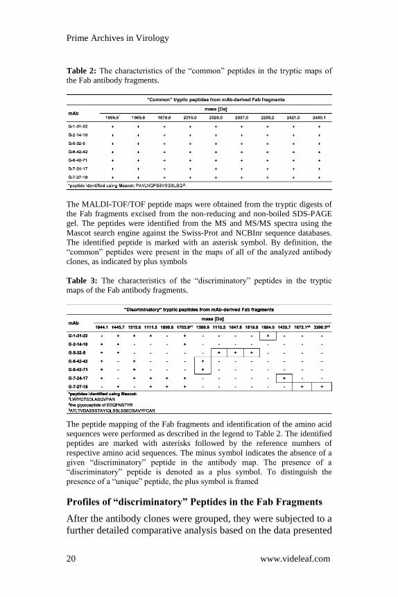
Table 2: The characteristics of the “common” peptides in the tryptic maps of
the Fab antibody fragments.
The MALDI-TOF/TOF peptide maps were obtained from the tryptic digests of
the Fab fragments excised from the non-reducing and non-boiled SDS-PAGE
gel. The peptides were identified from the MS and MS/MS spectra using the
Mascot search engine against the Swiss-Prot and NCBInr sequence databases.
The identified peptide is marked with an asterisk symbol. By definition, the
“common” peptides were present in the maps of all of the analyzed antibody
clones, as indicated by plus symbols
Table 3: The characteristics of the “discriminatory” peptides in the tryptic
maps of the Fab antibody fragments.
The peptide mapping of the Fab fragments and identification of the amino acid
sequences were performed as described in the legend to Table 2. The identified
peptides are marked with asterisks followed by the reference numbers of
respective amino acid sequences. The minus symbol indicates the absence of a
given “discriminatory” peptide in the antibody map. The presence of a
“discriminatory” peptide is denoted as a plus symbol. To distinguish the
presence of a “unique” peptide, the plus symbol is framed
Profiles of “discriminatory” Peptides in the Fab Fragments
After the antibody clones were grouped, they were subjected to a
further detailed comparative analysis based on the data presented

Prime Archives in Virology
21 www.videleaf.com
in Table 3. Only the Fab fragment maps were examined. Thus,
the same set of 3 “discriminatory” peptides was found in maps of
the G-6-42-42 and G-6-42-71 antibodies. This indicates that G-
6-42-42 and G-6-42-71 mAbs are the same clone. In the
corresponding Fab fragments of these antibodies, a “unique”
peptide at m/z 1580.9 was recognized. The simultaneous and
exclusive presence of the peptides with m/z 1515.8 and 1944.1
additionally distinguished the G-6-42-42 and G-6-42-71 mAbs
from the remaining 5 clones.
In the tryptic digest of the Fab fragment of the G-2-14-10 clone,
3 “discriminatory” peptides were obtained, including one peptide
identified using Mascot (m/z 1705.9). For this mAb, no “unique”
peptide was recognized. Analysis of the map generated for the
G-5-32-5 clone indicated 5 “discriminatory” peptides, including
3 “unique” for the clone, which had m/z values of 1110.5,
1647.8 and 1819.8. The maps of both the G-2-14-10 and G-5-32-
5 antibodies were characterized by the presence of 2 peptides
with m/z 1445.7 and 1944.1. Subsequently, the investigation of
the map for the G-1-31-22 clone demonstrated 5
“discriminatory” peptides, including 1 “unique” peptide with an
m/z of 1884.0 and 1 that was identified using Mascot (m/z
1705.9). The G-1-31-22 mAb was characterized by the
simultaneous presence of 3 peptides at m/z 1111.5, 1445.7 and
1515.8. The analysis of the data for the G-7-24-17 clone
identified 6 “discriminatory” peptides, with 1 “unique” peptide
at m/z 1430.7 and 1 that was identified using Mascot (m/z
1705.9). The G-7-24-17 mAb could be described by the presence
of 4 signals at m/z 1111.5, 1515.8, 1800.8 and 1944.1. In the G-
7-27-18 clone map, 6 “discriminatory” peptides were
distinguished. The three at m/z 1705.9, 1872.1 and 3300.5 were
identified using Mascot. The last two peptides were found to be
“unique” for this clone. The G-7-27-18 mAb was differentiated
by the simultaneous presence of 3 peptides at m/z 1111.5, 1445.7
and 1800.8.
Peptide maps of mAb-derived Fc Fragments
The main mAb discrimination was completed based the on the
profiles of the “discriminatory” peptides in the Fab fragment

Prime Archives in Virology
22 www.videleaf.com
maps, as described above. To extract all possible data, a
screening of Fc fragment-derived peptides was also performed.
As a result, a total of 13 peptides were recognized in the maps of
the G-1-31-22, G-2-14-10, G-5-32-5, G-6-42-42, G-6-42-71, G-
7-24-17 and G-7-27-18 antibody clones (Table 4). Among these,
9 were “common” and 4 were “discriminatory” for the analyzed
mAbs. According to data in Table 4, the G-6-42-42 and G-6-42-
71 mAbs shared the same profile of “discriminatory” peptides
within their Fc fragments. This confirms the indication
previously shown in the profiling of the Fab-derived peptides
(Table 3) that G-6-42-42 and G-6-42-71 mAbs represent a single
antibody clone subsequently designated G-6-42-42,71. In two
mAb groups, identical profiles of “discriminatory” peptides from
the Fc fragments were recognized (Table 4). The first one
comprises the previously mentioned G-6-42-42,71 antibodies
and the G-7-24-17 mAb, and the second includes the G-2-14-10
and G-5-32-5 antibody clones. Compared to the second group,
the G-7-27-18 mAb additionally contained a “unique” peptide.
No “discriminatory” peptide was identified in the map of the G-
1-31-22 clone Fc fragment. In the set of 13 peptides from the Fc
fragments, the amino acid sequences of 9 were identified as
fragments of IgG antibodies. These included 7 “common” and 2
“discriminatory” peptides.
Table 4: The characteristics of the peptides in the tryptic maps of the Fc
antibody fragments.

Prime Archives in Virology
23 www.videleaf.com
The MALDI-TOF/TOF peptide maps were obtained from the tryptic digests of
the Fc fragments excised from non-reducing and non-boiled SDS-PAGE gel.
The identification of the amino acid sequences was performed as described in
the legend to Table 2. The identified “common” and “discriminatory” peptides
are marked with asterisks followed by the reference numbers of the respective
amino acid sequences. The plus and minus symbols indicate the presence and
absence of a given peptide in the maps of the individual antibodies,
respectively. To distinguish the presence of a “unique” peptide, the plus symbol
is framed
The Role of Fab and Fc Fragment Mapping in mAb
Differentiation
Consistent with the results of immunoreactivity and isotyping
studies (Table 1), the peptide mapping of the mAb-derived
fragments using MS showed that the analyzed Igs are not
completely different. Individual “discriminatory” peptides were
repeated in the Fab and Fc fragment maps of different antibodies
along with the set of peptides “common” to all of them
(Tables 2, 3 and 4). However, the presence or absence of the
“unique” peptides within the Fab fragments and the pattern of
the remaining “discriminatory” peptides enabled effective
antibody differentiation (Table 3). Thus, 6 distinct clones were
found among 7 newly established mAbs against the H5 HA.
These 6 antibody clones probably recognize different epitopes in
the antigen molecule. The peptide mapping of the Fc fragments
from the anti-HA H5 IgG1 antibodies (Table 1) only grouped the
antibody clones (Table 4) that were clearly distinguished by the
profiling of the Fab-derived peptides (Table 3). On the other
hand, this process allowed the definitive conclusion that the G-6-
42-42 and G-6-42-71 mAbs, which shared the same Fab
fragment maps, represent one antibody clone (Tables 2, 3 and 4).
Discussion
Infection with HPAIVs of the H5 subtype leads to multi-organ
disease and death in domestic birds [2,4-7]. In addition, the
H5N1 viral strains pose a persistent pandemic threat [3]. To
prevent and treat H5N1 influenza virus infections and for
surveillance of H5N1 and other H5-subtype AIVs, mAbs against
H5 HA have been developed by many research groups (e.g., [23-
28]; for review, see [8]). Our work responded to the demand for

Prime Archives in Virology
24 www.videleaf.com
diagnostically valuable mAbs with broad strain specificity
against AIVs of the H5 subtype. These antibodies were produced
with hybridoma technology using recombinant, ectodomain-
based H5 HA protein with native-HA characteristics to
immunize mice.
The hybridomas generated by this process were screened for the
production of IgG antibodies against the H5 HA using ELISA.
The screening was performed against several forms of the HA
antigen that had various properties. The use of conformational
rHA1 proteins enabled the identification of antibodies that bound
to the highly variable HA1 subunit, which determines the HA
subtype. Distinguishing between conformation sensitive and
non-sensitive antibodies was achieved using a misfolded rHA
protein. In addition to the variations in the forms, the sequences
of the HA antigens that were used originated from highly
divergent H5-subtype influenza viruses. As a consequence, the
H5 HA antigens demonstrated substantial antigenic diversity,
which was confirmed by a homology search against the
immunogen.
From our screening strategy, we obtained a total of 64
hybridoma cell lines. These cell lines secreted antibodies that
were reactive with all of the H5 HA antigens that were used for
the specificity testing except for the non-conformational antigen.
A final set of 7 hybridoma clones was selected. Specifically, the
G-1-31-22, G-2-14-10, G-5-32-5, G-6-42-42, G-6-42-71, G-7-
24-17 and G-7-27-18 mAbs, all of IgG1 isotype, were further
analyzed. In the preliminary immunoreactivity studies, we were
able to show that the newly established mAbs specifically
recognized epitopes in the properly folded HA1 subunit of H5
HAs from multiple strains of the H5-subtype influenza viruses
(Table 1). Importantly, they did not cross-react with influenza
viruses of H1-H4 and H6-H16 subtypes (Table 1). However,
these studies did not allow for clear discrimination among the
finally selected mAbs and the relevant hybridoma cell lines. For
this reason, the exact number of the unique antibody and
hybridoma clones could not be inferred.

Prime Archives in Virology
25 www.videleaf.com
The differentiation of antibody clones and relevant antibody-
producing cell lines is of special importance for comprehensive
assessment of their possible applications. When used in
diagnostics or basic research, the set of mAbs that recognize
different epitopes in the H5 HAs potentially extends the range of
the target AIVs among the formerly and currently circulating
viral strains. It can also facilitate the identification of the novel
emerging H5-subtype AIVs. In addition, availability of different
antibody clones enables the choice of the ones that will be best
suited to specified method or technique. For example, two
distinct mAbs can be successfully used as detection and capture
antibodies in virus detection by a sandwich ELISA or
immunochromatography.
Insight into the antibody heterogeneity could be provided by a
comparison of the sequences encoding their variable regions,
especially the CDRs [29]. Sequencing is routinely used to
identify antibodies. However, it may be perceived as challenging
if the presence of pseudogenes and mRNAs encoding non-
functional antibody chains in hybridoma cells is considered [30].
Another method for differentiating mAbs is based on cross-
inhibition experiments (e.g., [31]). Antibodies that do not
compete for binding to the target antigen are considered to
recognize distinct, non-overlapping epitopes. Competition
between mAbs is interpreted as indicating that the tested
antibody clones bind to the same or to closely related epitopes.
Thus, cross-inhibition experiments may not give conclusive
results. As the first mass spectra characterizing the generated
antibody clones showed some differences between selected
antibodies, we decided to expand them with peptide mapping of
the Fab and Fc fragments. It was assumed that antibody
examination at the protein level would allow to avoid some
possible drawbacks related with their analyses at the genetic and
functional levels.
Digestion with the immobilized ficin produced Fc and Fab
fragments of the G-1-31-22, G-2-14-10, G-5-32-5, G-6-42-42,
G-6-42-71, G-7-24-17 and G-7-27-18 mAbs. Subsequently,
tryptic peptide maps of these fragments were generated. Based
on the resulting MS and MS/MS spectra, Mascot searches

Prime Archives in Virology
26 www.videleaf.com
against the Swiss-Prot and NCBInr sequence databases were
performed. This enabled identification of some peptides derived
from both Fc and Fab fragments, all of which belonged to the Ig
class of proteins (Tables 2, 3, and 4).
Most of the peptides detected in the Fc fragments were
“common” for the analyzed mAbs (69%; Table 4). The majority
of the amino acid sequences of these fragments were identified
within the protein databases. This is consistent with the widely
accepted view that the Fc fragments are species- and isotype-
conserved components of the Igs, which have no significance for
their specificity [9]. In contrast, “discriminatory” peptides
dominated the Fab fragment maps (61%; Table 3). Within these
antibody fragments, very few sequences could be identified with
the database searches: many fewer than for the conserved Fc
fragments (17% vs. 69%; Tables 2, 3 and 4). These different
proportions can be explained by the fact that the Fab fragments
exhibit considerable variation in the specificity-determining
sequences. For this reason, the protein databases are incomplete
in this area.
A close inspection of the peptide maps of the Fab fragments
revealed that the analyzed antibodies differed in the profiles of
their “discriminatory” peptides (Table 3). Accordingly, 6
different clones were distinguished among the 7 selected mAbs.
Presumably, these mAbs target distinct epitopes in the H5 HA
molecule. For the G-6-42-42 and G-6-42-71 clones, identical
peptide maps of the Fab and Fc fragments were obtained
(Tables 2, 3 and 4). This indicates that these two mAbs are the
same antibody clone. Interestingly, the G-6-42-42 and G-6-42-71
antibodies were the only clones among selected mAbs that
originated from subcloning of the same hybridoma. Conclusions
from the mass spectrometry approach are consistent with those
from the advanced immunoreactivity studies (Additional file 6:
Figures S10-S12).
Addendum
On 9 June 2016, G-1-31-22, G-2-14-10, G-5-32-5, G-6-42-42,
G-7-24-17, G-7-27-18 hybridoma cell lines were given the

Prime Archives in Virology
27 www.videleaf.com
following Accession Numbers by the International Depositary
Authority: DSM ACC3292, DSM ACC3293, DSM ACC3294,
DSM ACC3295, DSM ACC3296 and DSM ACC3297,
respectively. They are all held by the Leibniz Institute DSMZ-
German Collection of Microorganisms and Cell Cultures
(Braunschweig, Germany).
Conclusions
A unique panel of 6 different anti-H5 HA antibody clones was
generated and characterized. The newly established mAbs target
epitopes in the properly folded HA1 subunit of HAs from
multiple strains of the H5-subtype influenza viruses and do not
cross-react with AIVs of H1-H4 and H6-H16 subtypes.
Characterized by high specificity and broad-range activities
against the H5 HAs, the described mAbs constitute valuable
diagnostic and basic research tools. In the present study, the
mass spectrometry approach has been developed as a method for
antibody clone differentiation at the protein level. The method
may be successfully used for characterization of mAbs that are
poorly discriminated by immunological techniques as well as to
obtain supplementary or confirmatory results. It also enables
identification of unique antibody-producing cell lines.
References
1. Xu X, Subbarao, Cox NJ, Guo Y. Genetic characterization of
the pathogenic influenza A/Goose/Guangdong/1/96 (H5N1)
virus: similarity of its hemagglutinin gene to those of H5N1
viruses from the 1997 outbreaks in Hong Kong. Virology.
1999; 261: 15–19.
2. Verhagen JH, Herfst S, Fouchier RA. Infectious disease.
How a virus travels the world. Science. 2015; 347: 616–617.
3. World Health Organization. Cumulative number of
confirmed human cases for avian influenza A(H5N1)
reported to WHO, 2003-2017. Available Online at:
http://www.who.int/influenza/human_animal_interface/2017
_01_16_tableH5N1corrected.pdf.
4. Ip HS, Torchetti MK, Crespo R, Kohrs P, DeBruyn P, et al.
Novel Eurasian highly pathogenic avian influenza A H5

Prime Archives in Virology
28 www.videleaf.com
viruses in wild birds, Washington, USA, 2014. Emerg Infect
Dis. 2015; 21: 886–890.
5. Hvistendahl M. Avian influenza. Enigmatic bird flu strain
races across the U.S. Midwest. Science. 2015; 348: 741–742.
6. World Health Organization. Assessment of risk associated
with influenza A(H5N8) virus. Available Online at:
http://www.who.int/influenza/human_animal_interface/avian
_influenza/riskassessment_AH5N8_201611/en/.
7. United States Department of Agriculture, Animal and Plant
Health Inspection Service. Highly pathogenic avian
influenza infected premises 2014–2015. Available Online at:
https://www.aphis.usda.gov/animal_health/animal_dis_spec/
poultry/downloads/hpai-positive-premises-2014-2015.pdf.
8. Velkov T, Ong C, Baker MA, Kim H, Li J, et al. The
antigenic architecture of the hemagglutinin of influenza
H5N1 viruses. Mol Immunol. 2013; 56: 705–719.
9. Thompson NJ, Rosati S, Heck A. Performing native mass
spectrometry analysis on therapeutic antibodies. Methods.
2014; 65: 11–17.
10. Chirino AJ, Mire-Sluis A. Characterizing biological products
and assessing comparability following manufacturing
changes. Nat Biotechnol. 2004; 22: 1383–1391.
11. Köhler G, Milstein C. Continuous cultures of fused cells
secreting antibody of predefined specificity. Nature. 1975;
256: 495–497.
12. Steinitz M, Klein G, Koskimies S, Makel O. EB virus-
induced B lymphocyte cell lines producing specific
antibody. Nature. 1977; 269: 420–422.
13. Lanzavecchia A, Corti D, Sallusto F. Human monoclonal
antibodies by immortalization of memory B cells. Curr Opin
Biotechnol. 2007; 18: 523–528.
14. Borth N, Zeyda M, Kunert R, Katinger H. Efficient selection
of high-producing subclones during gene amplification of
recombinant Chinese hamster ovary cells by flow cytometry
and cell sorting. Biotechnol Bioeng 2000–2001; 71: 266-
273.
15. Kuhne M, Dippong M, Flemig S, Hoffmann K, Petsch K, et
al. Comparative characterization of mAb producing hapten-
specific hybridoma cells by flow cytometric analysis and
ELISA. J Immunol Methods. 2014; 413: 45–56.

Prime Archives in Virology
29 www.videleaf.com
16. Zhang Z, Pan H, Chen X. Mass spectrometry for structural
characterization of therapeutic antibodies. Mass Spectrom
Rev. 2009; 28: 147–176.
17. Chelius D, Ruf P, Gruber P, Plöscher M, Liedtke R, et al.
Structural and functional characterization of the trifunctional
antibody catumaxomab. MAbs. 2010; 2: 309–319.
18. Gahoual R, Burr A, Busnel JM, Kuhn L, Hammann P, et al.
Rapid and multi-level characterization of trastuzumab using
sheathless capillary electrophoresis-tandem mass
spectrometry. MAbs. 2013; 5: 479–490.
19. Srebalus Barnes CA, Lim A. Applications of mass
spectrometry for the structural characterization of
recombinant protein pharmaceuticals. Mass Spectrom Rev.
2007; 26: 370–388.
20. Yamaguchi Y, Kim H, Kato K, Masuda K, Shimada I, et al.
Proteolytic fragmentation with high specificity of mouse
immunoglobulin G. Mapping of proteolytic cleavage sites in
the hinge region. J Immunol Methods. 1995; 181: 259–267.
21. Parham P. On the fragmentation of monoclonal IgG1, IgG2a,
and IgG2b from BALB/c mice. J Immunol. 1983; 131:
2895–2902.
22. Mariani M, Camagna M, Tarditi L, Seccamani E. A new
enzymatic method to obtain high-yield F(ab’)2 suitable for
clinical use from mouse IgG1. Mol Immunol. 1991; 28: 69–
77.
23. Linke S, Neubauer K, Dorner MB, Dorner BG, Pauli G, et
al. Generation and characterisation of monoclonal antibodies
against influenza virus A, subtype H5N1. J Virol Methods.
2011; 175: 85–94.
24. Ohnishi K, Takahashi Y, Kono N, Nakajima N, Mizukoshi
F, et al. Newly established monoclonal antibodies for
immunological detection of H5N1 influenza virus. Jpn J
Infect Dis. 2012; 65: 19–27.
25. Sakurai A, Takayama K, Nomura N, Munakata T,
Yamamoto N, et al. Broad-spectrum detection of H5 subtype
influenza A viruses with a new fluorescent
immunochromatography system. PLoS One. 2013; 8:
e76753.
26. Wu R, Li X, Leung HC, Cao Z, Qiu Z, et al. A novel
neutralizing antibody against diverse clades of H5N1

Prime Archives in Virology
30 www.videleaf.com
influenza virus and its mutants capable of airborne
transmission. Antivir Res. 2014; 106: 13–23.
27. Fiebig P, Shehata AA, Liebert UG. Generation of
monoclonal antibodies reactive against subtype specific
conserved B-cell epitopes on haemagglutinin protein of
influenza virus H5N1. Virus Res. 2015; 199: 46–55.
28. Gronsang D, Bui AN, Trinh DQ, Bui VN, Nguyen KV, et al.
Characterization of cross-clade monoclonal antibodies
against H5N1 highly pathogenic avian influenza virus and
their application to the antigenic analysis of diverse H5
subtype viruses. Arch Virol. 2017.
29. Johnson G, Wu TT. Kabat database and its applications: 30
years after the first variability plot. Nucleic Acids Res. 2000;
28: 214–218.
30. Creative Biolabs. Antibody mRNA Sequencing Service.
Available Online at: http://www.creative-
biolabs.com/antibody-sequencing-service.html.
31. Lelli D, Moreno A, Brocchi E, Sozzi E, Capucci L, et al.
West Nile virus: characterization and diagnostic applications
of monoclonal antibodies. Virol J. 2012; 9: 81.