Embed Size (px)
Citation preview
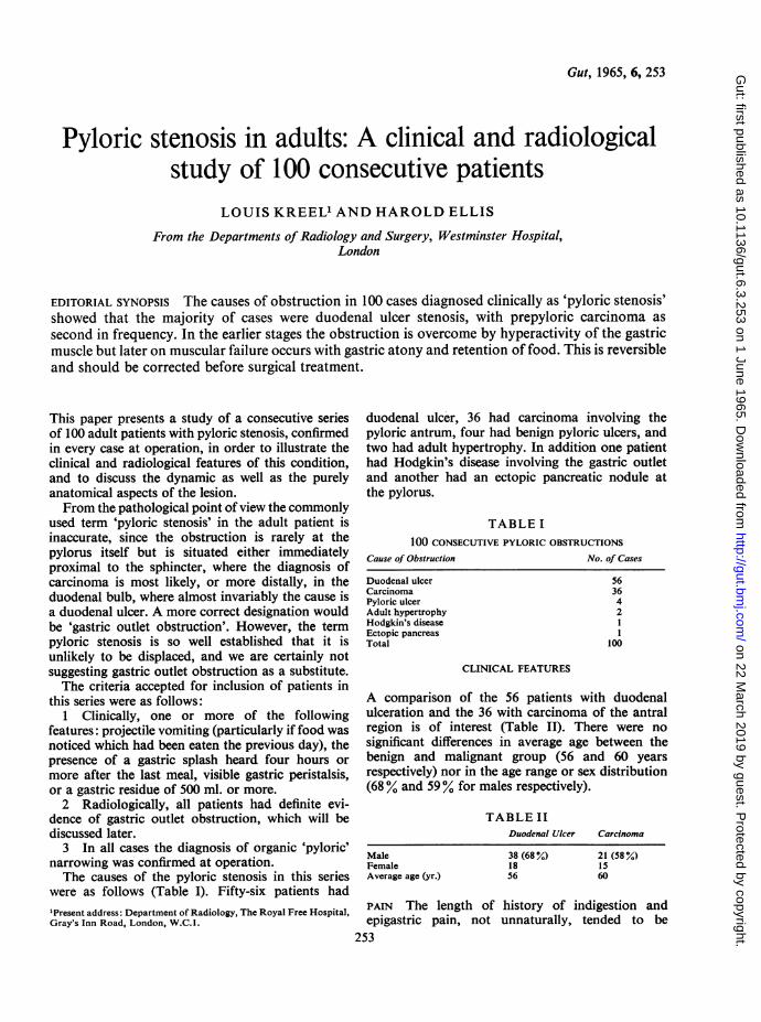
Gut, 1965, 6, 253
Pyloric stenosis in adults: A clinical and radiologicalstudy of 100 consecutive patients
LOUIS KREEL' AND HAROLD ELLIS
From the Departments of Radiology and Surgery, Westminster Hospital,London
EDITORIAL SYNOPSIS The causes of obstruction in 100 cases diagnosed clinically as 'pyloric stenosis'showed that the majority of cases were duodenal ulcer stenosis, with prepyloric carcinoma as
second in frequency. In the earlier stages the obstruction is overcome by hyperactivity of the gastricmuscle but later on muscular failure occurs with gastric atony and retention of food. This is reversibleand should be corrected before surgical treatment.
This paper presents a study of a consecutive seriesof 100 adult patients with pyloric stenosis, confirmedin every case at operation, in order to illustrate theclinical and radiological features of this condition,and to discuss the dynamic as well as the purelyanatomical aspects of the lesion.From the pathological point of view the commonly
used term 'pyloric stenosis' in the adult patient isinaccurate, since the obstruction is rarely at thepylorus itself but is situated either immediatelyproximal to the sphincter, where the diagnosis ofcarcinoma is most likely, or more distally, in theduodenal bulb, where almost invariably the cause isa duodenal ulcer. A more correct designation wouldbe 'gastric outlet obstruction'. However, the termpyloric stenosis is so well established that it isunlikely to be displaced, and we are certainly notsuggesting gastric outlet obstruction as a substitute.The criteria accepted for inclusion of patients in
this series were as follows:1 Clinically, one or more of the following
features: projectile vomiting (particularly if food wasnoticed which had been eaten the previous day), thepresence of a gastric splash heard four hours ormore after the last meal, visible gastric peristalsis,or a gastric residue of 500 ml. or more.2 Radiologically, all patients had definite evi-
dence of gastric outlet obstruction, which will bediscussed later.
3 In all cases the diagnosis of organic 'pyloric'narrowing was confirmed at operation.The causes of the pyloric stenosis in this series
were as follows (Table I). Fifty-six patients had'Present address: Department of Radiology, The Royal Free Hospital,Gray's Inn Road, London, W.C.1.
duodenal ulcer, 36 had carcinoma involving thepyloric antrum, four had benign pyloric ulcers, andtwo had adult hypertrophy. In addition one patienthad Hodgkin's disease involving the gastric outletand another had an ectopic pancreatic nodule atthe pylorus.
TABLE I100 CONSECUTIVE PYLORIC OBSTRUCTIONS
Cause of Obstruction
Duodenal ulcerCarcinomaPyloric ulcerAdult hypertrophyHodgkin's diseaseEctopic pancreasTotal
No. of Cases
56364211
100
CLINICAL FEATURES
A comparison of the 56 patients with duodenalulceration and the 36 with carcinoma of the antralregion is of interest (Table II). There were nosignificant differences in average age between thebenign and malignant group (56 and 60 yearsrespectively) nor in the age range or sex distribution(68 % and 59% for males respectively).
TABLE IIDuodenal Ulcer Carcinoma
MaleFemaleAverage age (yr.)
38 (68%)1856
21 (58%)1560
PAIN The length of history of indigestion andepigastric pain, not unnaturally, tended to be
253
on 22 March 2019 by guest. P
rotected by copyright.http://gut.bm
j.com/
Gut: first published as 10.1136/gut.6.3.253 on 1 June 1965. D
ownloaded from
Louis Kreel and Harold Ellis
considerably longer in the patients with benignulcers (Table III). It is interesting, however, that fivepatients with duodenal ulceration had a history ofpain for less than one year. One of these patientswith a history of only two months had had a previousthoraco-lumbar sympathectomy for hypertensionand may well have had a consequent gastric denerv-ation. Of the remaining duodenal ulcer patients,11 had a history of one to four years, 12 of five tonine years, and 27 had a history of from 10 to 20or more years; in one patient the length of historywas not recorded.
In contrast, 11 of the 36 patients with carcinomahad experienced no pain at any time, in 20 thehistory was under one year, in three the history wasup to two years, one was of three years' duration,and in one patient there had been mild dyspepsia forthe preceding eight years.
TABLE IIILENGTH OF HISTORY OF PAIN
Duodenal Ulcer Carcinoma
EXAMINATION
On clinical examination it is obvious that the'typical ulcer facies' of the old text books applied tomany of the duodenal ulcer patients with stenosis.Thirty-three patients were thin (59 %) and only oneof the whole group was described as obese. Abdom-inal examination revealed a splash in 36 patients ofthe ulcer group (64 %), in nine of whom, in addition,visible peristalsis was seen and in four of whom thegastric distension was great enough to produce alarge, palpable mass. In contrast, only five of thepatients with carcinoma had a splash (14%) andonly one had visible peristalsis. However, 19 of thepatients with carcinoma had a palpable mass in thepyloric region (Table IV).
TABLE IVDuodenal Ulcer Carcinoma
MassSplashStomach visibleStomach palpable
36 (64%)94
19 (53%)5 (14 %)1
Under 1 year1-4 years5-9 years10-19 years20+
No painTotals
511122341
56
2041
1136
VOMITING Vomiting was the commonest presentingsymptom in both groups. In the peptic ulcer patientsit occurred in 49 of the 56 cases (87-5 %) and wastypical projectile vomiting of large amounts in 39of these. In addition, 24 of the patients noted stalefood in the vomitus. In the carcinoma group 31 ofthe 38 patients (86%) had been vomiting; in 23 ofthem it was typical of stenosis and in nine stale foodhad been noted in addition.
OTHER SYMPTOMS Weight loss was a prominentfeature in both groups, occurring in 44 of theduodenal ulcer patients (78 %) and in 34 patients withcancer (94 %).
Recent constipation was recorded in 14 of theduodenal ulcer and in 12 of the carcinoma group.
Recent diarrhoea was recorded in three of theulcer and in three of the cancer series.
Gastrointestinal bleeding had occurred recentlyin five patients with duodenal ulcer and at sometime in the past in eight further cases.
In the carcinoma group eight patients had smallrecent haemorrhages; one indeed presented withmelaena and another with anaemia.Three of the ulcer series had perforations repaired
in the past but there was no case of associatedperforation in the cancer group.
BIOCHEMICAL CHANGES
Biochemical disturbances did not occur in patientswho had never vomited or in whom the vomitingwas not profuse or of almost daily occurrence. In44 patients in this series, vomiting had been copiousand frequent before admission; in 20 of these theblood biochemistry was completely normal, and in afurther 11 there was only a raised blood urea above50 mg.% as the sole biochemical abnormality, insix being above 100 mg.% and in one of thesereaching 243 mg. %. All were rapidly corrected afterrehydration. In the remaining 13 patients there weremore widespread biochemical alterations including,in nine of these, a raised blood urea. In these 13cases there were one or more of the followingchanges: sodium less than 135 mEq. %, potassiumless than 3-5 mEq. %, chloride less than 95 mEq. %or bicarbonate above 30 mEq. % (Table V).
It is interesting that only five of the 24 patientswith biochemical disturbance were admitted in aclinically obvious state of electrolytic imbalance-ill, dehydrated, weak and, in one case, with obvioustetany.Anaemia (haemoglobin of less than 80%) was
present in 11 of the 56 patients with duodenalulceration and in seven of the 36 with carcinoma:in one of the latter it was the presenting feature.
ASSOCIATED BENIGN GASTRIC ULCER
Four of the patients with duodenal ulceration hadan associated benign gastric ulcer confirmed at
254
on 22 March 2019 by guest. P
rotected by copyright.http://gut.bm
j.com/
Gut: first published as 10.1136/gut.6.3.253 on 1 June 1965. D
ownloaded from
Pyloric stenosis in adults: A clinical and radiological study of 100 consecutive patients
TABLE VPATIENTS WITH BIOCHEMICAL DISTURB 'NCES'and its Duration Na K Cl Bi
(mEq./l.) (mEq./l.) (mEq./l.) ct(rn
icarbon-tenEq.!l.)
Urea(mEq./l.)
255
Clinical State
2345678
Duodenal ulcerDuodenal ulcerDuodenal ulcerDuodenal ulcerDuodenal ulcerDuodenal ulcerDuodenal ulcerDuodenal ulcer
9 Duodenal ulcer10 Duodenal ulcer1 I Carcinoma12 Carcinoma13 Gastric ulcer'Other than raised blood urea onl
Large amounts, 3 mth.Large amounts, 3 mth.Large amounts, 2 mth.Large amounts, 2 yr.Large amounts, 2 mth.Large amounts, I mth.Large amounts, 5 mth.Large amounts, 6 mth.
Large amounts, 1 mth.Large amounts, I yr.Large amounts, 1 mth.Large amounts, 2 mth.Large amounts, 4 mth.
ly
146132136128126142140145
132148124137131
3-13-23-74-13-43-43-23-8
2-93-22-82950
82898012897757845
8789758091
34353130
504564
39
576158178310633
243
1193710424527
SatisfactorySatisfactorySatisfactorySatisfactorySatisfactorySatisfactorySatisfactoryDehydrated,irrational, tetanicThin, ill, dehydratedIll, dehydrated111, dehydratedlll, dehydratedSatisfactory
operation. Three of these were women. Two morepatients, also women, had radiological evidence of agastric ulcer, although as these patients had drainageprocedures and not partial gastrectomy, histologicalconfirmation was not obtained. Apart from thisquite marked sex bias for gastric ulcer associatedwith stenosis due to duodenal ulcer, analysisrevealed no relevant differences from the remainingpatients with benign pyloric obstruction.
RADIOLOGICAL AND BARIUM MEAL EXAMINATION
The radiological signs of pyloric stenosis can bedivided into those indicating the presence ofobstruction at the gastric outlet and those indicatingits site and possible nature. However, very un-commonly, the first radiological abnormality to bedetected is the presence of lymphangitis carcinoma-tosa on the chest film (one case).
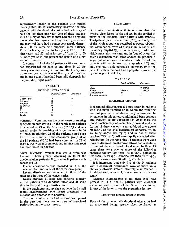
Occasionally there is evidence of gastric stasis onthe plain film of the abdomen in the form of a largegastric shadow with retained food particles producingpatchy translucencies (two cases). In these cases anerect film will show a large, high gastric fluid level.Retention of oral cholecystographic contrast fromthe previous evening with a 'non-functioning' gallbladder was the first radiological evidence ofobstruction at the gastric outlet in two cases (Fig. 1).Subsequent barium meal examination revealed adeformed, contracted duodenal cap as the cause ofthe obstruction.
SIGNS OF OBSTRUCTION AT THE GASTRIC OUTLETThere are several signs of obstruction at the gastricoutlet visible radiologically.Presence of excessive fasting gastric juice This is
immediately apparent on the first mouthful ofbarium reaching the stomach. Instead of the bolus
FIG. 1. Oral cholecystographic contrast medium retainedin the stomach 14 hours after ingestion of the tablets. Intwo cases in this series this was the first radiological signofgastric outlet obstruction.
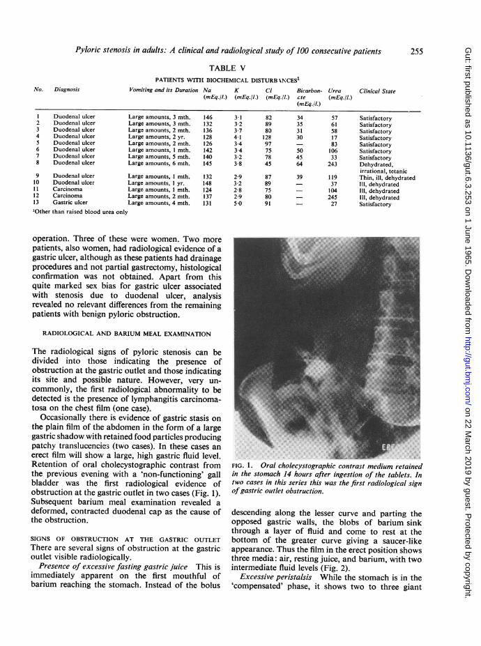
descending along the lesser curve and parting theopposed gastric walls, the blobs of barium sinkthrough a layer of fluid and come to rest at thebottom of the greater curve giving a saucer-likeappearance. Thus the film in the erect position showsthree media: air, resting juice, and barium, with twointermediate fluid levels (Fig. 2).
Excessive peristalsis While the stomach is in the'compensated' phase, it shows two to three giant
No. Diagnosis Vomiting
on 22 March 2019 by guest. P
rotected by copyright.http://gut.bm
j.com/
Gut: first published as 10.1136/gut.6.3.253 on 1 June 1965. D
ownloaded from
Louis Kreel and Harold Ellis
Delay in emptying A 24-hour barium residue isusually diagnostic but this is seldom necessary. Afour to six-hour film, using barium sulphate as thecontrast medium, normally shows less than 20%retention whereas in pyloric stenosis there is morethan 50% retention. It is, however, important toexclude pylorospasm or obstructive lesions in theproximal small bowel. Pylorospasm can be distin-guished by administering probanthine intravenously,after which the stomach will empty normally.
FIG. 2. Barium entering the stomach in the presence ofexcessive gastric juice showing three media barium,gastric juice, and air-and the two intervening fluid levels.
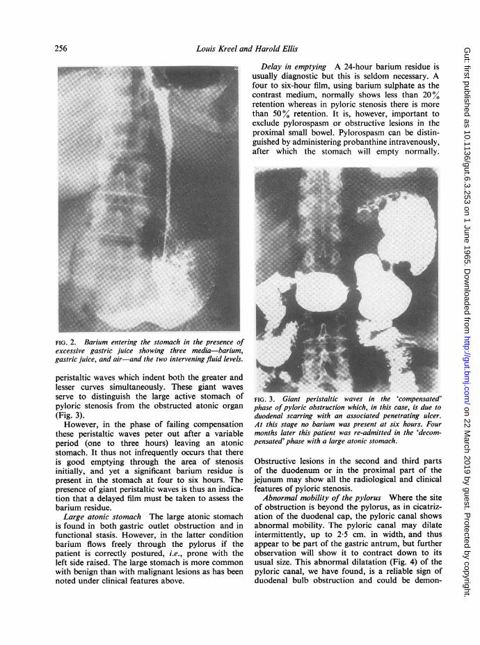
peristaltic waves which indent both the greater andlesser curves simultaneously. These giant wavesserve to distinguish the large active stomach ofpyloric stenosis from the obstructed atonic organ(Fig. 3).However, in the phase of failing compensation
these peristaltic waves peter out after a variableperiod (one to three hours) leaving an atonicstomach. It thus not infrequently occurs that thereis good emptying through the area of stenosisinitially, and yet a significant barium residue ispresent in the stomach at four to six hours. Thepresence of giant peristaltic waves is thus an indica-tion that a delayed film must be taken to assess thebarium residue.Large atonic stomach The large atonic stomach
is found in both gastric outlet obstruction and infunctional stasis. However, in the latter conditionbarium flows freely through the pylorus if thepatient is correctly postured, i.e., prone with theleft side raised. The large stomach is more commonwith benign than with malignant lesions as has beennoted under clinical features above.
FIG. 3. Giant peristaltic waves in the 'compensated'phase of pyloric obstruction which, in this case, is due toduodenal scarring with an associated penetrating ulcer.At this stage no barium was present at six hours. Fourmonths later this patient was re-admitted in the 'decom-pensated' phase with a large atonic stomach.
Obstructive lesions in the second and third partsof the duodenum or in the proximal part of thejejunum may show all the radiological and clinicalfeatures of pyloric stenosis.Abnormal mobility of the pylorus Where the site
of obstruction is beyond the pylorus, as in cicatriz-ation of the duodenal cap, the pyloric canal showsabnormal mobility. The pyloric canal may dilateintermittently, up to 2-5 cm. in width, and thusappear to be part of the gastric antrum, but furtherobservation will show it to contract down to itsusual size. This abnormal dilatation (Fig. 4) of thepyloric canal, we have found, is a reliable sign ofduodenal bulb obstruction and could be demon-
256
on 22 March 2019 by guest. P
rotected by copyright.http://gut.bm
j.com/
Gut: first published as 10.1136/gut.6.3.253 on 1 June 1965. D
ownloaded from
Pyloric stenosis in adults: A clinical and radiological study of 100 consecutive patients
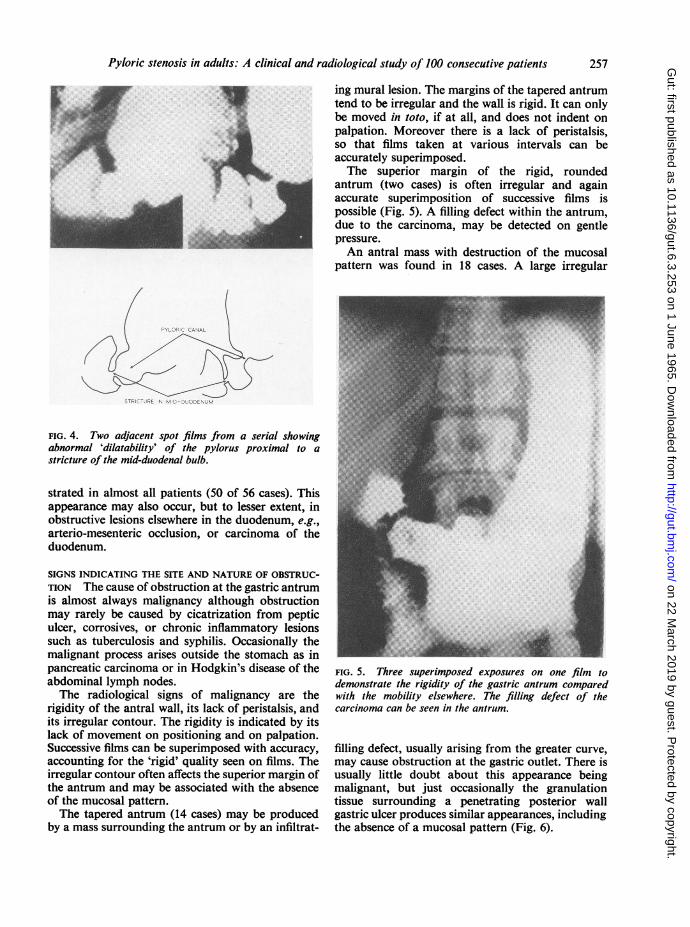
ing mural lesion. The margins of the tapered antrumtend to be irregular and the wall is rigid. It can onlybe moved in toto, if at all, and does not indent onpalpation. Moreover there is a lack of peristalsis,so that films taken at various intervals can beaccurately superimposed.The superior margin of the rigid, rounded
antrum (two cases) is often irregular and againaccurate superimposition of successive films ispossible (Fig. 5). A filling defect within the antrum,due to the carcinoma, may be detected on gentlepressure.An antral mass with destruction of the mucosal
pattern was found in 18 cases. A large irregular
PYLORIC CANAL
STRICTURE IN MID-DUODENUM
FIG. 4. Two adjacent spot films from a serial showingabnormal 'dilatability' of the pylorus proximal to astricture of the mid-duodenal bulb.
strated in almost all patients (50 of 56 cases). Thisappearance may also occur, but to lesser extent, inobstructive lesions elsewhere in the duodenum, e.g.,arterio-mesenteric occlusion, or carcinoma of theduodenum.
SIGNS INDICATING THE SITE AND NATURE OF OBSTRUC-TION The cause of obstruction at the gastric antrumis almost always malignancy although obstructionmay rarely be caused by cicatrization from pepticulcer, corrosives, or chronic inflammatory lesionssuch as tuberculosis and syphilis. Occasionally themalignant process arises outside the stomach as inpancreatic carcinoma or in Hodgkin's disease of theabdominal lymph nodes.The radiological signs of malignancy are the
rigidity of the antral wall, its lack of peristalsis, andits irregular contour. The rigidity is indicated by itslack of movement on positioning and on palpation.Successive films can be superimposed with accuracy,accounting for the 'rigid' quality seen on films. Theirregular contour often affects the superior margin ofthe antrum and may be associated with the absenceof the mucosal pattern.The tapered antrum (14 cases) may be produced
by a mass surrounding the antrum or by an infiltrat-
FIG. 5. Three superimposed exposures on one filnm todemonstrate the rigidity of the gastric antrum comparedwith the mobility elsewhere. The filling defect of thecarcinoma can be seen in the antrum.
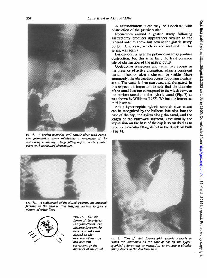
filling defect, usually arising from the greater curve,may cause obstruction at the gastric outlet. There isusually little doubt about this appearance beingmalignant, but just occasionally the granulationtissue surrounding a penetrating posterior wallgastric ulcer produces similar appearances, includingthe absence of a mucosal pattern (Fig. 6).
257
on 22 March 2019 by guest. P
rotected by copyright.http://gut.bm
j.com/
Gut: first published as 10.1136/gut.6.3.253 on 1 June 1965. D
ownloaded from
Louis Kreel and Harold Ellis
FIG. 6. A benign posterior wallsive granulation tissue mimickiantrum by producing a large fillcurve with associated obstruction.
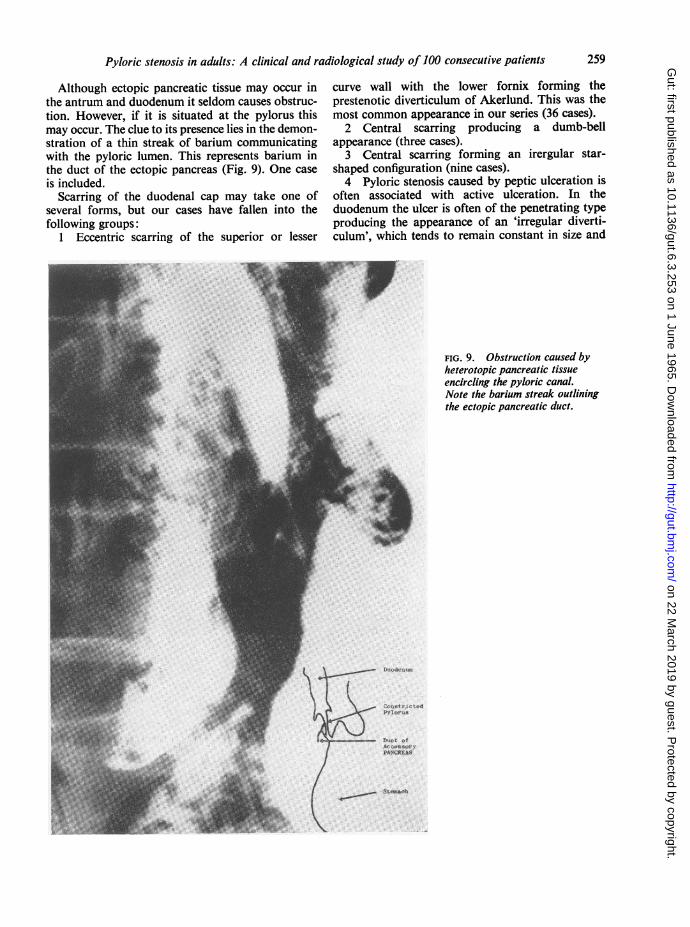
FIG. 7a. A radiograph of the clofurrows in the pyloric ring tra,picture of white lines.
i _ A carcinomatous ulcer may be associated withobstruction of the gastric outlet.Recurrence around a gastric stump following
gastrectomy produces appearances similar to thetapered antrum above but now at the gastric stumpoutlet. (One case, which is not included in thisseries, was seen.)
Lesions occurring at the pyloric canal may produceobstruction, but this is in fact, the least commonsite of obstruction of the gastric outlet.
Obstructive symptoms and signs may appear inthe presence of active ulceration, when a persistentbarium fleck or ulcer niche will be visible. Morecommonly, the obstruction occurs following cicatriz-ation. The canal is then narrowed and elongated. Inthis respect it is important to note that the diameterof the canal does not correspond to the width betweenthe barium streaks in the pyloric canal (Fig. 7) aswas shown by Williams (1962). We include four casesin this series.Adult hypertrophic pyloric stenosis (two cases)
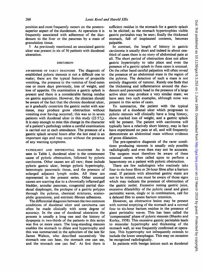
can be recognized by the bulbous intrusion into thebase of the cap, the spikes along the canal, and thelength of the narrowed segment. Occasionally theimpression on the base of the cap is so marked as toproduce a circular filling defect in the duodenal bulb(Fig. 8).
gastric ulcer with exces-ing a carcinoma of the'ing defect on the greater -
osed pylorus, the mucosalpping barium to give a
FIG. 7b. The slitlumen of the pylorusis asymmetrical. Thedistance between thebarium streaks willdepend on thedirection of the rays FIG. 8. Film of adult hypertrophic pyloric stenosis inand does not which the impression on the base of cap by the hyper-correspond to the trophied pylorus was so marked as to produce a circulardiameter of the canal. filling defect in the duodenal bulb.
258
on 22 March 2019 by guest. P
rotected by copyright.http://gut.bm
j.com/
Gut: first published as 10.1136/gut.6.3.253 on 1 June 1965. D
ownloaded from
Pyloric stenosis in adults: A clinical and radiological study of 100 consecutive patients
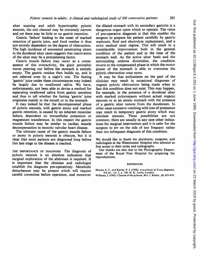
Although ectopic pancreatic tissue may occur inthe antrum and duodenum it seldom causes obstruc-tion. However, if it is situated at the pylorus thismay occur. The clue to its presence lies in the demon-stration of a thin streak of barium communicatingwith the pyloric lumen. This represents barium inthe duct of the ectopic pancreas (Fig. 9). One case
is included.Scarring of the duodenal cap may take one of
several forms, but our cases have fallen into thefollowing groups:
1 Eccentric scarring of the superior or lesser
curve wall with the lower fornix forming theprestenotic diverticulum of Akerlund. This was themost common appearance in our series (36 cases).
2 Central scarring producing a dumb-bellappearance (three cases).
3 Central scarring forming an irergular star-shaped configuration (nine cases).4 Pyloric stenosis caused by peptic ulceration is
often associated with active ulceration. In theduodenum the ulcer is often of the penetrating typeproducing the appearance of an 'irregular diverti-culum', which tends to remain constant in size and
FIG. 9. Obstruction caused byheterotopic pancreatic tissueencircling the pyloric canal.Note the barium streak outliningthe ectopic pancreatic duct.
259
on 22 March 2019 by guest. P
rotected by copyright.http://gut.bm
j.com/
Gut: first published as 10.1136/gut.6.3.253 on 1 June 1965. D
ownloaded from
Louis Kreel and Harold Ellis
position and most frequently occurs on the postero-superior aspect of the duodenum. At operation it isfrequently associated with adherence of the duo-denum to the liver or the pancreas by a mass ofgranulation tissue.
5 As previously mentioned an associated gastriculcer was present in six of 56 patients with duodenalulceration.
DISCUSSION
AWARENESS OF EARLY DIAGNOSIS The diagnosis ofestablished pyloric stenosis is not a difficult one tomake; there are the typical features of projectilevomiting, the presence in the vomitus of food eatenone or more days previously, loss of weight, andloss of appetite. On examination a gastric splash ispresent and there is a considerable fasting residueon gastric aspiration. However, the clinician shouldbe aware of the fact that the chronic duodenal ulcer,as it gradually constricts the gastric outlet with scar
tissue, may produce gross obstruction withoutvomiting ever having occurred; this was so in seven
patients with duodenal ulcer in this study (22 5 %).It is easy enough to miss this change in the characterof a patient's pathology unless a careful examinationis carried out at each attendance. The presence of a
gastric splash several hours after the last meal is animportant sign and may occur without the presence
of any warning symptoms.
PATHOLOGY AND DIFFERENTIAL DIAGNOSIS As is
seen in Table I, duodenal ulcer is the commonestcause of pyloric obstruction, followed by pyloriccarcinoma. Other causes are all rare; these includepyloric gastric ulcer, benign pyloric hypertrophy,heterotopic pancreatic tissue, and the pressure ofenlarged adjacent lymph nodes. All these arerepresented in the present series. Other unusualcauses are scarring due to a chronically inflamed gallbladder, annular pancreas, congenital partial duo-denal diaphragm, the prolapse of a gastric polypusthrough the pylorus, chemical strictures, eosino-philic granuloma, and extrinsic fibrous adhesions.The differential diagnosis between the two common
conditions of duodenal ulcer and carcinoma can
often be made clinically with some degree ofaccuracy. In the case of duodenal ulceration theprocess is usually a long one and the history ofdyspepsia in two-thirds of the patients in this serieswas five or more years. The long-standing stenosisenables the stomach to dilate and hypertrophy andthis was summarized in the aphorism of the late SirJames Walton, who described successively 'thestomach one can hear, the stomach one can see,
and the stomach one can feel'. At first there is
sufficient residue in the stomach for a gastric splashto be elicited; as the stomach hypertrophies visiblegastric peristalsis may be seen; finally the thickenedstomach, full of inspissated contents, becomespalpable.
In contrast, the length of history in gastriccarcinoma is usually short and indeed in about one-third of cases there is no story of abdominal pain atall. The short period of obstruction does not allowgastric hypertrophy to take place and even thepresence of a gastric splash in these cases is unusual.On the other hand careful palpation will often revealthe presence of an abdominal mass in the region ofthe pylorus. The detection of such a mass is notentirely diagnostic of tumour. Rarely one finds thatthe thickening and inflammation around the duo-denum and pancreatic head in the presence of a largeactive ulcer may produce a very similar mass; wehave seen two such examples, although none waspresent in this series of cases.To summarize, the patient with the typical
features of a duodenal ulcer which progresses topyloric stenosis will clinically have a long history,show marked loss of weight, and a gastric splashwill be present. The patient with carcinoma willtypically have a relatively short history, may in facthave experienced no pain at all, and will frequentlydemonstrate an abdominal mass without evidenceof gross dilatation.The pre-operative diagnosis of the rarer condi-
tions producing stenosis is usually only possibleradiologically and even then may not be accurate.The surgeon must therefore be aware of theseunusual causes when called upon to perform alaparotomy on a patient with pyloric obstruction.
There are few radiologists who routinely takefour to six-hour films or 24-hour films after a bariummeal. If patients with abnormal gastric stasis arenot to be missed, one must be aware of those signswhich may indicate the presence of obstruction ofthe gastric outlet. Excessive resting gastric juice,excessive dilatability of the pyloric canal and giantperistaltic waves, singly or in combination, call fora delayed film to assess barium retention.However, an obstructive lesion may be present
with normal emptying of the stomach and a normalfour to six-hour barium residue in the presence ofgiant peristaltic waves. This has been called the'compensated' phase of pyloric stenosis (Shanks andKerley, 1958). This excessive gastric peristalsis leadsto muscular hypertrophy and thickening of thestomach wall, as was frequently confirmed at opera-tion. This hypertrophy not infrequently extends toinclude the lower oesophagus. This may on occasionbe recognized radiologically.
In patients with benign lesions such as duodenal
260
on 22 March 2019 by guest. P
rotected by copyright.http://gut.bm
j.com/
Gut: first published as 10.1136/gut.6.3.253 on 1 June 1965. D
ownloaded from
Pyloric stenosis in adults: A clinical and radiological study of 100 consecutive patients 261
ulcer scarring and adult hypertrophic pyloricstenosis, the exit channel may be extremely narrowand yet there may be little or no gastric retention.
Gastric 'failure' leading to the onset of markedretention of gastric juice, and food residue is thusnot entirely dependent on the degree of obstruction.The high incidence of associated penetrating ulcersin the duodenal ulcer cases suggests that reactivationof the ulcer may be a precipitating factor.
Gastric muscle failure may occur as a conse-quence of this overactivity, the giant peristalticwaves petering out before the stomach is entirelyempty. The gastric residue then builds up, and isnot relieved even by a night's rest. The fasting'gastric' juice under these circumstances may indeedbe largely due to swallowed saliva. We have,unfortunately, not been able to devise a method forseparating swallowed saliva from gastric secretionand thus to tell whether the fasting 'gastric' juiceoriginates mainly in the mouth or in the stomach.
It may indeed be that the decompensated phaseof pyloric stenosis, with gastric atony and markedgastric retention, is caused by an inherent muscularfailure, dependent on intracellular potassium ormagnesium transference. In this respect the gastricmuscle failure may be similar to cardiac muscledecompensation in stenotic valvular heart disease.The ultimate cause of the gastric muscle failure
or atony in pyloric stenosis is obscure, but it isclear that most patients are diagnosed long beforethis late stage in the disease is reached.
THE IMPORTANCE OF DIAGNOSIS The diagnosis ofpyloric stenosis is an absolute indication thatsurgical exploration of the abdomen is required. Itis important that the clinician and radiologistestablish the diagnosis pre-operatively. Metabolicdisturbances may be present which will requirecareful correction before operation, and moreover
the dilated stomach with its secondary gastritis is adangerous organ upon which to operate. The valueof pre-operative diagnosis is that this enables thesurgeon to prepare his patient carefully by gastricwashouts, fluid and electrolyte replacement, and astrict medical ulcer regime. This will result in aconsiderable improvement both in the generalcondition of the patient and in the tone of thestomach wall. As the active ulcer heals and thesurrounding oedema diminishes, the conditionreverts to the compensated phase in which the motorpower of the stomach is able to overcome thepyloric obstruction once more.
It may be that enthusiasm on the part of theclinician may result in occasional diagnoses oforganic pyloric obstruction being made when infact this condition does not exist. This may happen,for example, in the presence of a duodenal ulcerwith marked pylorospasm without actual organicstenosis or in an atonic stomach with the presenceof a gastric ulcer remote from the duodenum. Inother cases excessive vomiting with loss of potassiummay result in temporary gastric atony which maysimulate stenosis. These possibilities are notcommon; there are usually in any case other indica-tions for surgical intervention and it is safer for thesurgeon to err on the side of too frequent ratherthan too infrequent diagnosis of this condition.
We should like to thank the physicians, surgeons, andradiologists at the Westminster Hospital who allowed usfree access to their notes and radiographs.Our thanks are also due to the Photographic Depart-
ment of the Royal Free Hospital for preparing thereproductions.
REFERENCES
Shanks, S. C., and Kerley, P. J. (1958). A text-book of X-ray diagnosis,3rd ed., vol. 3, p. 128. H. K. Lewis, London.
Williams, I. (1962). Closure of the pylorus. Brit. J. Radiol., 35, 653-670.
on 22 March 2019 by guest. P
rotected by copyright.http://gut.bm
j.com/
Gut: first published as 10.1136/gut.6.3.253 on 1 June 1965. D
ownloaded from