Embed Size (px)
Citation preview
Zahabiya KhorakiwalaManaging Director,Wockhardt Hospitals
Managing Director’s Desk
Dear Associates,
Another edition of Wocksynapse is out and once
again the published cases highlight some of our
clinical success stories. There is nothing better than
seeing someone get well and go home and the case
‘from Silence to sound’ is a testimony that our
associates are living our philosophy at Wockhardt
Hospitals Life Wins.
With Wockhardt Hospital, Nagpur completing its
first Liver Transplant, we now have 4 of our hospitals
where we offer both Liver and Kidney Transplant
Services. In the last one year itself we have completed
100 transplant Surgeries (36 Liver and 64 Kidney)
across our Group Hospitals. Our South Mumbai
Hospital has also now started doing paediatric liver
transplants and this is a big achievement considering
our Liver transplant program is just about in itsnd2 year. Organ Transplant is a very complex
speciality and I am happy with the progress and
outcomes our team led by Dr. Anurag Shrimal and Dr.
Gaurav Gupta has delivered in such a short span of
time. Congratulations to all our associates at these
hospitals who have made this possible.
I would like to wish all our Nursing Associates on
the occasion of Nursing Day celebrated globally on th12 May. At Wockhardt Group Hospitals we have
always stressed on the importance of the nursing
role in delivering holistic and complete care.
Quarterly Medical Bulletin
Vol 10: July, 2018
1
Table of Contents Page No.
From the Managing Director’s Desk 1
From silence to sound- A gift of hearing: Dr. Shweta Lohiya 2
Storm in a c-cup: Dr. Bhagyam Nagarajan, Dr Meghal Sanghvi 3
Rare case of aspergillus disictis-Suspicion is the key: 4
Dr. Priyesh Dhoke
An interesting case of endoscopic transnasal excision of Juvenile 5
nasopharyngeal angiofibroma - Success achieved due to team
work of specialists: Dr. Neepa Vellimuttam & Team South Mumbai
Leptospira- Things you should know: Dr. Prakash Jiandani 6
Laparoscopic treatment for liver hydatid cyst: Dr. Brijesh Dube 7
An interesting case of a cyst in the vocal cord: Dr. Neepa Vellimuttam 8
Awake craniotomy for deep brain stimulation: Dr. Vinita Sangai 9
World International Nurses Day celebration at 10thWockhardt Group Hospitals - 11 May 2018
Traumatic vertebral artery to vein fistula: treated by embolization: 11
Dr. Vikash Jain
Coronary artery fistula: Dr. Bhawan K. Paunipagar 12
Anaesthesia management of a case of congenital diaphragmatic 13-14
hernia for a bariatric surgery: Dr. Anjali Patki
Answers to medical quiz- Wocksynapse 9 14
Reaching the horizon for Fast-track Coronary Artery Bypass: 15-16
Dr. Chirantan Mangukia, Dr. Mehul Kachhadia
Abstract- Behavioural problems among children: Ms. Greeshma Nair 16
Non-union of tibia fracture with advanced knee arthritis: Dr. Niraj Kasat 17
Expert Column: Renal replacement therapies in Critical care with 18-20
specific emphasis on CRRT: Dr. Kedar Toraskar
New consultants who joined The Wockhardt Family 20
Medical Quiz: Dr. Sadaf Khan 21
Message from the Editor 22
2
From silence to sound: A gift of hearingBirth of a child in a family is a time to celebrate, but what if the child is born with multiple complications that you are not aware about.
First child of his parents, the boy was born at the 7th month of gestation with prematurity and low birth weight of 1.5kg, had to be kept in NICU for around a month. After recovering from his various neonatal complications as the child started developing his other milestones the parents realized that the child is not speaking at the age of 3 years. Also he did not respond to even loud sounds. So the worried parents consulted the pediatrician and then started the journey from silence to sound.
1 2All audiological evaluations in the form of OAE , BERA , Behavioural Audiometry, Impedance Audiometry were done. It was confirmed that the child had bilateral profound sensorineural hearing loss by our audiologist. Hence he was fitted with hearing aids and started on intensive speech therapy. The child was reevaluated every month with aided and unaided
3audiogram and CAPS score for speech. At the end of three months with no significant improvement, parents were started counselling for cochlear implant. It took some time for the parents to accept that their child has to undergo a surgery for implantation but looking at the advantages they accepted the situation and decided to go ahead with the implantation.
Cochlear implant is a true Bionic Implant wherein the work of Cochlea is done by the implant. In a normal hearing cochlea, the sound that we hear is converted in to electrical energy by the hair cells in the cochlea and transmitted to the brain. In 90% of congenital hearing loss, the hair cells are damaged, so the conversion of sound to electrical energy does not happen. Hence the cochlear implant does the job of cochlea where it converts the sound energy to electrical energy and then transmits it to the nerve endings. So it is truly a gift of sound.
4Then started the preparation for cochlear implantation. HRCT temporal bone was done which showed a normal cochlear th th thand other inner ear anatomy. MRI brain with 7 and 8 nerve complex was done which showed normal IAC and 7 ,
th8 nerve complex.
Vaccination with pneumococcal, meningococcal and HiB vaccine was done. The child was planned for right ear cochlear implantation under GA after 1 month of vaccination.
Under all aseptic precautions, through postural approach, cortical mastoidectomy & posterior tymanotomy was done. Round window exposed, chochleostomy done and implant inserted through it. Intraoperative impedance and telemetry done. All electrodes were found functional. Intaroperative C-ARM imaging was done, which showed implant well inside the cochlea. Wound was closed in layers . Procedure was uneventful.
After 3 weeks of surgery, the child's processor has been switched on. This is the begininning of his journey to the world of sound. He has a long way to go, with intensive speech therapy where he has to learn to listen through his implant and develop language at least for 2 years. Also he has to undergo regular mapping of the implant.
By implanting the child we also have a long way to go where we want to see him speak like his normal peers, study in a regular school and pursue his dreams in his life. We have just given feathers to his wings as at Wockhardt Hospitals, Life Wins.
st1Article
Dr. Shweta LohiyaConsultant ENTWockhardt Hospital, Nagpur
1. OAE- Otoacoustic emission. 2. BERA- Brainstem evoked response audiometry. 3. CAPS- Categories of Auditory Performance Test. 4. HRCT- High resolution computed tomography
3
Storm In A C-cup!A 44-year-old lady came for a consultation with history of ‘silicone implants’ for breast augmentation done in a Thailand clinic. The implant procedure was performed at an attractive discounted rate. Four months post the surgery, she noticed her breasts becoming red, discoloured and lumpy. As no topical creams were effective, she visited the Thailand clinic for a follow up. To her dismay, she found the clinic had shut down and the doctor had vanished!
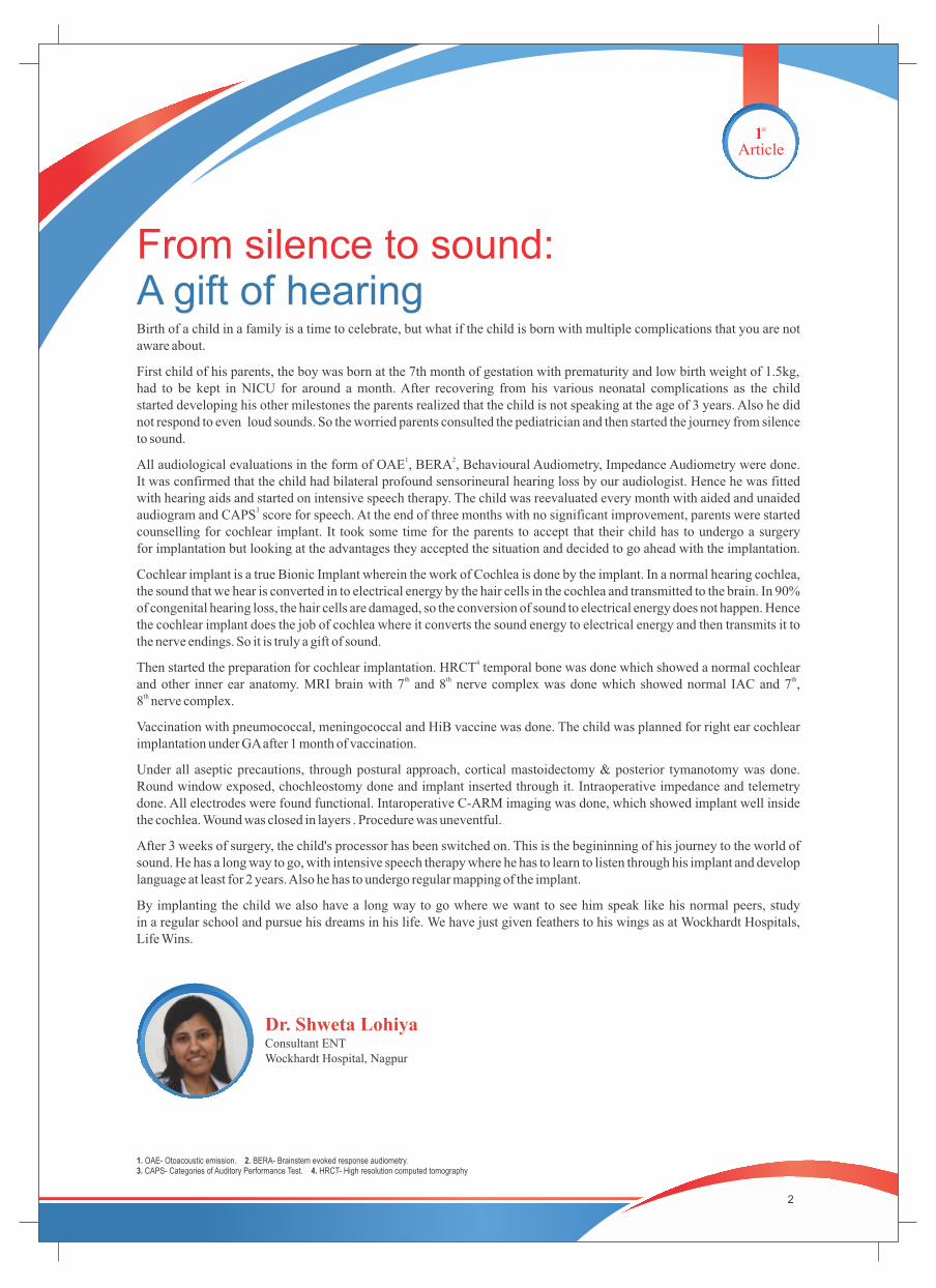
Worried, she visited Wockhardt Hospital, South Mumbai for a check up. On examination, the breast surgeon found her breasts discoloured and nodular. She was referred for a mammogram which revealed high-density masses scattered in both breasts. Interestingly, no implant/implant rupture could be seen. Ultrasound showed a classic ‘snow storm appearance’ with multiple cysts and echogenic free silicone causing dense shadowing. This ‘snowstorm appearance’ is classical of free silicone causing mastopathy. The patient had been given silicone injections in the breast rather than implants which explained the discounted rates!
nd2Article
General information:
This case is rare these days. Silicone injection for breast augmentation used to be performed in the 1950's and 60's. It has since been banned by the FDA due to adverse effects of silicone granulomas, mastitis and adenopathy. However, its practise continues by some unscrupulous practitioners notably in some South Asian countries.
Due to the dense shadowing from free silicone, mammography and ultrasound may miss small cancers in such cases. Hence MRI may be required as a problem-solving tool in some cases.
It is impossible to remove all the free silicone piecemeal embedded in the breast parenchyma hence this patient was offered bilateral subcutaneous mastectomy with breast reconstruction.
A heavy price to pay for cosmesis!
Dr. Bhagyam NagarajanConsultant RadiologistWockhardt Hospital, South Mumbai
Dr. Meghal Sanghavi Consultant OncosurgeonWockhardt Hospital, South Mumbai
4
A very rare case of fungal disictis: Suspicion the key
rd3Article
Dr. Priyesh DhokeConsultant - Spine SurgeonWockhardt Hospital , Nagpur
A 50 year old gentleman was referred to Wockhardt Hospital, Nagpur with gradually increasing low back pain and leg weakness since 3 months, not responding to conservative treatment elsewhere and since the last one month was on bed, wincing with pain. On examination his Lumbosacral spine was tender and he was unable to move or sleep in bed due to excruciating pain.
His MRI scan showed features of spondylodiscitis at L3-4 level. CT scan showed complete destruction of vertebral endplates and some part of vertebrae as well. Blood culture was negative. Blood investigations were normal except liver functions which were deranged as the patient was alcoholic.
Surgical intervention was planned considering the anterior vertebral column destruction and need of its reconstruction. Only posterior surgery was done with pedicle screw instrumentation, laminectomy, complete disc removal was done which was like necrotic material with pus, endplates were thoroughly curetted. Artificial titanium mesh cage with bone graft was put from back to front and also iliac crest strut bone graft for structural support and the remaining space was packed with one graft. The pathological sample was sent for various cultures and histopathology. To our surprise Aspergillus fungus was grown on culture and lactophenol cotton blue staining was done. Considering the positive culture of Aspergillus antifungal treatment was planned by infectious disease consultant, Dr. Nitin Shinde. Patient very well responded to Voriconazole and became symptom free. Post operatively he started walking on day 4 with support and without support in 3 weeks . He joined his duties at 6 weeks and became a happy patient for us.
General information:
Aspergillus Spondylodiscitis and vertebral osteomyelitis in immunocompetant host is very rare to occur and very few cases are reported in world literature.
Bacterial and tubercular discitis is however common in India and high degree of suspicion is needed to diagnose fungal discitis which has very similar presentation like the former but if diagnosis is missed than it is known to increase the morbidity and mortality.
High index of suspicion is the key for the diagnosis of Aspergillus infections of spine as the radiological features and blood investigations are not disease specific. We recommend of fungal cultures in all cases of vertebral osteomyelitis and Spondylodiscitis.
5
th4Article
An interesting case of endoscopic transnasal excision of Juvenile nasopharyngeal angiofibroma - Success achieved due to team work of specialists. A 19 year old boy presented to Wockhardt Hospital, Mumbai Central with chief complaints of nasal blockage and headache since 2 years. He had similar complaints 1 year back, for which he visited an ENT Surgeon at Miraj. On investigations, he was diagnosed with Juvenile Nasopharyngeal Angiofibroma. An endoscopic excision of the tumor was attempted at Miraj, however surgery was abandoned due to excessive bleeding.
At Wockhardt Hospital, we did a detailed CT scan and MRI of brain of the patient which showed an extensive lesion in the left pterygopalatine fossa, eroding the left lateral wall of sphenoid sinus and extending intracranially abutting the temporal cortex. The mass was seen extending into nasopharynx, left nasal cavity, left masticator space, eroding the pterygoid plates and invading the clivus with erosion of left internal carotid artery. The mass showed intense enhancement on post contrast images.
The case was discussed and surgery was planned by a team of ENT Surgeon, Head & Neck Oncosurgeon, Neurosurgeon and Interventional Radiologist. We decided to embolise the tumor pre- operatively to reduce the blood supply as it was a highly vascular tumour. The Interventional Radiologist did a DSA which showed blood supply to the tumour mainly by internal maxillary artery. Embolisation of sphenopalatine artery, branches of internal maxillary artery and ascending pharyngeal artery was done.
Surgery was planned 24 hours after the embolisation. Plan was to remove the entire tumour endoscopically, however if any part of tumour could not be accessed, an open approach using maxillary swing and subtemporal craniotomy was decided. After adequate counselling and consent, the patient was taken up for surgery. An endoscopic posteromedial maxillectomy was performed and entire tumour was removed endoscopically from the nasal cavity, nasopharynx, pterygopalatine fossa and left sphenoid sinus . The tumour was fibrous and less vascular due to adequate embolisation. Sphenopalatine area was cleared completely to avoid recurrence. Nasal packs were kept which were removed 3 days after surgery and patient was discharged on day 5.
Patient was followed up at 1 week and at 1 month, with no nasal symptoms. Post op MRI showed near complete excision of tumour.
General information:
Juvenile nasopharyngeal angiofibroma (JNA) is a rare benign tumour arising predominantly in nasopharynx of adolescent males. It is a highly vascular and aggressive neoplasm and shows propensity for destructive local spread often extending to base of skull and into the cranium.
JNA occurs exclusively in males. Patient may present with recurrent, unilateral nasal bleed or only nasal blockage, headache, facial swelling. It is diagnosed on CT scan and MRI. Clinically it may appear as a vascular polypoid mass. Biopsy should be avoided as it may lead to massive bleed since the tumour is composed of blood vessels without muscular coat. DSA ( Digital subtraction angiography) is done to see vascular supply of the tumour.
Treatment of juvenile nasopharyngeal is mainly surgical. Surgery can be done by endoscopic approach or external approach using various techniques such as transpalatine, transmaxillary or infratemporal approach. Endoscopic approach provides magnified visualisation and precise removal of the tumour and prevents morbidity and facial scars of open approach. Pre op embolisation is recommended for huge tumours to prevent intraoperative bleeding.
Proper planning and team effort are essential in management of such cases.
Dr. Neepa VellimuttamConsultant ENT Surgeon
Wockhardt Hospital , North Mumbai
Dr. Chandranath R. TiwariConsultant Neurosurgeon
Wockhardt Hospital , South Mumbai
Dr. Rahul VakhariaConsultant Radiologist MRI/CT
Wockhardt Hospital , South Mumbai
Dr. Rahul SethConsultant Interventional Radiologist Wockhardt Hospital, South Mumbai
Dr. Sayed Suhail Consultant Head & Neck Oncosurgeon Wockhardt Hospital, South Mumbai
6
Leptospira: Things you should knowLeptospirosis is a zoonosis of global distribution, caused by infection with pathogenic spirochetes of the genus Leptospira. The disease is greatly under reported, particularly in tropical regions, but attempts at surveillance suggest that it may be the most common zoonosis A syndrome of severe multisystem disease, presenting with profound jaundice and renal function impairment, was described by Weil in Heidelberg in 1886. “Leptospira” derives from the Greek word leptos word (thin) and Latin word spira (coiled). Leptospirosis is endemic throughout the world. Human infections are endemic in most regions; the peak incidence occurs in the rainy season in tropical regions and the late summer to early fall in temperate regions. Outbreaks may follow periods of excess rainfall. Leptospirosis is maintained in nature by chronic renal infection of carrier animals. The most important reservoirs are rodents and other small mammals, but livestock and companion animals are also significant sources of human infection. Direct contact is important in transmission to veterinarians, workers in milking sheds on dairy farms, abattoir works, butchers, hunters, and animal handlers (transmission has been reported to occur to children handling puppies and to dog handlers). Indirect contact is more common, and is responsible for disease following exposure to wet soil or water. A majority of cases are acquired by this route in the tropics, either through occupational exposure to water (rice or taro farming) flooding after heavy rains, or exposure to damp soil and water during avocational activities. Leptospires enter the body through cuts and abrasions, mucous membranes or conjunctiva, or aerosol inhalation of microscopic droplets.Leptospiral infection is associated with a very broad spectrum of severity, ranging from subclinical illness followed by seroconversion to two clinically recognizable syndromes-a self-limited systemic illness seen in approximately 90% of infections, and a severe, potentially fatal illness accompanied by any combination of renal failure, liver failure,and pneumonitis with hemorrhagic diathesis. The mean incubation period is 10 days (ranges from 5 to 14 days); determination of precise exposures may be difficult, leading to significant imprecision in estimated incubation times. The acute septicemic phase of illness begins abruptly with a high remittent fever (38° to 40°C) and headache, chills, rigors, and myalgias; conjunctival suffusion without purulent discharge; abdominal pain; anorexia, nausea, and vomiting; diarrhea; and cough and pharyngitis; a pretibial maculopapular cutaneous eruption occurs rarely. Prevention of leptospirosis may be achieved by avoidance of high-risk exposures, adoption of protective measures, immunization, and use of chemoprophylaxis, in varying combinations depending on environmental circumstances and the degree of human activity. High-risk exposures include immersion in fresh water, as in swimming, and contact with animals and their body fluids. Removal of leptospires from the environment is impractical, but reducing direct contact with potentially infected animals and indirect contact with urine-contaminated soil and water remains the most effective preventive strategy available.
th5Article
Dr. Prakash JiandaniDirector - Critical CareWockhardt Hospital, South Mumbai
Policy for post exposure prophylaxis for prevention of Leptospirosis
Category of Risk Definition
Low Risk Exposure Individuals with a single history of wading in flooded or contaminated water without wounds, cuts or open lesions of the skin.
Moderate Risk Exposure Adult individuals with a single history of wading in flooded or contaminated water and the presence of wounds, cuts or open lesions on the skin or accidental ingestion of contaminated water
Pregnant Women with Low Individuals with a single history of wading in flooded or moderate risk exposure to contaminated or moderate risk exposure water and with or without the presence of wounds, cuts or open lesions on the skin or accidental ingestion of contaminated water
Children - Age less than 8 years Individuals with a single history of wading in flooded or contaminated water and with or without presence of wounds, cuts or open lesions on the skin or accidental ingestion of contaminated water
High Risk Exposure Adult individuals with continuous exposure in flooded waters especially in urban areas infested with domestic, sewer rats and accidental ingestion of contaminated water. ex. Solid waste Management workers and other individuals working in flooded waters /marshy lands during relief and rescue operations
7
Laparoscopic Treatment For Liver Hydatid Cyst.
th6Article
Dr. Brijesh DubeConsultant – General SurgeryWockhardt Hospital, North Mumbai
A 55 year old gentleman, non diabetic, non hypertensive, presented to Wockhardt Hospital, North Mumbai with dull aching pain in the right upper quadrant of abdomen since 1 to 2 months. This was not associated with vomiting, fever, loose motion, constipation or jaundice. Clinically, he only had minimal distension in Right Hypochondriac region.
Blood investigation were absolutely normal. CT scan showed an approximately 10 cm × 10 cm cyst on the superior surface of the Right lobe of the liver abutting the diaphragm. The cyst contained hyperdense particulate matter/debris within suggestive of a Hydatid cyst.
The patient was counseled and posted for Laparoscopic Cyst excision/marsupialization. He was started on Tab. Albendazole 400 mg twice a day 5 days prior to the surgery.
Intra operatively, the cyst was found to be densely adherent to the under surface of the diaphragm, highly suggestive to a probable small rupture in the recent past. The cyst was instilled with 3% Hypertonic Saline and the debris was suctioned to clear the whole cyst. The cyst wall above the liver surface and adhered to diaphragm was excised and sent for Histopathology. Hemostasis was achieved and the cavity checked for any possible biliary communication. A drain was kept in the operated area.
Post op was uneventful. Patient resumed his feeds on Day 1 and drain was removed on Day 3. Patient was discharged the next day with advice to continue Tab. Albendazole for 28 days.
On regular follow up, patient has complete resolution with no suggestion of recurrence as yet.
General Information:Liver Hydatid Disease is a zoonosis caused by larvae of the dog tapeworm Echinococcus Granulosa, with man acting as accidental intermediate host.
The most common site of occurrence of Hydatid cyst is the liver (50% - 93%). Liver Hydatid cyst (LHC) if left untreated, grow and follow one of the several courses -
Develop fistulae with adjacent organs or biliary system Rupture into the peritoneal cavity seeding daughter cysts Develop daughter cyst within Rarely die Calcify
Although most patients may be asymptomatic for years or have non specific symptoms, one third of patients may present with effects or complications like infection or rupture.
Routine blood workup is non specific.
Indirect Haemagglutination test and ELISA have sensitivity of over 90% overall and are initial screening test of choice.
The advent of USG has repeated a breakthrough in diagnosis, treatment and follow up of patients with LHC. CT scan has 98% of sensitivity and specificity for LHC.
Treatment modalities:
Surgery: Surgery till date remains the treatment of choice.
Percutaneous: First introduced in mid-1980's has become an attractive alternative to surgical and medical management. PAIR (Puncture the cyst, Aspirate the cyst fluid, Inject the scolicidal agent and Re-aspirate the cyst content) is concluded to be effective and safe option for unilocular cysts with reports showing repeated failures of PAIR in multi-vesiculated cysts.
ERCP: Endoscopic management is useful in presence of intra-biliary rupture or after surgery in drainage of residual hydatid material in biliary tree.
Medical Treatment: Mebendazole, due to its poor efficacy, has largely been replaced by better absorbed Albendazole (10-15 mg/kg/day) in LHC.
Patients undergoing surgery receive Albendazole 5-7 days prior to surgery and continue to full course of 28 days post-op. This is known to give adequate result if no major intra-op spillage of cyst contents has occurred.
8
An interesting case of a cyst in the vocal cord
th7Article
A 30 year old male patient with no co-morbidities presented to Wockhardt Hospital with chief complaints of change in voice since 2 months. On Hopkins Rigid Laryngoscopy examination in OPD, a cystic lesion was seen in the left vocal cord near the anterior commissure. Both vocal cords were moving well bilaterally. There was no other abnormality or swelling in neck. His mean phonation time was only 6 seconds.
After proper counselling and consent, patient was taken up for surgery - Microlaryngoscopy and excision of vocal cord cyst. Patient was intubated with No.5.5 special MLS endotracheal tube, which has a larger cuff as compared to routine endotracheal tube. Special laryngoscope designed for Microlaryngoscopy was inserted to visualise the vocal cords. The cyst was excised using microflap technique using magnified vision of a microscope. Entire cyst with the sac was removed to avoid recurrence.
Patient was discharged the next day and advised strict voice rest for 5 days.
Patient was followed up at 1 week. Voice had returned to normal with mean phonation time of 22 seconds. Laryngoscopy showed complete healing of the vocal cord.
General Information:
Vocal cord cyst is a fluid filled sac within the substance of the vocal cord. It disrupts the vibratory pattern of the vocal fold mucosa resulting in hoarseness of voice and vocal fatigue. It grows slowly and can cause obstruction to the airway leading to difficulty in breathing in extreme cases. Treatment is mainly surgical. There is no role of medicines or voice therapy in resolution of the vocal cord cyst. Surgery is done using microscope or endoscope for precise and magnified vision. Microflap technique is used for vocal cord surgery to avoid damage to deeper structures of the vocal cord which can result in scarring and bad quality of voice. Entire cyst with surrounding sac should be removed to avoid recurrence. Aspiration of cyst is no longer recommended as it leads to recurrence.
Dr. Neepa VellimuttamConsultant ENT SurgeonWockhardt Hospital , North Mumbai
9
Awake craniotomy for deep brain stimulation
th8Article
A 60 year old man presented to Wockhardt Hospital, North Mumbai with a history of Parkinson's disease for the past 7 years on maximum drugs for control of symptoms like muscle rigidity and intentional tremors. The patient was also a hypertensive for the last 5 years which was well controlled on medications. This patient was a good candidate for the DBS* surgery and was counselled accordingly for the same. The surgery requires a very well motivated patient who understands the level of cooperation necessary for the surgery. Hence, a good doctor patient rapport is the key for the success of the procedure.
The patient was kept drug free for 12 hrs prior to surgery to elicit maximum disease related symptoms and to watch for its improvement on table during the surgery. After the patient was shifted to the preoperative holding area, the head was fixed onto the Leksell head frame which helps in localising the target areas in the brain to be stimulated on CT scan.
The patient was then shifted to the operating room from DBS.This infusion of the sedative along with the scalp block which was given at the start of the procedure produces a state of conscious sedation where the patient was made comfortable enough to drift into light sleep like state and easily arousable to follow instructions during the surgery. Dexmeditomidine is a uniquely effective drug that causes no interference with the recording of the electrode. After confirming the placement of the microelectrodes with CTscan, the placement of the battery and the pulse generator was done under general anesthesia. The patient was extubated and shifted to the ICU after the surgery. Eight weeks after placement the stimulation of the microelectrodes was commenced and the patient responded very well to the same. The quality of life improvement seen in this patient was remarkable and the dependence on medications is reduced considerably.
From an anesthetic point of view awake surgeries are challenging and not without risks. Risks like airway obstruction, seizures, involuntary movements causing scalp lacerations, claustrophobia and increased blood pressure are there. However, with the newer medications most of these complications are taken care of or avoided. The success of these surgeries is completely dependent on good patient rapport, patient selection and good communication between the surgical, anesthesia and neuromonitoring teams. The success of this procedure at our institute was due to all of the above.
General Information:
Parkinson's disease is a long term degenerative disorder of the central nervous system which develops slowly over a period of time. The symptoms include shaking, rigidity, slowness of movement, difficulty in walking, dementia, depression, anxiety. There is no cure for the disease and treatment is directed mainly at improving symptoms. As the disease progresses and neurons continue to be lost, the medications become less effective and the treatment itself starts to produce complications marked by involuntary writhing movements. Ablative intracranial surgery for Parkinson's disease has advanced to embedding microelectrodes into the precise areas of the brain and stimulating them with an external battery. The surgery is referred to as DEEP BRAIN STIMULATION ( DBS ) and is preferred in view of its reversibility, adjustability and capability to be safely performed bilaterally.
*. Deep Brain Stimulation
Dr. Vinita SangaiConsultant AnesthesiologistWockhardt Hospital, North Mumbai
RAB TE IL NGEC
55years
Sheetal Meshram
Nagpur
Harsha Gire
Nasik
Priya Muneshwar
Asmita Shende
Shubhangi Dhepe
Nitish Bhagat
Vrushali Sable
Ashwini Lote
Shiny Abraham
Nandini Sakharkar
Radhika Jaypurkar
Kanchan Daheriya
Neha Daheriya
Sapna Bagde
Leena Chitte
Alice Daisy Binu
Vashi
Shine Anil
Nobo
Heena Dudhagara
Rajkot
Saritha Shiny P
Kavita Titiya
Dipali Hirani
Usha Govil
Sobo
Devashree Karande
Vasthrawathy Retnamony
Jyotsna Bhagat
Suresh Arul
Ajo Mani
SuratSaumya N.v.
Divya Shetty
Mariamma Mathew
1010RAB TE IL NGEC
years
Nagpur
Maria Joseph
Nagpur
Monica Rao
Nagpur
Savita Chedge
Nagpur
Reena Biswas
Nagpur
Vandana Lokhande
Rajkot
Manorama Thakore
Rajkot
Nayana Ajani
Rajkot
Laly Jacob
Rajkot
Madhu H. Gosai
Surat
Bincy Mathew
Vashi
Vanita Chavan
10
World International Nurses DayCelebration at Wockhardt Group
thHospitals - 11 May 2018
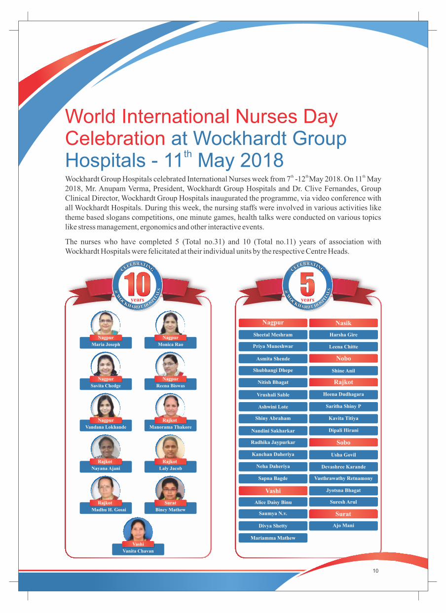
th th thWockhardt Group Hospitals celebrated International Nurses week from 7 -12 May 2018. On 11 May 2018, Mr. Anupam Verma, President, Wockhardt Group Hospitals and Dr. Clive Fernandes, Group Clinical Director, Wockhardt Group Hospitals inaugurated the programme, via video conference with all Wockhardt Hospitals. During this week, the nursing staffs were involved in various activities like theme based slogans competitions, one minute games, health talks were conducted on various topics like stress management, ergonomics and other interactive events.
The nurses who have completed 5 (Total no.31) and 10 (Total no.11) years of association with Wockhardt Hospitals were felicitated at their individual units by the respective Centre Heads.
11
Traumatic vertebral artery to vein fistula: Treated by embolization
th9Article
A 19- year- old male who had met with an accident in the factory was presented to Wockhardt Hospital, Rajkot. The accident caused a penetrating injury to the left side of the neck at around 10 pm. He was bleeding profusely and was taken to a hospital. He was evaluated by Oncosurgeon and ENT surgeon. His Pulse was 140/min, BP: 90/60 mm of Hg, Hemoglobin(Hb) was 5.6 gm/dl. They considered it as Carotid injury and explored the patient surgically. However, during surgery they found it as vertebral artery (VA) injury with active bleeding. Surgical wound was packed and referred to Wockhardt hospital for embolization.
When he reached his pulse was 162/min, BP 84/54 mm of Hg, Hb 4gm/dl. He was profusely bleeding from neck wound. Resuscitation was done with IV crystalloids and blood. He was taken to cathlab and an angiogram was done which revealed left vertebral (V2) segment transection with VA to vein fistula (AVF). Considering the urgency, balloon occlusion test of VA was not done and left VA proximal to fistula was occluded with coils. As expected there was steal from opposite VA and persistent bleeding. Right VA was catheterized and left vertebral (V2 segment) was accessed across vertebra basilar junction and embolized with coils, however there was persistent flow across coils with persistent bleeding. Considering this, the fistula was embolized with 25% n-butyl cyanoacrylate (NBCA) with complete occlusion of fistula. Patient did well, closure
thof neck wound was done the following day by the ENT surgeon. He was discharged on 7 day without any neurological deficit.
General information:
Vertebral arteriovenous fistulas are an uncommon pathology, either spontaneous or traumatic in origin. Traumatic lesions tend to occur in zone II of the neck (zone II lies between the cricoid and the angle of the mandible). Because in most penetrating injuries of the VA, there is extensive damage of the vessel wall, possibly with transection, a reconstructive endovascular approach is not possible and vascular sacrifice may be necessary. This technique of treatment is safe whenever the contralateral vessel is adequate to supply both intracranial vertebral circulations. Because the possibility of steal phenomena from the contralateral VA exists, it is important to perform a trapping technique, occluding both the proximal and distal aspects of the fistula by using detachable balloons or coils. In the cases in which the vessel distal to the site of fistula is patent, the use of a covered stent allows preservation of the normal arterial flow.
Endovascular techniques for occlusion of VA traumatic lesions are safe and effective. It is not associated with significant morbidity or mortality and could be the methods of choice for the treatment of most VA traumatic lesions, reserving surgery only for patients with failed embolization.
Dr. Vikash JainConsultant Interventional RadilogistWockhardt Hospital, Rajkot
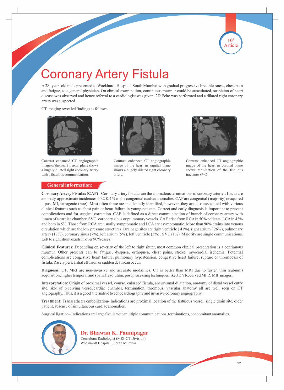
Coronary Artery Fistula A 28- year- old male presented to Wockhardt Hospital, South Mumbai with gradual progressive breathlessness, chest pain and fatigue, to a general physician. On clinical examination, continuous murmur could be auscultated, suspicion of heart disease was observed and hence referral to a cardiologist was given. 2D Echo was performed and a dilated right coronary artery was suspected.
CT imaging revealed findings as follows
12
th10Article
General information:
Coronary Artery Fistulas (CAF) Coronary artery fistulas are the anomalous terminations of coronary arteries. It is a rare anomaly, approximate incidence of 0.2-0.4 % of the congenital cardiac anomalies. CAF are congenital ( majority) or aquired – post MI, iatrogenic (rare) .Most often these are incidentally identified, however, they are also associated with various clinical features such as chest pain or heart failure in young patients. Correct and early diagnosis is important to prevent complications and for surgical correction. CAF is defined as a direct communication of branch of coronary artery with lumen of a cardiac chamber, SVC, coronary sinus or pulmonary vessels. CAF arise from RCA in 50% patients, LCA in 42% and both in 5%. Those from RCA are usually symptomatic and LCA are asymptomatic. More than 90% drains into venous circulation which are the low pressure structures. Drainage sites are right ventricle ( 41%), right atrium ( 26%), pulmonary artery (17%), coronary sinus (7%), left atrium (5%), left ventricle (3%) , SVC (1%). Majority are single communications. Left to right shunt exists in over 90% cases.
Clinical Features: Depending on severity of the left to right shunt, most common clinical presentation is a continuous murmur. Other presents can be fatigue, dyspnea, orthopnea, chest pains, stroke, myocardial ischemia. Potential complications are congestive heart failure, pulmonary hypertension, congestive heart failure, rupture or thrombosis of fistula. Rarely pericardial effusion or sudden death can occur.
Diagnosis: CT, MRI are non-invasive and accurate modalities. CT is better than MRI due to faster, thin (submm) acquisition, higher temporal and spatial resolution, post processing techniques like 3D VR, curved MPR, MIP images.
Interpretation: Origin of proximal vessel, course, enlarged fistula, aneurysmal dilatation, anatomy of distal vessel entry site, size of receiving vessel/cardiac chamber, termination, thrombus, vascular anatomy all are well seen on CT angiography. Thus, it is a good alternative to echocardiography and invasive coronary angiography.
Treatment: Transcatheter embolization- Indications are proximal location of the fistulous vessel, single drain site, older patient, absence of simultaneous cardiac anomalies.
Surgical ligation - Indications are large fistula with multiple communications, terminations, concomitant anomalies.
Contrast enhanced CT angiographic image of the heart in axial plane shows a hugely dilated right coronary artery with a fistulous communication.
Contrast enhanced CT angiographic image of the heart in sagittal plane shows a hugely dilated right coronary artery.
Contrast enhanced CT angiographic image of the heart in coronal plane shows termination of the fistulous tract into SVC
Dr. Bhawan K. PaunipagarConsultant Radiologist (MRI-CT Division)Wockhardt Hospital , South Mumbai
13
th11Article
Anaesthesia management of a case of congenital diaphragmatic hernia for bariatric surgery.A 68-year-old, morbidly obese female patient was presented to Wockhardt Hospital, South Mumbai for laparoscopic sleeve gastrectomy surgery. She had a chronic cough and breathlessness, subsequently diagnosed to have Diaphragmatic hernia and GERD.
She was a known case of Obstructive sleep apnoea since 4 years using CPAP at home, not good compliance and had limited physical activity.
On examination Weight 117 Kg; Height 147 cm; BMI 55, Vitals normal. RS ; Bilaterally decreased air entry mid & lower zone, SpO2 95% on room air. Difficult IV access, Oedema feet
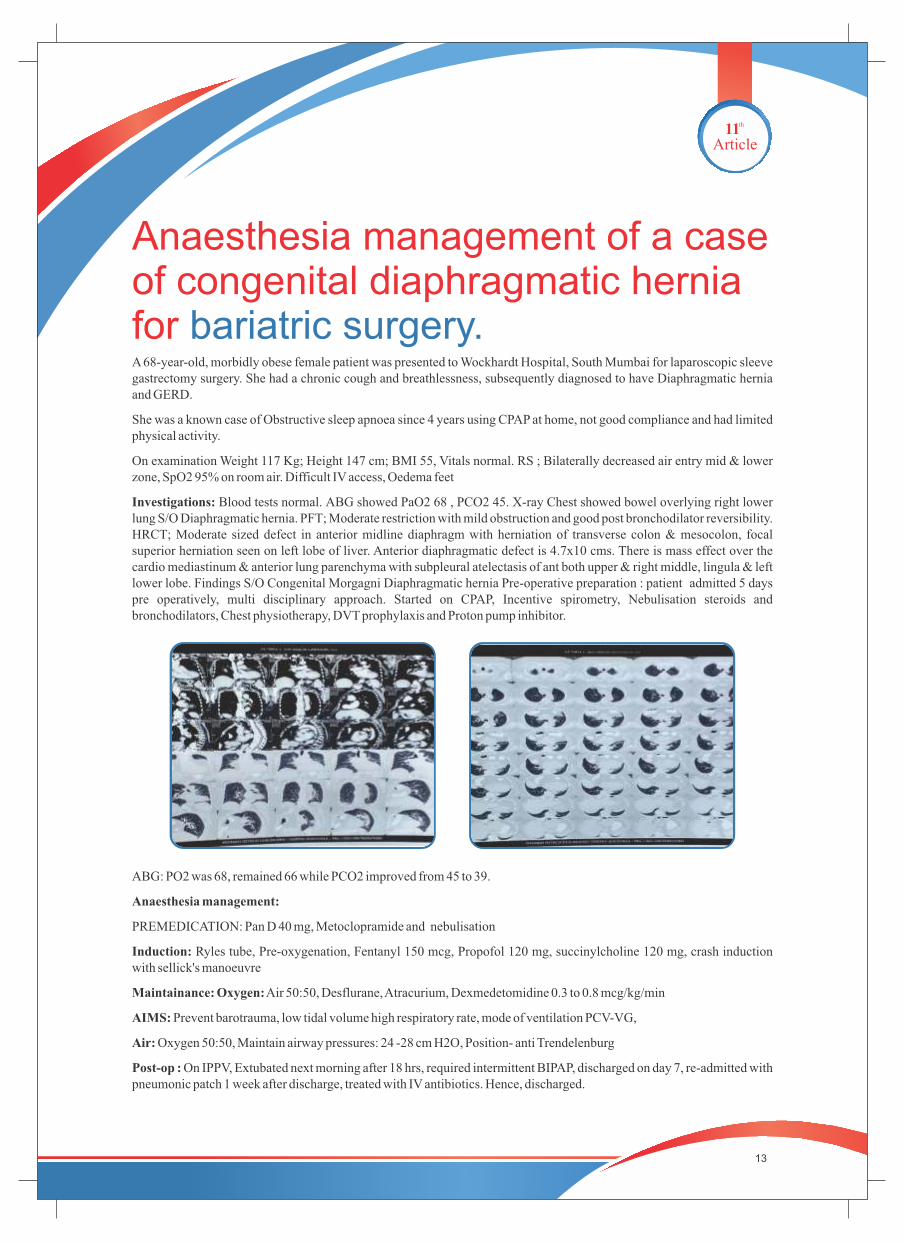
Investigations: Blood tests normal. ABG showed PaO2 68 , PCO2 45. X-ray Chest showed bowel overlying right lower lung S/O Diaphragmatic hernia. PFT; Moderate restriction with mild obstruction and good post bronchodilator reversibility. HRCT; Moderate sized defect in anterior midline diaphragm with herniation of transverse colon & mesocolon, focal superior herniation seen on left lobe of liver. Anterior diaphragmatic defect is 4.7x10 cms. There is mass effect over the cardio mediastinum & anterior lung parenchyma with subpleural atelectasis of ant both upper & right middle, lingula & left lower lobe. Findings S/O Congenital Morgagni Diaphragmatic hernia Pre-operative preparation : patient admitted 5 days pre operatively, multi disciplinary approach. Started on CPAP, Incentive spirometry, Nebulisation steroids and bronchodilators, Chest physiotherapy, DVT prophylaxis and Proton pump inhibitor.
ABG: PO2 was 68, remained 66 while PCO2 improved from 45 to 39.
Anaesthesia management:
PREMEDICATION: Pan D 40 mg, Metoclopramide and nebulisation
Induction: Ryles tube, Pre-oxygenation, Fentanyl 150 mcg, Propofol 120 mg, succinylcholine 120 mg, crash induction with sellick's manoeuvre
Maintainance: Oxygen: Air 50:50, Desflurane, Atracurium, Dexmedetomidine 0.3 to 0.8 mcg/kg/min
AIMS: Prevent barotrauma, low tidal volume high respiratory rate, mode of ventilation PCV-VG,
Air: Oxygen 50:50, Maintain airway pressures: 24 -28 cm H2O, Position- anti Trendelenburg
Post-op : On IPPV, Extubated next morning after 18 hrs, required intermittent BIPAP, discharged on day 7, re-admitted with pneumonic patch 1 week after discharge, treated with IV antibiotics. Hence, discharged.
14
Dr. Anjali PatkiConsultant AnaesthetistWockhardt Hospital , South Mumbai
Answer 1 : 30 minutes
Answer 2 : Hives
Answer 3 : Asperger syndrome
Answer 4 : Dysgerminoma
Answer 5 : Carotid body tumour
Answer 6 : Genu varum
Answer 7 : Extrahepatic Portal Venous Obstruction (EHPVO)
Answer8 : Subcutaneous fibrosis
Answer 9 : Receptor defect to gonadotropic hormones
Answer 10 : Mauriceau Smellie Veit maneuver
Answers to medical quiz Wocksynapse 9
Discussion:
Herniated viscera in the thorax may produce mass effect and can lead to cardiovascular impairment by compression of heart and mediastinal shift which can kink vena cavae, pulmonary veins, impair venous return to heart and cause cardiac output to decrease
Positive pressure ventilation with potential gastric insufflation and expansion of compressed lung may decrease venous return and cardiac output. For the same reason Loehning et al.[9] recommend low tidal volume and low airway pressure strategy. Nitrous oxide may also worsen mass effect should, therefore, be avoided
Collapsed lung should not be inflated to avoid the combined mass effect of herniated viscera and inflated lung, in this way, DLT is helpful
To avoid pneumothorax, intraabdominal pressure should be kept low, and one must avoid nitrous oxide. If there is a decrease in SpO2 during the procedure, ask for lowering the intra-abdominal pressure or stop surgery and ventilate the lung with 100% oxygen and PEEP. [10] Post-procedure chest radiograph should be checked
Reaching the horizon for Fast-track Coronary Artery Bypass
15
th12Article
A 58-year-old male diagnosed with Coronary Artery Disease/Single Vessel Disease/Mild LV Dysfunction/Old NSTEMI/Stable Angina II/Current Every day Smoker/Mild COPD was referred to Wockhardt Hospital, Rajkot for elective CABG. Pre-operative Ix were within satisfactory limits. CABG was done using Left Internal Mammary artery graft to LAD.
We used iGel Supraglottic airway for anesthesia which was removed on table after surgery ensuring adequate analgesia (visual analogue scale-VAS was on 3-4, mild pain). We used local anesthesia infiltration during closure, butorphanol (sos Morphine), i/v and oral paracetamol for pain management. Patient maintained stable and hemodynamics. Beta blockers and dual anti-platelets started in the evening on the same day. Drains removed on Day 1 morning with total drain output of 230 ml. Patient was ambulated on Day 1 evening, made to climb stair case on Day 2 morning. Stable hemodynamics and normal lab parameters and imaging along with excellent patient co-operation allowed us to send the patient on Day 2 afternoon, precisely 50 hours after the surgery. Patient came for follow up on Day 7 and Day 15 and was found to have normal convalescence with good sense of satisfaction and excitement for the fast tracking.
General information:
Fast-track surgery and anesthesia for CABG (Coronary artery bypass) was both found to be safe in terms of mortality, morbidity and re-admission rates as well as has shown to reduce cost significantly in several reports. We have been practicing fast track off pump CAB program since couple of years. We are proud to declare that we have achieved comparable results compared to conventional track CAB program with supreme priority to patient safety. Our definition of
thfast-track is – Extubation within 4 hours of closure and discharge by 4 post-operative day. We intend to enroll almost every elective case into the fast-track CAB program. After surgery, we keep on evaluating the clinical and psychological condition of the patient at regular intervals to determine eligibility of continuation in this program. We do not hesitate to keep the patient for few more days if deemed necessary.
Two weeks ago, we achieved a milestone – Off Pump CAB done under iGel supraglottic airway with on table extubation with discharged on day 2nd.
Summary of our results in last one year is as below.
No of Elective CABG - 248
Risk Factors
Mean age - 58.6 years, 203 Males, 45 Females
DM - 109, Hypertension - 175, Smokers - 112, Tobacco chewers - 80, COPD - 96
Mean STS (Society of Thoracic Surgeons) Mortality Risk score - 1.17
Mean STS Morbidity and early mortality (3 months) risk score (Predicts prolonged hospital stay, early mortality after discharge etc) - 8.3
Operative Record
Off Pump to On Pump Conversion - 0
IABP insertion - 4 (1 prophylactically at induction in Urgent case with ST changes, 3 Elective, intraoperatively; Salvage - 0)
Average number of grafts - 2.8
Ideal average number of graft - 2.89
Extubated within 4 hours of shifting - 247 (One patient developed prolonged recurarization)
In Hospital Mortality - 0
Total number of Blood Transfusion (Only LRCC) - 26
Mean Transfusion - 0.104 per case
16
Dr. Chirantan MangukiaConsultant- Cardiothoracic and Vascular SurgeryWockhardt Hospital, Rajkot
Dr. Mehul Kachhadia Consultant- Cardiac AnesthesiaWockhardt Hospital, Rajkot
Abstract: Behavioural problems among children
Ms. Greeshma NairNurse EducatorWockhardt Hospital, Nagpur
th13Article
Post-operative Course 3 month mortality-2 (0.8%)(one drug defaulter, second sudden death after 20 days)
Mean discharge time-81.36 hours
30-day readmission-3 (1.2 %), two had sternal wound infection, one had symptoms of heartfailure
AF - 26 cases (10.48%), managed with drugs
GI complication - 3 (1.2%) (Prolonged paralytic ileus - 2, GI bleed - 1)
CNS complication - Stroke - 1 (0.40%), discharged and rehabilitated subsequently
Sternal wound infection/dehiscence requiring readmission - 2 (0.8%) (Both Managed with Vacuum Assisted Closure and went home with complete healing with average stay of 36 days)
Renal Complications requiring Dialysis - 0
Pulmonary complications - 14 (5.64%) (Ventilator associated Pneumonia - 1, Pain induced Diastolic Heart Failure 13,; 2 were re-intubated, rest of were managed by NIV)
Problem Statement
A comparative study to assess the level of behavioral problems among children between the age group of 2½years to 5 years of employed and unemployed mothers in selected areas of Nagpur.
Abstract:
A mother's emotional adjustment when she assumes work outside home is the key to whether she succeeds or fails. Although working mothers have become the norm in the society, disapproving attitudes from various strata of the society contribute to the torn and guilty feelings many working mothers experience. The mother's status as a worker may consistently have either positive or negative effects on children's development and educational outcomes. In the present comparative study done on employed and unemployed mothers with children in the age group of 2½-5 years , it was seen that maximum children of employed mothers exhibit moderate behavioral problems 24 (80%) while those of unemployed mothers exhibit mild behavioral problems 22 (73.33%).
Interpretation And Conclusion
Findings of the study indicate no significant difference in the level of behavioral problems of children of employed and unemployed mothers but there is a significant association of the demographic variables with the behavioral problems of employed mothers.
Non-union of tibia fracture with advanced knee arthritis
17
th14Article
Dr. Niraj KasatConsultant Joint Replacement SurgeryWockhardt Hospital, North Mumbai
Stress fracture of tibia secondary to deformities from longstanding knee osteoarthritis is rare and difficult to manage. We treated one such patient with stress fracture of proximal tibia which went to non-union and had the patient bed bound for 2 years. Challenge was to treat her fracture non-union and knee arthritis together.
A 67-year-old female suffering from bilateral knee pain since last 10 years came to Wockhardt Hospital, Mira Road. Since, last 2 years she was bedbound and dependent on others for her day to day activities. She came on a wheelchair with inability to walk or stand on left leg.
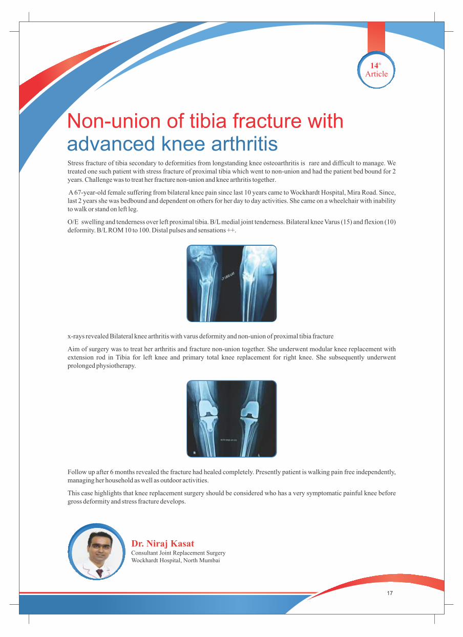
O/E swelling and tenderness over left proximal tibia. B/L medial joint tenderness. Bilateral knee Varus (15) and flexion (10) deformity. B/L ROM 10 to 100. Distal pulses and sensations ++.
x-rays revealed Bilateral knee arthritis with varus deformity and non-union of proximal tibia fracture
Aim of surgery was to treat her arthritis and fracture non-union together. She underwent modular knee replacement with extension rod in Tibia for left knee and primary total knee replacement for right knee. She subsequently underwent prolonged physiotherapy.
Follow up after 6 months revealed the fracture had healed completely. Presently patient is walking pain free independently, managing her household as well as outdoor activities.
This case highlights that knee replacement surgery should be considered who has a very symptomatic painful knee before gross deformity and stress fracture develops.
Expert Column Renal replacement therapies in Critical care with specific emphasis on CRRT
18
IntroductionThe management of patients with acute kidney injury (AKI) is supportive, with renal replacement therapy (RRT) indicated in patients with severe kidney injury. Multiple modalities of RRT are available. These include intermittent hemodialysis (IHD); continuous renal replacement therapies (CRRTs); and hybrid therapies, also known as prolonged intermittent renal replacement therapies (PIRRTs), such as sustained low-efficiency dialysis (SLED) and extended-duration dialysis (EDD). Despite these varied techniques, mortality in patients with AKI remains high, exceeding 40 to 50 percent in severely ill patients.
TimingRandomized, controlled trials that have compared strategies of early versus delayed initiation of RRT (in the absence of obvious indications) have yielded conflicting results. A meta-analysis of 10 randomized trials showed no benefit of early initiation on 30-, 60-, or 90-day mortality]. There was also no difference between early and late initiation on the risk of dialysis dependence, length of intensive care unit (ICU) or hospital stay, or recovery of renal function. However, the strength of this analysis is low in part because of heterogeneity due to variable definitions of early versus late initiation. In addition, most trials had an unclear or high risk of bias for allocation concealment.
Optimal ModalityA large number of modalities are available for RRT. These include intermittent hemodialysis (IHD), peritoneal dialysis, continuous renal replacement therapy (CRRT), and hybrid therapies such as sustained low-efficiency hemodialysis (SLED).
Data do not support the superiority of any particular mode of RRT in patients with AKI. In the majority of patients, selection of modality should therefore be based upon local expertise and availability of staff and equipment. However, in selected patients, other factors may prevail. As an example, in patients with acute brain injury or fulminant hepatic failure, continuous therapy may be associated with better preservation of cerebral perfusion. However, the costs associated with CRRT may be greater than with other modalities of RRT.
Continuous renal replacement therapies versus intermittent hemodialysis CRRT represents a family of modalities that provides continuous support for severely ill patients with AKI. These include continuous hemofiltration, hemodialysis, and hemodiafiltration, which involve both convective and diffusive therapies. Although superior clearance of middle- and larger-molecular-weight molecules is associated with convective therapies (hemofiltration) compared with diffusive therapies (hemodialysis), there are no studies clearly showing improved clinical outcomes compared with the type of solute transport.
19
Studies suggest that survival and recovery of renal function are similar with both CRRT and IHD, and the Kidney Disease: Improving Global Outcomes (KDIGO) Clinical Practice Guidelines for AKI suggest using intermittent and continuous RRT as complementary therapies in patients with AKI.
Advantages of CRRT
CRRT may be associated with the following advantages compared with IHD:
Enhanced hemodynamic stability, which may be particularly beneficial in hemodynamically unstable patients. Hemodynamic stability is thought to be related to slower solute and volume removal and the effects of modest hypothermia often associated with CRRT
More consistent net salt and water removal, particularly in hemodynamically unstable patients, thereby permitting superior management of volume overload and nutritional requirements
Enhanced clearance of inflammatory mediators, which may provide benefit in septic patients, particularly using convective modes of continuous therapy. However, a meta- analysis of convection versus diffusion demonstrated no benefit to convection. Open-label, randomized, controlled trials have also shown no benefit of high-volume hemofiltration in sepsis or cardiogenic shock following cardiac surgery
Among patients with acute brain injury or fulminant hepatic failure, continuous therapy may be associated with better preservation of cerebral perfusion
The actual importance of these benefits is uncertain, given the absence of a difference in survival between these modalities. As an example, although convective therapy may provide enhanced clearance of pro-inflammatory mediators, it may also result in removal of beneficial anti-inflammatory mediators. In addition, the maximal achieved extracorporeal clearance of these mediators is low relative to the rates of generation and endogenous clearance.
Based on clinical practice patterns, the major indication for choosing continuous renal replacement therapy (CRRT) over intermittent hemodialysis is hemodynamic instability. However, randomized trials have not proven that CRRT causes less hypotension than intermittent hemodialysis.
CRRT modalities include continuous venovenous hemofiltration (CVVH), continuous venovenous hemodialysis (CVVHD), and continuous venovenous hemodiafiltration (CVVHDF). The major difference among modalities is the underlying mechanism that drives solute removal
The choice of CRRT modality depends on availability and the expertise of the clinician. All modalities utilize venovenous circuits with blood flow through the dialyzer/hemofilter driven by an extracorporeal blood pump. Arteriovenous modalities, in which blood flow was driven by the gradient between the mean arterial pressure (MAP) and venous pressure, are no longer routinely used because of risks associated with the need for arterial access (embolization, bleeding)
CRRT requires reliable vascular access capable of blood flows of at least 200 to 250 mL/min. The standard is a double-lumen tunneled or non-tunneled dialysis catheter. Among end-stage renal disease patients who have arteriovenous fistulas (AVFs) or arteriovenous grafts (AVGs) for maintenance hemodialysis,it is suggested that the AVF or AVG not be used for CRRT unless no other access is possible. There is a risk of dislodging a needle causing bleeding or injury to the AVF or AVG.
20
Complications
Complications of CRRT include hypotension, infection, bleeding, and hypothermia. Common laboratory abnormalities include hypophosphatemia, hypokalemia, hypomagnesemia, and, depending on the method of anticoagulation, hypocalcemia.
ConclusionCRRT is an excellent modality of RRT in the critically ill patients who are hemodynamically unstable with multiorgan failure. It provides excellent flexibility of volume thereby facilitating delivery blood/blood products & nutrition which is not possible with conventional intermittent hemodialysis. Specific subgroup of patients with fulminant hepatic failure benefit with better preservation of cerebral function.
The only limiting factor remains the cost especially in our part of the world.
Dr. Kedar ToraskarConsultant Critical Care Wockhardt Hospitals, South Mumbai
New consultants who joined The Wockhardt FamilyName of the consultant Speciality Qualification Location
Dr. Vijaysinh Patil Cardiology DNB - Internal Medicine, Cardiology, Nashik FNB - Cardiology
Dr. Prashant Makhija Neurology M.B.B.S., M.D. (General Medicine), South Mumbai M. (Neurology)., PDF epilepsy and sleep Medicine
Dr. Mangesh Kohale CVTS MBBS, M.S (General Surgery), M.Ch. South Mumbai in Cardiovascular and Thoracic Surgery
Dr. Dinki Dharod Paediatric Anesthesia M.B.B.S., DNB (Anesthesia), Fellowship South Mumbai of Indian association of cardiac anesthesia
Dr. Nikhil Konde Anesthesia M.B.B.S., D.A. (Anaesthesiology), DNB South Mumbai (Anaesthesiology)., Fellow - Liver transplant
Dr. Urmi Shah Microbiologist MD Microbiology North Mumbai
Dr. Srushti Jibhkate Biochemistry MD Biochemistry North Mumbai
Dr. Bipin Jibhkate Critical care and pain medicine MD Anaesthesia, FNB Critical Care North Mumbai
Dr. Pritam Moon Internal Medicine MD Internal Medicine North Mumbai
Dr. Amit Sahu Interventional Radiology MD in Radio-Diagnosis, Fellowship in North Mumbai Interventional Neuro Radiology and Advanced training in Vascular and Interventional Radiology
Dr. Khyati Vadera Radiologist MD (Radio-Diagnosis), DNB (Radio-Diagnosis) Rajkot
Dr. Gaurang Raval Radiologist DNB (Radio-Diagnosis) Rajkot
Dr. Manisha Singh Obstetrician and Gynecologist MS(Obstetrics and Gynecology), Rajkot F. MAS, D.MAS
Dr. Vimal Dave Critical Care MD(Anesthesia), IDCCM Rajkot
Dr. Abhishek Raval Cardiologist DM(Cardiology) Rajkot
Medical Quiz
21
Dr. Sadaf KhanMedical AdministrationWockhardt Hospital. North Mumbai
Q1. ?In FSGS(Focal segmental glomerulosclerosis) pathological changes are most prominent in glomeruli located at
A. Corticomedullary junction B. Outer cortex
C. Middle cortex D. All of the above
Q2. Most IgA deposited in kidney is derived from?
A. Lymph node B. Bone marrow
C. Spleen D. Liver
Q3. Churg strauss syndrome distinguishes from other small vessel vasculitis by?
A. Pulmonary infiltrates B. Peripheral eosinophilia
C. Glomerulonephritis D. All of the above
Q4. Which of the following is an inactivated or killed vaccine?
A. Measles B. Chicken pox
C. BCG D. Hepatitis B
Q5. A 40-year-old woman has had several episodes of rheumatic fever as a child. She is currently afebrile and feels well,
and has come to a hospital for monitoring echocardiography. Which of the following would be most likely to be seen
in this patient's mitral valve?
A. Fish mouth valve B. Irregular beads of calcification in annulus
C. Ballooning of valve leaflets D. Large bulky vegetation with adjacent leaflet perforation
Q6. Which of the following occurs pathologically in malignant hypertension?
A. Diffuse necrotizing vasculitis B. Arterial thrombi
C. Fibrin deposition in arteriolar walls D. All of the above
Q7. In an 8-day-old child with no history of consanguinity in the parents, the mother reports blisters and bleeding off the
skin at the site of handling and pressure. There was a similar history in the previous child which proved to be fatal.
The diagnosis is?
A. Bullous pemphigoid B. Congenital syphilis
C. Congenital epidermolysis bullosa D. Letterer-Siwe disease
Q8. Splenomegaly with variceal hemorrhage in the absence of cirrhosis suggests?
A. Splenic vein thrombosis B. Portal vein thrombosis
C. Splenic Hemorrhage D. Refractory ascites
Message from the Editor
Disclaimer: “It is to be noted that the treatments being discussed above are informative in nature and case to case specific. Hence it should not be treated as medical advice. Readers are advised to consult clinicians before making any informed view or decision in this regard.”
Contact [email protected]. No. (+91)-22-7159 6509
Dr. Clive FernandesGroup Clinical DirectorWockhardt Group Hospitals
Dr. Clive Fernandes Editor
Editorial Board
Dr. Ieshu Razdan BKC
Dr. Ravi Maggo Rajkot
Dr. Sachin Mane Nashik
Dr. Manisha Pathak NOBO
Dr. Vikas Chaudhari Nagpur
Dr. Bhupendra Makwana Surat
Dr. Shobana Nair SOBO
Dr. Sadaf Khan NOBO
22
Dear Readers
Hope you enjoy the interesting and complex cases that are published in our Medical Bulletin ‘Wocksynapse’. The cases described here are just a snapshot of the wonderful clinical outcomes that our hospitals are delivering. Even as this edition is going to print Wockhardt Hospitals Nagpur just completed its first Liver transplant case. Congratulations to the entire team.
There is a lot of debate going on amongst healthcare organizations specially the private ones regarding the new healthcare scheme “Ayushman Bharat Yogna” with regards specially to some of the projected costs assigned for certain procedures. The scheme aims at providing healthcare insurance to an additional 41.3% of our population. The numbers are huge, the cost involved is phenomenal and like we always say in the quality domain, the devil lies in the details, we have to wait and see how this rolls out. This scheme when fully implemented as intended will be a giant step forward in the right direction and a game changer of magnanimous proportion.
thNursing Day was celebrated across the World on 12 May and we at Wockhardt Hospitals had a week-long celebration with different events including all nursing associates. Those Nursing associates who completed 10 years and 5 years were specially felicitated.
Hope you enjoy this edition of Wocksynapse. Looking forward to your inputs and feedback at wocksynapse @wockhardthospitals.com