Embed Size (px)
Citation preview
REVIEW Open Access
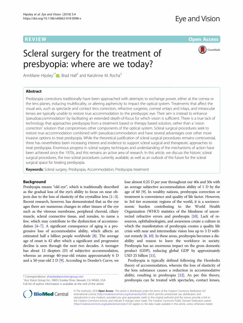
Scleral surgery for the treatment ofpresbyopia: where are we today?AnnMarie Hipsley1* , Brad Hall2 and Karolinne M. Rocha3
Abstract
Presbyopia corrections traditionally have been approached with attempts to exchange power, either at the cornea orthe lens planes, inducing multifocality, or altering asphericity to impact the optical system. Treatments that affect thevisual axis, such as spectacle and contact lens correction, refractive surgeries, corneal onlays and inlays, and intraocularlenses are typically unable to restore true accommodation to the presbyopic eye. Their aim is instead to enhance‘pseudoaccommodation’ by facilitating an extended depth-of-focus for which vision is sufficient. There is a true lack oftechnology that approaches presbyopia from a treatment based or therapy based solution, rather than a ‘visioncorrection’ solution that compromises other components of the optical system. Scleral surgical procedures seek torestore true accommodation combined with pseudoaccommodation and have several advantages over other moreinvasive options to treat presbyopia. While the theoretical justification of scleral surgical procedures remains controversial,there has nevertheless been increasing interest and evidence to support scleral surgical and therapeutic approaches totreat presbyopia. Enormous progress in scleral surgery techniques and understanding of the mechanisms of action havebeen achieved since the 1970s, and this remains an active area of research. In this article, we discuss the historic scleralsurgical procedures, the two scleral procedures currently available, as well as an outlook of the future for the scleralsurgical space for treating presbyopia.
Keywords: Scleral surgery, Presbyopia, Accommodation, Presbyopia treatment
BackgroundPresbyopia means “old eye”, which is traditionally describedas the gradual loss of the eye’s ability to focus on near ob-jects due to the loss of elasticity of the crystalline lens [1–3].Recent research, however, has demonstrated that as the eyeages there are numerous changes in other tissues of the eyesuch as the vitreous membrane, peripheral choroid, ciliarymuscle, scleral connective tissue, and zonules, to name afew, which may contribute to the dysfunction of accommo-dation [4–7]. A significant consequence of aging is a pro-gressive loss of accommodative ability, which affects anestimated half a billion people worldwide [8]. The averageage of onset is 42 after which a significant and progressivedecline is seen through the next two decades. A teenagerhas about 13 diopters (D) of subjective accommodation,whereas an average 40-year-old retains approximately 6 Dand a 50-year-old 2 D [9]. According to Donder’s Curve, we
lose almost 0.25 D per year throughout our 40s and 50s withan average subjective accommodation ability of 1 D by theage of 60 [9]. In wealthy nations, presbyopia correction ortreatment is convenience and quality of life factor. However,in 3rd tier economic regions of the world, it is a socioeco-nomic burden contributing to the World HealthOrganization (WHO) statistics of the blindness of uncor-rected refractive errors and presbyopia [10]. Lack of re-sources, ophthalmologists, and awareness create a culture inwhich the manifestation of presbyopia creates a quality lifecrisis with near and intermediate vision loss up to 3 D with-out remedy [8, 10]. In these areas, presbyopia becomes a dis-ability and reason to leave the workforce in society.Presbyopia has an enormous impact on the gross domesticproduct (GDP), reducing global GDP by approximatelyUSD 25 billion [11].Presbyopia is typically defined following the Hemholtz
theory of accommodation, wherein the loss of elasticity ofthe lens substance causes a reduction in accommodativeability, resulting in presbyopia [12]. As per this theory,presbyopia can be treated with spectacles, contact lenses,
* Correspondence: [email protected] Vision Group Inc, 39655 Eureka Drive, Newark, CA 94560, USAFull list of author information is available at the end of the article
© The Author(s). 2018 Open Access This article is distributed under the terms of the Creative Commons Attribution 4.0International License (http://creativecommons.org/licenses/by/4.0/), which permits unrestricted use, distribution, andreproduction in any medium, provided you give appropriate credit to the original author(s) and the source, provide a link tothe Creative Commons license, and indicate if changes were made. The Creative Commons Public Domain Dedication waiver(http://creativecommons.org/publicdomain/zero/1.0/) applies to the data made available in this article, unless otherwise stated.
Hipsley et al. Eye and Vision (2018) 5:4 https://doi.org/10.1186/s40662-018-0098-x

corneal surgery, or intraocular lenses. Spectacle and con-tact lens use are the conventional treatments, [13] howeverneither of these attempt to restore true accommodation tothe presbyopic eye.There are limitations to treating the real cause of presby-
opia or the loss of accommodative ability of the lens to dy-namically change focus power. Firstly, the early attempts toaddress presbyopia were to exchange either the power inthe cornea or the lens to achieve multifocality or changesin asphericity. Corneal presbyopic correction procedures,such as presbyLASIK, attempt to create a multifocal corneaby manipulating the optical properties of the eye, aspheri-city, and inducing higher-order aberrations, [14] while in-traocular lens (IOL) replacement may include multifocaland aspheric lenses. These vision correction proceduresmay compromise distance vision and degrade binocularityand stereopsis [15–17]. Performing these corrections withsurgical intervention carries additional risks of regression,scarring, and night vision problems [18]. These treatmentsalso only aim to enhance ‘pseudoaccommodation’ by facili-tating an extended depth-of-focus for which vision is suffi-cient, [19] rather than restoring true accommodation andpseudoaccommodation together.True accommodation is the ability of the eye to modify
the focal length of the lens to see objects clearly whenchanging focus from distance to near. During true ac-commodation the ciliary muscles contract, releasing ten-sion in the zonules, which allows the lens to return toits more natural convex shape [20]. Moreover, the ciliarymuscles, the zonular tensions on the lens, and the roleof the elastic choroid, in both the pre-stretch, disaccom-modated state and accommodated states, all play com-plex roles in the amount of accommodative range andbiomechanical functionality of the entire accommoda-tion complex [5]. The biomechanics of this functionalanatomy is directly proportional to the amount of ac-commodative amplitude and the central optical powerthat can be generated from the dynamic accommodativeforces [4]. Moreover, as we age, there is resultant
biomechanical dysfunction that is manifested with pres-byopia creating a dysadaptation of binocularity, whichfurther complicates the visual disturbances experiencedwith progressive presbyopia [21].There have also been strides to classify the treatment
paradigm for presbyopia, which assists the ophthalmic sur-geon to determine the stages of classification of presbyopiaand allow a more evidence-based decision-making tree forthe treatment of presbyopia. Dysfunctional Lens Syndrome(DLS) has been described by George Waring IV and col-leagues as a deterministic model to characterize the aginglens [22]. In DLS Stage I the lens becomes more rigid andless flexible, corresponding with presbyopia. In DLS StageII contrast sensitivity loss, increased higher-order aberra-tions and light scatter often affect night vision function. InDLS Stage III the lens clouding is significant, and severelyimpacts daily activities; this stage corresponds with cata-racts. Scleral surgeries are useful in as much as they canstill effectuate the molding of the lens. To achieve this, thelens must be clear and void of opacities and age-relateddamage. The most likely candidate to achieve the most im-provements with scleral surgeries would be a person whois classified as having Stage I DLS. However, candidateswho are in Stage II have also received benefits from scleralprocedures. Therefore, the relationship of DLS as it corre-lates to scleral procedure outcomes requires further inves-tigation and remains an open question.
ReviewScleral surgeryBackgroundDespite the different treatment options to restore pseu-doaccommodation, there remains a need for treatments torestore true accommodation combined with pseudoaccom-modation to the presbyopic eye. Scleral surgical procedureshave the potential to fulfill this requirement and have sev-eral advantages over other more invasive options to treatpresbyopia. Firstly, scleral procedures deviate from theparadigm of ‘vision correction’ (rectifying visual acuity
Fig. 1 VisAbility Micro-Insert surgical procedure. a) VisAbility Micro-Insert; b) Sclerotome and docking station creating a partial thickness tunnel inthe sclera. (Images courtesy of Refocus Group, Dallas, USA)
Hipsley et al. Eye and Vision (2018) 5:4 Page 2 of 11
deficits) to a therapeutic approach; aiming to restore staticand dynamic physiological function in the eye. The risk ofvision loss is lower, as the cornea, visual axis, and the nativecrystalline lens are not involved in these procedures, whichallows scleral procedures to be performed after or in com-bination with other corrective methods, such as cataractsurgery. While their theoretical justification may be contro-versial [23], there has nevertheless been increasing interestin scleral surgery to treat presbyopia. In this article, we willdiscuss the historical scleral surgical procedures and thetwo procedures currently available to treat presbyopia.
HistoryScleral surgical procedures, as a treatment for presbyopia,were followed from the surgical myopia treatments ofFyodorov in the 1970s. Fyodorov treated myopia with ra-dial keratotomy (RK) - radial or spoke cuts through thecornea [24]. Thornton later expanded RK surgery to thesclera, using a procedure known as anterior ciliary scler-otomy (ACS) [25]. In ACS, radial incisions are not madein the cornea, but in the sclera overlaying the ciliarymuscle [26]. The aim was to increase the space betweenthe lens and the ciliary muscle, tightening the zonules and
increasing accommodative ability [26]. Accommodationwas observed to improve slightly with ACS. However, amyopic shift of 0.5 D was also seen [25]. The accommoda-tive improvements were also short-term, with 0.8 D of theamplitude of accommodation remaining after 12 months[26]. To reduce this regression, Fukasaku used silicone im-plants in conjunction with ACS [27]. These treatmentoptions are no longer available.
Scleral implantsScleral implants are based on the accommodation modeldescribed by Schachar and colleagues [28–31]. This modeldescribes a decreasing gap between the lens perimeter andthe ciliary ring with age, due to a combination of anatom-ical changes, as the cause of presbyopia. This model re-mains controversial, as it differs from the widely acceptedHemholtz model of accommodation, [12] however it is sup-ported by experimental evidence [7, 32].Schachar and colleagues used scleral implants in an at-
tempt to increase the area between the ciliary muscleand the sclera to restore accommodation. The first in-stances used poly[methyl methacrylate] (PMMA) rodimplants to expand the sclera and were referred to as
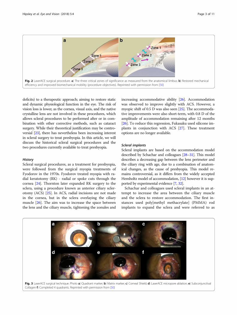
Fig. 3 LaserACE surgical technique. Photo a) Quadrant marker; b) Matrix marker; c) Corneal Shield; d) LaserACE micropore ablation; e) SubconjunctivalCollagen f) Completed 4 quadrants. Reprinted with permission from [50]
Fig. 2 LaserACE surgical procedure. a) The three critical zones of significance as measured from the anatomical limbus; b) Restored mechanicalefficiency and improved biomechanical mobility (procedure objectives). Reprinted with permission from [50]
Hipsley et al. Eye and Vision (2018) 5:4 Page 3 of 11

‘scleral expansion bands’ [3, 33]. Scleral expansion bands(SEBs) did achieve some success in restoring accommoda-tion but were ultimately retired due to mixed results andlow patient satisfaction [34]. There were also events ofanterior ischemia, which was not an acceptable risk for an‘elective procedure.’ This lead to a general decrease insupport and interest from the ophthalmology communityand almost complete abandonment of the idea that scleralprocedures were viable to treat presbyopia [23, 35].Despite early failures, using implants to expand the area
between the sclera and the ciliary muscle is still an activearea of research. The VisAbility Micro-Insert scleral implant(Refocus Group, Dallas, TX, USA), an updated version of
the PresView (Refocus Group, Dallas, TX, USA), remainsthe only scleral implant with the CE mark and is currentlyundergoing FDA clinical trials [36]. The procedure uses fourPMMA injection molded implants, each about the size of agrain of rice (Fig. 1). The implants are placed about 3000-4000 μm from the limbus and to a depth of 400 μm withinthe sclera. Patients are placed under monitored anesthesiacare for the duration of the procedure, approximately 1 h bi-laterally. The implants aim to lift the sclera and the ciliarymuscle to tighten the zonular fibers holding the lens [37].Results from a previous 24-month clinical trial with theVisAbility Micro-Insert were presented in 2013 [37, 38].The authors subjectively evaluated the visual function of 80
Fig. 4 Uncorrected (lightly colored) and distance-corrected (darkly colored) visual acuity at a distance 4 m, intermediate (60 cm), and near(40 cm) for a) Monocular and b) Binocular patient eyes. Error bars represent mean ± SD. Reprinted with permission from [50]
Hipsley et al. Eye and Vision (2018) 5:4 Page 4 of 11
patients after 24 months using a questionnaire. The partici-pants were asked to describe their unaided vision as ‘excel-lent’, ‘acceptable’, or ‘poor’, pre and postoperatively. Thepercentage of patients reporting at least ‘acceptable’ visionafter 24 months was 73% overall, and 99% for distance tasks[37, 38]. Preoperatively, 4% of patients reported at least‘acceptable’ vision when reading newspapers, whichimproved to 76% of patients 24 months postoperatively [37,38]. Approximately 83% of patients were able to completenear tasks (such as reading newspapers, prices, and medi-cine labels) without using reading spectacles [37, 38].Distance-corrected near visual acuity (DCNVA) data fromthe same clinical trial were presented in 2014 [39]. Theresults showed that 93% of patient eyes had DCNVA of 0.3logMAR (20/40 Snellen) or better [39].While the early VisAbility clinical trial results seem
promising, there are substantial risks involved for patientsundergoing this procedure. Anterior segment ischemia(ASI) due to mechanical vascular compression from theimplant can occur, as can subconjunctival erosion, moder-ate to severe subconjunctival hemorrhage, implant infec-tion, and endophthalmitis. There is also a significant riskthat the implants may become displaced [40]. An early USFederal Drug Administration (FDA) study showed thatabout 75% patients with the first generation of the nowVisAbility Micro-Insert implant had at least one implantmove or displace [40]. Other treatment options exist thatmay be safer for patients.
Scleral laser excisionScleral laser excision procedures as a treatment for presby-opia began with Lin in 1998 [41]. Lin argued that ACS wasunsuccessful due to the rapid healing of the sclera and
proposed instead to ablate nearly the full thickness of thesclera [42]. Termed laser presbyopia reversal (LAPR), Lin’ssurgical procedure involved radial sclerectomy with anerbium-doped yttrium aluminum garnet (Er:YAG) laser.Excisions were performed to a depth of 500-600 μm, with alength of approximately 4500 μm, and a width of 600-700 μm [42]. Results after 12 months showed 2 D of sub-jective accommodation. However, this may be explained bythe decrease in anterior chamber depth causing a myopicshift. This treatment option is no longer available.
Scleral laser micro-excisionScleral laser anterior ciliary excision (LaserACE, Ace VisionGroup, Newark, CA, USA) is the only scleral laser micro-
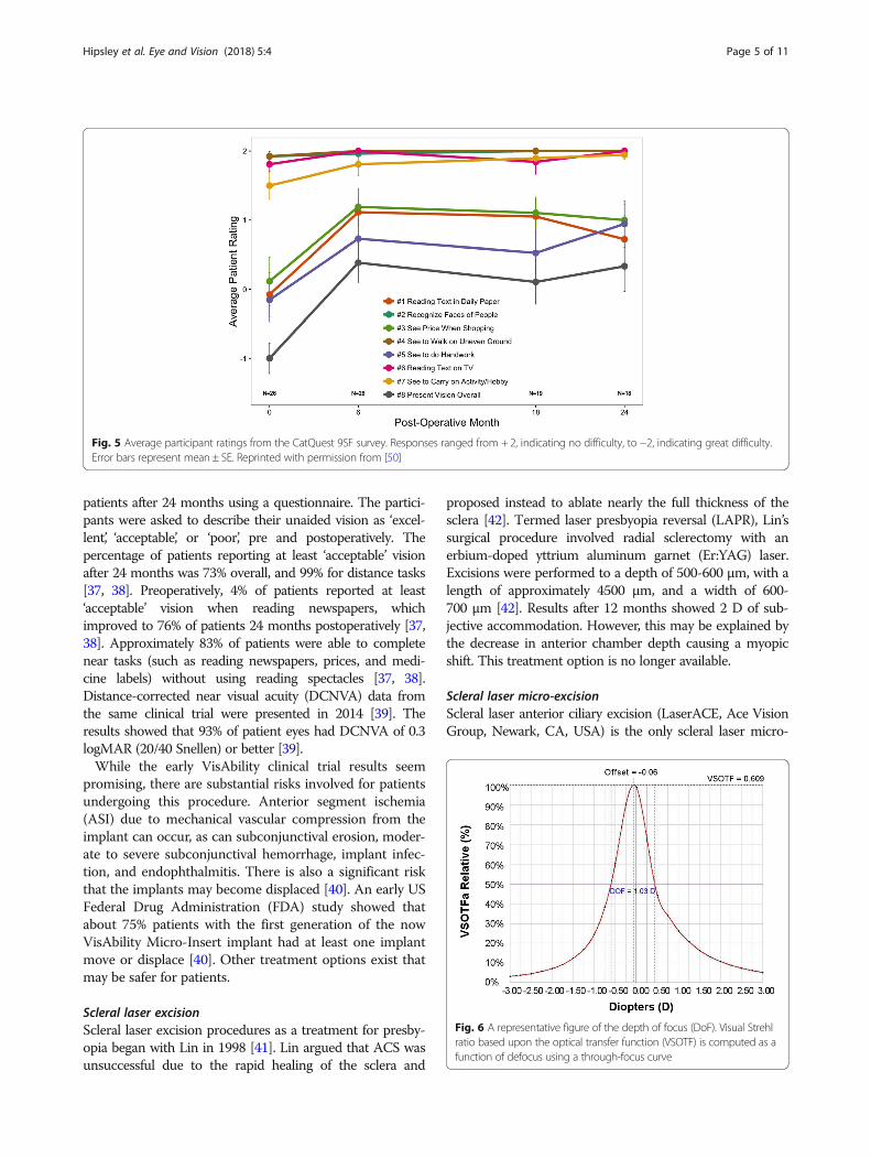
Fig. 5 Average participant ratings from the CatQuest 9SF survey. Responses ranged from + 2, indicating no difficulty, to −2, indicating great difficulty.Error bars represent mean ± SE. Reprinted with permission from [50]
Fig. 6 A representative figure of the depth of focus (DoF). Visual Strehlratio based upon the optical transfer function (VSOTF) is computed as afunction of defocus using a through-focus curve
Hipsley et al. Eye and Vision (2018) 5:4 Page 5 of 11
excision procedure currently available and has recently com-pleted phase III clinical trials [43]. LaserACE is not based onthe Schachar model but instead follows from VisioDynamicstheory, which is a biomechanical model for the aging eye[44]. VisioDynamics theory contends that presbyopia is nota refractive error or the loss of accommodation solely, butrather an aging disease limited by structural/mechanical,extracellular and intracellular, and physiological aspects ofthe eye. It argues that as the eye ages, the connective tissueswithin begin to change and impact ocular biomechanical ef-ficiency. This, in turn, influences visual function and ocularphysiology including ocular metabolic efficiency, and ocularbiotransport. A better approach to treat presbyopia may beto address these age-related changes rather than to increasescleral diameter across the globe since increasing scleraldiameter could induce unwanted biomechanical effects [45].Given the complexity of the accommodation mechanism,
the Helmholtz theory is an incomplete explanation for pres-byopia. Recent evidence has highlighted aging-relatedchanges in the vitreous membrane, peripheral choroid, ciliarymuscle, and zonules [4–7]. The sclera itself is known to beaffected by age, bowing inward [6]. Ocular rigidity and in-creasing stiffness of the zonular apparatus may also further
contribute to presbyopia [46, 47]. Proprioceptors in the vitre-ous zonular system have also been found to contribute tothe loss of accommodation with age [48]. Given the manysuggested contributions to the loss of accommodation, pres-byopia may be better described by age-related changes inresting muscle apex thickness and accommodative lensthickening together [2]. LaserACE was thus designed to bothalter the biomechanical properties of ocular tissue and im-prove the efficiency of the accommodation apparatus (Fig. 2).The LaserACE surgical technique is shown in Fig. 3.
In brief, an Er:YAG laser is used to create a matrix arrayof micro-excisions (micropores, 600 μm in diameter) inthe sclera, to a depth of 85-90% the thickness of thesclera (approximately 500-700 μm). The micro-excisionsare done in four oblique quadrants of the eye over threecritical zones of anatomical and physiological signifi-cance [44, 46, 49]. The procedure is performed undertopical anesthesia and takes approximately 10 min pereye. The 3 critical zones of anatomic and physiologic im-portance are as follows and range from 0.5 mm up to6.0 mm from the anatomical limbus (AL): 1) the scleralspur at the origin of the ciliary muscle (0.5 - 1.1 mmfrom AL); 2) the mid ciliary muscle body (1.1 – 4.9 mm
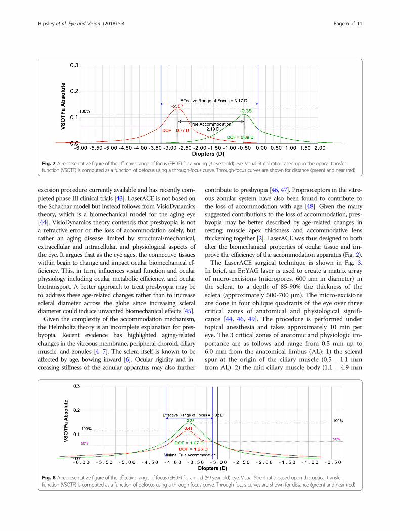
Fig. 7 A representative figure of the effective range of focus (EROF) for a young (32-year-old) eye. Visual Strehl ratio based upon the optical transferfunction (VSOTF) is computed as a function of defocus using a through-focus curve. Through-focus curves are shown for distance (green) and near (red)
Fig. 8 A representative figure of the effective range of focus (EROF) for an old (59-year-old) eye. Visual Strehl ratio based upon the optical transferfunction (VSOTF) is computed as a function of defocus using a through-focus curve. Through-focus curves are shown for distance (green) and near (red)
Hipsley et al. Eye and Vision (2018) 5:4 Page 6 of 11
from AL); and 3) insertion of the longitudinal muscle fi-bers of the ciliary, just anterior to the ora serrata at theinsertion of the posterior vitreous zonules (4.9 – 5.5 mmfrom AL) [44, 46, 49]. Within the matrix, there are areasof both positive stiffness (remaining interstitial tissue)and negative stiffness (removed tissue or micropores).The differential stiffness created in these areas increasesthe plasticity and compliance of the scleral tissue duringcontraction of the ciliary muscles, and thus improve theefficiency of the accommodation apparatus.The primary risk factor with LaserACE is accidental
micro-perforation of the sclera. This can be mitigated with acollagen biomatrix. If a micro-perforation does occur, intra-ocular pressure may be temporarily lowered. Non-persistentmild subconjunctival hemorrhages are also a risk factor.Data from a 24-month postoperative follow-up of the
LaserACE clinical study were published in 2017 andshow promising results [50]. Visual acuity at distance(4 m), intermediate (60 cm), and near (40 cm) was mea-sured using standard Early Treatment Diabetic Retin-opathy Study (ETDRS) charts, and statistical analysiswas done using an ANOVA and Tukey HSD test(Fig. 4). Monocular uncorrected near visual acuity(UNVA) improved from + 0.36 ± 0.20 logMAR pre-operatively, to + 0.25 ± 0.18 logMAR (p < 0.00005) at24 months postoperatively, and binocular DCNVA im-proved from + 0.21 ± 0.17 logMAR preoperatively, to +0.11 ± 0.12 logMAR at 24 months (p = 0.00026).DCNVA was also 0.2 logMAR (20/32 Snellen) or betterin 83% of patients at 24 months postoperatively [50].Stereoacuity (Randot stereoscopic test) also improved,averaging 58.8 ± 22.9 s of arc at 24 months postopera-tively compared to 75.8 ± 29.3 s of arc preoperatively[50]. There were no complications such as loss of best-corrected visual acuity, cystoid macular edema, or per-sistent hypotony. Patients surveyed using the CatQuest9SF Survey, [51] indicated reduced difficulty in areas ofnear vision, such as seeing when doing handwork, readingnewsprint text, and seeing prices while shopping (Fig. 5).Patients indicated overall satisfaction with the procedureand their mean satisfaction scores significantly im-proved from − 1.00 (SE = 0.22) preoperatively, to + 0.33(SE = 0.36) at 24 months postoperatively (p = 0.000016).With advances in diagnostic techniques, such as
wavefront aberrometry, it is now possible to objectivelyassess visual performance. Visual Strehl of the OpticalTransfer Function (VSOTF) is an optical wavefronterror-derived metric that predicts patient visual acuity[52]. It is defined as [53]:
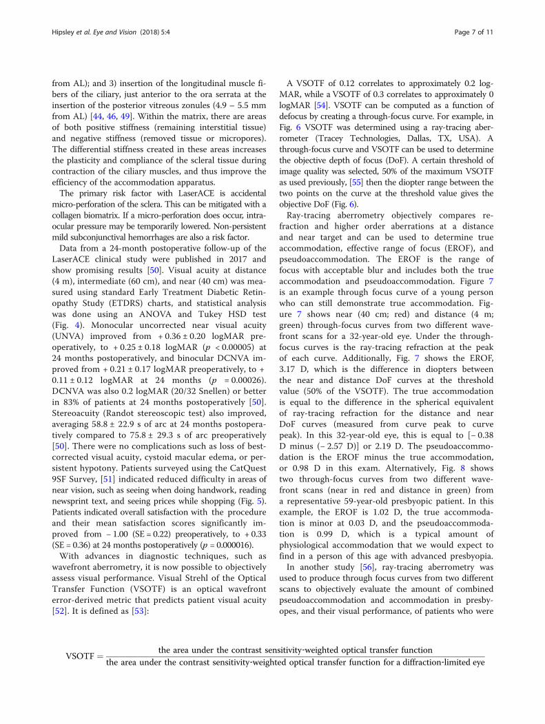
A VSOTF of 0.12 correlates to approximately 0.2 log-MAR, while a VSOTF of 0.3 correlates to approximately 0logMAR [54]. VSOTF can be computed as a function ofdefocus by creating a through-focus curve. For example, inFig. 6 VSOTF was determined using a ray-tracing aber-rometer (Tracey Technologies, Dallas, TX, USA). Athrough-focus curve and VSOTF can be used to determinethe objective depth of focus (DoF). A certain threshold ofimage quality was selected, 50% of the maximum VSOTFas used previously, [55] then the diopter range between thetwo points on the curve at the threshold value gives theobjective DoF (Fig. 6).Ray-tracing aberrometry objectively compares re-
fraction and higher order aberrations at a distanceand near target and can be used to determine trueaccommodation, effective range of focus (EROF), andpseudoaccommodation. The EROF is the range offocus with acceptable blur and includes both the trueaccommodation and pseudoaccommodation. Figure 7is an example through focus curve of a young personwho can still demonstrate true accommodation. Fig-ure 7 shows near (40 cm; red) and distance (4 m;green) through-focus curves from two different wave-front scans for a 32-year-old eye. Under the through-focus curves is the ray-tracing refraction at the peakof each curve. Additionally, Fig. 7 shows the EROF,3.17 D, which is the difference in diopters betweenthe near and distance DoF curves at the thresholdvalue (50% of the VSOTF). The true accommodationis equal to the difference in the spherical equivalentof ray-tracing refraction for the distance and nearDoF curves (measured from curve peak to curvepeak). In this 32-year-old eye, this is equal to [− 0.38D minus (− 2.57 D)] or 2.19 D. The pseudoaccommo-dation is the EROF minus the true accommodation,or 0.98 D in this exam. Alternatively, Fig. 8 showstwo through-focus curves from two different wave-front scans (near in red and distance in green) froma representative 59-year-old presbyopic patient. In thisexample, the EROF is 1.02 D, the true accommoda-tion is minor at 0.03 D, and the pseudoaccommoda-tion is 0.99 D, which is a typical amount ofphysiological accommodation that we would expect tofind in a person of this age with advanced presbyopia.In another study [56], ray-tracing aberrometry was
used to produce through focus curves from two differentscans to objectively evaluate the amount of combinedpseudoaccommodation and accommodation in presby-opes, and their visual performance, of patients who were
VSOTF ¼ the area under the contrast sensitivity‐weighted optical transfer functionthe area under the contrast sensitivity‐weighted optical transfer function for a diffraction‐limited eye
Hipsley et al. Eye and Vision (2018) 5:4 Page 7 of 11

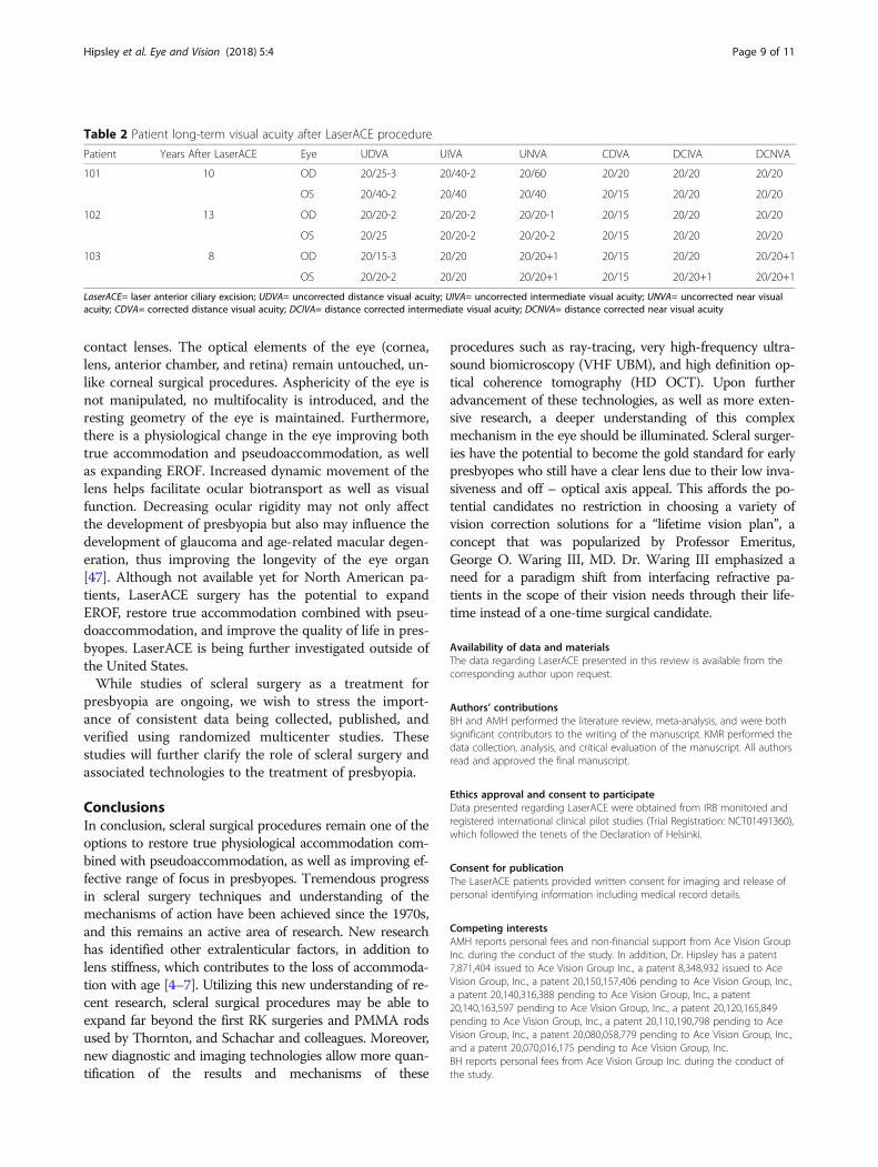
treated with LaserACE procedure up to 13 years postop-eratively. Patient demographics and visual performanceare shown in Table 1. Figure 9 shows the DoF for near(red) and distance (green) and the EROF measurementsfor one postoperative patient eye. The VSOTF, DoF,EROF, and objective accommodation were determinedas described above and shown in Table 1. Pupil contrac-tion can enhance DoF measurements. Figures 7, 8 and 9show that patient pupils did contract for near observa-tion. Since LaserACE does not affect pupil contraction,this will occur during both preoperative and postopera-tive accommodation measurements and can be elimi-nated by comparing the ranges of accommodation. Theeffective range of focus averaged 1.56 ± 0.36 D for all pa-tient eyes (n = 6), which was higher than preoperativeclinical accommodation averaging 0.92 ± 0.61 D. Pa-tients’ DoF also increased by 0.84 ± 0.74 D compared topreoperative DoF. Up to 13 years postoperatively, trueaccommodation and pseudoaccommodation averaged0.23 ± 0.24 D and 1.33 ± 0.38 D respectively. A one quar-ter diopter increase in true accommodation correspondsto a one-line improvement in near visual acuity. Pseu-doaccommodation also improved by approximately one-quarter diopter. Up to 13 years postoperatively, the 0.5 Dof restored accommodation after LaserACE was clinically
significant, and there was a corresponding increase inUNVA. UNVA was 20/20 or better in 66% of patient eyesup to 13 years postoperatively. Post-operative uncorrecteddistance visual acuity was 20/40 or better in all patienteyes, while 83% of eyes had + 1.25 D of sphere or greater.It is possible that these patients may have latent hyperopia,and thus the restored accommodative ability after Laser-ACE can correct a small degree of the hyperopia in thesepatients improving their distance vision [57]. Previousstudies have shown a similar result in hyperopic patienteyes after LaserACE [49, 50]. Patient postoperative visualacuities are shown in Table 2.DCNVA for all patients remained at 0 logMAR (20/
20 Snellen) or better up to 13 years postoperatively.It is interesting to note that these patients all hadprior laser vision correction (LVC) to correct theirdistance refraction to emmetropia before LaserACE.Since LaserACE does not touch the visual axis, thesepatients were able to achieve efficient near visual per-formance dynamically through combined accommoda-tion and pseudoaccommodation without affectingtheir previous LVC.LaserACE has many benefits compared to other presby-
opia treatments. Patients experience an increased qualityof life by decreasing their dependence on spectacles and
Table 1 Patient demographics and visual outcomes prior to and after LaserACE procedurePatient Age
LaserACE(years)
AgeLong-TermExam(years)
YearsSinceLaserACEProcedure
Pre-OPMRSE
Post-OPMRSE(Long-Term)
Eye Post-OPSphere(D)
Post-OPCylinder(D)
Post-OPAxis(degrees)
Post-OPIOP(mmHg)
Post-OPVSOTF
Pre-OPDepthofFocus(D)
Post-OPDepth ofFocus (D)
Pre-OPClinicalAccommodation(D)
Post-OPTrueAccommodation(D)
Post-OPPseudo-accommodation(D)
Post-OPEffectiveRange ofFocus (D)
101 49 59 10 20/20 20/25-3 OD +2.12 −1.12 150 12 0.555 0.5 1.23 0.5 0.31 1.14 1.45
OS +2.37 −1.12 31 14 0.281 0.5 2.82 0.5 0.06 2.10 2.16
102 48 59 13 20/20 20/20-2 OD +1.75 −0.75 178 14 0.616 1.07 1.32 1.7 0.06 1.21 1.27
OS +2.00 −0.25 166 13 0.426 0.75 1.44 1.7 0.19 1.30 1.49
103 52 60 8 20/20 20/15-3 OD +1.25 −0.50 151 11 0.609 0.5 1.03 0.5 0.68 1.09 1.77
OS +0.25 −0.25 14 17 0.405 0.6 1.12 0.6 0.06 1.14 1.20
LaserACE= laser anterior ciliary excision; MRSE= manifest refraction spherical equivalent; IOP= intraocular pressure; VSOTF= visual Strehl ratio based on the optical transfer function
Fig. 9 A representative figure of the effective range of focus (EROF) for a patient eye (60-year-old; 103-OD) after LaserACE. Visual Strehl ratio basedupon the optical transfer function (VSOTF) is computed as a function of defocus using a through-focus curve. Through-focus curves are shownfor distance (green) and near (red). Reprinted with permission from [56]
Hipsley et al. Eye and Vision (2018) 5:4 Page 8 of 11
contact lenses. The optical elements of the eye (cornea,lens, anterior chamber, and retina) remain untouched, un-like corneal surgical procedures. Asphericity of the eye isnot manipulated, no multifocality is introduced, and theresting geometry of the eye is maintained. Furthermore,there is a physiological change in the eye improving bothtrue accommodation and pseudoaccommodation, as wellas expanding EROF. Increased dynamic movement of thelens helps facilitate ocular biotransport as well as visualfunction. Decreasing ocular rigidity may not only affectthe development of presbyopia but also may influence thedevelopment of glaucoma and age-related macular degen-eration, thus improving the longevity of the eye organ[47]. Although not available yet for North American pa-tients, LaserACE surgery has the potential to expandEROF, restore true accommodation combined with pseu-doaccommodation, and improve the quality of life in pres-byopes. LaserACE is being further investigated outside ofthe United States.While studies of scleral surgery as a treatment for
presbyopia are ongoing, we wish to stress the import-ance of consistent data being collected, published, andverified using randomized multicenter studies. Thesestudies will further clarify the role of scleral surgery andassociated technologies to the treatment of presbyopia.
ConclusionsIn conclusion, scleral surgical procedures remain one of theoptions to restore true physiological accommodation com-bined with pseudoaccommodation, as well as improving ef-fective range of focus in presbyopes. Tremendous progressin scleral surgery techniques and understanding of themechanisms of action have been achieved since the 1970s,and this remains an active area of research. New researchhas identified other extralenticular factors, in addition tolens stiffness, which contributes to the loss of accommoda-tion with age [4–7]. Utilizing this new understanding of re-cent research, scleral surgical procedures may be able toexpand far beyond the first RK surgeries and PMMA rodsused by Thornton, and Schachar and colleagues. Moreover,new diagnostic and imaging technologies allow more quan-tification of the results and mechanisms of these
procedures such as ray-tracing, very high-frequency ultra-sound biomicroscopy (VHF UBM), and high definition op-tical coherence tomography (HD OCT). Upon furtheradvancement of these technologies, as well as more exten-sive research, a deeper understanding of this complexmechanism in the eye should be illuminated. Scleral surger-ies have the potential to become the gold standard for earlypresbyopes who still have a clear lens due to their low inva-siveness and off – optical axis appeal. This affords the po-tential candidates no restriction in choosing a variety ofvision correction solutions for a “lifetime vision plan”, aconcept that was popularized by Professor Emeritus,George O. Waring III, MD. Dr. Waring III emphasized aneed for a paradigm shift from interfacing refractive pa-tients in the scope of their vision needs through their life-time instead of a one-time surgical candidate.
Availability of data and materialsThe data regarding LaserACE presented in this review is available from thecorresponding author upon request.
Authors’ contributionsBH and AMH performed the literature review, meta-analysis, and were bothsignificant contributors to the writing of the manuscript. KMR performed thedata collection, analysis, and critical evaluation of the manuscript. All authorsread and approved the final manuscript.
Ethics approval and consent to participateData presented regarding LaserACE were obtained from IRB monitored andregistered international clinical pilot studies (Trial Registration: NCT01491360),which followed the tenets of the Declaration of Helsinki.
Consent for publicationThe LaserACE patients provided written consent for imaging and release ofpersonal identifying information including medical record details.
Competing interestsAMH reports personal fees and non-financial support from Ace Vision GroupInc. during the conduct of the study. In addition, Dr. Hipsley has a patent7,871,404 issued to Ace Vision Group Inc., a patent 8,348,932 issued to AceVision Group, Inc., a patent 20,150,157,406 pending to Ace Vision Group, Inc.,a patent 20,140,316,388 pending to Ace Vision Group, Inc., a patent20,140,163,597 pending to Ace Vision Group, Inc., a patent 20,120,165,849pending to Ace Vision Group, Inc., a patent 20,110,190,798 pending to AceVision Group, Inc., a patent 20,080,058,779 pending to Ace Vision Group, Inc.,and a patent 20,070,016,175 pending to Ace Vision Group, Inc.BH reports personal fees from Ace Vision Group Inc. during the conduct ofthe study.
Table 2 Patient long-term visual acuity after LaserACE procedure
Patient Years After LaserACE Eye UDVA UIVA UNVA CDVA DCIVA DCNVA
101 10 OD 20/25-3 20/40-2 20/60 20/20 20/20 20/20
OS 20/40-2 20/40 20/40 20/15 20/20 20/20
102 13 OD 20/20-2 20/20-2 20/20-1 20/15 20/20 20/20
OS 20/25 20/20-2 20/20-2 20/15 20/20 20/20
103 8 OD 20/15-3 20/20 20/20+1 20/15 20/20 20/20+1
OS 20/20-2 20/20 20/20+1 20/15 20/20+1 20/20+1
LaserACE= laser anterior ciliary excision; UDVA= uncorrected distance visual acuity; UIVA= uncorrected intermediate visual acuity; UNVA= uncorrected near visualacuity; CDVA= corrected distance visual acuity; DCIVA= distance corrected intermediate visual acuity; DCNVA= distance corrected near visual acuity
Hipsley et al. Eye and Vision (2018) 5:4 Page 9 of 11
Author details1Ace Vision Group Inc, 39655 Eureka Drive, Newark, CA 94560, USA. 2SengiData, Cambridge, ON, Canada. 3Storm Eye Institute, Medical University ofSouth Carolina, Charleston, SC, USA.
Received: 17 May 2017 Accepted: 28 January 2018
References1. Millodot M. Dictionary of optometry and visual science. Philadelphia:
Elsevier Health Sciences; 2014. p. 4.2. Croft MA, Glasser A, Kaufman PL. Accommodation and presbyopia. Int
Ophthalmol Clin. 2001;41:33–46.3. Marmer RH. The surgical reversal of presbyopia: a new procedure to restore
accommodation. Int Ophthalmol Clin. 2001;41:123–32.4. Richdale K, Sinnott LT, Bullimore MA, Wassenaar PA, Schmalbrock P, Kao CY,
et al. Quantification of age-related and per diopter accommodative changesof the lens and ciliary muscle in the emmetropic human eye. InvestOphthalmol Vis Sci. 2013;54:1095–105.
5. Croft MA, McDonald JP, Katz A, Lin TL, Lütjen-Drecoll E, Kaufman PL.Extralenticular and lenticular aspects of accommodation and presbyopia inhuman versus monkey eyes. Investi Ophthalmol Vis Sci. 2013;54:5035–48.
6. Croft MA, Nork TM, McDonald JP, Katz A, Lütjen-Drecoll E, Kaufman PL.Accommodative movements of the vitreous membrane, choroid, and sclerain young and presbyopic human and nonhuman primate eyes. InvestOphthalmol Vis Sci. 2013;54:5049–58.
7. Strenk SA, Strenk LM, Guo S. Magnetic resonance imaging of theanteroposterior position and thickness of the aging, accommodating, phakic,and pseudophakic ciliary muscle. J Cataract Refract Surg. 2010;36:235–41.
8. Holden BA, Fricke TR, Ho SM, Wong R, Schlenther G, Cronjé S, et al. Globalvision impairment due to uncorrected presbyopia. Arch Ophthalmol. 2008;126:1731–9.
9. American Optometric Association. Care of the Patient with Presbyopia. St.Louis, MO; 2010.
10. World Health Organization. Elimination of avoidable visual disabilitydue to refractive errors: report of an informal planning meeting,Geneva, 3-5 July. 2000.
11. Frick KD, Joy SM, Wilson DA, Naidoo KS, Holden BA. The Global Burden ofPotential Productivity Loss from Uncorrected Presbyopia. Ophthalmology.2015;122:1706–10.
12. von Helmholtz H. Mechanism of accommodation. In: Southall JPC, editor.Helmholtz's Treatise on Physiological Optics. Vol. 1, Trans. from the 3rdGerman Ed. Rochester: Optical Society of America; 1924. p. 143-72.
13. Charman WN. Developments in the correction of presbyopia I: spectacleand contact lenses. Ophthalmic Physiol Opt. 2014;34:8–29.
14. Vargas-Fragoso V, Alió JL. Corneal compensation of presbyopia: PresbyLASIK:an updated review. Eye Vis (Lond). 2017;4:11.
15. Gil-Cazorla R, Shah S, Naroo SA. A review of the surgical options for thecorrection of presbyopia. Br J Ophthalmol. 2016;100:62–70.
16. Evans BJ. Monovision: a review. Ophthalmic Physiol Opt. 2007;27:417–39.17. O’Keefe M, O’Keeffe N. Corneal surgical approach in the treatment of
presbyopia. J Clin Exp Ophthalmol. 2016;7:1.18. Baikoff G. Surgical treatment of presbyopia: scleral, corneal, and lenticular.
Curr Opin Ophthalmol. 2004;15:365–9.19. Charman WN. Developments in the correction of presbyopia II: surgical
approaches. Ophthalmic Physiol Opt. 2014;34:397–426.20. Vilupuru AS, Roorda A, Glasser A. Spatially variant changes in lens power
during ocular accommodation in a rhesus monkey eye. J Vis. 2004;4:299–309.21. Rozanova O. Seeing presbyopia formation in depth. In: The Internation
Society of Presbyopia Conference. Barcelona, Spain; 2015.22. Waring GO IV. Diagnosis and treatment of dysfunctional lens syndrome.
Cataract and Refractive Surgery Today. 2013;13:36–8.23. Glasser A. Restoration of accommodation: surgical options for correction of
presbyopia. Clin Exp Optom. 2008;91:279–95.24. Fyodorov SN, Durnev VV. Operation of dosaged dissection of corneal
circular ligament in cases of myopia of mild degree. Ann Ophthalmol. 1979;11(12):1885–90.
25. Thornton SP. Anterior ciliary sclerotomy (ACS), a procedure to reversepresbyopia. In: Sher NA, editor. Surgery for Hyperopia and Presbyopia.Baltimore: Williams &Wilkins; 1997. p. 33–6.
26. Hamilton DR, Davidorf JM, Maloney RK. Anterior ciliary sclerotomy fortreatment of presbyopia: a prospective controlled study. Ophthalmology.2002;109:1970–6.
27. Fukasaku H, Marron JA. Anterior ciliary sclerotomy with silicone expansionplug implantation: effect on presbyopia and intraocular pressure. IntOphthalmol Clin. 2001;41:133–41.
28. Schachar RA. Cause and treatment of presbyopia with a method forincreasing the amplitude of accommodation. Ann Ophthalmol. 1992;24:445–7. 452.
29. Schachar RA, Black TD, Kash RL, Cudmore DP, Schanzlin DJ. The mechanism ofaccommodation and presbyopia in the primate. Ann Ophthalmol. 1995;27:58–67.
30. Schachar RA, Anderson DA. The mechanism of ciliary muscle function. AnnOphthalmol. 1995;27:126–32.
31. Schachar RA. Zonular function: a new hypothesis with clinical implications.Ann Ophthalmol. 1994;26:36–8.
32. Strenk SA, Semmlow JL, Strenk LM, Munoz P, Gronlund-Jacob J, DeMarco JK.Age-related changes in human ciliary muscle and lens: a magneticresonance imaging study. Invest Ophthalmol Vis Sci. 1999;40:1162–9.
33. Schachar RA. The correction of presbyopia. Int Ophthalmol Clin. 2001;41:53–70.34. Malecaze FJ, Gazagne CS, Tarroux MC, Gorrand JM. Scleral expansion bands
for presbyopia. Ophthalmology. 2001;108:2165–71.35. Malecaze FJ, Soler V, Guillard C. Scleral expansion bands. In: Pallikaris IG,
Plainis S, Charman WN, editors. Presbyopia: origins, effects, and treatment.New Jersey: SLACK Incorporated; 2012. p. 213–7.
36. U.S. National Institutes of Health Clinical Trials. A Clinical Trial of TheVisAbility Micro Insert System for Presbyopic Patients. https://clinicaltrials.gov/ct2/show/NCT02374671. Accessed 17 Aug 17 2016.
37. Davidson RS, Dhaliwal D, Hamilton DR, Jackson M, Patterson L, Stonecipher K,et al. Surgical correction of presbyopia. J Cataract Refract Surg. 2016;42:920–30.
38. Stonecipher K. Results from a multi-center US clinical trial for the surgicaltreatment of Presbyopia. In: XXXI congress of the European Society ofCataract and Refractive Surgeons. Amsterdam, the Netherlands; 2013.
39. Soloway B, Schanzlin DJ. Effect of refinements in surgical instrumentationand scleral implant device and technique on presbyopia treatment. In: Theannual ASCRS and ASOA symposium and congress. ASCRS; 2014.
40. Charters L. Refocus Scleral Implants for presbyopia. Ophthalmology Times.http://ophthalmologytimes.modernmedicine.com/ophthalmologytimes/content/tags/barrie-soloway/refocus-scleral-implants-presbyopia. Published2013. Accessed 1 Sept 2016.
41. Lin JT, Kadambi V. The new mechanism of laser presbyopia reversal (LAPR)and accommodation. In: Agarwal A, editor. Presbyopia: a surgical textbook.Thorofare: SLACK; 2002. p. 63–70.
42. Lin JT, Mallo O. Treatment of presbyopia by infrared laser radial sclerectomy.J Refract Surg. 2003;19:465–7.
43. U.S. National Institutes of Health Clinical Trials. LaserACE® Procedure RestoreVisual Function and Range of Accommodation LaserACE® Procedure.NCT01491360. https://clinicaltrials.gov/ct2/show/NCT01491360. Accessed 17Aug 2016.
44. Hipsley A, Dementiev D. VisioDynamics theory: a biomechanical model forthe aging ocular organ. In: Ashok G, Urzua G, Dementiev D, Pinelli R, editors.Step by step innovations in Presbyopia management. New Delhi: JaypeeBrothers Medical Publishers; 2006. p. 269–315.
45. Hipsley A. Influence of biomechanics on age-related changes of the eye:clinical implications for sefractive surgery. In: American Society of Cataractand Refractive Surgery: ASCRS. Washington, DC; 2005.
46. Lütjen-Drecoll E, Kaufman PL, Wasielewski R, Ting-Li L, Croft MA.Morphology and accommodative function of the vitreous zonule in humanand monkey eyes. Invest Ophthalmol Vis Sci. 2010;51:1554–64.
47. Detorakis ET, Pallikaris IG. Ocular rigidity: biomechanical role, in vivomeasurements and clinical significance. Clin Exp Ophthalmol. 2013;41:73–81.
48. Flügel-Koch CM, Croft MA, Kaufman PL, Lütjen-Drecoll E. Anteriorly locatedzonular fibres as a tool for fine regulation in accommodation. OphthalmicPhysiol Opt. 2016;36:13–20.
49. Hipsley A, McDonald M. Laser scleral matrix microexcisions (LaserACE/erbiumYAG laser). In: Pallikaris IG, Plainis S, Charman WN, editors. Presbyopia: origins,effects, and treatment. New Jersey: Slack Incorporated; 2012. p. 219–25.
50. Hipsley A, Ma DH, Sun CC, Jackson MA, Goldberg D, Hall B. Visual outcomes24 months after LaserACE. Eye Vis (Lond). 2017;4:15.
51. Lundström M, Roos P, Jensen S, Fregell G. Catquest questionnaire for use incataract surgery care: description, validity, and reliability. J Cataract RefractSurg. 1997;23:1226–36.
Hipsley et al. Eye and Vision (2018) 5:4 Page 10 of 11
52. Iskander DR. Computational aspects of the visual Strehl ratio. Optom Vis Sci.2006;83:57–9.
53. Thibos LN, Hong X, Bradley A, Applegate RA. Accuracy and precision ofobjective refraction from wavefront aberrations. J Vis. 2004;4:329–51.
54. Cheng X, Bradley A, Thibos LN. Predicting subjective judgment of bestfocus with objective image quality metrics. J Vis. 2004;4:310–21.
55. Yi F, Iskander DR, Collins MJ. Estimation of the depth of focus fromwavefront measurements. J Vis. 2010;10:1–9.
56. Hipsley A, Hall B, Rocha KM. Long-term visual outcomes of laser anteriorciliary excision. Am J Ophthalmol Case Rep. 2018;10:38–47.
57. Morgan MW. Changes in refraction over a period of twenty years in anon-visually selected sample. Am J Optom Arch Am Acad Optom.1958;35:281–99.
• We accept pre-submission inquiries
• Our selector tool helps you to find the most relevant journal
• We provide round the clock customer support
• Convenient online submission
• Thorough peer review
• Inclusion in PubMed and all major indexing services
• Maximum visibility for your research
Submit your manuscript atwww.biomedcentral.com/submit
Submit your next manuscript to BioMed Central and we will help you at every step:
Hipsley et al. Eye and Vision (2018) 5:4 Page 11 of 11