Embed Size (px)
Citation preview
FACTA UNIVERSITATIS Series: Medicine and Biology Vol.12, No 3, 2005, pp. 164 - 169 UC 616-006:611.93-053.2-073
SONOGRAPHY OF CONGENITAL NECK MASSES IN CHILDREN
Sladjana Petrović¹, Dragan Petrović², Zoran Pešić², Predrag Kovačević3
1Institute of Radiology, Clinical Centre, Niš, Serbia and Montenegro 2Clinic of Stomatology, Department of Maxillofacial Surgery, Niš, Serbia and Montenegro 3Clinic of Plastic and Reconstructive Surgery, Clinical Centre, Niš, Serbia and Montenegro E-mail: [email protected]
Summary. Congenital neck masses in children are a relatively frequent finding. To the clinicians, they may pose various diagnostic and therapeutic dilemmas and high-resolution sonography is therefore a method of choice. Differential diagnosis of paediatric congenital neck masses primarily includes the presence of hemangioma, lymphangioma, thyreoglossal and branchial cleft cysts. The aim of this paper is to examine the ability of echosonography in differential diagnosis of various congenital neck masses in children and to determine the efficacy and significance of echosonography in preoperative patient preparation. Patients and methods: The investigation enrolled 53 paediatric patients with palpable masses in the neck, who have been examined by high-resolution sonography and color doppler sonography before surgical treatment. CT and MRI, as intra-operative and histopathological findings, confirmed echosonographic diagnosis. Results: Our experience showed that sonography is a sensitive method in differential diagnosis of congenital neck masses in children. Conclusion: We recommend this method as an accurate, cost-effective, noninvasive imaging modality in the preoperative evaluation.
Key words: Congenital, neck masses, Doppler, sonography
Introduction
Congenital neck masses in children are a relatively frequent finding. To the clinicians, they may pose vari-ous diagnostic and therapeutic dilemmas and high-resolution sonography is, therefore, a method of choice. Differential diagnosis of paediatric congenital neck masses primarily includes the presence of hemangioma, lymphangioma, thyreoglossal and branchial cleft cysts. High-resolution sonography with colour and power Ddoppler sonography is the method which can provide reliable differentiation of these changes.
Aim The aim of the paper is to examine the ability of
echosonography in differential diagnosis of various congenital neck masses in children and to determine the efficacy and significance of echosonography in preop-erative patient preparation.
Patients and Methods The investigation enrolled 53 paediatric patients
with palpable masses in the neck, referred to echosono-graphy by clinicians in the period from January 2000 till the end of December 2005. The age of the patients ranged from 3 months to 15 years. All examinations were performed at the Institute of Radiology, Clinical
Centre, Niš, using conventional machines for echosono-graphy with high-resolution multi-frequency probes (7.5-10 MHz) with colour and power Doppler option. The examinations were targeted and consisted of longi-tudinal, transversal and oblique grey scale sonograms, and after that colour and power Doppler options were applied and flow curves were registered. The results were postoperatively compared with intra-operative and histopathological findings, and, in some cases, ultra-sound findings were preoperatively compared with CT and MRI findings.
Results Out of the total proportion, 58% of lesions were lo-
cated at the medial line and 42% in the lateral portions of the neck.
Based on the echosonographic picture, all lesions were categorised as cystic, semisolid without vasculari-zation and solid vascularized lesions.
In 21 patients solid structures were found with ir-regular transonic spaces demonstrating vascularization at colour Doppler sonography. Power Doppler shows an increased high vessel density, whereas pulsed Doppler demonstrates high-flow velocity and low-resistance index – these changes were categorised as hemangiomas (Fig. 1). They presented as well-vascu-larized solid masses, which were hypoechogenic in 50%, hyperechogenic in 17% and heteroechogenic in
SONOGRAPHY OF CONGENITAL NECK MASSES IN CHILDREN 165
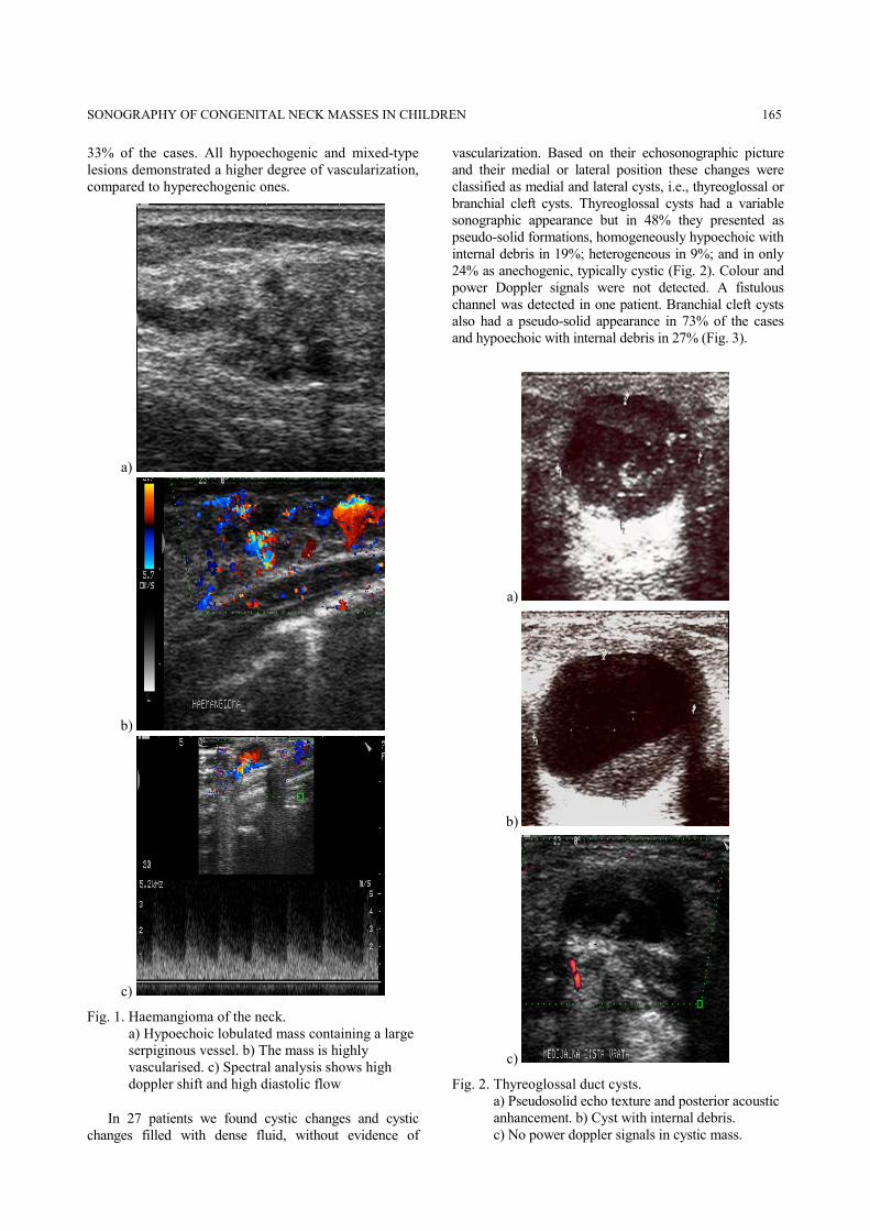
33% of the cases. All hypoechogenic and mixed-type lesions demonstrated a higher degree of vascularization, compared to hyperechogenic ones.
a)
b)
c)
Fig. 1. Haemangioma of the neck. a) Hypoechoic lobulated mass containing a large serpiginous vessel. b) The mass is highly vascularised. c) Spectral analysis shows high doppler shift and high diastolic flow
In 27 patients we found cystic changes and cystic changes filled with dense fluid, without evidence of
vascularization. Based on their echosonographic picture and their medial or lateral position these changes were classified as medial and lateral cysts, i.e., thyreoglossal or branchial cleft cysts. Thyreoglossal cysts had a variable sonographic appearance but in 48% they presented as pseudo-solid formations, homogeneously hypoechoic with internal debris in 19%; heterogeneous in 9%; and in only 24% as anechogenic, typically cystic (Fig. 2). Colour and power Doppler signals were not detected. A fistulous channel was detected in one patient. Branchial cleft cysts also had a pseudo-solid appearance in 73% of the cases and hypoechoic with internal debris in 27% (Fig. 3).
a)
b)
c)
Fig. 2. Thyreoglossal duct cysts. a) Pseudosolid echo texture and posterior acoustic anhancement. b) Cyst with internal debris. c) No power doppler signals in cystic mass.
166 S. Petrović, D. Petrović, Z. Pešić, P. Kovačević
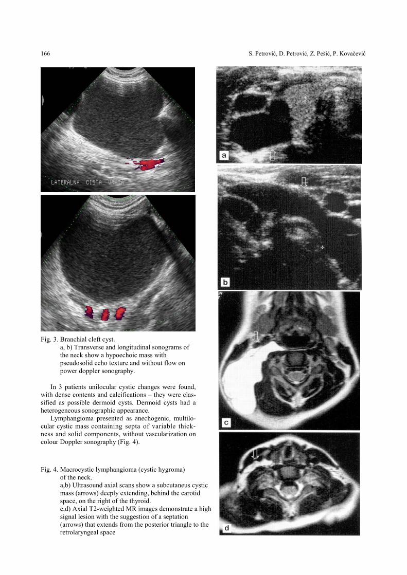
Fig. 3. Branchial cleft cyst.
a, b) Transverse and longitudinal sonograms of the neck show a hypoechoic mass with pseudosolid echo texture and without flow on power doppler sonography.
In 3 patients unilocular cystic changes were found, with dense contents and calcifications – they were clas-sified as possible dermoid cysts. Dermoid cysts had a heterogeneous sonographic appearance.
Lymphangioma presented as anechogenic, multilo-cular cystic mass containing septa of variable thick-ness and solid components, without vascularization on colour Doppler sonography (Fig. 4).
Fig. 4. Macrocystic lymphangioma (cystic hygroma) of the neck. a,b) Ultrasound axial scans show a subcutaneus cystic mass (arrows) deeply extending, behind the carotid space, on the right of the thyroid. c,d) Axial T2-weighted MR images demonstrate a high-signal lesion with the suggestion of a septation (arrows) that extends from the posterior triangle to the retrolaryngeal space
SONOGRAPHY OF CONGENITAL NECK MASSES IN CHILDREN 167
In 5 patients computerised tomography was also ap-plied, confirming echosonographic diagnosis: in 2 pa-tients branchial cleft cysts, in 1 patient thyreoglossal cysts, in 1 hemangioma, and in 1 patient lymphangioma.
In 2 patients magnetic resonance imaging was done, confirming the presence of branchial cleft cyst and lym-phangioma.
After surgical treatment and histopathological verifi-cation it was established that 17 (32.0%) patients had branchial cleft cysts, 10 (18.9%) thyreoglossal cysts, 3 (5.7%) dermoid cysts and 2 (3.8%) lymphangiomas (cystic hygromas). Out of 21 patients (39.6%) with he-mangiomas, 5 patients were treated surgically and other patients conservatively.
The intra-operative and histopathological findings completely correlated with the echosonographic find-ings.
Discussion Hemangiomas are the most common tumours occur-
ring in infancy (1,2). They usually appear in the first week of life and are located in the head and neck in al-most 60% of cases. Haemangiomas are characterised by three phases: rapid postnatal endothelial proliferation (3-9 months); stable period of variable length; and spontane-ous slow involution (approximately 18 months to 10 years). Histologically, in the proliferative phase, they are formed by well-delimited lobular masses of endothelial cells with an increased number of mast cells. Later, cap-illary-size lumina are often seen. During the involutive phase there is progressive perivascular deposition of fibrofatty tissue, enlargement of the vascular lumen and thinning of the endothelial lining. Diagnosis is usually made on the basis of clinical findings, and, in most in-stances, further investigations are not needed. Imaging is indicated either in the diagnosis of deep haemangiomas with normal overlying skin to evaluate their extent or in cases of "alarming haemangiomas", i.e. lesions which are dangerous to vital structures (e.g. obstruction of the air-way, impairment of vision, heart failure or thrombocyto-penic coagulopathy) (3,4). According to Dubois and Ga-rel, echosonography is the best imaging modality for de-fining haemangiomas (3). At echosonography, haeman-giomas may appear homogeneously hyperechoic (multi-ple tiny vascular channel interfaces, proteinaceous matrix and areas of thrombosis and fibrosis), or with a typical hypoechoic lobular pattern (Fig. 1), or even like a com-plex mass containing vascular spaces (5). The colour and/or power Doppler show an increased high vessel den-sity (defined as more than five structures per square cen-timetre), whereas the pulsed Doppler demonstrates high flow velocity (up to 90 cm/s) and a low-resistance index (RI: 0.4-0.7) with broadening of the spectrum (Fig. 1) (6). During the fibrolipomatous involution, the number of vessels decreases and RI progressively increases. Both CT and MRI provide a better tissue differentiation and, as a consequence, a better evaluation of the extent of the lesions if they need to be surgically treated. In a majority
of cases, however, no treatment is required because all haemangiomas undergo spontaneous involution.
Lymphangioma is believed to develop from se-questered lymphatic sacs that fail to communicate with peripheral draining channels (7). Approxi-mately 75% of all lymphangiomas occur in the neck, generally located in the posterior compartment (Fig. 4), and 3-10% may extend into the mediastinum (8). The presence of loose fatty tissue in the neck al-lows the formation of cystic hygroma, which consists of hugely dilated cystic lymphatic spaces, but a com-bination of the four histological types of lymphan-gioma (cystic hygroma, cavernous and capillary lym-phangioma, vasculolymphatic malformations) can often be seen in a single lesion (8,9). Actually, at echosonography, they appear as multilocular, pre-dominantly cystic, masses (Fig. 4) containing septa of variable thickness and solid components. A cor-relation between the sonogram and pathological specimen demonstrates that the echogenic compo-nent corresponds to a cluster of abnormal lymphatic channels, too small to be resolved with echosono-graphy (10). Haemorrhagic or infected cystic spaces are also more echogenic (11). Large lesions had ill-defined boundaries, with cystic components dissecting between normal tissue planes. Lymphangiomas are treated by surgery and MRI is the most accurate technique for evaluating the extent of the tumour and its relatedness to surgical planning (12,13,14). Scle-rosing therapy with echosonography guidance may be an alternative for macro-cystic lymphangiomas (15).
Sonographically, these tumours can usually be dif-ferentiated from other cervical masses, especially soft tissue haemangiomas and venous malformations. The differential diagnosis of a cystic neck mass in-cludes thyreoglossal duct cyst, branchial cleft cysts and dermoid tumours.
The thyroglossal duct is the most frequent midline cyst and the most common congenital neck mass, ac-counting for 70% of congenital neck masses (16). The echosonography diagnosis is readily made by follow-ing the base of the tongue in the midline towards the sternal notch. On echosonography, thyroglossal duct cysts in children are not simple cysts but have a com-plex pattern ranging from a typical anechoic cyst to a pseudo-solid appearance (most common; Fig. 2) (17). The origin, as well as a sinus to the base of the tongue, is sometimes identified (thyroglossal duct cysts move with movement of the tongue, whereas other masses generally do not) (18). Thyroglossal-duct remnant should be differentiated from ectopic thyroid prior to surgical excision. When the thyroid gland can be identified in the normal position, coexistent ectopic thyroid is seldom found, even if it is possible (19); however, when ectopic thyroid is suspected, Tc-99m and thyroid function tests are valuable (20).
The branchial cleft cysts are the most common non-inflammatory paediatric lateral neck masses; almost all arise from the second branchial cleft
168 S. Petrović, D. Petrović, Z. Pešić, P. Kovačević
(16,21). The usual position of these lesions is high in the lateral neck along the anterior border of the ster-nocleidomastoid muscle, just beneath the superficial cervical fascia. These cysts may occur anywhere along the developmental path of the second bran-chial complex and may be located as high as the ton-sillar fossa at the level of the hyoid bone. The most common location is lateral to the jugular vein at the level of the carotid artery bifurcation. The cysts can extend inward towards the lateral wall of the pharynx (22). Rarely, the cysts can be completely located deep in the carotid vessels. Clinically, branchial cleft cysts present as firm or fluctuant masses. Ultrasound reveals a complex mass with fluid and scattered debris (Fig. 3), which may be secondary not only to infec-tion, because these lesions are often filled with a tur-bid, yellowish fluid, which occasionally contains cho-lesterol crystals. A fluid-filled cyst usually can be dis-tinguished from a solid mass by the enhancement of the US beam posterior to the cyst (18).
Dermoid cysts are benign, usually unilocular, der-malined structures (23). In contrast to the epidermoid cyst, which has only a squamous epithelium, and to true teratomas, which contain tissue elements derived from all three germinal layers, the dermoid cysts contain only ectodermal and mesodermal elements (24). They include hair follicles, hair, sebaceous glands, sweat glands and dermal appendages. A unilocular cystic midline neck mass at the suprasternal notch in a child should suggest a dermoid cyst (23).
All the cysts have internal echoes, with a solid appear-ance with only slight or no posterior echo enhance-ment. Amorphous keratinous debris from keratinizing stratified squamous epithelium fills the lumen of each cyst, producing the internal echoes (25).
Epidermoid cysts are rare lesions in the head and neck. Most often they are located in the submental region. Commonly they have the same echosonogra-phy appearance as dermoid cysts. A cystic tumour with the unusual echosonography aspect of multiple smaller spherical formations caused by multiple spherical keratin formations has been described (26).
Conclusion The sonographic appearance of congenital cystic
masses of the neck in children is variable; to make correct preoperative assessment, the sonologist must be familiar with these characteristics. Cystic cervical masses do not seem to be simple cyst, as previously suggested, but in-stead they have a complex cystic pattern ranging from an anechoic to a pseudo-solid appearance. Solid and mixed cervical masses, like hemangiomas and cystic hygromas had a typical ultrasound and colour Doppler appearance. Our experience has shown that sonography is a sensitive method in differential diagnosis of congenital neck masses in children. We recommended this method as an accurate, cost-effective, noninvasive imaging modality in the preoperative evaluation.
References1. Mulliken JB, Glowacki J. Classification of paediatric vascular le-
sions. Plast Reconstr Surg 1982; 70: 120-121. 2. Mulliken JB, Fishman SJ, Burrows PE. Vascular anomalies. Curr
Probl Surg 2000; 37: 517-584. 3. Dubois J, Garel L. Imaging and therapeutic approach of hemangio-
mas and vascular malformations in the paediatric age group. Pediatr Radiol 1999; 29: 879-893.
4. Enjolras O. Classification and management of the various superfi-cial vascular anomalies: hemangiomas and vascular malformations. J Dermatol 1997; 24:701-710.
5. Toma P, Magnano GM, Oddone M. Practical approach to soft tis-sue vascular malformation. In: Theodoro-poulos BJ (ed) "Lectures". 35th Congress of the ESPR, Rhodes, 1998: 67-69.
6. Dubois J, Patriquin HB, Garel L, Powell J, Filiatrault D, David M, Grignon A. Soft-tissue hemangiomas in infants and children: diagnosis using Doppler sonography. Am J Roentgenol 1998; 171: 247-252.
7. Meza MP, Benson M, Slovis TL. Imaging of mediastinal masses in children. Radiol Clin North Am 1993; 31: 583-604.
8. Zadvinskis DP, Benson MT, Kerr HH, Mancuso AA, Cacciarelli AA, Madrazo BL, Mafee MF, Dalen K. Congenital malformations of the cervicothoracic lymphatic system: embryology and pathogenesis. Radiographics 1992; 12: 1175-1189.
9. Fordham LA, Chung CJ, Donnelly LF. Imaging of congenital vas-cular and lymphatic anomalies of the head and neck. Neuroimaging Clin North Am 2000; 10: 117-136.
10. Sheth S, Nussbaum AR, Hutchins GM, Sanders RC. Cystic hygro-mas in children: sonographic-pathologic correlation. Radiology 1987; 162: 821-824.
11. Borecky N, Gudinchet F, Laurini R, Duvoisin B, Hohlfeld J, Schnyder P. Imaging of cervico-thoracic lymphangiomas in chil-dren. Pediatr Radiol 1995; 25: 127-130.
12. Castellote A, Vazquez E, Vera J, Piqueras J, Lucaya J, Garcia-Pena P, Jimenez JA. Cervicothoracic lesions in infants and children. Radiographics 1999; 19: 583-600.
13. Oddone M, Toma P, Occhi M, Pelizza A. MR imaging of paediatric lymphangioma: report of 26 cases. Pediatr Radiol 1993; 23: 229.
14. Siegel MJ, Glazer HS, St Amour TE, Rosenthal DD. Lymphangio-mas in children: MR imaging. Radiology 1989; 170:467-470.
15. Dubois J, Garel L, Abela A, Laberge L, Yazbeck S. Lymphangio-mas in children: percutaneous sclerotherapy with an alcoholic solution of zein. Radiology 1997; 204: 651-654.
16. Koeller KK, Alamo L, Adair CF, Smirniotopoulos JG. Congenital cystic masses of the neck: radiologic-pathologic correlation. Ra-diographics 1999; 19: 121-146.
17. Ahuja AT, King AD, Metreweli C. Sonographic evaluation of thyroglossal duct cysts in children. Clin Radiol 2000; 55: 770-774.
18. Dubois J, Patriquin H. Doppler sonography of head and neck masses in children. Neuroimaging Clin North Am 2000; 10: 215-252.
19. Holland AJ, Sparnon AL, LeQuesne GW. Thyroglossal duct cyst or ectopic thyroid gland? J Paediatr Child Health 1997; 33: 346-348.
20. al-Dousary S. Current management of thyroglossal-duct remnant. J Otolaryngol 1997; 26: 259-265.
21. Stone JA, Figueroa RE. Embryology and anatomy of the neck. Neuroimaging Clin North Am 2000; 10: 55-73.
22. Mukherji SK, Fatterpekar G, Castillo M, Stone JA, Chung CJ. Imaging of congenital anomalies of the branchial apparatus. Neuroimaging Clin North Am 2000; 10: 75-93.
23. Vittore CP, Goldberg KN, McClatchey KD, Hotaling AJ. Cystic mass at the suprasternal notch of a newborn: congenital suprasternal dermoid cyst. Pediatr Radiol 1998; 28: 984-986.
24. Koch BL. Imaging extracranial masses of the paediatric head and neck. Neuroimaging Clin North Am 2000; 10: 193-214.
SONOGRAPHY OF CONGENITAL NECK MASSES IN CHILDREN 169
25. Yasumoto M, Shibuya H, Gomi N, Kasuga T. Ultrasonographic appearance of dermoid and epidermoid cysts in the head and neck. J Clin Ultrasound 1991; 19: 455-461.
26. Lohaus M, Hansmann J, Witzel A, Flechtenmacher C, Mende U, Reisser C. Uncommon sonographic findings of an epidermoid cyst in the head and neck. HNO 1999; 47: 737-740. (in German)
SONOGRAFIJA KONGENITALNIH MASA NA VRATU DECE
Sladjana Petrović¹, Dragan Petrović², Zoran Pešić², Predrag Kovačević3
1Institute za radiologiju, Klinički centar Niš 2Stomatološka klinika, Odeljenje maksilofacijalne hirurgije, Niš 3Klinika za plastičnu i rekonstruktivnu hirurgiju, Klinički centar, Niš E-mail: [email protected]
Kratak sadržaj: Kongenitalne mase na vratu u dece su relativno čest nalaz. Mogu zadavati kliničarima dijagnostičke i terapeutske dileme, zbog čega se visokorezolutivna sonografija preporučuje kao metoda izbora. Diferencijalna dijagnoza kongenitalnih masa na vratu u dece primarno uključuje prisustvo hemangioma, limfangioma, tireoglosalnih i branhijalnih cističnih promena. Cilj ovog rada je da se utvrditi mogućnost ehosonografije u diferencijalnoj dijagnostici različitih kongenitalnih masa na vratu u pedijatrijskih pacijenata, i odrediti efikasnost i značaj ehosonografije u preoperativnoj pripremi pacijenata. Ovo istraživanje obuhvata 53 pedijatrijska pacijenta sa palpabilnim masama na vratu, koji su bili podvrgnuti visokorezolutivnoj i color doppler sonografiji pre hirurškog tretmana. CT, MRI kao intraoperativni i patohistološki nalazi potvrdili su ehosonografske dijagnoze. Preporučujemo ovu metodu kao senzitivan, jeftin, neinvazivan modalitet imaginga-a u preopertivnoj pripremi pacijenata.
Ključne reči: Kongenitalne mase vrata, Doppler, sonografija