Embed Size (px)
Citation preview
The Rockefeller University Press, 0021-9525/97/06/1091/12 $2.00The Journal of Cell Biology, Volume 137, Number 5, June 2, 1997 1091–1102 1091
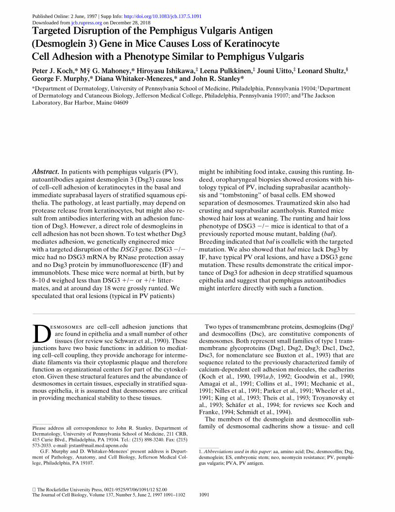
Targeted Disruption of the Pemphigus Vulgaris Antigen(Desmoglein 3) Gene in Mice Causes Loss of KeratinocyteCell Adhesion with a Phenotype Similar to Pemphigus Vulgaris
Peter J. Koch,* M G. Mahoney,* Hiroyasu Ishikawa,
‡
Leena Pulkkinen,
‡
Jouni Uitto,
‡
Leonard Shultz,
§
George F. Murphy,* Diana Whitaker-Menezes,* and John R. Stanley*
*Department of Dermatology, University of Pennsylvania School of Medicine, Philadelphia, Pennsylvania 19104;
‡
Department of Dermatology and Cutaneous Biology, Jefferson Medical College, Philadelphia, Pennsylvania 19107; and
§
The Jackson Laboratory, Bar Harbor, Maine 04609
y
Abstract.
In patients with pemphigus vulgaris (PV), autoantibodies against desmoglein 3 (Dsg3) cause loss of cell–cell adhesion of keratinocytes in the basal and immediate suprabasal layers of stratified squamous epi-thelia. The pathology, at least partially, may depend on protease release from keratinocytes, but might also re-sult from antibodies interfering with an adhesion func-tion of Dsg3. However, a direct role of desmogleins in cell adhesion has not been shown. To test whether Dsg3 mediates adhesion, we genetically engineered mice with a targeted disruption of the
DSG3
gene. DSG3
2
/
2
mice had no DSG3 mRNA by RNase protection assay and no Dsg3 protein by immunofluorescence (IF) and immunoblots. These mice were normal at birth, but by 8–10 d weighed less than DSG3
1
/
2
or
1
/
1
litter-mates, and at around day 18 were grossly runted. We speculated that oral lesions (typical in PV patients)
might be inhibiting food intake, causing this runting. In-deed, oropharyngeal biopsies showed erosions with his-tology typical of PV, including suprabasilar acantholy-sis and “tombstoning” of basal cells. EM showed separation of desmosomes. Traumatized skin also had crusting and suprabasilar acantholysis. Runted mice showed hair loss at weaning. The runting and hair loss phenotype of DSG3
2
/
2
mice is identical to that of a previously reported mouse mutant, balding (
bal
). Breeding indicated that
bal
is coallelic with the targeted mutation. We also showed that
bal
mice lack Dsg3 by IF, have typical PV oral lesions, and have a DSG3 gene mutation. These results demonstrate the critical impor-tance of Dsg3 for adhesion in deep stratified squamous epithelia and suggest that pemphigus autoantibodies might interfere directly with such a function.
D
esmosomes
are cell–cell adhesion junctions thatare found in epithelia and a small number of othertissues (for review see Schwarz et al., 1990). These
junctions have two basic functions: in addition to mediat-ing cell–cell coupling, they provide anchorage for interme-diate filaments via their cytoplasmic plaque and thereforefunction as organizational centers for part of the cytoskel-eton. Given these structural features and the abundance ofdesmosomes in certain tissues, especially in stratified squa-mous epithelia, it is assumed that desmosomes are criticalin providing mechanical stability to these tissues.
Two types of transmembrane proteins, desmogleins (Dsg)
1
and desmocollins (Dsc), are constitutive components ofdesmosomes. Both represent small families of type 1 trans-membrane glycoproteins (Dsg1, Dsg2, Dsg3; Dsc1, Dsc2,Dsc3, for nomenclature see Buxton et al., 1993) that aresequence related to the previously characterized family ofcalcium-dependent cell adhesion molecules, the cadherins(Koch et al., 1990, 1991
a
,
b
, 1992; Goodwin et al., 1990;Amagai et al., 1991; Collins et al., 1991; Mechanic et al.,1991; Nilles et al., 1991; Parker et al., 1991; Wheeler et al.,1991; King et al., 1993; Theis et al., 1993; Troyanovsky etal., 1993; Schäfer et al., 1994; for reviews see Koch andFranke, 1994; Schmidt et al., 1994).
The members of the desmoglein and desmocollin sub-family of desmosomal cadherins show a tissue- and cell
Please address all correspondence to John R. Stanley, Department ofDermatology, University of Pennsylvania School of Medicine, 211 CRB,415 Curie Blvd., Philadelphia, PA 19104. Tel.: (215) 898-3240. Fax: (215)573-2033. e-mail: [email protected]
G.F. Murphy and D. Whitaker-Menezes’ present address is Depart-ment of Pathology, Anatomy, and Cell Biology, Jefferson Medical Col-lege, Philadelphia, PA 19107.
1.
Abbreviations used in this paper
: aa, amino acid; Dsc, desmocollin; Dsg,desmoglein; ES, embryonic stem; neo, neomycin resistance; PV, pemphi-gus vulgaris; PVA, PV antigen.
on December 28, 2018jcb.rupress.org Downloaded from http://doi.org/10.1083/jcb.137.5.1091Published Online: 2 June, 1997 | Supp Info:
The Journal of Cell Biology, Volume 137, 1997 1092
type–specific expression pattern (e.g., Koch et al., 1992;Arnemann et al., 1993; Theis et al., 1993; Schäfer et al.,1994, 1996; Schmidt et al., 1994; Nuber et al., 1995, 1996;Amagai et al., 1996; North et al., 1996). Some tissues, e.g.,simple epithelia, express Dsg2 and Dsc2 only. In stratifiedsquamous epithelia, however, all three desmoglein anddesmocollin isoforms are present, although the expressionof some of these proteins is restricted to certain strata. Inhuman skin, for example, Dsg1 is expressed in suprabasalcell layers, Dsg2 in the basal cell layer only, and Dsg3 inthe basal as well as the immediate suprabasal cell layer(Amagai et al., 1996; Schäfer et al., 1996).
In this study we focused on the biological function ofone member of the desmoglein family, Dsg3. This proteinhas been shown to be the antigen recognized by autoanti-bodies from patients with the disease pemphigus vulgaris(PV), and therefore is also referred to as PV antigen (PVA)(Amagai et al., 1991).
PV is a life-threatening, autoantibody-mediated, blister-ing disease of the skin and mucous membranes (Stanley,1993
a
). Blisters in these patients result from loss of kerati-nocyte cell-to-cell adhesion in the basal and immediate su-prabasal level of stratified squamous epithelia. The typicalhistology of an early lesion in PV shows detachment of theepithelium just above the basal cells, usually with a fewacantholytic (detached and rounded-up) cells in the blistercavity. There is no, or minimal, inflammation in an earlylesion. In addition, the basal cells may detach slightly fromone another while maintaining their attachment to thebasement membrane, a histologic pattern referred to as a“row of tombstones” (Lever, 1965; also shown schemati-cally in Stanley, 1993
b
). Besides the resemblance of the in-dividual basal cells to tombstones, this designation of thehistologic appearance also reflected the dismal prognosisof the disease which, before the advent of corticosteroidtherapy, was almost uniformly fatal. Without therapy, pa-tients died because these blisters rapidly lost the superfi-cial epithelia, resulting in large areas of erosions on mu-cous membranes and skin. The mucous membrane lesionsprevented adequate food and fluid intake, while erosionson the skin resulted in protein and electrolyte loss as wellas infection. Histology of older lesions shows erosions withinflammation (which is characteristically seen in mucousmembrane or skin eroded from any cause, probably sec-ondary to infection, colonization with microbes, and/or ir-ritation due to loss of the barrier function) and attempts atreepithelialization.
IgG autoantibodies against the cell surface of kerati-nocytes of stratified squamous epithelia are present in theskin and sera of PV patients, as detected by direct and in-direct immunofluorescence, respectively (Stanley, 1993
a
).These antibodies are pathogenic, i.e., they cause blisterformation, as proven by several lines of evidence (for re-view see Stanley, 1990). In general, autoantibody titer, asdetermined by indirect immunofluorescence, correlateswith disease activity. That is, the higher the antibody titer,the more severe the disease. Furthermore, neonatal PVhas been reported in infants born to mothers with activePV. As the passively transferred maternal IgG is catabo-lized, the infant recovers. Similarly, PV IgG passivelytransferred to neonatal mice causes clinically and histolog-ically typical blisters. Finally, normal skin in organ culture
incubated with PV IgG develops typical blisters, withoutthe addition of complement or inflammatory cells.
Screening of a keratinocyte
l
gt11 expression librarywith patient sera was used to isolate cDNA encoding PVA(Amagai et al., 1991). Analysis of the predicted amino acidsequence defined PVA as Dsg3 (Buxton et al., 1993). An-tibodies raised in rabbits to recombinant Dsg3, as well aspatient sera, localized it, like the other desmogleins, todesmosomes, in particular to their extracellular face (Aki-yama et al., 1991; Karpati et al., 1993). Recent studies haveshown that antibodies against the extracellular domain ofDsg3 can cause suprabasilar blisters in neonatal mice(Amagai et al., 1992) and that the extracellular domain ofDsg3, expressed by baculovirus in Sf9 insect cells, can ad-sorb out all pathogenic antibodies from PV sera (Amagaiet al., 1994
a
; Memar et al., 1996). Therefore, the antibod-ies against Dsg3 in patient sera are pathogenic. Finally, ithas recently been shown that Dsg3 mRNA and antibodiesto Dsg3 in patients’ sera localize in epidermis to the basaland immediate suprabasal layer, exactly where blisters oc-cur in PV (Arnemann et al., 1993; Shimizu et al., 1995;Amagai et al., 1996).
It is unclear exactly how PV autoantibodies cause loss ofcell–cell adhesion. One possibility is that Dsg3 functions incell–cell adhesion in the deep stratified squamous epithe-lia, and that autoantibodies from PV patients might inter-fere with its adhesive function. However, several points donot support such a hypothesis. First, it has been suggestedthat PV autoantibodies do not directly cause loss of celladhesion, but function through stimulating release of pro-teases (specifically, plasminogen activator) from kerati-nocytes (Schiltz et al., 1978, 1979; Farb et al., 1978; Hashi-moto et al., 1983, 1989; Morioka et al., 1987; Naito et al.,1989). Furthermore, although the classical cadherins (e.g.,E-cadherin, N-cadherin, P-cadherin) have been directlyshown to be calcium-dependent cell adhesion molecules,the desmogleins have not. Expression of classical cad-herins in transfected L cells (mouse fibroblasts) mediatescalcium-dependent aggregation (Nagafuchi et al., 1987).However, experiments with desmogleins do not show sim-ilar aggregation (Kowalczyk et al., 1996) nor do studies us-ing desmocollins (Chidgey et al., 1996). Even a chimericmolecule of the extracellular domain of Dsg3 with the cy-toplasmic domain of E-cadherin, which allows for properinteraction with the actin cytoskeleton through catenins(an interaction shown to be critical for E-cadherin func-tion), mediates only weak aggregation when expressed inL cells (Amagai et al., 1994
b
). These studies suggest thatDsg3 might mediate adhesion but only does so effectivelywhen organized with other desmosomal molecules.
Another approach to investigate whether Dsg3 is impor-tant for adhesion of keratinocytes would be to eliminate itfrom desmosomes in tissue. Therefore, in this study we ge-netically engineered a mouse with a targeted disruption ofthe desmoglein 3 gene (
DSG3
). We hypothesized that ifthe PV autoantibodies directly interfere with an adhesionfunction of this molecule then such a mouse might have aphenotype resembling human disease. Our findings dem-onstrate that Dsg3 is critical for cell adhesion in the basaland immediate suprabasilar keratinocytes, especially inthe oral mucous membrane, thereby showing a differentia-tion- and tissue-specific adhesion function of one of the
Koch et al.
DSG3 Null Mice Resemble Pemphigus Patients
1093
desmogleins. Furthermore, because the pathology of thesemice strikingly resembles that of humans with PV, ourfindings are consistent with the idea that PV autoantibod-ies directly interfere with this adhesive function.
Materials and Methods
Cloning and Characterization of 129/Sv DSG3 Gene
We have previously cloned partial cDNAs corresponding to mouse Dsg3(Ishikawa et al., 1994). To initiate the cloning of the mouse
DSG3
gene,three different cDNA probes corresponding to mouse cDNA sequenceswere used for screening of a mouse 129/Sv genomic
l
FixII library (Strata-gene, La Jolla, CA) (Sambrook et al., 1989). These three cDNAs, 413, 405,and 404 bp in size, respectively, corresponded to the amino-terminal, cen-tral, and carboxyl-terminal regions of the coding sequences. (These se-quence data are available from EMBL/Genbank/DDBJ under accessionnumber U86016.) A total of nine unique genomic clones were isolated andcharacterized by restriction enzyme digestions and Southern analysis withsynthetic oligonucleotides based on exon sequences derived from thecDNA. The endonuclease digestion products were fractionated by elec-trophoresis on 1% agarose gels, and the sizes were estimated by compari-son with standard DNA markers (New England Biolabs, Beverly, MA).Subcloning and sequencing of the genomic DNA, in comparison withcDNA sequences, allowed identification of the intron–exon borders. Thesizes of the introns in the genomic DNA were determined by direct nucle-otide sequencing, estimated by generating PCR products using syntheticnucleotide primers placed on the flanking exons, or by Southern blot anal-ysis. (These sequence data [for exon 1/intron 1/exon 2] are available fromEMBL/Genbank/DDBJ under accession number U86015.)
Construction of Targeting Vector
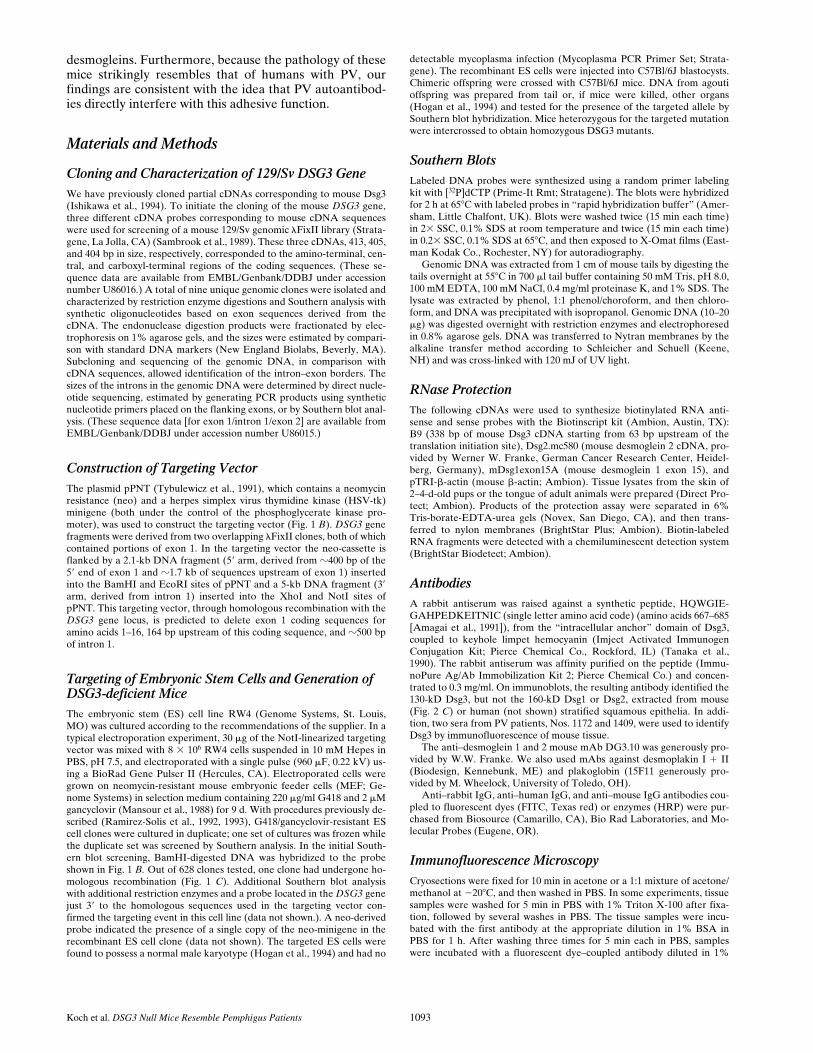
The plasmid pPNT (Tybulewicz et al., 1991), which contains a neomycinresistance (neo) and a herpes simplex virus thymidine kinase (HSV-tk)minigene (both under the control of the phosphoglycerate kinase pro-moter), was used to construct the targeting vector (Fig. 1
B
).
DSG3
genefragments were derived from two overlapping
l
FixII clones, both of whichcontained portions of exon 1. In the targeting vector the neo-cassette isflanked by a 2.1-kb DNA fragment (5
9
arm, derived from
z
400 bp of the5
9
end of exon 1 and
z
1.7 kb of sequences upstream of exon 1) insertedinto the BamHI and EcoRI sites of pPNT and a 5-kb DNA fragment (3
9
arm, derived from intron 1) inserted into the XhoI and NotI sites ofpPNT. This targeting vector, through homologous recombination with the
DSG3
gene locus, is predicted to delete exon 1 coding sequences foramino acids 1–16, 164 bp upstream of this coding sequence, and
z
500 bpof intron 1.
Targeting of Embryonic Stem Cells and Generation of DSG3-deficient Mice
The embryonic stem (ES) cell line RW4 (Genome Systems, St. Louis,MO) was cultured according to the recommendations of the supplier. In atypical electroporation experiment, 30
m
g of the NotI-linearized targetingvector was mixed with 8
3
10
6
RW4 cells suspended in 10 mM Hepes inPBS, pH 7.5, and electroporated with a single pulse (960
m
F, 0.22 kV) us-ing a BioRad Gene Pulser II (Hercules, CA). Electroporated cells weregrown on neomycin-resistant mouse embryonic feeder cells (MEF; Ge-nome Systems) in selection medium containing 220
m
g/ml G418 and 2
m
Mgancyclovir (Mansour et al., 1988) for 9 d. With procedures previously de-scribed (Ramirez-Solis et al., 1992, 1993), G418/gancyclovir-resistant EScell clones were cultured in duplicate; one set of cultures was frozen whilethe duplicate set was screened by Southern analysis. In the initial South-ern blot screening, BamHI-digested DNA was hybridized to the probeshown in Fig. 1
B.
Out of 628 clones tested, one clone had undergone ho-mologous recombination (Fig. 1
C
). Additional Southern blot analysiswith additional restriction enzymes and a probe located in the
DSG3
genejust 3
9
to the homologous sequences used in the targeting vector con-firmed the targeting event in this cell line (data not shown.). A neo-derivedprobe indicated the presence of a single copy of the neo-minigene in therecombinant ES cell clone (data not shown). The targeted ES cells werefound to possess a normal male karyotype (Hogan et al., 1994) and had no
detectable mycoplasma infection (Mycoplasma PCR Primer Set; Strata-gene). The recombinant ES cells were injected into C57Bl/6J blastocysts.Chimeric offspring were crossed with C57Bl/6J mice. DNA from agoutioffspring was prepared from tail or, if mice were killed, other organs(Hogan et al., 1994) and tested for the presence of the targeted allele bySouthern blot hybridization. Mice heterozygous for the targeted mutationwere intercrossed to obtain homozygous DSG3 mutants.
Southern Blots
Labeled DNA probes were synthesized using a random primer labelingkit with [
32
P]dCTP (Prime-It Rmt; Stratagene). The blots were hybridizedfor 2 h at 65
8
C with labeled probes in “rapid hybridization buffer” (Amer-sham, Little Chalfont, UK). Blots were washed twice (15 min each time)in 2
3
SSC, 0.1% SDS at room temperature and twice (15 min each time)in 0.2
3
SSC, 0.1% SDS at 65
8
C, and then exposed to X-Omat films (East-man Kodak Co., Rochester, NY) for autoradiography.
Genomic DNA was extracted from 1 cm of mouse tails by digesting thetails overnight at 55
8
C in 700
m
l tail buffer containing 50 mM Tris, pH 8.0,100 mM EDTA, 100 mM NaCl, 0.4 mg/ml proteinase K, and 1% SDS. Thelysate was extracted by phenol, 1:1 phenol/choroform, and then chloro-form, and DNA was precipitated with isopropanol. Genomic DNA (10–20
m
g) was digested overnight with restriction enzymes and electrophoresedin 0.8% agarose gels. DNA was transferred to Nytran membranes by thealkaline transfer method according to Schleicher and Schuell (Keene,NH) and was cross-linked with 120 mJ of UV light.
RNase Protection
The following cDNAs were used to synthesize biotinylated RNA anti-sense and sense probes with the Biotinscript kit (Ambion, Austin, TX):B9 (338 bp of mouse Dsg3 cDNA starting from 63 bp upstream of thetranslation initiation site), Dsg2.mc580 (mouse desmoglein 2 cDNA, pro-vided by Werner W. Franke, German Cancer Research Center, Heidel-berg, Germany), mDsg1exon15A (mouse desmoglein 1 exon 15), andpTRI-
b
-actin (mouse
b
-actin; Ambion). Tissue lysates from the skin of2–4-d-old pups or the tongue of adult animals were prepared (Direct Pro-tect; Ambion). Products of the protection assay were separated in 6%Tris-borate-EDTA-urea gels (Novex, San Diego, CA), and then trans-ferred to nylon membranes (BrightStar Plus; Ambion). Biotin-labeledRNA fragments were detected with a chemiluminescent detection system(BrightStar Biodetect; Ambion).
Antibodies
A rabbit antiserum was raised against a synthetic peptide, HQWGIE-GAHPEDKEITNIC (single letter amino acid code) (amino acids 667–685[Amagai et al., 1991]), from the “intracellular anchor” domain of Dsg3,coupled to keyhole limpet hemocyanin (Imject Activated ImmunogenConjugation Kit; Pierce Chemical Co., Rockford, IL) (Tanaka et al.,1990). The rabbit antiserum was affinity purified on the peptide (Immu-noPure Ag/Ab Immobilization Kit 2; Pierce Chemical Co.) and concen-trated to 0.3 mg/ml. On immunoblots, the resulting antibody identified the130-kD Dsg3, but not the 160-kD Dsg1 or Dsg2, extracted from mouse(Fig. 2
C
) or human (not shown) stratified squamous epithelia. In addi-tion, two sera from PV patients, Nos. 1172 and 1409, were used to identifyDsg3 by immunofluorescence of mouse tissue.
The anti–desmoglein 1 and 2 mouse mAb DG3.10 was generously pro-vided by W.W. Franke. We also used mAbs against desmoplakin I
1
II(Biodesign, Kennebunk, ME) and plakoglobin (15F11 generously pro-vided by M. Wheelock, University of Toledo, OH).
Anti–rabbit IgG, anti–human IgG, and anti–mouse IgG antibodies cou-pled to fluorescent dyes (FITC, Texas red) or enzymes (HRP) were pur-chased from Biosource (Camarillo, CA), Bio Rad Laboratories, and Mo-lecular Probes (Eugene, OR).
Immunofluorescence Microscopy
Cryosections were fixed for 10 min in acetone or a 1:1 mixture of acetone/methanol at
2
20
8
C, and then washed in PBS. In some experiments, tissuesamples were washed for 5 min in PBS with 1% Triton X-100 after fixa-tion, followed by several washes in PBS. The tissue samples were incu-bated with the first antibody at the appropriate dilution in 1% BSA inPBS for 1 h. After washing three times for 5 min each in PBS, sampleswere incubated with a fluorescent dye–coupled antibody diluted in 1%
The Journal of Cell Biology, Volume 137, 1997 1094
BSA/PBS for 30 min and washed as described above. Stained sectionswere examined and photographed with a BX60 photomicroscope (Olym-pus Corp., Lake Success, NY).
Western Blotting
Whole tissue extracts were prepared by homogenizing mouse tongue in ly-sis buffer (10 mM Tris-HCl, pH 7.4, 5 mM EDTA, 420 mM NaCl, 1% Tri-ton X-100, 100
m
g/ml leupeptin, 0.5 mM PMSF, 50
m
g/ml aprotinin) on ice.The lysates were centrifuged for 10 min (4
8
C 16,000
g
). The pellet was sus-pended in Laemmli buffer (Bio Rad Laboratories), incubated for 5 min at100
8
C, and then centrifuged for 5 min at 16,000
g.
The proteins from thesesupernatants were separated in 8% Tris-glycine gels (Novex) and thentransferred to nitrocellulose (Trans-Blot; Bio Rad Laboratories). Themembranes were incubated for 1 h in blocking buffer (5% fat-free milkpowder, 1% BSA, 0.1% Tween-20 in PBS).The first antibody (diluted inblocking buffer) was incubated overnight at 4
8
C. The membranes werewashed three times, for 15 min each, in 0.1% Tween-20/PBS, and then in-cubated with the secondary antibody (goat anti–mouse-HRP or goat anti–rabbit-HRP, 1:1,000 dilution in PBS/0.1% Tween-20). The membraneswere washed as described above. Binding of the secondary antibody wasdetected with the ECL system (Amersham Corp., Arlington Heights, IL).
Histology and EM
Paraffin-embedded, microtome-sectioned tissues were stained with hema-toxylin and eosin by routine methods (Lavker et al., 1991). We used rou-tine, previously described methods to examine the ultrastructure of le-sional mouse skin by transmission EM (Lavker et al., 1991).
Detection and Verification of a DSG3 Mutationin bal Mice
Information on the intron–exon organization of the mouse
DSG3
gene al-lowed us to develop a strategy to screen for sequence variants by hetero-duplex analysis using conformation-sensitive gel electrophoresis (Gangulyet al., 1993). For heteroduplex analysis, total DNA isolated from homozy-gous
bal
/
bal
, heterozygous
bal
/
1
, or wild-type mice (
1
/
1
) was used astemplate for amplification of exons within DSG3. Oligonucleotide prim-ers spanning each of the 15 exons were synthesized on the basis of intronicsequences and used to generate PCR products spanning the exons. Specif-ically, to amplify exon 14 containing the mutation, the following primerswere used: sense, 5
9
-GCCATAGCATGAACTGTTAG-3
9
; antisense, 5
9
-GTTGGCTTGTCTTGTGAGTT-3
9
.For PCR amplification, 250 ng of genomic DNA was used as a template
in the amplification buffer containing 20 pmol of each primer, 100 nmolMgCl
2
, 20 mmol of each nucleotide, and 2.5 U of Taq polymerase (GIBCOBRL, Gaithersburg, MD), in a total vol of 50
m
l. The amplification condi-tions were 94
8
C for 5 min, followed by 35 cycles of 94
8
C for 45 s, 55
8
C for45 s, and 72
8
C for 45 s. Aliquots of 5
m
l of the PCR products were ana-lyzed on 2% agarose gel electrophoresis, and 10
m
l of the sample was pre-pared for heteroduplex analysis. The PCR products demonstrating het-eroduplex formation were sequenced using an ABI automated sequencer.
Since the mutation in the
DSG3
gene did not create or abolish a restric-tion endonuclease site, its presence was verified by allele-specific oligonu-cleotide hybridization. The oligonucleotide probes used for hybridizationwere, for the wild-type (WT) sequence, 5
9
-TTGAAGGACTATGCT-GCGC-3
9
, and, for the mutant (M) allele, 5
9
-TGAAGGACTTATGCT-GCGC-3
9
. These oligomers were end labeled with [
g
-
32
P]ATP and hybrid-ized to PCR-amplified DNAs that had been immobilized to Zetabindnylon membrane. Hybridizations were carried out in 5
3
SSPE, 0.5% SDS,0.1% BSA, 0.1% polyvinyl pyrrolidone/Ficoll, at 37
8
C for 1 h, followed bywashing in 2
3
SSPE, 0.1% SDS at 58
8
C. Radioactive oligomer DNA hy-brids were visualized by autoradiography on exposure to x-ray film.
Results
Cloning of the 129/Sv Mouse DSG3 Gene
To obtain homologous DNA sequences to target the
DSG3
gene in embryonic stem cells derived from 129/Svmice, we cloned the gene from a 129/Sv
l
FixII genomic li-brary and determined the intron–exon structure, which re-
vealed 15 exons spanning
z
25 kb of DNA (Fig. 1
A
). Thesmallest exon (13) was 64 bp in size, while the last exon(15) consisted of 3,677 bp, including a segment corre-sponding to the 3
9
untranslated sequence ending at the
Figure 1. Targeting strategy and Southern blot confirmation oftargeted alleles. (A) Intron–exon organization of the mouse DSG3gene. The exons (vertical blocks) and introns (horizontal lines) aredrawn to scale, with the exception of intron 1 that is .6 kb in size.(B) Targeting strategy. Vertical box indicates exon 1 (lightershading indicates part of exon 1 that is deleted in targeting strat-egy). neo, neomycin-resistance cassette; tk, herpes thymidine kinasecassette; B, BamHI sites; NotI, restriction site used to linearizevector; (bold horizontal lines) portions of the DSG3 gene that wereused in targeting vector; (thickest horizontal line) pUC vector se-quences. Probe indicated was used in Southern blots describedbelow. (C) Southern blot of DNA from wild type (1/1) and tar-geted (1/2) ES cell clones. DNA was digested with BamHI. Tar-geted allele shows a 2.7-kb band; wild-type allele shows a 9-kbband. (D) Southern blot of tail DNA from pups of DSG3 1/2 3DSG3 1/2 mating. 2/2, both DSG3 alleles are targeted, result-ing in only a 2.7-kb band. 1/2 animals show both a recombinant(2.7 kb) and wild-type (9 kb) allele, whereas 1/1 animals onlyshow a wild-type allele. Lane C shows control ES cell DNA.
Koch et al. DSG3 Null Mice Resemble Pemphigus Patients 1095
polyadenylation signal. Exon 1 contained the putativetranslation initiation codon, ATG, as well as an upstream59 untranslated region.
Generation of Mice with a Targeted Disruption of the DSG3 Gene
The strategy underlying the experiments described belowwas to delete the coding sequence from the first exon ofthe DSG3 gene, thereby functionally inactivating the gene.
We constructed a targeting vector to delete, by homolo-gous recombination, about one-third of the 39 end of exon1 from one DSG3 allele in mouse 129/Sv ES cells (Fig. 1 B)(Horie et al., 1994; Thomas et al., 1992; Zhang et al., 1994).This part of exon 1 was replaced by a neo-cassette in therecombinant locus. The deleted part of exon 1 encodes thefirst 16 amino acids (aa) of the Dsg3 signal peptide and 164bp upstream of this coding sequence (see above and Ish-ikawa et al., 1994). The next methionine in the aa se-quence is located at position 177 (encoded by exon 6) inthe second extracellular domain (EC2) of Dsg3 (Ishikawaet al., 1994; Amagai et al., 1991). Therefore, even if theneo-cassette would be spliced out of a primary transcriptgenerated at a targeted gene locus, the resulting mRNAwould encode a truncated polypeptide lacking signal se-quences necessary for insertion into the cell membrane.Furthermore, the first extracellular domain of Dsg3 (EC1;Amagai et al., 1991) would be missing in this polypeptide.Although the specific function of EC1 in desmogleins isunknown, it has been shown for other cadherins (e.g.,E-cadherin) that this domain of the protein is crucial forhomophilic interactions (Nose et al., 1990; Blaschuk et al.,1990).
An ES clone with a targeted allele (Fig. 1 C) was in-jected into C57Bl/6J blastocysts and seven male chimeraswere generated, three of which showed germline transmis-sion of the ES cell genome as determined by the agouticoat color of offspring from chimera 3 C57Bl/6J breed-ings. As expected, Southern blot analysis revealed thatz50% of the agouti animals were heterozygous (1/2) forthe targeted mutation. The DSG31/2 animals werehealthy and indistinguishable from wild-type (1/1) litter-mates. Heterozygous mice (F1 generation) were then in-tercrossed to produce offspring homozygous (2/2) for thetargeted allele. 132 pups derived from these intercrosseswere genotyped (example in Fig. 1 D). Of those, 23% were1/1, 54% 1/2, and 23% 2/2, indicating inheritance ofthe targeted mutation according to Mendel’s laws with noindication of significant embryonic lethality of the ho-mozygous state. The litter size in the F2 generation wasnormal, and, at birth, 1/1, 1/2, or 2/2 animals could notbe distinguished by visual inspection.
Taken together, these findings indicated that the tar-geted mutation did not significantly interfere with prenataldevelopment. Furthermore, so far one male and one fe-male homozygous mutant mouse were able to breed, dem-onstrating fertility of the mutants.
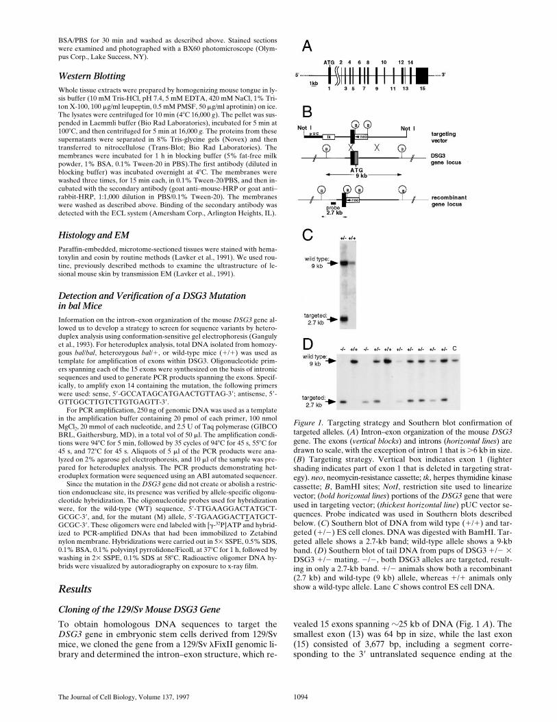
Mice Homozygous for the Targeted Mutation Do Not Synthesize Dsg3
To demonstrate that the gene targeting resulted in a func-tional null mutation, we studied the Dsg3 mRNA expres-
sion and protein synthesis in pups derived from inter-crosses between 1/2 animals. Dsg3 mRNA was absent in2/2 animals, whereas the mRNA was detected in 1/1and 1/2 animals, as determined by an RNase protectionassay. (Fig. 2 A). The levels of Dsg1 and Dsg2 mRNA ex-pression were not affected by the targeted mutation andwere similar in 1/1, 1/2, and 2/2 mice (data not shown).Immunofluorescence microscopy on tongue epithelium us-ing antibodies against extracellular or cytoplasmic epitopesof Dsg3 indicated the absence of the protein in 2/2 mu-tants (Fig. 2 B). Control antibodies against other desmo-gleins (e.g., mAb 3.10 that recognizes Dsg1 and Dsg2)(Fig. 2 B), as well as antibodies against the desmosomalplaque proteins plakoglobin (Fig. 2 B) or desmoplakin(not shown), showed no difference in the staining patternsin samples derived from 1/1, 1/2, and 2/2 mice. Fur-thermore, Western blot analysis of tongue extracts from2/2 mutants demonstrated the absence of the Dsg3 poly-peptide but the presence of Dsg1 and Dsg2, as indicatedby mAb 3.10 (Fig. 2 C).
These data indicate that the targeted mutation indeedrepresented a functional null mutation.
Figure 2. Lack of Dsg3 RNA and protein in homozygous tar-geted mice. (A) RNase protection assay of tongue lysates fromwild type (1/1) and targeted (2/2) mice. (B) Immunofluores-cence of tongue from wild-type and targeted mice shows absenceof Dsg3 from 2/2 mice, but presence of Dsg 1 and 2 (Dsg1/2,identified by mAb DG3.10), and plakoglobin. The submucosa of2/2 mice shows increased nonspecific fluorescence probably as aresult of inflammation. (C) Western blot of tongue lysates. Dsg3is absent in 2/2 mice (arrowhead), but Dsg1 and 2 are present(arrow) to the same degree as in 1/1 mice. Bar, 25 mm.
The Journal of Cell Biology, Volume 137, 1997 1096
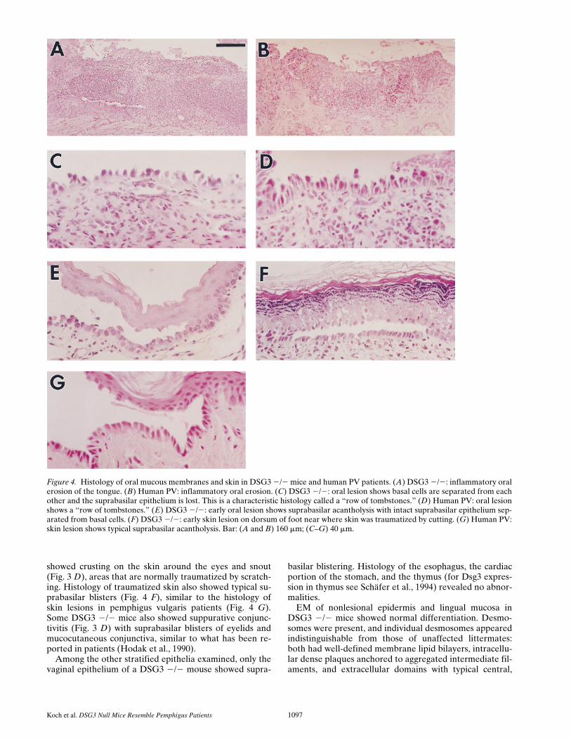
histological examination of the oral mucosa in DSG3 2/2mice showed a full spectrum of the types of lesions typicalof PV. The most common lesion was an inflammatory ero-sion, sometimes seen with reepithelialization (Fig. 4 A).This is typical of a late PV blister after the superficial epi-dermis is lost and the resulting irritation and/or coloniza-tion of the erosion results in acute inflammation and lossof the basal cells (Fig. 4 B). Further examination showedintermediate lesions with the superficial epidermis lost,leaving the basal cells still attached to the basement mem-brane, but slightly detached from each other (Fig. 4 C).This appearance has been called the “row of tombstones”in patients with PV (Fig. 4 D). Finally, we could also detectthe earliest lesion of PV in the DSG3 2/2 mice, namely asuprabasilar split in the epithelium, with minimal inflam-mation (Fig. 4 E). Oropharyngeal biopsies of essentiallyall DSG3 2/2 mice, examined at ages 3 d–5 mo, showedthese changes, but DSG3 1/2 and 1/1 mice nevershowed them. We speculate from this data that sucklingresulted in the trauma necessary to cause these lesions ini-tially, with the beginning of solid food at 16–20 d exacer-bating them. The resulting lesions presumably decreasedfood intake enough to result in the runting of these mice.
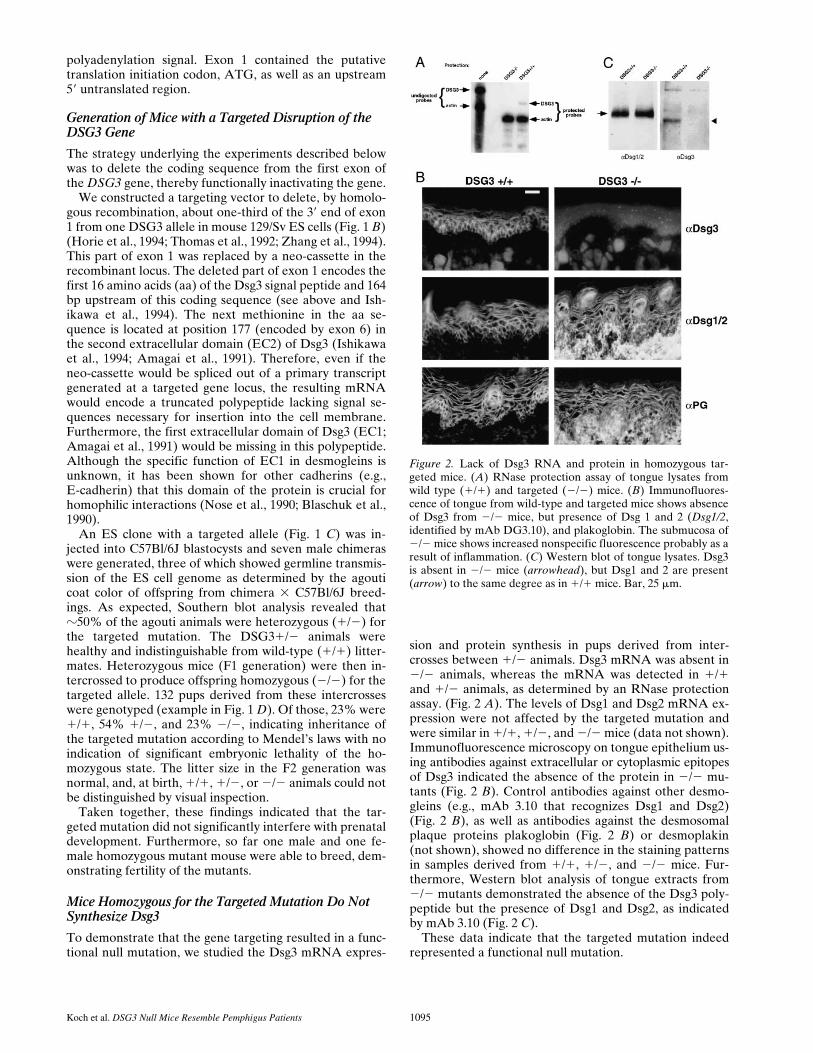
Interestingly, the epidermis of these mice did not showextensive spontaneous lesions. However, when a femaleDSG3 2/2 mouse delivered pups, their suckling causederosions around the nipples (Fig. 3 C) and some mice
Figure 3. DSG3 2/2 miceare runts and have skin ero-sions and eye lesions. (A)DSG3 2/2 mice are runts.Upper mouse is DSG3 2/2;lower mouse is a 1/1 litter-mate. (B) Weight graphshows that 2/2 mice (opencircles), compared with 1/1and 1/2 littermates (filledcircles), are born with equalweight but by about day 8–10are lagging in weight gain.Weight loss is seen about day20, approximately the time ofweaning and start of solidfood. (C) Nipple erosions ina DSG3 2/2 nursing mother.(D) Snout erosion and con-junctivitis in a DSG3 2/2mouse.
DSG3 2/2 Mice Show Loss of Keratinocyte Cell Adhesion Resulting in a Phenotype That Resembles That of Patients with PV
Around 15–20 d after birth, 2/2 animals differed in sizefrom 1/2 and 1/1 animals. The 2/2 mutants were muchsmaller than their littermates (Fig. 3 A). Autopsies re-vealed a dramatic reduction in body fat in the 2/2 animalsthat resembles what is seen in starvation.
Runting of the pups consistently sorted with the tar-geted mutation and was never observed in animals thatwere wild type or heterozygous for the mutant allele. Acloser analysis revealed that at birth all pups showed simi-lar weights, but, at 8–10 d after birth, 2/2 animals showedreduced bodyweight (Fig. 3 B). In the following days, themutants grew at a much slower rate than 1/2 and 1/1mice. Between days 18 and 25, the growth of 2/2 animalsslowed down even more with most mutants losing weightand a few dying (Fig. 3 B). However, .80% of mutantssurvived and again started to gain weight, but they werestill clearly smaller than their littermates. No significantweight difference between 1/2 and 1/1 mice was ob-served.
Since the most characteristic lesions in pemphigus vul-garis patients are painful oral erosions that may interferewith eating, we speculated that the DSG3 2/2 animalsmight have similar lesions preventing them from feedingsufficiently that would, in turn, result in runting. Indeed,
Koch et al. DSG3 Null Mice Resemble Pemphigus Patients 1097
showed crusting on the skin around the eyes and snout(Fig. 3 D), areas that are normally traumatized by scratch-ing. Histology of traumatized skin also showed typical su-prabasilar blisters (Fig. 4 F), similar to the histology ofskin lesions in pemphigus vulgaris patients (Fig. 4 G).Some DSG3 2/2 mice also showed suppurative conjunc-tivitis (Fig. 3 D) with suprabasilar blisters of eyelids andmucocutaneous conjunctiva, similar to what has been re-ported in patients (Hodak et al., 1990).
Among the other stratified epithelia examined, only thevaginal epithelium of a DSG3 2/2 mouse showed supra-
basilar blistering. Histology of the esophagus, the cardiacportion of the stomach, and the thymus (for Dsg3 expres-sion in thymus see Schäfer et al., 1994) revealed no abnor-malities.
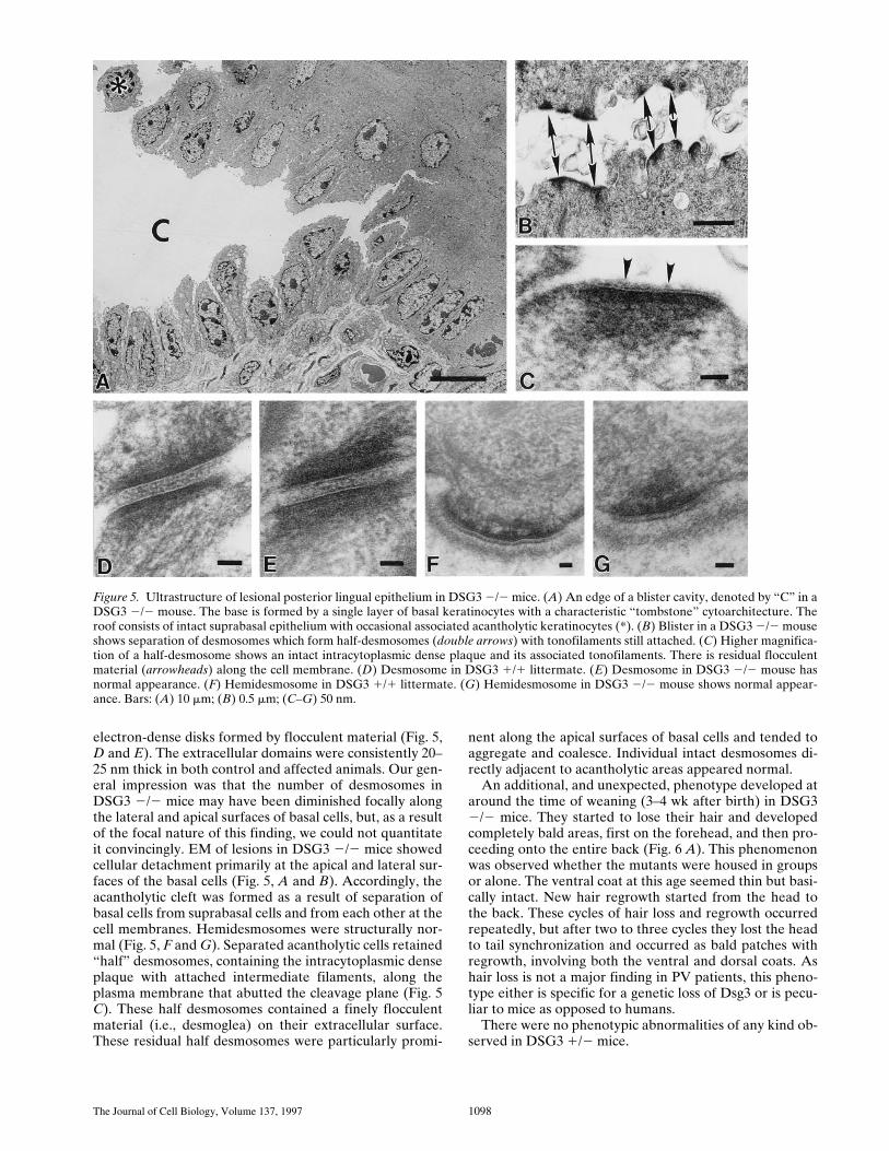
EM of nonlesional epidermis and lingual mucosa inDSG3 2/2 mice showed normal differentiation. Desmo-somes were present, and individual desmosomes appearedindistinguishable from those of unaffected littermates:both had well-defined membrane lipid bilayers, intracellu-lar dense plaques anchored to aggregated intermediate fil-aments, and extracellular domains with typical central,
Figure 4. Histology of oral mucous membranes and skin in DSG3 2/2 mice and human PV patients. (A) DSG3 2/2: inflammatory oralerosion of the tongue. (B) Human PV: inflammatory oral erosion. (C) DSG3 2/2: oral lesion shows basal cells are separated from eachother and the suprabasilar epithelium is lost. This is a characteristic histology called a “row of tombstones.” (D) Human PV: oral lesionshows a “row of tombstones.” (E) DSG3 2/2: early oral lesion shows suprabasilar acantholysis with intact suprabasilar epithelium sep-arated from basal cells. (F) DSG3 2/2: early skin lesion on dorsum of foot near where skin was traumatized by cutting. (G) Human PV:skin lesion shows typical suprabasilar acantholysis. Bar: (A and B) 160 mm; (C–G) 40 mm.
The Journal of Cell Biology, Volume 137, 1997 1098
electron-dense disks formed by flocculent material (Fig. 5,D and E). The extracellular domains were consistently 20–25 nm thick in both control and affected animals. Our gen-eral impression was that the number of desmosomes inDSG3 2/2 mice may have been diminished focally alongthe lateral and apical surfaces of basal cells, but, as a resultof the focal nature of this finding, we could not quantitateit convincingly. EM of lesions in DSG3 2/2 mice showedcellular detachment primarily at the apical and lateral sur-faces of the basal cells (Fig. 5, A and B). Accordingly, theacantholytic cleft was formed as a result of separation ofbasal cells from suprabasal cells and from each other at thecell membranes. Hemidesmosomes were structurally nor-mal (Fig. 5, F and G). Separated acantholytic cells retained“half” desmosomes, containing the intracytoplasmic denseplaque with attached intermediate filaments, along theplasma membrane that abutted the cleavage plane (Fig. 5C). These half desmosomes contained a finely flocculentmaterial (i.e., desmoglea) on their extracellular surface.These residual half desmosomes were particularly promi-
nent along the apical surfaces of basal cells and tended toaggregate and coalesce. Individual intact desmosomes di-rectly adjacent to acantholytic areas appeared normal.
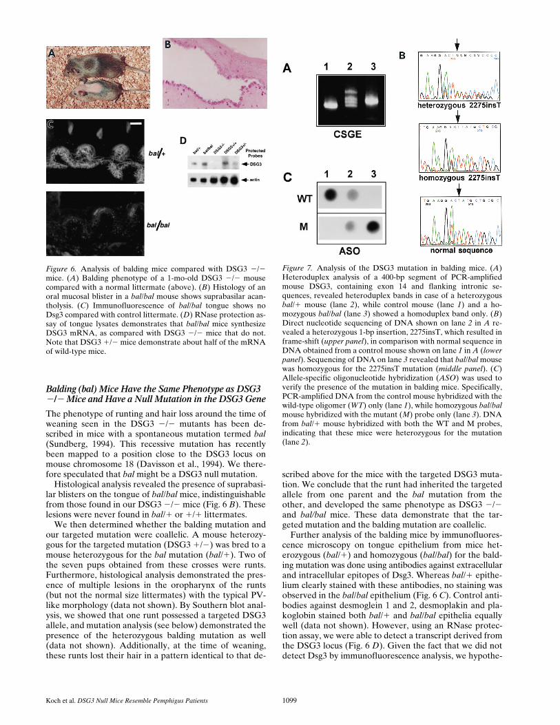
An additional, and unexpected, phenotype developed ataround the time of weaning (3–4 wk after birth) in DSG32/2 mice. They started to lose their hair and developedcompletely bald areas, first on the forehead, and then pro-ceeding onto the entire back (Fig. 6 A). This phenomenonwas observed whether the mutants were housed in groupsor alone. The ventral coat at this age seemed thin but basi-cally intact. New hair regrowth started from the head tothe back. These cycles of hair loss and regrowth occurredrepeatedly, but after two to three cycles they lost the headto tail synchronization and occurred as bald patches withregrowth, involving both the ventral and dorsal coats. Ashair loss is not a major finding in PV patients, this pheno-type either is specific for a genetic loss of Dsg3 or is pecu-liar to mice as opposed to humans.
There were no phenotypic abnormalities of any kind ob-served in DSG3 1/2 mice.
Figure 5. Ultrastructure of lesional posterior lingual epithelium in DSG3 2/2 mice. (A) An edge of a blister cavity, denoted by “C” in aDSG3 2/2 mouse. The base is formed by a single layer of basal keratinocytes with a characteristic “tombstone” cytoarchitecture. Theroof consists of intact suprabasal epithelium with occasional associated acantholytic keratinocytes (*). (B) Blister in a DSG3 2/2 mouseshows separation of desmosomes which form half-desmosomes (double arrows) with tonofilaments still attached. (C) Higher magnifica-tion of a half-desmosome shows an intact intracytoplasmic dense plaque and its associated tonofilaments. There is residual flocculentmaterial (arrowheads) along the cell membrane. (D) Desmosome in DSG3 1/1 littermate. (E) Desmosome in DSG3 2/2 mouse hasnormal appearance. (F) Hemidesmosome in DSG3 1/1 littermate. (G) Hemidesmosome in DSG3 2/2 mouse shows normal appear-ance. Bars: (A) 10 mm; (B) 0.5 mm; (C–G) 50 nm.
Koch et al. DSG3 Null Mice Resemble Pemphigus Patients 1099
Balding (bal) Mice Have the Same Phenotype as DSG3 2/2 Mice and Have a Null Mutation in the DSG3 Gene
The phenotype of runting and hair loss around the time ofweaning seen in the DSG3 2/2 mutants has been de-scribed in mice with a spontaneous mutation termed bal(Sundberg, 1994). This recessive mutation has recentlybeen mapped to a position close to the DSG3 locus onmouse chromosome 18 (Davisson et al., 1994). We there-fore speculated that bal might be a DSG3 null mutation.
Histological analysis revealed the presence of suprabasi-lar blisters on the tongue of bal/bal mice, indistinguishablefrom those found in our DSG3 2/2 mice (Fig. 6 B). Theselesions were never found in bal/1 or 1/1 littermates.
We then determined whether the balding mutation andour targeted mutation were coallelic. A mouse heterozy-gous for the targeted mutation (DSG3 1/2) was bred to amouse heterozygous for the bal mutation (bal/1). Two ofthe seven pups obtained from these crosses were runts.Furthermore, histological analysis demonstrated the pres-ence of multiple lesions in the oropharynx of the runts(but not the normal size littermates) with the typical PV-like morphology (data not shown). By Southern blot anal-ysis, we showed that one runt possessed a targeted DSG3allele, and mutation analysis (see below) demonstrated thepresence of the heterozygous balding mutation as well(data not shown). Additionally, at the time of weaning,these runts lost their hair in a pattern identical to that de-
scribed above for the mice with the targeted DSG3 muta-tion. We conclude that the runt had inherited the targetedallele from one parent and the bal mutation from theother, and developed the same phenotype as DSG3 2/2and bal/bal mice. These data demonstrate that the tar-geted mutation and the balding mutation are coallelic.
Further analysis of the balding mice by immunofluores-cence microscopy on tongue epithelium from mice het-erozygous (bal/1) and homozygous (bal/bal) for the bald-ing mutation was done using antibodies against extracellularand intracellular epitopes of Dsg3. Whereas bal/1 epithe-lium clearly stained with these antibodies, no staining wasobserved in the bal/bal epithelium (Fig. 6 C). Control anti-bodies against desmoglein 1 and 2, desmoplakin and pla-koglobin stained both bal/1 and bal/bal epithelia equallywell (data not shown). However, using an RNase protec-tion assay, we were able to detect a transcript derived fromthe DSG3 locus (Fig. 6 D). Given the fact that we did notdetect Dsg3 by immunofluorescence analysis, we hypothe-
Figure 6. Analysis of balding mice compared with DSG3 2/2mice. (A) Balding phenotype of a 1-mo-old DSG3 2/2 mousecompared with a normal littermate (above). (B) Histology of anoral mucosal blister in a bal/bal mouse shows suprabasilar acan-tholysis. (C) Immunofluorescence of bal/bal tongue shows noDsg3 compared with control littermate. (D) RNase protection as-say of tongue lysates demonstrates that bal/bal mice synthesizeDSG3 mRNA, as compared with DSG3 2/2 mice that do not.Note that DSG3 1/2 mice demonstrate about half of the mRNAof wild-type mice.
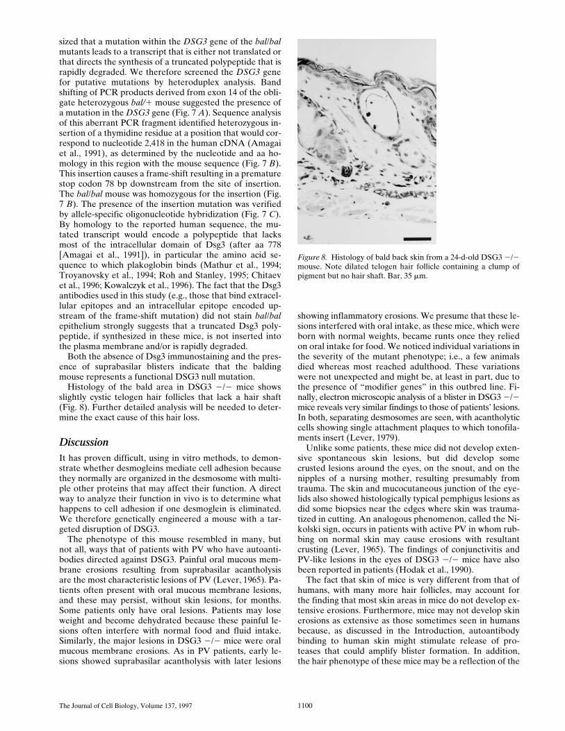
Figure 7. Analysis of the DSG3 mutation in balding mice. (A)Heteroduplex analysis of a 400-bp segment of PCR-amplifiedmouse DSG3, containing exon 14 and flanking intronic se-quences, revealed heteroduplex bands in case of a heterozygousbal/1 mouse (lane 2), while control mouse (lane 1) and a ho-mozygous bal/bal (lane 3) showed a homoduplex band only. (B)Direct nucleotide sequencing of DNA shown on lane 2 in A re-vealed a heterozygous 1-bp insertion, 2275insT, which resulted inframe-shift (upper panel), in comparison with normal sequence inDNA obtained from a control mouse shown on lane 1 in A (lowerpanel). Sequencing of DNA on lane 3 revealed that bal/bal mousewas homozygous for the 2275insT mutation (middle panel). (C)Allele-specific oligonucleotide hybridization (ASO) was used toverify the presence of the mutation in balding mice. Specifically,PCR-amplified DNA from the control mouse hybridized with thewild-type oligomer (WT) only (lane 1), while homozygous bal/balmouse hybridized with the mutant (M) probe only (lane 3). DNAfrom bal/1 mouse hybridized with both the WT and M probes,indicating that these mice were heterozygous for the mutation(lane 2).
The Journal of Cell Biology, Volume 137, 1997 1100
sized that a mutation within the DSG3 gene of the bal/balmutants leads to a transcript that is either not translated orthat directs the synthesis of a truncated polypeptide that israpidly degraded. We therefore screened the DSG3 genefor putative mutations by heteroduplex analysis. Bandshifting of PCR products derived from exon 14 of the obli-gate heterozygous bal/1 mouse suggested the presence ofa mutation in the DSG3 gene (Fig. 7 A). Sequence analysisof this aberrant PCR fragment identified heterozygous in-sertion of a thymidine residue at a position that would cor-respond to nucleotide 2,418 in the human cDNA (Amagaiet al., 1991), as determined by the nucleotide and aa ho-mology in this region with the mouse sequence (Fig. 7 B).This insertion causes a frame-shift resulting in a prematurestop codon 78 bp downstream from the site of insertion.The bal/bal mouse was homozygous for the insertion (Fig.7 B). The presence of the insertion mutation was verifiedby allele-specific oligonucleotide hybridization (Fig. 7 C).By homology to the reported human sequence, the mu-tated transcript would encode a polypeptide that lacksmost of the intracellular domain of Dsg3 (after aa 778[Amagai et al., 1991]), in particular the amino acid se-quence to which plakoglobin binds (Mathur et al., 1994;Troyanovsky et al., 1994; Roh and Stanley, 1995; Chitaevet al., 1996; Kowalczyk et al., 1996). The fact that the Dsg3antibodies used in this study (e.g., those that bind extracel-lular epitopes and an intracellular epitope encoded up-stream of the frame-shift mutation) did not stain bal/balepithelium strongly suggests that a truncated Dsg3 poly-peptide, if synthesized in these mice, is not inserted intothe plasma membrane and/or is rapidly degraded.
Both the absence of Dsg3 immunostaining and the pres-ence of suprabasilar blisters indicate that the baldingmouse represents a functional DSG3 null mutation.
Histology of the bald area in DSG3 2/2 mice showsslightly cystic telogen hair follicles that lack a hair shaft(Fig. 8). Further detailed analysis will be needed to deter-mine the exact cause of this hair loss.
DiscussionIt has proven difficult, using in vitro methods, to demon-strate whether desmogleins mediate cell adhesion becausethey normally are organized in the desmosome with multi-ple other proteins that may affect their function. A directway to analyze their function in vivo is to determine whathappens to cell adhesion if one desmoglein is eliminated.We therefore genetically engineered a mouse with a tar-geted disruption of DSG3.
The phenotype of this mouse resembled in many, butnot all, ways that of patients with PV who have autoanti-bodies directed against DSG3. Painful oral mucous mem-brane erosions resulting from suprabasilar acantholysisare the most characteristic lesions of PV (Lever, 1965). Pa-tients often present with oral mucous membrane lesions,and these may persist, without skin lesions, for months.Some patients only have oral lesions. Patients may loseweight and become dehydrated because these painful le-sions often interfere with normal food and fluid intake.Similarly, the major lesions in DSG3 2/2 mice were oralmucous membrane erosions. As in PV patients, early le-sions showed suprabasilar acantholysis with later lesions
showing inflammatory erosions. We presume that these le-sions interfered with oral intake, as these mice, which wereborn with normal weights, became runts once they reliedon oral intake for food. We noticed individual variations inthe severity of the mutant phenotype; i.e., a few animalsdied whereas most reached adulthood. These variationswere not unexpected and might be, at least in part, due tothe presence of “modifier genes” in this outbred line. Fi-nally, electron microscopic analysis of a blister in DSG3 2/2mice reveals very similar findings to those of patients’ lesions.In both, separating desmosomes are seen, with acantholyticcells showing single attachment plaques to which tonofila-ments insert (Lever, 1979).
Unlike some patients, these mice did not develop exten-sive spontaneous skin lesions, but did develop somecrusted lesions around the eyes, on the snout, and on thenipples of a nursing mother, resulting presumably fromtrauma. The skin and mucocutaneous junction of the eye-lids also showed histologically typical pemphigus lesions asdid some biopsies near the edges where skin was trauma-tized in cutting. An analogous phenomenon, called the Ni-kolski sign, occurs in patients with active PV in whom rub-bing on normal skin may cause erosions with resultantcrusting (Lever, 1965). The findings of conjunctivitis andPV-like lesions in the eyes of DSG3 2/2 mice have alsobeen reported in patients (Hodak et al., 1990).
The fact that skin of mice is very different from that ofhumans, with many more hair follicles, may account forthe finding that most skin areas in mice do not develop ex-tensive erosions. Furthermore, mice may not develop skinerosions as extensive as those sometimes seen in humansbecause, as discussed in the Introduction, autoantibodybinding to human skin might stimulate release of pro-teases that could amplify blister formation. In addition,the hair phenotype of these mice may be a reflection of the
Figure 8. Histology of bald back skin from a 24-d-old DSG3 2/2mouse. Note dilated telogen hair follicle containing a clump ofpigment but no hair shaft. Bar, 35 mm.
Koch et al. DSG3 Null Mice Resemble Pemphigus Patients 1101
difference in hair in rodents and humans, as humans withPV usually do not develop a balding phenotype. Prelimi-nary analysis of the hair phenotype in these mice shows anormal first hair cycle but loss of hair after this cycle. Inbald areas, histology of the skin reveals dilated telogen fol-licles lacking a hair shaft. Previous analysis of balding micehas shown necrosis immediately above the hair matrixleading to some scattered generalized follicular necrosis(Sundberg, 1994). Additional analysis will be required tobetter define the pathophysiology of hair loss in theseDSG3 2/2 and balding mice.
Moreover, considering the major difference in mouseand human stratified squamous epithelia and adnexalstructures, it is remarkable how similar the phenotype ofDSG3 2/2 mice is to PV patients.
The phenotype of Dsg3 2/2 mice is quite different fromthat of a recently described transgenic mouse that ex-presses an amino-terminal deleted Dsg3 under the controlof the K14 promoter (Allen et al., 1996). The transgeneused consisted of the cytoplasmic and transmembrane do-main as well as part of the extracellular domain of Dsg3and was expressed mainly in the lower layers of the epi-dermis. The transgenic mice, thought to express a domi-nant negative effect of the truncated Dsg3, showed swell-ing of paws and digits, focal flakiness of the skin, andnecrotic changes on the tips of the tails that ultimately re-sulted in tail degeneration. Furthermore, histological anal-ysis revealed epidermal thickening and widening of the in-tercellular space between keratinocytes but did not showloss of cell–cell adhesion (i.e., acantholysis). At the ultra-structural level, a reduction in the number of desmosomesand the occurrence of aberrant desmosomes were re-ported. Although these mice were noted to have a wet andmatted hair coat (probably because of excess grooming),they did not show an obvious loss of hair. Our DSG3 2/2mice clearly had a different phenotype and distinct histo-logic and ultrastructural abnormalities. DSG3 2/2 micedid not show any tail abnormality and did not have flakyskin, but they developed crusted erosions in areas oftrauma. They also had a striking balding phenotype. Histo-logically these mice showed obvious acantholysis. Finally, ul-trastructurally, the DSG3 2/2 mice showed normally ap-pearing desmosomes in intact skin and “half”-desmosomeswhere cells were acantholytic. We conclude from these dif-ferences between the transgenic and DSG3 2/2 mice thatthe truncated Dsg3 did not act in a dominant negativefashion to totally inactivate DSG3 function.
The phenotype of DSG3 knockout mice underscores theimportance of Dsg3 for cell adhesion and mechanical sta-bility in the deepest layers of stratified epithelia. The othertransmembrane adhesion molecules present, such as Dsg2,desmocollins, E-cadherin, and P-cadherin, apparently can-not compensate for the loss of Dsg3. In addition, Dsg3seems to be particularly important for adhesion in the oralmucous membrane, where lesions always occurred inDSG3 2/2 mice, and less so in other stratified squamousepithelia such as esophagus where lesions were never seen.The importance of Dsg3 in skin seems intermediate be-cause lesions were mainly seen secondary to trauma.These studies, then, show that specific desmogleins mayhave tissue- and differentiation-specific adhesion functions.
Finally, the phenotype of the Dsg3 knockout mice not
only demonstrates the importance of Dsg3 for cell adhe-sion in the deep stratified squamous epithelia, but also isconsistent with the idea that, at least in part, PV autoanti-bodies cause loss of cell adhesion by directly interferingwith the adhesive function of Dsg3.
We thank Dr. Chin Howe for valuable discussions and advice regardingknockout technology; Dr. Victor Tybulewicz for providing the targetingvector; Daniela Simon who did karyotyping; Dr. Jean Richa, from theUniversity of Pennsylvania Transgenic Facility, who performed the EScell injections; Qi Tian, Drs. Stephan Schäfer, and Werner Franke for pro-viding Dsg2 cDNA and antibodies; Dr. Margaret Wheelock for plakoglo-bin antibodies; Dr. Kehua Li for assistance in cloning the mouse DSG3gene; and Dr. Sarolta Karpati for helping with anti-Dsg3 antibody prepa-ration.
This work was supported by National Institutes of Health grants1RO1AR43776, PO1AR38923, and CA20408. P. Koch was supported byThe Thyssen Foundation and a research fellowship from the DermatologyFoundation.
Received for publication 3 February 1997 and in revised form 20 March1997.
References
Akiyama, M., T. Hashimoto, M. Sugiura, and T. Nishikawa. 1991. Ultrastruc-tural localization of pemphigus vulgaris and pemphigus foliaceus antigens incultured human squamous carcinoma cells. Br. J. Dermatol. 125:233–237.
Allen, E., Q.C. Yu, and E. Fuchs. 1996. Mice expressing a mutant desmosomalcadherin exhibit abnormalities in desmosomes, proliferation, and epidermaldifferentiation. J. Cell Biol. 133:1367–1382.
Amagai, M., V. Klaus-Kovtun, and J.R. Stanley. 1991. Autoantibodies against anovel epithelial cadherin in pemphigus vulgaris, a disease of cell adhesion.Cell. 67:869–877.
Amagai, M., S. Karpati, R. Prussick, V. Klaus-Kovtun, and J.R. Stanley. 1992.Autoantibodies against the amino-terminal cadherin-like binding domain ofpemphigus vulgaris antigen are pathogenic. J. Clin. Invest. 90:919–926.
Amagai, M., T. Hashimoto, N. Shimizu, and T. Nishikawa. 1994a. Absorptionof pathogenic autoantibodies by the extracellular domain of pemphigus vul-garis antigen (Dsg3) produced by baculovirus. J. Clin. Invest. 94:59–67.
Amagai, M., S. Karpati, V. Klaus-Kovtun, M.C. Udey, and J.R. Stanley. 1994b.The extracellular domain of pemphigus vulgaris antigen (desmoglein 3) me-diates weak homophilic adhesion. J. Invest. Dermatol. 102:402–408.
Amagai, M., P.J. Koch, T. Nishikawa, and J.R. Stanley. 1996. Pemphigus vul-garis antigen (Desmoglein 3) is localized in the lower epidermis, the site ofblister formation in patients. J. Invest. Dermatol. 106:351–355.
Arnemann, J., K.H. Sullivan, A.I. Magee, I.A. King, and R.S. Buxton. 1993.Stratification-related expression of isoforms of the desmosomal cadherins inhuman epidermis. J. Cell Sci. 104:741–750.
Blaschuk, O.W., R. Sullivan, S. David, and Y. Pouliot. 1990. Identification of acadherin cell adhesion recognition sequence. Dev. Biol. 139:227–229.
Buxton, R.S., P. Cowin, W.W. Franke, D.R. Garrod, K.J. Green, I.A. King, P.J.Koch, A.I. Magee, D.A. Rees, J.R. Stanley, and M.S. Steinberg. 1993. No-menclature of the desmosomal cadherins. J. Cell Biol. 121:481–483.
Chidgey, M.A., J.P. Clarke, and D.R. Garrod. 1996. Expression of full-lengthdesmosomol glycoproteins (desmocollins) is not sufficient to confer strongadhesion on transfected L929 cells. J. Invest. Dermatol. 106:689–695.
Chitaev, N.A., R.E. Leube, R.B. Troyanovsky, L.G. Eshkind, W.W. Franke,and S.M. Troyanovsky. 1996. The binding of plakoglobin to desmosomalcadherins: patterns of binding sites and topogenic potential. J. Cell Biol. 133:359–369.
Collins, J.E., P.K. Legan, T.P. Kenny, J. MacGarvie, J.L. Holton, and D.R. Gar-rod. 1991. Cloning and sequence analysis of desmosomal glycoproteins 2 and3 (desmocollins): cadherin-like desmosomal adhesion molecules with hetero-geneous cytoplasmic domains. J. Cell. Biol. 113:381–391.
Davisson, M.T., S.A. Cook, K.R. Johnson, and E.M. Eicher. 1994. Balding: anew mutation on mouse chromosome 18 causing hair loss and immunologi-cal defects. J. Hered. 85:134–136.
Farb, R.M., R. Dykes, and G.S. Lazarus. 1978. Anti-epidermal-cell-surfacepemphigus antibody detaches viable epidermal cells from culture plates byactivation of proteinase. Proc. Natl. Acad. Sci. USA. 75:459–463.
Ganguly, A., M.J. Rock, and D.J. Prockop. 1993. Conformation-sensitive gelelectrophoresis for rapid detection of single base differences in double-stranded PCR products and DNA fragments: evidence for solvent-inducedbends in DNA heteroduplexes. Proc. Natl. Acad. Sci. USA. 90:10325–10329.
Goodwin, L., J.E. Hill, K. Raynor, L. Raszi, M. Manabe, and P. Cowin. 1990.Desmoglein shows extensive homology to the cadherin family of cell adhe-sion molecules. Biochem. Biophys. Res. Commun. 173:1224–1230.
Hashimoto, K., K.M. Shafran, P.S. Webber, G.S. Lazarus, and K.H. Singer.
The Journal of Cell Biology, Volume 137, 1997 1102
1983. Anti-cell surface pemphigus autoantibody stimulates plasminogen ac-tivator activity of human epidermal cells. J. Exp. Med. 157:259–272.
Hashimoto, K., T.C. Wun, J. Baird, G.S. Lazarus, and P.J. Jensen. 1989. Char-acterization of keratinocyte plasminogen activator inhibitors and demon-stration of the prevention of pemphigus IgG-induced acantholysis by a puri-fied plasminogen activator inhibitor. J. Invest. Dermatol. 92:310–314.
Hodak, E., I. Kremer, M. David, B. Hazaz, A. Rothem, P. Feuerman, and M.Sandbank. 1990. Conjunctival involvement in pemphigus vulgaris: a clinical,histopathological and immunofluorescence study. Br. J. Dermatol. 123:615–620.
Hogan, B., R. Beddington, F. Costantini, and E. Lacy. 1994. Manipulating theMouse Embryo. Cold Spring Harbor Laboratory, Cold Spring Harbor, NY.497 pp.
Horie, K., S. Nishiguchi, S. Maeda, and K. Shimada. 1994. Structures of replace-ment vectors for efficient gene targeting. J. Biochem. (Tokyo). 115:477–485.
Ishikawa, H., S.A. Silos, K. Tamai, N.G. Copeland, D.J. Gilbert, N.A. Jenkins,and J. Uitto. 1994. cDNA cloning and chromosomal assignment of themouse gene for desmoglein 3 (Dsg3), the pemphigus vulgaris antigen.Mamm. Genome. 5:803–804.
Karpati, S., M. Amagai, R. Prussick, K. Cehrs, and J.R. Stanley. 1993. Pemphi-gus vulgaris antigen, a desmoglein type of cadherin, is localized within kera-tinocyte desmosomes. J. Cell Biol. 122:409–415.
King, I.A., J. Arnemann, N.K. Spurr, and R.S. Buxton. 1993. Cloning of thecDNA (DSC1) coding for human type 1 desmocollin and its assignment tochromosome 18. Genomics. 18:185–194.
Koch, P.J., and W.W. Franke. 1994. Desmosomal cadherins: another growingmultigene family of adhesion molecules. Curr. Opin. Cell Biol. 6:682–687.
Koch, P.J., M.J. Walsh, M. Schmelz, M.D. Goldschmidt, R. Zimbelmann, andW.W. Franke. 1990. Identification of desmoglein, a constitutive desmosomalglycoprotein, as a member of the cadherin family of cell adhesion molecules.Eur. J. Cell Biol. 53:1–12.
Koch, P.J., M.D. Goldschmidt, M.J. Walsh, R. Zimbelmann, and W.W. Franke.1991a. Complete amino acid sequence of the epidermal desmoglein precur-sor polypeptide and identification of a second type of desmoglein gene. Eur.J. Cell Biol. 55:200–208.
Koch, P.J., M.D. Goldschmidt, M.J. Walsh, R. Zimbelmann, M. Schmelz, andW.W. Franke. 1991b. Amino acid sequence of bovine muzzle epithelial des-mocollin derived from cloned cDNA: a novel subtype of desmosomal cad-herins. Differentiation. 47:29–36.
Koch, P.J., M.D. Goldschmidt, R. Zimbelmann, R. Troyanovsky, and W.W.Franke. 1992. Complexity and expression patterns of the desmosomal cad-herins. Proc. Natl. Acad. Sci. USA. 89:353–357.
Kowalczyk, A.P., J.E. Borgwardt, and K.J. Green. 1996. Analysis of desmo-somal cadherin-adhesive function and stoichiometry of desmosomal cad-herin-plakoglobin complexes. J. Invest. Dermatol. 107:293–300.
Lavker, R.M., G. Dong, P. Zheng, and G.F. Murphy. 1991. Hairless micropigskin. A novel model for studies of cutaneous biology. Am. J. Pathol. 138:687–697.
Lever, W.F. 1965. Pemphigus and Pemphigoid. Charles C. Thomas, Springfield,IL. 266 pp.
Lever, W.F. 1979. Pemphigus and pemphigoid. J. Am. Acad. Dermatol. 1:2–31.Mansour, S.L., K.R. Thomas, and M.R. Capecchi. 1988. Disruption of the
proto-oncogene int-2 in mouse embryo-derived stem cells: a general strategyfor targeting mutations to non-selectable genes. Nature (Lond.). 336:348–352.
Mathur, M., L. Goodwin, and P. Cowin. 1994. Interactions of the cytoplasmicdomain of the desmosomal cadherin dsg1 with plakoglobin. J. Biol. Chem.269:14075–14080.
Mechanic, S., K. Raynor, J.E. Hill, and P. Cowin. 1991. Desmocollins form adistinct subset of the cadherin family of cell adhesion molecules. Proc. Natl.Acad. Sci. USA. 88:4476–4480.
Memar, O.M., S. Rajaraman, R. Thotakura, S.K. Tyring, J.L. Fan, G.S. Seethar-amaiah, A. Lopez, R.E. Jordon, and B.S. Prabhakar. 1996. Recombinantdesmoglein 3 has the necessary epitopes to adsorb and induce blister-causingantibodies. J. Invest. Dermatol. 106:261–268.
Morioka, S., G.S. Lazarus, and P.J. Jensen. 1987. Involvement of urokinase-type plasminogen activator in acantholysis induced by pemphigus IgG. J. In-vest. Dermatol. 89:474–477.
Nagafuchi, A., Y. Shirayoshi, K. Okazaki, K. Yasuda, and M. Takeichi. 1987.Transformation of cell adhesion properties by exogenously introducedE-cadherin cDNA. Nature (Lond.). 329:341–343.
Naito, K., S. Morioka, S. Nakajima, and H. Ogawa. 1989. Proteinase inhibitorsblock formation of pemphigus acantholysis in experimental models of neo-natal mice and skin explants: effects of synthetic and plasma proteinase in-hibitors on pemphigus acantholysis. J. Invest. Dermatol. 93:173–177.
Nilles, L.A., D.A.D. Parry, E.E. Powers, B.D. Angst, R.M. Wagner, and K.J.Green. 1991. Structural analysis and expression of human desmoglein: a cad-herin-like component of the desmosome. J. Cell Sci. 99:809–821.
North, A.J., M.A. Chidgey, J.P. Clarke, W.G. Bardsley, and D.R. Garrod. 1996.Distinct desmocollin isoforms occur in the same desmosomes and show re-ciprocally graded distributions in bovine nasal epidermis. Proc. Natl. Acad.Sci. USA. 93:7701–7705.
Nose, A., K. Tsuji, and M. Takeichi. 1990. Localization of specificity determin-ing sites in cadherin cell adhesion molecules. Cell. 61:147–155.
Nuber, U.A., S. Schäfer, A. Schmidt, P.J. Koch, and W.W. Franke. 1995. Thewidespread human desmocollin Dsc2 and tissue-specific patterns of synthe-sis of various desmocollin subtypes. Eur. J. Cell Biol. 66:69–74.
Nuber, U.A., S. Schäfer, S. Stehr, H.R. Rackwitz, and W.W. Franke. 1996. Pat-terns of desmocollin synthesis in human epithelia: immunolocalization ofdesmocollins 1 and 3 in special epithelia and in cultured cells. Eur. J. CellBiol. 71:1–13.
Parker, A.E., G.N. Wheeler, J. Arnemann, S.C. Pidsley, P. Ataliotis, C.L. Tho-mas, D.A. Rees, A.I. Magee, and R.S. Buxton. 1991. Desmosomal glycopro-teins II and III. Cadherin-like junctional molecules generated by alternativesplicing. J. Biol. Chem. 266:10438–10445.
Ramirez-Solis, R., J. Rivera-Perez, J.D. Wallace, M. Wims, H. Zheng, and A.Bradley. 1992. Genomic DNA microextraction: a method to screen numer-ous samples. Anal. Biochem. 201:331–335.
Ramirez-Solis, R., A.C. Davis, and A. Bradley. 1993. Gene targeting in embry-onic stem cells. Methods Enzymol. 225:855–879.
Roh, J.Y., and J.R. Stanley. 1995. Plakoglobin binding by human Dsg3 (pem-phigus vulgaris antigen) in keratinocytes requires the cadherin-like intracy-toplasmic segment. J. Invest. Dermatol. 104:720–724.
Sambrook, J., E.F. Fritsch, and T. Maniatis. 1989. Molecular Cloning: A Labo-ratory Manual. Cold Spring Harbor Laboratory, Cold Spring Harbor, NY.545 pp.
Schäfer, S., P.J. Koch, and W.W. Franke. 1994. Identification of the ubiquitoushuman desmoglein, Dsg2, and the expression catalogue of a subfamily ofdesmosomal cadherins. Exp. Cell Res. 211:391–399.
Schäfer, S., S. Stumpp, and W.W. Franke. 1996. Immunological identificationand characterization of the desmosomal cadherin Dsg2 in coupled and un-coupled epithelial cells and in human tissues. Differentiation. 60:99–108.
Schiltz, J.R., B. Michel, and R. Papay. 1978. Pemphigus antibody interactionwith human epidermal cells in culture. J. Clin. Invest. 62:778–788.
Schiltz, J.R., B. Michel, and R. Papay. 1979. Appearance of “pemphigus acan-tholysis factor” in human skin cultured with pemphigus antibody. J. Invest.Dermatol. 73:575–581.
Schmidt, A., H.W. Heid, S. Schäfer, U.A. Nuber, R. Zimbelmann, and W.W.Franke. 1994. Desmosomes and cytoskeletal architecture in epithelial differ-entiation: cell type-specific plaque components and intermediate filamentanchorage. Eur. J. Cell Biol. 65:229–245.
Schwarz, M.A., K. Owaribe, J. Kartenbeck, and W.W. Franke. 1990. Desmo-somes and hemidesmosomes: constitutive molecular components. Annu.Rev. Cell Biol. 6:461–491.
Shimizu, H., T. Masunaga, A. Ishiko, A. Kikuchi, T. Hashimoto, and T. Nish-ikawa. 1995. Pemphigus vulgaris and pemphigus foliaceus sera show an in-versely graded binding pattern to extracellular regions of desmosomes in dif-ferent layers of human epidermis. J. Invest. Dermatol. 105:153–159.
Stanley, J.R. 1990. Pemphigus: skin failure mediated by autoantibodies. JAMA(J. Am. Med. Assoc.). 264:1714–1717.
Stanley, J.R. 1993a. Pemphigus. In Dermatology in General Medicine. T.B.Fitzpatrick, A.Z. Eisen, K. Wolff, I.M. Freedberg, and K.F. Austen, editor.McGraw-Hill, New York. 606–615.
Stanley, J.R. 1993b. Cell adhesion molecules as targets of autoantibodies inpemphigus and pemphigoid, bullous diseases due to defective epidermal celladhesion. Adv. Immunol. 53:291–325.
Sundberg, J.P. 1994. The balding (bal) mutation, chromosome 18. In Handbookof Mouse Mutations with Skin and Hair Abnormalities: Animal Models andBiomedical Tools. J.P. Sundberg, editor. CRC Press, Boca Raton. 187–191.
Tanaka, T., N.J. Korman, H. Shimizu, R.A.J. Eady, V. Klaus-Kovtun, K. Cehrs,and J.R. Stanley. 1990. Production of rabbit antibodies against carboxy-ter-minal epitopes encoded by bullous pemphigoid cDNA. J. Invest. Dermatol.94:617–623.
Theis, D.G., P.J. Koch, and W.W. Franke. 1993. Differential synthesis of type 1and type 2 desmocollin mRNAs in human stratified epithelia. Int. J. Dev.Biol. 37:101–110.
Thomas, K.R., C. Deng, and M.R. Capecchi. 1992. High-fidelity gene targetingin embryonic stem cells by using squence replacement vectors. Mol. Cell.Biol. 12:2919–2923.
Troyanovsky, S.M., L.G. Eshkind, R.B. Troyanovsky, R.E. Leube, and W.W.Franke. 1993. Contributions of cytoplasmic domains of desmosomal cad-herins to desmosome assembly and intermediate filament anchorage. Cell.72:561–574.
Troyanovsky, S.M., R.B. Troyanovsky, L.G. Eshkind, V.A. Krutovskikh, R.E.Leube, and W.W. Franke. 1994. Identification of the plakoglobin-bindingdomain in desmoglein and its role in plaque assembly and intermediate fila-ment anchorage. J. Cell Biol. 127:151–160.
Tybulewicz, V.L.J., C.E. Crawford, P.K. Jackson, R.T. Bronson, and R.C. Mul-ligan. 1991. Neonatal lethality and lymphopenia in mice with a homozygousdisruption of the c-abl proto-oncogene. Cell. 65:1153–1163.
Wheeler, G.N., A.E. Parker, C.L. Thomas, P. Ataliotis, D. Poynter, J. Arne-mann, A.J. Rutman, S.C. Pidsley, F.M. Watt, D.A. Rees et al. 1991. Desmo-somal glycoprotein DGI, a component of intercellular desmosome junctions,is related to the cadherin family of cell adhesion molecules. Proc. Natl. Acad.Sci. USA. 88:4796–4800.
Zhang, H., P. Hasty, and A. Bradley. 1994. Targeting frequency for deletionvectors in embryonic stem cells. Mol. Cell. Biol. 14:2404–2410.