Embed Size (px)
Citation preview
PEDIATRIC DENTISTRY/Copyright © 1991 byThe American Academy of Pediatric Dentistry
Volume 13, Number 2
Tissue-space emphysema, tissue necrosis, and infection followinguse of compressed air during pulp therapy: case reportKelly J. Wright, BSc, DMD Gary D. Derkson, DMD
Keith H. Riding, MD, FRCS(C)
AbstractThe in traoperative development of tissue-space emphysema
in a child undergoing restorative treatment under generalanesthesia is presented. Emphysema development seems to beconcomitant with the use of compressed air around patent rootcanals, complicated by tissue destruction due to movement ofcanal irrigants/medicaments into the periapical tissues andby secondary infection. Several recommendations for theprevention of tissue-space emphysema are presented includ-ing the use of a rubber dam, judicious use of compressed air,and maintenance of canal irrigants and medicaments withinthe root canal. Treatment recommendations vary from pal-liative care with follow up in cases of facial emphysema toimmediate medical attention in cases of pharyngeal or medi-astinal emphysema.
IntroductionTissue-space emphysema is defined as the passage
and collection of gas between the tissue space or fascialplanes (McGrannahan 1965). Tissue-space emphysemafollowing dental procedures is apparently a relativelyuncommon occurrence although numerous reports canbe found in the literature. Tissue space and mediastinalemphysema have been reported following surgical ex-tractions (LeRoy and Bregman 1968; Cardo et al. 1972),endodontic therapy (Lloyd 1975; Hirschmann andWalker 1983; Falomo 1984), periodontal therapy(McClendon and Hooper 1961; Feinstone 1971) andrestorative procedures (Duncan and Ferrillo 1967; Fisher1976; Quisling et al. 1977; Rosenberg et al. 1979; Geffner1980; Levy 1981; Kullaa-Mikkonen and Mikkonen 1982;Madden and Averett 1987). These cases are subsequentto the use of air-driven handpieces and air-water (tri-plex) dental syringes. Characteristic of these reports israpid swelling, erythema and crepitus, which may bepathognomonic of tissue-space emphysema (Hayduk etal. 1970). Less commonly reported sequelae are diffi-culty in swallowing (Lloyd 1975) and difficulty
breathing (Madden and Averett 1987) depending on thetracking of the emphysema. Pain is generally absent ordelayed. Emphysema resolved within a week with onlypalliative treatment in most of these reported cases. Inseveral cases, the patients were given prophylactic an-tibiotics to prevent infection (Duncan and Ferrillo 1967;Fisher 1976; Geffner 1980; Levy 1981; Kullaa-Mikkonenand Mikkonen 1982; Falomo 1984). Only two cases ofinfected emphysema have been reported (Feinstone1971; Cardo et al. 1972). Hospitalization for observationwas deemed necessary in several cases.
Case reports describing the sequelae of accidentalcontact of root canal irrigants or medicaments withperiapical tissues is much less frequently reported in theliterature than tissue-space emphysema.
Becker et al. (1974) describe a case of injection sodium hypochlorite beyond the root apex in a patient.Pain was an immediate reaction followed by edema,ecchymosis, and tissue necrosis. Hemorrhage throughthe canal was reported and lasted 6 min. Most of theswelling had subsided after one week; however theecchymotic appearance remained for three more weeks.Immediate and severe pain followed by edema andtrismus was a finding in a case where a 1.8 ml carpule of5.25% sodium hypochlorite inadvertently was placed ina syringe and used for mandibular block anesthesia(Herrmann and Heicht 1979).
The inflammatory potential of commonly used rootcanal medicaments including formocresol is well known(Schilder and Amsterdam 1959). These medicamentsmay cause tissue damage and necrosis if forced beyondthe root apex.
The following report describes a case of infectedtissue-space emphysema in a pediatric patient second-ary to the use of compressed air, sodium hypochloriteand formocresol during pulpal therapy of a cariousmaxillary right primary central incisor.
1 ] 0 PEDIATRIC DENTISTRY: MARCH/APRIL, 1991 - VOLUME ] 3, NUMBER 2
Case ReportA healthy, 22-month-old male of East Indian descent
had restorative dentistry under general anesthesia aturitish Columbia's Children's Hospital. Afternasoendotracheal intubation, packing the throat, tapingthe eyes, and placing a hood which covered the headand eyes, the oral cavity of the patient was examined,radiographs exposed, and a rubber dam placed. Duringpreparation of the carious maxillary right primary cen-tral incisor, the pulp was exposed. An access prepara-tion was made using an air-driven, high-speed handpiece(40 PSI) and the pulp tissue removed in toto withouthemorrhage, the canal rinsed with water from the tri-plex syringe and flushed with a stabilized 1 % sodiumhypochlorite irrigation solution (Hygeol™ —WampoleInc., Perth, Ontario, Canada). A one-inch, 27 gaugeneedle was inserted into the canal and approximately 4ccs of the sodium hypochlorite expressed. Paper pointswere used in an attempt to dry the canal: however a clearfluid continued to seep from the canal. A paper pointsoaked in formocresol (Buckley's Formocresol™ —Germiphene Co. Ltd., Brantford, Ontario, Canada) wasplaced in the canal for approximately 30 sec. The clearfluid continued to seep from the canal. In a furtherattempt to dry the canal, air from a triplex syringe at 65-70 PSI was directed into the canal. A ballooning of the lipwas noticed at each attempt using the air syringe. Due tothe ballooning of the lip and the fact the canal could notbe dried, the tooth was extracted and an absorbablegelatin sponge (Gelfoam® — The Upjohn Co. of Canada,Don Mills, Ontario, Canada) placed in the socket forhemostasis.
A swelling of the right cheek extending to the lateralborder of the nose and causing closure of the right eyewas apparent at extubation. The right eyelid was glossyand bulging as was the left eyelid (to a lesser degree).Crepitus was elicited on palpation of the periorbitalswelling. Total time from start to finish of general anes-thesia was 63 min. The patient was taken to postanestheticrecovery and later discharged on a seven-day regimenof amoxicillin (125 mg tid).
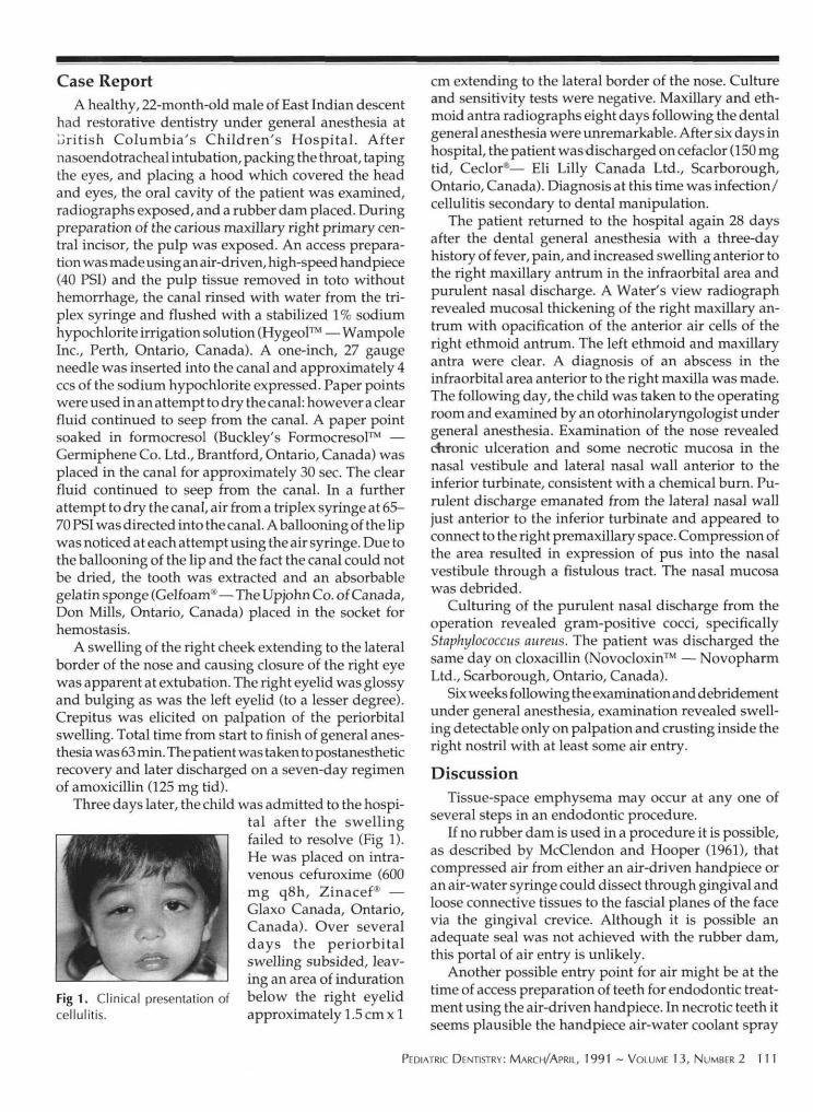
Three days later, the child was admitted to the hospi-tal after the swellingfailed to resolve (Fig 1).He was placed on intra-venous cefuroxime (600mg q8h, Zinacef® —Glaxo Canada, Ontario,Canada). Over severaldays the periorbitalswelling subsided, leav-ing an area of indurationbelow the right eyelidapproximately 1.5 cm x 1
Fig 1. Clinical presentation ofcellulitis.
cm extending to the lateral border of the nose. Cultureand sensitivity tests were negative. Maxillary and eth-moid antra radiographs eight days following the dentalgeneral anesthesia were unremarkable. After six days inhospital, the patient wasdischarged on cefaclor (150 mgtid, Ceclor®— Eli Lilly Canada Ltd., Scarborough,Ontario, Canada). Diagnosis at this time was infection/cellulitis secondary to dental manipulation.
The patient returned to the hospital again 28 daysafter the dental general anesthesia with a three-dayhistory of fever, pain, and increased swelling anterior tothe right maxillary antrum in the infraorbital area andpurulent nasal discharge. A Water's view radiographrevealed mucosal thickening of the right maxillary an-trum with opacification of the anterior air cells of theright ethmoid antrum. The left ethmoid and maxillaryantra were clear. A diagnosis of an abscess in theinfraorbital area anterior to the right maxilla was made.The following day, the child was taken to the operatingroom and examined by an otorhinolaryngologist undergeneral anesthesia. Examination of the nose revealedchronic ulceration and some necrotic mucosa in thenasal vestibule and lateral nasal wall anterior to theinferior turbinate, consistent with a chemical burn. Pu-rulent discharge emanated from the lateral nasal walljust anterior to the inferior turbinate and appeared toconnect to the right premaxillary space. Compression ofthe area resulted in expression of pus into the nasalvestibule through a fistulous tract. The nasal mucosawas debrided.
Culturing of the purulent nasal discharge from theoperation revealed gram-positive cocci, specificallyStaphylococcus aureus. The patient was discharged thesame day on cloxacillin (Novocloxin™ — NovopharmLtd., Scarborough, Ontario, Canada).
Six weeks following the examination and debridementunder general anesthesia, examination revealed swell-ing detectable only on palpation and crusting inside theright nostril with at least some air entry.
DiscussionTissue-space emphysema may occur at any one of
several steps in an endodontic procedure.If no rubber dam is used in a procedure it is possible,
as described by McClendon and Hooper (1961), thatcompressed air from either an air-driven handpiece oran air-water syringe could dissect through gingival andloose connective tissues to the fascial planes of the facevia the gingival crevice. Although it is possible anadequate seal was not achieved with the rubber dam,this portal of air entry is unlikely.
Another possible entry point for air might be at thetime of access preparation of teeth for endodontic treat-ment using the air-driven handpiece. In necrotic teeth itseems plausible the handpiece air-water coolant spray
PEDIATRIC DENTISTRY: MARCH/APRIL, 1991 ~ VOLUME 1 3, NUMBER 2 111

might result in tissue-space emphysema if directed intoempty canals. However, in this case, in which the pulpwas present, it seems unlikely a significant amount of aircould penetrate past the pulp.
The most likely cause of the tissue-space emphysemain this case was from the compressed air of the air-watersyringe directed into the empty canal. The close proxim-ity of the maxillary primary central incisors to the buccalbone in children generally (and in this case specifically),and the presence of the periapical resorption in theinvolved tooth (Fig 2), suggest that the air entered thefascial tissue planes of the child's face through a perfo-ration in the buccal bone at the apex of the centralincisor, and progressed to the periorbital area.
Fig 2. Preoperative (intraoperative) anterior periapical view.
Complicating this tissue-space emphysema, it seems,was the presence of sodium hypochlorite and/orformocresol in the periapical tissues. Both these chemi-cals have proven highly irritating to soft tissue (Schilderand Amsterdam 1959; Becker et al. 1973) and may havebeen responsible for the observed tissue destruction ifcarried apically by compressed air.
The secondary infection of the necrotic infraorbitaltissues by S. aureus is likely from one of two routes. TheS. aureus may have been carried apically from the oralcavity resulting in the periorbital infection or, a sterileabscess may have formed in the infraorbital area whicheventually tracked to the nasal cavity, resulting in sec-ondary infection of the necrotic infraorbital tissue. Thelatter route is more likely because S. aureus is usuallyfound in low numbers in the oral cavity as compared tothe nose or nasopharynx (Rosan 1988). The possibilitythat the culture was contaminated by S. aureus at the timeof collection in the nasal vestibule also exists.
Based on the experience described in this case reportand reports from the literature, we have several recom-mendations for prevention. Whenever possible, use arubber dam with a tight seal around the teeth to reduce
the possibility of emphysema as well as infection. Whenpreparing an endodontic access cavity, switch off the aircoolant on the air-driven high speed handpiece to mini-mize air being directed into the canal, especially whenthe canal is empty and the root apex is patent.
To prevent apical movement of air and bacteria, donot direct the dental air-water syringe into a canal oncepatency of the root canal is established. A case of fatal airembolism secondary to the use of compressed air to drya mandibular anterior tooth canal in a child has beenreported (Rickles and Joshi 1963).
When using canal irrigants, ensure the syringe needlefits loosely within the canal before expressing the irrigantto prevent forcing the irrigant beyond the apical foramen.Measuring the needle length and comparing it to theroot length on a radiograph will also help ensure theirrigant is not deposited beyond the canal apex. Canalmedicaments must similarly be contained within theroot canal.
The use of clear plastic drapes is recommended topermit intraoperative observation of the patient in pro-cedures under general anesthesia.
The pain associated with tissue emphysema can betreated palliatively with analgesics. Close follow-up bythe practitioner must follow.
If air tracks in an inferior direction, respiratory em-barrassment through pharyngeal emphysema and me-diastinal emphysema may create a medical emergency(McClendon and Hooper 1961). If the emphysema oc-curs while the patient is under general anesthesia, nitrousoxide should be immediately terminated and 95% oxy-gen administered (Levy 1981) to prevent enlargement ofthe air mass.
Infection represents a potential problem and the pa-tient should be covered prophylactically with antibiot-ics. As demonstrated by this case, antibiotics are notalways effective, so drainage of a localized infectionmay be required. Any infection of the face, whether ofprimary or secondary origin, must be given specialattention because the infection may extend directly tothe cavernous sinuses (Miller and Keane 1983).
The authors thank Shelly-Mae Boyd, CDA for her assistance in thiscase report, and Dawn Wright and Anne Der for manuscript prepa-ration.
Dr. Wright is pediatric dental resident, University of Connecticut,Farmington, CT. Dr. Derkson is associate professor, University ofBritish Columbia and head, department of dentistry, BritishColumbia's Children's Hospital. Dr. Riding is clinical assistant pro-fessor, University of British Columbia, Vancouver, Canada.
Becker GL, Cohen S, Borer R: The sequelae of accidentally injectingsodium hypochlorite beyond the root apex: report of a case. OralSurg 38:633-38,1974.
Cardo VA Jr, Mooney JW, Stratigos GT: latrogenic dental-air em-physema: report of case. J Am Dent Assoc 85:144—47,1972.
112 PEDIATRIC DENTISTRY: MARCH/APRIL, 1991 ~ VOLUME 1 3, NUMBER 2
Duncan JM, Ferrillo PJ: Interstitial emphysema after an amalgamrestoration. J Am Dent Assoc 74:407-10, 1967.
Falomo OO: Surgical emphysema following root canal therapy: reportof a case. Oral Surg 58:101-02, 1984.
Feinstone T: Infected subcutaneous emphysema: report of case. J AmDent Assoc 83:1309-11, 1971.
Fisher FJ: Surgical emphysema following a simple conservation pro-cedure: a case report. J Dent 4:129-30, 1976.
Geffner I: Subcutaneous facial emphysema following an amalgamrestoration. Br Dent J 148:192, 1980.
Hayduk S, Bennett CR, Monheim LM: Subcutaneous emphysemaafter operative dentistry: report of case. J Am Dent Assoc 80:1362,1970.
HerrmannJW, Heicht RC: Complications in therapeutic use of sodiumhypochlorite. J Endod 5:160, 1979.
Hirschmann PN, Walker RT: Facial emphysema during endodontictreatment -- two case reports. Int Endod J 16:130-32,1983.
Kullaa-Mikkonen A, Mikkonen M: Subcutaneous emphysema. Br JOral Surg 20:200-202, 1982.
LeRoy NB, Bregman AH: Subcutaneous emphysema. J Am DentAssoc 76:798-99, 1968.
Levy E: Iatrogenic subcutaneous emphysema during dental anesthesia:report of two cases. ASDC J Dent Child 48:272-74, 1981.
Lloyd RE: Surgical ecphysema as a complication in endodontics. BrDent J 138:393-94, 1975.
Madden PW, Averett JN: Subcutaneous emphysema. Gen Dent 35:474-75, 1987.
McClendon JL, Hooper WC Jr: Cervicofacial emphysema after airblown into a periodontal pocket: a case report. J Am Dent Assoc63:810-12, 1961.
McGrannahan WW: Tissue space emphysema from an air turbinehandpiece. J Am Dent Assoc 71:884-85, 1965.
Miller BF, Keane CB: Encyclopedia and Dictionary of Medicine,Nursing, and Allied Health, 3rd ed. Philadelphia: WB SaundersCo, 1983, p 206.
Quisling RW, Kangur TT, Jahrsdoerfer RA: Otologic complicationsfollowing the use of a high-speed air-turbine handpiece. J AmDent Assoc 94:895-97, 1977.
Rickles NH, Joshi BA: Death from air embolism during root canaltherapy. J Am Dent Assoc 67:397-404, 1963.
Rosan B: The staphylococci, in Oral Microbiology and Immunology.Newman MG, Nisengard R, eds. Philadelphia: WB Saunders Co,1988 p 167.
Rosenberg MB, Wunderlich BK, Reynolds RN: Iatrogenic subcutane-ous emphysema during dental anesthesia. Anesthesiology 51:80-81, 1979.
Schilder H, Amsterdam M: Inflammatory potential of root canalmedicaments: a preliminary report including nonspecific drugs.Oral Surg 12:211-21, 1959.
PEDIATRIC DENTISTRY: MARCH/APRIL, 1991 -- VOLUME 13, NUMBER 2 113


![Acute or chronic pulmonary emphysema? Or both?—A ......emphysema or acute alveolar dilation, respectively [3 , 5]. In some cases, an interstitial emphysema is described [, 636]](https://img.pdfslide.net/doc/110x75/6138f505a4cdb41a985b64ce/acute-or-chronic-pulmonary-emphysema-or-bothaa-emphysema-or-acute-alveolar.jpg)