Embed Size (px)
Citation preview
American Journal of Obstetrics and Gynecology Founded in 1920
volume 149 number 6 .JULY 15, 1984
GYNECOLOGY
Transplantation of normal and ectopic human endometrial
tissue into athymic nude mice
Nezaam M. Zamah, M.D., F.R.C.S.(C),* Melvin G. Dodson, M.D., Ph.D.,
L. Clifton Stephens, D.V.M., Ph.D., Veasy C. Buttram, Jr., M.D., Paige K. Besch, Ph.D.,
and Raymond H. Kaufman, M.D.
Houston, Texas
Implants or tiny circumscribed nodules of endometrial tissue were found in all female nude mice given
intraperitoneal injections of fragments of human normal (proliferative and secretory) or ectopic
(endometrioma) endometrium. Half of these animals received estrogen supplementation and the other half
received none. The endometriosis tissue present in these animals at 28 or 56 days after inoculation
consisted of glands and stroma with an infiltration of hemosiderin-laden macrophages. Glands in tissue
transplants of animals given supplemental estrogen tended to be larger, and the secretory endometrium
tended to revert to a proliferative pattern. Palpable nodules at the site of subcutaneous inoculations of
proliferative endometrium became undetectable grossly and microscopically within 24 to 32 days, whereas
endometrioma tissue remained detectable for up to 70 days and resembled the intraperitoneal tissue
microscopically. This study demonstrates that human endometrial tissue can be successfully transplanted
into the nude mouse and will retain its basic morphology. (AM. J. OssTET. GYNECOL. 149:591, 1984.)
In 1880, Breus first recognized endometriosis, and
16 years later Von Recklinghausen 1 first described en
dometriosis as a pathologic entity. In the first quarter
of this century, several theories regarding the etiology
From the Department of Obstetrics and Gynecology, Baylor College of Medicine, the Reproductive Research Laboratory, St. Luke's Episcopal Hospital, and the Section of Veterinary Pathology, M. D. Anderson Hospital Tumor Institute.
Supported in part by a grant from the Women's Fund of Houston, Texas.
Presented at the Thirty-first Annual Clinical Meeting of the American College of Obstetricians and Gynecologists, Atlanta, Georgia, May 7-12, 1983, and raeived the first-prize award for the best basic science research paper from The Upjohn Co., Kalamazoo, Michigan.
Received for publication December 14, 1983; accepted january 3, 1984.
Reprint requests: Paige K. Besch, Ph.D., Department of Obstetrics and Gynecology, Baylor College of Medicine, One Baylor Plaza, Houston, TX 77030.
*Research completed during the tenure of a Postdoctoral Fellowship in Reproductive Endocrinology and Infertility.
of endometriosis were proposed. The three most
commonly accepted theories are those of retrograde menstruation and implantation, coelomic metaplasia,
and vascular dissemination. However, the last hundred
years have not fully clarified the pathogenesis of endometriosis. One of the limitations has been the diffi
culty in doing controlled experiments in human
subjects. Endometriosis occurs naturally in the monkey, which
happens to be the only other animal that has cyclic
menstrual periods.2 Observations and experiments
with the use of monkeys and other species of animals
that do not naturally have the disease have concen
trated on evaluation of the proposed etiologic theories.
Scott et al.~ demonstrated the development of endome
triosis in five of 10 Macaca monkeys, but some animals required as long as 3 years before endometriosis was
noted. Merrili,4 using autologous rabbit endometrium
placed inside Millipore diffusion chambers, produced
growth of ectopic endometrial-like tissue in the adja-
591
592 Zamah et al. July 15, 1984 Am. J. Obstet. Gynecol.
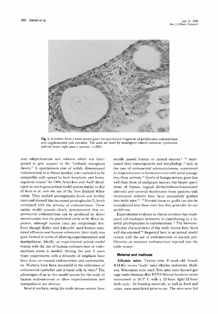
Fig. l. A nodule from a nude mouse given intraperitoneal fragments of proliferative endometrium and supplemented with estradiol. The acini are lined by malaligned ciliated columnar epithelium and the lower right area is necrotic. (x250.)
cent subperitoneum and subcutis which was interpreted to give support to the "coelomic metaplasia theory." A spontaneous case of widely disseminated endometriosis in a rhesus monkey was concluded to be compatible with spread by both lymphatic and hematogenous routes." In 1980, Schenken and Asch6 devel
oped an autologous animal model system similar to that of Scott et a!. with the use of the New Zealand White rabbit. They studied prostaglandin levels and fertility rates and showed that increased prostaglandin E2 levels correlated with the severity of endometriosis. These earlier model systems clearly demonstrated that experimental endometriosis can be produced by direct menstruation into the peritoneal cavity or by direct injection, although success rates are surprisingly low. Even though Ridley and Edwards' used human menstrual effluents and human volunteers, their study was quite limited in terms of allowing experimentation and
manipulation. Ideally, an experimental animal model
system with the use of human endometrium or endometriosis tissue is needed. During the past 5 years
many experiments with a diversity of emphasis have
been done on normal endometrium and endometriosis. Workers have been successful in the cultivation of
endometrial epithelial and stromal cells in vitro.8 The
advantages of an in vivo model system for the study of human endometriosis to allow experimentation and manipulation are obvious.
Several workers, using the nude mouse system, have
serially passed human or animal tumors, 9·
10 maintained their tumorigenicity and morphology, 11 and, in
the case of endometrial adenocarcinoma, maintained its responsiveness to hormones even with serial passage into these animals. 12 Grafts of benign tumors grow less
well than those of malignant tumors, but biopsy specimens of human vaginal diethylstilbestrol-associated
adenosis and synovial membranes from patients with rheumatoid arthritis have been successfully grafted into nude mice. 1
'L 14 Normal tissue or grafts can also be
transplanted into these mice but they generally do not proliferate.
Experimental evidence in rhesus monkeys has implicated cell-mediated immunity in contributing to a familial predisposition to endometriosis. 15 The immunedeficient characteristics of the nude mouse have been well documented. 16 Reported here is an animal model system with the use of endometriosis or normal pro
liferative or secretory endometrium injected into the
nude mouse.
Material and methods
Athymic mice. Twenty-nine 8-week-old female BALB/c nu/nu "nude" mice (Harlan Industries, Madi
son, Wisconsin) were used. Five mice were housed per cage under laminar-flow HEPA filtered hoods in rooms
maintained at 26.7° C with a 12-hour light/12-hour dark cycle. All housing materials, as well as food and water, were autoclaved prior to use. The mice were fed
Volume 149 Number 6
Transplantation of endometrial tissue into athymic mice 593
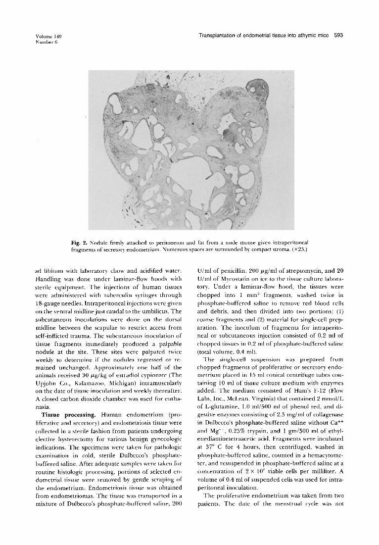
Fig. 2. Nodule firmly attached to peritoneum and fat from a nude mouse given intraperitoneal fragments of secretory endometrium. Numerous spaces are surrounded by compact stroma. (X25.)
ad libitum with laboratory chow and acidified water.
Handling was done under laminar-flow hoods with
sterile equipment. The injections of human tissues were administered with tuberculin syringes through
18-gauge needles. Intraperitoneal injections were given
on the ventral midline just caudal to the umbilicus. The
subcutaneous inoculations were done on the dorsal
midline between the scapulae to restrict access from
self-inflicted trauma. The subcutaneous inoculation of
tissue fragments immediately produced a palpable nodule at the site. These sites were palpated twice weekly to determine if the nodules 1·egressed or remained unchanged. Approximately one half of the animals received 30 p.g/ kg of estradiol cypionate (The
Upjohn Co., Kalamazoo, Michigan) intramuscularly on the date of tissue inoculatio n and weekly thereafter.
A closed carbon dioxide chamber was used for eutha
nasia. Tissue processing. Human endometrium (pro
liferative and secretory) and endometriosis tissue were
collected in a sterile fashion from patients undergoing elective hysterectomy for various benign gynecologic
indications. The specimens were taken for pathologic
examination in cold, sterile Dulbecco's phosphate
buffered saline. After adequate samples were taken fo r
routine histologic processing, po rtions of selected endometrial tissue were removed by gentle scraping of
the endometrium. Endometriosis tissue was obtained from endometriomas. The tissue was transported in a mixture of Dulbecco's phosphate-buffered saline, 200
Ulml of penicillin, 200 p.g/ ml of stre ptomycin, and 20
U/ ml o f Mycostatin on ice to the tissue culture laboratory. Under a laminar-flow hood, the tissues were
chopped into 1 mm'1 fragments, was hed twice in
phosphate-buffered saline to remove red blood cells
and debris, and then divided into two portions: (l)
coarse fragments and (2) material for single-cell prep
aration. The inoculum of fragments for intraperitoneal or subcutaneous injection consisted of 0.2 ml of
chopped tissues in 0 .2 ml of phosphate-buffered saline
(total volume, 0.4 ml) . T he single-cell suspension was prepared from
chopped fragments of proliferative or secretory endometrium placed in 15 ml conica l centrifuge tubes con
taining 10 ml of tissue culture medium with enzymes added. The medium consisted of Ham's F-12 (Flow Labs, Inc., McLean, Virginia) that contained 2 mmoi/L
of L-glutamine, 1.0 ml/500 ml of phenol red, and di
gestive enzymes consisting of 2.5 mg/ml of collagenase
in Dulbecco's phosphate-buffered saline without Ca++
and Mg++, 0.25% trypsi n, and 1 gm /500 ml of ethylenediaminetetraacetic acid. Fragments were incubated
at 37° C for 4 hours, then centrifuged, washed in
phosphate-buffered saline , counted in a hemacytome
ter, and resuspended in phosphate-buffered saline at a
concentration of 2 x 10⁷ viable cells per milliliter. A
volume of 0.4 ml of suspended cells was used for intraperitoneal inoculation.
The proliferative endometrium was taken from two
patients. The date of the menstrual cycle was not
594 Zamah et al. July 15, 1984 Am. J. Obstet. Gynecol.
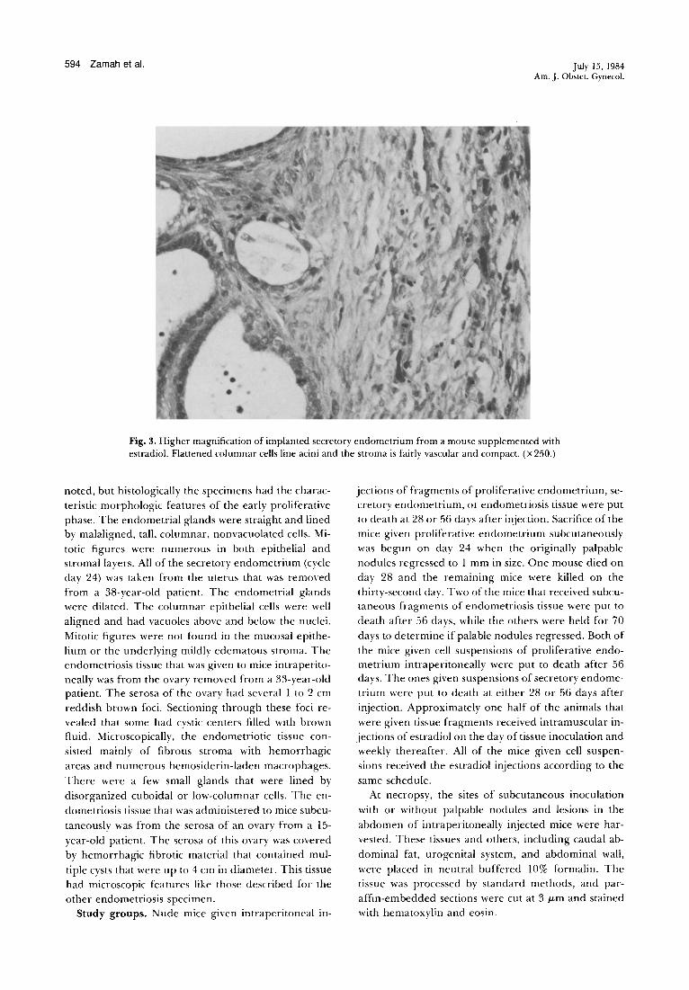
Fig. 3. Higher magnification of implanted secretory endometrium from a mouse supplemented with estradiol. Flattened columnar cells line acini and the stroma is fairly vascular and compact. (X250.)
noted, but histologically the specimens had the charac
teristic morphologic features of the early proliferative
phase. The endometrial glands were straight and lined by malaligned , tall , columnar, nonvacuolated cells. Mi
totic figures were numerous in both epithelial and stromal layers. All of the secretory endometrium (cycle
day 24) was taken from the uterus that was removed
from a 38-year-old patient. The endometrial glands
were dilated. The columnar epithelial cells were well
aligned and had vacuoles above and below the nuclei.
Mitotic figures were not found in the mucosal epithe
lium or the underlying mildly edematous stroma. The endometriosis tissue that was given to mice intra perito
neally was from the ovary removed from a 33-year-old patient. The serosa of the ovary had several 1 to 2 em
reddish brown foci. Sectioning through these foci re
vealed that some had cystic centers filled with brown fluid. Microscopically, the endometriotic tissue con
sisted mainly of fibrous stroma with hemorrhagic
areas and numerous hemosiderin-laden macrophages.
There were a few small glands that were lined by
disorganized cuboidal or low-columnar cells. The en
dometriosis tissue that was administered to mice subcu
taneously was from the serosa o f an ovary from a 15-
year-old patient. The serosa of this ovary was covered
by hemorrhagic fibrotic material that contained mul
tiple cysts that were up to 4 em in diameter. This tissue
had microscopic features like those described for the
other endometriosis specimen . Study groups. Nude mice given intraperitoneal in-
jections of fragments of proliferative endometrium, secretory endometrium, or endometriosis tissue were put
to death at 28 or 56 days after injection . Sacrifice of the
mice given proliferative endometrium subcutaneously
was begun on day 24 whe n the originally palpable
nodules regressed to 1 mm in size. One mouse died on
day 28 and the remaining mice were killed on the
thirty-second day. Two of the mice that received subcutaneous fragments of endometriosis tissue were put to
death after 56 days, while the others were held for 70
days to determine if palable nodules regressed. Both of
the mice given cell suspensions of proliferative endometrium intraperitoneally were put to death after 56 days. The ones given suspensions of secretory endome
trium were put to death at either 28 or 56 days after
injection. Approximately one half of the animals that
were given tissue fragments received intramuscular in
j ec tions of estradiol on the day of tissue inoculation and
weekly thereafter. All of the mice given cell suspen
sions received the estradiol injections according to the
same schedule. At necropsy, the sites of subcutaneous inoculation
with or without palpable nodules and lesions in the
abdo men of intraperitoneally injected mice were har
vested. These tissues and others, including caudal ab
dominal fat, urogenital system, and abdominal wall,
were placed in neutral buffered 10% formalin. The
tissue was processed by standard methods, and par
affin-embedded sections were cut at 3 J.Lm and stained
with hematoxylin and eosin.
Volume 149 Number 6
Results
Proliferative endometrium. Six nude m1ce were
given fragments of proliferative endometrium intra
peritoneally. Two were put to death after 28 days and the remaining four, at 56 days after inoculation. At necropsy, five of the six nude mice injected had single or multiple discrete, tan nodules 1 to 3 mm in diameter and loosely attached to the omentum, mesentery, or caudal abdominal fat. A nodule was found in histologic
sections of the caudal abdominal fat from the sixth mouse. Microscopically, the loosely attached nodules
harvested at 28 or 56 days from five of the mice were similar. The periphery of the nodules had a thin mem
branous capsule overlying densely cellular stroma that surrounded variable numbers of small acinar struc
tures. The nodules from mice that received estradiol had more acini that were larger than those from the
ones that were unsupplemented, but the microscopic
features of the acini in both groups of mice were similar. These structures were lined by malaligned
columnar cells that commonly had a ciliated luminal border (Fig. 1). Small numbers of lymphocytes and
plasma cells infiltrated the stroma. The centers of nodules from estradiol-supplemented and unsup
plemented mice were, in general, necrotic. The nodule found microscopically in an estradiol-supplemented mouse (C4) put to death at 56 days was embedded within the caudal abdominal fat and surrounded by
congested capillaries. This nodule was composed of several cystically dilated acini. Ciliated columnar epithelium lined these structures which were separated by thin fibrous trabeculae. This tissue contained no areas of necrosis.
The nodules produced by subcutaneous inoculation
of fragments of proliferative phase endometrium into
six mice began to regress within 3 to 4 weeks after inoculation. Tissues taken from the inoculation site 24 to 32 days after inoculation either had no microscopic lesions or contained only mild diffuse infiltrates of lymphoid cells and hemosiderin-laden macrophages.
No gross or microscopic lesions were found in the two mice put to death 28 days after being given an intraperitoneal cell suspension made from proliferative
endometrium. Secretory endometrium. Four mice were given in
traperitoneal fragments of secretory endometrium.
Two were put to death at 28 days and the other two at
56 days after inoculation. All had discrete nodules 1 mm in diameter in the abdomen. In addition to loosely
attached nodules in fat, such as were seen in mice given
proliferative endometrium, nodules of tissue were firmly attached to caudal abdominal fat or the serosa of abdominal viscera (Fig. 2). One mouse had a nodule on
the serosa of the stomach, another had a nodule attached to the capsule of the spleen, and a third had a
Transplantation of endometrial tissue into athymic mice 595
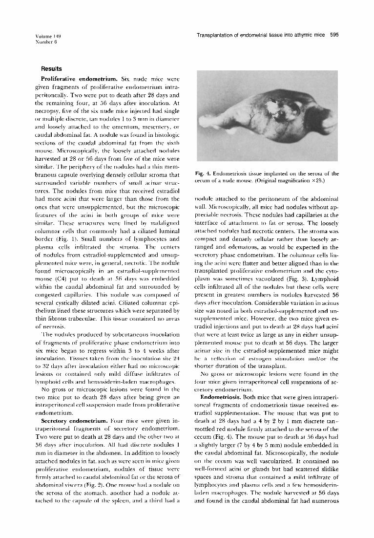
Fig. 4. Endometriosis tissue implanted on the serosa of the cecum of a nude mouse. (Original magnification x25.)
nodule attached to the peritoneum of the abdominal
wall. Microscopically, all mice had nodules without appreciable necrosis. These nodules had capillaries at the
interface of attachment to fat or serosa. The loosely
attached nodules had necrotic centers. The stroma was compact and densely cellular rather than loosely ar
ranged and edematous, as would be expected in the secretory phase endometrium. The columnar cells lin
ing the acini were flatter and better aligned than in the transplanted proliferative endometrium and the cytoplasm was sometimes vacuolated (Fig. 3). Lymphoid
cells infiltrated all of the nodules but these cells were present in greatest numbers in nodules harvested 56
days after inoculation. Considerable variation in acinus size was noted in both estradiol-supplemented and unsupplemented mice. However, the two mice given estradiol injections and put to death at 28 days had acini
that were at least twice as large as any in either unsup
plemented mouse put to death at 56 days. The larger acinar size in the estradiol-supplemented mice might be a reflection of estrogen stimulation and/or the shorter duration of the transplant.
No gross or microscopic lesions were found in the four mice given intraperitoneal cell suspensions of secretory endometrium.
Endometriosis. Both mice that were given intraperitoneal fragments of endometriosis tissue received es
tradiol supplementation. The mouse that was put to death at 28 days had a 4 by 2 by 1 mm discrete tan
mottled red nodule firmly attached to the serosa of the cecum (Fig. 4). The mouse put to death at 56 days had
a slightly larger (7 by 4 by 5 mm) nodule embedded in the caudal abdominal fat. Microscopically, the nodule
on the cecum was well vascularized. It contained no well-formed acini or glands but had scattered slitlike spaces and stroma that contained a mild infiltrate of lymphocytes and plasma cells and a few hemosiderinladen macrophages. The nodule harvested at 56 days and found in the caudal abdominal fat had numerous
596 Zamah et al. July 15, 1984 Am.]. Obstet. Gynecol.
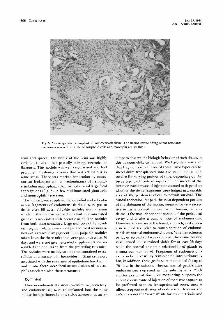
Fig. 5. An intraperitoneal implant of endometriosis tissue. The stroma surrounding acinar remnants contains a marked infiltrate of lymphoid cells and marcophages. (X 100.)
acini and spaces. The lining of the acini was highly
variable . It was either partially missing, necrotic, or
flattened. This nodule was well vascularized and had
prominent hyalinized stroma that was edematous in
some areas. There was marked infiltration by mono
nuclear leukocytes with a predominance of hemosid
erin-laden macrophages that formed several large focal
aggregations (Fig. 5). A few multinucleated giant cells
and neutrophils were seen.
Two mice given supplemental estradiol and subcuta
neous fragments of endometriosis tissue were put to
death after 56 days. Palpable nodules were present
which in the microscopic sections had multinucleated
giant cells associated with necrotic acini. The nodules
from both mice contained large numbet·s of hemosid
erin pigment-laden macrophages and focal accumula
tions of extracellular pigment. The palpable nodules
taken from the three mice that were put to death at 70
days and were not given estradiol supplementation re
sembled the ones taken from the preceding two mice.
The nodules were mainly stroma that contained extra
cellular and intracellular hemosiderin. Giant cells were
associated with the remnants of epithelium lined acini,
and in one there were focal accumulations of neutro
phils associated with these structures.
Comment
Human endometrial tissues (proliferative, secretory,
and endometriosis) were transplanted into the nude
mouse intraperitoneally and subcutaneously in an at-
tempt to observe the biologic behavior of such tissues in
this immune-deficient animal. We have demonstrated
that fragments of all three of these tissue types can be
successfully transplanted into the nude mouse and
survive for varying periods of time, depending on the
tissue type and route of injection. The success of the
intraperitoneal route of injection seemed to depend on
whether the tissue fragments were lodged in a suitable
area of the peritoneal cavity to permit survival. The
caudal abdominal fat pad, the most dependent portion
of the abdomen of the mouse, seems to be very recep
tive to tissue transplantation. In the human, the cul
de-sac is the most dependent portion of the peritoneal
cavity and is also a common site of endometriosis.
However, the serosa of the bowel, stomach, and spleen
also seemed receptive to transplantation of endome
triosis or normal endometrial tissue. When attachment
to fat or serosal surfaces occurred, the tissue became
vascularized and remained viable for at least 56 days
while the normal anatomic relationship of glands to
stroma was maintained. Fragments of endometrioma
can also be successfully transplanted intraperitoneally
but, in addition, these grafts were maintained for up to
70 days in the subcutis whereas normal proliferative
endometrium regressed in the subcutis in a much
shorter period of time. For monitoring purposes the
subcutaneous route of injection of the tissue appears to
be preferred over the intraperitoneal route, since it
allows frequent evaluation of nodule size. However, the
subcutis is not the "normal" site for endometriosis, and
Volume 149 Number 6
so studies of tissue behavior may not be comparable to
the in vivo situation. The effect of estrogen supplementation is striking in
that the tissue showed better morphologic features in the estrogen-stimulated group compared to the nonestrogen-stimulated group with proliferative but not
with secretory endometrium. Mitotic activity also appeared to be more common in the estrogen-treated
group of animals. Single-cell intraperitoneal trans
plants of proliferative endometrium did not survive in the presence or absence of estrogen stimulation.
It is well known that the nude mouse still has the
innate ability to mount a limited immune response because of its intact natural "killer" T-cell function. It was
not surprising, therefore, to see a marked lymphocytic
and plasma cell infiltrate in some of the tissues recovered. Interestingly, endometriosis tissue survived for a much longer period of time than did normal prolifera
tive endometrium when both were administered subcu
taneously. This marked variation in survival time in the nude mouse raises some very important questions re
garding the "normality" of the glands and stroma of endometriosis and suggests that ectopic endometrium is biologically different. Perhaps adaptation to an aberrant site in the patient resulted in this tissue becoming more autonomous and, as a result, more susceptible to
transplantation. It is to be noted that none of the single-cell suspen
sion preparations resulted in endometrial cellular pro
liferation grossly or microscopically. Perhaps repeated injections of the same preparation over a prolonged
period of time are necessary before such changes can
occur. Further studies will be necessary to determine the
usefulness of the nude mouse system for the study of
endometriosis and possibly other benign gynecologic diseases.
We wish to thank David Fink, B.A., B.S., Research Assistant, Department of Obstetrics and Gynecology, Baylor College of Medicine, Charles Castro, B.A., Denise Martin, B.A., Willie Virgil, and the staff of the M. D. Anderson Hospital and Tumor Institute Animal Research Facilities for their technical assistance.
Transplantation of endometrial tissue into athymic mice 597
REFERENCES
l. Von Recklinghausen F. Die Edenomyone und Cystadenomyone der uterus und Tukenwandrug; ihre Akkunft von Restur des Wolff'scher Korpers Berlin, Berlin: Hirschwald, 1896.
2. MacKenzie WF, Casey HW. Animal model of human disease: endometriosis in rhesus monkeys. Am J Pathol 1975;80:341.
3. Scott RB, TeLinde RW, Wharton LR Jr. Further studies on experimental endometriosis. AM J OBSTET GYNECOL 1953;66:5.
4. Merrill JA. Endometrial induction of endometriosis across Millipore filters. AM J 0BSTET GYNECOL 1966;94:780.
5. McClure HM, Graham CE, Guilloud NB. Widespread endometriosis in a rhesus monkey (Macaca mulatta). In: Hofer HO, ed. Proceedings of the Second International Congress of Primatology. New York: S. Karger AG, 1969:155. (vol 3).
6. Schenken RS, Asch RH. Surgical induction of endometriosis in the rabbit: effects on fertility and concentrations of peritoneal fluid prostaglandins. Fertil Steril 1980;34:6.
7. Ridley JH, Edwards JK. Experimental endometriosis in the human. AM J 0BSTET GYNECOL 1958;76:4.
8. Varma VA, Adamac TA, Dorman BH, Kaufman DG. Human endometrial epithelial and stromal cells in culture (Abstract). Lab Invest 1982;46:86A.
9. Rygaard J, Povlsen CO. Heterotransplantation of a human malignant tumor to "nude" mice. Acta Pathol Microbial Scand 1960;77:758.
10. Fogh J, Fogh JM, Orfed T. One hundred and twentyseven cultured human tumor cell lines producing tumors in nude mice. J Nat! Cancer Inst 1977;59:221.
11. Stiles CD, Desmond W, Chuman LM, Sato G, Saier MH. Growth control of heterologous tissue culture cells in the congenitally athymic nude mouse. Cancer Res 1976; 36:1353.
12. Satyaswaroop PG, Zaino RJ, Mortel R. Human endometrial adenocarcinoma transplanted into nude mice: growth regulation by estradiol. Science 1983;219:58.
13. Pienkowski MM, Mann LC, Rosloniec EF, Welch CW. Accretion biopsy specimens of vaginal adenosis from patients exposed in utero to diethylstilbesterol, when transplanted to nude mice. JNCI 1979;62:521.
14. Brinckerhoff CE, Harris ED. Survival of rheumatoid synovium implanted into nude mice. Am J Pathol 1981; 103:411.
15. Dmowski WP, Steele RW, Baker GF. Deficient cellular immunity in endometriosis. AMj 0BSTET GYNECOL 1981; 141:377.
16. Wortis HH. Immunological studies of nude mice. Contemp Top Immunobiol 1974;3:243.