Embed Size (px)
Citation preview
Case ReportTransumbilical Surgery for Duodenal Stenosis ina Child with Situs Inversus: The First Report
Isamu Saeki,1 Yu Ueno,1,2 Wataru Mukai,1,2 Reisuke Imaji,1 and Takashi Akiyama1,2
1Department of Pediatric Surgery, Hiroshima City Hospital, 7-33 Motomachi Naka-ku, Hiroshima-shi, Hiroshima 730-8518, Japan2Chugoku-Shikoku Pediatric Surgical Partners Organization, Japan
Correspondence should be addressed to Isamu Saeki; [email protected]
Received 22 November 2016; Accepted 22 December 2016; Published 12 March 2017
Academic Editor: Denis A. Cozzi
Copyright © 2017 Isamu Saeki et al. This is an open access article distributed under the Creative Commons Attribution License,which permits unrestricted use, distribution, and reproduction in any medium, provided the original work is properly cited.
Background. Situs inversus is a rare congenital anomaly with a reported incidence of only 1 in 5,000 to 10,000 live births. Congenitalduodenal stenosis complicated with situs inversus is an even rarer entity. Case Presentation. A 1-year-old girl with situs inversuswho had undergone a hemi-Fontan procedure against a single ventricle in our hospital was referred to our department forvomiting and failure to thrive. An upper gastrointestinal contrast study and endoscopy revealed duodenal stenosis. A transumbilicalradical operation as a minimally invasive surgery was successfully performed. After the surgery, she stopped vomiting, andthe postoperative course was uneventful with good cosmetic results. Conclusions. To our knowledge, this is the first report oftransumbilical surgery for congenital duodenal stenosis with situs inversus as minimally invasive surgery. Transumbilical surgeryto situs inversus patient can be performed safely and lead to good cosmetic outcome.
1. Introduction
Situs inversus (SI) is a rare clinical malformation with anincidence of 1 in 5,000 to 10,000 live births [1]. The sur-gical procedures for SI patients are technically challengingbecause of the mirror image anatomical construction andhigh frequency of other anomalies. Congenital duodenalobstruction, which includes atresia and stenosis, is estimatedto have an incidence of about 1 in 4,000 to 15,000 live births[2, 3]. Congenital duodenal stenosis (CDS) is relatively rarein comparison with duodenal atresia and sometimes has adelayed presentation [3–5]. CDS associated with SI is anextremely rare entity and only a few cases have been reportedthus far [2, 6]. We present a case of a 1-year-old girl with CDSand SI who successfully underwent a transumbilical radicaloperation as minimally invasive surgery.
2. Case Presentation
A 1-year-old Asian girl who had undergone a hemi-Fontanprocedure against a single ventricle in our hospital wasreferred to our department. She suffered from vomiting
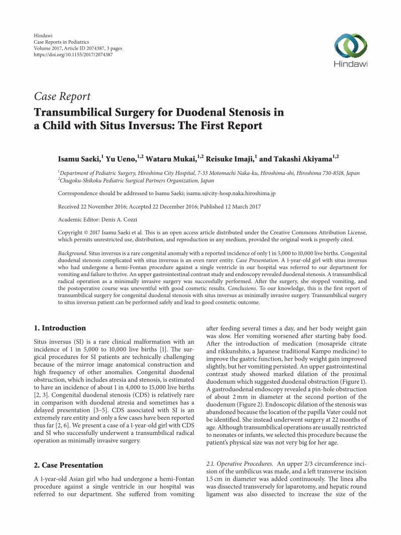
after feeding several times a day, and her body weight gainwas slow. Her vomiting worsened after starting baby food.After the introduction of medication (mosapride citrateand rikkunshito, a Japanese traditional Kampo medicine) toimprove the gastric function, her body weight gain improvedslightly, but her vomiting persisted. An upper gastrointestinalcontrast study showed marked dilation of the proximalduodenumwhich suggested duodenal obstruction (Figure 1).A gastroduodenal endoscopy revealed a pin-hole obstructionof about 2mm in diameter at the second portion of theduodenum (Figure 2). Endoscopic dilation of the stenosis wasabandoned because the location of the papilla Vater could notbe identified. She instead underwent surgery at 22 months ofage. Although transumbilical operations are usually restrictedto neonates or infants, we selected this procedure because thepatient’s physical size was not very big for her age.
2.1. Operative Procedures. An upper 2/3 circumference inci-sion of the umbilicus was made, and a left transverse incision1.5 cm in diameter was added continuously. The linea albawas dissected transversely for laparotomy, and hepatic roundligament was also dissected to increase the size of the
HindawiCase Reports in PediatricsVolume 2017, Article ID 2074387, 3 pageshttps://doi.org/10.1155/2017/2074387
2 Case Reports in Pediatrics
Figure 1: The upper gastrointestinal contrast study finding (supineposition). The proximal duodenum was markedly dilated, whichsuggested duodenal obstruction.
Figure 2: The gastroduodenal endoscopy findings. A pin-holeobstruction about 2mm in diameter was found at the secondportion of the duodenum (arrow).
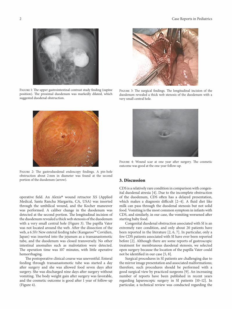
operative field. An Alexis� wound retractor XS (AppliedMedical, Santa Rancha Margarita, CA, USA) was insertedthrough the umbilical wound, and the Kocher maneuverwas performed. A caliber change in the duodenum wasdetected at the second portion. The longitudinal incision ofthe duodenum revealed a thickweb stenosis of the duodenumwith a very small central hole (Figure 3). The papilla Vaterwas not located around the web. After the dissection of theweb, a 6.5Fr New enteral feeding tube (Kangaroo� Covidien,Japan) was inserted into the jejunum as a transanastomotictube, and the duodenum was closed transversely. No otherintestinal anomalies such as malrotation were detected.The operation time was 107 minutes, with little operativehemorrhaging.
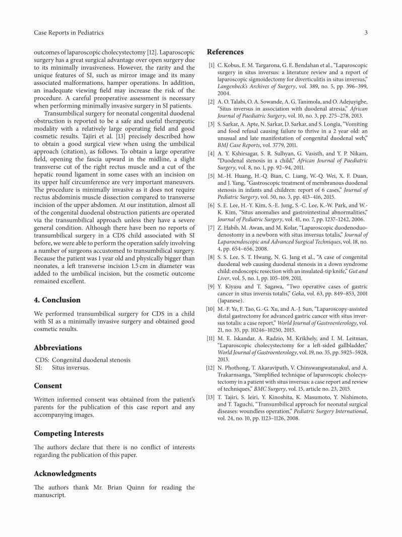
The postoperative clinical course was uneventful. Enteralfeeding through transanastomotic tube was started a dayafter surgery and she was allowed food seven days aftersurgery. She was discharged nine days after surgery withoutvomiting. The body weight gain after surgery was favorable,and the cosmetic outcome is good after 1 year of follow-up(Figure 4).
Figure 3: The surgical findings. The longitudinal incision of theduodenum revealed a thick web stenosis of the duodenum with avery small central hole.
Figure 4: Wound scar at one year after surgery. The cosmeticoutcome was good at the one-year follow-up.
3. Discussion
CDS is a relatively rare condition in comparisonwith congen-ital duodenal atresia [4]. Due to the incomplete obstructionof the duodenum, CDS often has a delayed presentation,which makes a diagnosis difficult [2–4]. A fluid diet likemilk can pass through the duodenal stenosis but not solidfood. Vomiting is themost common symptom in infants withCDS, and similarly, in our case, the vomiting worsened afterstarting baby food.
Congenital duodenal obstruction associated with SI is anextremely rare condition, and only about 20 patients havebeen reported in the literature [2, 6, 7]. In particular, only afew CDS patients associated with SI have ever been reportedbefore [2]. Although there are some reports of gastroscopictreatment for membranous duodenal stenosis, we selectedopen surgery because the location of the papilla Vater couldnot be identified in our case [5, 8].
Surgical procedures in SI patients are challenging due tothemirror image presentation and associatedmalformations;therefore, such procedures should be performed with agood surgical view by practiced surgeons [9]. An increasingnumber of reports have been published in recent yearsregarding laparoscopic surgery in SI patients [10–12]. Inparticular, a technical review was conducted regarding the
Case Reports in Pediatrics 3
outcomes of laparoscopic cholecystectomy [12]. Laparoscopicsurgery has a great surgical advantage over open surgery dueto its minimally invasiveness. However, the rarity and theunique features of SI, such as mirror image and its manyassociated malformations, hamper operations. In addition,an inadequate viewing field may increase the risk of theprocedure. A careful preoperative assessment is necessarywhen performing minimally invasive surgery in SI patients.
Transumbilical surgery for neonatal congenital duodenalobstruction is reported to be a safe and useful therapeuticmodality with a relatively large operating field and goodcosmetic results. Tajiri et al. [13] precisely described howto obtain a good surgical view when using the umbilicalapproach (citation), as follows. To obtain a large operativefield, opening the fascia upward in the midline, a slighttransverse cut of the right rectus muscle and a cut of thehepatic round ligament in some cases with an incision onits upper half circumference are very important maneuvers.The procedure is minimally invasive as it does not requirerectus abdominis muscle dissection compared to transverseincision of the upper abdomen. At our institution, almost allof the congenital duodenal obstruction patients are operatedvia the transumbilical approach unless they have a severegeneral condition. Although there have been no reports oftransumbilical surgery in a CDS child associated with SIbefore, we were able to perform the operation safely involvinga number of surgeons accustomed to transumbilical surgery.Because the patient was 1 year old and physically bigger thanneonates, a left transverse incision 1.5 cm in diameter wasadded to the umbilical incision, but the cosmetic outcomeremained excellent.
4. Conclusion
We performed transumbilical surgery for CDS in a childwith SI as a minimally invasive surgery and obtained goodcosmetic results.
Abbreviations
CDS: Congenital duodenal stenosisSI: Situs inversus.
Consent
Written informed consent was obtained from the patient’sparents for the publication of this case report and anyaccompanying images.
Competing Interests
The authors declare that there is no conflict of interestsregarding the publication of this paper.
Acknowledgments
The authors thank Mr. Brian Quinn for reading themanuscript.
References
[1] C. Kobus, E.M. Targarona, G. E. Bendahan et al., “Laparoscopicsurgery in situs inversus: a literature review and a report oflaparoscopic sigmoidectomy for diverticulitis in situs inversus,”Langenbeck’s Archives of Surgery, vol. 389, no. 5, pp. 396–399,2004.
[2] A.O. Talabi, O. A. Sowande, A.G. Tanimola, andO.Adejuyigbe,“Situs inversus in association with duodenal atresia,” AfricanJournal of Paediatric Surgery, vol. 10, no. 3, pp. 275–278, 2013.
[3] S. Sarkar, A. Apte, N. Sarkar, D. Sarkar, and S. Longia, “Vomitingand food refusal causing failure to thrive in a 2 year old: anunusual and late manifestation of congenital duodenal web,”BMJ Case Reports, vol. 3779, 2011.
[4] A. Y. Kshirsagar, S. R. Sulhyan, G. Vasisth, and Y. P. Nikam,“Duodenal stenosis in a child,” African Journal of PaediatricSurgery, vol. 8, no. 1, pp. 92–94, 2011.
[5] M.-H. Huang, H.-Q. Bian, C. Liang, W.-Q. Wei, X. F. Duan,and J. Yang, “Gastroscopic treatment of membranous duodenalstenosis in infants and children: report of 6 cases,” Journal ofPediatric Surgery, vol. 50, no. 3, pp. 413–416, 2015.
[6] S. E. Lee, H.-Y. Kim, S.-E. Jung, S.-C. Lee, K.-W. Park, and W.-K. Kim, “Situs anomalies and gastrointestinal abnormalities,”Journal of Pediatric Surgery, vol. 41, no. 7, pp. 1237–1242, 2006.
[7] Z. Habib, M. Awan, and M. Kolar, “Laparoscopic duodenoduo-denostomy in a newborn with situs inversus totalis,” Journal ofLaparoendoscopic and Advanced Surgical Techniques, vol. 18, no.4, pp. 654–656, 2008.
[8] S. S. Lee, S. T. Hwang, N. G. Jang et al., “A case of congenitalduodenal web causing duodenal stenosis in a down syndromechild: endoscopic resectionwith an insulated-tip knife,”Gut andLiver, vol. 5, no. 1, pp. 105–109, 2011.
[9] Y. Kiyasu and T. Sagawa, “Two operative cases of gastriccancer in situs inversis totalis,” Geka, vol. 63, pp. 849–853, 2001(Japanese).
[10] M.-F. Ye, F. Tao, G.-G. Xu, and A.-J. Sun, “Laparoscopy-assisteddistal gastrectomy for advanced gastric cancer with situs inver-sus totalis: a case report,”World Journal of Gastroenterology, vol.21, no. 35, pp. 10246–10250, 2015.
[11] M. E. Iskandar, A. Radzio, M. Krikhely, and I. M. Leitman,“Laparoscopic cholecystectomy for a left-sided gallbladder,”World Journal of Gastroenterology, vol. 19, no. 35, pp. 5925–5928,2013.
[12] N. Phothong, T. Akaraviputh, V. Chinswangwatanakul, and A.Trakarnsanga, “Simplified technique of laparoscopic cholecys-tectomy in a patient with situs inversus: a case report and reviewof techniques,” BMC Surgery, vol. 15, article no. 23, 2015.
[13] T. Tajiri, S. Ieiri, Y. Kinoshita, K. Masumoto, Y. Nishimoto,and T. Taguchi, “Transumbilical approach for neonatal surgicaldiseases: woundless operation,” Pediatric Surgery International,vol. 24, no. 10, pp. 1123–1126, 2008.

Submit your manuscripts athttps://www.hindawi.com
Stem CellsInternational
Hindawi Publishing Corporationhttp://www.hindawi.com Volume 2014
Hindawi Publishing Corporationhttp://www.hindawi.com Volume 2014
MEDIATORSINFLAMMATION
of
Hindawi Publishing Corporationhttp://www.hindawi.com Volume 2014
Behavioural Neurology
EndocrinologyInternational Journal of
Hindawi Publishing Corporationhttp://www.hindawi.com Volume 2014
Hindawi Publishing Corporationhttp://www.hindawi.com Volume 2014
Disease Markers
Hindawi Publishing Corporationhttp://www.hindawi.com Volume 2014
BioMed Research International
OncologyJournal of
Hindawi Publishing Corporationhttp://www.hindawi.com Volume 2014
Hindawi Publishing Corporationhttp://www.hindawi.com Volume 2014
Oxidative Medicine and Cellular Longevity
Hindawi Publishing Corporationhttp://www.hindawi.com Volume 2014
PPAR Research
The Scientific World JournalHindawi Publishing Corporation http://www.hindawi.com Volume 2014
Immunology ResearchHindawi Publishing Corporationhttp://www.hindawi.com Volume 2014
Journal of
ObesityJournal of
Hindawi Publishing Corporationhttp://www.hindawi.com Volume 2014
Hindawi Publishing Corporationhttp://www.hindawi.com Volume 2014
Computational and Mathematical Methods in Medicine
OphthalmologyJournal of
Hindawi Publishing Corporationhttp://www.hindawi.com Volume 2014
Diabetes ResearchJournal of
Hindawi Publishing Corporationhttp://www.hindawi.com Volume 2014
Hindawi Publishing Corporationhttp://www.hindawi.com Volume 2014
Research and TreatmentAIDS
Hindawi Publishing Corporationhttp://www.hindawi.com Volume 2014
Gastroenterology Research and Practice
Hindawi Publishing Corporationhttp://www.hindawi.com Volume 2014
Parkinson’s Disease
Evidence-Based Complementary and Alternative Medicine
Volume 2014Hindawi Publishing Corporationhttp://www.hindawi.com

![Case Report Congenital Duodenal - Semantic Scholar file40% cases have a duodenal diaphragm with or without central opening and 10% cases show stenosis [1]. Duodenal diaphragm develops](https://img.pdfslide.net/doc/110x75/5cc5dc3b88c99377368b665e/case-report-congenital-duodenal-semantic-scholar-cases-have-a-duodenal-diaphragm.jpg)