Embed Size (px)
Citation preview

Welcome to BIO 202
Human Anatomy and Physiology
with Dr. Fernandez
Please check to make sure you are in the correct course sections:
• Lecture (14950) MW 9:30-10:45am LS 104
• Lab (14885) MW 8:00-9:20am LS 310
OR
• Lab (14859) MW 11:00-12:20pm LS 310

Endocrine System
Chapter 17
• Mechanisms of Cell Communication
• Endocrine vs Exocrine
• Neuro-endocrine Relationship
• Major Organs of the Endocrine System
• Hormones and their Actions
• Eicosanoids and Paracrine Signaling
• Endocrine Disorders

Overview of Cell Communication • Communication among cells is necessary for coordinating
important cell activities like growth maintenance and reproduction.
• Mechanisms of cell communication include:
– gap junctions
• pores in cell membranes that signaling chemicals can move through from cell to cell
– neurotransmitters
• released from neuron travels across a small gap to a 2nd cell
– hormones
• Classical definition of a hormone is a chemical messenger that travels in the bloodstream
– paracrine hormones
• chemical messengers secreted into tissue fluids that effect nearby cells

Endocrine System
• Major endocrine organs include the: – pineal gland
– pituitary gland
– thyroid gland
– parathyroid glands
– thymus
– adrenal glands
– pancreas
• Endocrine System is composed of all of the major endocrine glands and hormone-secreting cells of other organs including the brain, heart, kidneys, organs of the digestive system, and reproductive organs.

Major Organs of the Endocrine System

Differences between
Exocrine Glands and Endocrine Glands
• Exocrine Glands
– Exocrine glands have ducts that carry a secretion to a body surface or an organ cavity.
– Exocrine glands produce extracellular effects.
– Example: sweat glands release sweat onto the skin.
• Endocrine Glands
– Endocrine glands do not have ducts.
– Endocrine glands release hormones into intercellular space and can be absorbed into the blood.
– Endocrine glands produce intracellular effects in target cells that will change the target cell’s metabolism.
– Example: insulin is a hormone that causes cells to absorb glucose that is used as fuel for cell metabolism.


• Endocrine Glands secrete hormones into the interstitial space between cells.
• Hormones can be carried in the bloodstream as “chemical messengers” that produce a response in target cells of another tissue or organ.
• Target Cells respond to hormones by having a complementary receptor that matches a particular hormone.
• A hormone may stimulate or inhibit a target cell.
Components of the Endocrine System
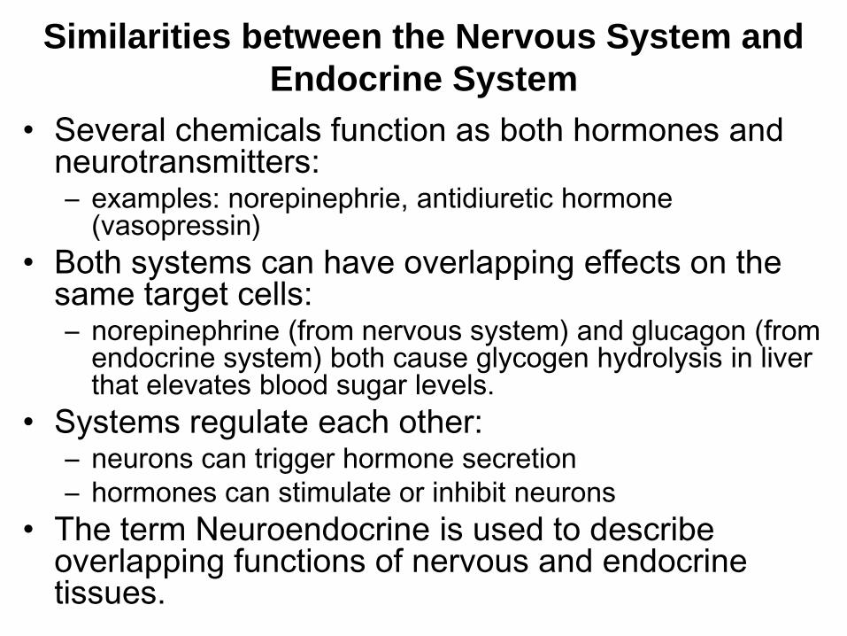
Similarities between the Nervous System and
Endocrine System
• Several chemicals function as both hormones and neurotransmitters: – examples: norepinephrie, antidiuretic hormone
(vasopressin)
• Both systems can have overlapping effects on the same target cells: – norepinephrine (from nervous system) and glucagon (from
endocrine system) both cause glycogen hydrolysis in liver that elevates blood sugar levels.
• Systems regulate each other: – neurons can trigger hormone secretion
– hormones can stimulate or inhibit neurons
• The term Neuroendocrine is used to describe overlapping functions of nervous and endocrine tissues.
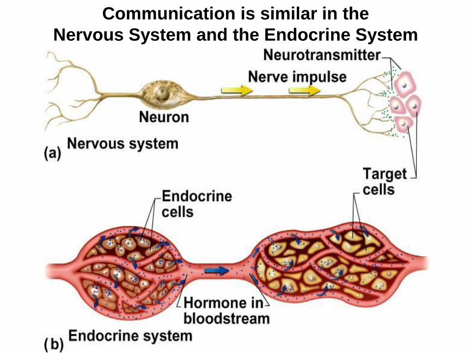
Communication is similar in the
Nervous System and the Endocrine System

Differences between the
Nervous System and Endocrine System • Means of communication:
– nervous system has both electrical and chemical methods
– endocrine system has only chemical methods
• Speed and persistence of response:
– nervous system reacts quickly (1 - 10 msec) and stops quickly
– endocrine system reacts more slowly (hormone release in seconds or days) and effect may continue for weeks
• Adaptation to long-term stimuli:
– nervous system adapts quickly and response declines quickly
– endocrine system has more persistent responses
• Area of effect:
– nervous system effects are very specific (one cell or organ)
– endocrine system usually has more general, widespread effects on many organs

Major Organs of the Endocrine System

Hypothalamus
17-13
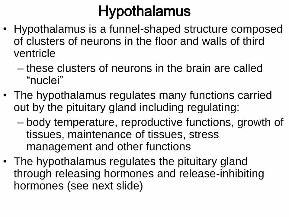
Hypothalamus • Hypothalamus is a funnel-shaped structure composed
of clusters of neurons in the floor and walls of third ventricle
– these clusters of neurons in the brain are called “nuclei”
• The hypothalamus regulates many functions carried out by the pituitary gland including regulating:
– body temperature, reproductive functions, growth of tissues, maintenance of tissues, stress management and other functions
• The hypothalamus regulates the pituitary gland through releasing hormones and release-inhibiting hormones (see next slide)

Hypothalamus / Pituitary Axis
Hypothalamic Hormone Effect on Anterior Pituitary
TRH: Thyrotropin-releasing H. TSH secretion
CRH: Corticotropin-releasing H. ACTH secretion
GnRH: Gonadotropin-releasing H. FSH and LH secretion
GHRH: Growth hormone-releasing H. GH secretion
Somatostatin inhibits GH and TSH secretion
PIH: Prolactin-inhibiting H. (dopamine) inhibits PRL secretion


Pituitary Gland
17-17

Pituitary Gland (Hypophysis)
• The Pituitary is connected to the hypothalamus by a stalk called the infundibulum.
• The Pituitary is located in the sella turcica of the sphenoid bone of the skull.
• The Pituitary Gland is about 1.3 cm (about ½ inch) diameter
• 3 lobes of the Pituitary Gland:
– Anterior Lobe (pars distalis, adenohypophysis) develops from oral ectoderm of the roof of the embryonic mouth (pharynx) forming Rathke’s (hypophyseal) pouch.
– Posterior Lobe (pars nervosa, neurohypophysis) develops from a neurohypophyseal bud of neural ectoderm from the floor of the diencephalon.
– Intermediate Lobe (pars intermedia) tissue is induced from oral ectoderm that contacts neural ectoderm.

Representation of the actual size of embryos from
zygote to the eighth week of development.
http://missinglink.ucsf.edu/lm/IDS_101_embryology_basics/CoreText.htm

Embryonic Development of Pituitary

(b) (c) (d)
Anterior
Lobe
Posterior Lobe
Hypophyseal Pouch
Floor of
Diencephalon
Roof of the
Mouth

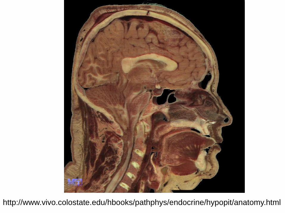
http://www.vivo.colostate.edu/hbooks/pathphys/endocrine/hypopit/anatomy.html

Pituitary Gland Anatomy and
Hormones of the Posterior Lobe
anterior posterior

• Posterior Lobe produces, stores and releases two hormones: – Oxytocin (OT)
– Vasopressin (Antidiuretic Hormone – ADH)
• OT and Vasopressin are produced by neurons in the hypothalamus and are transported down axons to the posterior lobe
• OT is produced by the paraventricular nucleus
• Vasopressin is produced by the supraoptic nucleus
• Note similarity in molecular structure of the two hormones
Pituitary Hormones of the Posterior Lobe

Posterior Lobe Hormone Actions: • Vasopressin (ADH)
– targets kidneys to water retention, reduce urine production
– called vasopressin because it can cause vasoconstriction
– also functions as a neurotransmitter in the brain
• Oxytocin
– stimulates labor contractions during childbirth
– stimulates flow of milk during lactation
– promotes emotional bonding between lactating mother and infant
– promotes feelings of sexual satisfaction and emotional bonding between partners
– surge of oxytocin is released during sexual arousal and orgasm causing smooth muscle contractions in uterus and and propulsion of semen

• Melaonotroph cells of the intermediate lobe produce Melanocyte-Stimulating Hormone (MSH)
– Intermediate Lobe is present in human fetuses but quickly degenerates into non-functional cysts in adults.
– Human brain neurons produce a large prohormone, POMC (pro-opiomelanocortin), which is cleaved into several smaller products including MSH, ACTH, endorphins, b-lipotropic hormone and other neurohormones. MSH functions as a neurotransmitter in the human brain.
– In other animals, MSH stimulates pigment cells to produce more melanin pigment which can darken skin, hair or feathers
Pituitary Intermediate Lobe

1. FSH (follicle stimulating hormone)
2. LH (luteinizing hormone)
3. TSH (thyroid stimulating hormone)
4. ACTH (adrenocorticotropic hormone)
5. PRL (prolactin)
6. GH (growth hormone)
6 Hormones of the Anterior Lobe

6 Anterior Pituitary Hormones and their Target Organs
• Axis refers to the regulatory interactions between the hypothalamus, pituitary and other endocrine glands.

Gonadotropin- releasing hormone controls FSH + LH release Thyrotropin- releasing hormone Corticotropin- releasing hormone Prolactin- releasing hormone Prolactin- inhibiting hormone GH- releasing hormone Somatostatin
Hypothalamic-Hypophyseal Portal System
• Releasing and inhibiting hormones secreted by the hypothalamus are absorbed by hypothalamic capillaries that drain into portal venules that connect to capillaries in the anterior pituitary

Actions of Anterior Lobe Hormones
• FSH (secreted by gonadotrope cells)
– female: stimulates development of follicles in ovaries
– male: stimulates production of sperm in testes
• LH (secreted by gonadotrope cells)
– female: stimulates ovulation and maintains the corpus
luteum to secrete progesterone and estrogen
– male: stimulates interstitial cells of testes to secrete
testosterone
• TSH (secreted by thyrotrope cells)
– stimulates growth of thyroid gland and the secretion of
thyroid hormone

• ACTH (secreted by corticotropes)
– regulates response to chronic stress, stimulates
adrenal cortex to secrete corticosteroids that
regulate glucose, fat and protein metabolism
• PRL (secreted by lactotropes)
– female: milk production by mammary glands
– male: enhances secretion of testosterone by testes
• GH (secreted by somoatotropes)
– see next 2 slides for functions of GH
Actions of Anterior Lobe Hormones continued:

• GH is also called somatotropin
• GH is secreted by somatotropes of the anterior pituitary
• GH promotes tissue growth through multiple mechanisms:
– directly affects mitosis and cellular differentiation
– stimulates liver to produce Insulin-like Growth Factors (IGF)
that are also called somatomedins.
– IGFs last 60x longer than than GH (20 hours vs 20 minutes)
• Functions of GH and IGF:
– protein synthesis and protein catabolism
– lipid metabolism which spares proteins and carbohydrates
– Electrolyte balance: promotes Na+, K+, Cl- retention and Ca+2
absorption
Growth Hormone

• During childhood and adolescence GH causes bone, cartilage and muscle growth
• GH in adults: – increases osteoblastic activity and appositional growth
resulting in bone thickening and remodeling
• GH in old age: – blood concentration of GH normally decreases by age 75
to ¼ of that of adolescents
– low GH in old age results in decrease protein synthesis that can cause wrinkling of skin and less muscle strength and muscle mass
• Levels of GH – are higher after high protein meals, after vigorous exercise,
and during the first 2 hours of deep sleep
– GH also increases after trauma
Growth Hormone and Aging

Robert Pershing Wadlow
(in 1938, at age 20 with
actresses Maureen
O’Sullivan, left, and Ann
Morris) had grown to a
record height of 8’11” by
the time of his death at
age 22. Smithsonian August 2005 p. 76.
Growth Hormone
Disorders Hypersecretion of growth
hormone during childhood can lead to gigantism.
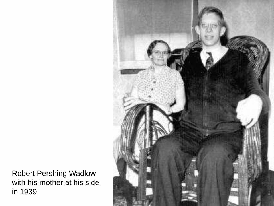
Robert Pershing Wadlow
with his mother at his side
in 1939.

In 2008, Leonid Stadnik
was named the world's
tallest man. Standing at 8
feet, 5 inches.
Here he walks near his
home in the tiny village of
Podolyantsi in central
Ukraine.

Growth Hormone Disorders continued
• Hypersecretion of growth hormone in old age leads to acromegaly. – symptoms of acromegaly include thickening of hand,
foot, jaw and brow bones and growth of soft tissues particularly in the nose and ears.

There are over 200 different types of dwarfism, all of which involve bone growth disorders (osteodysplasia) that result in short stature (adult height less than 4 ft. 10 in. tall).
Primordial Dwarfism is a group of disorders in which growth is proportional but severely delayed, beginning in the womb. This results in some of the smallest people in the world.
The individual pictured has Majewski osteodysplastic primordial dwarfism (MOPD) Type II. Only about 100 individuals worldwide have been identified as having MOPD type II. Both males and females of all ethnic backgrounds are affected. Some families have more than one child with MOPD Type II, which suggests that the disorder is inherited in an autosomal recessive manner.
• Majewski osteodysplastic primordial dwarfism type II (MOPD II): natural history and clinical findings. Hall, Flora, Scott, Pauli, Tanaka, Am J. Med Genet A. 2004 Sep 15 ; 130A(1):55-72. 7 pounds at age 2

Pineal Gland (Epiphysis) • Shaped like a PINE CONE
• The pineal gland secretes melatonin in darkness.
• The pineal is inhibited by light.
• Pinealocytes convert tryptophan into serotonin during the day (in bright light), then converts serotonin into melatonin at night (in darkness).
• Peak melatonin secretion in humans occurs at 1-5 years old.
• Melatonin inhibits puberty in humans. Levels drop 75% at puberty.
• Melatonin levels are correlated with human activity cycles (biorhythms).
• High melatonin levels are correlated with SAD and PMS with symptoms including:
– depression
– sleepiness, irritability
– carbohydrate craving
• Melatonin levels may be manipulated by phototherapy and can reduce the affects of jet lag.
• In other animals, high levels of melatonin inhibit reproduction and inhibit melanogenesis which affects seasonal reproductive timing, and fur or feather color.

Pineal Gland (Epiphysis)

Pineal Gland
Histology

Melatonin
Secretion

Winter: Long nights, pinealocytes uninhibited, high levels of
melatonin inhibit pigment cells and gonads.
snowshoe hare
snowy owl

Summer: Long days, pinealocytes inhibited, low levels of
melatonin, uninhibited pigment cells and gonads.
snowshoe hare
snowy owl

Thymus • Location: mediastinum,
superior to heart
• Involution after puberty (converts to fibrous connective tissue and adipose)
• Secretes the hormones Thymosin and Thymopoietin that regulate development of T-lymphocytes (type of white blood cell)

Thymus Gland
adult thymus
17-47 17-47
infant thymus

Athymic Nude Mice play an
important role in immunology
research and cancer research.
These mouse colonies must be
maintained in absolutely sterile
conditions.

Severe Combined Immunodeficiency
Disease
• Hereditary lack of T and B
lymphocytes
• Extreme vulnerability to
opportunistic infection

Thyroid Gland Anatomy
• Thyroid gland is located on the anterior and lateral sides of trachea.
• 2 large lobes of thyroid tissue are connected by an isthmus of thyroid tissue.
• Thyroid gland is highly vascular and receives a large volume of blood flow.

Thyroid Gland Histology

Cells of the Thyroid Gland • Thyroid Follicles
– Spheres of simple cuboidal cells (follicular cells) filled with a colloid of 2 thyroid hormones, T3 and T4 bound to thyroglobulin
– T3 = triiodothyronine T4 = tetraiodothyronine
– T3 contains 3 iodine atoms, T4 contains 4 iodine atoms
– Follicles produce 90% T4 and 10% T3
– T3 is more potent than T4
– In circulation and in cells, T4 is broken down into T3
– Thyroid hormone functions:
• metabolic rate, O2 consumption and heat production
• heart rate and respiratory rate
• stimulates appetite and breakdown of carbohydrates, lipids and proteins
• C-cells (calcitonin cells or parafollicular cells)
– cells are located in between the follicles.
– cells produce the hormone calcitonin that blood Ca+2 and promotes Ca+2 deposition in bone, especially in children.

Thyroid Hormone Synthesis
Release of T3 and T4 into the blood

Thyroid Hormone Synthesis Thyroglobulin
(3) Iodine added to a tyrosine forms monoiodothyronine (MIT). (4) Iodine added to MIT forms diiodothyronine (DIT). (5) DIT + MIT = T3.
(6) Lysosome enzymes cleave T3 from the thyroglobulin. Arrows
indicate where adding another iodine would form T4.
I I
I I

Thyroid Hormone Effects
• T3 and T4 dissociate from thyroxine-binding globulin (TBG), leave bloodstream and enter target cell. T4 is converted into T3.
• T3 can
– bind to receptors on mitochondria that rate of aerobic respiration
– activate ribosomes that protein synthesis
– enter the nucleus and activate genes and protein synthesis
• Example: one protein that can be produced is Na+-K+ ATPase and the activity of this protein generates heat
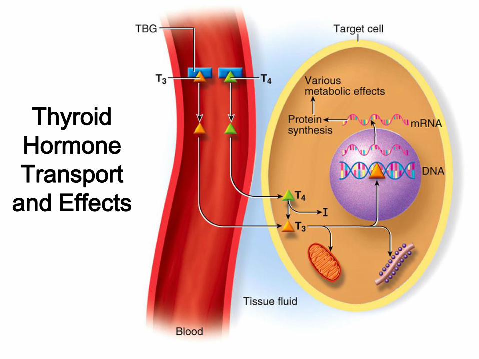
Thyroid
Hormone
Transport
and Effects

Thyroid Hormone Control
High thyroid hormone levels inhibit release of
tropic hormones (TRH, TSH).

Thyroid Gland Disorders
• Congenital hypothyroidism ( TH)
– infant suffers abnormal bone development, thickened
facial features, low body temperature, lethargy, brain
damage. Can be treated with synthroid.
• Myxedema (adult hypothyroidism, TH)
– low metabolic rate, sluggishness, sleepiness, weight
gain, puffy face, constipation, dry skin and hair, cold
sensitivity, blood pressure and tissue swelling
• Graves Disease (a type of hyperthyroidism)
– antibodies mimic TSH, TH secretion, exophthalmos
• Endemic Goiter (goiter = enlarged thyroid gland)
– dietary iodine deficiency, low TH, low - feedback, TSH

Congenital Hypothyroidism
Congenital hypothyroidism
results from chronically low
levels of TH.
Infants suffer abnormal
bone development,
thickened facial features,
low body temperature,
lethargy, brain damage.
Can be treated with
synthroid.

Myxedema
Myxedema (adult hypothyroidism) chronically low TH results
in low metabolic rate, sluggishness, sleepiness, weight gain,
puffy face, constipation, dry skin and hair, cold sensitivity,
increased blood pressure and tissue swelling.

Exophthalmos – a symptom of Graves’ Disease
Graves Disease (a type of hyperthyroidism) results from
production of antibodies that mimic TSH resulting in increased
TH secretion.

Endemic Goiter
Endemic Goiter (goiter = enlarged thyroid gland) due to dietary iodine
deficiency results in low TH synthesis and reduced feedback resulting in
increased TSH and thyroid growth.

Parathyroid Glands • Multiple lobes of tissue on
the posterior surface of the thyroid gland. There may also be other ectopic sites.
• Produces Parathyroid Hormone (PTH or parathormone)
– PTH increases blood Ca+2 levels and synthesis of calcitriol (Vitamin D) resulting in:
• absorption of Ca+2
• urinary Ca+2 excretion
• urinary PO-4 excretion
• bone erosion

Parathyroid Disorders
• Hypoparathyroid
– usually results from surgical excision during
thyroid surgery
– can result in fatal tetany in 3-4 days from low Ca+2
• Hyperparathyroid
– Excess PTH secretion can result from a tumor
– causes soft, fragile and deformed bones,
extremely high blood Ca+2, and renal calculi
(kidney stones).

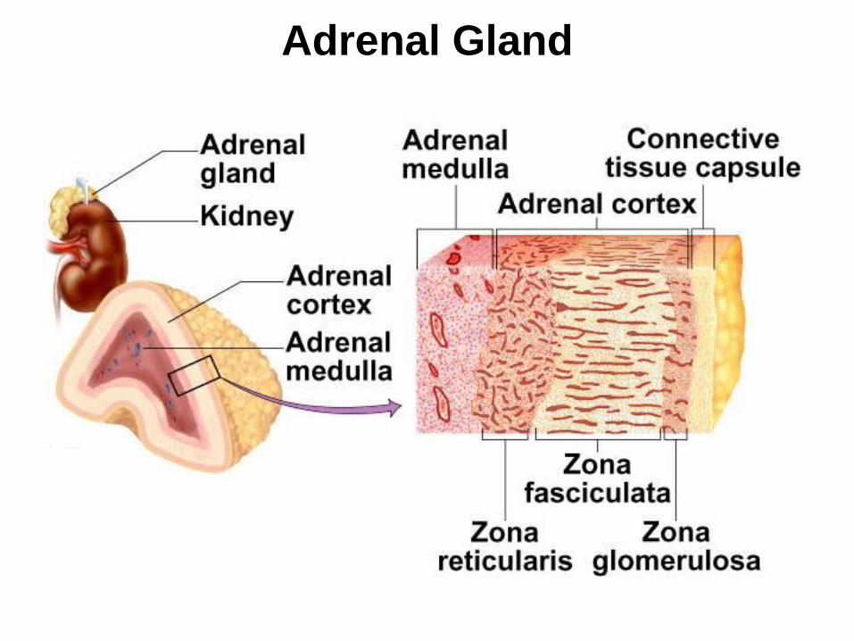
Adrenal (Suprarenal) Glands
17-65 17-65
Adrenal Gland
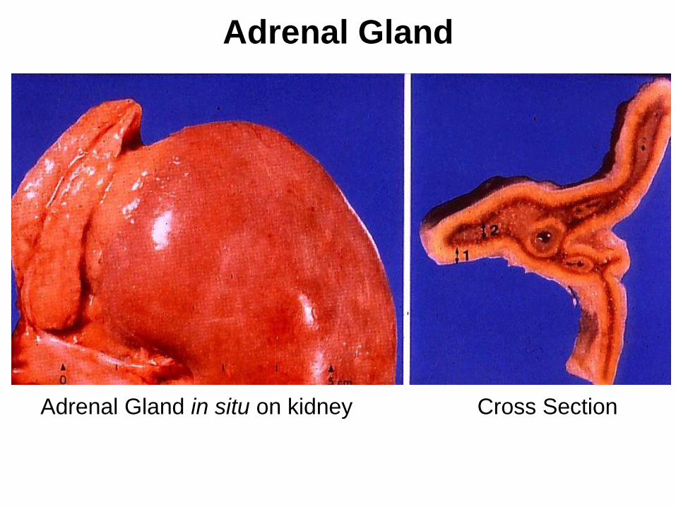
Adrenal Gland in situ on kidney Cross Section
Adrenal Gland

Adrenal Medulla
• Postganglionic sympathetic neurons innervated by
sympathetic preganglionic fibers.
– consists of modified neurons called chromaffin cells
– stimulation causes release of adrenaline into bloodstream
• Responds to ACUTE stress (fight or flight)
• Hormonal effect is relatively long lasting (minutes)
compared to nervous system (fractions of a second).
– increases BP and heart rate
– increases blood flow to skeletal muscle
– increases pulmonary air flow
– stimulates gluconeogenesis and glycogenolysis
– decreases digestion and urine formation

Adrenal Cortex
• Response to CHRONIC stress (long duration – weeks or more)
• Three Layers - (outer) zona glomerulosa, (middle) zona fasciculata, (inner) zona reticularis
• All three layers produce Corticosteroids from cholesterol
– Zona Glomerulosa produces the mineralocorticoid aldosterone
• controls electrolyte balance by conserving Na+
– Zona Fasciculata produces the glucocorticoids cortisol, corticosterone and cortisone
• cortisol stimulates fat and protein catabolism, gluconeogenesis (from amino acids and fatty acids) and release of fatty acids and glucose into blood
• anti-inflammatory effect suppresses the immune system
– Zona Reticularis produces the sex steroids testosterone and estrogen
• important source of estrogen in women after after menopause

Steroid Hormone Synthesis
All steroid hormones are synthesized from cholesterol – the various hormones differ in functional groups attached to a 4-ringed steroid backbone

Adrenal Cortex Disorders • Cushing’s Syndrome is excessive secretion of
cortisol
– can be caused by pituitary hypersecretion of ACTH, ACTH-secreting tumor, over activity of adrenal cortex
– causes hyperglycemia, hypertension, weakness, edema
– muscle and bone loss occurs with protein catabolism
– buffalo hump and moon face caused by abnormal fat deposition between shoulders and in face
• Adrenogenital Syndrome (AGS)
– adrenal androgen hypersecretion causes enlargement of external sexual organs in children and early onset of puberty
– can cause masculinizing effects on females (deeper voice, beard growth, male-like genitalia)
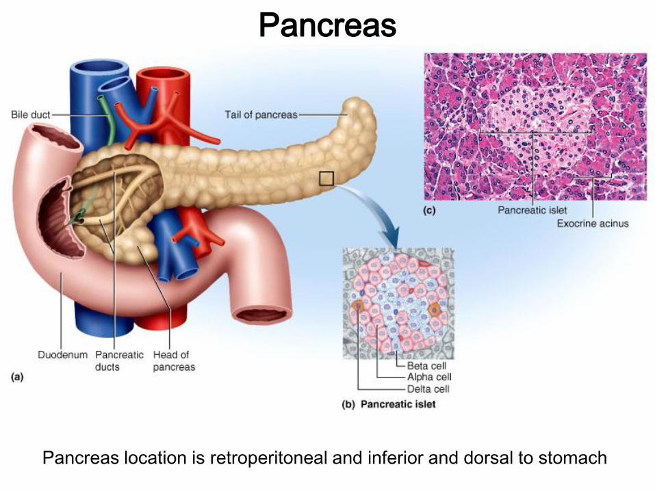
Pancreas
Pancreas location is retroperitoneal and inferior and dorsal to stomach

Products of the Pancreas
• 98% of pancreas is EXOCRINE – produces digestive enzymes and bicarbonate from acinar tissue.
• 2% of pancreas is ENDOCRINE – produces at least 5 hormones – best understood are insulin, glucagon and somatostatin.
• Endocrine tissue is in 1-2 million pancreatic islets of Langerhans.

Pancreatic Hormones • Insulin (from cells)
– insulin is secreted when blood sugar levels are high as in after a meal
– insulin receptors are on most cells of the body
– insulin stimulates cells to take up glucose and amino acids
– insulin antagonizes glucagon
• Glucagon (from cells) – glucagon is secreted when blood glucose levels are low as it is
between meals
– glucagon stimulates glycogenolysis (breakdown of glycogen), fat catabolism (release of free fatty acids) and promotes absorption of amino acids for gluconeogenesis (production of glucose from amino acids)
• Somatostatin (from delta cells) – secreted with rise in blood glucose and amino acids after a meal
– paracrine secretion modulates secretion of and cells
• Hyperglycemic hormones raise blood glucose: glucagon, epinephrine, norepinephrine, cortisol and corticosterone
• Hypoglycemic hormone, insulin, lowers blood glucose

Diabetes Mellitus
• Diabetes Mellitus is a disease resulting from the hyposecretion or inaction of insulin.
• Extremely high blood glucose levels can exceed the kidney tubule transport maximum of glucose reabsorption and cause osmotic diuresis: glucose remains in urine, osmolarity increases and draws water into urine.

Diabetes Mellitus • Signs and symptoms of Diabetes Mellitus
– polyuria, polydipsia, polyphagia
– hyperglycemia, glycosuria, ketonuria
– blood glucose levels up to 100 mg/dL are considered normal. (mg/dL = milligrams per deciliter)
– Diabetes is typically diagnosed when fasting blood glucose levels are 126 mg/dL or higher.
– Glucose is a major source of energy for most cells of the body. Some cells (for example, brain and red blood cells), are almost totally dependent on blood glucose as a source of energy. The brain requires that glucose concentrations in the blood remain within a certain range in order to function normally. Concentrations of less than 30 mg/dL or greater than 300 mg/dL can produce confusion or unconsciousness.
– Ketones are a metabolic breakdown product of fatty acid metabolism. Some cells will use fatty acids for fuel if glucose is not available and produce ketones as waste.

Types of Diabetes Mellitus • Type I (insulin dependent diabetes mellitus = IDDM)
– 10% of diabetes mellitus cases
– Caused by autoimmune destruction of cells
– Usually diagnosed at about age 12
– Treated with diet, exercise, monitoring of blood glucose and periodic injections of insulin (new delivery methods include pumps and inhalers).
• Type II (non-insulin dependent diabetes mellitus = NIDDM)
– 90% of diabetes mellitus cases
– Patients develop insulin resistance which is a failure of target cells to respond to insulin due to receptor downregulation (see next slide).
– 3 major risk factors are heredity, age (40+) and obesity
– Treated with programs for weight loss through diet and exercise.
– Some patients are helped by oral medications (like glucophage) that decrease intestinal absorption of glucose, decrease hepatic glucose production and improve target cell sensitivity to insulin.

Receptor Regulation on Target Cells
• Down-regulation: Long-term exposure to high levels of a hormone can result in target cells reducing the number of receptors to the hormone as in Type II Diabetes.
• Up-regulation results from chronically low levels of hormone.

Pathology of Diabetes
• Acute pathology: cells cannot absorb glucose, so
instead they rely on fat and proteins resulting in weight
loss and weakness.
– fat catabolism FFA’s and ketones in blood
– ketonuria promotes osmotic diuresis, loss of Na+, K+
– ketoacidosis occurs as ketones blood pH
• Chronic pathology:
– chronic hyperglycemia leads to neuropathy and
cardiovascular damage:
• retina and kidney damage (common in type I),
atherosclerosis leading to heart failure (common
in type II), and gangrene
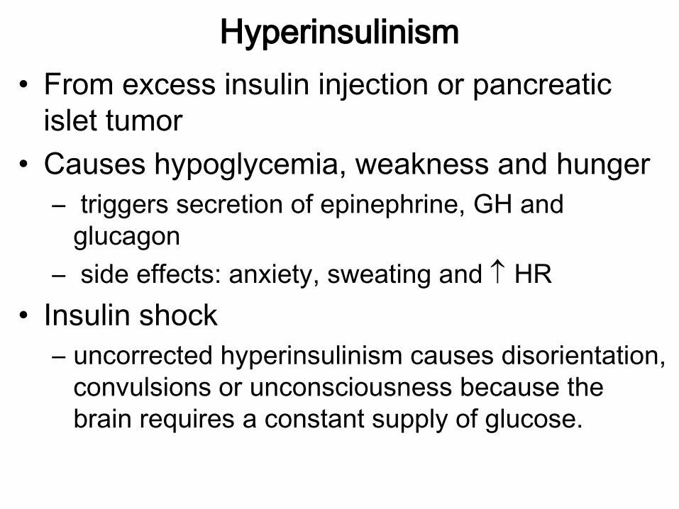
Hyperinsulinism
• From excess insulin injection or pancreatic
islet tumor
• Causes hypoglycemia, weakness and hunger
– triggers secretion of epinephrine, GH and
glucagon
– side effects: anxiety, sweating and HR
• Insulin shock
– uncorrected hyperinsulinism causes disorientation,
convulsions or unconsciousness because the
brain requires a constant supply of glucose.

Gonadal Hormones: the Ovary
Follicle = egg surrounded by granulosa cells
(source of
estrogen)
(source of estrogen
and progesterone)

Ovary
• Granulosa cells in wall of ovarian follicle
– stimulated by follicle-stimulating hormone (FSH)
– produce estrogen during the menstrual cycle
• Corpus Luteum: follicle after ovulation
– stimulated by luteinizing hormone (LH)
– produces estrogen and progesterone for 12 days if no fertilization or 8-12 weeks with pregnancy
• Functions of estrogen and progesterone
– development of female reproductive system and physique including bone growth
– regulates menstrual cycle, sustains pregnancy
– prepares mammary glands for lactation
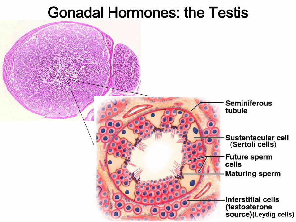
Gonadal Hormones: the Testis
(Sertoli cells)
(Leydig cells)

Testes
• Interstitial Cells (Leydig cells)
– between seminiferous tubules
– Stimulated by LH and FSH
– produce testosterone (and small amounts of estrogen)
– testosterone causes development of male reproductive system and physique
– sustains sperm production and sex drive
• Sustentacular Cells (Sertoli cells)
– secrete inhibin which regulates sperm production rate.

Chemical Classification of Hormones • Steroid Hormones are all derived from cholesterol
– aldosterone, cortisol, cortisone, corticosterone, progesterone, testosterone, estrogen, calcitrol
• Oligopeptides are chains of 3-10 amino acids
– angiotensin II, vasopressin (ADH), GnRH, Oxytocin, TRH
• Polypeptides are chains of 14-199 amino acids
– ACTH, atrial naturitic peptide, calcitonin, CRH, glucagon, growth hormone, GHRH, insulin, PTH, PRL, somatostatin
• Glycoproteins are two amino acid chains with a carbohydrate
– FSH, LH, TSH, HCG, inhibin
• Monoamines or Biogenic Amines are all made from the amino acid tyrosine except melatonin which is made from tryptophan
– dopamine, epinephrine, norepinephrine, serotonin, melatonin, T3, T4

Transport and Action of Hormones • Hydrophobic
hormones (steroids and thyroid hormone) are carried in the blood on carrier proteins to increase their solubility. Free hormone can penetrate plasma membrane and enter the nucleus.
• Hydrophilic hormones (monoamines and peptides, glycoproteins) are soluble in plasma, but can not pass through membranes so they must bind to cell-surface receptors.

Steroid Hormones are
derived from cholesterol
and diffuse easily through
the plasma membranes of
cells.
Once in the cell, the
hormone binds to a
receptor forming a
hormone-receptor
complex.
The complex can bind to
DNA in the nucleus and
either activate or inhibit the
genes.
Activation of genes leads
to protein synthesis.


http://highered.mcgraw-
hill.com/sites/0072437316/student_view0/chapter47/
animations.html#
Mechanism of Steroid Hormone Action Animation
Other Endocrine System Animations
http://nhscience.lonestar.edu/biol/ap1int.htm#endocrine


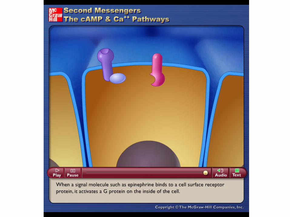
Hydrophilic Hormones using the
cAMP Second Messenger
note: caffeine inhibits
breakdown of cAMP


Hydrophilic hormones may also work through a phospholipase-
mediated second messenger system.


Eicosanoids • Diacylglycerol Arachidonic Acid Eicosanoids
• Diacylglycerol is converted into Arachidonic Acid by a phospholipase
enzyme and this process can be inhibited by steroidal anti-inflamitants like
cortisol and corticosterone
• Arachidonic acid is converted into various eicosanoids by 2 enzymes:
– lipoxygenase converts arachidonic acid into leukotrienes that
mediate allergic and inflammatory reactions.
– cyclooxygenase converts arachidonic acid into prostacyclins,
thromboxanes and prostaglandins and can be inhibited by NSAIDs:
• prostacyclins: produced by blood vessel walls, inhibits blood
clotting and vasoconstriction
• thromboxanes: produced by blood platelets after injury, they
override prostacyclin and stimulate vasoconstriction and clotting
• prostaglandins: diverse group including:
– PGE’s: relaxes smooth muscle in bladder, intestines,
bronchioles, uterus and stimulates contraction of blood vessels
– PGF’s: opposite effects

Eicosanoid
Synthesis
NSAIDs = non-steroidal anti-inflammatory drugs (aspirin, ibuprofen, naproxen,
indomethacin) are also called COX inhibitors since they inhibit cyclooxygenase
SAIDs = steroidal anti-inflammatory drugs like cortisol, cortisone.
Diacylglycerol

Eicosanoids and Headaches
• The vascular theory of headaches states that
the constriction of peripheral arteries and the
dilation of cerebral blood vessels lead to a
headache. The vasoconstiction leads to a loss
of cerebral oxygen in the blood, therefore, the
compensatory mechanism is vasodilation. The
dilation and constriction of vessels is sensed
as pain.


Endocrine Functions of Other Organs
• Heart produces atrial natriuretic peptide regulates blood pressure
• Liver – source of IGF that works with GH
– secretes about 15% of erythropoietin
– secretes angiotensinogen (a prohormone involved in blood pressure regulation)
• Kidneys produce 85% of erythropoietin which stimulates bone marrow to produce RBC’s
• Stomach and Small Intestines produce 10 enteric hormones that coordinate digestive motility and secretion
• Placenta secretes estrogen, progesterone and other hormones that regulate pregnancy, stimulate development of fetus and mammary glands