Malignant Clinical Course of Histologically BenignOvarian Struma: case report and literature reviewAleksandra Asaturova ( [email protected] )
FSBI National Medical Research Center for Obstetrics Gynecology and Perinatology named afterAcademician V.I.Kulakov: FGBU Naciona'lnyj medicinskij issledovatel'skij centr akuserstva ginekologii iperinatologii imeni Akademika V I Kulakova https://orcid.org/0000-0001-8739-5209A. Magnaeva
FSBI National Medical Research Center for Obstetrics Gynecology and Perinatology named afterAcademician V.I.Kulakov: FGBU Naciona'lnyj medicinskij issledovatel'skij centr akuserstva ginekologii iperinatologii imeni Akademika V I KulakovaA. Tregubova
FSBI National Medical Research Center for Obstetrics Gynecology and Perinatology named afterAcademician V.I.Kulakov: FGBU Naciona'lnyj medicinskij issledovatel'skij centr akuserstva ginekologii iperinatologii imeni Akademika V I KulakovaV. Kometova
FSBI National Medical Research Center for Obstetrics Gynecology and Perinatology named afterAcademician V.I.Kulakov: FGBU Naciona'lnyj medicinskij issledovatel'skij centr akuserstva ginekologii iperinatologii imeni Akademika V I KulakovaE. Karamurzin
Mediclinic Middle EastS. Martynov
FSBI National Medical Research Center for Obstetrics Gynecology and Perinatology named afterAcademician V.I.Kulakov: FGBU Naciona'lnyj medicinskij issledovatel'skij centr akuserstva ginekologii iperinatologii imeni Akademika V I KulakovaYu. Lipatenkova
FSBI National Medical Research Center for Obstetrics Gynecology and Perinatology named afterAcademician V.I.Kulakov: FGBU Naciona'lnyj medicinskij issledovatel'skij centr akuserstva ginekologii iperinatologii imeni Akademika V I KulakovaL. Adamyan
FSBI National Medical Research Center for Obstetrics Gynecology and Perinatology named afterAcademician V.I.Kulakov: FGBU Naciona'lnyj medicinskij issledovatel'skij centr akuserstva ginekologii iperinatologii imeni Akademika V I Kulakova
Case Report

Keywords: Struma ovarii, peritoneal strumosis, highly differentiated follicular carcinoma arising in strumaovarii
Posted Date: June 24th, 2021
DOI: https://doi.org/10.21203/rs.3.rs-646069/v1
License: This work is licensed under a Creative Commons Attribution 4.0 International License. Read Full License
Malignant Clinical Course of Histologically Benign Ovarian Struma: case report and
literature review
A.Asaturova1, A. Magnaeva1, A. Tregubova1, V. Kometova1, E. Karamurzin2, S.
Martynov1, Yu. Lipatenkova1, L. Adamyan1
1FSBI National Medical Research Center for Obstetrics Gynecology and Perinatology named after Academician V.I.Kulakov (Moscow, Russia)
2Mediclinic Middle East (Dubai, United Arab Emirates) Corresponding author: Aleksandra V Asaturova, FSBI National Medical Research Center for Obstetrics Gynecology and Perinatology named after Academician V.I.Kulakov, 117198, 4
Oparina st., Moscow, Russia; [email protected]
Abstract
Background: Struma ovarii is a variant of monodermal teratoma, consisting of morphologically
benign, atypical, or frankly malignant thyroid tissue. Morphologic features may or may not correlate
with biologic behavior. Albeit this case report is not unique, ovarian tumor developed with peritoneal
dissemination and bone metastasis, which is highly unlikely clinical complication. Additionally, we
summarized previously cases of struma ovarii with an emphasis on correlation between morphological
appearances, clinical course and providing treatment.
Case presentation: We present the 38-year-old patient who was hospitalized for ovarian tumor. The
diagnostic laparoscopy revealed lesions of peritoneum, sigmoid serosa and omentum and left ovarian
mass. We diagnosed left ovarian mature teratoma without struma tissue and metastatic lesions with
struma morphology which can be related to her history of left ovarian struma in 2016. Taking into
account the metastatic lesions revealed in 2020, the tumor removed in 2016 was assessed as highly
differentiated follicular carcinoma arising in struma ovarii.
Conclusions: Prediction of biologic behavior of struma ovarii is still to be to diagnostic challenge,
therefore multidisciplinary approach including clinical and laboratory findings, radiologic details and
histopathological features is required. Providing additional data, the present case report contributes to
expending the knowledge of these peculiar neoplasms.
Key words: Struma ovarii, peritoneal strumosis, highly differentiated follicular carcinoma arising in
struma ovarii.
Introduction
Struma ovarii is a monodermal ovarian teratoma composed predominantly of thyroid
tissue, a rare neoplasm comprising 1% of all ovarian tumors and 3% cases of ovarian teratomas (1–3).
The tumor is usually an incidental finding of a pelvic mass with or without abdominal pain. Uncommon
clinical manifestations may include the following: hyperthyroidism, dropsy and Meigs' syndrome (1).
Emerging evidence indicates that histological features and growth patterns of struma ovarii seem no
to correlate with biological behavior. For instance, morphologically benign struma ovarii may be
associated with extraovarian spreading, known as peritoneal strumosis. Peritoneal strumosis is an
infrequent condition, where both peritoneal implants and primary ovarian tumor are morphologically
bland (4,5). The phenomenon of extraovarian spreading is peculiar due to several reasons. Firstly, the
mechanisms of dissemination are not well understood, albeit accumulating data suggests the molecular
alterations occurring in benign peritoneal strumosis comprise ALK, EGFR and BRAF mutations may
contribute to spread (6,7). Secondly, the treatment approaches are not standardized, and currently may
include various doses of radioactive ablation with I-131 and thyroidectomy (4,8,9). Thirdly, the
differential diagnosis includes secondary metastatic involvement by an adenocarcinoma,
neuroendocrine carcinoma, follicular thyroid carcinoma etc. (10,11).
We report a clinical case of morphologically “benign” struma ovarii that subsequently
progressed with development of peritoneal deposits and a remote osseous metastatic involvement
following 4 years after struma ovarii resection.
Case report
A 38-year old woman with remote history of ovarian teratoma was admitted to
gynecological surgery department due to ovarian tumor recurrence. She had been operated 3 times
previously: in 1999 due to mature teratoma of the right ovary (right-side salpingo-oophorectomy); in
2006 due to mature teratoma of the left ovary (ovarian cystectomy); and in 2016 during pregnancy due
to proliferative struma of the left ovary (completion oophorectomy during caesarian section). The
mature teratomas resected in 1999 and 2006 consisted of ectodermal derivatives (squamous epithelium
and other adnexal structures) and mesodermal derivate (mature cartilage).
Clinical presentation.
In May of 2020 the patient was examined due to lower back pain. MRI revealed a lesion
involving the body of the L5 vertebrum measuring 6.2x3.4х2.7 cm, associated with a vertical fracture
line, spinal canal narrowing, left neural foramen reduction and nerve root compression. Then A99mTc
SestaMIBI whole body scan was performed. It demonstrated bone tissue remodeling in the lumbar
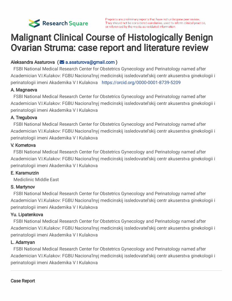
column but did not reveal any osteoblastic lesions. Core biopsy showed morphologically bland thyroid
tissue (Fig 1) and percutaneous image-guided vertebroplasty was done due to pathologic fracture L5
risk and severe back pain.
Figure 1. Core biopsy of the lumbar column. A. Hematoxylin and eosin stain, B. Thyroglobulin stain, x 200
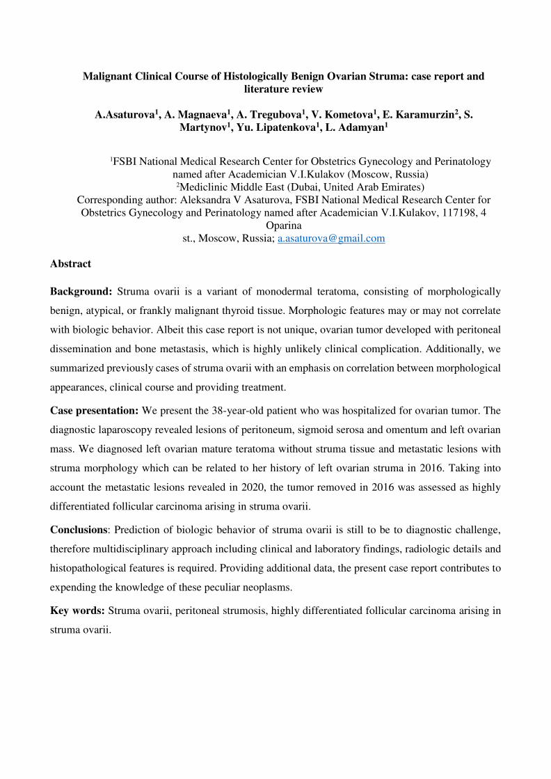
PET-CT was performed for TNM classification, revealing an osteolytic lesion in L5
vertebral body, as well as clearly outlined left ovarian mass sized 3.0х2.2 cm with
nonhomogeneous density and fat, highly suggestive of mature teratoma (Fig. 2).
Figure 2. A. PET image in transverse plane, confirming abnormal FDG uptake within the metastatic
lesion. B. CT image at the same level.
Taking into account the morphology of lumbar column metastasis thyroid gland USD
was performed to exclude thyroid gland pathology. It revealed a small nodule, however fine-needle
aspiration was benign (Bethesda diagnostic category II).
Given the patient’s past medical history, a metastasis from ovarian neoplasm was
suspected and the patient was admitted to surgery department of National medical center for
obstetrics, gynecology and perinatology named after V.I. Kulakov in September of 2020.
For preoperative preparation the pelvic USD was done. The ultrasound scan
demonstrated hyperechoic mass involving left ovary with the solid tumor sized 3.5 х 3.0 х 3.4 cm
with increased echogenicity and clear smooth contour, without vascularization zones. We
compared the current USD scan with previous USD scan (2016). The latter demonstrated solid and
cystic tumor with heterogeneous solid component, increased echogenicity and vascularization
zones at the periphery, 7.5 х 5.0 х 3.5 cm in diameter.
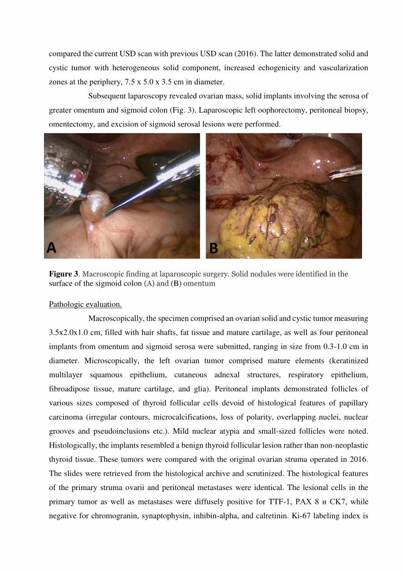
Subsequent laparoscopy revealed ovarian mass, solid implants involving the serosa of
greater omentum and sigmoid colon (Fig. 3). Laparoscopic left oophorectomy, peritoneal biopsy,
omentectomy, and excision of sigmoid serosal lesions were performed.
Figure 3. Macroscopic finding at laparoscopic surgery. Solid nodules were identified in the
surface of the sigmoid colon (A) and (B) omentum
Pathologic evaluation.
Macroscopically, the specimen comprised an ovarian solid and cystic tumor measuring
3.5x2.0x1.0 cm, filled with hair shafts, fat tissue and mature cartilage, as well as four peritoneal
implants from omentum and sigmoid serosa were submitted, ranging in size from 0.3-1.0 cm in
diameter. Microscopically, the left ovarian tumor comprised mature elements (keratinized
multilayer squamous epithelium, cutaneous adnexal structures, respiratory epithelium,
fibroadipose tissue, mature cartilage, and glia). Peritoneal implants demonstrated follicles of
various sizes composed of thyroid follicular cells devoid of histological features of papillary
carcinoma (irregular contours, microcalcifications, loss of polarity, overlapping nuclei, nuclear
grooves and pseudoinclusions etc.). Mild nuclear atypia and small-sized follicles were noted.
Histologically, the implants resembled a benign thyroid follicular lesion rather than non-neoplastic
thyroid tissue. These tumors were compared with the original ovarian struma operated in 2016.
The slides were retrieved from the histological archive and scrutinized. The histological features
of the primary struma ovarii and peritoneal metastases were identical. The lesional cells in the
primary tumor as well as metastases were diffusely positive for TTF-1, PAX 8 и CK7, while
negative for chromogranin, synaptophysin, inhibin-alpha, and calretinin. Ki-67 labeling index is
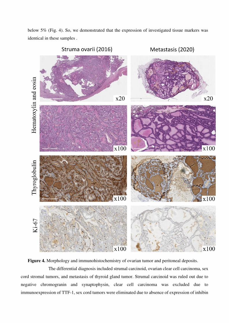
below 5% (Fig. 4). So, we demonstrated that the expression of investigated tissue markers was
identical in these samples .
Figure 4. Morphology and immunohistochemistry of ovarian tumor and peritoneal deposits.
The differential diagnosis included strumal carcinoid, ovarian clear cell carcinoma, sex
cord stromal tumors, and metastasis of thyroid gland tumor. Strumal carcinoid was ruled out due to
negative chromogranin and synaptophysin, clear cell carcinoma was excluded due to
immunoexpression of TTF-1, sex cord tumors were eliminated due to absence of expression of inhibin
A and calretinin negative stain, while thyroid gland tumor metastasis was rejected due to normal fine-
needle aspiration cytology and normal thyroid-related hormones values. No BRAF mutations in
ovarian struma or metastatic lesions were detected. Consequently, we can suppose that there is not
molecular evolution between the primary ovarian struma and the metastatic lesions.
Discussion
Struma ovarii is the most common type of mature monodermal teratoma. The entity is of
considerable interest because of its unique features. Foremost, histological appearance does not
accurately correlate with biological behavior. Somewhat paradoxically, morphological features that
characterized malignant potential for thyroid gland neoplasms cannot be extrapolated to struma ovarii
in terms of prediction of extraovarian dissemination. Struma ovarii without any evidence of
architectural or cytologic atypia could metastasize both to abdominal cavity and beyond (12,13). We
present a case of recurrent struma ovarii with development of peritoneal and extraperitoneal spread
following 4 years after the initial surgery. The resected tumor was devoid of histological features of
malignancy. The peritoneal deposits were found both in omentum and on sigmoid serosa,
histologically resembling a benign thyroid follicular lesion. Furthermore, an osteolytic metastasis
involving L5 vertebral body reveled similar morphology. Currently, the terminology of
morphologically benign struma ovarii with hematogenic, lymphogenic and peritoneal dissemination
remains controversial. The term metastatic MSO (malignant struma ovarii) does not fit the benign or
low-grade histologic appearances of the thyroid tissue in the anatomic sites of the tumor dissemination.
Some authors used the term strumosis for a long time (4,14,15), whereas others suggested the term of
“struma ovarii with extraovarian dissemination” (2). The most recent WHO classification of ovarian
tumors the term of highly differentiated follicular carcinoma of ovarian origin (HDFCO)/highly
differentiated follicular carcinoma arising in struma ovarii was coined as more appropriate for
describing extraovarian spreading, so the International Classification of Diseases for Oncology (ICD-
O) code is only presented for benign and malignant struma ovarii (16). It may seem feasible to use
ICD-O code 1 to reflect the difficulty to accurately predict the biologic behavior of struma ovarii (17).
Although it is exteremely difficult to suppose the further clinical course at the moment of struma
original diagnosis it is very important to make frozen sections. M. R. Quddus et al. shown that it helps
to improve the diagnostic accuracy when compared with gross examination, to differ ovarian struma
from other malignant and benign ovarian tumors and to provide the important information to the
surgeon in the operating room (35).
Due to the rarity of struma ovarii, the optimal management approaches and guidelines are
not well-established nor standardized. Putative initial treatment is resection of the primary tumor,
supplementing with thyroidectomy and radioactive ablation with I-131 (8,9,18) in order to exclude
metastatic thyroid gland malignancy, target the radioactive iodine to metastatic foci and to facilitate
monitoring of thyroglobulin levels after surgery. However, it is rather difficult to accurately evaluate
the benefits of the putative approach due to the rarity of the disease and, consequently, insufficient
data on long term follow up. Primary hyperthyroidism in some cases may occur simultaneously with
struma ovarii (19), providing additional diagnostic challenge in determining the cause of
hyperthyroidism in such patients. Moreover, some thyroid gland carcinoma coincides with struma
ovarii in the presence of circulating thyroid-stimulating hormone receptor antibodies (TSHR-Ab)
(20,21). In the presented case a small thyroid nodule was detected, however, fine needle aspiration
cytology was negative for neoplasia. Taking into account the FNA findings, as well as the patient’s
refusal of total thyroidectomy, the decision was made on “wait-and-see” approach, including delayed
radioiodine therapy. The effectiveness of the approach will be determined over some time in this case,
but it seems necessary to develop alternative management protocols due to certain disadvantages of
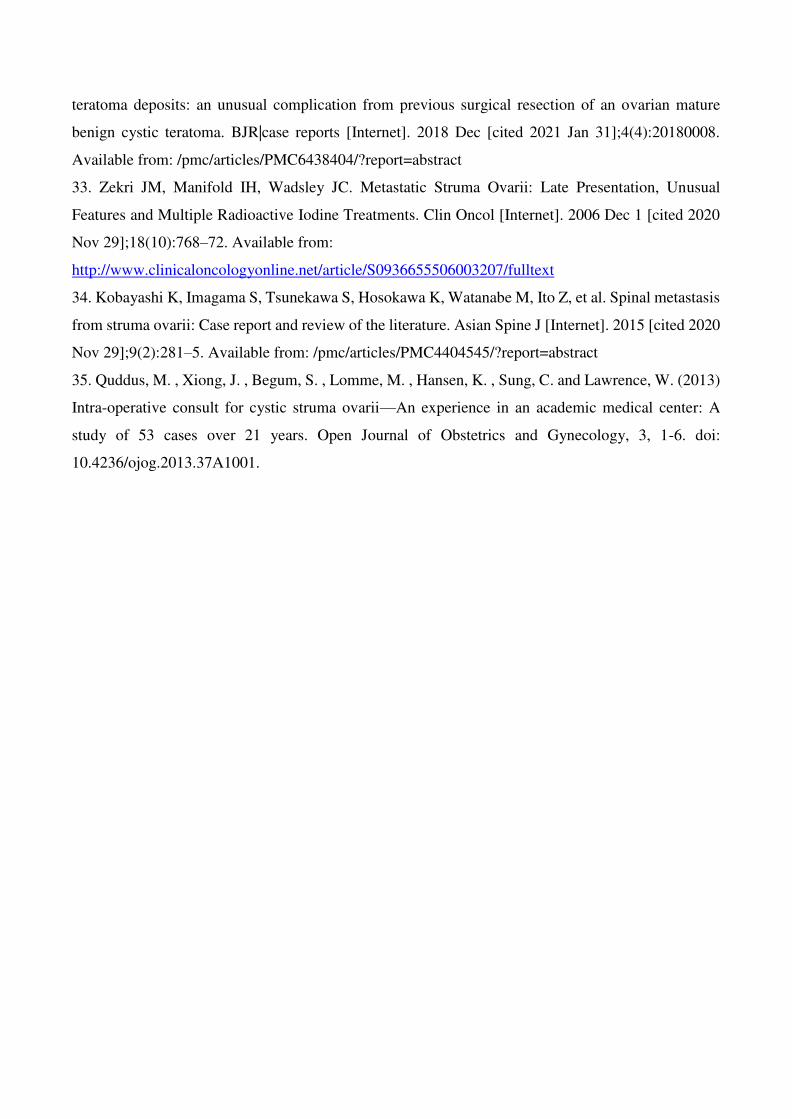
lifelong hormone replacement therapy. Some of the previously reported cases, their features and
outcomes are summarized in table 1. They demonstrated that benign strumas can have malignant
clinical course and malignant strumas can show long progression-free survival (e.g. Garg K. et al. and
Marti J.M. et al. described malignant strumas with no recurrence [13,26]; Akahira J. et al and .A.I.
Karseladze et al. reported histologically benign strumas with multiple metastases [5,14]). In addition,
morphology and clinical course did not correlate with patients’ age (median age varied from 41 to 46
years).The localization of the metastases was different and also did not correlate with morphological
features. Salpingoovarioectomy, thyroidectomy and hysterectomy were the most common treatment.
Although progression-free survival did not depend on the treatment approach.
Conclusion
Despite histologically bland appearance of tumor at the primary site, minority of struma
ovarii tumors are capable of extraovarian spread (peritoneal implants, distant metastasis). Due to this
reason, the last WHO edition recommends the term of "highly differentiated follicular carcinoma
arising in struma ovarii. The potential of late recurrences and extraovarian spread requires long-term
follow-up. Treatment protocols have not been established, but radioiodine therapy, oophorectomy, and
thyroidectomy remain as the most reasonable approach. The notorious challenge to predict the biologic
behavior necessitates multidisciplinary approach in management of struma ovarii, with the meticulous
correlation of clinical findings, laboratory data, radiologic details, and histopathological features.
List of Abbreviations
99mTc - Technetium 99m
FNA – Fine-needle aspiration
HDFCO -Highly differentiated follicular carcinoma of ovarian origin
I-131 – Iodine 131
ICD-O - International Classification of Diseases for Oncology
MRI – Magnetic resonance imaging
MSO - Malignant struma ovarii
PET-CT- Positron emission tomography–computed tomography
TSHR-Ab - Thyroid-stimulating hormone receptor antibodies
Declarations
Ethics approval and consent to participate:
Not applicable
Consent for publication:
The patient gave consent for publication.
Availability of data and materials:
All the data regarding the findings are available within the manuscript.
Competing interests:
The author(s) declared no conflicts of interest with respect to the authorship and/or publication of this
article.
Funding:
This manuscript was supported by National Medical Research Center for Obstetrics Gynecology and
Perinatology named after Academician V.I.Kulakov (Moscow, Russia).
Authors’ contributions: AA, AM and AT carried out the histochemical explorations; SM and JL participated in the USD,
surgery and the design of the study; AV, AM, EK, VK and LA conceived the study and have been
involved in the literature search. EK drafted the manuscript. All authors read and approved the final
manuscript.
Acknowledgements:
The authors would like to thank Anna Yemelyanova, MD (Weill Cornell Medicine, New York) and
Vadim Gushchin, MD (Mercy Medical Centre, Baltimor) for their brilliant consultations.
References
1. Roth LM, Talerman A. The enigma of struma ovarii. Pathology [Internet]. 2007 Feb [cited 2020
Nov 28];39(1):139–46. Available from:
https://linkinghub.elsevier.com/retrieve/pii/S0031302516338326
2. Shaco-Levy R, Peng RY, Snyder MJ, Osmond GW, Veras E, Bean SM, et al. Malignant struma
ovarii: A blinded study of 86 cases assessing which histologic features correlate with aggressive
clinical behavior. Arch Pathol Lab Med [Internet]. 2012 Feb [cited 2020 Nov 28];136(2):172–8.
Available from: https://pubmed.ncbi.nlm.nih.gov/22288964/
3. Navarro P, López L, González M, Sangrós M, Liévano P, Álvarez S, et al. Peritoneal strumosis: An
extension study with 99mTc-pertechnetate. Rev Española Med Nucl e Imagen Mol (English Ed. 2012
Mar 1;31(2):97–100.
4. Kaleem T, Peterson J, Krishna M. Peritoneal strumosis: Presentation and management with multiple
radioactive iodine treatments. J Clin Transl Endocrinol Case Reports. 2018 Jun 183;8:1–4
5.Akahira J, Endo M, Chiba R, Tanoguchi K, Yamauchi J, Ishiyama S, et al. Peritoneal strumosis, 10
years after laparoscopic surgery for mature cystic teratoma of the ovary: a case report. Int Cancer Conf
J [Internet]. 2013 Oct 14 [cited 2021 Jan 30];2(4):251–4. Available from:
https://link.springer.com/article/10.1007/s13691-013-0103-0
6.Schmidt J, Derr V, Heinrich MC, Crum CP, Fletcher JA, Corless CL, et al. BRAF in papillary thyroid
carcinoma of ovary (struma ovarii). Am J Surg Pathol [Internet]. 2007 Sep [cited 2021 Jan
30];31(9):1337–43. Available from: https://pubmed.ncbi.nlm.nih.gov/17721188/
7. Brockmann M, Schildgen V, Schildgen O, Lüsebrink J, Pieper M, Gudima A. NGS-dataset of
putative driver mutations associated with benign peritoneal strumosis. Data Br [Internet]. 2018 Oct 1
[cited 2020 Nov 28];20:468–70. Available from: https://pubmed.ncbi.nlm.nih.gov/30186896/
8. Janszen EWM, van Doorn HC, Ewing PC, de Krijger RR, de Wilt JHW, Kam BLR, et al. Malignant
struma ovarii: Good response after thyroidectomy and 131I ablation therapy. Clin Med Oncol
[Internet]. 2008 Feb 29 [cited 2021 Jan 30];2:147–52. Available from:
/pmc/articles/PMC3161682/?report=abstract
9. Aurore O, Eric L, Juliette B, Hélène KG. Treatment and follow-up of malignant struma ovarii:
Regarding two cases. Gynecol Oncol Reports. 2016 Aug 1;17:56–9.
10. Leite I, Cunha TM, Figueiredo JP, Félix A. Papillary carcinoma arising in struma ovarii versus
ovarian metastasis from primary thyroid carcinoma: A case report and review of the literature. J Radiol
Case Rep [Internet]. 2013 [cited 2021 Jan 30];7(10):24–33. Available from:
/pmc/articles/PMC3888341/?report=abstract
11. Robboy SJ, Scully RE. Strumal carcinoid of the ovary: An analysis of 50 cases of a distinctive
tumor composed of thyroid tissue and carcinoid. Cancer [Internet]. 1980 [cited 2021 Jan
30];46(9):2019–34. Available from: https://pubmed.ncbi.nlm.nih.gov/7427909/
12. Robboy SJ, Shaco-Levy R, Peng RY, Snyder MJ, Donahue J, Bentley RC, et al. Malignant struma
ovarii: An analysis of 88 cases, including 27 with extraovarian spread. Int J Gynecol Pathol [Internet].
2009 Sep [cited 2021 Jan 30];28(5):405–22. Available from:
https://pubmed.ncbi.nlm.nih.gov/19696610/
13. Garg K, Soslow RA, Rivera M, Tuttle MR, Ghossein RA. Histologically bland “extremely well
differentiated” thyroid carcinomas arising in struma ovarii can recur and metastasize. Int 215 J Gynecol
Pathol. 2009 May;28(3):222–30. 14. Karseladze AI, Kulinitch SI. Peritoneal Strumosis. Pathol Res
Pract [Internet]. 1994 [cited 2020 Nov 28];190(11):1082–5. Available
from:https://pubmed.ncbi.nlm.nih.gov/7746743/
15. Kim D, Cho HC, Park JW, Lee WA, Kim YM, Chung PS, et al. Struma ovarii and peritoneal
strumosis with thyrotoxicosis. Thyroid [Internet]. 2009 Mar 1 [cited 2021 Jan 30];19(3):305–8.
Available from: https://pubmed.ncbi.nlm.nih.gov/19265502/
16. Board WC of TE, editor. WHO Female genital tumors. Lyon; 2020.
17. Wu M, Hu F, Huang X, Tan Z, Lei C, Duan D. Extensive peritoneal implant metastases of
malignant struma ovarii treated by thyroidectomy and 131I therapy. Medicine (Baltimore) [Internet].
2018 Dec 1 [cited 2021 Jan 30];97(51):e13867. Available from: 225
http://journals.lww.com/00005792-201812210-00138
18. McGill JF, Sturgeon C, Angelos P. Metastatic struma ovarii treated with total thyroidectomy and
radioiodine ablation [Internet]. Vol. 15, Endocrine Practice. American Association of Clinical
Endocrinologists; 2009 [cited 2021 Jan 30]. p. 167–73. Available from:
https://pubmed.ncbi.nlm.nih.gov/19289330/
19. Teale E, Gouldesbrough DR, Peacey SR. Graves’ disease and coexisting struma ovarii: Struma
expression of thyrotropin receptors and the presence of thyrotropin receptor stimulating antibodies. In:
Thyroid [Internet]. Thyroid; 2006 [cited 2021 Jan 30]. p. 791–3. Available from:
https://pubmed.ncbi.nlm.nih.gov/16910883/
20. Anastasilakis AD, Ruggeri RM, Polyzos SA, Makras P, Molyva D, Campennì A, et al. Coexistence
of Graves’ disease, papillary thyroid carcinoma and unilateral benign struma ovarii: Case report and
review of the literature. Metabolism. 2013 Oct 1;62(10):1350–6.
21. Sitasuwan T, Hanamornroongruang S, Peerapatdit T, Thongtang N. Coexistence of Graves’ disease
and unilateral functioning Struma ovarii: A case report. BMC Endocr Disord [Internet]. 2015 Nov 4
[cited 2021 Jan 30];15(1). Available from: /pmc/articles/PMC4632472/?report=abstract
22.Wei S, Baloch ZW, LiVolsi VA. Pathology of Struma Ovarii: A Report of 96 Cases. Endocr Pathol
[Internet]. 2015 Dec 16 [cited 2020 Nov 28];26(4):342–8. Available from:
http://link.springer.com/10.1007/s12022-015-9396-1
23.Shaco-Levy R, Bean SM, Bentley RC, Robboy SJ. Natural history of biologically malignant struma
ovarii: Analysis of 27 cases with extraovarian spread. Int J Gynecol Pathol [Internet]. 2010 May [cited
2020 Nov 28];29(3):212–27. Available from: https://pubmed.ncbi.nlm.nih.gov/20407319/
24.Devaney K, Snyder R, Morris HJ, Tavassoli FA. Proliferative and histologically malignant struma
ovarii: A clinicopathologic study of 54 cases. Int J Gynecol Pathol. 1993;12(4):333–250
25. Yongxue, Wang, Lingya, Pan, Huifang, Huang, Keng, Shen, Ming, Wu, Jinghe L. Clinical study
on 68 cases with struma ovarii. Zhonghua Fu Chan Ke Za Zhi. 2014;49(6):451–4.
26. Marti JL, Clark VE, Harper H, Chhieng DC, Sosa JA, Roman SA. Optimal surgical management
of well-differentiated thyroid cancer arising in struma ovarii: A series of 4 patients and a review of 53
reported cases [Internet]. Vol. 22, Thyroid. Thyroid; 2012 [cited 2021 Jan 31]. p. 400–6. Available
from: https://pubmed.ncbi.nlm.nih.gov/22181336/
27. Muallem MZ, Harter P, Heitz F, El-Balat A, Fisseler-Eckhoff A, Menzel C, et al. Struma ovarii
recurrence with peritoneal strumosis: A case report. J Solid Tumors [Internet]. 2012 Nov 7 [cited 2020
Nov 29];2(6):46. Available from:
www.sciedu.ca/jstURL:http://dx.doi.org/10.5430/jst.v2n6p46www.sciedu.ca/jst
28. Ranade R, Rachh S, Basu S. Late manifestation of struma peritonei and widespread functioning
lesions in the setting of struma ovarii simulating highly differentiated follicular carcinoma. J Nucl Med
Technol [Internet]. 2015 [cited 2020 Nov 28];43(3):231–3. Available from:
https://pubmed.ncbi.nlm.nih.gov/25537757/
29. Hwu. A Rare Case of Histologic Benign Struma Ovarii With Distant Metastasis. J Clin Gynecol
a1 Hwu A Rare Case Histol Benign Struma Ovarii With Distant Metastasis J Clin Gynecol Obs
[Internet] 2014 Sep 17 [cited 2020 Oct 21];3(3)108–13 Available from
http//dx.doi.org/1014740/jcgo232wnd Obstet [Internet]. 2014 Sep 17 [cited 2020 Oct 21];3(3):108–
13. Available from: http://dx.doi.org/10.14740/jcgo232w
30. Riggs MJ, Kluesner JK, Miller CR. Management of highly differentiated thyroid follicular
carcinoma of ovarian origin with a minimally invasive approach. Gynecol Oncol Reports [Internet].
2018 May 1 [cited 2020 Nov 28];24:87–9. Available from:
https://pubmed.ncbi.nlm.nih.gov/29725612/
31. Oh SJ, Jung M, Kim YO. Late bone metastasis of histologically bland struma ovarii: The
unpredictability of its biologic behavior. J Pathol Transl Med [Internet]. 2015 [cited 2020 Oct
12];49(4):343–5. Available from: http://dx.doi.org/10.4132/jptm.2015.04.27
32. Prasad R, Sieberhagen C, Fenwick S, Katti A, Davis R. Intra-peritoneal mature benign cystic
teratoma deposits: an unusual complication from previous surgical resection of an ovarian mature
benign cystic teratoma. BJR|case reports [Internet]. 2018 Dec [cited 2021 Jan 31];4(4):20180008.
Available from: /pmc/articles/PMC6438404/?report=abstract
33. Zekri JM, Manifold IH, Wadsley JC. Metastatic Struma Ovarii: Late Presentation, Unusual
Features and Multiple Radioactive Iodine Treatments. Clin Oncol [Internet]. 2006 Dec 1 [cited 2020
Nov 29];18(10):768–72. Available from:
http://www.clinicaloncologyonline.net/article/S0936655506003207/fulltext
34. Kobayashi K, Imagama S, Tsunekawa S, Hosokawa K, Watanabe M, Ito Z, et al. Spinal metastasis
from struma ovarii: Case report and review of the literature. Asian Spine J [Internet]. 2015 [cited 2020
Nov 29];9(2):281–5. Available from: /pmc/articles/PMC4404545/?report=abstract
35. Quddus, M. , Xiong, J. , Begum, S. , Lomme, M. , Hansen, K. , Sung, C. and Lawrence, W. (2013)
Intra-operative consult for cystic struma ovarii—An experience in an academic medical center: A
study of 53 cases over 21 years. Open Journal of Obstetrics and Gynecology, 3, 1-6. doi:
10.4236/ojog.2013.37A1001.
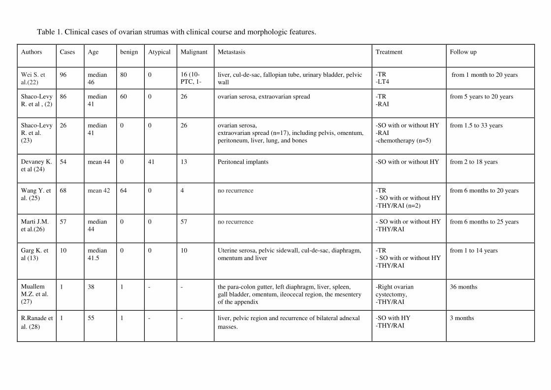
Table 1. Clinical cases of ovarian strumas with clinical course and morphologic features.
Authors Cases Age benign Atypical Malignant Metastasis Treatment Follow up
Wei S. et al.(22)
96 median 46
80 0 16 (10- PTC, 1-
liver, cul-de-sac, fallopian tube, urinary bladder, pelvic wall
-TR -LT4
from 1 month to 20 years
Shaco-Levy R. et al , (2)
86 median 41
60 0 26 ovarian serosa, extraovarian spread -TR -RAI
from 5 years to 20 years
Shaco-Levy R. et al. (23)
26 median 41
0 0 26 ovarian serosa, extraovarian spread (n=17), including pelvis, omentum, peritoneum, liver, lung, and bones
-SO with or without HY -RAI -chemotherapy (n=5)
from 1.5 to 33 years
Devaney K. et al (24)
54 mean 44 0 41 13 Peritoneal implants -SO with or without HY from 2 to 18 years
Wang Y. et al. (25)
68 mean 42 64 0 4 no recurrence -TR - SO with or without HY -THY/RAI (n=2)
from 6 months to 20 years
Marti J.M. et al.(26)
57 median 44
0 0 57 no recurrence - SO with or without HY -THY/RAI
from 6 months to 25 years
Garg K. et al (13)
10 median 41.5
0 0 10 Uterine serosa, pelvic sidewall, cul-de-sac, diaphragm, omentum and liver
-TR - SO with or without HY -THY/RAI
from 1 to 14 years
Muallem M.Z. et al. (27)
1 38 1 - - the para-colon gutter, left diaphragm, liver, spleen, gall bladder, omentum, ileocecal region, the mesentery of the appendix
-Right ovarian cystectomy, -THY/RAI
36 months
R.Ranade et
al. (28)
1 55 1 - - liver, pelvic region and recurrence of bilateral adnexal
masses.
-SO with HY -THY/RAI
3 months
Authors Cases Age Benign Atypical Malignant Metastasis Treatment Follow up
Hwua D.-
W. et al
(29)
1 28 1 - - bilateral ovarian tumors, metastases in the liver and
peritoneal space
-SO with HY -THY/RAI
4 months
Akahira J.
et al (5)
1 64 1 - - tumor tissue of the pelvic cavity including in the
uterus, rectum and mesentery
-TR 17 months
A.I.
Karseladze
et al. (14)
1 49 1 - - contrlateral ovary, omentum -SO with HY -3 consecutive cycles of polychemotherapy
36 months
McKayla J,
et al. (30)
1 32 1 - - anterior and posterior peritoneal reflections of the
uterus without evidence of malignancy
wait-and-see attitude 36 months
Sun-Ju O.
et al (31)
1 60 1 - - 12 level of the spine -TR
-THY+ spondylectomy/ (RAI)
not specified
R. Prasad et
al. (32)
1 yearly
forties
1 - - the right hemidiaphragm, adherent to the adjacent
segment 8 of the liver, adjacent to the right adrenal
gland, overlying the right perinephric tissue and on the
porta hepatis
TR 96 months
Zekri J. et
al. (33)
1 26 1 - - multiple pulmonary nodules THY/RAI - 60 months after the initial dose
of iodine therapy the mts
appeared. (treated with
bifrontal craniotomy)
Kobayashi
K. et al (34)
1 39 1 - - osteolysis at the Th7 level external beam
radiotherapy at a dose of
37.5 Gy for the
spinal lesion
9 months
*PTC- papillary thyroid carcinoma, HDFCO - highly differentiated follicular carcinoma of ovarian origin, SC- strumal carcinoid, TR- Tumor resection, LT4 – levothyroxine, THY/RAI -
thyroidectomy/radioactive iodine, SO – salpingoovarioectomy, HY-hysterectomy
2

Supplementary Files
This is a list of supplementary �les associated with this preprint. Click to download.
CAREchecklistEnglish.docx
Recommended


