
Virology 446 (2013) 95–101
Contents lists available at ScienceDirect
Virology
0042-68http://d
n CorrE-m1 Th
journal homepage: www.elsevier.com/locate/yviro
Rapid Communication
The charged residues in the surface-exposed C-terminusof the Soybean mosaic virus coat protein are criticalfor cell-to-cell movement
Jang-Kyun Seo a,b,1, Mi Sa Vo Phan a,1, Sung-Hwan Kang a, Hong-Soo Choi b,Kook-Hyung Kim a,n
a Department of Agricultural Biotechnology and Plant Genomics and Breeding Institute, Seoul National University, Seoul 151-921, Republic of Koreab Crop Protection Division, National Academy of Agricultural Science, Rural Development Administration, Suwon 441-707, Republic of Korea
a r t i c l e i n f o
Article history:Received 20 May 2013Returned to author for revisions8 June 2013Accepted 25 July 2013Available online 25 August 2013
Keywords:SMVPotyvirusCoat proteinMovementVirion assembly
22/$ - see front matter Crown Copyright & 20x.doi.org/10.1016/j.virol.2013.07.033
esponding author. Fax: +82 2 873 2317.ail address: [email protected] (K.-H. Kim).ese authors contributed equally to this work
a b s t r a c t
The Soybean mosaic virus (SMV) coat protein (CP) is necessary for virion assembly and viral cell-to-celland long-distance movements in plants. We previously showed that the C-terminal region of the SMV CPis required for CP self-interaction. In the present study, we generated SMV mutants containing CPs withsingle amino acid substitutions of the charged amino acids in the C-proximal region. Infectivity and cell-to-cell movement of the SMV mutants were examined in soybean plants. Through this genetic approach,we identified three charged amino acid residues (R245, H246, and D250) in the surface-exposedC-terminus of the SMV CP that are critical for virus cell-to-cell and long-distance movement. Our findingssuggest that the identified charged amino acids in the surface-exposed C-terminus of SMV CP are criticalfor CP intersubunit interactions and thereby for cell-to-cell and long-distance movement and virionassembly.
Crown Copyright & 2013 Published by Elsevier Inc. All rights reserved.
Introduction
Soybean mosaic virus (SMV), which is a member of the genusPotyvirus in the family Potyviridae, has a single-stranded positive-sense RNA genome of approximately 9.6 kb. The major hosts ofSMV are Glycine max and Glycine soja. Since SMV was firstclassified into seven strains (G1–G7) based on phenotypic reac-tions on different soybean cultivars (Cho and Goodman, 1979),emergence of variant SMV strains has been continuously reported(Cho et al., 1983; Choi et al., 2005; Hajimorad et al., 2003; Kim andLee, 1991; Kim et al., 2003; Lim, 1985). Like the genomes of othermembers in the genus Potyvirus, the SMV genome encodes onelarge polyprotein, which is cleaved to yield at least 10 matureproteins, including P1, helper component-proteinase (HC-Pro), P3,6K1, cylindrical inclusion (CI), 6K2, genome-linked viral protein(VPg), nuclear inclusion a proteinase (NIa-Pro), nuclear inclusion b(NIb), and coat protein (CP) (Riechmann et al., 1992). A small openreading frame embedded in the P3 cistron of potyviruses wasrecently discovered and named pipo (Pretty interesting PotyviridaeORF) (Chung et al., 2008). The PIPO protein is also encoded by SMVand is essential for SMV movement (Wen and Hajimorad, 2010).
13 Published by Elsevier Inc. All r
.
Virus movement in plants is an active process mediated byvirus-encoded proteins such as the movement proteins (MPs) andCPs. Viral MPs generally facilitate cell-to-cell movement of infec-tious entities by increasing the size-exclusion limits of plasmo-desmata (Lucas, 2006; Ueki and Citovsky, 2011; Verchot-Lubiczet al., 2010). In potyvirus movement, no dedicated MP, like thosefor other viruses, has been identified. However, some potyviralproteins including HC-Pro, CI, VPg, CP, and P3N-PIPO have beendemonstrated to be involved in viral cell-to-cell movement(Carrington et al., 1998; Cronin et al., 1995; Dolja et al., 1994,1995; Dunoyer et al., 2004; Hofius et al., 2007; Rojas et al., 1997;Shen et al., 2010; Vijayapalani et al., 2012; Wei et al., 2010). Theearlier studies demonstrated that CP is necessary for both cell-to-cell and long-distance movement of Tobacco etch potyvirus (TEV)(Dolja et al., 1994, 1995). The potyvirus CP has variable N- andC-terminal domains exposed on the virion surface and a conservedcentral core domain (Anindya and Savithri, 2003; Shukla andWard, 1989). Mutational analyses of TEV CP have revealed thatthe central core domain is critical for virus cell-to-cell movementand that the variable N- and C-terminal domains function duringlong-distance movement (Dolja et al., 1994, 1995).
We previously found a strong interaction between subunits ofSMV CP based on a yeast two-hybrid system (YTHS) and demon-strated that the C-terminal domain of SMV CP is required for theCP intersubunit interactions (Kang et al., 2004, 2006). In thecurrent study, we further verified the in planta significance of
ights reserved.

J.-K. Seo et al. / Virology 446 (2013) 95–10196
the C-terminal domain of SMV CP in virus cell-to-cell and long-distance movements by generating SMV mutants containing CPswith single amino acid substitution of the charged amino acids inthe C-terminal domain. Based on this approach, we demonstratethat some charged amino acid residues in the C-terminal domainof CP are critical not only for the intersubunit interactions (Kanget al., 2006) but also for virus movement (this study). Our findingsalso support the concept that potyviruses move as virions.
(AY216010)(AY216987)(FJ640982)(FJ640978)(FJ640977)(AB100442)(FJ640965)(AB100443)(FJ640961)(FJ640955)(FJ640967)(FJ640963)(FJ640956)(FJ640958)(EU871724)(EU871725)(S42280) (AJ619757)(FJ640979)(D00507) (FJ807700)(FJ548849)(AY294044)(FJ807701)(FJ640954)(FJ640962)(FJ640974)(FJ640975)(FJ640981)(AJ310200)(FJ640957)(FJ640970)(FJ640980)(AJ312439)
G7G7dG7a
G3G1Aa
WS135Aa15-M2
WS116WS37
WS145WS128WS84
WS105L
L-RBG2
CN18G4
NG7H
WS200G5
G5HWS32
WS117WS202WS205
G6HHH5
WS101WS155
G6HZ
pSMV-GUSpSMV-GUS-CPR245ApSMV-GUS-CPH246ApSMV-GUS-CPR249ApSMV-GUS-CPD250ApSMV-GUS-CPH256A
AP1 HC-pro P3 CI NIa NIb CP
5’UTR 6K1 6K2 3’UTR
pSMV-GUS
Rz NOSt
P35SX2
CP 1-265 AA
RHTARDVNQNMHTLLGMGPQQ
GUS
- - - DIQHY / SRTR TRESVSLQ / SQNPE - - -
P1in cis
NIa-proin trans
GUS
AHTARDVNQNMHTLLGMGPQQRATARDVNQNMHTLLGMGPQQRHTAADVNQNMHTLLGMGPQQRHTARAVNQNMHTLLGMGPQQRHTARDVNQNMATLLGMGPQQ
pipo
Fig. 1. Schematic representation of the construction of pSMV-GUS and the CPcistron showing the charged amino acid positions at the C-terminus. (A) SMVgenome organization and insertion of the gus gene between P1 and HC-Pro. Aminoacid sequences of the peptide cleavage sites recognized by either the P1 or NIa-Proare underlined, and arrowheads indicate the location of the cleaved peptide bond.In vivo transcription of SMV-GUS is under control of a double 35S promoter(P35SX2), a cis-cleaving ribozyme sequence (Rz), and a NOS terminator (NOSt). Theamino acid positions of alanine substitution mutations are shown in the schematicrepresentation of SMV CP. (B) Alignment of the C-terminal amino acid sequences ofCPs of various SMV strains and isolates. The names of SMV strains and isolates areindicated on the left side of the sequences together with their GenBank accessionnumbers. Sequences were aligned by ClustalX2.
Results and discussion
Introduction of single-substitution mutations into the C-terminaldomain of CP of pSMV-GUS
We previously developed an SMV-based viral vector (pSMV-MCS) to express foreign genes in soybean by inserting the cloningsites into the polyprotein open reading frame (ORF) between theP1 and HC-Pro cistrons (Seo et al., 2009a). In the current study, thebacterial β-glucuronidase (GUS) gene was inserted into pSMV-MCSto visualize virus infection and movement in plants, and theresulting construct was designated as pSMV-GUS (Fig. 1A).
The amino acid sequences of the CP cistron are highly con-served among various SMV strains and isolates (Seo et al., 2009b).In particular, comparison of the amino acid sequence revealed thatall of the charged amino acids (except H256) in the C-terminus ofSMV CP are conserved among 34 representative SMV strainsand isolates (Fig. 1B). Thus, to examine the significance of theC-terminal domain of SMV CP in virus cell-to-cell and long-distance movement, we introduced alanine substitutions into thecharged amino acids in the C-terminal domain of the CP cistron ofpSMV-GUS by site-directed mutagenesis. The resulting constructscontaining the single-substitution mutations in the CP cistronwere named pSMV-GUS–CPR245A, –CPH246A, –CPR249A, –CPD250A,and –CPH256A (Fig. 1A).
Effect of the mutations in the C-terminus of SMV CPon virus infectivity
We first examined the infectivity of SMV-GUS and the deriva-tive CP mutants. Plasmid DNAs of pSMV-GUS and the CP mutantswere rub-inoculated onto the primary leaves of soybean seedlings(cv. Lee68). The results of the infectivity tests are summarized inTable 1. Typical systemic mild mosaic symptoms appeared in theupper uninoculated leaves of all the soybean plants inoculatedwith pSMV-GUS, pSMV-GUS–CPR249A, or –CPH256A at 9–14 dayspost inoculation (dpi). However, no symptoms were observed inthe soybean plants inoculated with pSMV-GUS–CPR245A, –CPH246A,or –CPD250A even at 45 dpi. To verify whether the inoculatedsoybeans were systemically infected with each SMV mutant, weextracted total RNAs from upper uninoculated leaves at 45 dpi andsubjected the extracts to RT-PCR using SMV-specific primers. TheRT-PCR results confirmed that none of the soybean plants inocu-lated with pSMV-GUS–CPR245A, –CPH246A, or –CPD250A was infectedwith SMV while all soybean plants showing symptoms weresystemically infected (data not shown). We also verified that theintroduced mutations were maintained during systemic infectionby analyzing (by RT-PCR) sequences of progeny viruses of pSMV-GUS–CPR249A and –CPH256A, which were recovered from upperuninoculated leaves (data not shown). Because CP modifications(i.e. deletions of the N- or C-terminal region and several substitu-tion mutations in the central core domain) have no significanteffect on potyvirus genome amplification (Dolja et al., 1994, 1995),we suspected that the residues R245, H246, and D250 of SMV CPmay be critical for virus movement or assembly.
Effect of the mutations in the C-terminus of SMV CP on viruscell-to-cell movement
Because the loss of systemic infectivity of the CP mutants couldbe due to defects in virus movement, we next examined the cell-to-cell movement phenotypes of SMV-GUS and the CP mutants.To this end, the soybean leaves inoculated with pSMV-GUS or theCP mutants were subjected to a histochemical GUS assay at 3,5 and 8 dpi. This assay allows quantitative measurement of theextent of virus cell-to-cell movement because only infected cells

J.-K. Seo et al. / Virology 446 (2013) 95–101 97
express GUS activity. Results are summarized in Fig. 2 and Table 2.No infection foci were observed at 3 dpi in all leaves mechanicallyinoculated with the plasmid DNAs of pSMV-GUS and its derivativemutants. However, SMV-GUS spread radially to form infection fociwith average diameters of approximately 573.2764.9 and1463.47317.2 μm at 5 and 8 dpi, respectively. The mutants SMV-GUS–CPR249A and –CPH256A, which were previously determined tobe capable of systemic infection, formed infection foci of similarsizes to those formed by SMV-GUS at 5 and 8 dpi (Fig. 2 andTable 2). In contrast, movement of the mutants SMV-GUS–CPR245A,–CPH246A, and –CPD250A, which were previously determined to beincapable of systemic infection, was restricted to several epider-mal cells surrounding the initially infected cells, and the foci wereabout 14-fold smaller than those produced by SMV-GUS at 5 dpi
100μmSMV-GUS SMV-GUS-C
Inoculu
SMV-GUS-CPR249A SMV-GUS-C
563.1 μm36.4
Inoculu
100μm
54.8 μ530.3 μm
Fig. 2. Histochemical detection of GUS activity in soybean leaves inoculated with pSMVabove each image. The GUS foci were photographed at 5 dpi.
Table 1Effect of SMV CP mutations on systemic infection of soybean plants.
Inoculuma Infectivityb
1 2 3 Total
Mock 0/3 0/3 0/3 0/9pSMV-GUS 3/3 3/3 3/3 9/9pSMV-GUS–CPR245A 0/5 0/5 0/5 0/15pSMV-GUS–CPH246A 0/5 0/5 0/5 0/15pSMV-GUS–CPR249A 5/5 5/5 5/5 15/15pSMV-GUS–CPD250A 0/5 0/5 0/5 0/15pSMV-GUS–CPH256A 5/5 5/5 5/5 15/15
a Soybean cultivar Lee68 (rsv) plants were inoculated with the correspondingplasmids as described in Materials and methods section.
b Number of systemically infected plants/number of plants inoculated. Virusinfection of the upper non inoculated leaves was confirmed by RT-PCR using SMVspecific primers at 45 dpi.
(Fig. 2 and Table 2). Furthermore, sizes of infection foci weremarginally increased at 8 dpi when compared to those at 5 dpi inleaves inoculated with pSMV-GUS–CPR245A, –CPH246A, or –CPD250A.These results demonstrated that the charged amino acids R245,H246, and D250 are critical for the functioning of SMV CP in cell-to-cell movement.
Effect of the mutations in the C-terminus of SMV CP on virionassembly
Because the C-terminal domain of SMV CP is required forinteractions between CP subunits (Kang et al., 2006), we examinedwhether the charged amino acids in the C-terminus of SMV CP are
100μm 100μm
PR245A SMV-GUS-CPH246A
m
PD250A SMV-GUS-CPH256A
μm 40.4 μm
m
100μm100μm
m501.7 μm
-GUS and its CP mutants. The inocula used to infect soybean leaves are indicated
Table 2Diameters of infection foci on soybean leaves inoculated with SMV-GUS andCP mutants.
Inoculuma Infection foci diameter (μm)b
3 dpi 5 dpi 8 dpi
Mock ND ND NDpSMV-GUS ND 573.2764.9 1463.47317.2pSMV-GUS–CPR245A ND 36.576.1 54.8711.3pSMV-GUS–CPH246A ND 38.576.1 62.178.7pSMV-GUS–CPR249A ND 596.7791.7 1341.57411.3pSMV-GUS–CPD250A ND 41.878.6 82.4721.6pSMV-GUS–CPH256A ND 548.1795.4 1419.87271.6
ND¼not detected.a Soybean cultivar Lee68 (rsv) plants were inoculated with the corresponding
plasmids as described in Materials and methods section.b The diameters of foci were determined by GUS histochemical assays using
light microscopy. Each value is the mean7standard deviation of 410 foci.
J.-K. Seo et al. / Virology 446 (2013) 95–10198
required for virion assembly. Soybean leaves were inoculated withpSMV-GUS or the CP mutants, and leaf lysates were prepared fromthe inoculated leaves at 5 dpi and subjected to serologicallyspecific electron microscopy (SSEM) in order to determine thepresence of virions. Virions were easily detected in the leaf lysatesinoculated with pSMV-GUS, –CPR249A, or –CPH256A (Table 3).Despite extensive observations, however, no virions were detectedin the leaf lysates inoculated with the movement-defectivemutants SMV-GUS–CPR245A, –CPH246A, or –CPD250A (Table 3). Thisindicates that the charged amino acids R245, H246, and D250 inthe C-terminus of SMV CP are required for successful virionassembly. In our previous study, based on a yeast two-hybridsystem, we found that alanine substitution mutations at thecharged amino acids R245, H246, and R249 of SMV CP disruptedthe CP intersubunit interactions (Kang et al., 2006). In this regard,it is likely that the charged amino acids in the C-terminus of SMVCP mediate the intersubunit interactions that are critical for virionassembly. However, we cannot exclude the possibility that the lackof virions in leaf samples inoculated with the movement-defectiveCP mutants might be due to that the concentration of virionswas below the level of detection by the method that we used.Future technical advances in analysis of SMV virion assembly atsingle-cell level may demonstrate clearly the significance of theC-terminal charged amino acids of SMV CP in virion assembly.
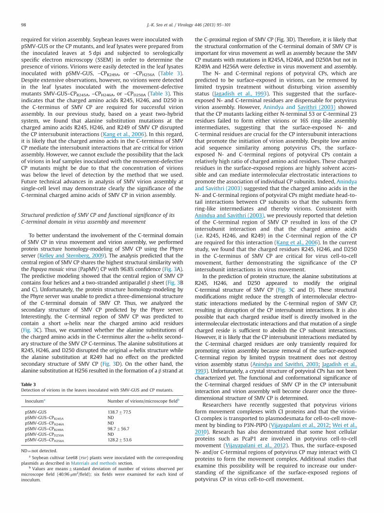
Structural prediction of SMV CP and functional significance of itsC-terminal domain in virus assembly and movement
To better understand the involvement of the C-terminal domainof SMV CP in virus movement and virion assembly, we performedprotein structure homology-modeling of SMV CP using the Phyreserver (Kelley and Sternberg, 2009). The analysis predicted that thecentral region of SMV CP shares the highest structural similarity withthe Papaya mosaic virus (PapMV) CP with 96.8% confidence (Fig. 3A).The predictive modeling showed that the central region of SMV CPcontains four helices and a two-stranded antiparallel β sheet (Fig. 3Band C). Unfortunately, the protein structure homology-modeling bythe Phyre server was unable to predict a three-dimensional structureof the C-terminal domain of SMV CP. Thus, we analyzed thesecondary structure of SMV CP predicted by the Phyre server.Interestingly, the C-terminal region of SMV CP was predicted tocontain a short α-helix near the charged amino acid residues(Fig. 3C). Thus, we examined whether the alanine substitutions ofthe charged amino acids in the C-terminus alter the α-helix second-ary structure of the SMV CP C-terminus. The alanine substitutions atR245, H246, and D250 disrupted the original α-helix structure whilethe alanine substitution at R249 had no effect on the predictedsecondary structure of SMV CP (Fig. 3D). On the other hand, thealanine substitution at H256 resulted in the formation of a β strand at
Table 3Detection of virions in the leaves inoculated with SMV-GUS and CP mutants.
Inoculuma Number of virions/microscope fieldb
pSMV-GUS 138.7777.5pSMV-GUS–CPR245A NDpSMV-GUS–CPH246A NDpSMV-GUS–CPR249A 98.7756.7pSMV-GUS–CPD250A NDpSMV-GUS–CPH256A 128.2753.6
ND¼not detected.a Soybean cultivar Lee68 (rsv) plants were inoculated with the corresponding
plasmids as described in Materials and methods section.b Values are means7standard deviation of number of virions observed per
microscope field (40.96 μm2/field); six fields were examined for each kind ofinoculum.
the C-proximal region of SMV CP (Fig. 3D). Therefore, it is likely thatthe structural conformation of the C-terminal domain of SMV CP isimportant for virus movement as well as assembly because the SMVCP mutants with mutations in R245A, H246A, and D250A but not inR249A and H256A were defective in virus movement and assembly.
The N- and C-terminal regions of potyviral CPs, which arepredicted to be surface-exposed in virions, can be removed bylimited trypsin treatment without disturbing virion assemblystatus (Jagadish et al., 1993). This suggested that the surface-exposed N- and C-terminal residues are dispensable for potyvirusvirion assembly. However, Anindya and Savithri (2003) showedthat the CP mutants lacking either N-terminal 53 or C-terminal 23residues failed to form either virions or 16S ring-like assemblyintermediates, suggesting that the surface-exposed N- andC-terminal residues are crucial for the CP intersubunit interactionsthat promote the initiation of virion assembly. Despite low aminoacid sequence similarity among potyvirus CPs, the surface-exposed N- and C-terminal regions of potyviral CPs contain arelatively high ratio of charged amino acid residues. These chargedresidues in the surface-exposed regions are highly solvent acces-sible and can mediate intermolecular electrostatic interactions topromote the association of individual CP subunits. Indeed, Anindyaand Savithri (2003) suggested that the charged amino acids in theN- and C-terminal regions of potyviral CPs might mediate head-to-tail interactions between CP subunits so that the subunits formring-like intermediates and thereby virions. Consistent withAnindya and Savithri (2003), we previously reported that deletionof the C-terminal region of SMV CP resulted in loss of the CPintersubunit interaction and that the charged amino acids(i.e. R245, H246, and R249) in the C-terminal region of the CPare required for this interaction (Kang et al., 2006). In the currentstudy, we found that the charged residues R245, H246, and D250in the C-terminus of SMV CP are critical for virus cell-to-cellmovement, further demonstrating the significance of the CPintersubunit interactions in virus movement.
In the prediction of protein structure, the alanine substitutions atR245, H246, and D250 appeared to modify the originalC-terminal structure of SMV CP (Fig. 3C and D). These structuralmodifications might reduce the strength of intermolecular electro-static interactions mediated by the C-terminal region of SMV CP,resulting in disruption of the CP intersubunit interactions. It is alsopossible that each charged residue itself is directly involved in theintermolecular electrostatic interactions and that mutation of a singlecharged reside is sufficient to abolish the CP subunit interactions.However, it is likely that the CP intersubunit interactions mediated bythe C-terminal charged residues are only transiently required forpromoting virion assembly because removal of the surface-exposedC-terminal region by limited trypsin treatment does not destroyvirion assembly status (Anindya and Savithri, 2003; Jagadish et al.,1993). Unfortunately, a crystal structure of potyviral CPs has not beencharacterized yet. The functional and conformational significance ofthe C-terminal charged residues of SMV CP in the CP intersubunitinteraction and virion assembly will become clearer once the three-dimensional structure of SMV CP is determined.
Researchers have recently suggested that potyvirus virionsform movement complexes with CI proteins and that the virion-CI complex is transported to plasmodesmata for cell-to-cell move-ment by binding to P3N-PIPO (Vijayapalani et al., 2012; Wei et al.,2010). Research has also demonstrated that some host cellularproteins such as PcaP1 are involved in potyvirus cell-to-cellmovement (Vijayapalani et al., 2012). Thus, the surface-exposedN- and/or C-terminal regions of potyvirus CP may interact with CIproteins to form the movement complex. Additional studies thatexamine this possibility will be required to increase our under-standing of the significance of the surface-exposed regions ofpotyvirus CP in virus cell-to-cell movement.

CP wt2° structure
CP R245A
CP H246A
CP R249A
CP D250A
CP H256A
2° structure
2° structure
2° structure
2° structure
2° structure
PapMV CP (4dox)SMV CP
29 144
77 2001 265
77
200
90
PapMV CP (4dox)SMV CP
77
200
90
Fig. 3. Structural prediction of SMV CP. (A) Alignment coverage between SMV CP and PapMV CP. The central region (124 residues from the position 77 to 200) of SMV CP wasaligned to the central region (116 residues from the position 29 to 144) of PapMV CP (4DOX) with 96.8% confidence. The homology alignment was performed using the Phyreserver. (B) Three-dimensional structure model of the central region of SMV CP. The three-dimensional structure of the central region of SMV CP was determined byhomology-modeling using the Phyre server. The three-dimensional images were generated using the Protein Workshop. (C) Secondary structure of wild-type (wt) SMV CP.The secondary structure elements predicted using the Phyre server are shown under the amino acid sequence of wt SMV CP. (D) Secondary structures of the C-terminalregions of the SMV CP mutants. The introduced substitution mutations are indicated with arrows.
J.-K. Seo et al. / Virology 446 (2013) 95–101 99
Materials and methods
Insertion of the gus gene into the SMV viral genome
The coding region of the gus gene was amplified by PCR using aprimer pair harboring XbaI sites (5′-GCTCTAGAATGTTACGTCCTG-TAGAAACCC-3′ and 5′-GCTCTAGATTGTTTGCCTCCCTGCTG-3′). Theamplified fragments were digested with XbaI and cloned intopSMV-MCS (Seo et al., 2009a), which was opened with XbaI.
The resulting construct with the GUS insert in the correctorientation was named pSMV-GUS.
Construction of SMV mutants
Individual mutations R245A, H246A, R249A, D250A, andH256A in the C-terminus of CP were introduced into pSMV-GUSby site-directed mutagenesis using appropriate primers (the list ofprimers used for the site-directed mutagenesis is available on

J.-K. Seo et al. / Virology 446 (2013) 95–101100
request) (Nassal and Rieger, 1990; Seo et al., 2009a). The resultingconstructs were named pSMV-CPR245A, –CPH246A, –CPR249A,–CPD250A, and –CPH256A, respectively.
Plant growth and inoculation
Soybean plants were grown in a growth chamber at 25 1Cunder a 16/8-h photoperiod. Seedlings were selected for inocula-tion when the cotyledons were fully expanded. Plasmid DNAs ofpSMV-GUS and its derivative mutants were prepared using thePlasmid Maxi Kit (QIAGEN, Valencia, CA). Each cDNA plasmidwas rub-inoculated as described previously (Seo et al., 2009a).To detect virus accumulation in the inoculated and upper unin-oculated leaves, RT-PCR was performed using an SMV-specificprimer pair designed to amplify the CP region (5′- TCAGGTAAG-GAGAAGGAAGGA-3′ and 5′–CTGCTGTGGACCCATGCC-3′) and theresulting PCR products were sequenced to verify that the intro-duced mutations were maintained.
Histochemical GUS assays
GUS expression driven by virus infection was monitored byhistochemical GUS assays as described previously (Dolja et al.,1992). Briefly, the inoculated soybean leaves were vacuum-infiltrated with the colorimetric GUS substrate 5-bromo-4-chloro-3-indoyl β-D-glucuronic acid, cyclohexylammonium salt (X-gluc)(1.2 mM) in 0.5 mM potassium ferricyanide/0.5 mM potassiumferrocyanide/10 mM EDTA. After overnight incubation at roomtemperature, the leaves were bleached in 70% ethanol and exam-ined with a microscope to assess the diameters of GUS foci.
Serologically specific electron microscopy (SSEM)
To obtain crude virion preparations, the inoculated soybeanleaves were ground in five volumes of a grinding buffer (10 mMTris–HCl, 1 mM EDTA [pH 7.6]). Tissue debris was removed bycentrifugation at 17,000� g for 10 min. Anti-SMV serum-coatedgrids were prepared as described previously with minor modifica-tions (Dolja et al., 1995). Formvar/carbon-coated EM grids wereincubated with anti-SMV serum (1:500 dilution; Agdia, USA) for1 h. The grids were rinsed with a washing buffer (50 mM Tris–HCl,150 mM NaCl [pH 7.2]) and then incubated with the crude virionpreparation for 2 h. The grids were rinsed with the washing bufferand incubated with 2% uranyl acetate for 5 min. The grid prepara-tions were examined with a transmission electron microscopeoperated at 80 kV.
Structural prediction of SMV CP
Protein structure homology-modeling of SMV CP was per-formed using the Phyre server (Kelley and Sternberg, 2009) andwas based on the crystal structure of PapMV CP (4DOX) (Yanget al., 2012), the closest related protein with available crystalstructure. Prediction of secondary structures of SMV CP andmutants was also performed using the Phyre server. The three-dimensional images of the central region of SMV CP weregenerated using the Protein Workshop (Moreland et al., 2005).
Acknowledgments
This research was supported in part by grants from the AgendaProgram (PJ008579), Rural Development Administration; theNational Research Foundation of Korea funded by the Ministry ofEducation, Science, and Technology (MEST; Grant 20110012328);and the Vegetable Breeding Research Center through the Agriculture
Research Center program from the Ministry for Food, Agriculture,Forestry and Fisheries (Grant 710001-03). MSVP was supported by agraduate fellowship from the MEST through the Brain Korea 21Project.
References
Anindya, R., Savithri, H.S., 2003. Surface-exposed amino- and carboxy-terminalresidues are crucial for the initiation of assembly in Pepper vein banding virus:a flexuous rod-shaped virus. Virology 316, 325–336.
Carrington, J.C., Jensen, P.E., Schaad, M.C., 1998. Genetic evidence for an essentialrole for potyvirus CI protein in cell-to-cell movement. Plant J. 14, 393–400.
Cho, E.K., Choi, S.H., Cho, W.T., 1983. Newly recognized soybean mosaic virusmutants and sources of resistance in soybeans. Res. Rep. ORD (S.P.M.U.) 25,18–22.
Cho, E.K., Goodman, R.M., 1979. Strains of soybean mosaic virus: classification basedon virulence in resistant soybran cultivars. Phytopathology 69, 467–470.
Choi, B.K., Koo, J.M., Ahn, H.J., Yum, H.J., Choi, C.W., Ryu, K.H., Chen, P., Tolin, S.A.,2005. Emergence of Rsv-resistance breaking Soybean mosaic virus isolates fromKorean soybean cultivars. Virus Res. 112, 42–51.
Chung, B.Y., Miller, W.A., Atkins, J.F., Firth, A.E., 2008. An overlapping essential genein the Potyviridae. Proc. Nat. Acad. Sci. U.S.A. 105, 5897–5902.
Cronin, S., Verchot, J., Haldeman-Cahill, R., Schaad, M.C., Carrington, J.C., 1995. Long-distance movement factor: a transport function of the potyvirus helpercomponent proteinase. Plant Cell 7, 549–559.
Dolja, V.V., Haldeman-Cahill, R., Montgomery, A.E., Vandenbosch, K.A., Carrington, J.C.,1995. Capsid protein determinants involved in cell-to-cell and long distancemovement of tobacco etch potyvirus. Virology 206, 1007–1016.
Dolja, V.V., Haldeman, R., Robertson, N.L., Dougherty, W.G., Carrington, J.C., 1994.Distinct functions of capsid protein in assembly and movement of tobacco etchpotyvirus in plants. EMBO J. 13, 1482–1491.
Dolja, V.V., McBride, H.J., Carrington, J.C., 1992. Tagging of plant potyvirus replica-tion and movement by insertion of beta-glucuronidase into the viral poly-protein. Proc. Nat. Acad. Sci. U.S.A. 89, 10208–10212.
Dunoyer, P., Thomas, C., Harrison, S., Revers, F., Maule, A., 2004. A cysteine-richplant protein potentiates Potyvirus movement through an interaction with thevirus genome-linked protein VPg. J. Virol. 78, 2301–2309.
Hajimorad, M.R., Eggenberger, A.L., Hill, J.H., 2003. Evolution of Soybean mosaicvirus-G7 molecularly cloned genome in Rsv1-genotype soybean results inemergence of a mutant capable of evading Rsv1-mediated recognition. Virology314, 497–509.
Hofius, D., Maier, A.T., Dietrich, C., Jungkunz, I., Bornke, F., Maiss, E., Sonnewald, U.,2007. Capsid protein-mediated recruitment of host DnaJ-like proteins isrequired for Potato virus Y infection in tobacco plants. J. Virol. 81, 11870–11880.
Jagadish, M.N., Huang, D., Ward, C.W., 1993. Site-directed mutagenesis of apotyvirus coat protein and its assembly in Escherichia coli. J. Gen. Virol. 74,893–896.
Kang, S.H., Lim, W.S., Hwang, S.H., Park, J.W., Choi, H.S., Kim, K.H., 2006. Importanceof the C-terminal domain of soybean mosaic virus coat protein for subunitinteractions. J. Gen. Virol. 87, 225–229.
Kang, S.H., Lim, W.S., Kim, K.H., 2004. A protein interaction map of soybean mosaicvirus strain G7H based on the yeast two-hybrid system. Mol. Cells 18, 122–126.
Kelley, L.A., Sternberg, M.J., 2009. Protein structure prediction on the Web: a casestudy using the Phyre server. Nat. Protoc. 4, 363–371.
Kim, J.S., Lee, E.J., 1991. A new virulent strain of soybean mosaic virus infecting SMVresistant soybean cultivar, Deogyou. Korea J. Plant Pathol. 7, 37–41.
Kim, Y.H., Kim, O.S., Lee, B.C., Moon, J.K., Lee, S.C., Lee, J.Y., 2003. G7H, a new soybeanmosaic virus strain: its virulence and nucleotide sequence of CI gene. Plant Dis.87, 1372–1375.
Lim, S.M., 1985. Resistance to soybean mosaic virus in soybeans. Phytopathology 75,199–201.
Lucas, W.J., 2006. Plant viral movement proteins: agents for cell-to-cell traffickingof viral genomes. Virology 344, 169–184.
Moreland, J.L., Gramada, A., Buzko, O.V., Zhang, Q., Bourne, P.E., 2005. The MolecularBiology Toolkit (MBT): a modular platform for developing molecular visualiza-tion applications. BMC Bioinf. 6, 21.
Nassal, M., Rieger, A., 1990. PCR-based site-directed mutagenesis using primerswith mismatched 3′-ends. Nucleic Acids Res. 18, 3077–3078.
Riechmann, J.L., Lain, S., Garcia, J.A., 1992. Highlights and prospects of potyvirusmolecular biology. J. Gen. Virol. 73, 1–16.
Rojas, M.R., Zerbini, F.M., Allison, R.F., Gilbertson, R.L., Lucas, W.J., 1997. Capsidprotein and helper component-proteinase function as potyvirus cell-to-cellmovement proteins. Virology 237, 283–295.
Seo, J.K., Lee, H.G., Kim, K.H., 2009a. Systemic gene delivery into soybean by simplerub-inoculation with plasmid DNA of a Soybean mosaic virus-based vector.Arch. Virol. 154, 87–99.
Seo, J.K., Ohshima, K., Lee, H.G., Son, M., Choi, H.S., Lee, S.H., Sohn, S.H., Kim, K.H.,2009b. Molecular variability and genetic structure of the population of soybeanmosaic virus based on the analysis of complete genome sequences. Virology393, 91–103.
Shen, W., Yan, P., Gao, L., Pan, X., Wu, J., Zhou, P., 2010. Helper component-proteinase (HC-Pro) protein of Papaya ringspot virus interacts with papayacalreticulin. Mol. Plant Pathol. 11, 335–346.

J.-K. Seo et al. / Virology 446 (2013) 95–101 101
Shukla, D.D., Ward, C.W., 1989. Structure of potyvirus coat proteins and its application inthe taxonomy of the potyvirus group. Adv. Virus Res. 36, 273–314.
Ueki, S., Citovsky, V., 2011. To gate, or not to gate: regulatory mechanisms forintercellular protein transport and virus movement in plants. Mol. Plant 4, 782–793.
Verchot-Lubicz, J., Torrance, L., Solovyev, A.G., Morozov, S.Y., Jackson, A.O., Gilmer, D.,2010. Varied movement strategies employed by triple gene block-encodingviruses. Mol. Plant Microbe Interact. 23, 1231–1247.
Vijayapalani, P., Maeshima, M., Nagasaki-Takekuchi, N., Miller, W.A., 2012. Interac-tion of the trans-frame potyvirus protein P3N-PIPO with host protein PCaP1facilitates potyvirus movement. PLoS Pathog. 8, e1002639.
Wei, T., Zhang, C., Hong, J., Xiong, R., Kasschau, K.D., Zhou, X., Carrington, J.C., Wang, A.,2010. Formation of complexes at plasmodesmata for potyvirus intercellularmovement is mediated by the viral protein P3N-PIPO. PLoS Pathog. 6, e1000962.
Wen, R.H., Hajimorad, M.R., 2010. Mutational analysis of the putative pipo ofsoybean mosaic virus suggests disruption of PIPO protein impedes movement.Virology 400, 1–7.
Yang, S., Wang, T., Bohon, J., Gagne, M.E., Bolduc, M., Leclerc, D., Li, H., 2012. Crystalstructure of the coat protein of the flexible filamentous papaya mosaic virus.J. Mol. Biol. 422, 263–273.
Recommended


