Embed Size (px)
Citation preview
Med. Microbiol. lmmunol. 164, 179-195 (1977) Medical . . . . . .
Microbiology and munology �9 by Springer-Verlag 1977
AKR-MuLV-Ass0ciated Cell Surface Antigens*
J.N. Ihle, J.D. Longstreth, N.H. Pazmi~o, W.L. McLellan, Jr.,
and M.G. Hanna, Jr.
Basic Research Program, NCI Frederick Cancer Research Center, Frederick, Maryland 21701, USA
Murine C-type viruses are endogenous, genetically transmitted viruses, which have been etiologically associated with spontaneous leukemias in strains of mice such as AKR/J [19]. Virus replication can be detected in normal and transformed mouse cells [29], or can be induced by various means [20,25,29]. In an attempt to understand the role of C-type viruses in the biology of various mouse tumors, there have been numerous attempts to characterize the cell surface expression o f virus-associated antigens using syngeneic or allogeneic antisera produced by immunization with either intact virus or virus- replicating cells (Table 7). Because of the complexity o f the antisera used, these exper- iments have generated considerable literature, much controversy, and incomprehensible nomenclature, which, in turn, have resulted in an abstract view of virus-associated cell surface antigens [2]. These studies are also complicated by the expression of viral components on the cell surface as an integral event in the virus budding. Therefore, the distinction between an integral membrane component and a virion component is difficult to assess.
Several recent reports have suggested the widespread expression of oncornavirus glycoproteins in the absence o f complete virus expression [6,18,27]. The conclusion that there is non-coordinate viral glycoprotein expression is probably premature. These studies failed to identify seroIogically or biochemically the cell surface antigens detected with the viral antigens. Thus, there is no information to suggest that the surface antigens detected are coded for by 'viral genes'. It is conceivable that both host and viral genes code for serologically related, but structurally distinct proteins. The data do suggest, however, that viral-like proteins are normally expressed and may be involved in cell differentiation. Moreover, the association and/or function of these virion cell surface antigens may be characterized by using anti-viral protein-specific antisera.
There is a need for extensive, defined studies of the role o f C-type viruses in the biology of normal as well as tumor cells of the mouse. This is especially true since it has been demonstrated recently that another consequence of spontaneous expression
*Dedicated to Professor Werner Schiifer on the occasion of his 65th birthday Research sponsored by the National Cancer Institute under Contract No. NO1-CO-25423 with Litton Bionetics, Inc.
180 J.N. lhle et al.
of endogenous murine C-type viruses is the natural development of a chronic immune response [5,8,13,17]. Thus, in the host there may exist both genetic and systemic modes of regulation of endogenous RNA tumor virus as well as cell surface virus expression. Within the past few years, many investigators have described the purification and sero- logical characterization of most of the known viral proteins. The availability of these proteins and monospecific antisera has allowed a more realistic approach to the questions of the organization of the virus and expression of virion components on the cell surface of infected ceils.
Materials and Methods
Animals C57BL/6, C3H, BALB/c and AKR mice were used in these studies. All animals were specific pathogen-free (SPF) and were obtained from the Experimental Animal Breeding Facility of the Frederick Cancer Research Center, Frederick, MD, USA. The animals were maintained in a SPF environment throughout the course of these studies. All animals were housed 5 - 1 0 to a cage, and fed Purina Laboratory Chow and water ad lib.
Cells. Long-term transplantable leukemias, EL-4, EClG2 and K-36, were passaged in vivo in syngeneic recipients. EL-4 and Ec~G2 are, respectively, a chemically induced ascites leukemia and a Gross passaged A virus-induced splenic leukemia of C57BL/6 mice. K-36 is a spontaneous AKR thymoma, which converted to the ascites form is passaged in AKR males. Tissue culture cell lines of ultraviolet radiation-induced fibrosarcomas were provided by Dr. Margaret Kripke (Frederick Cancer Research Center).
Antisera. Xenogeneic antisera to the AKR and FLV glycoprotein, gp71, and to the AKR proteins, p30, p15, p12, and plO, were made by injecting NZW rabbits (Charles River, Wilmington, MA) subcutaneously (sc) at multiple sites with 200--300//g of antigen in complete Freund's adjuvant. A booster injection of approximately 200/ag of antigen was required to obtain antisera to AKR p30.
Methods. The procedures used in these studies have been extensively described in previous communications, which include more extensive descriptions of control ex- periments. The references for the methods are~ radioimmune precipitation assay of the intact virus [ 17 ] and disrupted virus or virus proteins [ 13 ], competition radioimmune precipitation assay for intact virus [12], purification of AKR-MuLV virion components [ 11 ], neutralization assays [ 16], complement dependent cytotoxicity assays [ 15 ], immunoelectron microscopy (IEM) [5] and immunofluorescence studies [23].
Results
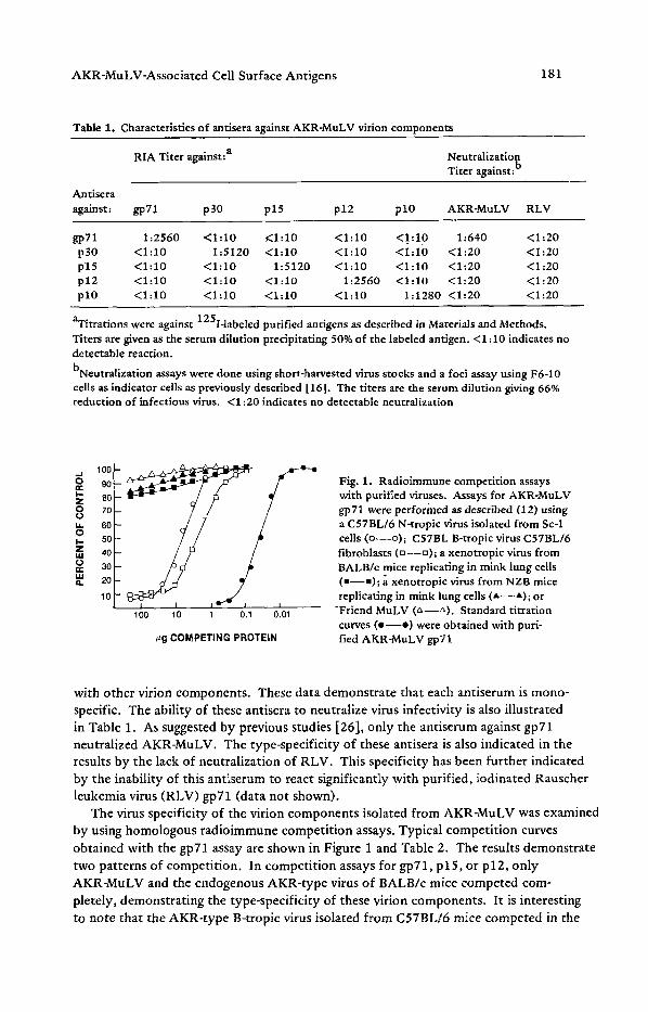
Immunologic Specificity of Antisera. The characteristics of rabbit monospecific anti- sera to the purified viral antigens of AKR virus are shown in Table 1. Each antiserum reacted with its homologous protein with titers of 1:1280-1:5120 and did not react
AKR-MuLV-Associated Cell Surface Antigens 181
Table 1. Characteristics of antisera against AKR-MuLV virion components
�9 �9 a
RIA Tlter against: Neutralization Titer against: b
Antisera against: gp71 p30 p15 p12 plO AKR-MuLV RLV
gp71 1:2560 <1:10 KI:10 <1:10 <1.:1.0 1:640 <1:20 p30 <1:10 1:5120 <1:10 <1:10 <1:10 <1:20 <1:20 p15 <1:10 <1:10 1:5120 <1:10 <1:10 <1:20 <1:20 p12 <1:10 <1:10 <1:10 1:2560 <1:10 <1:20 <1:20 pl0 <1:10 <1:10 <1:10 <1:10 1:1280 <1:20 <1:20
aTitrations were against 125I-labeled purified antigens as described in Materials and Methods. Titers are given as the serum dilution precipitating 50% of the labeled antigen. <1:10 indicates no detectable reaction.
bNeutralization assays were done using short-harvested virus stocks and a foci assay using F6-10 cells as indicator cells as previously described 116]. The titers are the serum dilution giving 66% reduction of infectious virus. <1:20 indicates no detectable neutralization
1001
e,- 00 l.- z 80 O 70 O u.. 60 o ~. 50 z 40 14.1
30 e,.- LU ,', 20
10
100 10 1 0,1 0.01
~g COMPETING PROTEIN
Fig. 1. Radioimmune competition assays with purified viruses. Assays for AKR-MuLV gp71 were performed as described (12) using a C57BL/6 N-tropic virus isolated from Sc-1 cells (o--o) ; C57BL B-tropic virus C57BL/6 fibroblasts (u - -u ) ; a xenotropic virus from BALB/e mice replicating in mink lung cells (a--m); a xenotropic virus from NZB mice replicating in mink lung cells ( A - - A ) ; or
-Friend MuLV (&--LX). Standard titration curves ( e - - e ) were obtained with puri- fied AKR-MuLV gp71
with other virion components. These data demonstrate that each antiserum is mono- specific. The ability of these antisera to neutralize virus infectivity is also illustrated in Table 1. As suggested by previous studies [26], only the antiserum against gp71 neutralized AKR-MuLV. The type-specificity of these antisera is also indicated in the results by the lack of neutralization of RLV. This specificity has been further indicated by the inability of this antiserum to react significantly with purified, iodinated Rauscher leukemia virus (RLV) gp71 (data not shown).
The virus specificity of the virion components isolated from AKR-MuLV was examined by using homologous radioimmune competition assays. Typical competit ion curves
obtained with the gpT1 assay are shown in Figure 1 and Table 2. The results demonstrate
two patterns of competition. In competit ion assays for gp71, p15, or p12, only
AKR-MuLV and the endogenous AKR-type virus of BALB/c mice competed com- pletely, demonstrating the type-specificity of these virion components. It is interesting to note that the AKR-type B-tropic virus isolated from C57BL/6 mice competed in the
182 J.N. Ihle et al.
Table 2. Summary of the presence of AKR-MuLV specific proteins in various murine virus isolates
Virus a Competition BALB BALB C57 assay for.. AKR X-tropic MoLV FLV B-tropic B-tropic
g p 7 1 + - - - + +
p 3 0 + + + + + +
p 1 5 + - - - - + +
p 1 2 + - - - + -
p l O + + + + + +
aviruses were assayed in competition radioimmunoassays as illustrated in Figure 1. (+) indicates the presence of a serotogically indistinguishable antigen from the AKR-MuLV protein. (-) indicates the absence of competition
u
e,
a.
1 0 0 -
90
80
70
60
50
40
30
20
10
�9
I I I I 100 1000 10,000 100,000
l lSerum Dilution
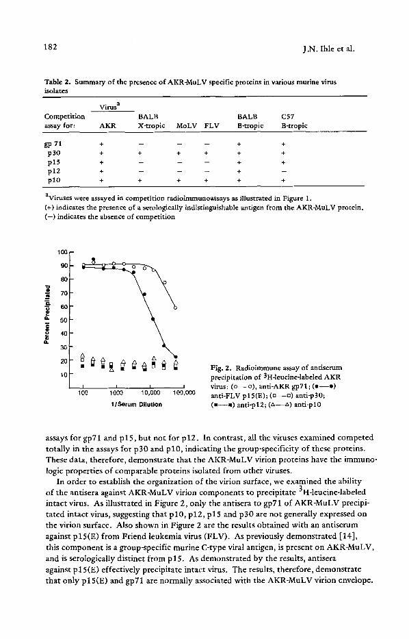
Fig. 2. Radioimmune assay of antiserum precipitation of 3H-leucine-labeled AKR virus: (o--o), anti-AKR gp71; (o- -o) anti-FLV plS(E); (n--o) anti-p30; (m--m) anti-pl2; (zx--z~) anti-pl0
assays for gp71 and p15, but not for p12. In contrast, all the viruses examined competed totally in the assays for p30 and p l0 , indicating the group-specificity of these proteins. These data, therefore, demonstrate that the A K R - M u L V virion proteins have the immuno- logic properties of comparable proteins isolated from other viruses.
In order to establish the organization of the virion surface, we examined the ability of the antisera against AKR-MuLV virion components to precipitate 3H-leucine-labeled intact virus. As illustrated in Figure 2, only the antisera to gp71 of AKR-MuLV precipi- tated intact virus, suggesting that p 10, p 12, p 15 and p 30 are not generally expressed on the virion surface. Also shown in Figure 2 are the results obtained with an antiserum against plS(E) from Friend leukemia virus (FLV). As previously demonstrated [14], this component is a group-specific murine C-type viral antigen, is present on AKR-MuLV, and is serologically distinct from p l 5. As demonstrated by the results, antisera against p 15(E) effectively precipitate intact virus. The results, therefore, demonstrate that only p l 5(E) and gpT1 are normally associated with the AKR-MuLV virion envelope.
AKR-MuLV-Associated Cell Surface Antigens 183
O
r
| i t .
Spontaneous AKR Thymoma
H ogp71 100 ~ C---o ~p12
\ \ ~ ~p30 90 \ \ ~ apl0 80 ~ _ ~ O--Gap15
7O
60
5O
30
2O
10
1/10 1/40 1/160 1/640 Serum Dilution
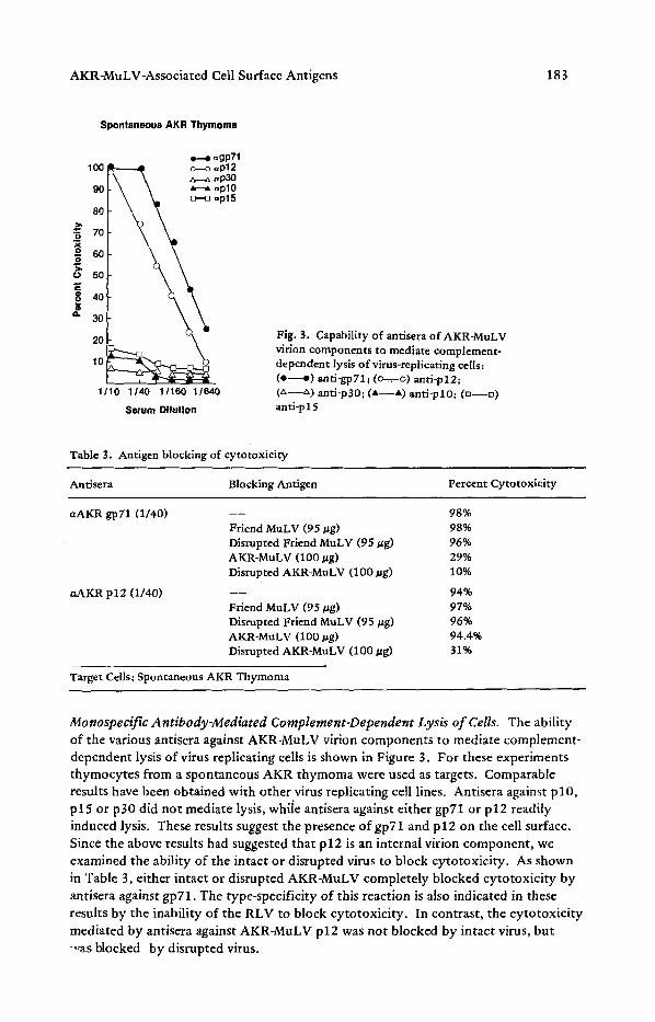
Fig. 3. Capability of antisera of AKR-MuLV virion components to mediate complement- dependent lysis of virus-replicating cells: ( e - - e ) anti-gp71; (o-=-. o) anti-p12; (~,--z~) anti-p30; (A--A) anti-pl0; (n--o) anti-p15
Table 3. Antigen blocking of cytotoxicity
AntJsera Blocking Antigen Percent Cytotoxicity
aAKR gp71 (1/40)
aAKR p12 (1/40)
- - - 98% Friend MuLV (95 Vg) 98% Disrupted Friend MuLV (95/~g) 96% AKR-MuLV (100 #g) 29% Disrupted AKR-MuLV (100 #g) 10%
-- - 94% Friend MuLV (95 t~g) 97% Disrupted Friend MuLV (95/~g) 9696 AKR-MuLV (100 ~g) 94.4% Disrupted AKR-MuLV (100/~g) 31%
Target Cells; Spontaneous AKR Thymoma
Monospecific Antibody-Mediated Complement-Dependent Lysis of Cells. The ability of the various antisera against AKR-MuLV virion components to mediate complement- dependent lysis of virus replicating cells is shown in Figure 3. For these experiments thymocytes from a spontaneous AKR thymoma were used as targets. Comparable results have been obtained with other virus replicating cell lines. Antisera against p l0 , p 15 or p 30 did not mediate lysis, whiie antisera against either gp71 or p 12 readily induced lysis. These results suggest the presence of gp71 and p 12 on the cell surface. Since the above results had suggested that p12 is an internal virion component, we examined the ability of the intact or disrupted virus to block cytotoxicity. As shown in Table 3, either intact or disrupted AKR-MuLV completely blocked cytotoxicity by antisera against gpT1. The type-specificity of this reaction is also indicated in these results by the inability of the RLV to block cytotoxicity. In contrast, the cytotoxicity mediated by antisera against AKR-MuLV p12 was not blocked by intact virus, but -,,as blocked by disrupted virus.
184 J.N. Ihle et al.
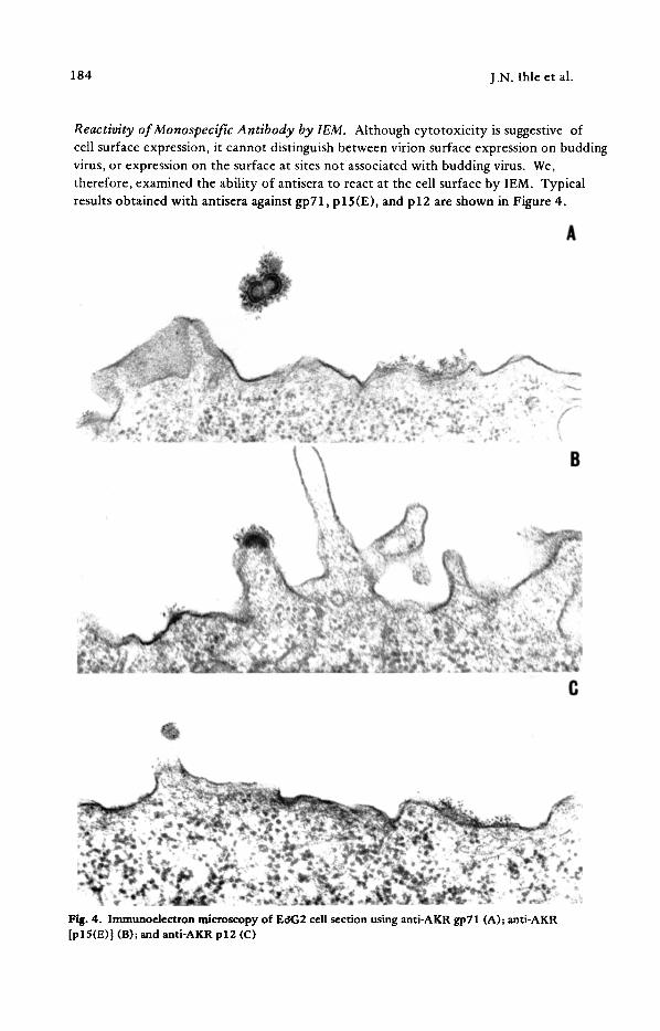
Reactivity of Monospecific Antibody by IEM. Although cytotoxicity is suggestive of cell surface expression, it cannot distinguish between virion surface expression on budding virus, or expression on the surface at sites not associated with budding virus. We, therefore, examined the ability of antisera to react at the cell surface by IEM. Typical results obtained with antisera against gp71, p 15(E), and p 12 are shown in Figure 4.
Fig. 4. Immunoelectron microscopy of EOG2 cell section using anti-AKR gp71 (A); anti-AKR [pl5(E)] (B); and anti-AKR p12 (C)
�9
- a
Tab
le 4
. A
ctiv
ity
of
rab
bit
im
mu
ne
sera
to
war
d m
urm
e le
uk
emia
cel
ls
EcS
G2
K-3
6 E
L-,
* F
LC
-745
A
KR
-T b
Ser
um
C
S
VE
C
S
VE
C
S
VE
C
S
VE
C
S
VE
>,
<
anti
-FM
R g
p71
90
/11
7
52
/60
9
9/1
18
6
1/7
1
75
/91
8
/9
49
/60
3
6/4
0
77
/94
6
0/7
6
anti
-AK
R g
p71
94
/11
3
53
/60
3
3/4
5
29
/35
5
/11
1
1/10
.
..
.
10
1/1
14
6
8/8
5
t~
anti
-FM
R p
30
8
7/1
02
6
/90
5
/77
3
/28
4
/85
0
/7
3/49
2
/23
6
/10
1
3/53
an
ti-F
MR
pl5
(E) c
4
/44
2
3/2
5
5/1
03
5
9/6
9
1/47
2/
3 0
/47
3
1/3
7
..
..
e~
an
ti-A
KR
p1
2
85
/11
4
3/55
.
..
.
2/5
0
0/3
.
..
.
46
/63
1/
32
alm
mu
no
ferr
itin
lab
elin
g o
f le
uk
emia
cel
ls a
nd C
-ty
pe
viru
s.
Pos
itiv
e ce
ll se
ctio
ns
wer
e th
ose
hav
ing
3 o
r m
ore
fer
riti
n-la
bele
d su
rfac
e si
tes.
P
osit
ive
viru
ses
wer
e th
ose
hav
ing
>
10 f
erri
tin
grai
ns
bA
KR
-T w
ere
pri
mar
y A
KR
th
ym
om
as,
gene
rall
y fr
om
on
e an
imal
CT
his
anti
seru
m w
as t
he
gift
of
Dr.
D.P
. B
olog
nesi
, D
uk
e U
niv
ersi
ty,
Du
rham
, N
ort
h C
arol
ina,
US
A
~t~
> g
186 J.N. lhle et al.
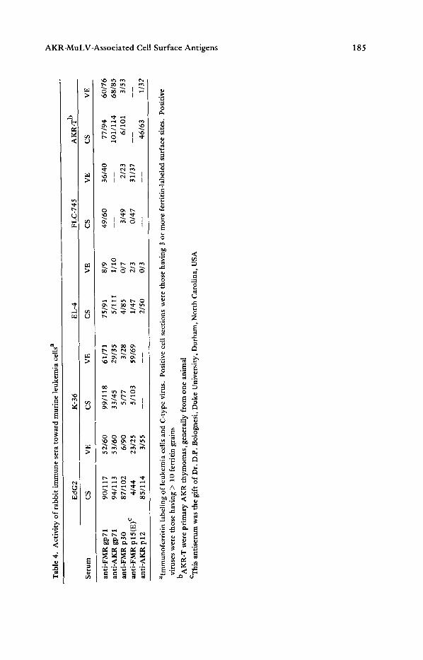
Antisera to gp71 and p12 labeled the cell surface, while only the antisera against gp71 also labeled the virion surface. Also shown are our results with antisera against p 15(E), which under the conditions used only labeled the virion surface. The results of these experiments and others are summarized in Table 4. In all the replicating cell lines examined antisera against gp71 labeled both virion and cell surfaces. In contrast, antisera against p12 only labeled the cell surface of the cells examined. Lastly, as suggested by the above results, antisera against p30 generally did not react with either the virion or cell surface with the exception of one cell line, Ec~G2. These results demonstrate that of the virion components examined, only gp71 and p12 are consistently expressed on the cell surface.
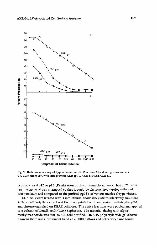
The exceptional anti-p30 reactivity with Ec~G2 was of particular interest since this was the test cell for the original description for the GCSA typing system. The typing antiserum was a C57BL/6 anti-K-36 serum. This was used to describe the classical Gross (G), Gross antigen (GA), and GCSA on EdG2 cells [4,22]. Our results indicate that three proteins can be found on Ec~G2: gp71, p30, and p12, and only two of these proteins are displayed on the surface of other G + cells, namely gp71 and p12. We tested the anti-K-36 serum to determine which proteins of the virion can be recognized. Figure 5 shows radioimmune assay (RIA) titration curves of the.hyperimmune anti-K-36 serum and the autgenous immune BL-6 serum with the viral proteins AKR gp71, AKR p30 and AKR p12. The reactions of the typing serum anti-K-36 with p30 is confirmed and it is apparent that the hyperimmune serum recognizes p12, while normal serum does not.
Thus, the anti-K-36 typing serum differs from natural immune serum in three ways: 1) it has a higher titer to gp71; 2) it has a detectable titer to p30; and 3) it recognizes p12. Therefore, it appears that the reaction of the anti-K-36 typing serum with EgG2 is dependent on at least three proteins: gp71, p30, and p12. Furthermore, in the GCSA typing system of EC~G2 and anti-K-36 serum, a cell or tissue having any combination of these proteins could type as G-positive. Thus, these data take us from the abstract definitions of GCSA to a definition in terms of viral proteins, AKR gp71, AKR p30, and AKR p12.
It is interesting that EC~G2 is the only transplantable murine leukemia tested which grows as a splenic lymphoma. It is not unlikely that p30 fortuitously binds to these transformed cells by virtue of the high content of p30 in the spleen. Thus, the broad reactivity of the anti-K-36 typing serum with the three viral proteins would inherently make this a good typing serum for a tumor cell line which has two expressed antigens and one fortuitous viral antigen on its surface.
An interesting exception in this study was the reactivity of anti-FMR gp71, but not anti-AKR gp71, with the cell surface of EL-4. This cell line is derived from a benzo- pyrene-induced C57BL/6 lymphoma passaged in ascites form. Originally it was con- sidered the prototype G- cell line, not replicating C-type virus, GCSA- and GIX-. Table 5 shows a comparison of antigens of EL-4 and two virus producing cell lines (G +) Ec~G2 and spontaneous AKR thymoma. Specific competition radioimmune assays (CRIA) of extracts of EL-4 showed no competition with AKR gp71, Rauscher or Friend gp71 in homologous assays, but there was competition in an interspecies assay using 125I-Rauscher gp71 and anti-feline leukemia virus (FeLV) antisera. The amount of p30 in EL-4 was low compared to the other cell lines and there was no detectable
AKR-MuLV-Associated Cell Surface Antigens 187
t -
O
t~
o .
"6 2
a .
=u
a .
90
80
70
60
50
4C
3C
2C
1C
9C
8s
70
60
50
40
30
20
10
A
R gp71
B ~ p 3 0 pl 2 AKR R t _
T . . . . - ~ -= - " lo 20 ~ ~o 1~0 ' ' " 51'2o 320 640 1280 2560
Reciprocal of S e r u m D i lu t ion
Fig. S. Radioimmune assay of hyperimmune anti-K-36 serum (A) and autogenous immune C57BL/6 serum (B), with viral proteins AKR gp71, AKR p30 and AKR p12
ecotropic viral p12 or plS..Purification of this presumably non-viral, but gp71 cross- reactive material was attempted so that it could be characterized serologically and biochemically and compared to the purified gp71's of various murine C-type viruses.
EL-4 ceils were treated with 3 mm lithium diiodosalicylate to selectively solubilize surface proteins; the extract was then precipitated with ammonium sulfate, dialyzed and chromatographed on DEAE cellulose. The active fractions were pooled and applied to a column of Lentil lectin (LcH) Sepharose. The material eluting with alpha- methylmannoside was 300-to 600-fold purified. On SDS polyacrylamide gel electro- phoresis there was a prominent band at 70,000 daltons and other very faint bands.
188 J.N. lhle et al.
Table 5. Competition radioimmunoassay of extracts of murine leukemia cell lines
ng/mg Protein EL4 AKR-T EdG2
gp71 AKR ND a 65 47 RLV ND ND ND lnterspecies 350 950 375
p 30 48 1270 502 p12 ND 77 2OO p15 ND 100 16
aND = not detectable
The 70,000 dalton protein labeled with 3H-borohydride after neuraminidase and galactose oxidase treatment, suggesting it is a gly_coprotein.
The purified glycoprotein was labeled with 1251 and the reactivity with antisera to
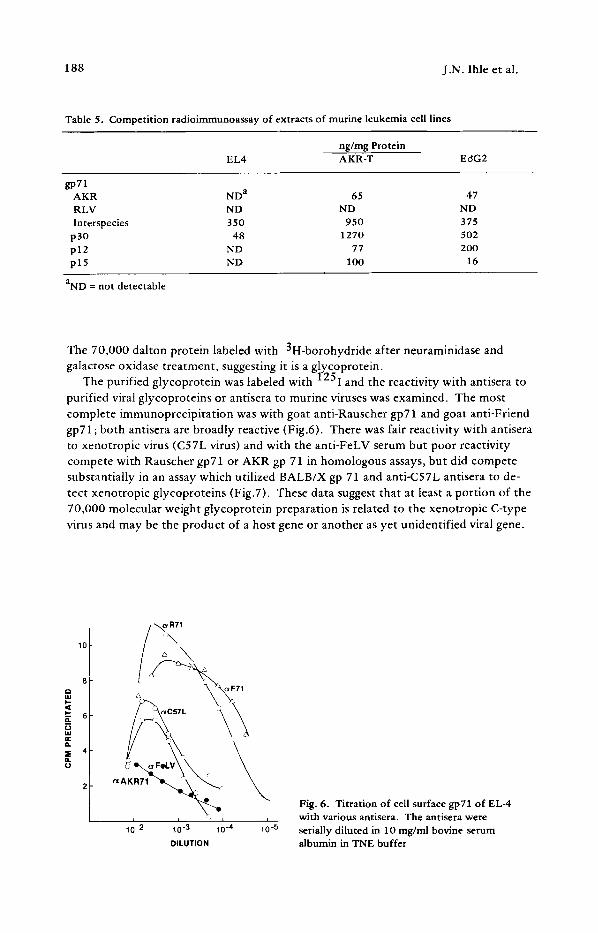
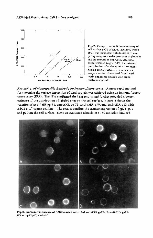
purified viral glycoproteins or antisera to murine viruses was examined. The most complete immunoprecipitation was with goat anti-Rauscher gp71 and goat anti-Friend gp71; both antisera are broadly reactive (Fig.6). There was fair reactivity with antisera to xenotropic virus (C57L virus) and with the anti-FeLV serum but poor reactivity compete with Rauscher gp71 or AKR gp 71 in homologous assays, but did compete substantially in an assay which utilized BALB/X gp 71 and anti-C57L antisera to de- tect xenotropic glycoproteins (Fig.7). These data suggest that at least a portion of the 70,000 molecular weight glycoprotein preparation is related to the xenotropic C-type
virus and may be the product of a host gene or another as yet unidentified viral gene.
10
i i
10-2 10-3 10-4 10-5 DILUTION
Fig. 6. Titration of cell surface gp71 of EL4 with various antisera. The antisera were serially diluted in 10 mg/ml bovine serum albumin in TNE buffer
AKR-MuLV-Associated Cell Surface Antigens 189
1 O0
80
60
40
20
o11 1 .o l o lOO lOOO
MICROGRAMS COMPETITOR
Fig. 7. Competition radioimmunoassay of cell surface gp71 of EL-4. BALB/X tropic gp71 was incubated with dilutions of com- peting antigens, carrier goat gamma globulin and an amount of anti-C57L virus IgG predetermined to give 50% of maximum precipitation of antigen; DEAE fraction- pooled active fractions in interspecies assay; LcH-fraction eluted from Lentil lectin Sepharose column with alpha- methylmannoside
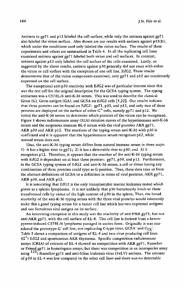
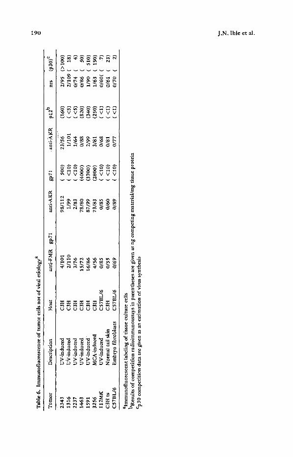
Reactivity, of Monospecific Antibody by Immunofluorescence. A more rapid method for screening the surface expression of viral protein was achieved using an immunofluores- cence assay (IFA). The 1FA confirmed the IEM results and further provided a better estimate of the distribution of labeled sites on the cell surface. Figure 8 shows the reaction of anti-FMR gp 71, anti-AKR gp 71, anti-FMR p30, and anti-AKR p12 with EdG2 a G § tumor cell line. The results confirm the surface expression of gp71, p 12 and p30 on the cell surface. Next we evaluated ultraviolet (UV) radiation-induced
Fig. 8. Immunofluorescence of Ec~G2 reacted with: (A) anti-AKR gp71; (B) anti-FLV gpT1; (C) anti-pl2; (D) anti-p30
Tab
le 6
. lm
mun
oflu
ores
cenc
e of
tum
or c
ells
not
of
vira
l eti
olog
y a
Tum
or
Des
crip
tion
H
ost
~nti
-FM
R
gp71
an
ti-A
KR
gp
71
. ant
i-,~
KR
p1
2 b
nrs
(p30
) e
~O
2343
U
V-i
ndue
ed
C3H
4/
101
98/1
12
(50
0)
23/5
6 (1
60)
2/95
(>
100)
13
16
UV
-ind
uced
C
3H
2/11
0 1/
99
(<~
10)
1/10
1 (<
5)
2/10
9 (
18)
2237
U
V-i
nduc
ed
C3H
3/
76
2/83
(<
10
) 1/
64
(<5
) 0/
74
( 4)
14
63
UV
-ind
uced
C
3H
15/7
2 78
/80
(400
0)
0/88
(8
20)
0/86
(
50)
1591
U
V-i
nduc
ed
C3H
16
/86
87/9
9 (3
700)
2/
99
(340
) 1/
90
(51
0)
3256
M
CA
-ind
uced
C
3H
4/56
73
/83
(280
0)
3/81
(2
50)
1/63
(1
50
) 11
2MK
U
V-i
nduc
ed
C57
BL
/6
0/85
0/
85
(<1
0)
0/68
(<
1)
0/60
( (
7)
C3H
ts
Nor
mal
tai
l sk
in
C3H
0/
55
0/60
(<
10
) 0/
81
(<i)
0/
61
( 23
) C
57B
L/6
E
mbr
yo f
ibro
blas
ts
C57
BL
/6
0/69
0/
89
((1
0)
0/7
7
((1
) 0/
70
( 2)
alm
mun
oflu
ores
cent
labe
ling
of
tiss
ue c
ultu
re c
ells
bR
esul
ts o
f co
mpe
titi
on r
adio
imm
unoa
ssay
s in
par
enth
eses
are
giv
en a
s ng
com
peti
ng m
ater
ial/
rag
tiss
ue p
rote
in
Cp 3
0 co
mpe
titi
on d
ata
are
give
n as
an
esti
mat
ion
of v
irus
syn
thes
is
Z
AKR-MuLV-Assoeiated Cell Surface Antigens 191
fibrosarcoma lines established from primary tumors. Many of these lines were only recently adapted to growth in tissue culture. The results are shown in Table 6. It can be seen that several of the cell lines showed immunofluorescent reactivity with anti-AKR gp71 and p12. These cell lines were examined both by IEM and by p30 competition assays. The cell lines that were negative for reactivity with anti-AKR gp71 and p12 were also negative for virus expression as .determined by p30 competition. Thus, in these recently established UV-induced fibrosarcomas of C3H mice the expression of virus- associated cell surface antigen appears to be coordinate with virus replication.
Discussion
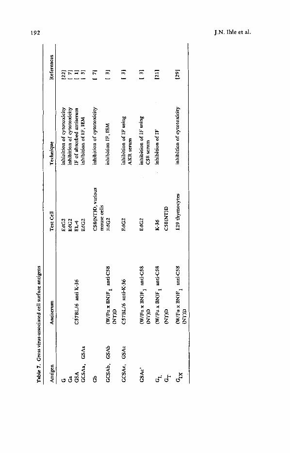
Previous studies of the cell surface antigens associated with endogenous, ecotropic RNA C-type virus infection have primarily depended on the use of broadly reactive antisera made against virus-replicating cells (Table 7). The reaction of one such antiserum, anti- K-36 (the Gross typing serum), with the surface of Ec~G2 (G +) cells has been charac- terized [4,221 . This has been the major test system for the detection and characteriza- tion of both the cell surface and soluble antigens. Because of the complexity of these and other reagents there has been a controversial and often conflicting nomenclature associated with these antigens with a lack of precise serological or biochemical corre- lation of the detected cell surface antigens with viral antigens.
The specificity of the immunological reactions was a major consideration in this series of experiments. We have attempted to bring this aspect of the virus-cell inter- action from the abstract to the defined by utilizing monospecific antisera to purified
viral proteins. The titration data using the purified AKR proteins indicate that they are excellent reagents for analyzing antigens recognizable by autogenous immune or the monospecific hyperimmune sera. The specificity of the reactivities of the monospecific antisera to the individual AKR proteins was established by RIA and thus the distinctive reactivities could be used to characterize the expression of virus-associated antigens on the surface of virus-infected cells. Our results using these specific reagents demon- strated that of the virion components examined, only gp71 and p12 are consistently ex- pressed on the cell surface. The anti-AKR gp71 also labeled the virion sites on G + cells and the anti-AKR p12 labeled only the cell surface sites of G + cells. Data from the use of anti-FMR p30 are also interesting due to the exceptional display of this antigen on EdG2 cells. It has been suggested that p30 is absorbed to the surfaces of cells that are heavy virus producers [10]. Such an absorption could have occurred with EdG2. Also, the presence of p30 on EdG2 may be fortuitous since this tumor line grows in the spleen, an organ demonstrated by Huebner et al. [9] to be rich in p30.
Data from the analysis of surface p12 expression, using anti-AKR p12, raise some interesting questions. P30 and p12 are processed from the same precursor molecule, yet on leukemic lymphocytes the expression of surface AKR p12 consistently ac- companies that of AKR gp71, with p30 being found only rarely. Like p30, p12 is an internal component of the virion which one would not expect to find on the surface of the cell. As yet there are no data to explain why antibody to p12 recognizes the cell surface and not the virion. One hypothesis that has been advanced to explain a similar finding in the Friend virus system is that the membrane perturbation which occurs
Tab
le 7
. G
ross
vir
us-a
ssoc
iate
d ce
ll s
urfa
ce a
ntig
ens
Ant
igen
A
ntis
erum
T
est
Cel
l T
echn
ique
R
efer
ence
s
G
Ec~G
2 in
hibi
tion
of
cyto
toxi
city
[2
2 J
Ga
EdG
2 in
hibi
tion
of
cyto
toxi
city
[
7]
GS
A
C57
BL
/6
anti
K-3
6 E
L-4
IF
of
abso
rbed
ant
iser
um
[ 1]
G
CS
Aa,
G
SA
a E
dG2
inhi
biti
on o
f IF
,IE
M
1 3]
Gb
C58
(NT
)D,
vari
ous
inhi
biti
on o
f cy
toto
xici
ty
[ 7
I m
ouse
cel
ls
GC
SA
b,
GS
Ab
(W/F
ux
BN
)F 1
an
ti-C
58
Ec~G
2 in
hibi
tion
IF
, IE
M
[ 3]
(N
T)D
GC
SA
c,
GS
Ac
C57
BL
/6
anti
-K-3
6 E
tG2
in
hibi
tion
of
IF u
sing
[
31
AK
R s
erum
inhi
biti
on o
f IF
usi
ng
[ 3]
C
58 s
erum
inhi
biti
on o
f IF
[2
1]
GS
Ac )
(W
/Fu
x B
N)F
1
anti
-C58
E
SG
2 (N
T)D
G L
(W
/Fu
x B
N)F
1
anti
-C58
K
-36
G T
(N
T)D
C
58(N
T)D
GIX
(W
/Fu
x B
N)F
1
anti
-C58
12
9 th
ymoc
ytes
(N
T)D
in
hibi
tion
of
cyto
toxi
city
12
91
FO
t~
r
AKR-MuLV-Associated Cell Surface Antigens 193
during virus budding places the p12 molecule in a site where it is inaccessible to anti- body, perhaps because it is blocked by the glycoprotein knobs [24].
The results of these studies utilizing known GCSA systems and extended to neoplastic cells from tumor systems not of viral etiology indicate that the expression of certain viral proteins, namely gp71 and p12, on tumor cell surfaces may be a widespread phenomenon. The purified MuLV proteins and their monospecific antisera present unparalleled reagents for further studies of this type.
References
1. Aoki, T., Boyse, E.A., Old, L.J.: Wild type Gross leukemia virus. I. Soluble antigen (GSA) in the plasma and tissues of infected mice. J. Natl. Cancer Inst. 41, 89-96 (1968)
2. Aoki, T., Herberman, R.B., Hartley, T.W., Liu, M., Walling, M.J., Nunn, M.: Surface antigens on transplantable tumor cell lines producing mouse type C viruses. J. Natl. Cancer Inst. 58, 1069-1078 (1977)
3. Aoki, T., Herberman, R.B., Johnson, P.A., Liu, M., Sturm, M.M.: Wild type Gross leukemia virus: Classification of soluble antigens (GSAs). J. Virol. 10, 1208- 1219 (1972)
4. Aoki, T., Takahashi, T.: Viral and cellular surface antigens of murine leukemias and myelomas. J. Exp. Med. 135,433-436 (1972)
5. Batzing, B.L., Yurconic, M., Jr., Hanna, M.G., Jr.: Autogenous immunity to endogenous RNA tumor virus: Chronic humoral immune response to virus envelope antigens in B6C3F 1 mice. J. Natl. Cancer Inst. 52 ,117-131 (1974)
6. Del Villano, B.C., Nave, B., Croker, B.P., Lerner, R.A., Dixon, F.J.: The oncorna- virus gp69/71. A constituent of the surface of normal and malignant thymocytes. J. Exp. Med. 141,172-187 (1975) Geering, G., Old, L.J., Boyse, E.A.: Antigens of leukemias induced by naturally occurring murine leukemia virus: Their relation to the antigens of Gross virus and other immune leukemia viruses. J. Exp. Med. 124, 753-772 (1966)
8. Hanna, M.G., Jr., Tennant, R.W., Yuhas, T., Clapp, N.K., Batzing, B.L., Snodgrass, M.J.: Autogenous immunity to endogenous RNA tumor virus antigens in mice with a low natural incidence of lymphoma. Cancer Res. 32, 2226--22234 (1972)
9. Huebner, R.J., Sarma, P.S., KeUoff, G.J., Gilden, R.V., Meier, H., Myers, D.D., Peters, R.L.: Immunological tolerances to RNA tumor virus genome expressions: Significance of tolerance and prenatal expression in embryogenesis and tumor- genesis. Ann. NY Acad. Sci. 181,246-271 (1970)
10. Hunsmann, G., Claviez, M., Moennig, V., Schwarz, H., Sch/ifer, W.: Properties of mouse leukemia viruses. X. Occurrence of viral structural antigens on the cell surface as revealed by a cytotoxicity test. Virology 69, 157-168 (1976)
11. lhle, J.N., Denny, T., Bolognesi, D.P.: Purification and serological characterization of the major envelope glycoprotein from AKR murine leukemia virus and its reactivity with autogenous immune sera from mice. J. Virol. 17 ,727-736 (1976)
12. Ihle, J.N., Domotor, J.J., Jr., Bengali, K.M.: Strain-dependent development of an autogenous immune response in mice to endogenous C-type viruses. Bibl. Haematol. 43,177--179 (1976)
194 J.N. Ihle et al.
13. Ihle, J.N., Hanna, M.G., Jr., Roberson, L.E., Kenney, F.T.: Autogenous immunity to endogenous RNA tumor virus: Identification of antibody reactivity to select viral antigens. J. Exp. Med. 139, 1568--1581 (1974)
14. Ihle, J.N., Hanna, M.G., Jr., Sch~ifer, W., Hunsmann, G., Bolognesi, D.P., Hiiper, G.: Polypeptides of mammalian oncornaviruses. III. Localization and reactivity of p15 with natural antibody. Vir_ology 63 ,60 -67 (1975)
15. Ihle, J.N., Joseph, D.R., Pazmi~o, N.H.: Radiation leukemia in C57BL/6 mice. lI. Lack of ecotropic virus expression in the majority of lymphomas. J. Exp. Med. 144, 1406--1423 (1976)
16. Ihle, J.N., Lazar, B.: Natural immunity in mice to the envelope glycoprotein of endogenous, ecotropic type C viruses: Nautralization of virus infectivity. J. Virol. 21 ,974-980 (1977)
17. Ihle, J.N., Yurconic, M., Jr., Hanna, M.G., Jr.: Autogenous immunity to endogenous RNA tumor virus: Radioimmune precipitation assay of mouse serum antibody levels. J. Exp. Med. 1'38,194--208 (1973)
18. Lerner, R.A., Wilson, C.B., Del Villano, B.C., McConahey, P.J., Dixon, F.J.. Endogenous oncornaviral gene expression in adult and fetal mice: Quantitative, histologic, and physiologic studies of the major viral glycoprotein, gpT0. J. Exp. Med. 143,151--166 (1976)
19. Lowy, D.R., Chattopadhyay, S.K., Teich, NAVI., Rowe, W.P., Levine, A.S.: AKR murine leukemia virus genome: Frequency of sequences in DNA of high- and low-, and non-virus-yielding mouse strains. Proc. Natl. Acad. Sci. USA 71, 3555-3559 (1974)
20. Lowy, D.R., Rowe, W.P., Teich, N., Hartley, J.W.: Murine leukemia virus: High frequency activation in vitro b)~ 5-iododeoxyuridine and 5-bromodeoxyuridine. Sience 174, 155-156 (1971)
21. Nowinski, R.C., Peters, E.D.: Cell surface antigens associated with murine leukemia virus definition of G L and G T antigenic systems. J. Virol. 12, 1104-1117 (1973)
22. Old, L.J., Boyse, A.E., Stockert, E.: The G (Gross) leukemia antigen. Cancer Res. 25 ,813-819 (1965)
23. Peterson, D.E., Bucana, C.D., Fidler, I.J.: Immunologic specificity and reactivity of goat anti-guinea pig and goat anti-mouse macrophage sera. J. Res. Soc. 21 ,119-129 (1977)
24. Schwarz, H., Hunsmann, G., Moennig, V., Sch~ifer, W.: Properties of mouse leukemia viruses. XI. Immunoelectron microscopic studies of viral structural antigens on the cell surface. J. Virol. in press (1977)
25. Sherr, C.J., Lieber, M.M., Todaro, G.J.: Mixed splenoeyte cultures and graft versus host reactions selectively induce an "S-tropic" murine type C virus. Cell 1 ,55-58 (1974)
26. Steeves, R.A., Strand, M., August, J.T.: Structural proteins of mammalian oncogenic RNA viruses: Murine leukemia virus neutralization by antisera prepared against purified envelope glycoprotein. J. Virol. 14, 187-189 (1975)
27. Strand, M., Lilly, F., August, J.T.: Host control of endogenous murine leukemia virus gene expression: Concentrations of viral proteins in high and low leukemia mouse strains. Proc. Natl. Acad. Sci. USA 71, 3682-3686 (1974)
AKR-MuLV-Associated Cell Surface Antigens 195
28. Stockert, E., Old, L.J., Boyse, E.A.: The GIX system. A cell surface allo-antigen associated with murine leukemia virus: implications regarding chromosomal integration of the viral genome. J. Exp. Med. 133, 1334--1355 (1971)
29. Todaro, G.J.: "Spontaneous" release of type C viruses from clonal lines of "spontaneously" transformed BALB/3T3 cells. Nature 240, 157--160 (1972)


