Embed Size (px)
Citation preview

Ceramide Kinase Regulates the Production of Tumor NecrosisFactor � (TNF�) via Inhibition of TNF�-converting Enzyme*
Received for publication, October 3, 2011, and in revised form, October 17, 2011 Published, JBC Papers in Press, October 18, 2011, DOI 10.1074/jbc.M111.310169
Nadia F. Lamour‡1, Dayanjan S. Wijesinghe‡§1,2, Jennifer A. Mietla‡3, Katherine E. Ward¶4, Robert V. Stahelin¶�**,and Charles E. Chalfant‡§‡‡5
From the ‡Department of Biochemistry and Molecular Biology, Virginia Commonwealth University, Richmond, Virginia 23298, the§McGuire Veterans Administration Medical Center, Richmond, Virginia 23249, the �Department of Biochemistry and MolecularBiology, Indiana University School of Medicine-South Bend, South Bend, Indiana 46617, the ¶Department of Chemistry andBiochemistry and the **Walther Center for Cancer Research, University of Notre Dame, Notre Dame, Indiana 46556, and ‡‡TheMassey Cancer Center, Richmond, Virginia 23298
Background: Pro-TNF� is transformed into the active/soluble form through proteolysis by TNF�-converting enzyme(TACE).Results: Genetic ablation of ceramide kinase induces an increase in TACE activity and secreted TNF�.Conclusion: Ceramide 1-phosphate (C1P) negatively regulates the activity of TACE.Significance: The TACE/C1P interaction is a viable drug target for the treatment of heart disease and sepsis.
Tumor necrosis factor � (TNF�) is a well known cytokineinvolved in systemic and acute inflammation. In this study, wedemonstrate that ceramide 1-phosphate (C1P) produced by cer-amide kinase (CERK) is a negative regulator of LPS-inducedTNF� secretion. Specifically, bone marrow-derived macro-phages isolated from CERK knock-out mice (CERK�/�) gener-ated higher levels of TNF� than the wild-type mice (CERK�/�)in response toLPS.An increase in basalTNF� secretionwas alsoobserved in CERK�/� murine embryonic fibroblasts, which wasrescued by re-expression ofwild-typeCERK.This effect was dueto increased secretion and not transcription. The secretion ofTNF� is regulated by TNF�-converting enzyme (TACE alsoknown as ADAM17), and importantly, the activity of TACEwashigher in cell extracts from CERK�/� as compared with wildtype. In vitro analysis also demonstrated that C1P is a potentinhibitor of this enzyme, in stark contrast to ceramide andsphingosine 1-phosphate. Furthermore, TACE specificallybound C1P with high affinity. Finally, several putative C1P-binding sites were identified via homology throughout the pro-tein sequence of TACE. These results indicate that C1P pro-ducedbyCERKhas a negative effect on the processing/secretionof TNF� via modulation of TACE activity.
Lipopolysaccharide (LPS) is the glycolipid of the outer mem-brane of Gram-negative bacteria (1–3). Recognition of LPS byimmune cells is essential in mounting responses against invad-ing microbes (1, 4); however, hyperactivation of the immuneresponse can also lead to the overwhelming production of tis-sue-damaging cytokines inducing septic shock. Sepsis or septicshock is a major cause of mortality with �120,000 deaths/yearin the United States (5), and currently, there are no effectivetherapeutics available.LPS is recognized by immune cells via Toll-like receptors.
Toll-like receptors are evolutionarily conserved proteins thatrecognize pathogen-associated molecular patterns (6), andmolecular studies coupled with genetic mouse models haveidentified TLR4 as an essential receptor for LPS signaling (7).Specifically, genetic mouse models lacking key moleculesinvolved in TLR4-mediated signaling are protected from LPS-induced septic shock (8), and one key molecule produced inresponse to LPS is the proinflammatory/tissue-damaging cyto-kine, tumor necrosis factor � (TNF�) (9). As alluded above,excessive production of proinflammatory cytokines like TNF�can cause deleterious effects on normal body functions (10).Injection of neutralizing antibodies against TNF� was consid-ered a promising therapeutic approach to block LPS-inducedseptic shock (11–14), but this therapeutic approach in septicpatients did not achieve the same therapeutic efficiency asobserved in animal models (15–17). Therefore, characteriza-tion of TNF� downstream signaling cascades may aid in devel-oping better therapeutics to modulate inflammatory cascadesduring the development of sepsis.With regard to signaling cascades, there are two main levels
of regulation for TNF� production/secretion in response toLPS as follows: 1) induction of TNF� synthesis via the NF-�Bpathway (18); and 2) induction of TNF� secretion via increasedprocessing (e.g. shedding) of TNF� (19). With regard to secre-tion/“shedding,” TNF� is produced in the body as pro-TNF�,an inactive precursor of the cytokine in the formof a 233-aminoacid membrane-anchored propeptide. Pro-TNF� is trans-
Author’s Choice—Final version full access.* This work was supported, in whole or in part, by National Institutes of Health
Grants HL072925 and CA117950 (to C. E. C.). This work was also supportedby a research grant supplied by ONO Pharmaceutical Co., Ltd. (to C. E. C.)and by American Heart Association Grant SDG0735350N (to R. V. S.).
1 Both authors contributed equally to this work.2 Recipient of National Institutes of Health NRSA Post-doctoral Fellowship (in
Wound Healing) Grant T32 GM008695.3 Recipient of National Institutes of Health NRSA Pre-doctoral Fellowship (in
Functional Lipidomics in Cardiovascular and Respiratory Diseases) GrantT31 HL094290.
4 Recipient of National Institutes of Health Training Fellowship CBBI GrantT32GM075762 and an American Heart Association pre-doctoralfellowship.
5 To whom correspondence should be addressed: Virginia CommonwealthUniversity, Dept. of Biochemistry and Molecular Biology, 1101 East Mar-shall St., P. O. Box 980614, Richmond, VA 23298-0614. Tel.: 804-828-9526;E-mail: [email protected].
THE JOURNAL OF BIOLOGICAL CHEMISTRY VOL. 286, NO. 50, pp. 42808 –42817, December 16, 2011Author’s Choice Printed in the U.S.A.
42808 JOURNAL OF BIOLOGICAL CHEMISTRY VOLUME 286 • NUMBER 50 • DECEMBER 16, 2011
at VIV
A, V
A C
omm
onwealth U
niv, on Septem
ber 7, 2012w
ww
.jbc.orgD
ownloaded from

formed into the active and soluble formby limited proteolysis atthe Ala76 and Val77 bond by the protease, TNF�-convertingenzyme (TACE6/ADAM17) (20).TACE/ADAM17 belongs to the ADAM family of protei-
nases, and the enzyme is a type I transmembrane protein syn-thesized as a zymogen (21). It contains a prodomain, a catalyticdomain, a disintegrin and cysteine-rich region, a transmem-brane segment, and a cytoplasmic tail (21). Mature, activeTACE is sequestered within cholesterol-rich membranemicrodomains, otherwise known as lipid rafts or detergent-re-sistant microdomains (22, 23). Numerous substrates of TACEare localized in these rafts, and depletion of cholesterol fromthe lipid rafts by cyclodextrin or high density lipoprotein treat-ment increased the shedding of TACE substrates withoutincreasing TACE activity (24).AsTACE regulates the secretion of TNF�, orally bioavailable
TACE inhibitorsmay have the potential to effectively treat sep-sis and other inflammatory diseases by limiting the levels ofsoluble TNF� release by cells (25). Indeed, inhibition of TACEby small molecular weight compounds has been more effectivethan TNF� neutralizing antibodies on alleviating septic shockin animal models (26). Hence, researchers are actively pursuingthe development of small molecules as orally bioavailableTACE inhibitors (27).In this study, we find that genetic ablation of the enzyme,
ceramide kinase (CERK), leads to a significant increase inTNF�production in response to LPS. Mechanistic studies demon-strate that the increase in TNF� is due to an increase in TNF�processing/shedding. Finally, this study demonstrates that theloss ofCERK leads to an increase in TACE activity, andTACE isdirectly and specifically inhibited by the product of CERK, cer-amide 1-phosphate (C1P).
EXPERIMENTAL PROCEDURES
Materials—Macrophage colony-stimulating factor wasobtained from Sigma. Dulbecco’s modified Eagle’s medium(DMEM), RPMI, fetal bovine serum (FBS), and penicillin/strep-tomycin (100 units/ml penicillin G sodium and 100 �g/mlstreptomycin sulfate) were obtained from Invitrogen. HPLCused was a Shimadzu Prominence LC-20-AD system, and themass spectrometer was a 4000 QTRAP from ABSciex. Prior tomass spectrometric analysis, lipids were separated by reversephase chromatography using a PhenomenexKinetex 2.6-�C18100A 50 � 2.1-mm reverse phase HPLC column (Torrance,CA). HPLC grademethanol, HPLC grade chloroform, andACSgrade formic acid (EMD Chemicals) were purchased fromVWR (Bridgeport, NJ). The TNF� assay kit was obtained fromPeproTech. 1-Palmitoyl-2-oleoyl-sn-glycero-3-phosphocho-line (POPC), 1-palmitoyl-2-oleoyl-sn-glycero-3-phosphoetha-nolamine (POPE), 1-oleoyl-2-oleoyl-sn-glycero-3-phosphate,1-palmitoyl-2-oleoyl-sn-glycero-3-phosphate, C18:1 C1P, C16
C1P, sphingosine 1-phosphate (S1P), C18:1 ceramide, C16 cer-amide, the lipid extruder, and 100-nm polycarbonate filterswere from Avanti Polar Lipids (Alabaster, AL). Triton X-100,CHAPS, and octyl glucoside were from Sigma and Fisher,respectively. L1 sensor chips were from GE Healthcare. Full-length TACEwas obtained fromEMDChemicals, and the fluo-rogenic substrate was obtained from R&D Systems.Bone Marrow-derived Macrophages (BMDMs)—Primary
monocytes were isolated from CERK�/� and CERK�/� mice(28) and placed in RPMI 1640 medium containing 10% serumand MCSF at 20 ng/ml for differentiation into BMDMs asdescribed previously (29). For experiments, BMDMs wereseeded in 6-well plates (5 � 105 cells/35-mm well), and incu-bated for 24 h at standard incubator conditions. The BMDMswere then stimulated by LPS (0.5 ng/ml) or treated with PBSsham control. The medium was collected at various times forTNF� quantification. For mass spectrometric quantification oflipids, the BMDM (1 � 106 cells) were grown in 100-mm platesunder the same condition as above for the same length of time.The cells were harvested and lipids quantified by HPLC ESI-MS/MS as described previously (30).Mouse Embryonic Fibroblasts (MEFs)—Primary MEFs were
isolated from 13 or 14 days pregnant females, as described pre-viously (31), and grown in high glucose Dulbecco’s modifiedEagle’s medium (Invitrogen) supplemented with 20% fetalbovine serum (Invitrogen) and 2% penicillin/streptomycin(BioWhittaker) at standard incubation conditions. The cellswere passaged every 3 days, and after 20 serial passages of theprimary MEFs, immortalized MEFs were obtained. For experi-ments, immortalized MEFs were seeded in 6-well plates (5 �105 cells/35 mm well) and incubated for 24 h at standard incu-bator conditions. The medium was changed to Dulbecco’smodified Eagle’s medium (Invitrogen) supplemented with 2%fetal bovine serum before treatment and TNF� quantification.TNF� Assay—TNF� levels were measured using mouse
TNF� EIA kit (PeproTech) following the manufacturer’sinstructions. The kit uses a monoclonal antibody to mouseTNF� immobilized on a microtiter plate to bind the mouseTNF� in the samples. After a short incubation, polyclonal anti-body to mouse TNF� was added. This antibody bound to themouse TNF� captured on the plate. After another short incu-bation, a donkey anti-rabbit IgG conjugated to HRPwas added,which bound to the polyclonal mouse TNF� antibody. Thereaction was stopped, and the color generated was read at 450nm using a microplate spectrophotometer (BMG LabtechFLUOStar Optima). The measured optical density was directlyproportional to the concentration of TNF�.RNA Isolation, Reverse Transcription-PCR, and Quantitative
PCR—Toevaluate the transcript level of TNF�, total RNA fromBMDMs was isolated using RNeasy kit (Qiagen) according tothemanufacturer’s instructions. Total RNA (1�g) was reverse-transcribed using Superscript III reverse transcriptase (Invitro-gen) as described previously (32–38), and the levels of TNF�mRNA transcript were determined using quantitative PCR inaccord with the TaqMan technology (Applied Biosystems) (33,37, 38) specific toTNF� andGAPDH.The cDNAwas amplifiedusing ABI 7900HT.
6 The abbreviations used are: TACE, TNF�-converting enzyme; C1P, ceramide1-phosphate; CERK, ceramide kinase; BMDM, bone marrow-derivedmacrophage; MEF, murine embryonic fibroblasts; SPR, surface plasmonresonance; POPC, 1-palmitoyl-2-oleoyl-sn-glycero-3-phosphocholine;POPE, 1-palmitoyl-2-oleoyl-sn-glycero-3-phosphoethanolamine; aSMase,acid sphingomyelinase; S1P, sphingosine 1-phosphate; CMC, criticalmicelle concentration; PA, phosphatidic acid; D-e-, D-erythro-.
Ceramide Kinase Regulates TNF� Production
DECEMBER 16, 2011 • VOLUME 286 • NUMBER 50 JOURNAL OF BIOLOGICAL CHEMISTRY 42809
at VIV
A, V
A C
omm
onwealth U
niv, on Septem
ber 7, 2012w
ww
.jbc.orgD
ownloaded from

TACE Activity Assay—BMDM cell extracts (10 �g) werediluted in 50 mM Tris, pH 7.9, and incubated with 10 �M of thefluorogenic substrate III (7-methoxycoumarin-PLAQAV-N-3-(2,4-dinitrophenyl)-l-2,3-diaminopropionyl-RSSSR-NH2) asspecified by the manufacturer’s instructions. For examinationof lipid effects on the enzymatic activity of TACE, a TritonX-100 mixed micelle approach was utilized as described previ-ously (39, 39–48). Briefly, recombinant human TACE (0.2ng/ml) was premixed for 5 min with Triton X-100 micelles (10times CMC) at 37 °C prior to the addition of substrate. Of note,the addition of Triton X-100 below CMC and �20� CMC hadno effect on in vitro TACE activity. To prepare the lipid-con-taining micelles, an appropriate volume of the indicated phos-pholipids (previously solubilized in an appropriate organic sol-vent) was dried under nitrogen. Triton X-100 at 20� CMC inTACE assay buffer was added to the dried lipid to produce a 2�concentrated lipid/micelle solution for each designated mol %.The solution was vortexed vigorously for 2 min followed byprobe sonication (1 min on and 1min off for 3 min) on ice. Thelipid/micelle solution was then added to the enzyme to a finalconcentration of 10� CMC for Triton X-100 containing thedesignated mol % of lipid. The fluorescence emitted from thecleavage product was quantified by spectrofluorometry usingexcitation and emission wavelength of 320 and 405 nm, respec-tively and was used to calculate specific activity of TACE fol-lowing the manufacturer’s protocol. The data were plottedusing SigmaPlot Version 12 (Systat Software).Expression of Ceramide Kinase by Adenovirus-mediated
Transfection—Recombinant adenovirus for ceramide kinasewas generated using Adeno-XTM Tet-Off system (Clontech)followed by purification and titration using Adeno-XTM virusmini purification kit (Clontech) and Adeno-XTM rapid titer kit(Clontech), respectively. For overexpression of CERK, cellswere treated with the recombinant CERK adenovirus togetherwith the Tet-Off adenovirus at a multiplicity of infection of 20as described previously (40). Assays requiring CERK ectopicexpression were carried out 48 h postinfection, and expressionof CERK was verified by western immunoblotting for the His6tag.SPR-binding Protein-Lipid Interaction—All SPR measure-
ments were performed at 25 °C in 20 mM HEPES, pH 7.4, con-taining 0.16 M KCl as described previously (40, 44–46, 49).Following washing of the sensor chip surfaces, POPC/POPE/X(70:20:10, where X � C1P, PA, S1P, or ceramide) and POPC/POPE (80:20) vesicles were injected at 5 �l/min onto the activesurface and the control surface, respectively, to give the sameresonance unit (6000 resonance units) values. Under our exper-imental conditions, no binding to the control surface wasdetected beyond the refractive index change for all proteins.Each lipid layer was washed three times with 10 �l of 50 mM
NaOH at 100 �l/min. Typically, no decrease in lipid signal wasseen after the first injection. Equilibrium SPR measurementswere done at the flow rate of 5 �l/min to allow sufficient timefor the R values of the association phase to reach equilibriumvalues (Req) for 1 �M TACE (44, 49).CERK Knock-out Mouse and Animal Welfare Assurances—
Breeding pairs for the CERK�/� and wild-type counterpartswere obtained fromNovartis Pharma as a gracious gift fromDr.
Frederic Bornancin. The phenotype and biological character-ization of these mice are reported elsewhere by Bornancin andco-workers (28, 51–53). All cells derived from these mice weregenetically verified for each presented n for each experiment asdescribed by Bornancin and co-workers (28). Animal experi-ments were conducted at Virginia Commonwealth University(Virginia Commonwealth University) (Richmond, VA). Theresearch protocol IACUC AM10089 was approved by the Vir-ginia CommonwealthUniversity Institutional Animal Care andUse Committee (IACUC) in accordance with the USDA Ani-mal Welfare Regulations; the Public Health Service Policy onthe Humane Care and Use of Laboratory Animals; and TheUnited StatesGovernment Principles for the Utilization andCare of Vertebrate Animals Used in Testing, Research, andTraining. Virginia Commonwealth University is in compliancewith all provisions of the AnimalWelfare Act and other federalstatutes and regulations relating to animals. Virginia Common-wealthUniversity is registered under theAnimalWelfareAct asa Class “R” Research Facility with the USDA-APHIS-AnimalCare (Registration number: 52-R-0007). The Office of Labora-tory Animal Welfare has approved the Virginia Common-wealth University’s Animal Welfare Assurance in compliancewith the United States Public Health Policy (assurance numberA3281-01).
RESULTS
Ex Vivo Cells from CERK�/� Mice Secrete Increased Levels ofTNF�—Compounds structurally related to C1P have beenshown previously to blockTNF� production in cells challengedwith LPS in culture and also in vivo in septic shock models(54–58). Our laboratory hypothesized that these compoundswere acting as C1P mimetics and inhibiting TNF� productionvia an unknown mechanism. Therefore, our hypothesis infersthat TNF� production would be increased in ex vivo cellsderived from CERK�/� mice (e.g. cells lacking CERK-derivedC1P). To test this hypothesis, MEFs from CERK�/� andCERK�/� mice were grown under standard incubator condi-tions, and basal TNF� secretion was analyzed. Fig. 1A demon-strates that secreted TNF� was significantly increased in themedia fromMEFs derived from theCERK�/� mouse. To verifythe phenotype and to specifically examine the role of CERK inthe production of TNF�, “rescue” experiments were concomi-tantly undertaken. Specifically, CERK cDNAwas re-introducedin CERK �/� MEFs by adenovirus-mediated transfection, andindeed, re-expression of CERK reduced the levels of secretedTNF� in accord with wild-type cells (Fig. 1A). These data dem-onstrate that the levels of TNF� are increased due to the spe-cific loss of CERK expression.During the course of these studies, Nikolova-Karakashian
and co-workers reported a role for ceramide derived from acidsphingomyelinase (aSMase) in LPS-induced secretion of TNF�in primary macrophages (59). This study coupled to our abovefindings suggested the hypothesis that the conversion ofaSMase-derived ceramide to C1P may be a key step in this reg-ulatorymechanism. To determinewhetherCERK regulated theproduction of TNF� in a relevant cell model and for compari-son with theNikolova-Karakashian studies(59), we utilized pri-mary BMDMs to determine whether loss of CERK produced
Ceramide Kinase Regulates TNF� Production
42810 JOURNAL OF BIOLOGICAL CHEMISTRY VOLUME 286 • NUMBER 50 • DECEMBER 16, 2011
at VIV
A, V
A C
omm
onwealth U
niv, on Septem
ber 7, 2012w
ww
.jbc.orgD
ownloaded from

the same phenotype. The treatment of BMDMs with LPSinduced TNF� secretion, with amaximumproduction after 8 hof treatment (Fig. 1B). However, TNF� levels were significantlyelevated in CERK�/� macrophages as compared withCERK�/� cells analogous to the findings of Nikolova-Kara-kashian and co-workers (59) with the aSMase ablation model(Fig. 1B). These results suggest that the conversion of ceramideto C1P is a key step in the secretion of TNF�.CERK Deficiency Does Not Affect the Rate of Synthesis for
TNF� mRNA—Nikolova-Karakashian and co-workers (59)reported that genetic ablation of aSMase affected the secretion/processing of TNF� and not the synthesis of TNF� mRNA. To
determine whether the differences in TNF� levels upon LPSstimulation were due to a change in the transcription rate ofTNF�, the levels of TNF� mRNA from CERK�/� andCERK�/� BMDMs challenged with LPS were analyzed byquantitative PCR. CERK deficiency did not have the effect ofincreasing the rate of synthesis of TNF� mRNA (Fig. 2A).
As the loss of CERK did not have an effect on the transcrip-tion rate of TNF� mRNA, we examined whether the amount ofTNF� secreted by shedding was increased in CERK�/�
BMDMs subjected to LPS. Indeed, the amount of TNF�retained in the CERK�/� BMDMs was significantly less thanthe CERK�/� BMDMs (Fig. 2B). These data correlated with anincrease in the amount of TNF� secreted into the media byCERK�/� BMDMs exposed to LPS in comparison withCERK�/� BMDMs (Fig. 2B). Furthermore, the total levels ofTNF� (when the cell and media extracts are combined) werenot significantly different between CERK�/� and CERK�/�
BMDMs. Therefore, these data demonstrate a role for CERK-derived C1P in the post-translational mechanism of TNF�processing/shedding in response to LPS.
FIGURE 1. Genetic ablation of ceramide kinase increases the levels ofTNF�. A, genetic ablation of ceramide kinase increases the levels of TNF� inimmortalized MEFs. MEFs (2 � 105 cells) derived from either wild-type mice orCERK�/� mice were seeded overnight in 35-mm plates. The following day,cells were transfected with either control adenovirus (adCMV) (20 multiplici-ties of infection) or CERK adenovirus (adCERK) (20 multiplicities of infection).After 48 h, cells were then washed and placed in complete media supple-mented with 2% FBS. After 4 h, the media were collected and analyzed forTNF� content by ELISA. Data presented is the mean of n � 7 � S.E. repeatedon three separate occasions. Student’s t test was used to assess statisticalsignificance (statistical significance � p � 0.01). B, genetic ablation of cer-amide kinase increases the levels of TNF� in response to LPS. BMDMs (2 � 105
cells) derived from either wild-type mice or CERK�/� mice were seeded over-night in 35-mm plates. Cells were treated for 0, 4, 8, 24, and 48 h with LPS (0.5ng/ml). The media were then collected at the appropriate time points andanalyzed for TNF� content by ELISA. The data presented are representative ofthe means � S.E. of an n � 7 repeated on three separate occasions (asteriskindicates a statistical significance of p � 0.01).
FIGURE 2. TNF� secretion but not expression is significantly different inCERK null macrophages compared with wild type. Primary monocytes iso-lated from the bone marrow of CERK null mice and their wild-type littermateswere differentiated into BMDMs as described under “Experimental Proce-dures.” The BMDMs (5 � 105 cells), thus obtained, were seeded overnight in35-mm plates. Cells were then treated with LPS (0.5 ng/ml) or PBS sham, andtotal RNA and protein were harvested 4 h post-treatment. A, TNF� andGAPDH mRNA levels were quantified via quantitative PCR with TNF� mRNAlevels normalized to GAPDH expression. Data presented in this figure are themeans of three separate experiments � S.E. (n � 3). B, protein extracts fromcells (10 �g) (labeled as Intracellular TNF�) or media (20 �l of cell media)(labeled as Secreted TNF�) were subjected to SDS-PAGE and Western immu-noblotting analysis for TNF� and �-actin expression. Of note: the cell mediademonstrated no �-actin expression (data not shown). The lower panellabeled Total TNF� represents a mixture of cell extracts (5 �g) combined withcell media (10 �l) from the same samples utilized to generate the data pre-sented in the upper panels. Data presented in this figure are representative ofn � 6 on two separate occasions.
Ceramide Kinase Regulates TNF� Production
DECEMBER 16, 2011 • VOLUME 286 • NUMBER 50 JOURNAL OF BIOLOGICAL CHEMISTRY 42811
at VIV
A, V
A C
omm
onwealth U
niv, on Septem
ber 7, 2012w
ww
.jbc.orgD
ownloaded from

Ceramide Kinase Regulates TNF� Production
42812 JOURNAL OF BIOLOGICAL CHEMISTRY VOLUME 286 • NUMBER 50 • DECEMBER 16, 2011
at VIV
A, V
A C
omm
onwealth U
niv, on Septem
ber 7, 2012w
ww
.jbc.orgD
ownloaded from

Enzymatic Activity of TACE Is Higher in BMDMs fromCERK�/� Mice—Thematuration process of TNF� is regulatedby the protease, ADAM17, also known as TNF� convertingenzyme (TACE). Nikolova-Karakashian and co-workers (59)reported that TACE activity was higher in ex vivo cells fromaSMase�/� mice (36). To determine whether the increasedTNF� secretion in CERK�/� cells was due to differences inTACE activity, we examined the TNF� converting activity ofthe enzyme in BMDM extracts from CERK�/� and CERK �/�
mice. CERK�/� BMDMs exhibited higher TACE activity ascompared with their respective CERK �/� control cells (Fig.3A). These differences were not due to alterations in TACEprotein content as Western blot analysis of TACE protein inmacrophages from CERK�/� and CERK�/� mice did not showany genotype-related differences (Fig. 3B). However, asexpected, CERK�/� BMDM contained significantly loweramounts of themajor CERK-derived C1P species (d18:1/16:0 andd18:1/24:1 C1P) compared with CERK�/� BMDM (Fig. 3C), sug-gesting that the increase in TACE activity is related to the cel-lular levels of CERK-derived C1P. Although the d18:1/16:0 cer-amide levels were higher for these BMDM compared with wildtype, the total ceramide was not significantly changed betweenthe two cell types (Fig. 3D) arguing against a direct role forceramide in the regulation of TACE activity. Insignificant dif-ferences were also observed between the two cell types for thetotal levels of the sphingolipids, d18:1 monohexosyl ceramides,sphingomyelins, sphingosine, sphinganine, sphingosine1-phosphate, and sphinganine 1-phosphate (Fig. 3, E–J). Thesedata demonstrate that CERK-derived C1P negatively regulatesthe activity of TACE, which would explain the effects of CERKablation on secreted TNF� levels.Ceramide 1-Phosphate Is a Specific Inhibitor of TACE inVitro—
Previously, our laboratory demonstrated that C1P binds to theC2domain of group IVphospholipaseA2 (cPLA2�) at a cationicpatch (Arg57, Lys58, and Arg59) in the �-groove (43, 44, 46, 48).As our data suggested that CERK regulated TACE activity, weexamined the primary sequence of TACE for possible C1Pinteraction sites. Indeed, several potential C1P-binding motifswere identified in TACE (Fig. 4A) suggesting that C1P directlyinteracts with the enzyme to affect the enzymatic activity.To determinewhetherC1Pdirectly affects TACE activity, we
analyzed the in vitro activity of the enzyme in the presence of
C1P (D-e-C18:1/16:0 C1P). Fig. 4B demonstrates that C1P is apotent inhibitor of TACE inducing �90% inhibition at 0.85mol %. With �120 molecules of Triton X-100 per micelle, thisequates to an estimated stoichiometry of one C1P molecule/micelle to inactivate TACE. More importantly, this inhibitoryeffect was highly specific as the closely related lipids such asceramide and S1P had no effect on TACE activity (Fig. 4B).Furthermore, the more soluble dioeloyl-PA was only a minorinhibitor of the enzyme failing to reach the IC50 value of theenzymatic activity (Fig. 4B). Thus, C1P is a direct, specific, andpotent inhibitor of TACE activity.Asmainly the C18:1/16:0 subspecies of C1P was reduced in the
CERK�/� BMDMs, we further characterized the chain lengthspecificity of C1P inhibition of TACE activity. Fig. 4C demon-strates that TACE activity is equally inhibited by the C1P sub-species, D-e-C18:1/12:0 C1P, D-e-C18:1/16:0 C1P, D-e-C18:1/18:1
C1P, and D-e-C18:1/24:0 C1P. D-e-C18:1/8:0 C1P significantlyinhibited TACE but to a lesser extent than the longer C1P sub-species. D-e-C18:1/2:0 C1P had a minor effect on TACE enzymeactivity, which was even less than the effect of PA at the samemol %. These data demonstrate that C1P inhibition of TACErequires an acyl chain of�12 carbons for full inhibitory activity.Furthermore, these data also demonstrate that the inhibition ofTACE enzyme activity by C1P is irrespective of the saturationstate of the acyl chain.Ceramide 1-Phosphate Specifically and Directly Interacts
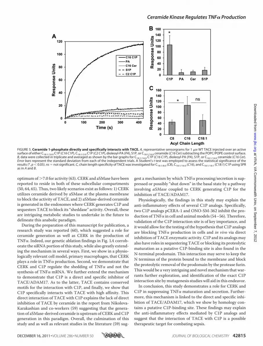
with TACE—To quantitatively determine the affinity of TACEfor liposomes harboring C1P, PA, S1P, and ceramide, weemployed a robust analysis by SPR. In these experiments, anactive surface was coated with POPC/POPE/X (70:20:10 X �C1P, S1P, PA, or ceramide), whereas a control surface wascoated with POPC/POPE (80:20). TACE was injected at 1 �M
across the active surfaces to determine the relative binding foreach of the lipids. As shown in Fig. 5, A and B, TACE onlydisplayed detectable and significant binding to liposomes con-taining C1P. As with the effect of C1P subspecies on TACEenzyme activity, the interaction of C1P was irrespective of thesaturation state of the acyl chain, and the interaction alsorequired a �12 carbon acyl chain for high affinity to beobserved (Fig. 5C). Hence, C1P specifically and directly binds toTACE.
FIGURE 3. Higher levels of TACE enzymatic activity are observed in primary macrophages isolated from CERK null mice. A, primary monocytes isolatedfrom the bone marrow of CERK null mice and their wild-type littermates were differentiated into BMDMs and treated with LPS concentration (0.5 ng/ml) for 4 has described under “Experimental Procedures.” Protein homogenates were harvested from these cells, and TACE specific activity (units of emission at 405 nmof cleaved fluorogenic substrate/20 min/10 �g of protein homogenate) was measured as described under “Experimental Procedures.” The data are represen-tative of the mean � S.E. of an n � 6 repeated on three separate occasions. B, mixture of cell extract (5 �g) combined with cell media (10 �l) (from Fig. 2B, lowerpanel) were subjected to SDS-PAGE and Western immunoblotting analysis for TACE and �-actins. Note: the depicted �-actin data show an exact match to the�-actin data depicted in Fig. 2B, lower panel, as the same Western immunoblot utilized in this figure was stripped and reprobed for TACE expression. C–J,primary monocytes isolated from the bone marrow of CERK null mice and their wild-type littermates were differentiated into BMDMs. Lipids were extractedfrom these cells 24 h later, and the lipids species of interest were quantified via HPLC ESI-MS/MS as described under “Experimental Procedures.” C, ceramide1-phosphate levels are represented in units of picomoles per 1 � 106 cells and are the means of n � 6 of individual experiments � S.D. Student’s t test was usedto assess statistical significance (*, p � 0.05; **, p � 0.01). D, ceramides were also measured from the same samples and are similarly represented in units ofpicomoles per 1 � 106 cells and are the means of n � 6 individual experiments � S.D. Student’s t test was used to assess statistical significance (*, p � 0.05; **,p � 0.01). E, monohexosyl ceramide levels represented as the mean of three experiments � S.D. and given in units of picomoles per 1 � 106 cells. Student’s ttest was used to assess statistical significance (*, p � 0.05). F, sphingomyelin levels represented as the mean of three experiments � S.D. and given in units ofpicomoles per 1 � 106 cells. Student’s t test was used to assess statistical significance (*, p � 0.05). G, sphingosine levels represented as the mean of threeexperiments � S.D. and given in units of picomoles per 1 � 106 cells. Student’s t test was used to assess statistical significance (*, p � 0.05). H, sphinganine levelsrepresented as the mean of three experiments � S.D. and given in units of picomoles per 1 � 106 cells. Student’s t test was used to assess statistical significance(NS means not significant). I, sphingosine 1-phosphate levels represented as the mean of three experiments � S.D. and given in units of picomoles per 1 � 106
cells. Student’s t test was used to assess statistical significance. J, sphinganine 1-phosphate levels represented as the mean of three experiments � S.D. andgiven in units of picomoles per 1 � 106 cells.
Ceramide Kinase Regulates TNF� Production
DECEMBER 16, 2011 • VOLUME 286 • NUMBER 50 JOURNAL OF BIOLOGICAL CHEMISTRY 42813
at VIV
A, V
A C
omm
onwealth U
niv, on Septem
ber 7, 2012w
ww
.jbc.orgD
ownloaded from

DISCUSSION
In this report, our laboratory found that CERK and its prod-uct C1P play a role in the suppression of TNF� production.Specifically, we found that genetic ablation of CERK led to anincrease in the amount of TNF� released, basally in MEFs andin response to LPS in primarymacrophages. Indeed, these stud-ies complement the recent report byNikolova-Karakashian andco-workers (59), which demonstrated that genetic ablation ofacid sphingomyelinase (aSMase) gave the same phenotype inprimarymacrophages as we observed inCERK�/� cells. Hence,the culmination of this report and the studies presented herestrongly suggest that CERK utilizes ceramide generated byaSMase in response to LPS.
Our study also suggests that the topography in the cell of C1Pis most important for the inhibition of TACE. Specifically, wefound that the in vitro inhibition of TACE was irrespective ofthe acyl chain length and saturation state of long chain C1Psubspecies (e.g. D-e-C18:1/16:0 C1P versus D-e-C18:1/24:1 C1P). Asour data coupled with the findings of Nikolova-Karakashianand co-workers (59) strongly suggest that CERK utilizes cera-mide-derived from aSMase, the subcellular compartment sug-gested is the lysosomes. Indeed, CERKhas a pHoptimumbelowpH 7.0 (61), and thus, it is not heretical that C1Pmay be gener-ated in a low pH environment preferred by aSMase. Suggestinga separate subcellular compartment are the reports that TACEis localized to the cell surface and endosomes (62) and has a pH
FIGURE 4. Ceramide 1-phosphate is a direct and specific inhibitor of TACE. A, TACE demonstrates the presence of highly conserved sequences homologousto the C1P-binding site of cytosolic phospholipase A2. The primary structure of human TACE was scrutinized for a potential C1P-binding site in the form of threeto four consecutive basic amino acids. Six such sequences were observed from 53 to 58 (N-terminal prodomain), 210 to 215 (mature TACE), 233 to 237 (matureTACE), 625 to 628 (mature TACE), 640 to 645 (mature TACE), and 753 to 758 (mature TACE). A multiple alignment of the corresponding region revealed severalhighly conserved motifs. B, effect of phospholipids on in vitro TACE activity. TACE activity was measured in vitro using human recombinant TACE enzyme andfluorogenic substrate as described using a Triton X-100 mixed micelle approach to deliver the individual lipids (26). C18:1/18:1 C1P, dioleoyl PA, S1P, and C18:1/18:1ceramide was used at the designated mol % in regard to Triton X-100. The data are plotted as the mol % of the specific lipid versus TACE specific activity(picomoles of cleaved fluorogenic substrate/min/�g of recombinant TACE protein). The data are representative of the mean � S.E. of an n � 7 repeated onthree separate occasions. C, acyl chain length requirement of C1P for inhibition of TACE activity. TACE activity was measured in vitro using human recombinantTACE enzyme and fluorogenic substrate as described using a Triton X-100 mixed micelle approach to deliver the individual lipids (26). The fluorescence wasmonitored over time on a fluorescence plate reader. C18:1/2:0 (C2:0 C1P), C18:1/8:0 (C8:0 C1P), C18:1/12:0 (C12:0 C1P), C18:1/16:0 (C16:0 C1P), C18:1/18:1 (C18:1 C1P), andC18:1/24:0 (C24:0 C1P)C1P was used at 0.6 mol % with respect to Triton X-100. The data are plotted as the treatment of the specific lipid and controls versus TACEspecific activity (picomoles of cleaved fluorogenic substrate/min/�g of recombinant TACE protein). The data are representative of the mean � S.E. of ann � 4 repeated on two separate occasions.
Ceramide Kinase Regulates TNF� Production
42814 JOURNAL OF BIOLOGICAL CHEMISTRY VOLUME 286 • NUMBER 50 • DECEMBER 16, 2011
at VIV
A, V
A C
omm
onwealth U
niv, on Septem
ber 7, 2012w
ww
.jbc.orgD
ownloaded from
optimumof�7.0 for activity (63). CERK and aSMase have beenreported to reside in both of these subcellular compartments(50, 64, 65). Thus, two likely scenarios exist as follows: 1) CERKutilizes ceramide derived by aSMase at the plasma membraneto block the activity of TACE, and 2) aSMase-derived ceramideis generated in the endosomes where CERK generates C1P andsequesters TACE to block its “sheddase” activity. Overall, theseare intriguing metabolic studies to undertake in the future todelineate this anabolic paradigm.During the preparation of this manuscript for publication, a
research study was reported (60), which suggested a role forceramide generation as well as CERK in the production ofTNF�. Indeed, our genetic ablation findings in Fig. 1A corrob-orate the siRNAportion of this study, while also greatly extend-ing the mechanism in several ways. First, we show in a physio-logically relevant cell model, primary macrophages, that CERKplays a role in TNF� production. Second, we demonstrate thatCERK and C1P regulate the shedding of TNF� and not thesynthesis of TNF� mRNA. We further extend the mechanismto demonstrate that C1P is a direct and specific inhibitor ofTACE/ADAM17. As to the latter, TACE contains conservedmotifs for the interaction with C1P, and finally, we show thatC1P specifically interacts with TACE with high affinity. Thisdirect interaction of TACE with C1P explains the lack of directinhibition of TACE by ceramide in the report from Nikolova-Karakashian and co-workers (59) suggesting that the genera-tion of aSMase-derived ceramide is upstreamof CERK andC1Pgeneration in this paradigm. Overall, the culmination of thisstudy and as well as relevant studies in the literature (59) sug-
gest a mechanism by which TNF� processing/secretion is sup-pressed or possibly “shut down” in the basal state by a pathwayinvolving aSMase coupled to CERK generating C1P for theinhibition of TACE/ADAM17.Physiologically, the findings in this study may explain the
anti-inflammatory effects of several C1P analogs. Specifically,two C1P analogs pCERA-1 and ONO-SM-362 inhibit the pro-duction of TNF� in cell and animalmodels (54–56). Therefore,validation of the C1P interaction site is of key importance, andit would allow for the testing of the hypothesis that C1P analogsare blocking TNF� production in cells and in vivo via directinhibition of TACE enzymatic activity. C1P and its analogsmayalso have roles in sequestering TACE or blocking its proteolyticmaturation as a putative C1P-binding site is also found in theN-terminal prodomain. This interaction may serve to keep theN terminus of the protein bound to the membrane and blockthe proteolytic removal of the prodomain by the protease furin.This would be a very intriguing and novel mechanism that war-rants further exploration, and identification of the exact C1Pinteraction site bymutagenesis studies will aid in this endeavor.In conclusion, this study demonstrates a role for CERK and
C1P in suppressing TNF� maturation and secretion. Further-more, this mechanism is linked to the direct and specific inhi-bition of TACE/ADAM17, which we show by homology con-tains a putative C1P-binding site. These findings may explainthe anti-inflammatory effects mediated by C1P analogs andsuggest that the interaction of TACE with C1P is a possibletherapeutic target for combating sepsis.
FIGURE 5. Ceramide 1-phosphate directly and specifically interacts with TACE. A, representative sensorgrams for 1 �M WT TACE injected over an activesurface of either C18:1/16:0 C1P (C16 C1P), C18:1/2:0 C1P (C2 C1P), dioleoyl-PA (PA), S1P, or C18:1/16:0 ceramide (C16 Cer) subtracting the POPC/POPE control surface.B, data were collected in triplicate and averaged as shown by the bar graphs for C18:1/16:0 C1P (C16 C1P), dioleoyl-PA (PA), S1P, or C18:1/16:0 ceramide (C16 Cer).Error bars represent the standard deviation from each of the independent trials. A Student’s t test was employed to assess the statistical significance of theresults (*, p � 0.05). ns � not significant. C, chain length specificity of TACE was investigated for C18:1/8:0 (C8), C18:1/16:0 (C16), and C18:1/18:1 (C18:1) C1P using SPRas in A and B.
Ceramide Kinase Regulates TNF� Production
DECEMBER 16, 2011 • VOLUME 286 • NUMBER 50 JOURNAL OF BIOLOGICAL CHEMISTRY 42815
at VIV
A, V
A C
omm
onwealth U
niv, on Septem
ber 7, 2012w
ww
.jbc.orgD
ownloaded from

Acknowledgments—We thank Dr. Frederick Bornancin of NovartisPharma for providing breeding pairs of the CERK�/� mouse alongwith wild-type counterparts. Virginia CommonwealthUniversity wasrecipient of National Institutes of Health Grant NH1C06-RR17393(for renovation).
REFERENCES1. Watson, R. W., Redmond, H. P., and Bouchier-Hayes, D. (1994) J. Leuko-
cyte Biol. 56, 95–1032. Raetz, C. R., and Whitfield, C. (2002) Annu. Rev. Biochem. 71, 635–7003. Raetz, C. R. (1990) Annu. Rev. Biochem. 59, 129–1704. Cohen, J. (2002) Nature 420, 885–8915. Martin, G. S., Mannino, D. M., Eaton, S., and Moss, M. (2003) N. Engl.
J. Med. 348, 1546–15546. Takeda, K., Kaisho, T., and Akira, S. (2003) Annu. Rev. Immunol. 21,
335–3767. Lu, Y. C., Yeh, W. C., and Ohashi, P. S. (2008) Cytokine 42, 145–1518. Akira, S. (2000) Biochem. Soc. Trans. 28, 551–5569. Lin, W. J., and Yeh, W. C. (2005) Shock 24, 206–20910. Ramesh, G., and Reeves, W. B. (2002) J. Clin. Invest. 110, 835–84211. Beutler, B., Milsark, I. W., and Cerami, A. C. (1985) Science 229,
869–87112. Bhola,M., Goto,M., Chen, H. Y., andMyers, T. F. (2000)Biol. Neonate 78,
207–21113. Hinshaw, L. B., Tekamp-Olson, P., Chang,A.C., Lee, P. A., Taylor, F. B., Jr.,
Murray, C. K., Peer, G. T., Emerson, T. E., Jr., Passey, R. B., and Kuo, G. C.(1990) Circ. Shock 30, 279–292
14. Tracey, K. J., Fong, Y., Hesse, D. G., Manogue, K. R., Lee, A. T., Kuo, G. C.,Lowry, S. F., and Cerami, A. (1987) Nature 330, 662–664
15. Abraham, E., Anzueto, A., Gutierrez, G., Tessler, S., San Pedro, G., Wun-derink, R., Dal Nogare, A., Nasraway, S., Berman, S., Cooney, R., Levy, H.,Baughman, R., Rumbak, M., Light, R. B., Poole, L., Allred, R., Constant, J.,Pennington, J., and Porter, S. (1998) Lancet 351, 929–933
16. Abraham, E., Laterre, P. F., Garbino, J., Pingleton, S., Butler, T., Dugernier,T.,Margolis, B., Kudsk, K., Zimmerli,W., Anderson, P., Reynaert,M., Lew,D., Lesslauer, W., Passe, S., Cooper, P., Burdeska, A., Modi, M., Leighton,A., Salgo, M., and Van der Auwera, P. (2001) Crit. Care Med. 29, 503–510
17. Panacek, E. A.,Marshall, J. C., Albertson, T. E., Johnson, D. H., Johnson, S.,MacArthur, R.D.,Miller,M., Barchuk,W.T., Fischkoff, S., Kaul,M., Teoh,L., VanMeter, L., Daum, L., Lemeshow, S., Hicklin, G., andDoig, C. (2004)Crit. Care Med. 32, 2173–2182
18. Collart, M. A., Baeuerle, P., and Vassalli, P. (1990) Mol. Cell. Biol. 10,1498–1506
19. Horiuchi, K., Kimura, T., Miyamoto, T., Takaishi, H., Okada, Y., Toyama,Y., and Blobel, C. P. (2007) J. Immunol. 179, 2686–2689
20. Hoareau, L., Bencharif, K., Rondeau, P.,Murumalla, R., Ravanan, P., Tallet,F., Delarue, P., Cesari, M., Roche, R., and Festy, F. (2010) J. Inflamm. 7:1
21. Black, R.A., Rauch,C. T., Kozlosky, C. J., Peschon, J. J., Slack, J. L.,Wolfson,M. F., Castner, B. J., Stocking, K. L., Reddy, P., Srinivasan, S., Nelson, N.,Boiani, N., Schooley, K. A., Gerhart, M., Davis, R., Fitzner, J. N., Johnson,R. S., Paxton, R. J., March, C. J., and Cerretti, D. P. (1997) Nature 385,729–733
22. Tellier, E., Canault, M., Poggi, M., Bonardo, B., Nicolay, A., Alessi, M. C.,Nalbone, G., and Peiretti, F. (2008) J. Cell. Physiol. 214, 687–693
23. Wakatsuki, S., Kurisaki, T., and Sehara-Fujisawa, A. (2004) J. Neurochem.89, 119–123
24. Tellier, E., Canault, M., Rebsomen, L., Bonardo, B., Juhan-Vague, I., Nal-bone, G., and Peiretti, F. (2006) Exp. Cell Res. 312, 3969–3980
25. DasGupta, S., Murumkar, P. R., Giridhar, R., and Yadav, M. R. (2009)Bioorg. Med. Chem. 17, 444–459
26. Newton, R. C., Solomon, K. A., Covington, M. B., Decicco, C. P., Haley,P. J., Friedman, S.M., andVaddi, K. (2001)Ann. Rheum.Dis. 60, iii25–iii32
27. Le, G. T., and Abbenante, G. (2005) Curr. Med. Chem. 12, 2963–297728. Graf, C., Zemann, B., Rovina, P., Urtz, N., Schanzer, A., Reuschel, R.,
Mechtcheriakova, D., Müller, M., Fischer, E., Reichel, C., Huber, S., Daw-son, J., Meingassner, J. G., Billich, A., Niwa, S., Badegruber, R., Van Veld-
hoven, P. P., Kinzel, B., Baumruker, T., and Bornancin, F. (2008) J. Immu-nol. 180, 3457–3466
29. Eske, K., Breitbach, K., Köhler, J., Wongprompitak, P., and Steinmetz, I.(2009) J. Immunol. Methods 342, 13–19
30. Wijesinghe, D. S., Allegood, J. C., Gentile, L. B., Fox, T. E., Kester, M., andChalfant, C. E. (2010) J. Lipid Res. 51, 641–651
31. vom Brocke, J., Schmeiser, H. H., Reinbold, M., and Hollstein, M. (2006)Carcinogenesis 27, 2141–2147
32. Chalfant, C. E., Rathman, K., Pinkerman, R. L., Wood, R. E., Obeid,L. M., Ogretmen, B., and Hannun, Y. A. (2002) J. Biol. Chem. 277,12587–12595
33. Goehe, R. W., Shultz, J. C., Murudkar, C., Usanovic, S., Lamour, N. F.,Massey, D. H., Zhang, L., Camidge, D. R., Shay, J. W., Minna, J. D., andChalfant, C. E. (2010) J. Clin. Invest. 120, 3923–3939
34. Massiello, A., and Chalfant, C. E. (2006) J. Lipid Res. 47, 892–89735. Massiello, A., Roesser, J. R., and Chalfant, C. E. (2006) FASEB J. 20,
1680–168236. Massiello, A., Salas, A., Pinkerman, R. L., Roddy, P., Roesser, J. R., and
Chalfant, C. E. (2004) J. Biol. Chem. 279, 15799–1580437. Shultz, J. C., Goehe, R. W., Murudkar, C. S., Wijesinghe, D. S., Mayton,
E. K., Massiello, A., Hawkins, A. J., Mukerjee, P., Pinkerman, R. L., Park,M. A., and Chalfant, C. E. (2011)Mol. Cancer Res. 9, 889–900
38. Shultz, J. C., Goehe, R.W.,Wijesinghe, D. S.,Murudkar, C., Hawkins, A. J.,Shay, J. W., Minna, J. D., and Chalfant, C. E. (2010) Cancer Res. 70,9185–9196
39. Wijesinghe, D. S., Massiello, A., Subramanian, P., Szulc, Z., Bielawska, A.,and Chalfant, C. E. (2005) J. Lipid Res. 46, 2706–2716
40. Lamour, N. F., Subramanian, P.,Wijesinghe, D. S., Stahelin, R. V., Bonven-tre, J. V., and Chalfant, C. E. (2009) J. Biol. Chem. 284, 26897–26907
41. Wijesinghe, D. S., Subramanian, P., Lamour, N. F., Gentile, L. B., Granado,M. H., Bielawska, A., Szulc, Z., Gomez-Munoz, A., and Chalfant, C. E.(2009) J. Lipid Res. 50, 1986–1995
42. Wijesinghe, D. S., Lamour, N. F., Gomez-Munoz, A., and Chalfant, C. E.(2007)Methods Enzymol. 434, 265–292
43. Subramanian, P., Vora, M., Gentile, L. B., Stahelin, R. V., and Chalfant,C. E. (2007) J. Lipid Res. 48, 2701–2708
44. Stahelin, R. V., Subramanian, P., Vora, M., Cho, W., and Chalfant, C. E.(2007) J. Biol. Chem. 282, 20467–20474
45. Lamour, N. F., Stahelin, R. V., Wijesinghe, D. S., Maceyka, M., Wang, E.,Allegood, J. C., Merrill, A. H., Jr., Cho, W., and Chalfant, C. E. (2007) J.Lipid Res. 48, 1293–1304
46. Subramanian, P., Stahelin, R. V., Szulc, Z., Bielawska, A., Cho, W., andChalfant, C. E. (2005) J. Biol. Chem. 280, 17601–17607
47. Pettus, B. J., Bielawska, A., Subramanian, P., Wijesinghe, D. S., Maceyka,M., Leslie, C. C., Evans, J. H., Freiberg, J., Roddy, P., Hannun, Y. A., andChalfant, C. E. (2004) J. Biol. Chem. 279, 11320–11326
48. Pettus, B. J., Bielawska, A., Spiegel, S., Roddy, P., Hannun, Y. A., and Chal-fant, C. E. (2003) J. Biol. Chem. 278, 38206–38213
49. Stahelin, R. V., Long, F., Diraviyam, K., Bruzik, K. S., Murray, D., and Cho,W. (2002) J. Biol. Chem. 277, 26379–26388
50. Jenkins, R. W., Canals, D., Idkowiak-Baldys, J., Simbari, F., Roddy, P.,Perry, D. M., Kitatani, K., Luberto, C., and Hannun, Y. A. (2010) J. Biol.Chem. 285, 35706–35718
51. Boath,A., Graf, C., Lidome, E., Ullrich, T.,Nussbaumer, P., andBornancin,F. (2008) J. Biol. Chem. 283, 8517–8526
52. Graf, C., Niwa, S., Müller, M., Kinzel, B., and Bornancin, F. (2008)Biochem. Biophys. Res. Commun. 373, 159–163
53. Tsuji, K., Mitsutake, S., Yokose, U., Sugiura, M., Kohama, T., and Igarashi,Y. (2008) FEBS J. 275, 3815–3826
54. Avni, D., Goldsmith, M., Ernst, O., Mashiach, R., Tuntland, T., Meijler,M. M., Gray, N. S., Rosen, H., and Zor, T. (2009) Immunol. Lett. 123, 1–8
55. Ogata, T., Yamashita, K., Horiuchi, H., Okuda, K., and Todo, S. (2008)Surgery 143, 545–555
56. Goldsmith, M., Avni, D., Levy-Rimler, G., Mashiach, R., Ernst, O., Levi,M., Webb, B., Meijler, M. M., Gray, N. S., Rosen, H., and Zor, T. (2009)Immunology 127, 103–115
57. Matsui, T., Kondo, T., Nishita, Y., Itadani, S., Nakatani, S., Omawari, N.,Sakai, M., Nakazawa, S., Ogata, A., Ohno, H., Obata, T., Nakai, H., and
Ceramide Kinase Regulates TNF� Production
42816 JOURNAL OF BIOLOGICAL CHEMISTRY VOLUME 286 • NUMBER 50 • DECEMBER 16, 2011
at VIV
A, V
A C
omm
onwealth U
niv, on Septem
ber 7, 2012w
ww
.jbc.orgD
ownloaded from

Toda, M. (2002) Bioorg. Med. Chem. Lett. 12, 903–90558. Matsui, T., Kondo, T., Nishita, Y., Itadani, S., Tsuruta, H., Fujita, S.,
Omawari, N., Sakai, M., Nakazawa, S., Ogata, A., Mori, H., Ohno, H.,Obata, T., Nakai, H., and Toda, M. (2002) Bioorg. Med. Chem. Lett. 12,907–910
59. Rozenova, K. A., Deevska, G. M., Karakashian, A. A., and Nikolova-Kara-kashian, M. N. (2010) J. Biol. Chem. 285, 21103–21113
60. Józefowski, S., Czerkies, M., Łukasik, A., Bielawska, A., Bielawski, J., Kwi-atkowska, K., and Sobota, A. (2010) J. Immunol. 185, 6960–6973
61. Sugiura, M., Kono, K., Liu, H., Shimizugawa, T., Minekura, H., Spiegel, S.,and Kohama, T. (2002) J. Biol. Chem. 277, 23294–23300
62. Doedens, J. R., and Black, R. A. (2000) J. Biol. Chem. 275, 14598–1460763. Willems, S. H., Tape, C. J., Stanley, P. L., Taylor, N. A., Mills, I. G., Neal,
D. E., McCafferty, J., and Murphy, G. (2010) Biochem. J. 428, 439–45064. Baumruker, T., Bornancin, F., and Billich, A. (2005) Immunol. Lett. 96,
175–18565. Mitsutake, S., Kim, T. J., Inagaki, Y., Kato, M., Yamashita, T., and Igarashi,
Y. (2004) J. Biol. Chem. 279, 17570–17577
Ceramide Kinase Regulates TNF� Production
DECEMBER 16, 2011 • VOLUME 286 • NUMBER 50 JOURNAL OF BIOLOGICAL CHEMISTRY 42817
at VIV
A, V
A C
omm
onwealth U
niv, on Septem
ber 7, 2012w
ww
.jbc.orgD
ownloaded from


