Embed Size (px)
Citation preview
1
Digitally Signed by: Content manager’s Name
DN : CN = Weabmaster’s name
O= University of Nigeria, Nsukka
OU = Innovation Centre
Fred Attah
Faculty of Veterinary Medicine
Department of Veterinary Pathology and Microbiology,
HAEMATOLOGY AND SERUM BIOCHEMICAL FINDINGS
ASSOCIATED WITH SOME PATHOLOGICAL CONDITIONS IN
SLAUGHTER CATTLE AT NSUKKA ABATTOIR, NIGERIA.
UDEANI, IKECHUKWU JOHN
PG/M.Sc/10/57665
2
HAEMATOLOGY AND SERUM BIOCHEMICAL FINDINGS ASSOCIATED
WITH SOME PATHOLOGICAL CONDITIONS IN SLAUGHTER CATTLE AT
NSUKKA ABATTOIR, NIGERIA.
BY
UDEANI, IKECHUKWU JOHN
PG/M.Sc/10/57665
DEPARTMENT OF VETERINARY PATHOLOGY AND MICROBIOLOGY,
FACULTY OF VETERINARY MEDICINE,
UNIVERSITY OF NIGERIA, NSUKKA
SUPERVISOR
PROF. J. I. IHEDIOHA
3
HAEMATOLOGY AND SERUM BIOCHEMICAL FINDINGS ASSOCIATED
WITH SOME PATHOLOGICAL CONDITIONS IN SLAUGHTER CATTLE AT
NSUKKA ABATTOIR, NIGERIA.
BY
UDEANI, IKECHUKWU JOHN
PG/M.Sc/10/57665
A DISSERTATION PRESENTED TO THE SCHOOL OF POSTGRADUATE
STUDIES, UNIVERSITY OF NIGERIA, NSUKKA IN PARTIAL FULFILLMENT
OF REQUIREMENTS FOR THE AWARD OF MASTER OF SCIENCE IN
VETERINARY PATHOLOGY.
MARCH, 2014
SUPERVISOR
PROF. J. I. IHEDIOHA
DEPARTMENT OF VETERINARY PATHOLOGY AND MICROBIOLOGY,
FACULTY OF VETERINARY MEDICINE,
UNIVERSITY OF NIGERIA, NSUKKA
4
DECLARATION
I hereby declare that the work described herein is my original work and has not been
previously submitted for any degree to any University or similar Institution.
Name: UDEANI, IKECHUKWU JOHN
Registration number: PG/M. Sc./10/57665
Sign________________________________
Date___________________________________
5
CERTIFICATION
This is to certify that UDEANI, IKECHUKWU J (PG/M. Sc./10/57665), a postgraduate
student in the Department of Veterinary Pathology and Microbiology, Faculty of
Veterinary Medicine, University of Nigeria Nsukka has satisfactorily completed the
requirements for research work for the Degree of Master of Science in Veterinary
Pathology. The work embodied in this dissertation is original and has not been submitted
in part or in full for any other degree of this or any other University. The dissertation has
therefore been approved for the award of Master of Science Degree in the Department of
Veterinary Pathology and Microbiology, University of Nigeria Nsukka.
BY
_____________ _____________ ____________ ___________
Sign Date Sign Date
Prof. J. I. Ihedioha Prof. K. F. Chah
(Supervisor) (Head of Department)
_____________ _____________ ____________ ___________
Sign Date Sign Date
Prof. S. V. O Shoyinka
(External Examiner) (Dean of Faculty)
6
ABSTRACT
This study evaluated the haematological and serum biochemical changes associated with
diseases and disorders of cattle billed for slaughter at Nsukka abattoir. It also compared
the haematological and serum biochemical findings in cattle with specific diseases and
conditions to that of cattle with no obvious abnormalities or lesions (apparently healthy
cattle) and evaluated the influence of age and sex on the haematology and serum
biochemistry profile of the apparently healthy cattle at the Nsukka abattoir, Enugu State,
Nigeria. The study was a disease surveillance survey. The study population was 8,100
trade cattle billed for slaughter at the Nsukka abattoir, Enugu State, Nigeria during the
ten-month period of study. The research (disease surveillance) visits were made once
every two weeks during the study period. All cattle billed for slaughter on the days of
research visit (a total of 567 cattle) were studied and constituted the sample population.
They were subjected to comprehensive physical examination. Cattle with grossly
observable signs or lesions of disease or disorder were followed up and blood samples
were collected from them. Blood samples were also collected from cattle with no obvious
abnormalities or lesions (apparently healthy cattle) to serve as control. Blood sample
collection was by jugular venipuncture. The haematology and serum biochemical tests on
the blood samples and confirmatory tests for specific diseases observed were done
following standard procedures. Cattle with specific diseases, disorders and conditions
were grouped accordingly. The apparently healthy cattle were segregated according to
their age and sex. Results showed that out of the 567 cattle investigated, 91 (16.05%) had
specific diseases, disorders and conditions while 476 (83.95%) had no obvious
abnormalities or lesions. The diseases, disorders and conditions and their percentage
occurrence were fasciolosis (8.47%), tuberculosis (1.76%), trypanosomosis (1.41%),
7
cachexia unassociated with any disease (1.06%), skin disorders (0.88%), rumen fluke-
infestation (0.71%), benign neoplasm (0.18%) and pregnancy (1.59%). Cattle with
fasciolosis, tuberculosis, trypanosomosis and rumen fluke-infestation had significantly (p
< 0.05) lower mean packed cell volume (PCV), red blood cell count and haemoglobin
concentration and significantly (p < 0.05) higher erythrocyte sedimentation rate (ESR)
when compared with the apparently healthy cattle. Fasciola and abomasal worm infested
cattle also had significantly (p < 0.05) lower serum total protein. The means for the total
leukocyte, lymphocyte and eosinophil counts of cattle with fasciolosis, tuberculosis and
trypanosomosis were significantly (p < 0.05) higher than those of the apparently healthy
cattle. In addition, cattle with fasciolosis had significantly (p < 0.05) lower serum alanine
aminotransferase (ALT) and significantly (p < 0.05) higher alkaline phosphatase (ALP)
activity, while those with tuberculosis had significantly (p < 0.05) higher serum globulin.
Cachectic cattle had significantly (p < 0.05) lower serum ALT, creatinine, urea,
monocyte, eosinophil and basophil counts. Pregnant females had significantly (p < 0.05)
higher ESR, serum ALP, globulin, total leukocyte and lymphocyte counts, and
significantly (p < 0.05) lower aspartate amino transferase (AST), ALT, creatinine and
urea than non-pregnant apparently healthy females. The mean RBC count, ESR,
eosinophil counts, serum ALP and creatinine levels of the apparently healthy male cattle
were significantly (p < 0.05) higher than that of the females, while serum globulin levels
of the apparently healthy adult cattle were significantly (p < 0.05) higher than that of the
young. Based on the results, it was concluded that among all diseases, disorders and
conditions recorded for cattle in this study, fasciolosis ranked topmost as the commonest,
followed by tuberculosis and then pregnancy. It was also noted that the disease, disorders
and conditions were associated with specific haematological and serum biochemical

8
findings that were considered to be of clinical diagnostic importance. The haematology
and serum biochemistry of the apparently healthy cattle in this study were in most
instances comparable to those reported for cattle in available literature, but some of the
minimum and maximum values recorded in this present study were different from the
upper and lower reference limits reported in available literature.

9
DEDICATION
In loving memory of my dearly departed father (Rt. Hon. H. C. Udeani) and brother (Mr.
J. J. Udeani). May your souls and those of all faithful departed through God’s mercy rest
in peace, Amen!
10
ACKNOWLEDGEMENT
I thank the Almighty God for his gift of mental and physical health throughout the
duration of this programme. I am grateful for and appreciative of the inestimable support
of my project supervisor (Prof. J. I. Ihedioha), my family, friends and colleagues in their
varied contributions towards the success of this study. I also thank the Department of
Veterinary Pathology and Microbiology, University of Nigeria Nsukka for giving me an
opportunity for an M. Sc programme in the department.
Udeani, Ikechukwu John (2014).

11
TABLE OF CONTENTS
Title page-----------------------------------------------------------------------------------------------
- i
Declaration---------------------------------------------------------------------------------------------
iii
Certification--------------------------------------------------------------------------------------------
- iv
Abstract-------------------------------------------------------------------------------------------------
- vi
Dedication ---------------------------------------------------------------------------------------------
- vii
Acknowledgement------------------------------------------------------------------------------------
viii
Table of content---------------------------------------------------------------------------------------
-- ix
CHAPTER ONE - INTRODUCTION -----------------------------------------------------------
-- 1
1.1. STATEMENT OF PROBLEM-----------------------------------------------------------------
--- 7
1.2. RESEARCH OBJECTIVES---------------------------------------------------------------------
-- 7
CHAPTER TWO - REVIEW OF RELATED LITERATURE -----------------------------
-- 8
2.1. CATTLE - HISTORICAL PERSPECTIVE---------------------------------------------------
---8
12
2.2. CATTLE BREEDS AND THEIR USES------------------------------------------------------
---9
2.3. DISEASE BURDEN IN ANIMALS AND ITS SOCIAL AND ECONOMIC
IMPLICATIONS---------------------------------------------------------------------------------------
-10
2.4. CHANGES IN PATHOGENICITY OF DISEASE CAUSING AGENTS---------------
--12
2.5. VALUE OF HAEMATOLOGY AND SERUM BIOCHEMISTRY IN CLINICAL
VETERINARY PRACTICE--------------------------------------------------------------------------
-14
2.5.1. Erythrocytic parameters------------------------------------------------------------------------
-15
2.5.2. Erythrocyte sedimentation rate---------------------------------------------------------------
- 16
2.5.3. Leukocytic parameters-------------------------------------------------------------------------
- 17
2.5.4. Serum biochemistry parameters--------------------------------------------------------------
- 20
2.6. HAEMATOLOGY AND SERUM BIOCHEMISTRY FINDINGS ASSOCCIATED
WITH SOME DISEASES OF CATTLE-----------------------------------------------------------
--25
2.6.1. BACTERIA DISEASES-----------------------------------------------------------------------
25
2.6.2. VIRAL DISEASES----------------------------------------------------------------------------
34
13
2.6.3. PROTOZOAN DISEASES -------------------------------------------------------------------
37
2.6.4. RICKETTSIAL DISEASES------------------------------------------------------------------
- 39
2.6.5. FUNGAL DISEASES--------------------------------------------------------------------------
41
2.6.6. DISEASES CAUSED BY HELMINTH PARASITES------------------------------------
-41
2.6.6a. Nematodiasis ----------------------------------------------------------------------------------
41
2.6.6b. Trematodiasis and Cestodiasis--------------------------------------------------------------
- 43
2.6.7. METABOLIC DISEASES--------------------------------------------------------------------
- 45
2.6.8. NUTRITIONAL DISEASES-----------------------------------------------------------------
- 47
CHAPTER THREE - MATERIALS AND METHODS -------------------------------------
50
3.1. Study location-------------------------------------------------------------------------------------
- 50
3.2. Animals for study---------------------------------------------------------------------------------
-50
3.3. Blood sample collection--------------------------------------------------------------------------
50
14
3.4. Haematology methods----------------------------------------------------------------------------
51
3.4.1 Packed cell volume (PCV)---------------------------------------------------------------------
- 51
3.4.2 Haemoglobin concentration (Hb)-------------------------------------------------------------
- 51
3.4.3 Erythrocyte (Red blood cell (RBC )) count--------------------------------------------------
- 51
3.4.4 Total Leukocyte (White blood cell (WBC))count------------------------------------------
--52
3.4.5 Differential leukocyte count-------------------------------------------------------------------
- 52
3.4.6 Erythrocyte sedimentation rate (ESR)--------------------------------------------------------
- 53
3.5. Serum biochemistry methods--------------------------------------------------------------------
53
3.5.1 Total protein--------------------------------------------------------------------------------------
53
3.5.2 Albumin-------------------------------------------------------------------------------------------
-54
3.5.3 Calculation of Globulin------------------------------------------------------------------------
54
3.5.4 Total cholesterol--------------------------------------------------------------------------------
55
15
3.5.5 Urea-----------------------------------------------------------------------------------------------
-55
3.5.6 Creatinine----------------------------------------------------------------------------------------
56
3.5.7 Alkaline phosphatase (ALP)------------------------------------------------------------------
56
3.5.8 Total bilirubin------------------------------------------------------------------------------------
57
3.5.9 Aspartate amino transferase (AST) and Alanine amino transferase (ALT)------------
57
3.6. Data Analysis--------------------------------------------------------------------------------------
-58
CHAPTER FOUR - RESULTS ------------------------------------------------------------------
59
4.1. Distribution of diseases, disorders and conditions in the cattle studied-------------------
--59
4.2. Cattle with fasciolosis----------------------------------------------------------------------------
-59
4.3. Cattle with tuberculosis--------------------------------------------------------------------------
-60
4.4. Trypanosome-infected cattle--------------------------------------------------------------------
- 61
4.5. Cattle with cachexia of unknown aetiology---------------------------------------------------
-62
4.6. Cattle with skin disorders------------------------------------------------------------------------
63
16
4.7. Cattle with rumen fluke infestation (Paramphistomosis)------------------------------------
-63
4.8. Cattle with benign tumor-------------------------------------------------------------------------
64
4.9. Pregnant cows-------------------------------------------------------------------------------------
65
4. 10. Apparently healthy cattle----------------------------------------------------------------------
-65
4. 11. Comparison of the haematology and serum biochemistry profile of cattle of
different sexes------------------------------------------------------------------------------------------
------------66
4. 12. Comparison of the haematology and serum biochemistry profile of cattle of
different age groups------------------------------------------------------------------------------------
------------66
CHAPTER 5 - DISCUSSION AND CONCLUSION ----------------------------------------
112
5.0. DISCUSSION------------------------------------------------------------------------------------
112
5.1. CONCLUSIONS----------------------------------------------------------------------------------
122
REFERENCES -------------------------------------------------------------------------------------
123
17
LIST OF TABLES
Table 1. Distribution of diseases and disorders and conditions in the trade cattle
slaughtered at the Nsukka abattoir, Enugu State, Nigeria.------------------------------------
---------- 68
Table 2. Comparison of the haematological profile of cattle with fasciolosis to apparently
healthy cattle-----------------------------------------------------------------------------------
- 69
Table 3. Comparison of the clinical biochemistry profile of fasciola-infected cattle with
those
of cattle with apparently healthy cattle.----------------------------------------------------
- 70
Table 4. The haematological profile of cattle with tuberculosis, compared to apparently
healthy cattle-----------------------------------------------------------------------------------
- 71
18
Table 5. The clinical biochemistry profile of cattle with tuberculosis, compared to that of
apparently healthy cattle---------------------------------------------------------------------
- 72
Table 6. Comparison of the haematological profile of cattle infected with trypanosomes
with those with those of apparently healthy cattle.---------------------------------------------
- 73
Table 7. The clinical biochemistry profile of cattle infected with trypanosomes, compared
to those of apparently healthy cattle.----------------------------------------------------------
74
Table 8. Comparison of the haematological profile of cachectic cattle of unknown
aetiology with those of apparently healthy cattle--------------------------------------------
--------- 75
Table 9. The clinical biochemistry profile of cachectic cattle of unknown aetiology
compared to that of apparently healthy cattle.------------------------------------------------
---------- 76
Table 10. Comparison of the haematological profile of cattle with skin disorders and
those of apparently healthy cattle.------------------------------------------------------------
--------- 77
Table 11. The clinical biochemistry profile of cattle with skin disorders, compared to that
of apparently healthy cattle.---------------------------------------------------------------------
78
Table 12. The haematological profile of cattle with rumen fluke infestation, compared
with that of apparently healthy cattle.------------------------------------------------------
- 79
19
Table 13. The clinical biochemistry profile of cattle with rumen fluke infestation,
compared to that of apparently healthy cattle.------------------------------------------------
---------- 80
Table 14. The haematological profile of cattle with benign neoplasm, compared to that of
apparently healthy cattle.---------------------------------------------------------------------
81
Table 15. The clinical biochemistry profile of cattle with benign neoplasm, compared to
that of apparently healthy cattle.-----------------------------------------------------------------
- 82
Table 16. The haematological profile of pregnant cattle, compared to that of non pregnant
apparently healthy female cattle.------------------------------------------------------------
- 83
Table 17. The clinical biochemistry profile of pregnant cattle, compared to non pregnant
apparently healthy female cattle.------------------------------------------------------------
- 84
Table 18. The haematological profile of apparently healthy cattle compared with
reference vales in available literature.---------------------------------------------------------
---------- 85
Table 19. The clinical biochemistry of apparently healthy cattle compared with reference
vales in available literature.------------------------------------------------------------------
- 86
Table 20. Comparison of the haematological profile of male and female apparently
healthy cattle.-----------------------------------------------------------------------------------
--------- 87
20
Table 21. Comparison of the clinical biochemistry profile of male and female apparently
healthy cattle.----------------------------------------------------------------------------------
- 88
Table 22. Comparison of the haematological profile of young and adult apparently
healthy cattle.-----------------------------------------------------------------------------------
--------- 89
Table 23. Comparison of the clinical biochemistry profile of young and adult apparently
healthy cattle.----------------------------------------------------------------------------------
- 90
21
LIST OF FIGURES
Figure 1. Large number of Fasciola in bile ducts and liver of cattle infected with Fasciola
gigantic-----------------------------------------------------------------------------------------
- 91
Figure 2. A single Fasciola gigantica.--------------------------------------------------------------
--92
Figure 3a. Tuberculous lungs obtained from cattle with tuberculosis.--------------------------
-93
Figure 3b. An incised Tuberculous lung showing tubercles in lung parenchyma------------
- 94
Figure 3c. Tuberculous lungs obtained from cattle with tuberculosis.--------------------------
-95
Figure 4a. Tuberculous liver obtained from cattle with tuberculosis.---------------------------
96
Figure 4b. Tuberculous liver (incised) obtained from cattle with tuberculosis.---------------
- 97
Figure 4c. Tuberculous gall bladder (incised) obtained from cattle with tuberculosis.-------
98
22
Figure 5. Spleen with tubercles, obtained from cattle with tuberculosis.-----------------------
99
Figure 6. Tuberculous mediastinal lymph node (incised)----------------------------------------
100
Figure 7a. Thin blood smear obtained from cattle with trypanosomosis showing a
trypanosome.----------------------------------------------------------------------------------
101
Figure 7b. Another thin blood smear obtained from cattle with trypanosomosis showing a
trypanosome.----------------------------------------------------------------------------------
102
Figure 8a. Cattle with cachexia of unknown aetiology.-----------------------------------------
103
Figure 8b. Another cattle with cachexia of unknown aetiology.-------------------------------
104
Figure 9. Skin of cattle with skin disorder.-------------------------------------------------------
105
Figure 10a. Paramphistomum spp on rumen mucosa of rumen-fluke infested cattle.-------
106
Figure 10b. Paramphistomum spp on rumen mucosa of rumen-fluke infested cattle--------
107
Figure 11. Tissue mass (benign tumour) beside the right forelimb of a cow.----------------
108
Figure 12a. Pregnant uterus collected from a pregnant cow.------------------------------------
109
Figure 12b. Pregnant uterus obtained from another pregnant cow.-----------------------------
110

23
Figure 13. Apparently healthy bull in the lariage-------------------------------------------------
111
24
CHAPTER ONE
1.0. INTRODUCTION
Cattle (Bos primigenus) are large grass-eating herd animals with cloven hooves (two-
toed). There are two sub species, Bos taurus and Bos indicus. Cattle have a four
chambered stomach, an adaptation which helps them digest grass. Depending on breed,
they may be horned or polled (hornless). The females usually give birth to one calf a year,
though twins are also known to be born (Grubb, 2005; Wikipedia, 2012).
Cattle is a major source of meat (beef and veal). Beef is rich in both macronutrients and
micronutrients, and therefore an essential part of a healthy diet (Neumann et al., 2002).
Beef is a good food source of protein, zinc, vitamin B12, selenium, phosphorous, niacin,
vitamin B6, iron, and riboflavin (USDA, 2002; Ndlov, 2010). Cattle is also a major source
of dairy products (milk, cheese, butter, yoghurt, ice cream etc). The health benefits of
cow milk include bone and teeth health (Flynn, 2003), reduction of blood pressure and the
risk of cardiovascular disease (Elwood, 2005), prevention of obesity (Zemel, 2005), type
2 diabetes (Choi, 2005), and cancer (Larson et al., 2005). Cattle are also used as draft
animals (oxen/bullocks) for pulling carts and plows, and also for transportation. They are
also used in different recreational activities like bull fighting, bull riding, and agricultural
competitions (Wikipedia, 2012). Cattle dung is used as manure and substitute for
25
synthetic fertilizers in crop production (Shapouri, 2002). The skin and hide of cattle is
used in the production of shoes, belts, couches, and clothing (Clay, 2004). In medicine,
cattle nasal septum is processed into chondrotin sulphate, an alternative medical treatment
for arthritis. Tissues from the small intestine of cattle are used for making catgut for
surgical sutures. Heparin, an anticoagulant used to prevent the clotting of blood is made
from cow lungs and intestines. Epinephrine from the adrenal gland is used in the
treatment of hay fever, asthma or other allergies, or stimulate the heart in the event of
cardiac arrest. Cholesterol used in making male sex hormone, comes from cattle spinal
cord (Woodward, 2012). In countries like India, a distillate of cow urine (gomutra) is
consumed by patients seeking treatment for a wide range of ailments (Dinkan, 2012).
Fractions of cattle urine obtained by solvent extraction has been shown to possess anti-
microbial activities (Dinkan, 2012). A distillate of cow urine was also shown to have a
bioenhancer activity and availability facilitator for bioactive molecules (Dinkan, 2012).
Cow urine was also shown to increase the phagocytic activity of macrophages and thus
helpful in the prevention and control of bacterial infections (Dinkan, 2012). Cattle
products also have industrial applications. They include: soap bars are made from cow
tallow which is a solid fat; car tyres are made from cow oils; asphalt roads contain bovine
fatty acids; explosive nitroglycerine is manufactured from glycerine which is an extract
from cow fat (this is used in warfare in bomb making); glue made from cow blood is
widely used in making plywood. Extracted protein from cattle horns and hooves is used
in making foam for fire extinguishers (Palmer, 2012).
Cattle rearing has been given the greatest prominence in discussions of Nigeria’s
livestock industry. The country’s cattle territory is essentially in the sudan savannah
where the limiting factors are the amount of water supply available as one moves from
26
the middle belt of guinea savannah towards the sahara and the existence of tse-tse fly
infested forests to the south. The main cattle territory accounts for about 90% of the
country’s cattle population. The two other cattle-producing areas are the southern forest
zone where the trypano-tolerant Muturu cattle is found, and the guinea savannah where
the Ndama cattle and crosses of Muturu and northern Zebu cattle are found. The two
lesser areas contain the remaining 10% of the country’s cattle population (Omofema,
2007).
Disease is an alteration in an organism or some of its organs or parts, which interrupts or
disturbs the performance of its vital function and constitutes a departure from its normal
health state (Cheville, 1988). Diseases may be caused by environmental factors, specific
infectious agents, nutritional deficiencies, inherent genetic defects of the organism, or a
combination of these factors (Gibbons, 1963; Ihedioha, 2003; Berry, 2012).
Economic losses due to disease occur in many ways. Some are obvious such as
mortalities, medication costs, and condemnations at the processing plants and abattoirs
during meat inspection. Others are sometimes less obvious such as poor growth, poor
productivity, reduced feed conversion, and down grading (Berry, 2012). For instance, in a
study in Ireland carried out by Richardson and More (2009) on dairy cattle with Johne’s
disease, there was significant decrease in milk yield and a decrease in cull price. These
direct effect of Johne’s disease, in combination with increased culling for infertility and
increased replacement rates, had a negative impact on farm output. Also, contagious
bovine pleuropneumonia has been associated with heavy financial losses to cattle owners
in Africa. These losses were attributed to high morbidity and mortality due to the disease
to cattle (Tambi, 2006). In an abattoir study conducted in Zaria Nigeria by Raji et al.,
27
(2010) where he sampled 7,812 cattle within a period of 8 to 9 months (January to
September), 5,758 organs had lesions, 598 organs were totally condemned and 5,160
carcasses partially condemned. This led to a financial loss of N 915,500 within the study
period (liver- �269,500; lungs- N527,500; heart- N 118,500, Total- N 915,500).
The relationship between a host and a pathogen is dynamic since each modifies the
activities and functions of the other. The outcome of such a relationship is dependent on
the virulence of the pathogen and the relative degree of resistance or susceptibility of the
host (Todar, 2009; Green, 1984a). Hosts have developed elaborate defense mechanisms,
both externally and internally to repel invading foreign microbes. External and internal
mechanisms consists of both non-specific, and specific protective mechanisms (Green,
1984a). Host-microbe interactions can be parasitic or commensal (Green, 1984
a). The
interaction between host and pathogenic microbes have undergone and is still undergoing
a complex evolutionary process. More recent or less well-adapted host-microbe
interactions are characterized by the production of pathologic disturbances in the host
during infection. Microbes evolve at a relatively rapid rate, and new pathogenic strains
constantly appear (Green, 1984b).
Pathogenicity is the ability of a microorganism to produce disease in a host organism.
Microbes express their pathogenicity by means of their virulence. Virulence is the degree
of pathogenicity of a microorganism (Todar, 2009). Considered broadly, two factors
determine the pathogenic activity of a microbe: invasiveness and toxigenesis (Wadsworth
& Kirkbride, 1918; Todar, 2009). Changes in the virulence or pathogenicity of microbes
and parasites had been severally reported. Parasites decreasing their virulence to
intermediate levels was seen in the “myxoma-rabbit’’ system where the myxoma virus
28
isolated from a South American rabbit was introduced to rabbits in Australia and Europe
to control their increasing densities. Within a few years, the virus decreased its virulence
to intermediate level (Toft & Karter, 1990). Also, syphilis which was an acute and
extremely unpleasant disease when it first appeared in Europe changed from high
virulence to low virulence in a space of 5 – 7 years (Knell, 2003). Parasites increasing
their virulence had been reported in some zoonotic infections (disease introduced to
humans from wild and domestic animals). The disease produced by these zoonotic
parasites are often severe or lethal in humans and milder in the reservoir host (animals)
(Toft & Karter, 1990). Parasites may decrease virulence to zero (commensalism) as
shown by Tashiro et al., (1987) when he and others infected quails with influenza virus
A/turkey/Ontario/7732/66 (H5N9) which is highly pathogenic to chicken but was non
pathogenic to quails. Parasites may become positive (mutualism) as stated by Porco et al.,
(2005) when he explained the development of resistance by host organisms to invading
pathogen, thereby reducing the pathogens pathogenicity and virulence. Other causes of
change to high virulence include: chemotherapy (Krynski et al., 1964), host nutritional
status (Beck et al., 2001), and transmission from one host to another (Tashiro et al., 1987;
Knell, 2003).
The clinical assessment of the haematological and serum biochemistry profile of animals
and humans is of immense diagnostic value. Since blood is the major transport system of
the body, both input and output substances of almost all the body’s metabolic processes
and deviations from normal caused by invasion of the body by pathogens, other forms of
injury, deprivation and stress are commonly reflected by changes in the blood picture
(Schalm et al.,1975; Ihedioha et al., 2004). The haematological parameters of utmost
importance include the erythrocyte count, packed cell volume (PCV), haemoglobin
29
concentration (Hbc), mean corpuscular values, total leukocyte count, differential
leucocyte count, and erythrocyte sedimentation rate (ESR) (Schalm et al., 1975; Coles,
1986). The erythrocyte parameters (erythrocyte count, PCV, Hbc, MCV, and ESR) are a
set of haematological indices used to evaluate the state of the erythron and thus determine
whether an animal is anaemic, normal, or polycythemic (Coles, 1986; Ihedioha &
Chineme, 2004). The leukocytes constitute an important part of the defence and immune
system of the body and as such act mainly outside the blood vessels (in tissues). Also,
some leukocytes function mainly in detoxification, initiation and maintainance of
inflammation. The assessment of leukocytic profile enables the clinician evaluate the
animals response to challenge by infectious agents, toxins and toxic chemicals, physical
injury and neoplastic proliferation (Schalm et al., 1975; Coles, 1986; Dein, 1986;
Ihedioha & Chineme, 2004).
Serum biochemistry is important because of its predictive value of pathologic changes in
vital internal organs such as kidney, liver, heart, muscles, and pancreas (Tyson &
Sawney, 1985; Coles, 1986; Harr, 2002). It is also useful in the evaluation of the nature
and extent of a disease process, response to therapeutic interventions and prognosis
(Coles, 1986; Stockham & Scott, 2008). These evaluations are thus important in arriving
at a diagnosis, assessment of the efficacy of therapy, toxicity of drugs and chemical
substances, and making a prognosis. Some of the serum biochemistry parameters of
importance in the clinical assessment of animals include: serum alanine amino transferase
(ALT), aspartate amino transferase (AST), and alkaline phosphatase (ALP) activities,
serum total protein, albumin, cholesterol and bilirubin, creatinine and blood urea nitrogen
(BUN) (Coles, 1986; Stockham & Scott, 2008).
30
Most diseases and disorders are usually associated with changes in haematology and
serum biochemistry profile of affected animals (Coles, 1986; Stockham & Scott, 2008).
With the reported changes in pathogenicity and virulence of several infectious agents
(Tashiro et al., 1987; Toft & Karter, 1990; Knell, 2003), it is believed that the
haematological and serum biochemical changes associated with the diseases they cause
may also be affected. Thus, there is a need to continually re-evaluate the haematology and
serum biochemistry findings associated with diseases in every specific environment.
1.1. STATEMENT OF PROBLEM
There is little or no information on haematology and serum biochemical changes
associated with diseases and disorders of cattle in Nigeria. Where basic information is
present, there have not been reasonable update to accommodate or take care of possible
changes in pathogenicity and virulence of infectious organisms across time as animals are
being treated or as the organisms are transmitted/passaged from one animal to another.
1.2. RESEARCH OBJECTIVES
1. To investigate/evaluate the haematological changes associated with diseases and
disorders of cattle billed for slaughter at Nsukka abattoir.
2. Evaluate the effect of age, sex, and season on the haematological and serum
biochemical profile of apparently healthy cattle slaughtered at Nsukka abattoir.
3. To compare the haematological and serum biochemical changes recorded in this
study with those earlier reported in literature.
31
CHAPTER TWO
2.0. REVIEW OF RELATED LITERATURE
2.1. CATTLE - HISTORICAL PERSPECTIVE
Cattle was originally identified as three separate species, the humpless Bos taurus
(European or taurine cattle), the humped Bos indicus (zebu), and the extinct Bos
primigenus (aurochs). Aurochs is ancestral to both zebu and taurine cattle. Of recent,
these three have been grouped as one species, with Bos primigenus taurus, Bos
primigenus indicus and Bos primigenus primigenus as the sub species (Garfield, 1995;
Hirst, 2012). Evidence indicates that cattle domestication occurred approximately 10,000
years ago in many parts of the world as a result of wild cattle being attracted to grain
fields being cultivated by early farmers (Ajmone-Marson et al., 2010). In Africa however,
evidence shows that the herding of cattle occurred regardless of agricultural activities.
Bos remains dating back 9,000 years have been found at sites such as Nabta playa and Bir
Kisieba (now Egypt) with evidence showing that they may have been domesticated. If so,
they may represent the first event of cattle domestication (Ajmone-Marson et al., 2010).
People kept cattle for easy access to food (milk, blood and meat) and for use as beasts of
burden (Adekunle et al., 2002). The usefulness of these animals encouraged humans to
capture and keep as many of them as possible. The long process of domestication and
keeping of different types of wild cattle in pens resulted in reduction in size of the
32
animals as they were cross bred. Not only did they become smaller, their temperaments
became more docile and naturally variations in markings and genetic characteristics also
evolved (Ajmone-Marson et al., 2010).
2.2. CATTLE BREEDS AND THEIR USES
In Nigeria, some of the indigenous cattle breeds are Red Bororo, White Fulani, Sokoto
Gudali, Muturu, Keteku, Ndama, Bunaji and Adamawa Gudali (Adekunle et al., 2002).
These are kept by traditional owners as a source of food and as draught animals (Payne,
1990; Tawa and Rege, 1996; Hanotte et al., 2002). The Ndama and Muturu breeds have a
low productive capacity in terms of milk and meat production; they are however trypano
tolerant and of reasonable beef conformation and are therefore mainly used as beef
animals (Payne, 1990; Nweze et al., 2012). In the South-East part of Nigeria, the Muturu
breed is prided among other cattle as they are used for cultural activities. Due to their
limited stamina, they are seldom used as draft animals (Nweze et al., 2012). The White
Fulani cattle are used as a source of meat, milk and as draft animals. The traditional
owners keep the White Fulani mainly for milk since their dairy potential is better than
most Zebu and is comparable to Kenana breed of Sudan which is a good milker (Tawa
and Rege, 1996; Hanotte et al., 2000).
Some of the exotic breeds of cattle include the Chianiana breed of Italy used for beef and
as draft animal; the Irish Dexter breed used for beef and milk; the French Limousine
breed used for cross breeding and lean tender beef production; the South Devon breed of
Britain (gentle giants or orange elephants) used for beef, milk and as draft animals; Indian
Braham breed named as sacred cow of Hindu is a good milker and also a source of beef;
South African Afrikaner breed used for milk, meat and as draft animal; Jersey breed of
33
Channel Island of Jersey used as dairy cow. It has a high butter fat milk content and is
therefore also used in cheese production. Switzerland’s Brown swiss breed is an excellent
milker and is used for milk and cheese production. The Texan Longhorn breed of Texas
U. S. A is used for bull riding and is a good beef source (McDonald, 2011).
2.3. DISEASE BURDEN IN ANIMALS AND ITS SOCIAL AND ECONOMIC
IMPLICATIONS
More than a billion people around the world living in poverty, depend on livestock for
their livelihoods. In Africa, this number is estimated at about 300 million people. Animals
provide these people with food protein, traction power and manure for crop production
(AU-IBAR, 2013). In arid and semi-arid areas of Africa, livestock play a crucial role in
food production. Here, they serve as banks for cash provision derived from sales of their
products or the animals themselves in times of demand, to raise funds needed to purchase
food and meet other family needs (AU-IBAR, 2013). In Nigeria, the agricultural sector
generates one-third of its gross domestic product (GDP) and employs two-thirds of the
workforce. Livestock is the second largest sector in the country (Fadiga et al., 2011). Poor
animal productivity is widely attributed to the occurrence and endemicity of certain
animal diseases. Economic analysis estimates that the current annual financial burden of
pestes des petits ruminants (PPR), contagious bovine pleuropneumonea (CBPP),
trypanosomosis, New castle disease (ND) and African swine fever (ASF) amounts to 29.2
billion Nigerian Naira (Fadiga et al., 2011). As at 2001, agricultural produce worth USD
4.75 billion is estimated to be lost each year as a result of trypanosomosis and the annual
value of lost milk due to trypanosomosis in Africa is estimated at USD 2.7 billion (Fadiga
et al., 2011). At a global level, average economic loss due to animal disease is more than
34
20%. In sub-saharan Africa, it is estimated that this percentage could be higher with
overall economic losses being estimated at USD 2 billion per annum. Losses due to
morbidity as reflected by reduction in growth, lactation, work output and reproduction are
probably of same magnitude. The poor run more risk of animal diseases since they lack
the capacity to tackle disease risks and outbreaks thus reducing their chances of escaping
poverty (AU-IBAR, 2013).
Contagious bovine pleuropneumonea (CBPP) is regarded as one of the most serious trans-
boundry diseases affecting cattle production in Africa, and outbreaks result in an
estimated economic loss of up to USD 2 billion per annum. An outbreak of PPR in
Nigeria in 1979 killed 10 - 20% of the nation’s small ruminant flock that was estimated at
USD 75 million (Otte et al., 2004). Bovine respiratory diseases caused by bovine herpes
virus type 1 is a source of economic loss in both dairy and beef industries in Canada due
to a decrease in production, higher susceptibility to secondary infections, and occurrence
of abortions (Bowland and Stephen, 2000). The particularly great cost associated with
bovine herpes virus type 1 involves its contribution to causing shipping fever, which is
estimated to cost USD 500 million to U. S feedlots annually (Bowland and Stephen,
2000).
Measurable effects of diseases on livestock productivity include premature deaths which
decreases potential market value of carcass. Diseased animals have lower market values
due to visible lesions or due to changes in appearance or body conformation which makes
them less attractive to buyers (Sykes et al., 1977). Values of offals may reduce due to
pathologic changes caused by agents such as Faciola hepatica or Echinococcus
granulosus. Presence of lesions of a zoonotic disease may render the animal totally unfit
35
for consumption (Sykes et al., 1980). Diseases which affect the skin may reduce market
value of hide or their value to the user (Britt et al., 1986). Yield and quantity of products
such as milk, wool and eggs may be reduced by disease. These decreases market value of
their products (Moris and Marsh, 2013). Parasitic infestations have been shown to affect
the taste of meat (Garriz et al., 1987). In Africa, cattle dung is a vital source of manure;
disease and death of animals influence human nutrition by reducing dung supply needed
for manure production (Moris and Marsh, 2013).
The direct effect of animal diseases on human well-being is through reducing the supply
of milk and notable minerals and vitamins needed for good growth. Animal diseases can
reduce both the total supply of animal products and modify the composition of animal
products in ways which reduce their nutritional value (Huss-Ashmore and Curry, 1992).
2.4. CHANGES IN PATHOGENICITY OF DISEASE CAUSING AGENTS
The relationship between a parasite and host is a story of benefit and harms. The parasite
benefits from host by living in and on it and by using host resources to reproduce. The
parasite benefit, gives rise to the hosts harm (Regoes et al., 2000). Defence mechanisms
of host evolve in order to reduce parasite accessibility to host resources, while
mechanisms increasing parasite accessibility to host resources also evolve in parasites
(host-parasite co-evolution) (Soler et al., 1998).
In an infection, the life span of the host is usually shortened and important fitness traits of
the host such as fecundity are often negatively affected by the parasite (Edward, 1994). A
parasite by reducing the life span or fitness of its host may inflict harm upon itself. A
parasite that doesn’t kill host has more time to exploit its host’s resources and be
transmitted, thus increasing its own fitness. Under such a circumstance, the parasite on
36
the long run should evolve (Regoes et al., 2000). Infectious agents therefore trade-off the
benefits and costs associated with virulence, and selection will favor those that achieve
balance between the cost and benefits associated with virulence. The optimal virulence is
expected to be at an intermediate level. Infectious agents that cause intermediate degrees
of damage to their hosts, rather than minimal or maximum damage, will often evolve
(Knell, 2003). In a host-parasite interaction, virulence of parasite may increase as seen in
serial passage experiments (SPEs) where a parasite used to infect a host is extracted from
the host and used to infect the next host of same specie etc. Serial passage in a new host
strain often increases virulence there, but decreases virulence in former host (virulence is
increased in new host but attenuated in previous host). This principle is used in vaccine
development (Ebert, 1998). Previously non virulent organisms can become virulent and
cause disease by inter specific transfer of toxin gene that will change the previously
benign micro organism into an important pathogen (Knell, 2006). Bacteria are well
known for their genetic promiscuity, and the horizontal transfer of virulent gene is now
recognized as being a significant phenomenon in many important diseases (Ochman et
al., 2000). For example, the causative organism of cholera, the bacterium Vibrio cholera
only became virulent when a lysogenic bacteriophage virus carrying the cholera toxin
gene inserts itself into the V. cholera genome. Also, the ancestor of the bacteria that cause
tuberculosis in humans and animals only became able to cause significant pathology once
it had acquired a gene that enhanced its ability to bind to host cells early on in infection
(Knell, 2006).
Host-parasite interaction may lead to a change in pathogenicity to intermediate levels
(Knell, 2003); avirulent levels (Ebert, 1998); or highly virulent levels (Ebert, 1998).
37
Avirulent parasites can become pathogenic by genetic mutation (Ochman et al., 2000;
Knell, 2006).
2.5. VALUE OF HAEMATOLOGY AND SERUM BIOCHEMISTRY IN
CLINICAL VETERINARY PRACTICE
Blood is a tissue which functions principally as a vehicle for the transport of gases,
nutrients, metabolic waste products, cells, and hormones throughout the body (Ihedioha
and Chineme, 2004). Circulating blood is made up of three types of mature cells
suspended in the plasma medium, they include: red blood cells (erythrocytes), white
blood cells (leukocytes), and platelets (thrombocytes) (Ihedioha and Chineme, 2004;
Mohan, 2010). Red blood cells are primarily involved in the transport of oxygen and
carbon dioxide and function exclusively in the vascular system. The white blood cells
constitute an important part of the defense and immune systems of the body and act
mainly outside blood vessels (in the tissues). White blood cells found in circulation are
merely in transit between their various sites of activity. There are five classes of white
blood cells present in circulation, they include neutrophils, lymphocytes, monocytes,
eosinophils, basophils. Platelets play a vital role in maintaining the integrity of blood
vessels and prevent blood loss (haemostasis). The plasma medium is an aqueous solution
of inorganic salts and proteins, which are constantly exchanged with the extracellular
fluid in the body tissues (Ihedioha and Chineme, 2004; Sink and Feldman, 2004; Mohan,
2010). The routine examination of blood is performed as a screening procedure to assess
general health and the body’s ability to fight infection (Gutierrez et al., 1971; Jain, 1993;
Peinado et al., 1999). The complete blood count is an important and powerful diagnostic
tool; it can be used to monitor the body’s response to therapy, guage severity of an illness,
38
or form a list of differential diagnosis (Roubies et al., 2006; Aengwanich et al., 2009;
Piccione et al., 2010; Ihedioha et al., 2012).
In veterinary practice, the parameters of utmost importance include the erythrocytic
parameter (erythrocyte count, packed cell volume (pcv), hemoglobin concentration (Hbc),
mean corpuscular values), erythrocyte sedimentation rate (ESR), total leukocyte count
and differential leukocyte count (Schalm et al., 1975; Coles, 1986; Sink and Feldman,
2004).
2.5.1. Erythrocytic parameters
Total erythrocyte count, packed cell volume and hemoglobin concentration are used to
determine the functional state of the erythron. They are also used to calculate the mean
corpuscular volume (MCV), mean corpuscular hemoglobin (MCH) and mean corpuscular
hemoglobin concentration (MCHC) (Coles, 1986; Aengwanich et al., 2009; Mohan,
2010). Mean corpuscular values are used in the morphological classification of anemias.
The mean corpuscular values present alterations in size and hemoglobin concentration of
individual red blood cells. Red cells may be normocytic (normal sized cells), macrocytic
(larger than normal) or microcytic (smaller than normal). Hemoglobin concentration may
be normochromic (normal concentration) or hypochromic (less than normal
concentration) (Sink and Feldman, 2004; Mohan, 2010). In anaemic conditions,
alterations in the average size of red cells (MCV) may be in line with changes in mean
corpuscular hemoglobin (MCH) and mean corpuscular hemoglobin concentration
(MCHC) (Coles, 1986). Microcytic cells may have a decreased hemoglobin concentration
and this is refared to as microcytic hypochromic anemia. This is usually seen in iron
deficiency or failure to properly utilize iron in the synthesis of hemoglobin, chronic blood
39
loss, copper deficiency and pyridoxine deficiency (Coles, 1986; Barger, 2003;).
Normocytic anemias have normal MCV, MCH and MCHC and are seen only when there
is a decrease in the number of erythrocytes, decreased packed cell volume (PCV) and
decreased hemoglobin concentrations (Hbc). Such anemia occur in the event of depressed
erythrogenesis (Coles, 1986; Jain, 2002; Barger, 2003; Sink and Feldman, 2004). This is
the most common form of anemia in domestic animals and is an indication of the
presence of certain disease conditions (Coles, 1986). Macrocytic anemia may be
hypochromic or normochromic and is usually seen when an animal have had an acute
haemorrhagic blood loss or an acute hemolytic crisis (Coles, 1986; Ihedioha, 2003;
Stockham and Scott, 2008). An increased number of reticulocytes in peripheral
circulation results in macrocytosis. This is indicative of good bone marrow response to
anemia. Persistent macrocytosis seldom occur, when it does, it is associated with an arrest
of the maturation cycle with a resultant increased size of the macrocytic cells
(reticulocytes) released into the peripheral circulation (Coles, 1986; Jain, 2002; Stockham
and Scott, 2008).
2.5.2. Erythrocyte sedimentation rate
The speed or erythrocyte fall is relatively slow in normal cattle, but fast in conditions of
anemia and inflammatory diseases in which there is tissue necrosis and degeneration.
This alteration in suspension stability probably results from changes that occur in the
physiochemical properties of the erythrocyte surface and the plasma (Coles, 1986; Roper,
1999; Mohan, 2010). Alterations in these properties of the erythrocyte surface cause red
blood cells to aggregate and form roleaux. The larger the aggregations that occur, the
more rapid is the fall of erythrocytes (Coles, 1986). The presence of reticulocytes and
40
other immature erythrocyte form brings about a diphasic sedimentation. This type of
sedimentation occur because these erythrocyte forms are larger and do not actively
participate in roleaux formation (Coles, 1986).
2.5.3. Leukocytic parameters
The blood stream is a channel for transport of leukocytes from the bone marrow to the
tissues (Mohan, 2010). Leukocytosis is an increase in total leukocyte count above normal
upper limit for an animal specie (Coles, 1986; Mohan, 2010). An increase in leukocyte
count is commonly due to an increase in number of circulating neutrophils although in
some cases other cells may be increased (Coles, 1986; Mohan, 2010). Normally, only
mature leukocytes (lymphocytes, monocytes, basophils, eosinophils and neutrophils) are
found in peripheral circulation (Mohan, 2010). Leukocytosis may be physiologic as seen
in fear and excitement due to an increase in epinephrine production, exercise, estrus in
cow, digestion in pigs and dogs, pregnancy in humans and cow, and age in the young of
most animal species and humans (Reece, 1997; Ihedioha and Chineme, 2004; Mohan,
2010). Pathologic leukocytosis is seen in generalized infections, localized infections,
intoxications with drugs, chemicals, venoms and metabolic disturbances, rapid growing
neoplasms, acute hemorrhage (internal or external), acute hemolysis, myeloproliferative
disorders (myeloid leukemia, polycythemia vera), and following corticosteroid therapy
(Coles, 1986; Ihedioha and Chineme, 2004; Mohan, 2010). Leucopenia is the reduction in
leukocyte count below normal value for a given specie. It may be balanced or may be due
to a single cell type (neutropenia, eosinopenia, or lymphopenia) (Coles, 1986; Mohan,
2010). The general cause of leucopenia are related to alterations in the bone marrow; they
include degeneration or depletion of the bone marrow (Coles, 1986; Sink and Feldman,
41
2004; Mohan, 2010). Leucopenia is seen in viral infections, overwhelming bacterial
infections, nutritional deficiencies, wide spread radiation, corticosteroid and
immunosuppressive therapy (Coles, 1986; Mohan, 2010).
Monocytosis is seen in chronic infections, acute stress and neutrophilic defects where
they perform primary phagocytic functions (Coles, 1986; Sink and Feldman, 2004).
Monocytopenia is not clinically significant in all animals (Latimer, 2012).
Neutrophilia (increased number of circulating neutrophils beyond what is considered
normal for a species) is usually associated with acute infections, acute inflammations,
neoplasia, traumatic states, chemical and metabolic intoxications, and haemolytic
anaemia (Ihedioha and Chineme, 2004). Neutropenia (decreased number of circulating
neutrophils below what is considered normal for a species) is associated with systemic
viral infections (in which case the neutropenia is usually replaced by a neutrophilia
attributable to secondary bacterial infection), overwhelming septicaemic bacterial
infection, prolonged inanition and cachexia, and certain chemical poisoning and exposure
to x-rays and radioactive substances (Coles, 1986; Ihedioha and Chineme, 2004).
Basophil granules are rich in heparin, hyaluronic acid and histamine, and are believed to
function principally in inhibiting the clotting mechanism and in initiating and modifying
the inflammatory response (Coles, 1986; Ihedioha and Chineme, 2004). They also
promote fat clearance from plasma. Significant changes in basophil numbers are rare.
Basophilia is usually associated with eosinophilia and is seen in chronic antigenic
stimulations of the skin or mucosal surfaces. Basopenia (decrease below normal in the
number of circulating basophils in the peripheral blood) is seen following
adrenocorticotrophic hormone or glucocorticoid administration and urticaria. Basopenia
42
is rare and therefore is of no diagnostic importance. Basopenia is in most cases associated
with stress (Coles, 1986; Ihedioha and Chineme, 2004).
Eosinophilia is associated with chronic infections or inflammatory processes that affect
the skin or mucosal surfaces and following hypersensitivity associated with parasitic
infestations. Eosinophilia is seen in acute stress of an infectious process (Coles, 1986;
Sink and Feldman, 2004). Eosinopenia, which is a decrease below the normal in the
number of circulating eosinophils, occurs in any stress condition, in which case, the
eosinophils may completely disappear after the stress is withdrawn. Other conditions that
lead to eosinopenia include acute infections, long term administration of
adrenocorticotrophic hormone or corticosteroids (Latimer, 2012) and hyperactivity of the
adrenal gland due to hyperplasia or neoplasia (Coles, 1986; Ihedioha and Chineme,
2004). Epinephrine release also promotes eosinopenia (Latimer, 2012).
Lymphocytosis is seen in chronic infectious diseases and occurs with neutrophilia and
monocytosis. Lymphopenia is seen in acute infections and stress (Coles, 1986).
Thrombocytosis (an increase in the number of thrombocytes in the peripheral blood
beyond normal in given species), is usually a response to any form of bone marrow
stimulation and usually accompanies anaemia and neutrophilia. Thrombocytopenia is
associated with leucopenia and is common in acute systemic infections as well as in
endotoxemia and septicemia (Neame et al., 1980; Schalm et al., 1975; Stockham & Scott,
2008).
The leukocytic parameters (total and differential cell count) if properly interpreted will
aid, confirm or eliminate a differential diagnosis, enable the making of an accurate
prognosis, aid selection of appropriate therapy, assess host susceptibility to a pathogen,
virulence of invading pathogen, nature and severity of a disease process, systemic
43
response of the host organism, and the duration of the disease process (Coles, 1986;
Padilla, 2000; Aengwanich et al., 2009; Piccione, 2010).
2.5.4. Serum biochemistry parameters
Since the clinical manifestation of diseases of organs like the kidneys, liver and pancreas
are not often grossly characteristic, the functional state of these organs can only be
evaluated by laboratory tests (Coles, 1986; Stockham & Scott, 2008). In combination
with haematology and urinalysis, the serum biochemistry profile provides a useful data
base for most diagnostic investigations. Many serum biochemical parameters tend to have
a specificity for an organ and or a limited range of pathologic processes (Kaneko et al.,
1997; Sink and Feldman, 2004). Certain cytochemical alterations accompany necrosis,
such as leaking out from damaged cells of substances such as intracellular ions, proteins
and soluble enzymes (alanine aminotransferase, aspartate aminotransferase, lactic
dehydrogenase, creatinine phosphokinase e. t. c) (Ihedioha, 2003). Assay of these in
blood enables the recognition of dead or dying tissues in a living animal and this is of
diagnostic importance (Ihedioha, 2003). Serum biochemistry analysis includes many
different tests, each of which provides information about one or more organs in the body.
If a test result is abnormal, it may indicate the presence of disease. The result may also
provide information about the nature and severity of the problem (Kaneko et al., 1997;
Ruotsalo and Tant, 2012).
Tests carried out in a typical serum biochemistry panel include: tests for proteins, liver
enzymes and bilirubin, kidney function tests, tests for pancreatic enzymes, glucose,
calcium, phosphorous, muscle enzymes, cholesterol, and electrolytes.
2.5.4a. Test for proteins
44
The main types of proteins found in blood are albumin and globulin. These can be
measured individually or combined in a single test for serum total protein (Kaneko et al.,
2008). Almost all proteins are produced in the liver with the exception of
immunoglobulins which are produced by the lymphoid tissues (Ruotsalo and Tant, 2012).
Hypoalbuminemia is often accompanied by a relative hyperglobulinemia. However, such
hyperglobulinemia is not enough to maintain the plasma protein level hence,
hypoproteinemia (Coles, 1986). Hypoproteinaemia is seen in chronic liver disease which
results in atrophy or fibrosis, and in protein loosing enteropathies (Stockham & Scott,
2008). Increased albumin levels (hyperalbuminaemia) can indicate that a patient is
dehydrated and can provide information about the liver, kidneys and digestive system.
Hyperalbuminemia is also seen in lactating animals and is a common occurrence in dairy
cattle. Hypoalbuminaemia is seen in primary or secondary intestinal malabsorptions,
exocrine pancreatic insufficiency, malnutrition (dietary or parasitic), chronic liver disease
(atrophy or fibrosis), glomerulonephritis resulting in proteinuria, acute inflammation and
severe exudative skin diseases or burns (Stockham and Scott, 2008; Ruotsalo and Tant,
2012). The globulin component of serum proteins is divided into alpha, beta and gamma
fractions. Alpha and beta fractions are important carriers of lipids, lipid soluble hormones
and vitamins. Gamma globulins are primarily associated with antibodies. Conditions of
infections, inflammations, immune mediated diseases and some neoplastic conditions
lead to hyperglobulinemia (Boyd, 1984).
2.5.4b. Tests for liver enzymes
These include tests for alanine aminotransferse (ALT), aspartate aminotranferase (AST),
alkaline phosphatase (ALP), and gamma glutamyltransferase (GGT). Alanine
45
aminotransferse (ALT) and aspartate aminotranferase (AST) are often increased when
there is liver cell inflammation, injury or destruction while ALP and GGT tend to
increase with decreased bile flow as a result of cholestasis (Boyd, 1984). Skeletal muscle
is the second largest source of AST in animals and therefore is a prerequisite to eliminate
extrahepatic tissue damage as a possible source of serum AST when evaluating enzymes
in relation to the liver. There is little hepatic ALT activity in large domestic animals.
Elevated serum AST and ALT activities may be indicative of muscular damage or
degeneration. Creatinin kinase levels should be checked in serum to eliminate or include
muscle damage as a source of increased serum AST and ALT (in muscle damage,
creatinine kinase activity is increased) (Boyd, 1984; Latimer, 2012). Alkaline
phosphatase concentrations are also increased in immature animals and is likely as a
result of bone growth. It is also increased in bone tumors. Gamma glutamyltransferase
has been suggested as a test of choice for the diagnosis of cholestasis in cattle and sheep
(Ruotsalo and Tant, 2012; Latimer, 2012).
2.5.4c. Test for bilirubin
Bilirubin is the pigment primarily produced in the liver and is associated with the
breakdown of hemoglobin derived from red blood cells. Bilirubin is stored in the gall
bladder as a component of bile. Increases in bilirubin levels is indicative of either an
increase in red blood cell destruction or decreased bile flow (cholestasis) (Sink and
Feldman, 2004; Latimer, 2012).
2.5.4.d. Kidney tests
The two substances most commonly measured to assess kidney function are urea [blood
urea nitrogen (BUN)] and creatinine. Urea is formed by the liver while creatinine
46
originates from the muscles. Both are filtered and excreted from the body by the kidneys.
Decreased glomerular filtration rate increases serum levels of both (Sink and Feldman,
2004). Increased urea concentration is considered under three categories: pre-renal, renal
and post-renal causes. Pre-renal causes include fever, infection, tissue necrosis,
corticosteroid administration, increased digestion of protein following gastrointestinal
bleeding, and high protein diet. Renal causes are seen in kidney disorders due to non-
functional nephrons. Post-renal causes include urinary tract obstruction (Sink and
Feldman, 2004; Latimer, 2012). Urinalysis should be carried out to differentiate the cause
of increased BUN. Urine specific gravity is usually increased in pre-renal causes than
renal causes (Sink and Feldman, 2004). An increased serum creatinine level is seen in
muscle damage and renal malfunctions (Abenga & Anosa, 2005).
2.5.4e. Test for pancreatic enzymes
The commonly measured pancreatic enzymes are amylase and lipase. Increases in these
enzymes may occur when the pancrease is inflamed and therefore, their assay is useful in
the diagnosis of pancreatitis (Latimer, 2012).
2.5.4f. Glucose test
Glucose is the sugar found in blood. Persistently elevated fasting glucose level is typically
associated with diabetes mellitus. Stress can cause a temporary rise in blood glucose level
(Boyd, 1984; Ruotsalo and Tant, 2012). Low blood sugar is associated with some types of
tumors and bacterial infections, or with insulin overdose in diabetic patients (Boyd,
1984).
2.5.4g. Test for calcium and phosphorous
47
These minerals are present in small amounts in blood and changes in their serum levels
may be associated with a variety of diseases or conditions. High blood levels of calcium
is seen in young and growing animals, hypervitaminosis D, hyperalbuminemia, primary
renal failure, hypercalcemia of malignancy, osteomyelitis, and hypoadrenocorticism
(Boyd, 1984; Latimer, 2012). Hypocalcemia is seen in hypoparathyroidism,
parathyroidectomy, increased phosphate intake, hypervitaminosis D, and
hypoalbuminemia (Latimer, 2012). Some conditions associated with low blood levels of
phosphorous include increased intestinal absorption, decreased phosphate excretion,
vitamin D toxicity, secondary renal hyperparathyroidism, hypoparathyroidism, phosphate
enemas, and rhabdomyolysis (Latimer 2012). Hyperphosphatemia is seen in decreased
phosphorous intake, diabetes mellitus, hypervitaminosis D, renal tubular defects, primary
hyperparathyroidism, hypercalcemia of malignancy, and enclampsia (Boyd, 1984).
In mammals, calcium concentrations in serum are primarily regulated by parathyroid
hormone and vitamin D. Alterations in the serum concentrations of vitamin D3 and / or
parathyroid hormone can result in hypercalcemia or hypocalcemia (Radostits et al.,
2002).
2.5.4h. Test for muscle enzymes
The enzyme most frequently measured to assess muscle health is creatinine kinase. This
enzyme has a short half-life and its elevation is indicative of an active muscle damage. If
it remains elevated, it means muscle damage is still ongoing (Radostits et al., 2002).
Creatinine kinase levels are elevated in conditions of increased muscular activity
(exercise, convulsions e.t.c), trauma and muscular inflammation. Assay of serum
48
creatinine kinase levels is useful in the diagnosis of skeletal or cardiac muscle
degeneration (Kaneko et al., 2008).
2.5.4i. Test for cholesterol
Cholesterol is produced in the liver. Increases in serum cholesterol are associated with
hormonal and metabolic diseases, and serious liver disease (Ruotsalo and Tant, 2012).
Hypocholesterolemia is seen in inherited lipoprotein deficiencies, intestinal
malabsorption or maldigestion, and advanced liver disease. Hypercholesterolemia is seen
in hypothyroidism, hyperadrenocorticism, extra-hepatic billiary obstruction, liver disease,
protein-loosing enteropathy, nephrotic syndrome, diabetes mellitus, pancreatitis, and post-
prandial sampling or starvation (Kaneko et al., 1997).
2.5.4j. Test for electrolytes
The most important serum electrolytes of diagnostic importance are potassium, chloride,
sodium and bicarbonate. They are present in blood in small quantities where they
collectively help maintain blood and tissue fluids in a stable and balanced electrolyte
state. Disturbances in electrolyte balance are often caused by vomiting or diarrhea, and
are encountered with many metabolic diseases (Ruotsalo and Tant, 2012).
2.6. HAEMATOLOGY AND SERUM BIOCHEMISTRY FINDINGS
ASSOCCIATED WITH SOME DISEASES OF CATTLE
2.6.1. BACTERIA DISEASES OF CATTLE
2.6.1a. Anthrax
49
Anthrax is a disease caused by bacteria Bacillus anthracis in cattle, which is characterized
by fever, scepticemia and sudden death. It is a reportable disease and is of zoonotic
importance (Lucey, 2007). Gross post-moterm findings associated with the disease
include a striking absence of rigour mortis with carcass undergoing gaseous
decomposition and natural orifices exuding dark, tarry blood which does not clot.
Necropsy is not usually carried out if anthrax is suspected since the causative organism is
an aerobic spore former. If carried out, there is lack of blood clot, widespread eccymoses,
blood stained serous fluids in body cavities, severe enteritis, and splenomegaly (Radostits
et al., 2002; Beyer and Turnbull, 2009).
Haematology and serum biochemistry findings associated with anthrax are non specific.
A decreased platelet count may be seen (Booth et al., 2010). Leukocytosis with a shift to
the left which is not marked because of the short course of the disease may be seen
(Andrews et al., 2008).
2.6.1b. Tuberculosis
Bovine tuberculosis is caused by the organism Mycobacterium bovis (Vallero et al.,
2008). The disease in cattle is characterized by progressive emaciation, inappetence,
chronic cough, dyspnoea and noisy breathing, mastitis and reproductive disorders. It is
also characterized by the formation of granulomas in tissues and organs, especially in the
lungs, lymph nodes, intestines, liver and kidneys (Shitaye et al., 2007; Abubakar et al.,
2011). The disease is zoonotic and therefore of public health importance (O‘Reilly and
Darbon, 1995; Ayele et al., 2004). Post motern findings associated with tuberculosis
include; presence of granulomatous lesions in the lungs, spleen, liver, retropharyngeal,
mediastinal and bronchial lymph nodes. Tubercules on incision contain a thick, yellow,
50
caseous material which is often calcified and surrounded by a thick fibrous capsule
(Ayele et al., 2004; Michel, 2011).
Haematology findings associated with bovine tuberculosis include decreased red blood
cell counts, decreased erythrocyte sedimentation rate, increased lymphocyte counts,
increased monocyte and eosinophil counts (Javed et al., 2006). Shetter et al., 2011
recorded an increased ESR, MCV, MCH, WBC, eosinophilia and monocytosis. Serum
biochemistry findings include increased total protein and globulin concentrations, , (Javed
et al., 2006; Shetter et al., 2011). Shetter et al., 2011 also recorded increases in levels of
serum cholesterol AST, ALT, ALP, globulin, calcium and phosphorous levels, and
decreased serum albumin levels.
2.6.1c. Paratuberculosis (Johne’s disease)
Johne’s disease, also called paratuberculosis, is caused by the organism Mycobacterium
avium sub specie paratuberculosis (MAP) (Richardson and More, 2009). The disease is
characterized by chronic diarrhea (water hose/ pipe stream diarrhea) and unthriftiness in
adult cattle. Animals are infected as calves but present clinical signs much later in life
(Collins, 2003). The disease may be zoonotic since concerns have been raised over the
link between Johne’s disease and Crohn’s disease in humans (O’Reilly et al., 2004;
Naser et al., 2004). At necropsy, the intestinal wall is thickened with corrugations in the
mucosa. There is also a thickening of the serosal lymphatics. Mesenteric and illiocecal
lymph nodes are enlarged and edematous lesions are confined to the posterior part of the
alimentary canal and its lymph nodes (Radostits et al., 2002).
51
Haematology findings seen in Johne’s disease include low packed cell volume, low
hemoglobin concentrations and low red blood cell counts. Serum biochemistry findings
include decreased serum iron and iron binding capacity (Senturk et al., 2009).
2.6.1d. Actinomycosis (Lumpy jaw)
Actinomycosis is caused by the organism Actinomyces bovis. The organism is a common
inhabitat of the bovine mouth which become pathogenic when they invade wounds on the
buccal mucosa or through dental alveoli (Nashiruddullah et al., 2004). Visceral
actinomycosis occurs when there is an invasion of lacerations caused by sharp foreign
bodies on the alimentary mucosa. The disease is characterized by the appearance of a
painless bony swelling on the mandible or maxilla usually at the level of the central molar
teeth. These swellings later become hard and immovable, painful to the touch and usually
break through the skin and discharge through one or more openings. With visceral
involvement, there may be impaired digestion and diarrhea with passage of undigested
food materials (Radostits et al., 2002). At necropsy, rarefraction of the affected area
reveals presence of thin whey-like pus with small gritty granules (Nashiruddullah et al.,
2004).
Haematology and serum biochemistry of affected animals are generally normal, but an
increase in eosinophils numbers may be recorded (Nashiruddullah et al., 2004).
2.6.1e. Actinobacillosis (wooden tongue)
Actinobacillosis is caused by the organism Actinobacillus lignieresii, a normal inhabitat
of the oral cavity and rumen (Borsberry, 2002). Infection occurs through ulcerating and
penetrating lesions of the tongue. The disease is characterized by excessive salivation and
52
gentle chewing of the tongue, lymphadenitis with enlargement of the retropharyngeal
lymph node, and may interfere with swallowing and may cause a loud snoring respiration.
On examination, the tongue is hard and swollen (Dhand et al., 2003).
Haematology and serum biochemistry of cattle affected by actinobacillosis are generally
normal (Smith, 2011).
2.6.1f. Dermatophilosis (Krichi, Cutaneous streptotrichosis)
Dermatophilosis is caused by the organism Dermatophilus congolensis (Babara et al.,
2010). The disease is characterized with the formation of pustles with the hair over
infected site being erect and matted with tufts (paint-brush lesions); with greasy exudates
forming crusts which are hard to remove. These crusts form scabs which are greesy and
fissure at flexion points (Babara et al., 2010). Gross examination reveals presence of
exudative dermatitis (Radostits et al., 2002; Babara et al., 2010).
Haematology and serum biochemistry findings in dermatophilosis include decreased RBC
counts, decreased MCV, HB and MCHC concentrations, decreased PCV, and increased
WBC count. There is also a decreased serum cholesterol, total protein, calcium and
globulin levels with an increased serum phosphorous level (Hamid and Musa, 2009).
2.6.1g. Contagious bovine pleuropneumonea (CBPP)
Contagious bovine pleuropneumonea is caused by the organism Mycoplasma mycoides
sub specie mycoides small colony (sc). This disease is characterized by high fever, drop in
milk yield, anorexia, depression, coughing and thoracic pain. Affected animal usually
stands with elbows out and is disinclined to move, with arched back and extended head,
exhibiting shallow respiration accompanied by respiratory grunts (Tambi et al., 2006).
53
Post motern lesions associated with CBPP are confined to the thoracic cavity and lungs
and is usually unilateral. There are pleural effusions, caseous fibrinous deposits on the
parietal and visceral surfaces of the lungs, marbling of the lungs and pleural adhesions
(Maunsell, 2007; Swai et al., 2013).
There are no information in available literature on the haematology and serum
biochemistry findings associated with CBPP.
2.6.1h. Brucellosis (Bang’s disease)
Brucellosis in cattle is caused by the organism Brucella abortus. The disease is
characterized by abortion in first calf heifers, especially in the last three months of
gestation. In bulls, there is orchitis, epididymitis, and synovitis (Radostits et al., 2002).
Brucellosis is zoonotic and is a cause of undulant fever in humans (Erduran et al., 2010).
Gross lesions associated with brucellosis include necrotizing placentitis, and
inflammatory reactions in the aborted fetal tissues (Sharifiyazdia et al., 2012).
Haematology findings in brucellosis include increased WBC count, monocytes and
eosinophil counts, decreased RBC and neutrophils counts, increased MCV and MCH
concentrations (Suchandan et al., 2012). Serum biochemistry findings include
hypertriglyceridemia (Bouhroum et al., 2012) and increased serum amyloid A
(Sharifiyazdia et al., 2012).
2.6.1i. Tetanus (Saw horse)
Tetanus is caused by an endotoxin, tetanospasmin produced by the organism Clostridium
tetani (Radostits et al., 2001). The disease is characterized by generalized muscular
rigidity, restriction of jaw movement, prolapsed of the nictitating membrane, convulsion,
54
respiratory arrest, and death (Malone, 2004). There is no obvious post motern findings
seen in tetanus (Novak and Thomas, 2012).
Haematology findings in tetanus include anaemia (decreased hemoglobin concentration
and decreased PCV) and leukocytosis. Serum biochemistry findings include increased
alanine aminotransferase activity, lactate dehydrogenase activity, and alkaline
phosphatase activity. There is also decreased serum urea concentration and an increase in
serum bilirubin concentration (Sahal et al., 2004).
2.6.1j. Botulism
Botulism is caused by an endotoxin produced by organism Clostridium botulinum type C
during vegetative growth (Malone, 2004). The disease is characterized by progressive,
symmetrical muscular paralysis affecting muscles of the limbs, jaw, tongue, and throat.
The paralysis is of an ascending type. There is weak tongue and retraction, mydriasis and
ptosis (Bohnel et al., 2001). Cattle become infected by feeding on poultry litter containing
poultry carcasses or following direct carrion ingestion when there is phosphorous
deficiency (osteophagia) (Braun et al., 2005). At necropsy, no lesion is seen except for
the presence of suspicious feed material in the forestomach (Bohnel et al., 2001).
Haematology findings in botulism include leukocytosis with a left shift with neutrophilia
and an increased PCV (Jean et al., 1995; Heider et al., 2001). Serum biochemistry
findings include an increase in muscle enzyme activity (creatinine kinase and AST),
increased serum calcium, phosphorous, urea, creatinine, and total protein, increased
plasma fibrinogen, mild hypokalemia, mild hypomagnesemia and hyperglycemia (Jean et
al., 1995; Heider et al., 2001; Senturk and Cihan, 2007). Senturk and Cihan (2007) also
recorded leukocytosis with neutropenia.
55
2.6.1k. Black leg (Infectious myositis)
Blackleg is caused by the organism Clostridium chauvoei. The disease is characterized by
lameness with pronounced swelling of the upper part of affected limb. The skin is usually
discolored, dry and may crack. There is myonecrosis of skeletal and cardiac muscles, and
high case fatality (Radostits et al., 2002; Malone, 2004). At necropsy, affected limb sticks
out stiffly, the carcass is bloated and putrifaction occurs quickly, and blood stained froth
exudes from the mouth, nostrils and anus. Affected muscles on incision reveals dark-red
to black swollen tissue with a rancid odour and thin, sanguinous fluid containing gas
bubbles. Lungs are congested and may be atelectic (Singh et al., 1993).
Haematologic findings in blackleg include increased RBC count, PCV, and hemoglobin
concentrations, decreased leukocyte count, and decreased platelet count, neutrophilia and
eosinophilia (Singh et al., 1993; Useh et al., 2008; 2010). Serum biochemistry findings
include increased free sialic acid levels, and nuraminidase activity in plasma, decreased
mean erythrocyte surface sialic acid concentration, increased creatinine phosphokinase,
AST, ALT, and LDH activity in serum (Singh et al., 1993; Useh et al., 2008; 2010).
2.6.1l. Colibacillosis in calves
Colibacillosis in calves is caused by the pathologic serotypes of the organism Escherichia
coli (Samad et al., 2003). The disease is characterized by diarrhea, dehydration,
depression and death (Samad et al., 2003). Necropsy findings in colibacillosis include
flaccid-fluid filled intestines, enteritis, edema of the mesenteric lymph nodes. The carcass
is usually dehydrated (Samad et al., 2003).
56
Haematology findings in colibacillosis include increased PCV and leukocytosis due to
dehydration. As the disease progresses, there is low hemoglobin concentration, low PCV,
RBC, and WBC counts (Radostits et al., 2002; Samad et al., 2003). Serum biochemistry
findings include increased BUN, serum immunoglobulins, magnesium, iron and
phosphorous levels (Radostits et al., 2002; Samad et al., 2003).
2.6.1m. Salmonellosis (Parathyphoid)
Salmonellosis is caused by the organism Salmonella typhimurium. The disease is
characterized by septicemia which is a common form of the disease in neonates. In
affected animals, there is depression, toxemia, weakness, fever, dyspnoea, nervous signs
and death. Acute enteritis occurs in older animals, and is characterized by dysentery with
passage of whole blood in clots. In chronic enteritis, there is reduced weight gain and
terminal dry gangrene of the extremities. In pregnant and lactating animals, there may be
abortions and agalactia (Radostits et al., 2002). At necropsy, in septicemic cases, there is
extensive subserosal and submucosal petechial hemorrhages, spenomegally and pin point
white foci in the liver (paratyphoid nodules). In acute enteritis, there is muco-enteritis
with submucosal petechiation to diffuse hemorrhagic enteritis. Intestinal contents are
watery and have a putrid odour and may contain mucus or whole blood (Radostits et al.,
2002). The mesenteric lymph nodes are enlarged, edematous and hemorrhagic. In chronic
enteritis,. Mesenteric lymph nodes and spleen are swollen, with areas of necrosis being
seen on walls of the cecum and colon. Pneumonia, polyarthritis, and osteomelitis may be
seen (Radostits et al., 2002).
Haematology and serum biochemistry findings in salmonellosis include an increased PCV
and RBC count, increased hemoglobin concentration associated with transitory
57
leucopenia characterized by neutropenia and lymphopenia (Santos et al., 2002; Smith et
al., 1979). Serum biochemistry findings include hypoglycemia, increased BUN,
creatinine, and fibrinogen concentrations and total bilirubin, decreased conjugated
bilirubin concentrations, increased total protein concentrations. There is also an increase
in ceruloplasmin, haptoglobin and acid glycoprotein (Santos et al., 2002; Silvia et al.,
2011).
2.6.2. VIRAL DISEASES OF CATTLE
2.6.2a. Footh and mouth disease (Aphthous fever)
Footh and mouth disease is a disease of cloven footed animals caused by an aphthovirus
of the family Picornaviridae (Nahed, 2010). The disease is characterized by painful
stomatitis with vesicles appearing in the mouth and feet. These may also be seen on the
teat leading to mastitis. The animal drools saliva and smacks the lips (Radostits et al.,
2002). These vesicles and erosions in the mouth, feet and udder may become ulcers
following a secondary bacteria infection (Nahed, 2010).
Haematology findings in foot and mouth disease include a decrease in RBC count,
increased MCV, leukocytosis and lymphocytosis (Gokce et al., 2004; Krupakaran et al.,
2009). Serum biochemistry findings include decreased glucose level, calcium, total
protein, albumin, and cholesterol. Serum nitric oxide level (NO3) was also increased
(Krupakaran et al., 2009). Decreased gamma globulin, decreased serum insulin, increased
AST and cortisol level has also been reported (Nahed, 2010).
2.6.2b. Rinderpest (cattle plague)
58
Rinderpest in caused by a morbilivirus the rinderpest virus (Anderson et al., 1996;
Radostits et al., 2002). The clinical characteristics of the disease may be peracute, acute
or subacute. Peracute cases are characterized by high fever, congestion of the mucous
membranes, respiratory distress and death within 1-3 days. Acute cases have two phases;
Phase of prodromal fever, which is characterized by high fever, anorexia, hyperemia of
the buccal, nasal and conjuctival mucosae, hyperemia of the vaginal mucosa and swelling
of the vulva, profuse lacrimation which become purulent and accompanied by
blepharospasm, blood stained saliva which become purulent with the presence of
halitosis, serous nasal discharges which may become purulent, discrete, grayish, raised
necrotic lesions in the mouth, nasal, vaginal and vulva mucosae, which may coalesce to
form shallow ulcers. Diarrhea and sometimes with tenesmus may occur. Scabs on skin,
respiratory difficulties and death may occur within 6-12 days (Anderson et al., 1996;
Radostits et al., 2002). Subacute cases are seen in enzootic areas and is characterized by
mild anorexia, mild temperature rise. Mucosal inflammation is catarrhal only and there is
no dysentery. Skin lesion is mild (Maurer et al., 1956). At necropsy, carcass is
dehydrated, emaciated and soiled with fetid feaces. Small discrete necrotic areas are seen
on the oral, pharyngeal, upper oesophageal and abomasal mucosae particularly on the
pyloric region of the abomasal mucosa. Peyer’s patches are swollen, haemorrhagic and
necrotic. Nasal turbinates and septa are coated with a tenacious mucopurulent exudates
overlying an ulcerated surface. There may also be congestion, swelling and erosion of the
vulva and vaginal mucosa (Maurer et al., 1956; Radostits et al., 2002).
Haematologic findings in rinderpest include leucopenia, lymphopenia, terminal
degenerative left shift (presence of immature neutrophils), eosinophilia and increased
PCV (Radostits et al., 2002). Serum biochemistry findings include decreased serum
59
chloride levels (Heuschele and Barber, 1996). Decreased serum protein levels (French,
1934), increased AST, total direct and indirect billirubin levels, hypoglycemia,
hypochloremia, hyponatremia, hypercalcemia has been reported (Al-Ani, 1992).
2.6.2c. Bovine viral diarrhea (BVD) (Mucosal disease, Bovine pestevirus disease
complex)
Bovine viral diarrhea is caused by the bovine pestivirus. The disease is characterized by
respiratory distress, profuse watery diarrhea, dysentery, conjunctivitis, fever, oral
erosions, late gestation abortions and agalactia in lactating animals (Anderson et al.,
1996). Post motern lesions seen include erosive stomatitis, gastroenteritis, and congenital
defects of calves. Heamorrhagic disease is also seen in calves (Anderson et al., 1996;
Radostits et al., 2002).
Haematologic findings in BVD include thrombocytopenia, severe leucopenia, high PCV,
and lymphopenia. Serum biochemistry findings include an increased fibrinogen and
haptoglobin concentration (Alsaad et al., 2012). Clotting time, prothrombin time and
activated partial thromboplastin time is usually high (Alsaad et al., 2012).
2.6.2d. Rabies
Rabies is caused by a Rhabdovirus, the rabies virus. The disease is characterized by
excessive salivation, behavioural change, muscle tremors, vocalization, aggression,
pharyngeal paralysis, knuckling of the feltlock joint, incoordinated gaits, paralysis and
death (Radostits, 1964; Peters, 2009). Normal pulse rate, normal respiratory rate and
normal temperature have been seen in heifers with rabies (Radostits, 1964). There is a
60
reduction in ruminal movement and tenesmus with an inability to pass feaces or urine
(Radostits, 1964).
Rabies is zoonotic. No haematology and serum biochemistry changes have been recorded
in cattle rabies (Radostits et al., 2002; Dee Whitter, 2006).
2.6.2e. Bovine ephemeral fever
Bovine ephemeral fever is caused by an arthropod-borne rhabdovirus of the family
ephemeroviridae. The disease is acute and is characterized by fever, anorexia, sharp fall
in milk yield in dairy cattle, respiratory distress, muscular shivers with stiffness. Affected
animals who go down assume the posture reminiscent of parturient paresis, and is
associated with hypocalcemia (hind legs stick out with head turned to the flank) (Basson
et al., 1970; Boonyayatra, 2011). At necropsy, there is serofibrinous polyserositis
involving synovial, pericardial,, pleural and peritoneal cavities. Lymph nodes are
enlarged and edematous. There is pulmonary emphysema. Serous membranes show
patchy congestion with some effusions and occasionally some petechiation. There are
also degenerative changes in the spinal cord similar to those produced by physical
compression (Basson et al., 1970; Boonyayatra, 2011).
Haematology findings in bovine ephemeral fever include leukocytosis with marked
neutrophilia. There is a left shift and lymphopenia (Boonyayatra, 2011). Serum
biochemistry findings include increased fibrinogen levels, marked increase in creatinine
kinase and hypocalcemia (Radostits et al., 2002; Boonyayatra, 2011).
2.6.3. PROTOZOAN DISEASES OF CATTLE
2.6.3a. Tryponosomosis (nagana)
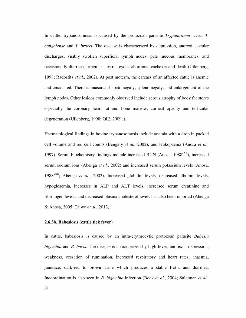
61
In cattle, trypanosomosis is caused by the protozoan parasite Trypanosoma vivax, T.
congolense and T. brucei. The disease is characterized by depression, anorexia, ocular
discharges, visibly swollen superficial lymph nodes, pale mucous membranes, and
occasionally diarrhea, irregular estrus cycle, abortions, cachexia and death (Uilenberg,
1998; Radostits et al., 2002). At post moterm, the carcass of an affected cattle is anemic
and emaciated. There is anasarca, hepatomegaly, splenomegaly, and enlargement of the
lymph nodes. Other lesions commonly observed include serous atrophy of body fat stores
especially the coronary heart fat and bone marrow, corneal opacity and testicular
degeneration (Uilenberg, 1998; OIE, 2009a).
Haematological findings in bovine trypanosomosis include anemia with a drop in packed
cell volume and red cell counts (Bengaly et al., 2002), and leukopaenia (Anosa et al.,
1997). Serum biochemistry findings include increased BUN (Anosa, 1988a&b
), increased
serum sodium ions (Abenga et al., 2002) and increased serum potassium levels (Anosa,
1988a&b
; Abenga et al., 2002). Increased globulin levels, decreased albumin levels,
hypoglcaemia, increases in ALP and ALT levels, increased serum creatinine and
fibrinogen levels, and decreased plasma cholesterol levels has also been reported (Abenga
& Anosa, 2005; Taiwo et al., 2013).
2.6.3b. Babesiosis (cattle tick fever)
In cattle, babesiosis is caused by an intra-erythrocytic protozoan parasite Babesia
bigemina and B. bovis. The disease is characterized by high fever, anorexia, depression,
weakness, cessation of rumination, increased respiratory and heart rates, anaemia,
jaundice, dark-red to brown urine which produces a stable froth, and diarrhea.
Incoordination is also seen in B. bigemina infection (Bock et al., 2004; Sulaiman et al.,
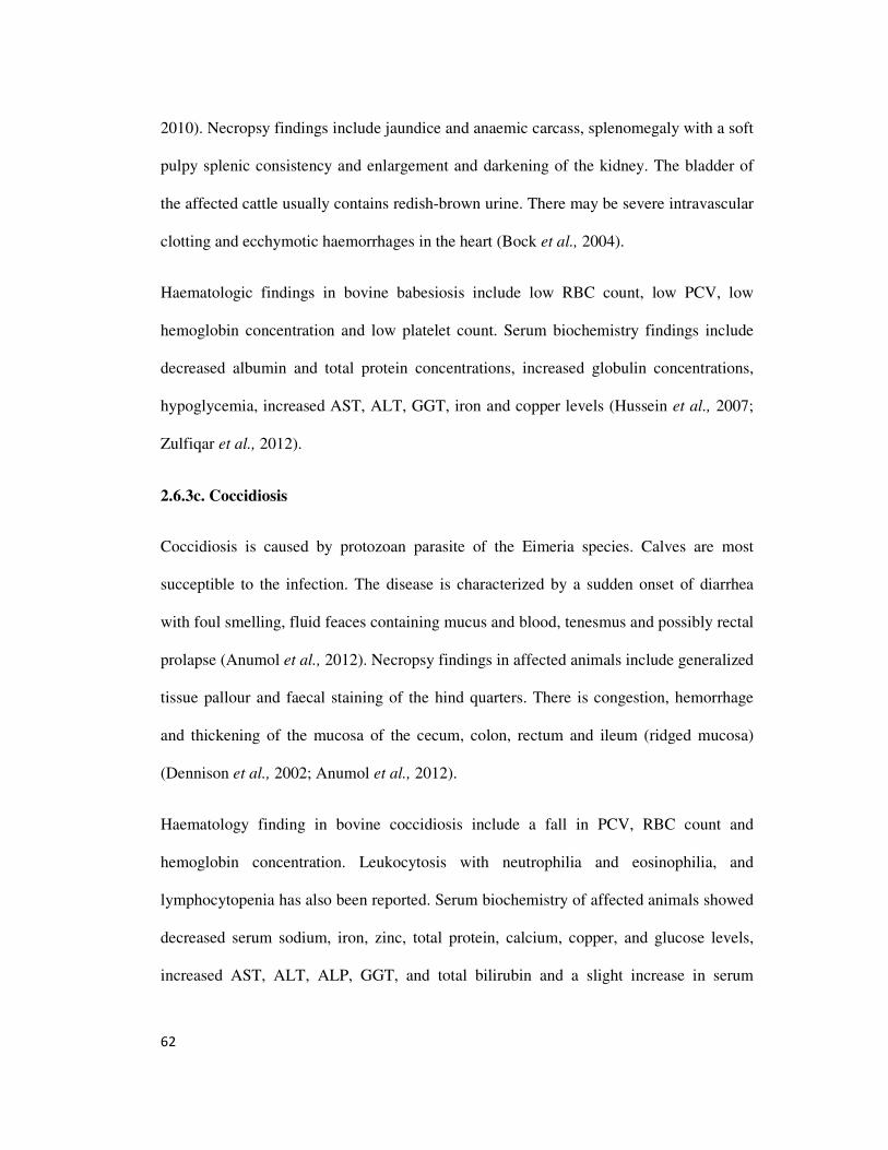
62
2010). Necropsy findings include jaundice and anaemic carcass, splenomegaly with a soft
pulpy splenic consistency and enlargement and darkening of the kidney. The bladder of
the affected cattle usually contains redish-brown urine. There may be severe intravascular
clotting and ecchymotic haemorrhages in the heart (Bock et al., 2004).
Haematologic findings in bovine babesiosis include low RBC count, low PCV, low
hemoglobin concentration and low platelet count. Serum biochemistry findings include
decreased albumin and total protein concentrations, increased globulin concentrations,
hypoglycemia, increased AST, ALT, GGT, iron and copper levels (Hussein et al., 2007;
Zulfiqar et al., 2012).
2.6.3c. Coccidiosis
Coccidiosis is caused by protozoan parasite of the Eimeria species. Calves are most
succeptible to the infection. The disease is characterized by a sudden onset of diarrhea
with foul smelling, fluid feaces containing mucus and blood, tenesmus and possibly rectal
prolapse (Anumol et al., 2012). Necropsy findings in affected animals include generalized
tissue pallour and faecal staining of the hind quarters. There is congestion, hemorrhage
and thickening of the mucosa of the cecum, colon, rectum and ileum (ridged mucosa)
(Dennison et al., 2002; Anumol et al., 2012).
Haematology finding in bovine coccidiosis include a fall in PCV, RBC count and
hemoglobin concentration. Leukocytosis with neutrophilia and eosinophilia, and
lymphocytopenia has also been reported. Serum biochemistry of affected animals showed
decreased serum sodium, iron, zinc, total protein, calcium, copper, and glucose levels,
increased AST, ALT, ALP, GGT, and total bilirubin and a slight increase in serum
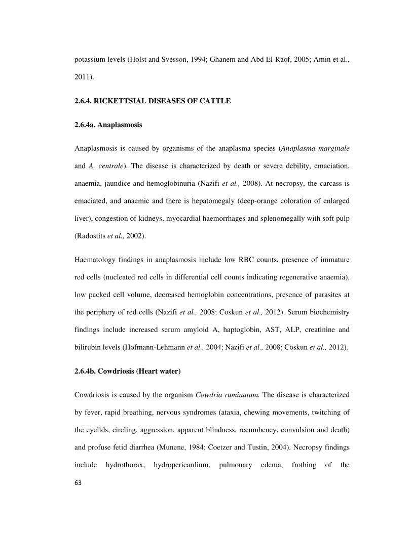
63
potassium levels (Holst and Svesson, 1994; Ghanem and Abd El-Raof, 2005; Amin et al.,
2011).
2.6.4. RICKETTSIAL DISEASES OF CATTLE
2.6.4a. Anaplasmosis
Anaplasmosis is caused by organisms of the anaplasma species (Anaplasma marginale
and A. centrale). The disease is characterized by death or severe debility, emaciation,
anaemia, jaundice and hemoglobinuria (Nazifi et al., 2008). At necropsy, the carcass is
emaciated, and anaemic and there is hepatomegaly (deep-orange coloration of enlarged
liver), congestion of kidneys, myocardial haemorrhages and splenomegally with soft pulp
(Radostits et al., 2002).
Haematology findings in anaplasmosis include low RBC counts, presence of immature
red cells (nucleated red cells in differential cell counts indicating regenerative anaemia),
low packed cell volume, decreased hemoglobin concentrations, presence of parasites at
the periphery of red cells (Nazifi et al., 2008; Coskun et al., 2012). Serum biochemistry
findings include increased serum amyloid A, haptoglobin, AST, ALP, creatinine and
bilirubin levels (Hofmann-Lehmann et al., 2004; Nazifi et al., 2008; Coskun et al., 2012).
2.6.4b. Cowdriosis (Heart water)
Cowdriosis is caused by the organism Cowdria ruminatum. The disease is characterized
by fever, rapid breathing, nervous syndromes (ataxia, chewing movements, twitching of
the eyelids, circling, aggression, apparent blindness, recumbency, convulsion and death)
and profuse fetid diarrhea (Munene, 1984; Coetzer and Tustin, 2004). Necropsy findings
include hydrothorax, hydropericardium, pulmonary edema, frothing of the
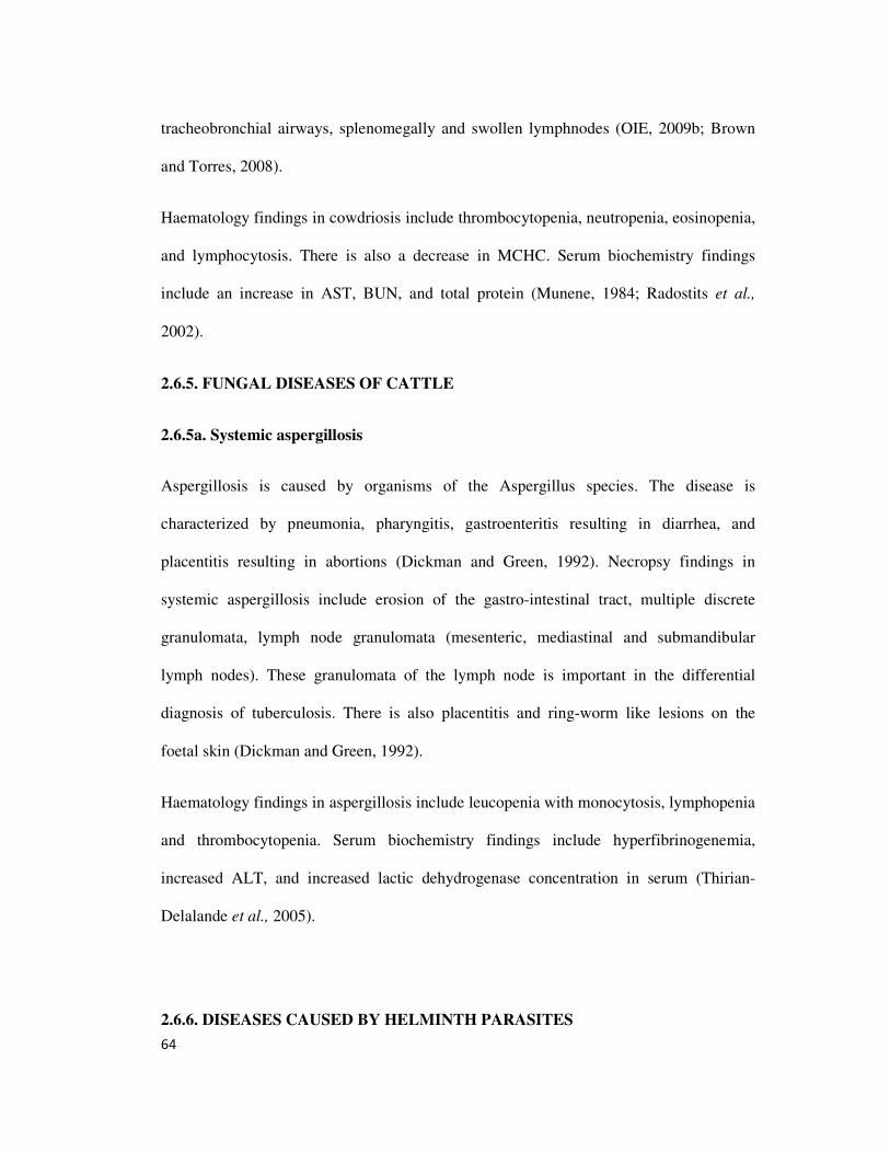
64
tracheobronchial airways, splenomegally and swollen lymphnodes (OIE, 2009b; Brown
and Torres, 2008).
Haematology findings in cowdriosis include thrombocytopenia, neutropenia, eosinopenia,
and lymphocytosis. There is also a decrease in MCHC. Serum biochemistry findings
include an increase in AST, BUN, and total protein (Munene, 1984; Radostits et al.,
2002).
2.6.5. FUNGAL DISEASES OF CATTLE
2.6.5a. Systemic aspergillosis
Aspergillosis is caused by organisms of the Aspergillus species. The disease is
characterized by pneumonia, pharyngitis, gastroenteritis resulting in diarrhea, and
placentitis resulting in abortions (Dickman and Green, 1992). Necropsy findings in
systemic aspergillosis include erosion of the gastro-intestinal tract, multiple discrete
granulomata, lymph node granulomata (mesenteric, mediastinal and submandibular
lymph nodes). These granulomata of the lymph node is important in the differential
diagnosis of tuberculosis. There is also placentitis and ring-worm like lesions on the
foetal skin (Dickman and Green, 1992).
Haematology findings in aspergillosis include leucopenia with monocytosis, lymphopenia
and thrombocytopenia. Serum biochemistry findings include hyperfibrinogenemia,
increased ALT, and increased lactic dehydrogenase concentration in serum (Thirian-
Delalande et al., 2005).
2.6.6. DISEASES CAUSED BY HELMINTH PARASITES
65
2.6.6a. NEMATODIASIS
i. Haemonchosis
Haemonchosis is caused by the nematode Hemonchus contortus. The disease is
characterized by pale mucous membrane and conjunctivae, anarsarca especially under the
lower jaw and abdomen. There may be a chronic wasting of affected animal (Qamar and
Maqbool, 2012). Necropsy findings in affected animals include anaemic carcass,
generalized edema, large numbers of H. contortus in abomasum, hyperemic abomasal
wall with blood clots in the mucosa, brownish colour of abomasal content due to free
blood (Qamar and Maqbool, 2012).
Haematologic findings in haemonchosis include reduced RBC count, hemoglobin and
packed cell volume (Radostits et al., 2002). There may also be reduced WBC counts with
neutropenia, lymphocytosis, monocytosis and basophilia, and a decrease in serum
albumin : globulin ratio (Qamar and Maqbool, 2012).
ii. Ascariosis
Ascariosis is a calf-hood disease caused by the nematode parasite Toxocara vitulorum.
The disease is characterized by poor hair coat, diarrhea, and colic (Sarma et al., 2010). At
necropsy, there is subpleural hemorrhages, oedema and congestion of the lungs,
hepatomegally with hemorrhages under the capsule. There is also a lot of adult worm in
the lumen of the small intestine (Radostits et al., 2002).
Haematology findings in ascariosis include eosinophilia which is marked, low RBC,
MCH and lymphocyte counts, low PCV, and hemoglobin concentration, basophilia, high
MCV, ESR, and WBC counts. Serum biochemistry findings include low phosphorous,
66
sodium, potassium, bicarbonate, glucose, total protein, and glutathion levels. There is
high serum calcium, chloride, cholesterol, ALP, AST, ALT, zinc, copper, and iron levels
(Chaudhry et al., 1999; Sarma et al., 2012).
iii. Bovine verminous bronchitis
Bovine verminous bronchitis is caused by the nematode Dictyocaulus viviparous. The
condition is characterized by frequent bronchial cough, slight nasal discharge, tachypnea,
dyspnea (Ganheim, 2004). Necropsy findings include the presence of adult worms in the
lumen of the trachea and bronchi, enlargement of the lungs due to oedema and
emphysema, froth in the trachea and bronchi, enlargement of the regional lymph nodes,
and the presence of areas of dark-pink consolidations in the diaphragmatic lobe of the
lungs (Radostits et al., 2002).
Haematology findings in bovine verminous bronchitis include eosinophilia. Serum
biochemistry findings include increase in acute phase proteins (haptoglobin and serum
amyloid A) and fibrinogen (Ganheim, 2004).
2.6.6b. Trematodiasis and Cestodiasis
i. Bovine cyticercosis
Bovine cyticercosis is an infection of cattle caused by the larva stage of Cyticercus bovis
of the human intestinal cestode, Taenia saginata. The disease is characterized by
myocarditis or heart failure in heavy infections if these parasitic cysts lodge in the heart
muscles (Gracey and Collins, 1992; Ashwani et al., 2011).
67
Serum biochemistry findings in cysticercosis include decreased total protein, albumin and
alpha 1-globulin concentrations, decreased AST, increased cholesterol and decreased urea
concentrations (Omnia et al., 2011).
ii. Hepatic facioliasis (Liver fluke disease)
Facioliasis is caused by infection by the parasite Faciola gigantica. The disease is
characterized by anaemia,weight loss, chronic diarrhea, and submandibular oedema
(Radostits et al., 2002). Necropsy findings include thickening of the bile ducts,
calcification of the bile duct walls, anaemic carcass and presence of bile duct walls,
anaemic carcass and presence of leaf-like flukes in the lumen of the bile ducts (Radostits
et al., 2002).
Haematologic findings in acute facioliasis include eosinophilia and severe normochromic
anemia. Serum biochemistry findings include hypoalbuminemia, increased gamma
glutamyl dehydrogenase concentration when immature flukes are migrating, and AST
increases which is useful in the determination of the extent of immature fluke infestation
(Al-Quaraishy and Al-Moussawi, 2011). In subacute and chronic infections, haematology
findings include hypochromic, macrocytic anemia. Serum biochemistry findings include
hypoalbuminemia, hypoglobulinemia, increased serum gamma glutamyl transpeptidase
concentration. There is also an increase in WBC counts, increased ALP, total bilirubin
and AST levels (Al-Quaraishy and Al-Moussawi, 2011).
iii. Coenurosis (gid, sturdy)
Coenurosis is a disease caused by the invasion of the brain and spinal cord by the
intermediate stage of Taenia multiceps called Ceonurus cerebralis. This diseae is
68
characterized by incordination, blindness, irritation phenomena, convulsions, circling,
head deviation with blind side tilted downward (Ozkan et al., 2011). Necropsy findings in
coenurosis include the presence of thin walled cyst on the external surface of the cerebral
hemisphere and spinal cord, pressure atrophy of nervous tissue and bone softening
(Ozkan et al., 2011).
Haematology and serum biochemistry findings in coenurosis include monocytosis,
lymphocytosis and increased serum creatinine kinase activity (Ozmen et al., 2005).
2.6.7. METABOLIC DISEASES OF CATTLE
2.6.7a. Parturient paresis (milk fever)
Parturient paresis is a disease of cattle which occurs around the time of parturition (3rd
trimester and 48 hours post paturm) and is caused by hypocalcemia. The disease is
characterized by weakness, recumbency, shock and death (Parish, 2012). There are no
obvious necropsy findings except in occasions where the animal was down for a long
time, in which case, there may be ischaemic muscle necrosis (Radostits et al., 2002).
Haematology findings in parturient paresis include eosinophilia, neutrophilia,
lymphopenia and increases PCV. Serum biochemistry findings include hypocalcemia,
hypophosphatemia, increased AST and increased creatine phosphokinase activity (Dima
et al., 1999).
2.6.7b. Downer cow syndrome
Downer cow syndrome ia a complication of milk fever caused by ischemic necrosis of
large muscles of pelvic limbs secondary to prolonged recumbency associated with milk
fever (Cox, 1982). Other causes of prolonged recumbency can also result in downer cow
69
syndrome (Barrington, 2011). Necropsy findings in downer cow syndrome include
ischemic necrosis, oedema and haemorrhage of large medial muscles of the hind limbs
(Cox, 1982; Radostits et al., 2002; Barrington, 2011).
Serum biochemistry findings in downer cow syndrome include increased creatine
phosphokinase activity, increased AST, decreased phosphorous and potassium levels,
hypomagnesemia (Cox, 1982; Dima et al., 1999; Asl et al., 2010; Barrington, 2011).
2.6.7c. Transit recumbency
Transit recumbency normally happens after prolonged transport of animals. Risk factors
for this condition include heavy feeding before shipment, deprivation of feed and water
for more than 24 hours during transit, unrestricted access to water, and exercise
immediately after unloading. Cows at late pregnancy are prone to this condition. It is
characterized by recumbency, alimentary tract stasis, coma and death 3-4 days after
development of the condition (Barrington, 2011). Necropsy findings include ischemic
muscle necrosis and bloat (Barrington, 2011).
Serum biochemistry findings in transit recumbency include mild hypocalcemia and
hypomagnesemia and increased serum creatine kinase activity (Dima et al., 1999; Asl et
al., 2010).
2.6.7d. Hypomagnesemic tetany (grass tetany)
The aetiology of hypomagnesemic tetany is related to magnesium concentrations in the
diet and the presence of competing cations such as potassium and sodium that affect
either herbage magnesium status or magnesium absorption. The condition is characterized
by an unusuall alertness, hyperesthesia, staggering, chronic convulsions, wildness of
70
facial expressions, spasmodic urination. In chronic cases, there is a parturient paresis-like
syndrome which is unresponsive to calcium treatment (Kvasnicka and Kysl, 2010). There
are no reported gross post motern findings seen in this disease (Radostits et al., 2002).
Clinical chemistry findings include low magnesium levels in serum, urine and
cerebrospinal fluid, low levels of serum total protein, albumin, globulin, creatinine and
urea. Serum cholesterol and blood glucose levels are increased (Adaikpoh et al., 2004;
Kvasnicka and Kysl, 2010).
2.6.8. NUTRITIONAL DISEASES OF CATTLE
2.5.8a. Deficiency of energy (incomplete starvation, protein-deficiency malnutrition)
Protein-energy malnutrition (PEM) occurs when an insufficient quantity or quality of feed
is fed livestock. In this form of deficiency, energy and protein are present in diet in
suboptimal quantities (Mashishi, 2007). This condition is characterized in young animals
by retarded growth and a delay in onset of puberty. In mature animals, there is a decline
in productivity and milk yield, loss of body weight, birth of undersized neonates with a
high mortality rate (Oetzel and Berger, 1985). Post moterm findings include oedema due
to hypoproteinemia (Oetzel and Berger, 1985).
Haematology and serum biochemistry findings in PEM include low hemoglobin
concentration, low PCV and low RBC counts. Serum biochemistry findings include low
serum total protein and low serum albumin concentrations (Oetzel and Berger, 1985).
2.6.8b. White muscle disease (Selenium-Vitamin E responsive disease)
White muscle disease (WMD) is caused by a dietary deficiency of selenium and vitamin
E and conditioning factors like dietary polysaturated fatty acids (Pavlata et al., 2001). The
71
disease is characterized by sternal recumbency, trembling of the hind limbs, rotation
movement of the hocks if animal tries to walk, dyspnoea with labored abdominal-type
respiration, flying scapular, and myoglobinuria (Suttle, 1992; Pavlata et al., 2001;
Kommisurd et al., 2005). Necropsy findings include the presence of pale streaks in
skeletal and myocardial muscles (Radostits et al., 2002).
Serum biochemistry findings in WMD include increased creatine phosphokinase and AST
activities, decreased vitamin E and selenium levels, decreased erythrocyte glutathione
peroxidase activity, increased lactate dehydrogenase activity and increased potassium
levels (Kennedy et al., 1987; Pavlata et al., 2001).
2.6.8c. Deficiencies that lead to osteodystrophy (calcium, phosphorous and vitamin D
deficiencies).
Calcium deficiency may be primary (following an outright deficiency of calcium in diet)
or secondary (following excess intake of phosphorous). This condition is characterized by
poor growth, poor dentition, fracture of long bones and arthropathy (Mashishi, 2007). At
necropsy, there is osteoporosis and hypertrophy of the parathyroid gland (Mashishi,
2007). There is also rickets, osteomalacia and osteodystrophia fibrosa (Radostits et al.,
2002).
Serum biochemistry findings in calcium deficiency include low serum calcium and high
serum potassium levels (Horst et al., 1999; Khan, 2005).
Phosphorous deficiency is caused by a primary deficiency of phosphorous in diet or may
be caused by a deficiency of vitamin D. in young cattle. The deficiency is characterized
by retarded growth and delayed sexual maturity. In adults, there is osteomalacia, low milk
72
yield and reduced fertility, there is also pica appetite (osteophagia in this case) (Iqbal and
Sajid, 2005; Mashishi, 2007). Post moterm findings include rickets in young animals and
osteomalacia in adult animals (Radostits et al., 2002; Mashishi, 2007). Serum
biochemistry findings in phosphorous deficiency include hypophosphatemia in severe
cases. Mild cases have normal phosphorous levels (Ternout, 1990; Gibson, 2013).
Vitamin D deficiency occurs when there is a lack of preformed vitamin D complex in
diet, coupled with a lack of ultra violet solar radiation of the skin. The deficiency is
characterized by reduced productivity, reduced reproductive efficiency, stiff gait and
lameness of the fore limbs which is accompanied by bending of long bones and
enlargement of joints in young animals (rickets), and osteomalacia in adults (Underwood
et al., 1999; Blezinger, 2001). Gross necropsy findings are those seen for rickets in young
animals and osteodystrophia fibrosa (Blezinger, 2001).
Serum biochemistry findings in vitamin D deficiency include hypophosphatemia,
hypocalcemia and increased ALT levels (Radostits et al., 2002)
73
CHAPTER THREE
MATERIALS AND METHODS
3.1. Study location
The abbatoir at the Ikpa New Commodity Market, Nsukka Local Government Area of
Enugu State Nigeria was the location of the study. Nsukka is situated within the derived
savanna belt of Eastern Nigeria between latitudes 5⁰ 50′ and 7⁰ 00′ north and longitudes
6⁰ 52′ and 7⁰ 54′ east at an average elevation of approximately 500 meters above sea level
(FMNAR, 2009)
3.2. Animals for study
The study population was 567 cattle presented for slaughter during 27 research visits
made once in two weeks to the abbatoir between March 2012 and March 2013.
The cattle were physically examined for clinical signs of disease. Blood samples were
collected from clinically unhealthy and apparently healthy cattle in the population.
3.3. Blood sample collection
Blood for haematology was collected from the jugular vein at point of slaughter into
plastic tubes containing ethylene diamine tetracetic acid (EDTA), while blood for serum
biochemistry was collected into plain glass test tubes and allowed to stand for one hour to
74
clot. Clotted blood was separated from serum by centrifugation at 3000 revolutions per
minute for 10 minutes.
3.4. Determination of Haematological parameters
3.4.1 Packed cell volume (PCV)
The PCV was determined by the microhaematocrit method (Thrall & Weiser, 2002).
Capillary tubes were almost filled with anticoagulated blood and one end was sealed with
plasticine. Filled tubes were centrifuged at 10,000 revolutions per minute for 5 minutes
using a microhaematocrit centrifuge (Hawksley, England). The PCV was read as
percentage using a microhematocrit reader (Thrall & Weiser, 2002 ).
3.4.2 Haemoglobin concentration (Hb)
The Hb concentration was determined by the cyanomethaemoglobin method (Higgins et
al., 2008a). Twenty microlitres of anti-coagulated blood was added to 5 ml of Drabkin’s
reagent. This was mixed well and allowed to stand for 20 minutes to react. The
absorbance of both the samples and standard were read against the Drabkin’s reagent
blank at a wavelength of 540 nm using a digital colorimeter (Higgins et al., 2008a).
3.4.3 Erythrocyte (Red blood cell, RBC ) count
The erythrocyte counts were done with a haemocytometer using a diluting fluid (Coles,
1986; Thrall & Weiser, 2002). Twenty microlitres of blood was added to 4 ml of diluting
fluid (a combination of sodium citrate, formaline and distilled water) to make a 1: 200
dilution. This was mixed and a drop of the diluted blood was charged onto the Neubaeur
chamber and allowed to settle for 2 – 3 minutes. The Neubaeur chamber was mounted on
75
a light microscope and the erythrocytes were enumerated at an X40 objective. The
number of cells enumerated in the 5 central squares for each sample was multiplied by
10,000 to obtain the erythrocyte counts per microlitre of blood (Coles, 1986; Thrall &
Weiser, 2002).
3.4.4 Total Leukocyte (White blood cell, WBC)count
The total leukocyte counts were also done with a haemocytometer, using a diluting fluid
(Coles, 1986; Thrall & Weiser, 2002). Twenty microlitres of the blood was added to 380
microlitre of diluting fluid (a combination of glacial acetic acid, gentian violet and
distilled water) to make a 1: 20 dilution. This was mixed and a drop of the diluted blood
was charged onto the Neubaeur chamber and allowed to settle for 2 – 3 minutes. The
Neubaeur chamber was mounted on a light microscope and the leukocyte count
enumerated at X10 objective. The number of cells enumerated in the four big side squares
for each sample was multiplied by 50 to obtain the leukocyte counts per microlitre of
blood (Coles, 1986; Thrall & Weiser, 2002).
3.4.5 Differential leukocyte count
The differential leukocyte counts were performed on thin smear stained with Leishman
stain (Coles, 1986; Thrall & Weiser, 2002). A drop of blood was placed on a clean
grease-free slide, and then smeared on the slide using a cover slip to make a thin smear.
The smear was air dried and stained by the Leishman technique. The different leukocyte
types were enumerated by the battlement counting method. The X100 (oil immersion)
objective of the microscope was used for the differential leukocyte count. The percentage
76
values obtained were converted to absolute counts by multiplying out with the total white
blood cell counts (Coles, 1986; Thrall & Weiser, 2002).
3.4.6 Erythrocyte sedimentation rate (ESR)
The ESR was determined by the wintrobe method (Coles, 1986; Thrall & Weiser,2002).
Capillary tubes were filled with anticoagulated blood and one end sealed with plasticine.
Filled tubes were stood in a vertical position and left for 24 hours. The ESR was
calculated by dividing the length of clear plasma space by the whole length previously
occupied by unsedimented blood (i. e clear plasma space + packed cells), and the value
obtained multiplied by 100. These lengths are obtained by the use of a ruler (Coles, 1986;
Thrall & Weiser,2002).
ESR (mm/hr) = length of clear plasma space X 100
clear plasma space + PCV
3.5. Serum biochemistry methods
All serum biochemical determinations were done following standard procedures, using
Quimica Clinica Aplicada reagent kits (QCA) ( Quimica Clinica Aplicada, Spain).
3.5.1 Total protein
Serum total protein was determined by the direct Biuret method (Johnson, 2008) using a
reagent kit. Clean test tubes were labeled for samples, standards, and blank. Biuret
reagent (1.2 ml) was added to the test tubes (samples, standards and blank). Twenty
microlitres of serum sample was then added to test tubes labeled for samples and twenty
microlitres of the standard added to those labeled for standard; nothing was added to test
tube for blank. The contents of the test tubes were well mixed and allowed to stand for 10
77
minutes at room temperature. The absorbance of both the test tubes labeled for samples
and standard were read against the working reagent blank at a wavelength of 540 nm
using a digital colorimeter. The total protein was calculated as follows.
Total protein (g/dl) = absorbance of sample X 5
absorbance of standard
3.5.2 Albumin
Serum albumin was determined by the bromocresol green method (Johnson, 2008) using
a reagent kit. Clean test tubes were labeled for samples, standards, and blank. To these
test tubes, 2.5 ml of bromocresol green reagent was added (samples, standards and
blank). Ten microlitres of serum sample was added to test tubes labeled for sample and
10 microlitres of the standard reagent was added to those labeled for standard. Nothing
was added to test tube for blank. The contents of the test tubes were well mixed and
allowed to stand for 5 minutes at room temperature. The absorbance of reagent mixture
labeled for both samples and standard were read immediately against the blank at a
wavelength of 620 nm using a digital colorimeter. The serum albumin was calculated as
follows.
Albumin (g/dl) = absorbance of sample X5
absorbance of standard
3.5.3 Calculation of globulin
Globulin was calculated by subtracting the value of albumin obtained from the value of
total protein (Johnson, 2008).
Globulin (g/dl) = total protein – albumin
78
3.5.4 Total cholesterol
Serum total cholesterol was determined by the enzymatic colorimetric method (Rifai et
al., 2008) using reagent kit. Clean test tubes were labeled for samples, standards, and
blank. To these test tubes, 1.2 ml of reagent was added (samples, standards and blank).
Twelve microlitres of the serum sample was then added to test tubes labeled for sample
and 12 microlitres of the standard reagent was added to those labeled for standard.
Nothing was added to test tube labeled for blank. The contents of the test tubes were well
mixed and allowed to stand for 10 minutes at room temperature. The absorbance of the
contents of both the test tubes labeled for samples and standard was read against the
working reagent blank at a wavelength of 520 nm using a digital colorimeter. The serum
cholesterol was calculated as follows.
Cholesterol (mg/dl) = absorbance of sample X 200
absorbance of standard
3.5.5 Urea
Serum urea was determined by the modified Berthelot-Searcy method (Lamb & Price,
2008) using reagent kit. One mlilitre of reagent A was added to clean test tubes labeled
for samples, standards and blank. Ten microlitres of serum samples and standard reagent
were added to test tubes labeled for samples and standards respectively. Nothing was
added to test tube labeled for blank. The contents of the test tubes were mixed and
allowed to stand for 5 minutes at room temperature. One mlilitre of reagent B was then
added to the test tubes labeled for samples, standard and blank. This was futher mixed
and allowed to stand at room temperature for 5 minutes. The absorbance of both the
contents of the test tubes labeled samples and standard were read against the reagent
79
blank at a wavelength of 590 nm using a digital colorimeter. The serum urea
concentration was calculated as follows.
Urea (mg/dl) = absorbance of sample X 40
absorbance of standard
3.5.6 Creatinine
The serum creatinine was determined by the modified Jaffe method (Blass et al., 1974)
using reagent kit. One hundred microlitres of serum sample and standard respectively will
be reacted with 1ml of creatinine working reagent. The absorbance of these were read
immediately at the 20th
and 80th
seconds against a reagent blank at 520 nm using a digital
colorimeter. The creatinine concentration of the sample was calculated as follows.
Creatinine (mg/dl) = change in absorbance of sample (80th-20th
sec) X 2
change in absorbance of standard (80th
-20th
sec)
3.5.7 Alkaline phosphatase (ALP)
The serum ALP activity was determined by the phenolphthalin monophosphate method
(Colville, 2002) using reagent kit. One milliliter of deionized water was added to clean
test tubes labeled for samples, standards and blank. One drop of chromogenic substrate
was then added to test tubes for samples and standards (none to blank). The tubes with
their contents were incubated at 37⁰C for 5 minutes. After the incubation, 100 microlitres
of serum sample and standard were added to test tubes labeled for samples and standards
respectively. The test tubes and their contents were further incubated at 37⁰C for 20
minutes. Five mlilitres of ALP colour developer was added to these test tubes. The
absorbance of the contents of the test tubes labeled for samples and standards were read
80
against a deionized water blank at 540 nm using a digital colorimeter. The ALP
concentration of the sample was calculated as follows.
ALP (IU/L) = absorbance of sample X 30
absorbance of standard
3.5.8 Total bilirubin
The serum total bilirubin was determined by the modified Jendrassik-Grof method
(Higgins et al., 2008b) using reagent kit. Two hundred microlitres of sulfanilic acid was
added to clean test tubes labeled for sample and blank. One drop of sodium nitrite was
then added to test tubes labeled for sample (none to blank). One ml of caffeine was futher
added to test tubes labeled for samples and blank. Two hundred microlitres of serum
sample was then added to these test tubes, mixed and kept at room temperature for 10
minutes. After this, 1 ml of tartarate was added to these test tubes, futher mixed and
allowed to stand at room temperature for 5 minutes. The absorbance of the contents of the
test tubes labeled for samples were read against a working reagent blank at 540 nm using
a digital colorimeter. The total bilirubin concentration of the sample was calculated as
follows.
Total bilirubin (mg/dl) = absorbance of sample X 43.2
3.5.9 Aspartate amino transferase (AST) and Alanine amino transferase (ALT)
The serum AST and ALT activities were determined by the Reitman-Frankel colorimetric
method (Reitman & Frankel, 1957; Colville, 2002) using reagent kits. To the test tubes
labeled for samples, 500 microlitres of AST and ALT substrate A were added
respectively and initially activated by incubation at 37⁰C for 5 minutes. Serum samples
81
(0.1ml) were then added to the activated substrates and incubated for 1 hour for AST ,
and 30 minutes for ALT. Afterwards, 500 microlitres of color developer and 5ml of
dilute sodium hydroxide (1 :10 dilution) were added to these test tubes. Test tubes
containing varied quantities of standard (0, 0.1, 0.15, 0.2, 0.25, 0.3ml for AST; 0, 0.5, 0.1,
0.15, 0.2ml for ALT), were subjected to the same procedure. Fifteen minutes after
addition of sodium hydroxide, the transmittance of both the contents of test tubes labeled
for samples and standards were read against a deionized water blank at 520 nm (AST)
and 490 nm (ALT) respectively. The transmittance readings of the samples were then
interpolated in a calibration curve from the standards in order to obtain the serum AST
and ALT in IU/L.
3.6. Data analysis
Data generated from the study were subjected to appropriate statistics using SPSS
statistical package (version 16.0). The haematological and serum biochemical parameters
of the animals in each specific disease condition was compared with that of the apparently
healthy ones using Student’s t- test. The haematological and serum biochemistry findings
in comparable disease like fasciolosis were subjected to a one way analysis of variance.
Variant means were separated using the least significant difference post hoc. Sex and age
related differences in the apparently healthy cattle were analysed using Student’s t- test.
Significance was accepted at p < 0.05
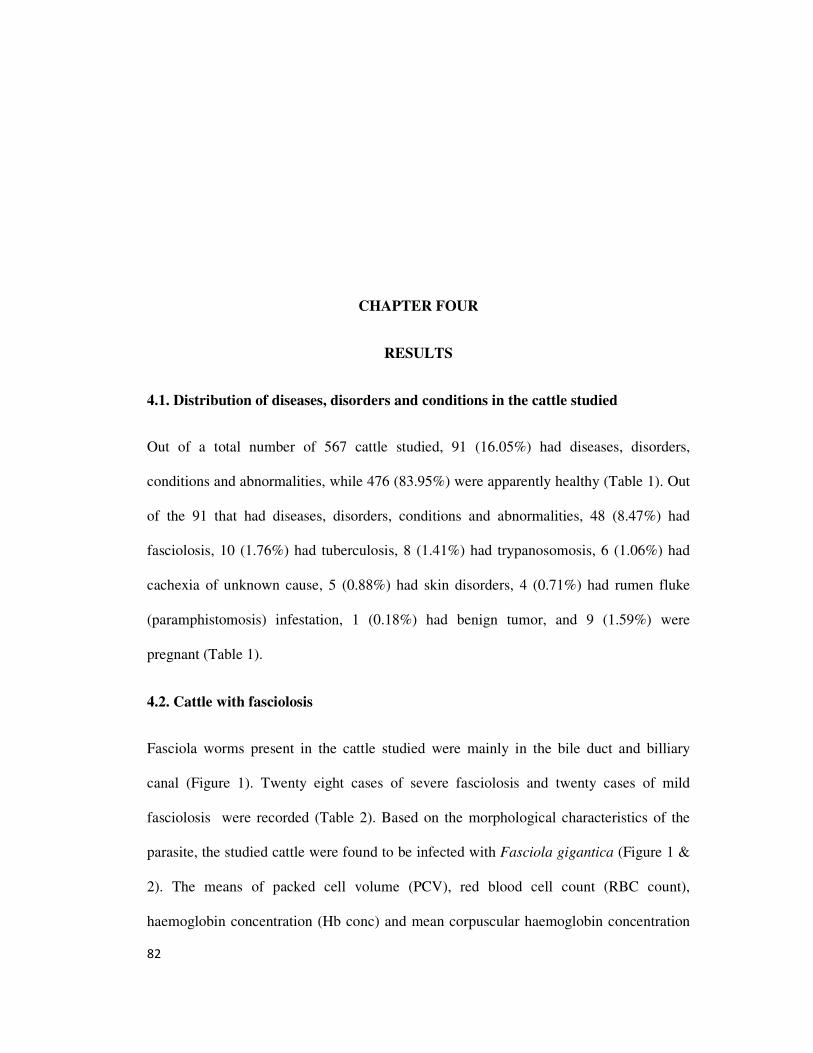
82
CHAPTER FOUR
RESULTS
4.1. Distribution of diseases, disorders and conditions in the cattle studied
Out of a total number of 567 cattle studied, 91 (16.05%) had diseases, disorders,
conditions and abnormalities, while 476 (83.95%) were apparently healthy (Table 1). Out
of the 91 that had diseases, disorders, conditions and abnormalities, 48 (8.47%) had
fasciolosis, 10 (1.76%) had tuberculosis, 8 (1.41%) had trypanosomosis, 6 (1.06%) had
cachexia of unknown cause, 5 (0.88%) had skin disorders, 4 (0.71%) had rumen fluke
(paramphistomosis) infestation, 1 (0.18%) had benign tumor, and 9 (1.59%) were
pregnant (Table 1).
4.2. Cattle with fasciolosis
Fasciola worms present in the cattle studied were mainly in the bile duct and billiary
canal (Figure 1). Twenty eight cases of severe fasciolosis and twenty cases of mild
fasciolosis were recorded (Table 2). Based on the morphological characteristics of the
parasite, the studied cattle were found to be infected with Fasciola gigantica (Figure 1 &
2). The means of packed cell volume (PCV), red blood cell count (RBC count),
haemoglobin concentration (Hb conc) and mean corpuscular haemoglobin concentration
83
(MCHC) of the severely infected cattle was significantly (p < 0.05) lower than that of the
apparently healthy cattle (Table 2). The mean corpuscular volume (MCV) and erythrocyte
sedimentation rate (ESR) of both the severely and mildly infested cattle were significantly
(p < 0.05) higher than that of the apparently healthy cattle (Table 2). Also, the total white
blood cell count (WBC count), lymphocyte and eosinophil counts of the infected cattle
were significantly (p < 0.05) higher than that of the apparently healthy cattle (Table 2).
There was also a significantly (p < 0. 05) higher mean basophil count in the mildly
infested group when compared with the apparently healthy cattle. There were no
significant (p > 0.05) differences in the mean corpuscular haemoglobin (MCH) and
neutrophil counts in infected cattle when compared to the apparently healthy cattle (Table
2).
The means of serum alanine aminotransferase (ALT) activity, total bilirubiin and total
protein levels were significantly (p < 0.05) higher in cattle with severe fasciolosis when
compared to the apparently healthy cattle (Table 3). Means of serum alkaline phosphatase
activity were significantly (p < 0.05) higher in both cattle with severe and those with
mild fasciolosis when compared to the apparently healthy cattle (Table 3). The serum
globulin level of cattle with mild fasciolosis was significantly (p < 0.05) higher than that
of both the severely infested and apparently healthy cattle groups (Table 3). The serum
albumin level was significantly (p < 0.05) lower in both cattle with severe and those with
mild fasciolosis when compared to the apparently healthy cattle (Table 3). There was no
significant (p > 0.05) differences in the means of serum aspartate aminotransferase (AST)
activity, total cholesterol, creatinine and urea levels of the fasciola-infested and
apparently healthy cattle (Table 3).

84
4.3. Cattle with tuberculosis
Tubercles in cattle studied were observed mainly in the lungs, liver, spleen and lymph
nodes draining these areas (Figure 3a-c, 4a-c, 5 & 6). The means of PCV, RBC count and
Hb concentration were significantly (p < 0.05) lower in cattle with tuberculosis when
compared to the apparently healthy cattle (Table 4). The means of ESR was significantly
(p < 0.05) higher in cattle with tuberculosis when compared to apparently healthy cattle.
The means of total WBC count, lymphocyte and eosinophils counts was significantly (p <
0.05) higher in cattle with tuberculosis when compared to apparently healthy cattle, but
the basophil counts of cattle with tuberculosis was significantly lower (p < 0.05) than that
of the apparently healthy cattle (Table 4). There was no significant (p > 0.05) differences
in the means of MVC, MCH, MCHC, neutrophil count and monocyte count of cattle with
tuberculosis when compared with apparently healthy cattle (Table 4).
The mean of serum ALT activity, albumin and urea levels of cattle with tuberculosis was
significantly lower than that of the apparently healthy cattle, but the serum globulin levels
of cattle with tuberculosis was significantly (p < 0.05) higher than that of apparently
healthy cattle (Table 5). The means of serum AST, ALP activities, total protein, total
cholesterol, creatinine and total bilirubin levels showed no significant (p > 0.05)
differences between cattle with tuberculosis and apparently healthy cattle (Table 5).
4.4. Trypanosome-infected cattle
Eight cattle were found to have trypanosomes in blood. Based on the morphologic
characteristics of the trypanosomes as observed on stained thin blood smears, the species
of trypanosomes were Trypanosoma vivax (Figure 7a & b). The blood parasite
85
concentration were low. The means of PCV, RBC count, Hb concentration, MCHC, and
eosinophil counts of the trypanosome-infected cattle were significantly (p < 0.05) lower
than that of apparently healthy cattle, while the ESR, lymphocyte and monocyte counts of
the trypanosome infected cattle were significantly (p < 0.05) higher than that of the
apparently healthy cattle (Table 6).
The means of serum albumin and creatinine levels of the trypanosome infected cattle
were significantly (p < 0.05) lower than that of the apparently healthy cattle, while the
means of serum urea level was significantly (p < 0.05) higher in trypanosome-infected
cattle when compared with apparently healthy cattle (Table 7). There were no significant
(p > 0.05) differences in the means of serum AST, ALT, ALP activities, total protein,
globulin, total cholesterol and total bilirubin levels of trypanosome-infected cattle when
compared with apparently healthy cattle (Table 7).
4.5. Cattle with cachexia of unknown aetiology
Among the 567 cattle studied, six were emaciated and cachexic, and possible cause of
their emaciation was not obvious. A wet mount, and staining of thin blood smear did not
show the presence of haemoparasites. Inspection of the vicera showed no obvious gross
lesions or presence of endoparasite infestation (Figure 8a & b ). The means of MCHC of
cachexic cattle was significantly (p < 0.05) higher, while the means of monocyte,
eosinophil and basophil counts were significantly (p < 0.05) lower than that of the
apparently healthy cattle (Table 8). There were however no significant (p > 0.05)
differences in the means of PCV, RBC count, Hb concentration, MCV, MCH, ESR, total
WBC count, neutrophil and lymphocyte count of the cachexic cattle when compared with
the apparently healthy cattle group (Table 8).
86
The means of serum ALT activity, and creatinine and urea levels were significantly (p <
0.05) lower in the cachexic cattle, while the means of serum ALP and globulin activity
were significantly (p < 0.05) higher when compared to that of the apparently healthy
cattle. There were no significant (p > 0.05) differences between the means of serum AST
activity, and levels of total protein, albumin, total cholesterol and total bilirubin of
cachexic cattle when compared to those of apparently healthy cattle (Table 9).
4.6. Cattle with skin disorders
Skin lesions in cattle studied were predominantly found on the flank and dorsum. They
appeared as scabs and crusts on the affected areas (Figure 9). This was found on 5 out of
the 567 cattle studied (Table 1). The means of total WBC, neutrophil and monocyte
counts of the cattle with skin disorders were significantly (p < 0.05) lower than those of
the apparently healthy cattle (Table 10). There were no significant (p > 0.05) differences
in the means of PCV, RBC count, Hb concentration, MCV, MCH, ESR, lymphocyte,
eosinophil and basophil counts of cattle with skin conditions when compared to those of
apparently healthy cattle (Table 10).
The means of serum ALP activity, total protein, globulin and total cholesterol levels were
significantly (p < 0.05) higher, while the means of serum AST and ALT activities, and
creatinine level were significantly (p < 0.05) lower in cattle with skin condition when
compared with the apparently healthy cattle group (Table 11). There were no significant
(p > 0.05) differences in the means of serum albumin, total bilirubin and urea levels
between cattle with skin conditions and apparently healthy cattle (Table 11).
4.7. Cattle with rumen fluke infestation (Paramphistomosis)
87
Paramphistomum in the cattle studied were predominantly found on the mucosal surface
of the rumen and abomasums of affected animal (Figure 10a & b). The means of PCV,
Hb concentration, MCV, MCH, MCHC, neutrophil, monocyte and basophil counts were
significantly (p < 0.05) lower in cattle with paramphistomosis when compared with
apparently healthy cattle (Table 12). There were no significant (p > 0.05) differences
between the means of RBC, total WBC, lymphocyte and eosinophils counts of cattle with
paramphistomosis when compared with apparently healthy cattle (Table 12).
The means of serum AST activity was significantly (p < 0.05) higher, while the means of
serum total protein and total bilirubin levels were significantly (p < 0.05) lower in cattle
with paramphistomosis when compared with the apparently healthy cattle (Table 13).
There were no significant (p > 0.05) difference between the means of serum ALT and
ALP activities, albumin, globulin, total cholesterol, creatinine and urea levels of cattle
with paramphistomosis when compared with apparently healthy cattle (Table 13).
4.8. Cattle with benign tumor
In the study population, only one cattle out of the 567 studied was found with a hard
circumscribed tissue mass on the right fore limb (Figure 11). The RBC count, Hb
concentration, MCHC and neutrophil count of the cattle with benign tumor was higher
than that of apparently healthy cattle (far higher than the upper limit of the reference
value obtained), but the PCV, MCV, MCH, ESR, total WBC count, lymphocyte,
monocyte, eosinophils and basophil counts were also higher in the cattle with benign
tumor when compared with those of apparently healthy cattle, but were within normal
reference ranges in the study population (Table 14).
88
The serum activities of AST and ALT, and the levels of protein, albumin, total cholesterol
and creatinin were lower, while ALP activity, globulin, total bilirubin and urea levels
were higher in the cattle with benign tumor when compared with those of the apparently
healthy cattle (Table 15). The serum levels of total bilirubin in the animal with benign
tumor higher than the normal reference range in the study population (Table 15).
4.9. Pregnant cows
Out of twenty three (23) female cattle studied, nine (9) were found to be pregnant (Figure
12a & b). The means of RBC count of the pregnant cows were significantly (p < 0.05)
lower, while their means of MCV, MCH, ESR, total WBC count, lymphocyte and
monocyte counts were significantly (p < 0.05) higher when compared with non-pregnant
apparently healthy females (Table 16). There was no significant (p < 0. 05) difference
between the means of the PCV, Hb concentration, MCHC, neutrophil count, eosinophils
and basophil counts of the pregnant cows and that of apparently healthy non-pregnant
females (Table 16). The means of serum AST and ALT activity, and creatinine and urea
levels were significantly (p < 0.05) lower in the pregnant cows, while the means of serum
ALP activity, and total bilirubin level were significantly (p < 0.05) higher in pregnant
cows when compared with non pregnant apparently healthy females (Table 17). There
were no significant (p > 0.05) differences between the means of serum total protein,
albumin, globulin and total cholesterol levels of pregnant cows when compared with
apparently healthy non-pregnant females (Table 17).
4. 10. Apparently healthy cattle
89
The values obtained for haematology and serum biochemistry parameters of the
apparently healthy trade cattle (Figure 13) studied, with their minimum and maximum
values are presented in Tables 18 and 19 side by side with the reference values for these
parameters as reported in literature (cited). The minimum and maximum values of
haematology and serum biochemistry parameters of apparently healthy cattle in this study
concurred in most instances with the ranges in available literature, but there were great
differences in the upper and/or lower reference limits of the means of MCV, ESR, total
WBC count, neutrophil and lymphocyte counts, AST and ALT activity, globulin,
bilirubin and creatinine levels recorded in this study when compared to that available in
literature (Table 18 & 19).
4. 11. Comparison of the haematology and serum biochemistry profile of cattle of
different sexes
The means of RBC count, ESR and eosinophils counts of male cattle were significantly (p
< 0.05) higher than that of females (Table 20). There were no significant (p > 0.05)
differences between the means of PCV, Hb concentration, MCV, MCH, MCHC, total
WBC count, neutrophil, lymphocyte, monocyte and basophil counts of males and those of
the females (Table 20).
The means of the serum ALP activity and levels of creatinine were significantly (p <
0.05) higher in males than in females (Table 21). The means of serum activities of AST
and ALT, serum levels of total protein, albumin, globulin, total cholesterol, total bilirubin
and urea showed no significant (p > 0.05) differences between males and females (Table
21).
90
4. 12. Comparison of the haematology and serum biochemistry profile of cattle of
different age groups
The means of MCHC and eosinophil counts of young cattle were significantly (p < 0.05)
higher than those of the adults (Table 22). There were no significant (p > 0.05)
differences between the means of PCV, RBC count, Hb concentration, MCV, MCH, ESR,
total WBC count, neutrophil, lymphocyte, monocyte and basophil count of young cattle
when compared with adult cattle (Table 22).
The mean serum level of globulin was significantly (p < 0.05) lower in young cattle when
compared with that of adult cattle. There were no significant (p > 0.05) differences
between the means of serum activities of AST, ALT and ALP and levels of total protein,
albumin, total cholesterol, total bilirubin, creatinine and urea of young cattle when
compared to those of adult cattle (Table 23).
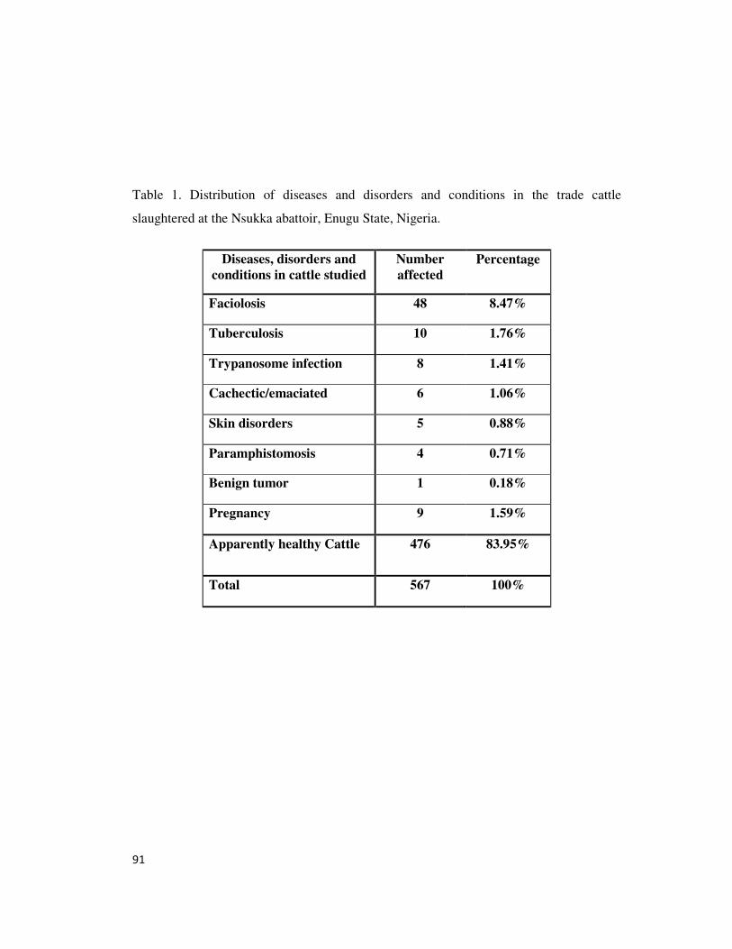
91
Table 1. Distribution of diseases and disorders and conditions in the trade cattle
slaughtered at the Nsukka abattoir, Enugu State, Nigeria.
Diseases, disorders and
conditions in cattle studied
Number
affected
Percentage
Faciolosis 48 8.47%
Tuberculosis 10 1.76%
Trypanosome infection 8 1.41%
Cachectic/emaciated 6 1.06%
Skin disorders 5 0.88%
Paramphistomosis 4 0.71%
Benign tumor 1 0.18%
Pregnancy 9 1.59%
Apparently healthy Cattle 476 83.95%
Total 567 100%
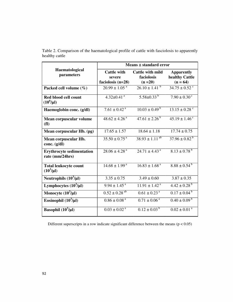
92
Table 2. Comparison of the haematological profile of cattle with fasciolosis to apparently
healthy cattle
Haematological
parameters
Means ± standard error
Cattle with
severe
faciolosis (n=28)
Cattle with mild
faciolosis
(n =20)
Apparently
healthy Cattle
(n = 64)
Packed cell volume (%) 20.99 ± 1.05 a 26.10 ± 1.41
b 34.75 ± 0.52
c
Red blood cell count
(106/µl)
4.32±0.41 a 5.58±0.33
b 7.90 ± 0.30
c
Haemoglobin conc. (g/dl) 7.61 ± 0.42 a 10.03 ± 0.49
b 13.15 ± 0.28
c
Mean corpuscular volume
(fl)
48.62 ± 4.26 a 47.61 ± 2.26
b 45.19 ± 1.46
c
Mean corpuscular Hb. (pg) 17.65 ± 1.57 18.64 ± 1.18 17.74 ± 0.75
Mean corpuscular Hb.
conc. (g/dl)
35.50 ± 0.75 a 38.93 ± 1.11
ab 37.96 ± 0.82
b
Erythrocyte sedimentation
rate (mm/24hrs)
28.06 ± 4.28 a
24.71 ± 4.43 a
8.13 ± 0.78 b
Total leukocyte count
(103/µl)
14.68 ± 1.99 a 16.83 ± 1.68
a 8.88 ± 0.54
b
Neutrophils (103/µl)
3.35 ± 0.75 3.49 ± 0.60 3.87 ± 0.35
Lymphocytes (103/µl) 9.94 ± 1.45
a 11.91 ± 1.42
a 4.42 ± 0.28
b
Monocyte (103/µl)
0.52 ± 0.28 ab
0.61 ± 0.23 a 0.17 ± 0.04
b
Eosinophil (103/µl)
0.86 ± 0.08 a 0.71 ± 0.06
a 0.40 ± 0.09
b
Basophil (103/µl)
0.03 ± 0.02 a 0.12 ± 0.03
b 0.02 ± 0.01
a
Different superscripts in a row indicate significant difference between the means (p < 0.05)
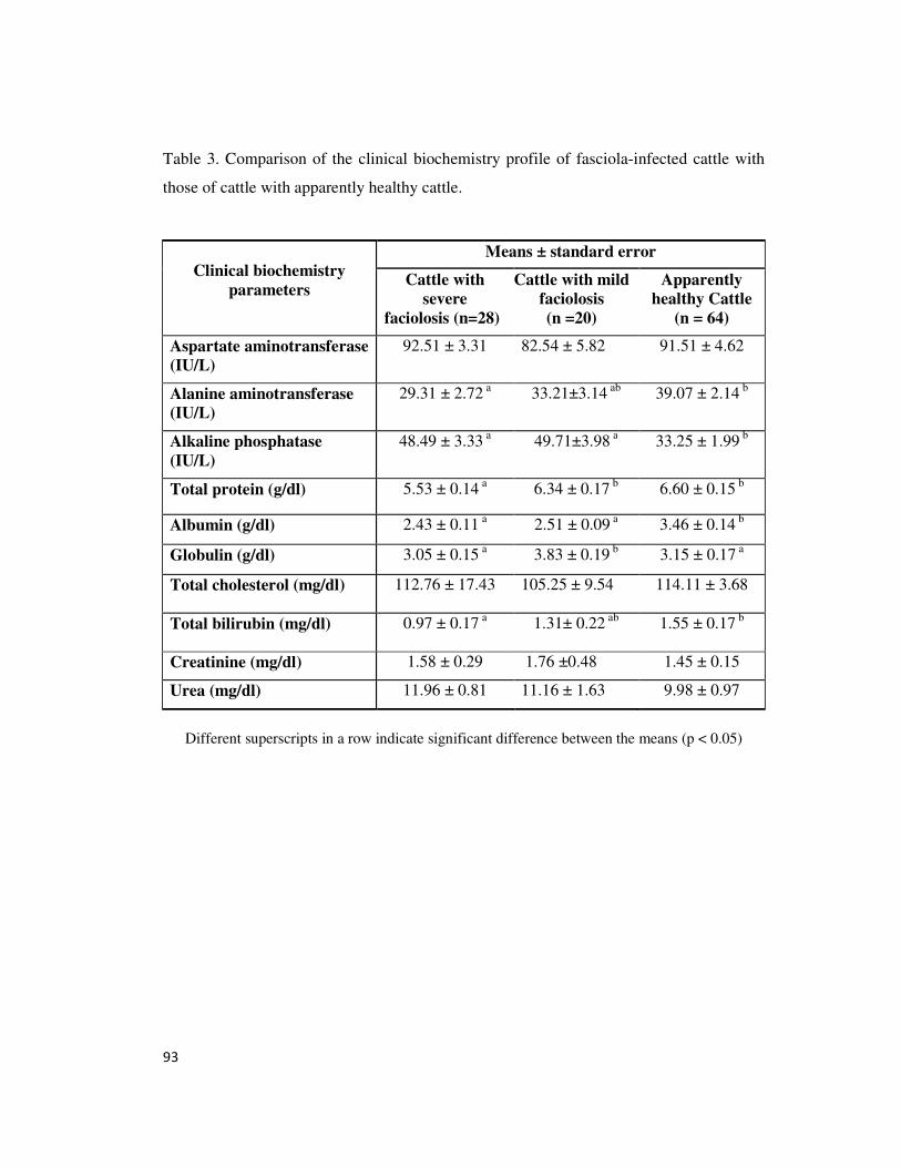
93
Table 3. Comparison of the clinical biochemistry profile of fasciola-infected cattle with
those of cattle with apparently healthy cattle.
Clinical biochemistry
parameters
Means ± standard error
Cattle with
severe
faciolosis (n=28)
Cattle with mild
faciolosis
(n =20)
Apparently
healthy Cattle
(n = 64)
Aspartate aminotransferase
(IU/L)
92.51 ± 3.31 82.54 ± 5.82 91.51 ± 4.62
Alanine aminotransferase
(IU/L)
29.31 ± 2.72 a 33.21±3.14
ab 39.07 ± 2.14
b
Alkaline phosphatase
(IU/L)
48.49 ± 3.33 a 49.71±3.98
a 33.25 ± 1.99
b
Total protein (g/dl) 5.53 ± 0.14 a 6.34 ± 0.17
b 6.60 ± 0.15
b
Albumin (g/dl) 2.43 ± 0.11
a 2.51 ± 0.09
a 3.46 ± 0.14
b
Globulin (g/dl) 3.05 ± 0.15
a 3.83 ± 0.19
b 3.15 ± 0.17
a
Total cholesterol (mg/dl) 112.76 ± 17.43 105.25 ± 9.54 114.11 ± 3.68
Total bilirubin (mg/dl) 0.97 ± 0.17 a 1.31± 0.22
ab 1.55 ± 0.17
b
Creatinine (mg/dl) 1.58 ± 0.29 1.76 ±0.48 1.45 ± 0.15
Urea (mg/dl) 11.96 ± 0.81 11.16 ± 1.63 9.98 ± 0.97
Different superscripts in a row indicate significant difference between the means (p < 0.05)
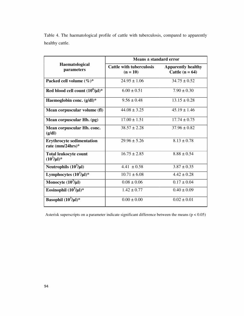
94
Table 4. The haematological profile of cattle with tuberculosis, compared to apparently
healthy cattle.
Haematological
parameters
Means ± standard error
Cattle with tuberculosis
(n = 10)
Apparently healthy
Cattle (n = 64)
Packed cell volume (%)* 24.95 ± 1.06 34.75 ± 0.52
Red blood cell count (106/µl)* 6.00 ± 0.51 7.90 ± 0.30
Haemoglobin conc. (g/dl)* 9.56 ± 0.48 13.15 ± 0.28
Mean corpuscular volume (fl) 44.08 ± 3.25 45.19 ± 1.46
Mean corpuscular Hb. (pg) 17.00 ± 1.51 17.74 ± 0.75
Mean corpuscular Hb. conc.
(g/dl)
38.57 ± 2.28 37.96 ± 0.82
Erythrocyte sedimentation
rate (mm/24hrs)*
29.96 ± 5.26 8.13 ± 0.78
Total leukocyte count
(103/µl)*
16.75 ± 2.85 8.88 ± 0.54
Neutrophils (103/µl)
4.41 ± 0.58 3.87 ± 0.35
Lymphocytes (103/µl)* 10.71 ± 6.08 4.42 ± 0.28
Monocyte (103/µl)
0.08 ± 0.06 0.17 ± 0.04
Eosinophil (103/µl)*
1.42 ± 0.77 0.40 ± 0.09
Basophil (103/µl)*
0.00 ± 0.00 0.02 ± 0.01
Asterisk superscripts on a parameter indicate significant difference between the means (p < 0.05)
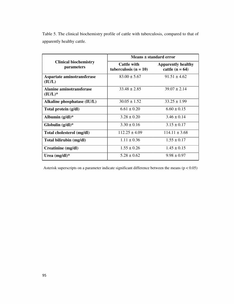
95
Table 5. The clinical biochemistry profile of cattle with tuberculosis, compared to that of
apparently healthy cattle.
Clinical biochemistry
parameters
Means ± standard error
Cattle with
tuberculosis (n = 10)
Apparently healthy
cattle (n = 64)
Aspartate aminotransferase
(IU/L)
83.00 ± 5.67 91.51 ± 4.62
Alanine aminotransferase
(IU/L)*
33.48 ± 2.85 39.07 ± 2.14
Alkaline phosphatase (IU/L) 30.05 ± 1.52 33.25 ± 1.99
Total protein (g/dl) 6.61 ± 0.20 6.60 ± 0.15
Albumin (g/dl)* 3.28 ± 0.20 3.46 ± 0.14
Globulin (g/dl)* 3.30 ± 0.16 3.15 ± 0.17
Total cholesterol (mg/dl) 112.25 ± 4.09 114.11 ± 3.68
Total bilirubin (mg/dl) 1.11 ± 0.36 1.55 ± 0.17
Creatinine (mg/dl) 1.55 ± 0.26 1.45 ± 0.15
Urea (mg/dl)* 5.28 ± 0.62 9.98 ± 0.97
Asterisk superscripts on a parameter indicate significant difference between the means (p < 0.05)
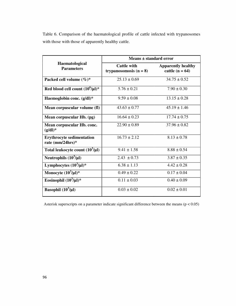
96
Table 6. Comparison of the haematological profile of cattle infected with trypanosomes
with those with those of apparently healthy cattle.
Haematological
Parameters
Means ± standard error
Cattle with
trypanosomosis (n = 8)
Apparently healthy
cattle (n = 64)
Packed cell volume (%)* 25.13 ± 0.69 34.75 ± 0.52
Red blood cell count (106/µl)* 5.76 ± 0.21 7.90 ± 0.30
Haemoglobin conc. (g/dl)* 9.59 ± 0.08 13.15 ± 0.28
Mean corpuscular volume (fl) 43.63 ± 0.77 45.19 ± 1.46
Mean corpuscular Hb. (pg) 16.64 ± 0.23 17.74 ± 0.75
Mean corpuscular Hb. conc.
(g/dl)*
22.90 ± 0.89 37.96 ± 0.82
Erythrocyte sedimentation
rate (mm/24hrs)*
16.73 ± 2.12 8.13 ± 0.78
Total leukocyte count (103/µl) 9.41 ± 1.58 8.88 ± 0.54
Neutrophils (103/µl)
2.43 ± 0.73 3.87 ± 0.35
Lymphocytes (103/µl)* 6.38 ± 1.13 4.42 ± 0.28
Monocyte (103/µl)*
0.49 ± 0.22 0.17 ± 0.04
Eosinophil (103/µl)*
0.11 ± 0.03 0.40 ± 0.09
Basophil (103/µl)
0.03 ± 0.02 0.02 ± 0.01
Asterisk superscripts on a parameter indicate significant difference between the means (p < 0.05)

97
Table 7. The clinical biochemistry profile of cattle infected with trypanosomes, compared
to those of apparently healthy cattle.
Clinical biochemistry
parameters
Means ± standard error
Cattle with
trypanosomosis (n = 8)
Apparently healthy
cattle (n = 64)
Aspartate aminotransferase
(IU/L)
89.32 ± 4.45 91.51 ± 4.62
Alanine aminotransferase
(IU/L)
41.15 ± 5.55 39.07 ± 2.14
Alkaline phosphatase (IU/L) 39.65 ± 2.63 33.25 ± 1.99
Total protein (g/dl) 6.20 ± 0.29 6.60 ± 0.15
Albumin (g/dl)* 2.84 ± 0.16 3.46 ± 0.14
Globulin (g/dl) 3.36 ± 0.24 3.15 ± 0.17
Total cholesterol (mg/dl) 94.73 ± 17.64 114.11 ± 3.68
Total bilirubin (mg/dl) 2.16 ± 0.47 1.55 ± 0.17
Creatinine (mg/dl)* 0.69 ± 0.11 1.45 ± 0.15
Urea (mg/dl)* 19.21 ± 4.43 9.98 ± 0.97
Asterisk superscripts on a parameter indicate significant difference between the means (p < 0.05)
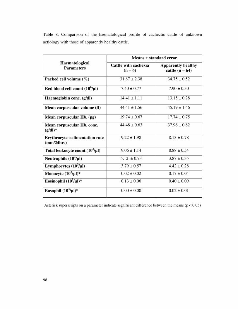
98
Table 8. Comparison of the haematological profile of cachectic cattle of unknown
aetiology with those of apparently healthy cattle.
Haematological
Parameters
Means ± standard error
Cattle with cachexia
(n = 6)
Apparently healthy
cattle (n = 64)
Packed cell volume (%) 31.87 ± 2.38 34.75 ± 0.52
Red blood cell count (106/µl) 7.40 ± 0.77 7.90 ± 0.30
Haemoglobin conc. (g/dl) 14.41 ± 1.11 13.15 ± 0.28
Mean corpuscular volume (fl) 44.41 ± 1.56 45.19 ± 1.46
Mean corpuscular Hb. (pg) 19.74 ± 0.67 17.74 ± 0.75
Mean corpuscular Hb. conc.
(g/dl)*
44.48 ± 0.63 37.96 ± 0.82
Erythrocyte sedimentation rate
(mm/24hrs)
9.22 ± 1.98 8.13 ± 0.78
Total leukocyte count (103/µl) 9.06 ± 1.14 8.88 ± 0.54
Neutrophils (103/µl)
5.12 ± 0.73 3.87 ± 0.35
Lymphocytes (103/µl) 3.79 ± 0.57 4.42 ± 0.28
Monocyte (103/µl)*
0.02 ± 0.02 0.17 ± 0.04
Eosinophil (103/µl)*
0.13 ± 0.06 0.40 ± 0.09
Basophil (103/µl)*
0.00 ± 0.00 0.02 ± 0.01
Asterisk superscripts on a parameter indicate significant difference between the means (p < 0.05)
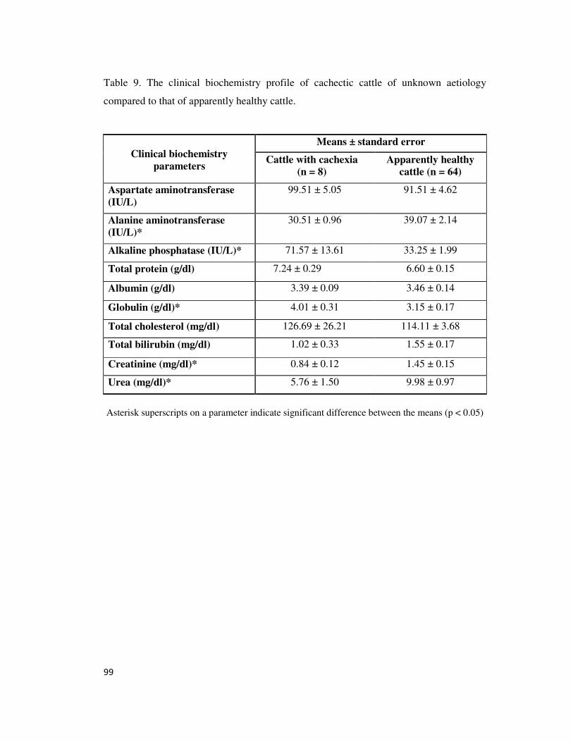
99
Table 9. The clinical biochemistry profile of cachectic cattle of unknown aetiology
compared to that of apparently healthy cattle.
Clinical biochemistry
parameters
Means ± standard error
Cattle with cachexia
(n = 8)
Apparently healthy
cattle (n = 64)
Aspartate aminotransferase
(IU/L)
99.51 ± 5.05 91.51 ± 4.62
Alanine aminotransferase
(IU/L)*
30.51 ± 0.96 39.07 ± 2.14
Alkaline phosphatase (IU/L)* 71.57 ± 13.61 33.25 ± 1.99
Total protein (g/dl) 7.24 ± 0.29 6.60 ± 0.15
Albumin (g/dl) 3.39 ± 0.09 3.46 ± 0.14
Globulin (g/dl)* 4.01 ± 0.31 3.15 ± 0.17
Total cholesterol (mg/dl) 126.69 ± 26.21 114.11 ± 3.68
Total bilirubin (mg/dl) 1.02 ± 0.33 1.55 ± 0.17
Creatinine (mg/dl)* 0.84 ± 0.12 1.45 ± 0.15
Urea (mg/dl)* 5.76 ± 1.50 9.98 ± 0.97
Asterisk superscripts on a parameter indicate significant difference between the means (p < 0.05)
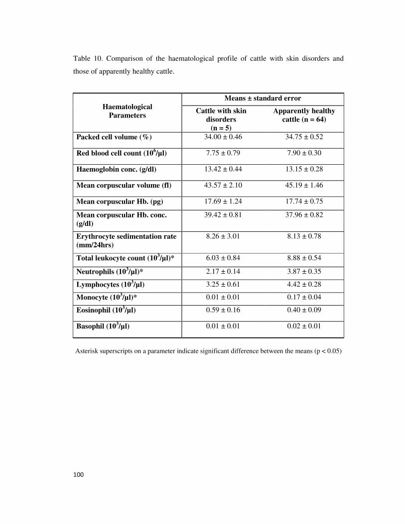
100
Table 10. Comparison of the haematological profile of cattle with skin disorders and
those of apparently healthy cattle.
Haematological
Parameters
Means ± standard error
Cattle with skin
disorders
(n = 5)
Apparently healthy
cattle (n = 64)
Packed cell volume (%) 34.00 ± 0.46 34.75 ± 0.52
Red blood cell count (106/µl) 7.75 ± 0.79 7.90 ± 0.30
Haemoglobin conc. (g/dl) 13.42 ± 0.44 13.15 ± 0.28
Mean corpuscular volume (fl) 43.57 ± 2.10 45.19 ± 1.46
Mean corpuscular Hb. (pg) 17.69 ± 1.24 17.74 ± 0.75
Mean corpuscular Hb. conc.
(g/dl)
39.42 ± 0.81 37.96 ± 0.82
Erythrocyte sedimentation rate
(mm/24hrs)
8.26 ± 3.01 8.13 ± 0.78
Total leukocyte count (103/µl)* 6.03 ± 0.84 8.88 ± 0.54
Neutrophils (103/µl)*
2.17 ± 0.14 3.87 ± 0.35
Lymphocytes (103/µl) 3.25 ± 0.61 4.42 ± 0.28
Monocyte (103/µl)*
0.01 ± 0.01 0.17 ± 0.04
Eosinophil (103/µl)
0.59 ± 0.16 0.40 ± 0.09
Basophil (103/µl)
0.01 ± 0.01 0.02 ± 0.01
Asterisk superscripts on a parameter indicate significant difference between the means (p < 0.05)
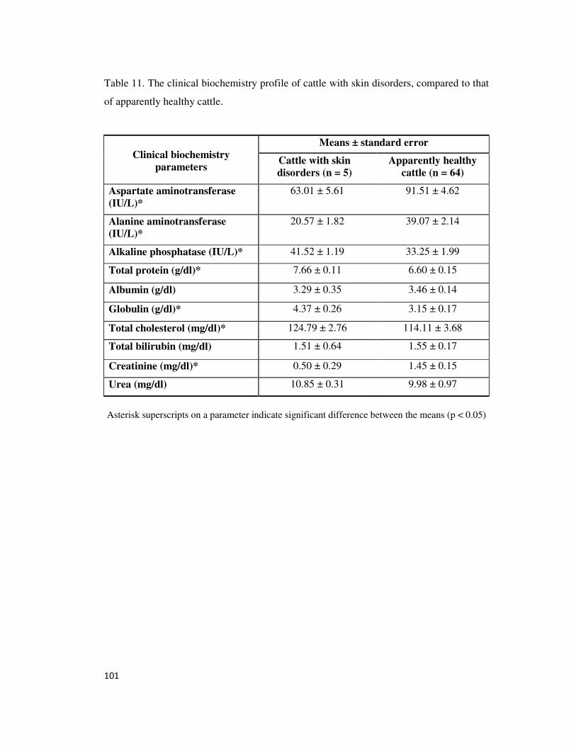
101
Table 11. The clinical biochemistry profile of cattle with skin disorders, compared to that
of apparently healthy cattle.
Clinical biochemistry
parameters
Means ± standard error
Cattle with skin
disorders (n = 5)
Apparently healthy
cattle (n = 64)
Aspartate aminotransferase
(IU/L)*
63.01 ± 5.61 91.51 ± 4.62
Alanine aminotransferase
(IU/L)*
20.57 ± 1.82 39.07 ± 2.14
Alkaline phosphatase (IU/L)* 41.52 ± 1.19 33.25 ± 1.99
Total protein (g/dl)* 7.66 ± 0.11 6.60 ± 0.15
Albumin (g/dl) 3.29 ± 0.35 3.46 ± 0.14
Globulin (g/dl)* 4.37 ± 0.26 3.15 ± 0.17
Total cholesterol (mg/dl)* 124.79 ± 2.76 114.11 ± 3.68
Total bilirubin (mg/dl) 1.51 ± 0.64 1.55 ± 0.17
Creatinine (mg/dl)* 0.50 ± 0.29 1.45 ± 0.15
Urea (mg/dl) 10.85 ± 0.31 9.98 ± 0.97
Asterisk superscripts on a parameter indicate significant difference between the means (p < 0.05)
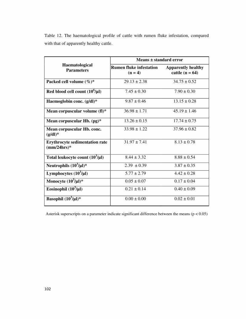
102
Table 12. The haematological profile of cattle with rumen fluke infestation, compared
with that of apparently healthy cattle.
Haematological
Parameters
Means ± standard error
Rumen fluke infestation
(n = 4)
Apparently healthy
cattle (n = 64)
Packed cell volume (%)* 29.13 ± 2.38 34.75 ± 0.52
Red blood cell count (106/µl) 7.45 ± 0.30 7.90 ± 0.30
Haemoglobin conc. (g/dl)* 9.87 ± 0.46 13.15 ± 0.28
Mean corpuscular volume (fl)* 36.98 ± 1.71 45.19 ± 1.46
Mean corpuscular Hb. (pg)* 13.26 ± 0.15 17.74 ± 0.75
Mean corpuscular Hb. conc.
(g/dl)*
33.98 ± 1.22 37.96 ± 0.82
Erythrocyte sedimentation rate
(mm/24hrs)*
31.97 ± 7.41
8.13 ± 0.78
Total leukocyte count (103/µl) 8.44 ± 3.32 8.88 ± 0.54
Neutrophils (103/µl)*
2.39 ± 0.39 3.87 ± 0.35
Lymphocytes (103/µl) 5.77 ± 2.79 4.42 ± 0.28
Monocyte (103/µl)*
0.05 ± 0.07 0.17 ± 0.04
Eosinophil (103/µl)
0.21 ± 0.14 0.40 ± 0.09
Basophil (103/µl)*
0.00 ± 0.00 0.02 ± 0.01
Asterisk superscripts on a parameter indicate significant difference between the means (p < 0.05)
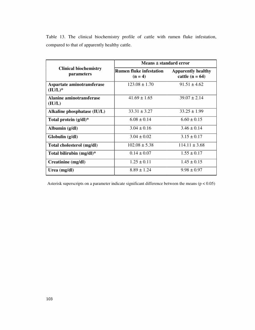
103
Table 13. The clinical biochemistry profile of cattle with rumen fluke infestation,
compared to that of apparently healthy cattle.
Clinical biochemistry
parameters
Means ± standard error
Rumen fluke infestation
(n = 4)
Apparently healthy
cattle (n = 64)
Aspartate aminotransferase
(IU/L)*
123.08 ± 1.70 91.51 ± 4.62
Alanine aminotransferase
(IU/L)
41.69 ± 1.65 39.07 ± 2.14
Alkaline phosphatase (IU/L) 33.31 ± 3.27 33.25 ± 1.99
Total protein (g/dl)* 6.08 ± 0.14 6.60 ± 0.15
Albumin (g/dl) 3.04 ± 0.16 3.46 ± 0.14
Globulin (g/dl) 3.04 ± 0.02 3.15 ± 0.17
Total cholesterol (mg/dl) 102.08 ± 5.38 114.11 ± 3.68
Total bilirubin (mg/dl)* 0.14 ± 0.07 1.55 ± 0.17
Creatinine (mg/dl) 1.25 ± 0.11 1.45 ± 0.15
Urea (mg/dl) 8.89 ± 1.24 9.98 ± 0.97
Asterisk superscripts on a parameter indicate significant difference between the means (p < 0.05)
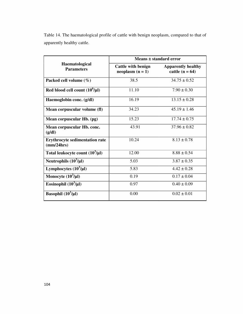
104
Table 14. The haematological profile of cattle with benign neoplasm, compared to that of
apparently healthy cattle.
Haematological
Parameters
Means ± standard error
Cattle with benign
neoplasm (n = 1)
Apparently healthy
cattle (n = 64)
Packed cell volume (%) 38.5 34.75 ± 0.52
Red blood cell count (106/µl) 11.10 7.90 ± 0.30
Haemoglobin conc. (g/dl) 16.19 13.15 ± 0.28
Mean corpuscular volume (fl) 34.23 45.19 ± 1.46
Mean corpuscular Hb. (pg) 15.23 17.74 ± 0.75
Mean corpuscular Hb. conc.
(g/dl)
43.91 37.96 ± 0.82
Erythrocyte sedimentation rate
(mm/24hrs)
10.24 8.13 ± 0.78
Total leukocyte count (103/µl) 12.00 8.88 ± 0.54
Neutrophils (103/µl)
5.03 3.87 ± 0.35
Lymphocytes (103/µl) 5.83 4.42 ± 0.28
Monocyte (103/µl)
0.19 0.17 ± 0.04
Eosinophil (103/µl)
0.97 0.40 ± 0.09
Basophil (103/µl)
0.00 0.02 ± 0.01
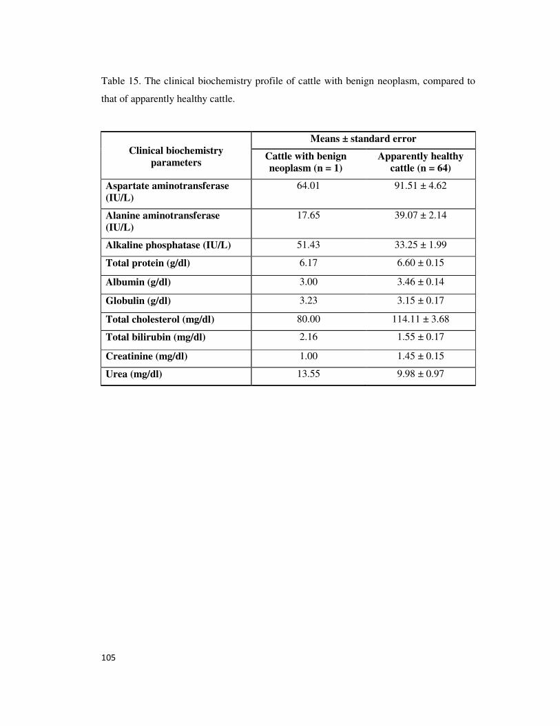
105
Table 15. The clinical biochemistry profile of cattle with benign neoplasm, compared to
that of apparently healthy cattle.
Clinical biochemistry
parameters
Means ± standard error
Cattle with benign
neoplasm (n = 1)
Apparently healthy
cattle (n = 64)
Aspartate aminotransferase
(IU/L)
64.01 91.51 ± 4.62
Alanine aminotransferase
(IU/L)
17.65 39.07 ± 2.14
Alkaline phosphatase (IU/L) 51.43 33.25 ± 1.99
Total protein (g/dl) 6.17 6.60 ± 0.15
Albumin (g/dl) 3.00 3.46 ± 0.14
Globulin (g/dl) 3.23 3.15 ± 0.17
Total cholesterol (mg/dl) 80.00 114.11 ± 3.68
Total bilirubin (mg/dl) 2.16 1.55 ± 0.17
Creatinine (mg/dl) 1.00 1.45 ± 0.15
Urea (mg/dl) 13.55 9.98 ± 0.97
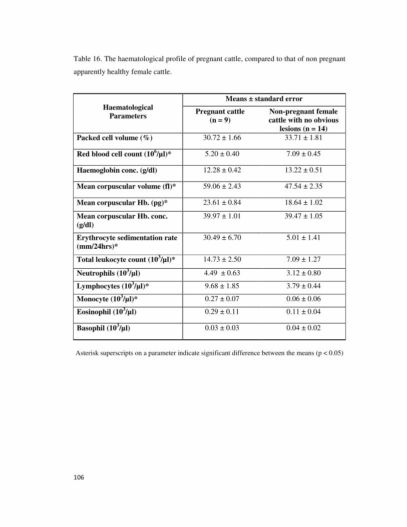
106
Table 16. The haematological profile of pregnant cattle, compared to that of non pregnant
apparently healthy female cattle.
Haematological
Parameters
Means ± standard error
Pregnant cattle
(n = 9)
Non-pregnant female
cattle with no obvious
lesions (n = 14)
Packed cell volume (%) 30.72 ± 1.66 33.71 ± 1.81
Red blood cell count (106/µl)* 5.20 ± 0.40 7.09 ± 0.45
Haemoglobin conc. (g/dl) 12.28 ± 0.42 13.22 ± 0.51
Mean corpuscular volume (fl)* 59.06 ± 2.43 47.54 ± 2.35
Mean corpuscular Hb. (pg)* 23.61 ± 0.84 18.64 ± 1.02
Mean corpuscular Hb. conc.
(g/dl)
39.97 ± 1.01 39.47 ± 1.05
Erythrocyte sedimentation rate
(mm/24hrs)*
30.49 ± 6.70 5.01 ± 1.41
Total leukocyte count (103/µl)* 14.73 ± 2.50 7.09 ± 1.27
Neutrophils (103/µl)
4.49 ± 0.63 3.12 ± 0.80
Lymphocytes (103/µl)* 9.68 ± 1.85 3.79 ± 0.44
Monocyte (103/µl)*
0.27 ± 0.07 0.06 ± 0.06
Eosinophil (103/µl)
0.29 ± 0.11 0.11 ± 0.04
Basophil (103/µl)
0.03 ± 0.03 0.04 ± 0.02
Asterisk superscripts on a parameter indicate significant difference between the means (p < 0.05)
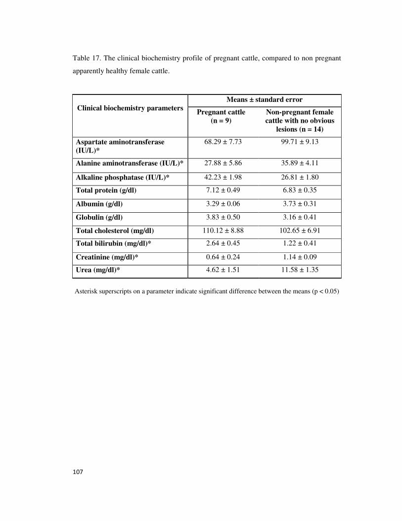
107
Table 17. The clinical biochemistry profile of pregnant cattle, compared to non pregnant
apparently healthy female cattle.
Clinical biochemistry parameters
Means ± standard error
Pregnant cattle
(n = 9)
Non-pregnant female
cattle with no obvious
lesions (n = 14)
Aspartate aminotransferase
(IU/L)*
68.29 ± 7.73 99.71 ± 9.13
Alanine aminotransferase (IU/L)* 27.88 ± 5.86 35.89 ± 4.11
Alkaline phosphatase (IU/L)* 42.23 ± 1.98 26.81 ± 1.80
Total protein (g/dl) 7.12 ± 0.49 6.83 ± 0.35
Albumin (g/dl) 3.29 ± 0.06 3.73 ± 0.31
Globulin (g/dl) 3.83 ± 0.50 3.16 ± 0.41
Total cholesterol (mg/dl) 110.12 ± 8.88 102.65 ± 6.91
Total bilirubin (mg/dl)* 2.64 ± 0.45 1.22 ± 0.41
Creatinine (mg/dl)* 0.64 ± 0.24 1.14 ± 0.09
Urea (mg/dl)* 4.62 ± 1.51 11.58 ± 1.35
Asterisk superscripts on a parameter indicate significant difference between the means (p < 0.05)
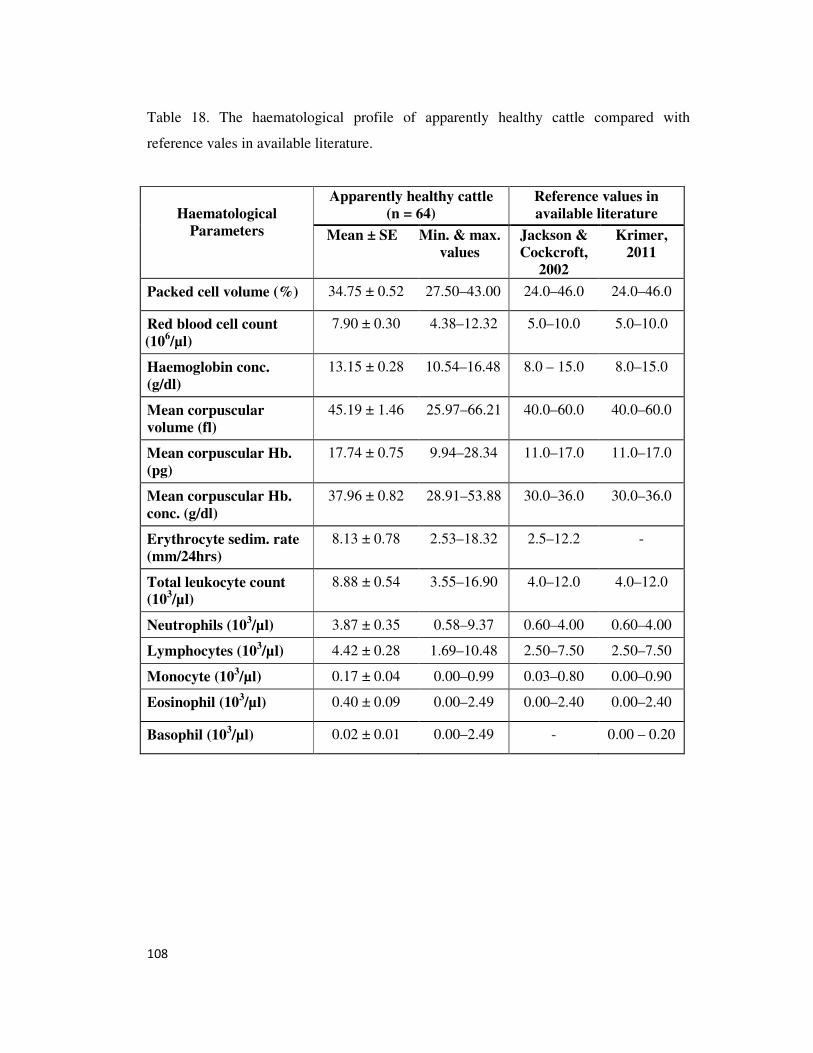
108
Table 18. The haematological profile of apparently healthy cattle compared with
reference vales in available literature.
Haematological
Parameters
Apparently healthy cattle
(n = 64)
Reference values in
available literature
Mean ± SE Min. & max.
values
Jackson &
Cockcroft,
2002
Krimer,
2011
Packed cell volume (%) 34.75 ± 0.52 27.50–43.00 24.0–46.0 24.0–46.0
Red blood cell count
(106/µl)
7.90 ± 0.30 4.38–12.32 5.0–10.0 5.0–10.0
Haemoglobin conc.
(g/dl)
13.15 ± 0.28 10.54–16.48 8.0 – 15.0 8.0–15.0
Mean corpuscular
volume (fl)
45.19 ± 1.46 25.97–66.21 40.0–60.0 40.0–60.0
Mean corpuscular Hb.
(pg)
17.74 ± 0.75 9.94–28.34 11.0–17.0 11.0–17.0
Mean corpuscular Hb.
conc. (g/dl)
37.96 ± 0.82 28.91–53.88 30.0–36.0 30.0–36.0
Erythrocyte sedim. rate
(mm/24hrs)
8.13 ± 0.78 2.53–18.32 2.5–12.2 -
Total leukocyte count
(103/µl)
8.88 ± 0.54 3.55–16.90 4.0–12.0 4.0–12.0
Neutrophils (103/µl)
3.87 ± 0.35 0.58–9.37 0.60–4.00 0.60–4.00
Lymphocytes (103/µl) 4.42 ± 0.28 1.69–10.48 2.50–7.50 2.50–7.50
Monocyte (103/µl)
0.17 ± 0.04 0.00–0.99 0.03–0.80 0.00–0.90
Eosinophil (103/µl)
0.40 ± 0.09 0.00–2.49 0.00–2.40 0.00–2.40
Basophil (103/µl)
0.02 ± 0.01 0.00–2.49 - 0.00 – 0.20
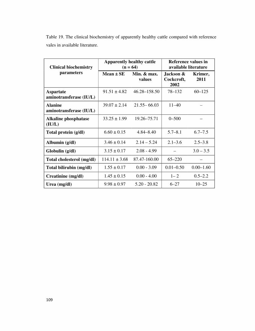
109
Table 19. The clinical biochemistry of apparently healthy cattle compared with reference
vales in available literature.
Clinical biochemistry
parameters
Apparently healthy cattle
(n = 64)
Reference values in
available literature
Mean ± SE Min. & max.
values
Jackson &
Cockcroft,
2002
Krimer,
2011
Aspartate
aminotransferase (IU/L)
91.51 ± 4.82 46.28–158.50 78–132 60–125
Alanine
aminotransferase (IU/L)
39.07 ± 2.14 21.55– 66.03 11–40 –
Alkaline phosphatase
(IU/L)
33.25 ± 1.99 19.26–75.71 0–500 –
Total protein (g/dl) 6.60 ± 0.15 4.84–8.40 5.7–8.1 6.7–7.5
Albumin (g/dl) 3.46 ± 0.14 2.14 – 5.24 2.1–3.6 2.5–3.8
Globulin (g/dl) 3.15 ± 0.17 2.08 - 4.99 – 3.0 – 3.5
Total cholesterol (mg/dl) 114.11 ± 3.68 87.47-160.00 65–220 –
Total bilirubin (mg/dl) 1.55 ± 0.17 0.00 - 3.09 0.01–0.50 0.00–1.60
Creatinine (mg/dl) 1.45 ± 0.15 0.00 - 4.00 1– 2 0.5–2.2
Urea (mg/dl) 9.98 ± 0.97 5.20 - 20.82 6–27 10–25
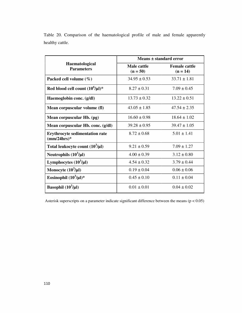
110
Table 20. Comparison of the haematological profile of male and female apparently
healthy cattle.
Haematological
Parameters
Means ± standard error
Male cattle
(n = 50)
Female cattle
(n = 14)
Packed cell volume (%) 34.95 ± 0.53 33.71 ± 1.81
Red blood cell count (106/µl)* 8.27 ± 0.31 7.09 ± 0.45
Haemoglobin conc. (g/dl) 13.73 ± 0.32 13.22 ± 0.51
Mean corpuscular volume (fl) 43.05 ± 1.85 47.54 ± 2.35
Mean corpuscular Hb. (pg) 16.60 ± 0.98 18.64 ± 1.02
Mean corpuscular Hb. conc. (g/dl) 39.28 ± 0.95 39.47 ± 1.05
Erythrocyte sedimentation rate
(mm/24hrs)*
8.72 ± 0.68 5.01 ± 1.41
Total leukocyte count (103/µl) 9.21 ± 0.59 7.09 ± 1.27
Neutrophils (103/µl)
4.00 ± 0.39 3.12 ± 0.80
Lymphocytes (103/µl) 4.54 ± 0.32 3.79 ± 0.44
Monocyte (103/µl)
0.19 ± 0.04 0.06 ± 0.06
Eosinophil (103/µl)*
0.45 ± 0.10 0.11 ± 0.04
Basophil (103/µl)
0.01 ± 0.01 0.04 ± 0.02
Asterisk superscripts on a parameter indicate significant difference between the means (p < 0.05)
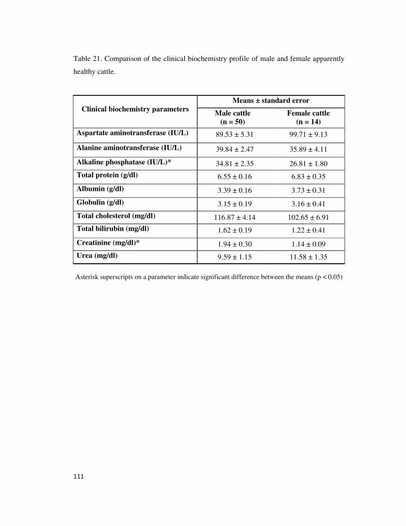
111
Table 21. Comparison of the clinical biochemistry profile of male and female apparently
healthy cattle.
Clinical biochemistry parameters
Means ± standard error
Male cattle
(n = 50)
Female cattle
(n = 14)
Aspartate aminotransferase (IU/L) 89.53 ± 5.31 99.71 ± 9.13
Alanine aminotransferase (IU/L) 39.84 ± 2.47 35.89 ± 4.11
Alkaline phosphatase (IU/L)* 34.81 ± 2.35 26.81 ± 1.80
Total protein (g/dl) 6.55 ± 0.16 6.83 ± 0.35
Albumin (g/dl)
3.39 ± 0.16 3.73 ± 0.31
Globulin (g/dl)
3.15 ± 0.19 3.16 ± 0.41
Total cholesterol (mg/dl) 116.87 ± 4.14 102.65 ± 6.91
Total bilirubin (mg/dl) 1.62 ± 0.19 1.22 ± 0.41
Creatinine (mg/dl)* 1.94 ± 0.30 1.14 ± 0.09
Urea (mg/dl) 9.59 ± 1.15 11.58 ± 1.35
Asterisk superscripts on a parameter indicate significant difference between the means (p < 0.05)
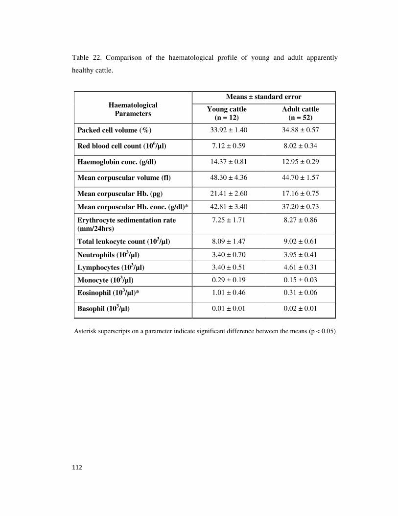
112
Table 22. Comparison of the haematological profile of young and adult apparently
healthy cattle.
Haematological
Parameters
Means ± standard error
Young cattle
(n = 12)
Adult cattle
(n = 52)
Packed cell volume (%) 33.92 ± 1.40 34.88 ± 0.57
Red blood cell count (106/µl) 7.12 ± 0.59 8.02 ± 0.34
Haemoglobin conc. (g/dl) 14.37 ± 0.81 12.95 ± 0.29
Mean corpuscular volume (fl) 48.30 ± 4.36 44.70 ± 1.57
Mean corpuscular Hb. (pg) 21.41 ± 2.60 17.16 ± 0.75
Mean corpuscular Hb. conc. (g/dl)* 42.81 ± 3.40 37.20 ± 0.73
Erythrocyte sedimentation rate
(mm/24hrs)
7.25 ± 1.71 8.27 ± 0.86
Total leukocyte count (103/µl) 8.09 ± 1.47 9.02 ± 0.61
Neutrophils (103/µl)
3.40 ± 0.70 3.95 ± 0.41
Lymphocytes (103/µl) 3.40 ± 0.51 4.61 ± 0.31
Monocyte (103/µl)
0.29 ± 0.19 0.15 ± 0.03
Eosinophil (103/µl)*
1.01 ± 0.46 0.31 ± 0.06
Basophil (103/µl)
0.01 ± 0.01 0.02 ± 0.01
Asterisk superscripts on a parameter indicate significant difference between the means (p < 0.05)
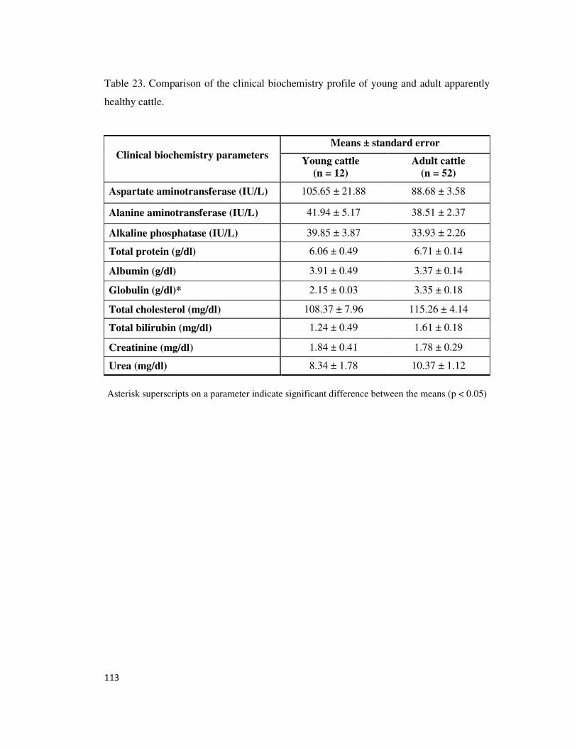
113
Table 23. Comparison of the clinical biochemistry profile of young and adult apparently
healthy cattle.
Clinical biochemistry parameters
Means ± standard error
Young cattle
(n = 12)
Adult cattle
(n = 52)
Aspartate aminotransferase (IU/L) 105.65 ± 21.88 88.68 ± 3.58
Alanine aminotransferase (IU/L) 41.94 ± 5.17 38.51 ± 2.37
Alkaline phosphatase (IU/L) 39.85 ± 3.87 33.93 ± 2.26
Total protein (g/dl) 6.06 ± 0.49 6.71 ± 0.14
Albumin (g/dl) 3.91 ± 0.49 3.37 ± 0.14
Globulin (g/dl)* 2.15 ± 0.03 3.35 ± 0.18
Total cholesterol (mg/dl) 108.37 ± 7.96 115.26 ± 4.14
Total bilirubin (mg/dl) 1.24 ± 0.49 1.61 ± 0.18
Creatinine (mg/dl) 1.84 ± 0.41 1.78 ± 0.29
Urea (mg/dl) 8.34 ± 1.78 10.37 ± 1.12
Asterisk superscripts on a parameter indicate significant difference between the means (p < 0.05)
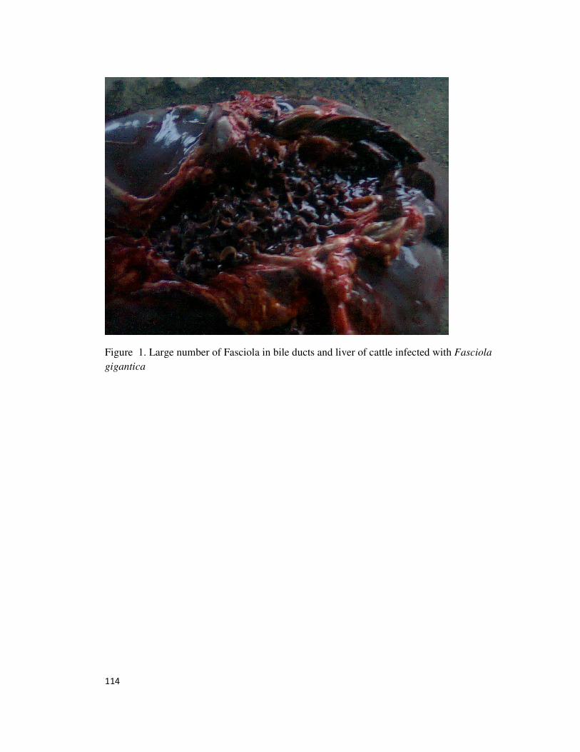
114
Figure 1. Large number of Fasciola in bile ducts and liver of cattle infected with Fasciola
gigantica
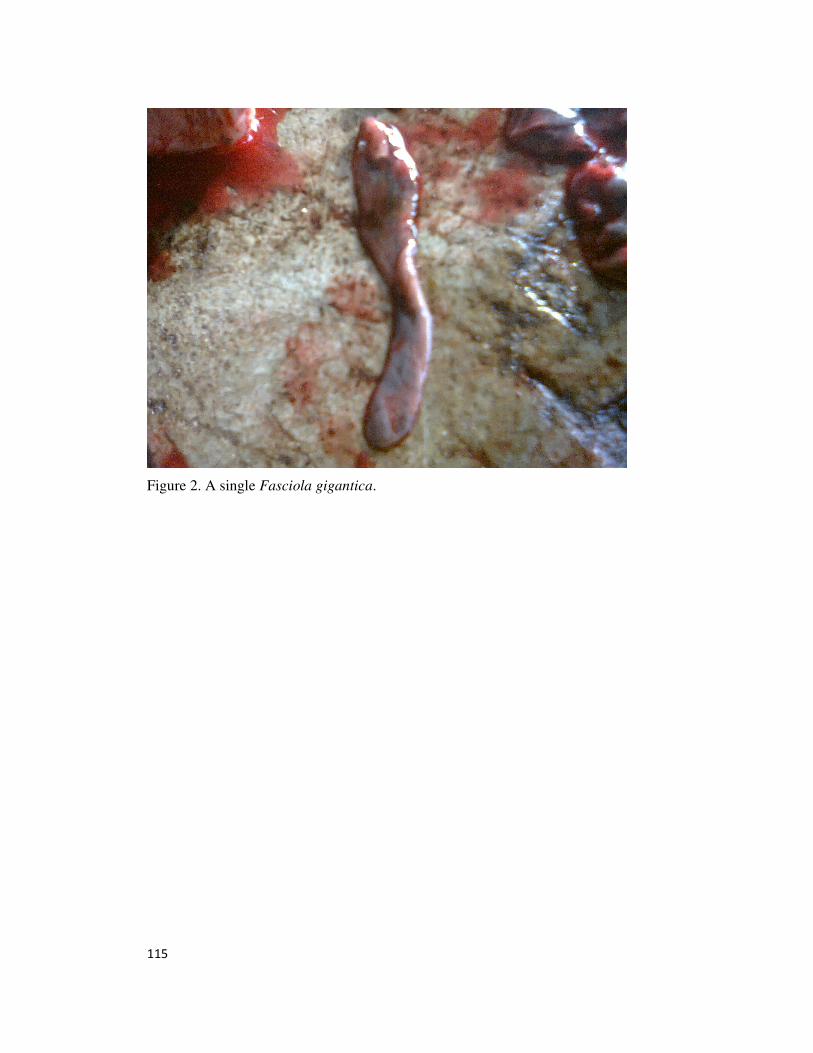
115
Figure 2. A single Fasciola gigantica.
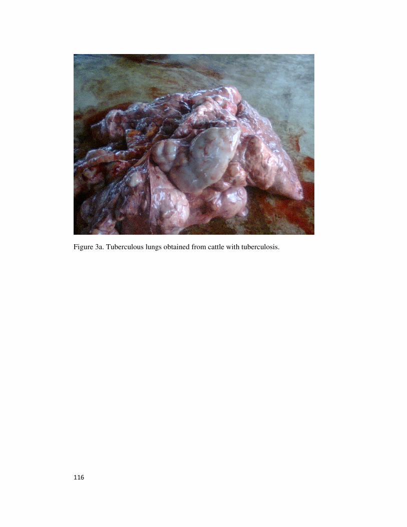
116
Figure 3a. Tuberculous lungs obtained from cattle with tuberculosis.
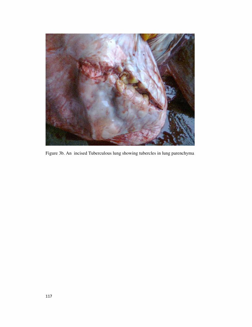
117
Figure 3b. An incised Tuberculous lung showing tubercles in lung parenchyma
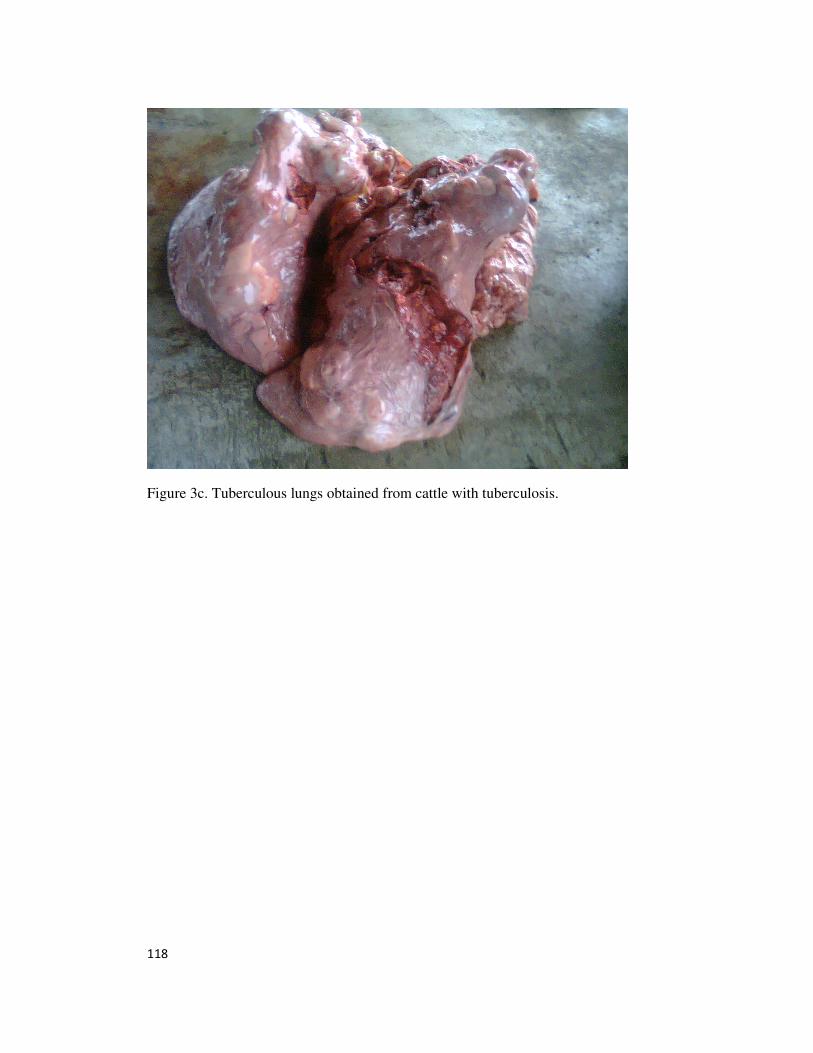
118
Figure 3c. Tuberculous lungs obtained from cattle with tuberculosis.
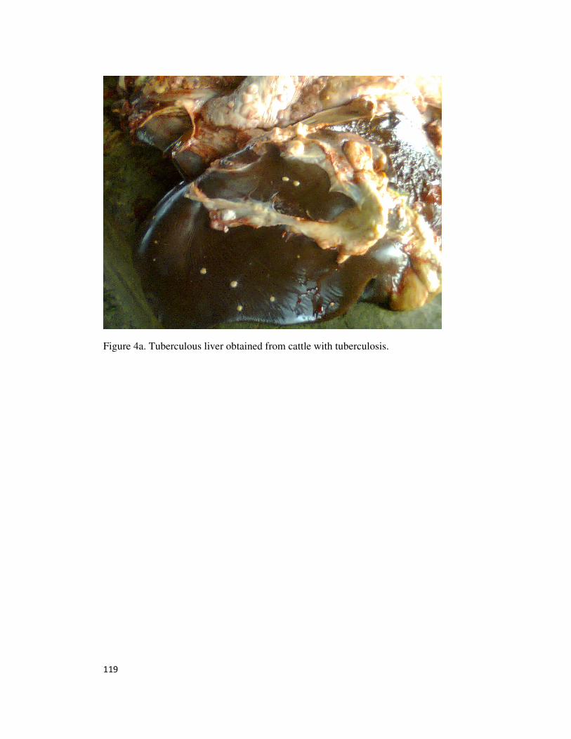
119
Figure 4a. Tuberculous liver obtained from cattle with tuberculosis.
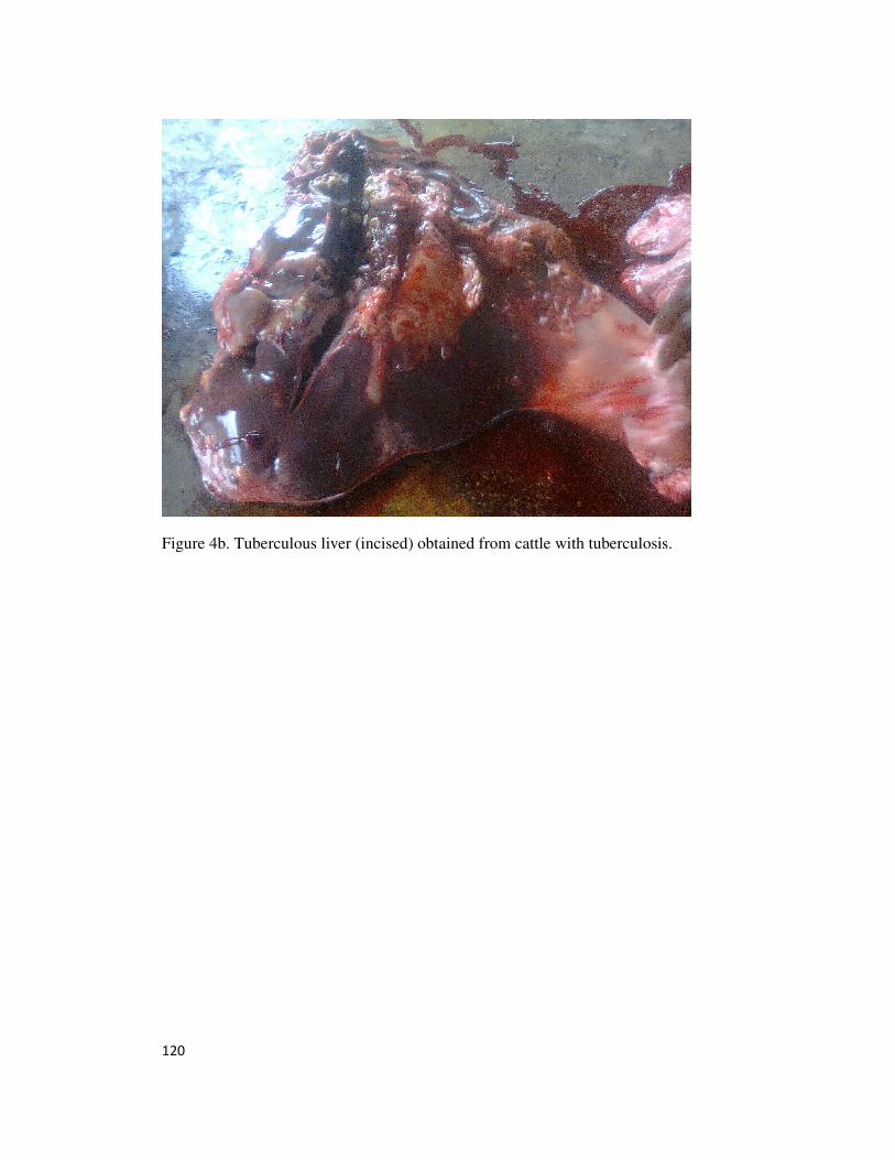
120
Figure 4b. Tuberculous liver (incised) obtained from cattle with tuberculosis.
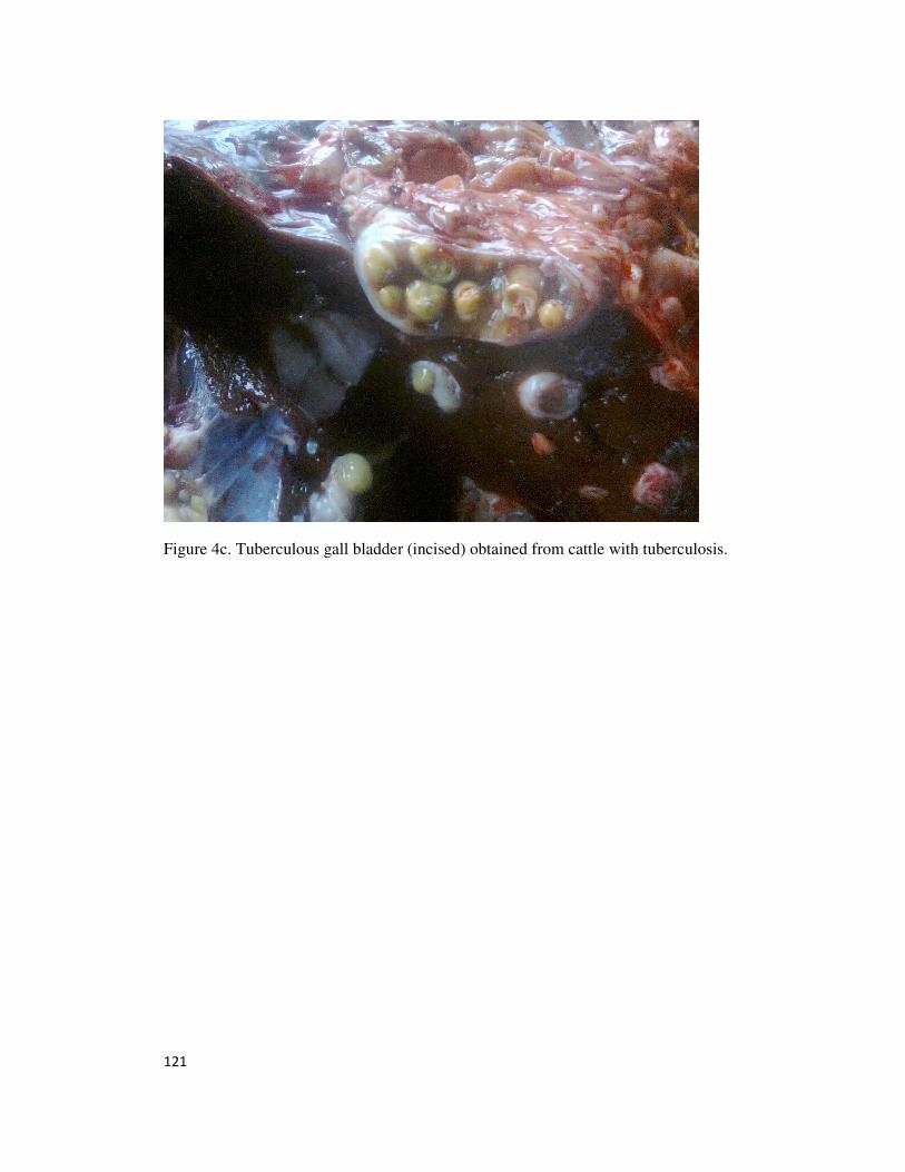
121
Figure 4c. Tuberculous gall bladder (incised) obtained from cattle with tuberculosis.
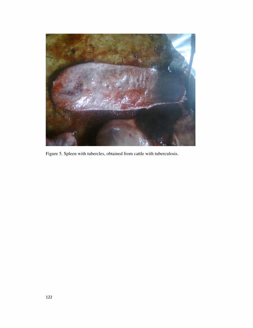
122
Figure 5. Spleen with tubercles, obtained from cattle with tuberculosis.
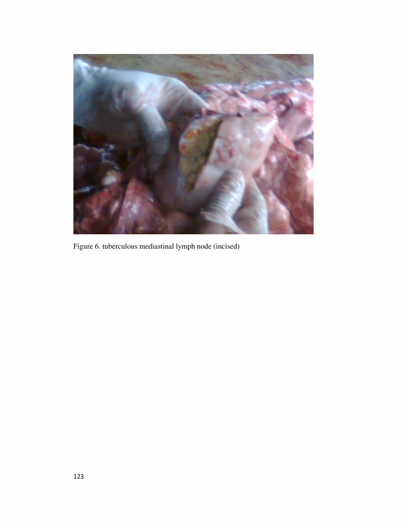
123
Figure 6. tuberculous mediastinal lymph node (incised)
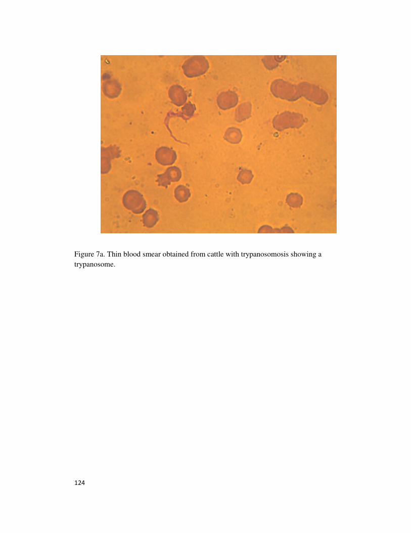
124
Figure 7a. Thin blood smear obtained from cattle with trypanosomosis showing a
trypanosome.

125
Figure 7b. Another thin blood smear obtained from cattle with trypanosomosis showing a
trypanosome.
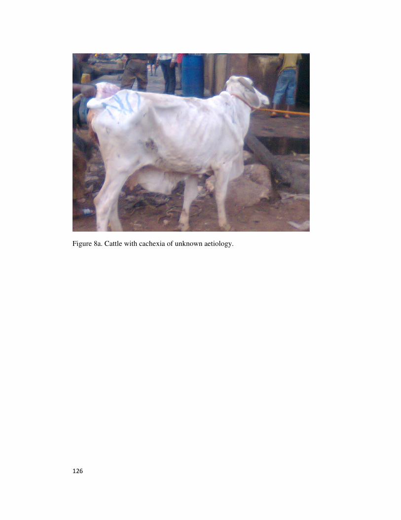
126
Figure 8a. Cattle with cachexia of unknown aetiology.
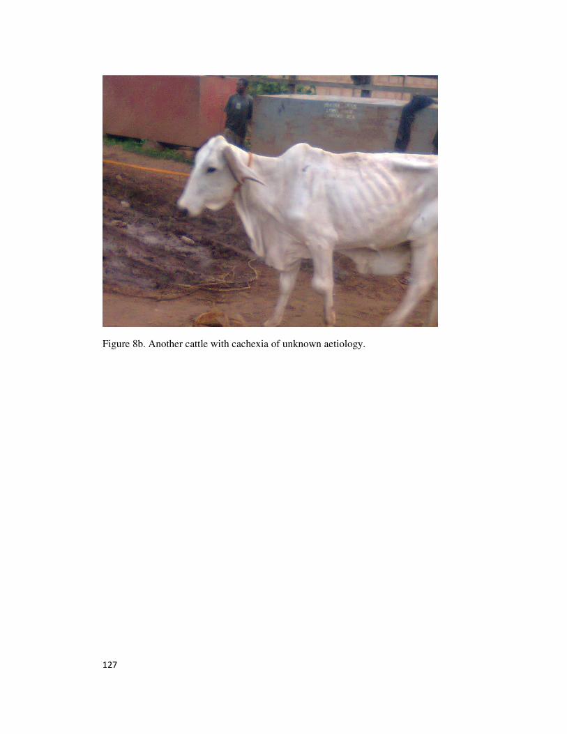
127
Figure 8b. Another cattle with cachexia of unknown aetiology.
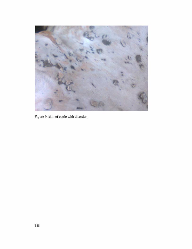
128
Figure 9. skin of cattle with disorder.
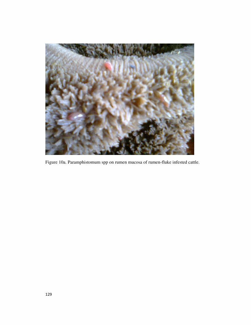
129
Figure 10a. Paramphistomum spp on rumen mucosa of rumen-fluke infested cattle.
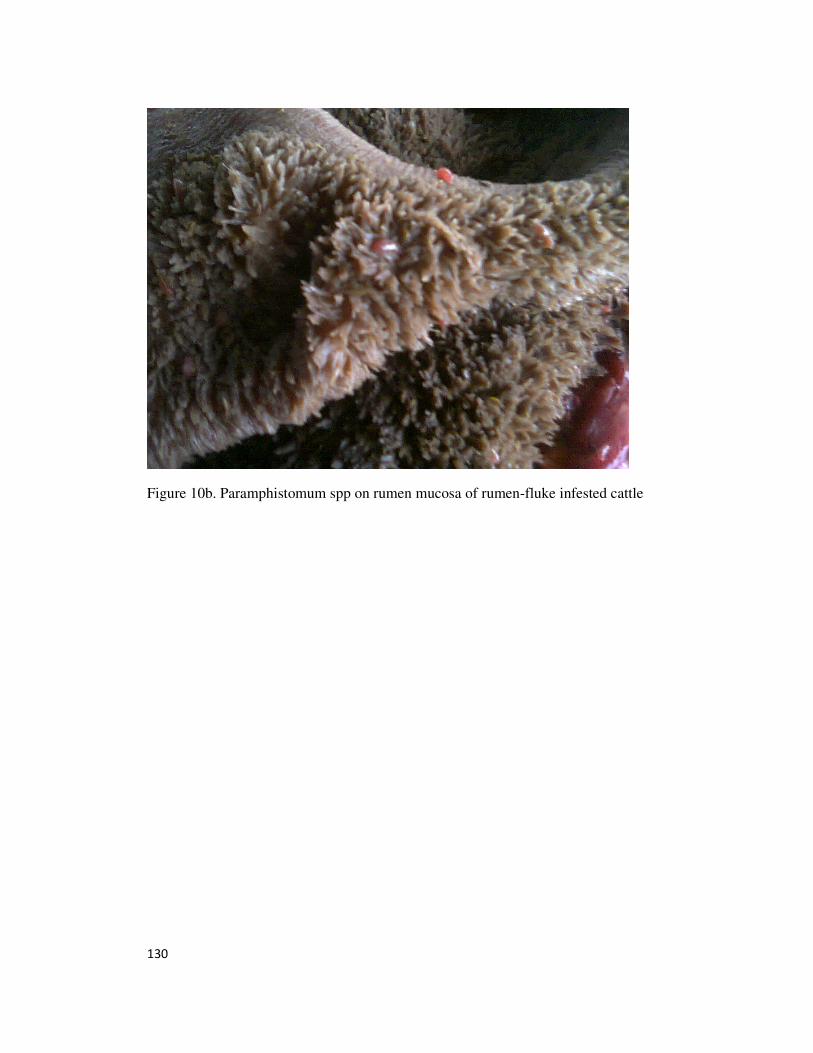
130
Figure 10b. Paramphistomum spp on rumen mucosa of rumen-fluke infested cattle
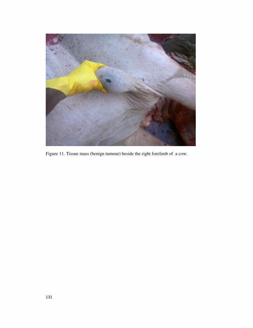
131
Figure 11. Tissue mass (benign tumour) beside the right forelimb of a cow.
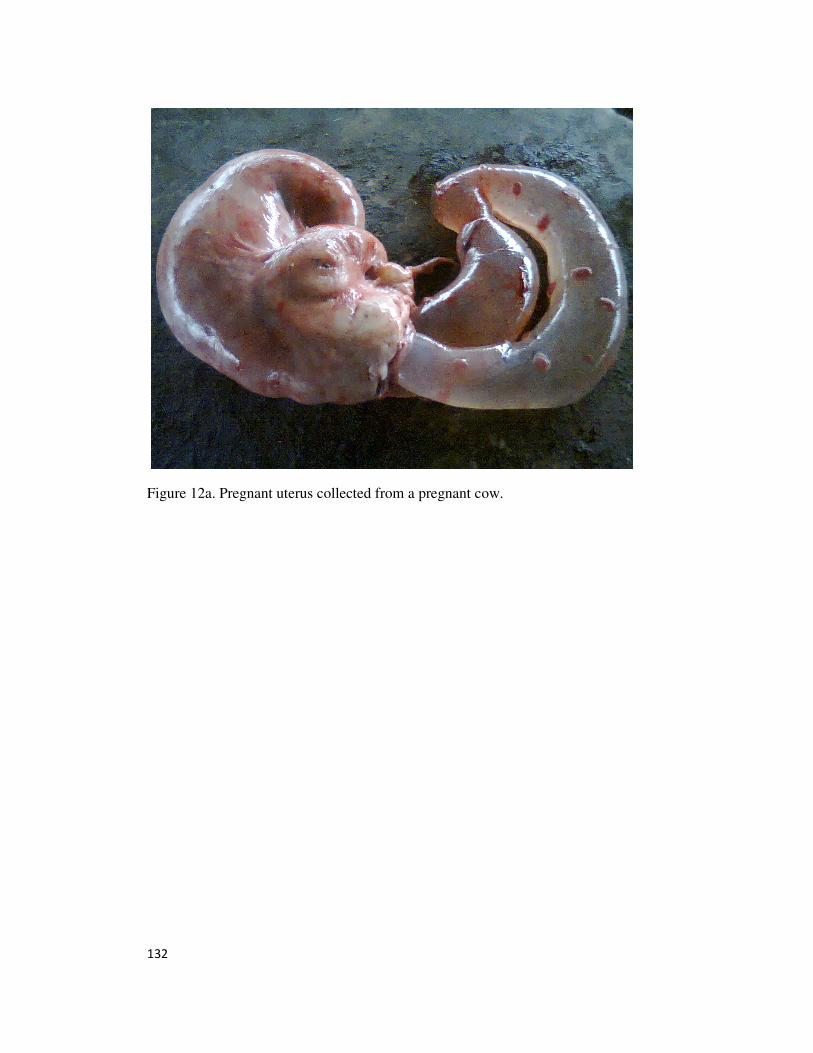
132
Figure 12a. Pregnant uterus collected from a pregnant cow.
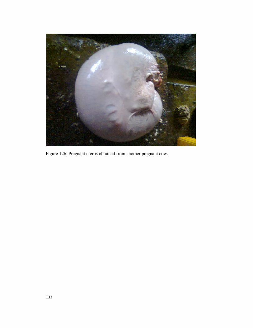
133
Figure 12b. Pregnant uterus obtained from another pregnant cow.
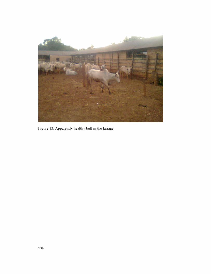
134
Figure 13. Apparently healthy bull in the lariage
135
CHAPTER 5
DISCUSSION AND CONCLUSION
5.0. DISCUSSION
The findings that fasciolosis ranks topmost in the list of diseases of cattle recorded in this
study is in agreement with the reports of Hammond and Sewell (1990), and that of Molina
et al., (2005) which stated that infection with Fasciola gigantica is one of the most
common single helminth infection of cattle and ranks topmost among diseases that lead to
condemnation of livers in abattoir. The 8.47% prevalence for fasciolosis recorded in this
study is relatively lower
than the 47.4% reported by Adedokun et al., (2008), but is comparable to 10.34%
reported by Marques and Scroferneker (2003). The 1.76% prevalence recorded for bovine
tuberculosis is worthy of note because of the public health (zoonotic) significance of its
occurrence and is comparable to 1.3% reported by Shirma et al., (2003), but lower than
the 3.85% reported by Proano-Perez et al., (2006). The relatively low prevalence of
trypanosomosis (1.47%) in the studied cattle is worthy of note, and is thought to be due to
the recent very high usage of trypanocides by herdsmen and animal health workers
(Holmes et al., 2004). The 1.56% occurrence of pregnancy among the studied cattle is
also worthy of note as it brings to the fore the high level of foetal wastage at abattoirs.
The alterations in the erythrocytic parameters recorded for cattle with fasciolosis in this
study, showed anaemia of the hypochromic macrocytic type (Ihedioha, 2003; Stockham
and Scott, 2008) as shown by decreases in PCV, RBC count, Hb concentration, and
MCHC, with an increase in MCV. Anaemia is a hallmark finding in fasciolosis of cattle
(Molina et al., 2008; Valerio et al., 2008). The cause of the anaemia in fasciolosis is
blood loss caused by the haematophagic activity of the parasite (Valerio et al., 2008;
136
Lotfollalizadeh et al., 2008), leakage of blood from the bile duct to the intestines with
resultant iron deficiency (Lotfollalizadeh et al., 2008), and/or release of an amino acid
proline by the fluke (Robert and Hadar, 1981). Anaemia of the macrocytic hypochromic
type recorded in fasciola-infected cattle in this study is in agreement with the findings of
Taimur et al., (1993); Behm and Sangster (1999); Teleb et al., (2007); Molina et al.,
(2008); Al-Quaraishy and Al-Moussawi (2011). The increase in ESR of cattle infected
with fasciola in this study may be due to the anaemia caused by the haematophagic
activity of the flukes, tissue destruction and inflammatory reactions caused by the
presence of the flukes in the billiary canal and bile duct (Coles, 1986; Roper, 1999;
Mohan, 2010).
The findings in this study of leukocytosis in cattle with fasciolosis associated with
lymphocytosis and monocytosis, eosinophilia and basophilia may be attributed to the
chronicity of the infection, and the antigenic stimulation caused by the flukes larval stage
migration (prehepatic and hepatic) and localization in the bile duct where they initiate an
inflammatory reaction (Taimur et al., 1993; Molina et al., 2008). These leukocytic
alterations recorded in fasciola infected cattle in this study is in agreement with findings
of Bashady et al., (1990); Molina et al., (2008); Al-Quaraishy and Al-Moussawi (2011).
The increase in ALP and total bilirubin recorded for cattle with fasciolosis in this study is
believed to be due to cholestasis caused by bile duct localization and blockade by the
adult flukes. This finding is in agreement with the reports of Lotfollizadeh et al., (2008)
and Al-Quaraishy and Al-Moussawi (2011). The hypoproteinaemia and
hypoalbuminaemia recorded for the fasciola infested cattle may be due to protein loss
caused by the haematophagic activity of the adult fluke or the reduced hepatic synthesis
137
of albumin due to damage to hepatic tissues (Ruotsalo and Tant, 2012). This finding is in
agreement with the reports of Sheikh et al., (2006); Al-Quaraishy and Al-Moussawi
(2011) and Okey et al., (2013). The hyperglobulinaemia recorded in the mildly infected
cattle may be due to immune response to inflammations (cholangitis) caused by migrating
immature flukes and localized adult flukes in the bile duct and billiary canals (Matanovic
et al., 2007; Ruotsalo and Tant, 2012). This finding is in agreement with that of Bashady
et al., (1990) who reported an increase in beta globulin levels in experimental fasciolosis
in sheep.
The erythrocytic alterations recorded in cattle with tuberculosis showed anaemia of the
normocytic hypochromic type as shown by decreases in PCV, RBC count, and Hb
concentration (Ihedioha, 2003; Stockham and Scott, 2008). The anaemia recorded for the
tuberculosis-infected cattle may be due to the haemolytic effect of the bacterial agent,
chronicity of the disease condition or bone marrow atrophy (Ihedioha and Chineme,
2004; Shetter et al., 2011). This finding is in agreement with the reports of Rao et al.,
(1992) and Kumar et al., (1994), but was in contrast to the reports of Shetter et al.,
(2011). The increased ESR recorded in cattle with tuberculosis may be due to tissue
destruction associated with the formation of granulomas in the parenchyma of the lungs,
liver, spleen and lymph nodes (Mohan, 2010). This finding is in agreement with the
reports of Amin et al., (1990) in a study on buffaloes, Olivia et al., (2008) in a study on
humans and Shetter et al., (2011) in a study in cattle. The findings in the present study is
however contrary to the reports of Javed et al (2006) in a study on buffaloes.
The findings of leukocytosis associated with lymphocytosis and eosinophilia recorded in
this study may be due to antigenic stimulation caused by active chronic tuberculosis
138
infection (Duncan and Prasse, 1986; Sink and Feldman, 2004). This finding is in
agreement with the reports of Javed et al., (2006) and Shetter et al., (2011).
The hypoalbuminaemia recorded in this study for cattle with tuberculosis may be due to
inadequate liver function due to the presence of granulomas in the liver parenchyma, and
reduced albumin synthesis due to damage of hepatic tissues (Ruotsalo and Tant, 2012).
The increase in serum globulin level seen in cattle with tuberculosis may be due to high
levels of immunoglobulins stimulated by chronic antigenic challenge, and the increased
serum globulin levels recorded in this study is in agreement with the reports of Shetter et
al., (2011).
The erythrocytic alterations recorded in cattle infected with trypanosomes in this study is
an anaemia of the normocytic hypochromic type (Miale, 1982; Brill and Baumgardner,
2000). This form of anaemia is at variance with the reports of Takeet and Fagbemi (2009)
who reported anaemia of the macrocytic normochromic type in early infections and later
normocytic normochromic type in advanced stages of infection of rabbits experimentally
infected with Trypanosome congolense. The cause of the anaemia recorded in cattle
infected with trypanosomosis may be due to the haemolytic activity of the protozoan
parasite (Anosa, 1988; Ihedioha and Chineme, 2004; Takeet and Fagbemi, 2009). This
finding is in agreement with the reports of Bengaly et al., (2002) who reported anaemia in
T. vivax infected cattle, Saror (1980) on T. vivax infected goats, Bisalla et al., (2007) on
T. congolense infected sheep, and Takeet and Fagbemi (2009) on T. congolense infected
rabbits. The increase in ESR recorded for trypanosome infected cattle in this study may
be due to anaemia (Coles, 1986; Mohan, 2010), or damage of the host tissues by the
protozoan parasite (Abenga and Anosa, 2005).
139
The lymphocytosis recorded in cattle infected with trypanosomes may be due to
exaggerated antigenic stimulation as a result of the antigenic variation reported to occur
in trypanosomosis (Borst et al., 1996; Barry and McCulloch, 2001; Vanhamme et al.,
2001). The monocytosis recorded in the present study may be due to response to the
presence of protozoan parasites, where the monocytes transform to macrophages and
function in phagocytosis of these parasites (Ihedioha and Chineme, 2004). Takeet and
Fagbemi (2009) reported leucopaenia characterized by neutropaenia, eosinopaenia and
lymphocytosis in an experimental infection of rabbits with T. congolense.
The hypoalbuminaemia recorded in cattle infected with trypanosomes may be due to
decreased protein synthesis due to hepatic dysfunction (Ruotsalo and Tant, 2012). This
finding is in agreement with the reports of Abenga and Anosa (2005) in vervet monkeys
experimentally infected with T. gambiensis and Taiwo et al., (2013), who reported same
in sheep experimentally infected with T. congolense and T. brucei. Significant lower
plasma creatinine recorded in trypanosome infected cattle may be due to emaciation,
unthriftiness and cachexia that usually characterize cattle trypanosomosis. Since
creatinine originates from muscles, a decrease in muscle mass may cause a decrease in
creatinine concentration (Coles, 1986; Sink and Feldman, 2004;Stockham and Scott,
2008; Ruotsalo and Tant, 2012). The finding in this present study of a lower serum
creatinine contrasts with the reports of Abenga and Anosa (2005) who reported a high
serum creatinine level in T. gambiensis infected vervet monkeys. The significantly higher
serum urea levels recorded in cattle with trypanosomosis may be due to fever caused by
the infection, renal dysfunction caused by the trypanosomes and/or the trypanocides
administered to infected cattle (Coles, 1986; Anosa, 1988a&b
; Latimer, 2003). This finding
is in agreement with the reports of Takeet and Fagbemi (2009) on rabbits experimentally
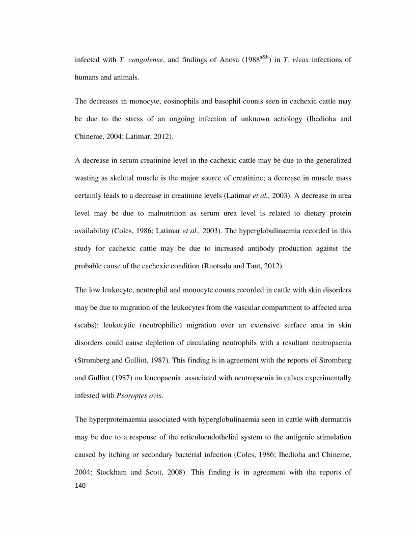
140
infected with T. congolense, and findings of Anosa (1988a&b
) in T. vivax infections of
humans and animals.
The decreases in monocyte, eosinophils and basophil counts seen in cachexic cattle may
be due to the stress of an ongoing infection of unknown aetiology (Ihedioha and
Chineme, 2004; Latimar, 2012).
A decrease in serum creatinine level in the cachexic cattle may be due to the generalized
wasting as skeletal muscle is the major source of creatinine; a decrease in muscle mass
certainly leads to a decrease in creatinine levels (Latimar et al., 2003). A decrease in urea
level may be due to malnutrition as serum urea level is related to dietary protein
availability (Coles, 1986; Latimar et al., 2003). The hyperglobulinaemia recorded in this
study for cachexic cattle may be due to increased antibody production against the
probable cause of the cachexic condition (Ruotsalo and Tant, 2012).
The low leukocyte, neutrophil and monocyte counts recorded in cattle with skin disorders
may be due to migration of the leukocytes from the vascular compartment to affected area
(scabs); leukocytic (neutrophilic) migration over an extensive surface area in skin
disorders could cause depletion of circulating neutrophils with a resultant neutropaenia
(Stromberg and Gulliot, 1987). This finding is in agreement with the reports of Stromberg
and Gulliot (1987) on leucopaenia associated with neutropaenia in calves experimentally
infested with Psoroptes ovis.
The hyperproteinaemia associated with hyperglobulinaemia seen in cattle with dermatitis
may be due to a response of the reticuloendothelial system to the antigenic stimulation
caused by itching or secondary bacterial infection (Coles, 1986; Ihedioha and Chineme,
2004; Stockham and Scott, 2008). This finding is in agreement with the reports of
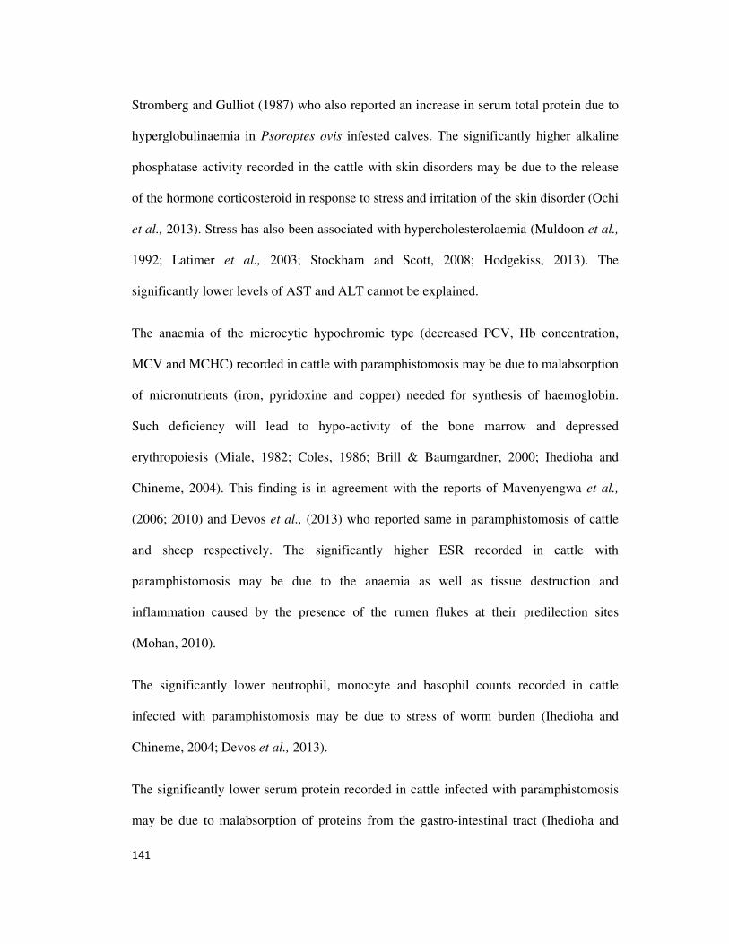
141
Stromberg and Gulliot (1987) who also reported an increase in serum total protein due to
hyperglobulinaemia in Psoroptes ovis infested calves. The significantly higher alkaline
phosphatase activity recorded in the cattle with skin disorders may be due to the release
of the hormone corticosteroid in response to stress and irritation of the skin disorder (Ochi
et al., 2013). Stress has also been associated with hypercholesterolaemia (Muldoon et al.,
1992; Latimer et al., 2003; Stockham and Scott, 2008; Hodgekiss, 2013). The
significantly lower levels of AST and ALT cannot be explained.
The anaemia of the microcytic hypochromic type (decreased PCV, Hb concentration,
MCV and MCHC) recorded in cattle with paramphistomosis may be due to malabsorption
of micronutrients (iron, pyridoxine and copper) needed for synthesis of haemoglobin.
Such deficiency will lead to hypo-activity of the bone marrow and depressed
erythropoiesis (Miale, 1982; Coles, 1986; Brill & Baumgardner, 2000; Ihedioha and
Chineme, 2004). This finding is in agreement with the reports of Mavenyengwa et al.,
(2006; 2010) and Devos et al., (2013) who reported same in paramphistomosis of cattle
and sheep respectively. The significantly higher ESR recorded in cattle with
paramphistomosis may be due to the anaemia as well as tissue destruction and
inflammation caused by the presence of the rumen flukes at their predilection sites
(Mohan, 2010).
The significantly lower neutrophil, monocyte and basophil counts recorded in cattle
infected with paramphistomosis may be due to stress of worm burden (Ihedioha and
Chineme, 2004; Devos et al., 2013).
The significantly lower serum protein recorded in cattle infected with paramphistomosis
may be due to malabsorption of proteins from the gastro-intestinal tract (Ihedioha and
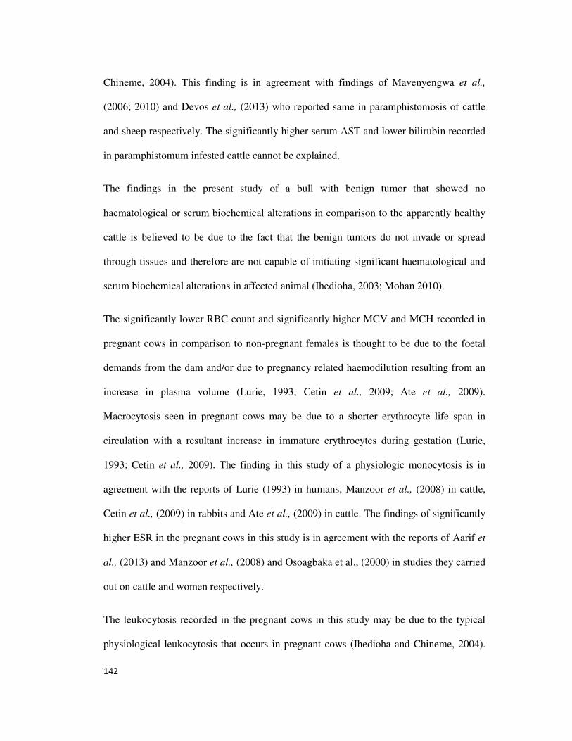
142
Chineme, 2004). This finding is in agreement with findings of Mavenyengwa et al.,
(2006; 2010) and Devos et al., (2013) who reported same in paramphistomosis of cattle
and sheep respectively. The significantly higher serum AST and lower bilirubin recorded
in paramphistomum infested cattle cannot be explained.
The findings in the present study of a bull with benign tumor that showed no
haematological or serum biochemical alterations in comparison to the apparently healthy
cattle is believed to be due to the fact that the benign tumors do not invade or spread
through tissues and therefore are not capable of initiating significant haematological and
serum biochemical alterations in affected animal (Ihedioha, 2003; Mohan 2010).
The significantly lower RBC count and significantly higher MCV and MCH recorded in
pregnant cows in comparison to non-pregnant females is thought to be due to the foetal
demands from the dam and/or due to pregnancy related haemodilution resulting from an
increase in plasma volume (Lurie, 1993; Cetin et al., 2009; Ate et al., 2009).
Macrocytosis seen in pregnant cows may be due to a shorter erythrocyte life span in
circulation with a resultant increase in immature erythrocytes during gestation (Lurie,
1993; Cetin et al., 2009). The finding in this study of a physiologic monocytosis is in
agreement with the reports of Lurie (1993) in humans, Manzoor et al., (2008) in cattle,
Cetin et al., (2009) in rabbits and Ate et al., (2009) in cattle. The findings of significantly
higher ESR in the pregnant cows in this study is in agreement with the reports of Aarif et
al., (2013) and Manzoor et al., (2008) and Osoagbaka et al., (2000) in studies they carried
out on cattle and women respectively.
The leukocytosis recorded in the pregnant cows in this study may be due to the typical
physiological leukocytosis that occurs in pregnant cows (Ihedioha and Chineme, 2004).
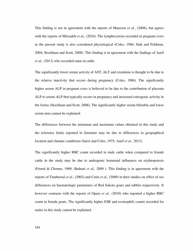
143
This finding is not in agreement with the reports of Manzoor et al., (2008), but agrees
with the reports of Mirzadeh et al., (2010). The lymphocytosis recorded in pregnant cows
in the present study is also considered physiological (Coles, 1986; Sink and Feldman,
2004; Stockham and Scott, 2008). This finding is in agreement with the findings of Aarif
et al., (2013) who recorded same in cattle.
The significantly lower serum activity of AST, ALT and creatinine is thought to be due to
the relative inactivity that occurs during pregnancy (Coles, 1986). The significantly
higher serum ALP in pregnant cows is believed to be due to the contribution of placenta
ALP to serum ALP that typically occurs in pregnancy and increased osteogenic activity in
the foetus (Stockham and Scott, 2008). The significantly higher serum bilirubin and lower
serum urea cannot be explained.
The differences between the minimum and maximum values obtained in this study and
the reference limits reported in literature may be due to differences in geographical
location and climatic conditions (Saror and Coles, 1975; Aarif et al., 2013).
The significantly higher RBC count recorded in male cattle when compared to female
cattle in the study may be due to androgenic hormonal influences on erythropoiesis
(Friend & Chomet, 1969; Shahani et al., 2009 ). This finding is in agreement with the
reports of Tambuwal et al., (2002) and Cetin et al., (2009) in their studies on effect of sex
differences on haematologic parameters of Red Sokoto goats and rabbits respectively. It
however contrasts with the reports of Opara et al., (2010) who reported a higher RBC
count in female goats. The significantly higher ESR and eosinophils counts recorded for
males in this study cannot be explained.
144
The significantly higher serum ALP levels recorded in males in comparism to females
may be due to the fact that males commonly have longer and bigger bones than females
and bone ALP contributes significantly to normal serum ALP (Bain, 2003; Stockham and
Scott, 2008). The significantly higher serum creatinine level recorded in males when
compared to females may also be attributed to the fact that males usually have more
muscle mass in comparison to females; skeletal muscles are the source of creatinine
(Latimer et al., 2003; Stockham and Scott, 2008).
The finding in this present study that age did not significantly influence most of the
haematological parameters (except MCHC and eosinophils counts) is in agreement with
the reports of Opara et al., (2010) who reported same in West African dwarf goats. This
finding however contrasts with the reports of Klinkan and Jezek (2012) who reported
increases in RBC count, Hb concentration and WBC counts with a decrease in MCV with
advancing age in a study carried out on calves, though their study did not include adults.
The findings of a significantly higher serum globulin in adults when compared to the
young cattle in this present study is thought to be due to the fact that with age there is
greater exposure to antigens which will elicit greater antibody formation as
immunoglobulins constitute a large proportion of circulating globulins (Franca et al.,
2011). This finding is in agreement with the reports of Franca et al., (2011) in buffaloes
but contrasts with the reports of Opara et al., (2010) in West African dwarf goats.
5.1. CONCLUSIONS
Among all diseases, disorders and conditions recorded for cattle in this study, fasciolosis
ranked topmost as the commonest disease (8.47%), followed by tuberculosis (1.76%), and
then pregnancy (1.59%).
145
The disease, disorders and conditions recorded in this study were associated with note
worthy haematological and serum biochemical findings which were considered to be of
diagnostic importance.
The haematology and serum biochemistry findings of the apparently healthy cattle in this
study were in most instances comparable to those reported for cattle in available
literature, but some of the minimum and maximum values recorded in this present study
were different from the upper and lower reference limits reported in available literature.
146
REFERENCE
Aarif, O., Ahmed, S., Sheikh, A.A. & Ahmed, N. (2013) Comparison of haematology in
various physiological states in Sahiwal Cattle. Indian Journal of Applied
Research. 3 (9): 134-135.
Abeng, J. N. and Anosa V.O (2005). Serum total proteins and creatinine levels in
experimental Gambian trypanosomosis of vervet monkeys. African Journal of
Biotechnology. 4 (2):187-190
Abenga, J. N., Sauda, S. A., Idowu, T. B., Lawani, F. A. G. (2002). Effects of acute
caprine trypanomosis on haemoglobin, urea and serum electrolytes. African
Journal of Clinical and Experimental Microbiology. 3(1):45-47.
Abubakar, U. B., Aneh, J. I., Abdulkadir, I. A., Salisu, I., Okaiyeto, S. O., Kudi, A. C.
(2011). Bovine tuberculosis in Nigeria: A review. Veterinary Research. 4(1):24-
27.
Adaikpoh, M. A., Onyeneke, E. C., Eviyamemu, G. E., Asagba, S. O., Nwajei, G. E.,
Oyeaburu, S. I. (2004). Changes in some serum and haematological indices in
magnesium and riboflavin deficiency in rats. Pakistan Journal of Nutrition
3(6): 348-352.
Adedokun, O. A, Ayinmode, A. B. and Fagbemi, B. O. (2008) A Comparative study of
three methods of detecting Fasciola infections in Nigerian cattle. Veterinarski
Archiv. 78: 411-416.
Adekunle, O. A., Oladele, O. I., Olukaiyeja, T. D. (2002). Indigenous control methods for
pests and diseases of cattle in Northen Nigeria. Livestock Research for Rural
Development. 14(2)
Aengwanich, W., Chantivatikul, A., Pamok, S. (2009). Effects of seasonal variations on
Hematological values and health monitor of own bred Beef cattle at slaughter
house in north eastern part of Thailand. American – Eurasian J. Agric. &
Environ. Sci. 5(5):644–48
Ajmone–Marsan, P., Garcia, J. F., Leastra, J. A. (2010). On the Origin of cattle: How
aurochs became cattle and colonized the world. Evolutionary Anthropology:
Issues, News and Reviews. 19 (4):148-57
Ajuogu, P. K., Ehine, O. A., Ajayi, F. O. (2011). Comparative study of serum cholesterol
status of dermatophilosis resistant and susceptible (healthy and infected) cattle
breeds in Von Plateau State Nigeria: New Clues in Sciences .1:84-87.
Al-Ani, F. K. (1992). Haematological, serum electrolytes, blood gases and acid-base
values of calves with rinderpest. Iraqi. J. vet. Sci. 5:187-196.
Allain C. C., Poon L. S., Richmond W and Fu P.U. (1974). Enzymatic determination of
the total cholesterol. Clinical Chemistry. 20 : 470-475.
147
Al-Quaraishy, L. O. & Al-Moussawi, A. M.(2011). Effects of Faciola gigantica Infection
on some blood physiological and biochemical aspects of infected cows in Babylon
governorate. http://www.uobabylon.edu. Accessed 14/3/2013.
Alsaad, K.A., Al-Obaidi, Q. T., Hassan, S. D. (2012). Clinical haematological and
coagulative studies of bovine viral diarrhea in local Iraqi calves. Bulgarian
Journal of Veterinary Medicine. 15(1):44-50.
Amin, E. F., Eman, A. A., Nesieen, A. S. & Fagr, A. M. (2011). Field trial for treatment
of coccidiosis in goat kids in Sharkia Governorate. Zag. Vet. J. 39(1): 136-143.
Amin, S., Khan, M.A., Hashmi, H.A., Rashid, J. & Khan, M.S. (1990) Effects of
tuberculosis on certain blood indicies in buffaloes. Pakistan Veterinary Journal.
10: 92-93.
Anderson, J., Barret, T., and Scott, G. R. (1996). Manual of Diagnosis of Rinderpest.
FAO Animal Health Manual- 1. 2nd
ed.
Andrews, A. H., Blowey, R. W., Boyd, H., and Eddy, R. G. (2008). Bovine Medicine :
Diseases and Husbandry of Cattle. John Wiley and sons. Page 718.
Anosa, V. O. (1988b). Haematological and biochemical changes in human and animal
trypanosomosis. Part II. Revue Elevage et de Medicine Veterinaire des Pays
Tropicaux. 4(2) : 151-164.
Anosa, V. O., Logan-Heafrey, L. L. and Wells, C. W. (1997) The haematology of
Trypanosona congolense infection in cattle II. Macrophage structure and function
in the bone marrow of Boran cattle. Comparative Haematology. 7 (1): 23-29.
Anosa, V.O. (1988a) Haematological and Biochemical changes in human and animal
trypanosomosis. Part I. Revue Elevage et de Medicine Veterinaire des Pays
Tropicaux. 41 (1): 67-71.
Anumol, J., Tresamol, P. V., Vinodkumar, K. and Saseendranath, M. R. (2012).
Haematobiochemical alterations in goats infected with coccidiosis. Tamilnadu J.
Veterinary & Animal Sciences. 8(3)163-165.
Ashwani, K. & Gebrehiwot, T. (2011). Bovine cysticercosis in Ethiopia: A review. Ethip.
Vet. J. 15(1):15-35.
Asl, A. N., Ghasrodashti, A. R., Nazifi , S. (2010). Successful management of a case of
downer syndrome in a cross bred cow after 45 days of recumbency. Comparative
Clinical Pathology. 19 (3): 241-242.
Ate, I.U., Rekwot P.I., Nok, A. J. and Tekdek, L. B. (2009) Haematological values of
cows during third trimester of pregnancy and early lactation in settled cattle
herds in Zaria, Northern Nigeria. African Journal of Biomedical Research.
12(3) 225-231.
AU–IBAR (African Union Interafrican Bureau for Animal Resources) (2013). Impact of
Livestock Diseases in Africa. http://www.au-ibar.org/. Accessed 3rd
April 2013.
148
Ayele, W. Y., Neill, S. D., Zinsstagg, J., Weiss, M. G., Pavlik, I. (2004). Bovine
tuberculosis: An old disease but a new threat to Africa. Int. J. Tuberc. Lung. Dis.
8:924-937.
Babara, A. B., Courtney, L. R., Valerie, R. M., Eileen, M. S. and Stephanie, A. B. (2010).
A typical Dermatophylosis congolensis infection in a three year old pony. J. Vet.
Diagn. Invest. 22(1):141-143.
Bain, P.J. (2003) Liver. In: Latimer, K.S., Mahaffey, E.A., Prasse, K.W. : Duncan &
Prasse’s Veterinary Laboratory Medicine: Clinical Pathology. 4th
ed. Ames, Iowa
State Press. Pp 193-214.
Barger, A. M., (2003): The complete blood cell count: A powerful diagnostic tool.
Veterinary Clinics of North America, Small Animal Practice. 33:1207-1222
Barrington, G. M., (ed)(2011). Overview of transport tetany in ruminants. The Merk
Veterinary Manual. http// www.merckmanuals.com. Accessed 10th
june 2013.
Barry, J. D. and McCulloch, R. (2001) Antigenic variation in trypanosomes: enhanced
phenotypic variation in a eukorytic parasite. Advances in Parasitology. 49: 1-70.
Bashady, M. M., Yassein, S, Lotfy., M. M., El-Bahi, M., Mohamed, A. M. and Dessouky,
M. I. (1990) Haematological and serum biochemical profiles in experimental
fasciolosis in sheep. Egyptian Journal of Comparative Pathology and Clinical
Pathology. 3 (2): 357- 374.
Basson, P. A., Pienar, J. C. & Van Der Westhuizen, B. (1970). The pathology of
ephemeral fever: A study of the experimental disease in cattle. J. S. Afr. Vet. Med.
Assoc. 40: 385-397.
Beck M.A. (2001). Selenium deficiency increases the pathology of an influenza virus
infection . The Journal of the Federation of American society of
Experimental Biology. 15, 1481-1483.
Behm, G.A and Sangster, W.C. (1999) Pathology, pathophysiology and clinical aspects.
In: Dalton, J.P. (Ed) Fasciolosis. CAB International publishing, wallinaford UK.
pp 185-224.
Bengaly, Z., Sidibe, I., Ganaba, R., Desquesues, M., Boly, H., Sawadogo, L. (2002).
Comparative pathogenicity of three genetically distinct types of Trypanosoma
congolense in cattle: Clinical observations and haematological changes. Vet
Parasitol. 108(1):1-19.
Berry J.G. (2012) . Livestock Diseases: Cause and Control. http://osufacts.okstate.edu.
Accessed 17 July 2012.
Beyer, W. and Turnbull, P. C. B. (2009) Anthrax in animals: A review. Molecular
Aspects of Medicine. 30(6): 481-489.
Bisalla, M., Ibrahim, N.D.G., Lawal, I.A. & Esievo, K.A.N (2007) Serum total protein,
albumin and globulin ratio in Yankassa sheep experimentally infected with
149
Trypanosome congolense and immunomodulated with levamisole. Journal of
Protozoology Research.17: 39-43.
Blass K.A., Thiebert R.J., and Lam L.K. (1974). A study of the mechanism of Jaffe
reaction. Journal of Clinical Chemistry. 12: 336-343.
Blezinger, S. L. (2001). Small vitamin imbalances can be critical.
http://www.cattletoday.com. Accessed 15/3/2013.
Bock, R., Jackson, L., De Vos, A., Jorgensen, W. (2004). Babesiosis of cattle. Cambridge
University Press. 129:247-269.
Bohnel, H., Schwagerick, B., Gessler, F. (2001). Viceral botulism – A new form of
bovine clostridum botulinum toxication. Journal of Veterinary Medicine. Series
A. 48:373-383.
Boonyayatra, S. (2011). Viral disease in ruminants. http://www.ask.com. Accessed
12/03/2013.
Booth, M. G., Hood, J., Brooks, T. J. & Hart, A. (2010) Anthrax infection in drug users.
Lancet; 375 (99723):1345-1346.
Borsberry, S., (2002). A typical actionbacillosis in a dairy replacement herd. Veterinary
Record. 151(10:)308.
Borst, P., Rudenko, G., Taylor, M. C., Blundell, P. A., VanLeeuwen, F., Bitter, W.,
Cross, M. and McCulloch, R. (1996) Antigenic variation intrypanosomes.
Archives of Medical Research. 27 (3): 379-388.
Bouhroum, N., Bensahli, B., Niar, A. (2012). Evolution of biochemical parameters in post
partum cows affected with Brucella in the Wilaya of Relzane. African Journal of
Biochemistry. 11(7)1818-1822.
Bowland, S. L. & Stephen, P. E. (2000). Bovine respiratory disease: Commercial
vaccines currently available in Canada. Canadian Veterinary Journal. 41:33-48.
Boyd, J. W. (1984). The interpretation of serum biochemistry test results in domestic
animals. Veterinary Clinical Pathology. 13(2):7-14.
Braun, U., Feige, K., Schweizer, G., Pospischil, A. (2005). Clinical findings and
treatment of 30 cattle with botulism. Veterinary Records. 156:438-441.
Brill, J.R. & Baumgardner, D.J. (2000). Normocytic anaemia. Am Fam Physician. 62
(10): 2255–2264.
Britt, A. G., Cotton, C. L., Pitman, I. H. & Sinclair, A. M. (1986). Effects of sheep –
chewing louse (Damalinia ovis) on the epidermis of Australian Merino.
Australian Journal of Biological Sciences. 37:137-143.
Brown, C., & Torres, A. (2008) Eds: USAHA Foreign Animal Diseases. 7th
ed.
Committee of Foreign and Emerging Diseases of the U.S Animal Health
Association. Boca Publications Group, Inc.
150
Çetin, N., Bekyürek, T. & Çetin, E. (2009) Effects of Sex, Pregnancy and Season on
some Haematological and Biochemical Blood Values in Angora Rabbits. Scand.
J. Lab. Anim. Sci. 36( 2): 155-162.
Chaudhry, C. S., Muhammad, K., Zafar, Q., Masood, A. (1999). Haematological and
biochemical disturbances associated with Toxocara vitulorum infection in buffalo
calves. International Journal of Agriculture and Biology. 1(4): 247-249.
Cheville N.F (1988) Introduction to Veterinary Pathology. Iowa State University Press,
Ames.
Choi H.K. (2005). Dairy consumption and risk of type 2 diabetes mellitus in men.
Archives of Internal Medicine, 165: 997-1003.
Clay J. (2004). World Agriculture and the environment. A Commodity by Commodity
Guide to Impacts and Practices . Island Press, Washington D.C. U.S.A.
Coetzer, J. A. W., & Tustin, R. C. (2004) Infectious Diseases of livestock. 2nd
ed. Oxford
University Press.
Coles E.H. (1986). Veterinary clinical pathology 4th
ed. W. B. Saunders. Philadelphia.
Collins, M. T. (2003). Update in paratuberculosis : epidemiology of Johne’s disease and
the biology of Mycobacterium paratuberculosis. Irish Veterinary Journal.56:565-
574.
Colville, J. (2002) Blood chemistry. In: Hendrix, C.M. (Ed), Laboratory Procedures for
Veterinary Technicians. 4th
ed. Mosby Inc. Missouri, USA. Pp 75-103.
Coskun, A., Derinbay, E. O., Guzelbektes, H., Ayodogbu, U., Sen. I. (2012). Acute phase
preoteins, clinical, haematological and biochemical parameters in dairy cattle
naturally infected with Anaplasma marginale. Kafkas Univ. Vet. Fak. Derg. 18(3):
497-502.
Cox, V. S., (1982). Pathogenesis of the downer cow syndrome. Vet Rec. 111 (44): 76-79.
DeeWhitter, W. (2006) Rabies in cattle a continual threat in Virginia. Virginia State
University. http://www.ext.vt.edu/news/. Accessed 24/6/2013.
Dein F.J. (1986) Haematology. In: Harrison G.J, Harrison L.R (Eds) Clinical Avian
Medicine and Surgery. Saunders Philadelphia.
Dennison, A. C., VanMetre, D. C., Callan, R. J., Dinsmore, P., Mason, G. L., and Ellis,
R.P. (2002). Hemorrhagic bowel Syndrome in dairy cattle: 22 cases(1997-2000).
JAVMA. 221(5): 686-689.
Devos, J., Vassiloglou, B., Amenna-Bernard, N., Marcotty, T. (2013) Paramphistomosis
in sheep; natural infection of lambs by Calicophoron daubneyi. Revue Méd.
Vét. 164,(11): 528-535.
151
Dhand, N. D., Sandhu, K. S., Singh, J. & Randhawa, S. S. (2003): Outbreak of
actinobacillosis in dairy cows. Veterinary Record. 15(9):280.
Dickman, D.A., and M.L. Green. (1992). Mycotoxins and reproduction in domestic
livestock. J. Anim. Sci. 70. 1615-1627.
Dima, G., Monea, L., V.Popescu, I., Stihi, C., Badica, T., Olariu, A. (1999). Pixe analysis
of calcium and phosphorous in the downer cow syndrome. International Journal
of Pixe. 9(1&2):35-42.
Dinkan (2012). Therapeutic uses of cow urine (gomutra). http//dinkan53.hubpages.com.
Accessed 14th
June 2012.
Doumas B. T., Perry B. W., Sasse E. A. and Stramfjord Jr. J. R. (1973). Standardization
in bilirubin assays: evaluation of selection methods and stability of bilirubin
solutions. Clinical Chemistry. 19: 984-993.
Doumas B. T., Watson W. A. and Biggs H.G. (1971) albumin standards in the
measurement of serum albumin with bromocresol green. Clinical Chimica Acta.
31: 87-96.
Duncan, J. R and Prasse, K. W. (1986) Veterinary Laboratory Medicine: Clinical
Pathology, 2nd
ed., Iowa State Press, Ames, IA. Pp 44-57, 229-233.
Ebert, D. (1998). Evolution – experimental evolution of parasites. Science. 282.1432-
1435.
Edward, P. W. (1994). The evolution of infectious disease. Oxford University Press.
Oxford.
Elwood P.C. (2005). Milk consumption, stroke and heart attack risk: evidence from the
caerphilly cohort of older men. Journal of Epidermiology and
Community Health, 59: 502-505.
Erdurand, E., Makuloglu, M., Mutlu, M. (2010). A rare haematological manifestation of
Brucellosis: Reactive hemophagocytic syndrome. Journal of Microbiology,
Immunology and Infection. 43(2): 159-162.
Fadiga, M., Jost, C., and Ihedioha, J. (2011). Financial costs of disease burden, morbidity
and mortality from priority livestock diseases in Nigeria: Disease burden and cost
– benefit analysis of targeted interventions. International Livestock Research
Institute (ILRI). Nairobi, Kenya.
Flynn A. (2003). The role dietary calcium and health. Proceedings of the Nutrition
Society, 62: 851-858.
FMANR (Federal Ministry of Agriculture and Natural Resource) (2009) Geographic data.
Federal Ministry of Agriculture and Natural Resource. Enugu. Nigeria.
152
Franca, R. T., Costa, M. M., Martins, D. B., Pagnoncelli, M., Leal, M. L., Mazzanti, C.
M., Palma, H. E., Kunert, C. P., Palm, F. C. and Lopes, S. T. D. (2011) Protein
profile of buffaloes of different ages. Acta Scientiae Veterinariae. 39 (4): 995.
French, M. H. (1934). Variations of serum protein fractions during an attack of rinderpest.
Annual Report Department Veterinary Science and Animal Husbandry. pp 42-48.
French, M. H. (1936). Serum protein changes induced by rinderpest virus. J. comp. path.
Ther. 49: 118-140.
Friend, W. and Chomet, B. (1969) The effect of androgens on erythropoiesis and on the
kidney. Annals of Internal Medicine. 70 (5): 1083-1088.
Ganheim, C. (2004). Studies on the Acute phase reaction during respiratory infections in
calves. Acta Universities Agriculturae Sueciae Veterina. 180: p 25.
Garfield, R. V. (1995) Aurochs. ICZN (International Commission on Zoological
Nomenclature). Conserved Bull. Zool. Nomencl. 60: 81-84.
Garriz, C. A., Gallinger, M. M., Touraille, C., Steffan, P. E., Fiel, C. A., Ambrustulo, R.
R., Bioadani, C. A., Zamorano, M. & Bulman, G. M. (1987). Gastrointestinal
parasitium: Its effects on muscle, fat and bone composition of the carcass and
orgaroleptic characteristics of meat. In: Proceedings MSD Ag Vet symposium on
Gastrointestinal parasitism. August 19, 1987. PP 59-68.
Ghanem, M. M., & Abd El – Raof, Y. M. (2005). Clinical and haematobiochemical
studies on lamb coccidiosis and changes following amprolium and
sulphadimthoxine therapy. Benha Vet. Med. J. 16(2): 286-300.
Gibbons J.W. (1963). Diseases of Cattle. American Veterinary Publications, Inc, 2nd
ed.
United States of America.
Gibson, C. M. (2013). Hypophosphatemia.
http//www.wikidoc.org/index.php/hypophosphatemia. Accesses 12th
june 2013.
Gokce, G., Gokce, H. I., Gunes, V., Erdogan, H. M., Citil, M., (2004). Alterations in
some haematological and biochemical parameters in cattle suffering from foot-
and-mouth disease. Turk. J. vet. Anim. Sci. 28:723-727.
Gracey, F. J & Collins, S. D. (1992). Meat Hygiene. 5th
ed. Bailliere Tindall. London.
Green, C. E. (1984a). Host–microbe interactions. In: Green C.E (eds): Clinical
Microbiology and Infectious Diseases of Dogs and Cats. W.B. Saunders
Company. Philadelphia. Pp 67-94.
Green, C. E. (1984b). Antimicrobial resistance of the host. In: Green C. E. (eds): Clinical
Microbiology and Infectious Diseases of Dogs and Cats. W. B. Saunders
Company. Pp 35- 66.
Grubb P. (2005) Bos taurus primigenus. In: Wilson D.E and Reeder D.N (eds): Mammal
Species of the World: A taxonomic and Geographic Reference, 3rd
ed.
John Hopkins University press Baltimore, vol. 2. Pp. 2142.
153
Gutierrez – DeLaw, J. H., Warnick, A. C., Carley, J. J. and Hentages, Jr. J. F. (1971).
Environmental physiology in the sub-tropics .I. Effects of continuous
environmental stress on some hematological values of beef cattle. Journal of
Animal Science. 32:969-973.
Hamid, M. E. and Musa, M. S. (2009). The treatment of bovine dermatophilosis and its
effects on some haematological and blood biochemical parameters. Science and
Technical Review of the office International des Epizooties.28(3): 1111-1118.
Hammond, J. A. and Sewell, M. M. H. (1990) Diseases caused by helminthes. In: Sewell,
M. M. H. and Brocklesdy, D. W. (Eds), Handbook of Animal Diseases in the
Tropics. 4th
ed. College of Tropical Veterinary Medicine, Edinburgh
University. pp 119-123.
Hanotte, O., Tawa, C. L., Bradley, D. G., Okano, M., Vergee, Y., Ochieng, J., and Rege,
J. E. O. (2000). Geographic distribution and frequency of taurine Bos Taurus and
an indicine Bos indicus Y specific allele amongst sub-saharan African cattle
breeds. Molecular Ecology. 9(4):387-396.
Harr, K.E. (2002). Clinical chemistry of companion avian species – A review. Veterinary
Clinical Pathology, 31:140-151.
Heider, L. C., McClure, J. T., Leger, E. R. (2001). Presumptive diagnosis of Clostridium
botulinum type D intoxication in a herd of feedlot cattle. Can. Vet. J. 42:210-212.
Heuschele, W. P. & Barber, T. L. (1966). Changes in certain blood components of
rinderpest infected cattle. Am. J. vet. Res. 27:1001-1006.
Higgins, T., Beutler, E. & Doumas, B.T. (2008a) Measurement of haemoglobin in blood.
In: Burtis, C.A., Ashwood, E.R. & Bruns, D.E. (Eds). Tietz Fundamentals of
Clinical Chemistry. 6th
ed. Saunders Elsevier, Missouri. Pp 514-515.
Higgins, T., Beutler, E. & Doumas, B.T. (2008b) Bilirubin. Analytical Methodology-
Serum bilirubin. In: Burtis, C.A., Ashwood, E.R. & Bruns, D.E. (Eds). Tietz
Fundamentals of Clinical Chemistry. 6th
ed. Saunders Elsevier, Missouri. Pp 524-
525.
Hirst, K. K. (2012). Cattle (Boss spp). http://archeology.about.com. Accessed 12th
Feb
2013
Hodgekiss, A. (2013). A stressful job realy can kill you by raising your cholesterol level.
http://www.dailymail.co.uk/health. assessed 5/2/2014.
Hofmann-Lehmann, R., Meli, M. L., Drehver, U. M., Gonczi, E., Deplazes, P., Braun, U.,
Engels, M., Schupbach, J., Jorger, K., Thoma, R., Groit, C., Stark, K. O. C., Willi,
B., Schmidt, J., Kocan, K. M., and Lutz, H. (2004). Concurrent infections with
Vector-borne pathogens associated with fatal hemolytic anemia Journal of in a
cattle herd in Switzerland. Clinical Microbiology. 42(8):3775-3780.
154
Holmes, P. H, Eisler, M. C. and Geerts, S. (2004). Current Chemotherapy of animal
trypanosomosis. In: Maudlin, I., Holmes, P. H. and Miles, M. A. (Eds). The
Trpanosomiases, CAB international, Wallingford. pp 413-444 .
Holst, H., and Svensson, C. (1994). Changes in blood composition of calves during
experimental and natural infections with Eimeria alabamensis. Research in
Veterinary Science. 7(3):377-383.
Horst, R.L., Goff, J. P., Reinhardt, T. A., and Buxton, D. R. (1999). Strategies for
preventing milk fever in dairy cattle. J. Dairy. Sci. 80: 139-141.
Huss–Ashmore, R. & Curry, J. J. (1992). Impact of improved livestock disease control on
house hold diet and welfare: A story in Uasin Gishu District, Kenya: In:
International Laboratory Research for Animal Diseases (ILRAD) Technical
Report. No 2: Nairobi, Kenya. Page 97.
Hussein, A. H., Mohammed, N. A.– E.– S., & Mohammed, H. K. (2007). Theileriosis and
Babesiosis in cattle: Haemogram and some biochemical parameters. ISAH Tartu.
Estonia. 143-150.
Igbal, U. M. & Sajid, M. S. (2005). Phosphorous deficiency in dairy animals.
http://www.achives.dawn.com. Accessed 15/3/2013.
Ihedioha, J. I. (2003). Basic Principles of Generals Pathology. Nsukka, Nigeria: AP
Express Publishers Limited, Nigeria.
Ihedioha, J. I., and Chineme C.N. (2004) Hematopoietic System. In: Fundamentals of
Systematic Veterinary Pathology Vol. 1 Great AP Express Publishers
Limited, Nigeria, pp 107-160.
Ihedioha, J. I., Okafor C., and Ihedioha T.E. (2004). The hematological profile of the
Sparague –Dawley outbred albino rats in Nsukka, Nigeria. Animal Research
International, 1:125-132.
Ihedioha, J. I., Ugwuja, J. I., Noel-Uneke, O. A., Udeani, I. J., & Daniel-Igwe, G. (2012).
Reference values for the haematology profile of conventional grade outbred
albino mice (Mus musculus) In Nsukka, eastern Nigeria. Animal Research
International. 9(2): 1601-1612.
Jackson, P.G.G & Cockcroft, P.D. (2002) Clinical examination of farm animals.
Blackwell Science Ltd, Oxford. Pp 302-305.
Jain, N. C. (1993). Essentials of Veterinary Hematology. Lea & Febiger. Philadelphia.
PP1- 18
Jain, N. C. (2002). Schlam’s Veterinary Haematology. Lippincott Williams & Wilkins.
Philadelphia. Baltimore. New York, London. Buenos Aires. Hong Kong. Sydney.
Tokyo.
155
Javed, M. T., Usman, M., Irfan, M., Cagiola, M. (2006). A study on tuberculosis in
buffaloes: Some epidemiological aspects along with haematological and serum
protein changes. Veterinarski Arhiv. 76(3):193-206.
Jean , D., Fecteau, G., Scott, D., Higgins, R. and Quessy, S. (1995). Clostridium
botulinum type C intoxication in feedlot steers being fed ensiled poultry litter.
Can. Vet . J. 36: 626-628.
Johnson, A.M. (2008) Amino acids and proteins. In: Burtis, C.A., Ashwood, E.R. &
Bruns, D.E. (Eds). Tietz Fundamentals of Clinical Chemistry. 6th
ed. Saunders
Elsevier, Missouri. Pp 206-316.
Kaneko, J. J., Harvey, J. W., Bruss, M. L. (1997). Clinical Biochemistry of Domestic
Animals. Academic Press, Inc. San Diego, London, Boston, New York, Sydney,
Tokyo, Toronto.
Kennedy, S., Rice, D. A., Davidson, W. B. (1987). Experimental myopathy in Vitamin E
and Selenium depleted calves with and without added dietary polysaturated fatty
acids as a model for nutritional degenerative myopathy in ruminant cattle. Res.
Vet. Sci.43(3):384394.
Khan, M. A. (ed) (2005). Calcium deficiency. Merck Veterinary Manual. 9th
ed. Merck
and Co., Inc. Whitehouse Station.
Klinkan, M. and Ježek, J. (2012). Values of Blood Variables in Calves, A Bird's-Eye
View of Veterinary Medicine, Dr. Carlos C. Perez-Marin (Ed.), ISBN: 978-953-
51-0031-7, InTech, Available from: http://www.intechopen.com/books/a-bird-s-
eye-view-of-veterinary-medicine/values-of-blood-variables-in-calves.
Knell, R. J. (2003). Syphilis in renaissance Europe: Rapid evolution of an introduced
sexually transmitted disease. The Royal Society. London B (suppl). 271:174-176.
Knell, R. J. (2006). Evolution of Virulence: New gene, new disease. Heredity. 97:315.
Kommisurd, E., Osteras, O., Voton, T. (2005). Blood Selenium association with health
and fertility in Norwegian dairy herds. Acta Veterinaria Scandinavica.
46(4):229-240.
Krimer, P.M. (2011) Generating and interpreating test results: Test validity, quality
control, reference values and basic epidermiology. In: Duncan & Prasse’s
Veterinary Laboratory Medicine. Clinical pathology. 5th
Ed (Latimer, K.S.
Ed). Wiley-Blackwell, Iowa. Pp 365- 382.
Krupakaran, R. P., Porcheziyan, T. and Sivseelan, S. (2009) Biochemical and
haematological profile of foot and mouth disease affected crossbred cows in Karur
district of Tamil Nadu. Veterinary Practitioner. 10(1):37-38.
Krynski S., Kedzia W., and Kaminska M. (1964). Some difference between staphylococci
isolated from pus and from healthy carriers. Journal of Infectious Diseases,
114:193-197
156
Kumar, G.S., Lyre, P.K.R., Prassad, M.C. & Sarma, A.K. (1994) Tuberculosis in cattle.
Haematological studies. Indian Journal of Veterinary pathology. 38: 20- 42.
Kvasnicka, B., and Kysl, L. J. (2010). Grass tetany in beef cattle.
http//www.iowabeefcentre.org. Accessed 10th
june 2013.
Lamb, E.J & Price, C.P. (2008) Creatinine, urea and uric acid. In: Burtis, C.A., Ashwood,
E.R. & Bruns, D.E. (Eds). Tietz Fundamentals of Clinical Chemistry. 6th
ed.
Saunders Elsevier, Missouri. Pp 363-372.
Larson S.C., Bergkvist L., and Wolk A. (2005). High-fat dairy food and conjugated
linoleic acid intakes in relations to colorectal cancer incidence in the Swedish
mammography cohort. American Journal of Clinical Nutrition, 82; 894-900.
Latimer, K. S. (2012). Duncan and Prasse’s Veterinary Laboratory Medicine: Clinical
Pathology. 5th
ed. John Wiley and Sons. Hoboken.
Latimer, K. S., Mahaffey, E. A. and Prasse, K. W. (eds.) (2003). Duncan and Prasse’s
Veterinary Laboratory Medicine: Clinical Pathology, 4th
ed., Iowa State Press,
Ames, IA.
Lotfollalizadeh, S., Mohri, M., Ranjbar Bahadari, S.H., Mokhbar Dezfouly, M. R. &
Tajik, P. (2008) The relationship between normocytic, hypochromic anaemia and
iron concentration together with hepatic enzymes in cattle infected with
Fasciola hepatica, Journal of Helminthology. 82 (1) 85-88.
Lucey, D. R. (2007). Anthrax. In: Goldman, L., Ausiello, D. (eds). Cecil Medicine. 23rd
ed. Saunders Elsavier. Philadelphia.
Lurie, S. (1993) Changes in age distribution of erythrocytes during pregnancy: a
longitudinal study. Gynecol Obstet Invest. 36. 141-144.
Malone, F. (2004). Clostridial diseases of cattle and sheep. Disease Surveillance and
Investigation Branch, Veterinary Science Division, department of Agriculture and
Rural Development. Northern Ireland.
Manzoor, R.M., Zahoor, A.P., Saleem, I., Javeed, I.A.B., Pal, M.A & Manzoor, A.K.
(2008) Haemato-biochemical indicies of crossbred cows during different stages of
pregnancy. Interntional Journal of Diary Science. 3(3) 154-159.
Marques, S. M. T. and Scroferneker, M. L. (2003) Fasciola hepatica infection in cattle
and buffaloes in the state of Rio Grande do Sul, Brazil. Parasitology Latinoam.
58: 167-172.
Mashish, M. S. K. (2007). My cow is unable to stand up. Department of Agriculture.
South Africa. PP1-6.
Matanovic, K., Severin, K., Martinkovic, F., Simpraga, M., Janicki., C. &Barisic, J.
(2007) Haematology and serum biochemistry changes in organicaly farmed sheep
naturally infected with Fasciola hepatica. Journal of Parasitological Research.
100(6) 1463-1731.
157
Maunsell, F. P. (2007). Mycoplasma bovis infection in dairy calves. University of
Florida. U.S.A. PP 40-45.
Maurer, F. D., Jones, T.C., Easterday, B., Detray, D. E. (1956). The pathology of
rinderpest. Proceedings of the 92nd
Annual meeting of the American Veterinary
Medical Association, Minneapolis, MN, U.S.A., 1955.. Chicago, K, U.S.A,
American Veterinary Medical association. P. 201-211.
Mavenyengwa, M., Mukaratirwa, S., Monrad, J. (2010): Influence of Calicophoron
microbothrium amphistomosis on the biochemical and blood cell counts of cattle.
J. Helminthol. 2010, 84, 355-361.
Mavenyengwa, M., Mukaratirwa, S., Obwolo M. & Monrad, J. (2006).Observations on
mass production of Calicophoron microbothrium metacercariae from
experimentally and naturally infested Bulinus tropicus. Onderstepoort Journal
of Veterinary Research. 73:95–100.
McDonald, K. (2011). Thirty cattle breeds described. http://www.bigpictureagriculture
/06/thirty-cattle-breeds-described. Html. Accessed 1/2/2013.
Miale, J.B. (1982). Laboratory Medicine: Hematology. (6th ed.) The CV Mosby
Company, St. Louis.
Michel, A. (2011) – Bovine tuberculosis. University of Pretoria.
Mirzadeh, K.H., Tabalebaei, S., Bojarpour, M. & Mamoei, M. (2010) Comparative study
of haematological parameters according to strain, age, sex, physiological status &
season in Iranian cattle. Journal of Animal and Veterinary Advances. 9 (16) 2123-
2127.
Mohan, H. (2010). Text Book of Pathology. 6th
ed. Jaypee Brothers Medical Publishers
Limited. PP 284-342.
Molina, E. C., Gonzao, E. A. and Lumbao, L. A. (2005) Prevalence of infection with
Fasciola gigantica and its relationship to carcasse and liver weights, and fluke and
egg gounts in slaughter cattle and buffaloes in Southern Mindanao, Philipines.
Tropical Animal Health and Production. 37: 215-221.
Molina, E.C., Skerratt, L.F. & Campbell, R. (2008) Pathology of fasciolosis in large
ruminants. In; Overcoming liver fluke as a constraint to ruminant production in
South- East Asia. ACIAR monograph series, Australian Centre for International
Agricultural Research (ACIAR), Canaberra, ACT Australia. Pp 94-98.
Morris, R. S. & Marsh, W. E. (2013). Relationship between infections, diseases and their
economic effects. Food and Agriculture Organisation (FAO). http://www.fao.org.
Accessed 3rd
April 2013.
Muldoon, M. F. Bachen, E. A., Manuck, S. B, Waldstein, S. R., Bricker, P. L and
Bennett, J. A. (1992) Acute cholesterol responses to mental stress and change in
posture. Archives of Internal Medicine. 152 (4): 775-780.
158
Munene, J. N. (1984). Pathogens and pathology of heartwater (cowdriosis) in sheep and
goats. A thesis submitted in part fulfillment for the degree of master of science in
veterinary medicine of the university of Nairobi, Kenya. pp 1-8.
Nahed, S. T. (2010) Investigation of serum insulin and cotisol concentrations in foot and
mouth disease-infected cattle in relation to changes in serum biochemical
variables and protein electrophoretic fractionation profile. Global Veterinaria. 4
(5): 450-455.
Naser, A. S., Globrial, G., Romero, C., Valentine, J. F. (2004). Culture of Mycobacterium
avium subspecies paratuberculosis from the blood of patients with Chron’s
disease. The Lancet. 364:1039-1044.
Nashiruddullah, N., Darzi, M. M., Kamil, S. A., Mir M. S. and Peer, F. (2004).
Comparative efficacy of fine needle aspiration and incicional biopsies in the
diagnosis of bovine actinomycosis. Vet. Archive. 74:443-452.
Nazifi, S., Rezakhami, A., Kcohimoghadam, M., Ansari – Lavi, M., Esmailnezhad, Z.
(2008). Evaluation of serum haptoglobin in clinically healthy cattle and cattle with
inflammatory disease in Shiraz, a tropical area in Southern Iran. Bulgarian
Journal of Veterinary Medicine. 11(2):95-101.
Ndlov, L. (2010). The role of foods of animals origin in human nutrition and health. In :
Swane Poel F., Stroebel A., & Mayos (eds) : The Role of Livestock in
Developing Communities: Enhancing Muiltifuntionality. UFS and CTA,
Bleomfontein South Africa, pp. 77-92.
Neame, P. B., Kelton, J. G. and Walker, I. R. (1980) Thrombocytopaenia in septicemia:
The role of disseminated intravascular coagulation. Blood. 56: 88-92.
Neumann C., Harris D.M., & Rogers L.M. (2002). Contributions of animals source food
in improving diet quality and functions in children in the developing
world. Nutrition Research, 22:193-220.
Nweze, B. O., Ekwe, O. O., Aluka, S. O. and Omeje, S. I. (2012): Productivity of two
indigenous Nigerian cattle breeds and their crossbred under range grazing
management. World Journal Life Science and Medical Research. 2: 1-7.
O’Reilly, C. E., O’Conor, L., Anderson, W. (2004). Surveillance of bulk raw and
commercially pasteurized cow milk from approved Irish liquid + milk
pasteurization plants to determine the incidence of Mycobacterium
paratuberculosis. Applied and Environmental Microbiology. 70: 5138-5144.
O’Reilly, L. M. & Daborn, C. J. (1995). Epidemiology of Mycobacterium bovis infection
in animals and man : A review. Tubercle Lung Dis. 76:1-46.
Ochi, T., Nishiura, I., Tatsumi, M., Hirano, Y., Yahagi, K., Sakurai, Y., Sudo. Y.,
Koyoama, H., Hagita, Y., Fugimoto, Y., Kitamura, S., Hashimoto, M., Nishina,
N. (2013) Effects of transport stress on serum alkaline phosphatase activity in
beagle dogs. Experimental Animals. 6 (4): 329-332.
159
Ochman, H., Lawrence, J.G & Grolsman, E. A. (2000). Lateral gene transfer and the
nature of bacterial innovation. Nature. 405: 299-304.
Oetzel, G. and Berger, L. L. (1985) Protein-energy related malnutrition in domestic
ruminants. Part 1. Predisposing factors and pathophysiology. Comp. Cont. Educ.
Pract. Vet. 7: 672-679.
OIE (2009a) Trypanosomosis (TseTse-transmitted). http://www.oieint/international-
standard-setting/terrestrial-manual. Accessed 12/03/2013.
OIE (2009b). Heartwater – cowdriosis. http://www.oie.int/international-standard-
setting/terrestrial-manual. Accessed 13/3/2013.
Okey, I.C., Egbu, F.M.I., Ubachukwu, P.O., Okafor, F.C. (2013) Biochemical alterations
due to bovine fasciolosis. International Journal of Scientific Research. 2 (11):
2277-8179.
Olivia, V.M., Cezario, G.A.G., Cocato, R.A. & Marcondes-Machado, J. (2008)
Pulmonary tuberculosis: Haematology, serum biochemistry and the relation with
the disease duration. J. Venom. Anim. Toxins incl. Trop. Dis. 14 (1): 71-81.
Omnia, M. K., Soad, M. N., Somia, A. N., El-Metanawey, T. M., Abo El-Aziz, M. H. N.
& Nadia, M. T. A. (2012). Serological and biochemical studies on cattle naturally
infected with Taenia Saginata cysticercosis. Global Veterinaria. 9(5):571-579.
Omofema O.S. (2007). Nigeria’s Agriculture sector: Opportunities in Nigerians
Agriculture sector. http://NigeriaBusinessInfo.com. Accessed July 14th
2012.
Opara, M.N., Udevi, N. and Okoli, I.C. (2010) Haematological Parameters And Blood
Chemistry Of Apparently Healthy West African Dwarf (Wad) Goats In Owerri,
South Eastern Nigeria. New York Science Journal. 3(8) 69-72.
Osoagbaka, O.U, Haruna-Rashid, H. and Anokwuru, O.C. (2000) Observation on some
haematological parameters of Nigerian women during pregnancy. J. Med. Invest.
Practice. 1. 45-48.
Otte, M. J., Nugent, R. & McLeod, A. (2004). Transboundry animal disease: Assessment
of socio-economic impacts and institutional responses. Food and Agriculture
Organization – Livestock Information and Policy Branch, AGAL. Livestock
policy discussion paper No 9. PP 7.
Ozkan, C., Yildirim, S., Kaya, A. (2011). Clinical coenurosis (Coenurus cerebralis) and
associated pathological findings in a calf. Pakistan Veterinary Journal. 31(x):
xxx.
Ozmen, O., Sahinduran, S., Haligur, M., Serzer, K (2005). Clinicopathologic observations
on Coenurus Cerebralis in naturally infected sheep. Schweiz Arch. Tierheilkd.
147(3):129-134.
160
Padilla, S., Bouda, J., Quiroz – Rocha, G. F., Davalos, J. L., and Sanchez, A. (2000).
Biochemical and haematological values in venous blood of Captive Red Deer
(Cervus elephus) at high attitude. Act Vet. Brno. 69:327-331.
Palmer B. (2012) Beef: its what for dinner and detergent, and explosives, and floor wax.
What are cows used for besides meat and milk. http://mobile.slate.com/articles/.
Accessed April 25th
2012.
Parish, S. (2012). Milk fever: calcium and hypocalcemia in dairy cows.
http://www.probioticsmart.com. Accessed 15/3/2013.
Pavlata, L., Pechova, A., Illek, J. (2001). Muscular dystrophy in dairy cows following a
change in housing technology. Acta Vet. Brno. 70: 269–275.
Payne, W. J. A. (1990). Cattle husbandry. In: Introduction to Animal Husbandry in the
Tropics. 4th
edition. Rigman. p 42.
Peinado, V. I., Celdran, J. F., Palomeque, J. (1999). Basic hematological values in some
wild ruminants in captivity. Comparative Biochemical and Physiology part A.
124:199-203.
Peters, D. (2009) Rabies in cattle: Symptoms and prevention.
http://www.helium.com/items. Accessed 27th April 2013.
Piccione, G., Casella, S., Lutri, L., Vazzana, I., Ferrantelli, V., Caola, G. (2010).
Reference values for some haematological, haematochemical and electrophoretic
parameters in the Givgentana goat. Turk .J. Vet. Anim. Sci. 34(2):197-204.
Porco T.C., ILoyd-Smith J.O., and Galvani A.P. (2005). The effect of treatment on
pathogen virulence. Journal of Theoretical Biology, 233: 91-102.
Proano-Perez, F., Rigouts, L., Brondt, J., Dorny, P., Ron, J., Chavez, M., Rodriguez. R.,
Fissette, K., Aerde, A. V., Portaels, F. and Benitez-Ortiz, W. (2006) Preliminary
observations on Mycobacterium spp in Dairy cattle in Ecuador. American Journal
of Tropical Medicine. 75 (2): 318-323.
Qamar, M. F. and Maqbool, A. (2012) Biochemical studies and sero-diagnosis of
Haemonchosis in sheep and goats. The Journal of Animal and Plant Sciences. 22
(1): 32-38.
Radostits, O. M. (1964). Clinical diagnosis of rabies in cattle. Canadian University
Journal. 5(4)76-83.
Radostits, O. M., Gay, C. C., Blow, D. C., Hinchelifit, K. W. (2002) Veterinary
Medicine: A Text Book of Diseases of Cattle, Sheep, Pigs, Goats and Horses. 9th
ed. Harcourt Publisher’s Limited. London.
Raji M.A., Salami S.O., and Ameh J.A. (2010) Pathological conditions and lesions
observed in slaughtered cattle in Zaria abattoir. Journal of Clinical
Pathology and Forensic Medicine, 1; 9-12
161
Rao, V.N.A., Radmadas, P. and Dhinakran, M. (1992) a study on the effect of
tuberculosis on body weight and haemogram values of cattle. Cheiron. 21: 19-22.
Reece, W.O., (1997). Physiology of domestic animals. Williams & Wilkins. New York.
Regoes, R. R., Nowak, M. A. & Bonhoeffer, S. (2000). Evolution of virulence in a
heterogenous host population. Evolution. 54(1):64-71.
Reitman S. and Frankel S. (1957). A colorimetric method for the determination of serum
glutamic oxaloacetic and glutamic pyruvic transaminase. American Journal of
Clinical Pathology. 28: 56-62.
Richardson, E. K. B. & More, S. J. (2009). Direct and indirect effects of Johne’s disease
on farm and animal productivity in an Irish dairy herd. Irish Veterinary Journal.
62:526-532.
Rifai, N., Waenick, G.R. & Remaley, A.T. (2008) Lipids, lipoproteins, apolipoproteins
and other cardiovascular risk factors. In: Burtis, C.A., Ashwood, E.R. & Bruns,
D.E. (Eds). Tietz Fundamentals of Clinical Chemistry. 6th
ed. Sounders
Elsevier, Misseuri. Pp 402-430.
Robert, N.S. & Hadar, I. (1981) Fasciolosis: is the anaemia caused by hematophagia?.
The Journal of Parasitology. 67 (6) 886-892.
Roper, N. (1999): Erthrocytes. In: Pocket medical dictionary. Churchill Livingstone.
Edinburgh, London, Melbourne and New York. 14th
ed. PP100.
Roubies, N.,Panaisis, N., Fytinou, A., Katsoulos, P. D., Giadinis, N., Karatzias, H.
(2006). Effects of age and reproductive stage on certain serum biochemical
parameters of Chios sheep under Greek rearing conditions. Journal of
Veterinary Medicine .A. Physiol. Pathol.clin.med. 53:277-281
Ruotsalo, K. and Tant, M. S. (2012). Serum biochemistry. http://www.vca
hospitals.com/main/pet-health. Accessed 2/2/13.
Sahal, M., Altintas, A., Arslan, H. H., Ural, K. and Aksoy, E. (2004) Serum hepatitis
associated with administration of tetanus toxin in serum producing horses and
therapy. Revue Med. Vet. 155(10):476-482.
Samad, M. A., Islam, K. A., Hossain, M. T., Saha, S. (2003). Haemato-biochemical
changes and antibiotic sensitivity to Escherichia coli associated with
concurrent enteric and septicaemic infection in calves. Bangl. J. Vet. Med.
1(1):39-43.
Santos, R. L., Tsolis, R. S., Baumler, A. J., Adams, L. G. (2002). Haematological and
serum biochemical changes in Salmonella ser typhimurium -infected calves. Am.
J. vet. Res. 63(8):1145-1150.
Sarma, K., Saravanan, M., Mandal, D. B., De U. K., Kumar, M. (2012) Influence of
natural infection of Toxocara Vitulorum on markers of oxidative stress in Indian
buffalo calves. Indian Journal of Animal Science. 82(10).
162
Saror, D. I. and Coles, E. H. (1975). Haematological parameters of zebu cattle under
native husbandry practices. J. Nig. Vet. Med. Assoc., 4 (2): 89 – 92.
Saror, D.I. (1980) Observation of the course and pathology of Trypanosoma vivax in red
Sokoto goats. Research in Veterinary Science. 28: 36-38.
Schalm, O. W., Jain, N. C., Carroll, E. J. (1975). Veterinary Hematology. 3rd
ed. Lea and
Febiger. Philadelphia.
Senturk, S. & Cihan, H. (2007). Outbreak of botulism in dairy herd in turkey. Irish
Veterinary Journal. 60(8):481-484.
Senturk, S., Metcitoglu, Z., Ulgen, M., Borum, E., Temizel, E. and Kasap, S. (2009).
Evaluation of serum iron and iron binding capacity of cows with paratuberculosis.
Tierarztliche Praxis. Ausgabe G, Grosstiere/Nutziere. 379(6):375-378.
Shahani, S., Brago-Bosaria, M., Maggio, M. and Bosaria, S. (2009) Androgens and
erythropoiesis: past and present. Journal of Endocrinological Investigation. 32
(8): 704-716.
Shapouri H (2002). The energy balance of corn ethanol: An update. USDFA
Agricultural Economics Report 814.
Sharifiyazdia, H., Nazifi, S., Nikseresht, K., Shahriari, R. (2012). Evaluation of serum
amyloid A and haptoglobin in dairy cows actually infected with brucellosis.
Journal of Bacteriology and Parasitology. 3:157.
Sheikh, G.N., Qadir, S.G.J., Willayat, M.M. and Gunjan, D. (2006) Biochemical profile
of cattle naturally infected with Fasciola gigantica and F. hepatica. Journal of
Veterinary Parasitology. 20 (1) 41-43.
Shetter, M., Nalini, T.S., Anjan Kumar, K.R., Ravikumar P., & Azeemulla., H.R. (2011)
Haematological and biochemical studies in tuberculine test positive reactors.
International Journal of Pharma and Bio Sciences. 2 (4) 16-22.
Shirma, G. M., Kazwala, R. R. and Kambarage, D. M. (2003) Prevalence of bovine
tuberculosis in cattle in different farming systems in the eastern zone of
Tanzania. Preventive Veterinary Medicine. 57: 167-172.
Shitaye, J. E., Tsegaye, W., Pavlik, I. (2007). Bovine tuberculosis infection in animal and
human populations in Ethiopia: A review. Vet. Med. 52:317-332.
Silvia, D. G., Silva, P. R. L and Fagliari, J. J. (2011) Serum protein concentrations,
including acute phase proteins, in calves experimentally infected with Salmonella
dublin. Pesquisa Veterinaria Brasileira. 31 (7): 551-554.
Singh, K. P., Pavihor, N. S., Charan, K. and Tripatti, B. N. (1993) Haematological and
biochemical alterations in hill bulls infected with Clostridium chauvoei. Acta.
Vet. Brno. 62:89-94.
163
Sink, S. A. & Feldman, B.F. (2004). Laboratory urinalysis and hematology for the small
animal practitioners. Teton New Media. U.S.A
Smith, B. P., Habasha, F., Reina-Guerra, M. and Hardy, A. J. (1979) Bovine
salmonellosis: experimental production and characterization of the disease in
calves, using oral challenge with Salmonella typhimurium. Am J Vet Res.
40(11):1510.
Smith, G. W. (2011) Overview of Actinobacillosis. The Merck Veterinary Manual.
http://www.merckmanuals.com. Accessed 6/4/2013.
Soler, J. J., Moller, A. P. & Soler, M. (1998). Mafia behaviour and evolution of
facultative virulence. Journal of Theoretical Biology. 191.267-277.
Stockham S.L., and Scott M.A. (2008). Fundamentals of Veterinary Clinical Pathology,
2nd
ed. Blackwell Publishing, Iowa, U.S.A.
Stromberg, P.C. & Guillot, F.S. (1987) Hematology in the Regressive Phase of Bovine
Psoroptic Scabies. Vet. Pathol. 24:371-377.
Suchandan, S., Rahman, S. M. M., Alim, M. A., Shubhagata, D. (2012) Haematological
variations in brucella abortus antibody positive cross-bred cattle at Chittagong,
Bangladesh. Yuzuncu Universities Veteriner Fakultesi Dergisi. 23:125-128.
Sulaiman, E. G., Arslan, S. H., Al–Obaidi, O. T., Daham, E. (2010). Clinical,
haematological and biochemical studies of babesiosis in native goats in Mosul.
Iraqi Journal of Veterinary Sciences. 24(1):31-35.
Suttle, N. F. (1992). Trace elements disorders. In: Bovine Medicine: Diseases and
Husbandry. Andrews A. H, Blowey R. W, Boyd H, Eddy L. G (eds). Blackwell
Scientific Publications Limited. Oxford. UK:261-276.
Swai , E., Mwezimpya, I., Ulicky, E., Mbaise, A. and Moshy, W. (2013). An abbatoir
survey of contagious pleuropneumonea lesions in slaughtered cattle in selected
districts in northern Tanzania. African Pacific Journal of Tropical Biomedicine.
3(4): 303-306.
Sykes, A. R., Coop, R. L. & Angus, K. W. (1977). The influence of chronic Ostertagia
circumcincta infection on the skeleton of graving sheep. Journal of Comparative
Pathology. 87:521-529.
Sykes, A. R., Coop, R. L. & Rushton, B. (1980). Chronic subclinical facioliasis in sheep:
effects on food intake, food utilization and blood constituents. Research in
Veterinary Science. 28:63-70.
Taimur, M.J.F.A., Halder, A.K., Chowdhury, S.M.Z.H., Akhter, N, Islam, M.S., Komal,
A.H.M. & Islam, K.S. (1993) Haematological studies on cattle exposed to
Fasciola gigantica infestation. Asian-Australian Journal of Animal Sciences. 6 (2)
301-303.
164
Taiwo, V.O, Olaniyi, M.O. & Ogunsanmi, A.O. (2013) Comparative plasma biochemical
changes and succeptibility of erythrocytes to in vitro peroxidation during
Trypanosoma congolense and T. brucei infections in sheep. Isreal Journal of
Veterinary Medicine. 58(4): 112-117
Takeet, M. I. and Fagbemi, B. O. (2009) Haematological, pathological and plasma
biochemical changes in rabbits experimentally infected with Trypanosome
congolense. Science World Journal. 4 (2): 29-36.
Tambi N.E., Maina W.O., and Ndi C. (2006). An estimation of the economic impact of
contagious bovine pleuropneumonia in Africa. Science and Technical Review of
the Office International des Epizooties, 3: 999-1012.
Tambuwal, F.M., Agale, B.M. & Bangana, A. (2002) Haematological and serum
biochemical values of apparently healthy red sokoto goats. Proceedings of 27th
annual conference. Nigerian Society of Animal Production (NSAP). March, 17-
21, 2002.FUTA, Akure, Nigeria. Pp 50-52.
Tashiro M., Reinacher M., and Rott R. (1987). Aggravation of pathogenicity of an avian
influenza virus by adaption to quails. Archives of Virology, 93: 81-95.
Tawa, C. L., Rege, J. E. O. (1996). White fulani cattle of west and central Africa. Animal
Genetic Resources Information Bulletin. 17:137–158.
Teleb, D.F., Soliman, E.K. & Abd El-Khalek, T.M.M. (2007) Effects of fasciolosis on
haematological, serum biochemical and histopathological changes in sheep.
Egyptian Journal of sheep and Goat Sciences. 2(2) 15-34.
Ternout, J. H. (1990). Phosphorous and beef cattle production in northern Australia.
Tropical Grasslands. 24:159-169.
Thiriam-Delalande, C., Guillot, J., Jensen, H. E., Crespeau, F. L. & Bernex, F. (2005).
Disseminated acute Concomitant Aspergillosis and Mucormycosis in a pony. J.
vet. Med. A(52):121-24.
Thrall, M.A. and Weiser, M.G. (2002) Haematology. In: Hendrix, C.M. (Ed), Laboratory
Procedures for Veterinary Technicians. 4th
ed. Mosby Inc. Missouri, USA. Pp 29-
74.
Todar K. (2009). The mechanism of bacterial pathogenicity. University of Winscosin-
Madison. Department of bacteriology. The microbial world.
http//testbookofbacteriology.net/themicrobialworld/pathogensis.html.
Accessed 14th
June, 2012.
Toft A.C. and Karter J.A. (1990). Parasite- Host Co-evolution. Free, 5, no. 10
Tyson C.A. and Sawhney D.S. (1985) Organ Function Tests in Toxicology Evaluation.
Noyes Publications, New Jersey, USA.
Uilenberg G. (1998). A field guide for the diagnosis treatment and prevention of African
animal trypanosomosis. FAO Rome. pp158.
165
Underwood, E. J., and Shuttle, N. F. (1999). The mineral nutrition of livestock. 3rd
ed.
CABI, Wallingford.
USDA (United States Department of Agriculture) (2002) USDA Nutrient Data base for
standard reference, Release 15. Nutrient Data Laboratory Homepage
http://www.nal.usda.gov/fanic /goodcomp. Retrieved 17th July 2012.
Useh, N. M., Nok, A. J., Ibrahim, N. D. C., Adamu, S., Esiero, K. A. N. (2010).
Leukocyte response in Zebu cattle experimentally infected with Clostridium
chauvoei. Bulgarian Journal of Veterinary Medicine. 13(3)169-178.
Useh, N. M., Nok, A. J., Ibrahim, N. D. G., Adamu, S., Esiero, K. A. N. (2008).
Haematological and some novel serum biochemical findings in Zebu cattle with
black leg. Bulgarian Journal of Veterinary Medicine. 11(3)205-211.
Valerio, M.A., Girones, N., Garcia-Bodelan, M.A., Periago, M.V., Chico-calero, I.,
Khoubbane, M., Fresino, M. & Mas-Coma, S. (2008) Anaemia in advanced
chronic fasciolosis. Acta Tropica. 108 (1) 35-43.
Vanhamme, L., Pays, E., McCulloch, R. and Barry, J. D (2001) An update on antigenic
variation in African trypanosomes. Trends in Parasitology. 17 (7): 338-343.
Varello, K., Bezzolato, M., Mascarino, D., Ingravalle, E., Caramelli, M. and Bozzetta, E.
(2008). Comparism in histologic techniques for the diagnosis of bovine
tuberculosis in the frame work of eradication programmes. J. Vet. Diagn. Invest.
20:164-169.
Wadsworth A.B., Kirkbride M.B. (1918) A Study of the Changes in Virulence of the
Pneumococcus at Different Periods of growth and under different
conditions of cultivation in media. Division of Laboratories and Research
of the New York State Department of Health, Abbany.
Wikipedia (2012). Cattle. http://en.wikipedia.org/wiki/cattle. Accessed on 23rd June
2012.
Woodward S. (2012). One cow, hundreds, of use.
http://www.oregonlive.com/nes/oregonian/index.ssf?/base/frant-
page/1073135194312870.Xml. Accessed on 23rd of June 2012.
Zemel M.B. (2005). Effects of calcium and dairy on body composition and weight loss
in African-American adults. Obesity Research 13:1218-1225
Zulfiqar, S., Schahnawaz, S., Ali, M., Bhutta Mahmood, A., Iqbal, S., Hayats, S., Qadir,
S., Latif, M., Kiran, N., Saeed, A., Ali, M., Iqbal, F. (2012). Detection of Babersia
bovis in blood samples and its effects on the haematological and serum
biochemical profile in large ruminants from Southern Punjab. Asian Pacific
Journal of Tropical Biomedicine. 2(2):104-108.