Embed Size (px)
Citation preview
ARTICLE
Loss-of-Function Mutations of ILDR1 CauseAutosomal-Recessive Hearing Impairment DFNB42
Guntram Borck,1,2,* Atteeq Ur Rehman,3,4,29 Kwanghyuk Lee,5,29 Hans-Martin Pogoda,6,29
Naseebullah Kakar,7,29 Simon von Ameln,1,2,8 Nicolas Grillet,9 Michael S. Hildebrand,10
Zubair M. Ahmed,11 Gudrun Nurnberg,2,12,13 Muhammad Ansar,14 Sulman Basit,14 Qamar Javed,14
Robert J. Morell,3 Nabilah Nasreen,7 A. Eliot Shearer,10 Adeel Ahmad,15 Kimia Kahrizi,16
Rehan S. Shaikh,4,17 Rana A. Ali,4 Shaheen N. Khan,4 Ingrid Goebel,1,2,8 Nicole C. Meyer,10
William J. Kimberling,18 Jennifer A. Webster,19 Dietrich A. Stephan,20,21,22 Martin R. Schiller,23
Melanie Bahlo,24 Hossein Najmabadi,16 Peter G. Gillespie,25 Peter Nurnberg,2,12,13 Bernd Wollnik,1,2,13
Saima Riazuddin,26 Richard J.H. Smith,10,27 Wasim Ahmad,14 Ulrich Muller,9
Matthias Hammerschmidt,2,6,13 Thomas B. Friedman,3 Sheikh Riazuddin,28 Suzanne M. Leal,5
Jamil Ahmad,7 and Christian Kubisch1,2,8,13
By using homozygosity mapping in a consanguineous Pakistani family, we detected linkage of nonsyndromic hearing loss to a 7.6 Mb
region on chromosome 3q13.31-q21.1 within the previously reported DFNB42 locus. Subsequent candidate gene sequencing identified
a homozygous nonsense mutation (c.1135G>T [p.Glu379X]) in ILDR1 as the cause of hearing impairment. By analyzing additional
consanguineous families with homozygosity at this locus, we detected ILDR1 mutations in the affected individuals of 10 more families
from Pakistan and Iran. The identified ILDR1 variants include missense, nonsense, frameshift, and splice-site mutations as well as a start
codon mutation in the family that originally defined the DFNB42 locus. ILDR1 encodes the evolutionarily conserved immunoglobulin-
like domain containing receptor 1, a putative transmembrane receptor of unknown function. In situ hybridization detected expression
of Ildr1, the murine ortholog, early in development in the vestibule and in hair cells and supporting cells of the cochlea. Expression in
hair cell- and supporting cell-containing neurosensory organs is conserved in the zebrafish, in which the ildr1 ortholog is prominently
expressed in the developing ear and neuromasts of the lateral line. These data identify loss-of-function mutations of ILDR1, a gene with
a conserved expression pattern pointing to a conserved function in hearing in vertebrates, as underlying nonsyndromic prelingual
sensorineural hearing impairment.
Introduction
Hearing is mediated by highly specialized cell types within
the inner ear that ultimately convert sound into electrical
signals. The high degree of conservation of hearing
processes in vertebrates is reflected by homologies in the
anatomy of the ear as well as by the conservation of molec-
ular pathways underlying hearing and balance.1 Sound is
1Institute of Human Genetics, University of Cologne, 50931 Cologne, Germany
50931 Cologne, Germany; 3Laboratory of Molecular Genetics, National Institu
Health, Rockville, MD 20850, USA; 4National Centre of Excellence in Molecula
Molecular and Human Genetics, Baylor College of Medicine, Houston, TX 7703
Cologne, Germany; 7Department of Biotechnology and Informatics, BUITEMS
89081 Ulm, Germany; 9Dorris Neuroscience Center and Department of Cell Bio
of Otolaryngology, Head and Neck Surgery, University of Iowa, Iowa City, IA
Hospital Research Foundation, and the Department of Ophthalmology, Colleg
for Genomics (CCG), University of Cologne, 50931 Cologne, Germany; 13Co
Diseases (CECAD), University of Cologne, 50674 Cologne, Germany; 14Depart
sity, Islamabad 45320, Pakistan; 15King EdwardMedical University,MayoHosp
Welfare and Rehabilitation Sciences, Tehran 19834, Iran; 17Institute of Biotech
ment of Genetics, Boys Town National Research Hospital, Omaha, NE 68131, U
Phoenix, AZ 85004, USA; 20Institute for Individualized Health (IGNITE), Palo A
Hospital Boston and Harvard Medical School, Boston, MA 02115, USA; 22Th
19111, USA; 23School of Life Sciences, University of Nevada Las Vegas, Las Ve
Institute of Medical Research, Parkville, Victoria 3052, Australia; 25Oregon Hear
sity, Portland, OR 97239, USA; 26Laboratory of Molecular Genetics, Division o
Hospital Research Foundation, and the Department of Otolaryngology, Coll
mental PhD Program in Genetics, Department of Otolaryngology, Universit
College/Jinnah Hospital Complex, University of Health Sciences, Lahore 545529These authors contributed equally to this work
*Correspondence: [email protected]
DOI 10.1016/j.ajhg.2010.12.011. �2011 by The American Society of Human
The America
transduced by hair cells embedded in layers of supporting
cells and the resulting electrical signal is then transmitted
to specific areas of the brain for information processing.
Any perturbation of the development, structure, function,
or maintenance of hair cells, supporting cells, or the audi-
tory nerve can lead to hearing impairment.2–4
In humans, hearing impairment can be due to environ-
mental influences, but in industrialized countries most
; 2Center for Molecular Medicine Cologne (CMMC), University of Cologne,
te on Deafness and Other Communication Disorders, National Institutes of
r Biology, University of the Punjab, Lahore 54500, Pakistan; 5Department of
0, USA; 6Institute for Developmental Biology, University of Cologne, 50674
, Quetta 78300, Pakistan; 8Institute of Human Genetics, University of Ulm,
logy, The Scripps Research Institute, La Jolla, CA 92037, USA; 10Department
52242, USA; 11Division of Pediatric Ophthalmology, Cincinnati Children’s
e of Medicine, University of Cincinnati, OH 45229, USA; 12Cologne Center
logne Excellence Cluster on Cellular Stress Responses in Aging-Associated
ment of Biochemistry, Faculty of Biological Sciences, Quaid-I-Azam Univer-
ital, Lahore 54000, Pakistan; 16Genetics Research Center, University of Social
nology, Bahauddin Zakariya University, Multan 60800, Pakistan; 18Depart-
SA; 19Neurogenomics Division, Translational Genomics Research Institute,
lto, CA 94301, USA; 21Children’s Hospital Informatics Program, Children’s
e Cancer Genome Institute at Fox Chase Cancer Center, Philadelphia, PA
gas, NV 89052, USA; 24Bioinformatics Division, The Walter and Eliza Hall
ing Research Center and Vollum Institute, Oregon Health & Science Univer-
f Pediatric Otolaryngology, Head and Neck Surgery, Cincinnati Children’s
ege of Medicine, University of Cincinnati, OH, 45229 USA; 27Interdepart-
y of Iowa, Iowa City, Iowa City, IA 52242, USA; 28Allama Iqbal Medical
0, Pakistan
Genetics. All rights reserved.
n Journal of Human Genetics 88, 127–137, February 11, 2011 127
cases of early-onset hearing impairment have a genetic
cause,with autosomal-recessive inheritance beingobserved
more frequently than autosomal-dominant, X-linked, or
mitochondrial inheritance patterns. More than 80 loci for
nonsyndromic autosomal-recessive hearing impairment
(nonsyndromic ARHI; designated as DFNB loci [MIM
220700])5 have been mapped and causative mutations
have been identified at more than 30 of these loci (Heredi-
tary Hearing Loss Homepage). Genome-wide mapping
and identification of genes that are mutated in hearing
impairment allows for an unbiased knowledge of proteins
and molecular pathways necessary for hearing. This has
led to the identificationof sets of proteins important forme-
chanotransduction and the structure of stereocilia, mainte-
nanceofhighpotassiumconcentrations in the endolymph,
and structure and function of the inner ear ribbon synapse
and the auditory nerve, among others.2 Many genes associ-
ated with hearing impairment have been conserved during
evolution, and targeted, chemically induced, or knock-
down-mediated inactivation of their orthologs in model
organisms such as mouse and zebrafish often leads to
hearing loss and related phenotypes (e.g., circling
behavior).6–10 Here, we report the identification of muta-
tions of ILDR1 (MIM 609739) as the cause of autosomal-
recessive hearing impairment DFNB42 (MIM 609646)11
and evaluate the expression of this gene in mouse and
zebrafish.
Subjects and Methods
Family Ascertainment and Clinical EvaluationsThe study was approved by the Institutional Review Boards (IRB)
at the University of Cologne, Germany; BUITEMS, Quetta,
Pakistan; the National Center for Excellence in Molecular Biology
(NCEMB), Lahore, Pakistan; the Quaid-I-Azam University, Islama-
bad, Pakistan; the University of Social Welfare and Rehabilitation
Sciences, Tehran, Iran; the Baylor College of Medicine and Affili-
ated Hospitals, Houston, TX; the University of Iowa, Iowa City,
IA; and the Combined Neuroscience IRB at the National Institutes
of Health, Bethesda, MD. Written informed consent was obtained
from participating individuals or their parents. Some of the
hearing-impaired individuals were evaluated by medical history
interviews and a physical examination. In general, several affected
individuals from each family underwent an otological examina-
tion and pure-tone audiometry with air and bone conduction
measurements. Some individuals were further evaluated by an
ophthalmologic examination with funduscopy and by serum
chemistry, blood count, urinanalysis, and an electrocardiogram.
Blood samples were obtained from each participating individual,
and genomic DNA was extracted by standard procedures.
SNP Genotyping and Linkage AnalysesWe performed a genome-wide linkage analysis with homozygosity
mapping by using Affymetrix GeneChip Human Mapping 250K
Sty arrays and genomic DNA samples from consanguineous family
PKDF637, which was ascertained in Balochistan province of
Western Pakistan. Relationship errors were evaluated with the
help of the program Graphical Relationship Representation.12
128 The American Journal of Human Genetics 88, 127–137, February
The program PedCheck was applied to detect Mendelian errors13
and data for SNPs with such errors were removed from the data
set. Non-Mendelian errors were identified with the program
MERLIN14 and unlikely genotypes for related samples were
deleted. Linkage analysis was performed assuming autosomal-
recessive inheritance, full penetrance, and a disease gene
frequency of 0.0001. Multipoint LOD scores were calculated
with ALLEGRO,15 which was also used for haplotype reconstruc-
tion. All data handling was performed with the graphical user
interface ALOHOMORA.16 In family PKDF637 and in several other
families, we performed fine mapping with short tandem repeat
(STR) markers from the chromosome 3q candidate region.
Results of linkage analysis and fine mapping in family
DEM4012, the family in which the DFNB42 locus was originally
mapped, have been reported previously.11 A total of 388 STR
markers were genotyped in families PKDF223, DEM4089,
DEM4098, and DEM4207. Family DEM4430 was genotyped with
the Illumina Infinium HumanLinkage-12 Panel, which contains
6090 single-nucleotide polymorphism (SNP) markers. Linkage
analysis in families L-867 and L-1621 was performed with 412
STR markers of the Applied Biosystems Linkage mapping set v2.5
and approximately 50,000 SNPs of the Affymetrix 50K XBA Gene-
Chip, respectively. Screening of additional Pakistani families with
STRmarkers from the chromosome 3q13.31-q21.1 region revealed
two additional DFNB42-linked families, PKDF790 and PKDF899.
Candidate Gene ScreeningFor candidate gene screening in the linkage interval in family
PKDF637, we designed intronic primers to PCR amplify coding
exons and the respective exon-intron boundaries by using
genomic DNA of an affected individual. Primer pairs for amplifica-
tion of the eight ILDR1 coding exons and their approximately
50 base pairs (bp) of flanking intronic sequences (RefSeq accession
NM_001199799.1 and GenBank transcript AY672838.1) are listed
in Table S1, available online. PCR products were sequenced on an
ABI 3730 DNA Analyzer with BigDye chemistry v1.1 or v3.1
(Applied Biosystems). Sequence traces were assembled, aligned,
and analyzed with the Seqman software (DNASTAR Lasergene).
Mutation nomenclature is based on transcript NM_001199799.1.
Cosegregation of the mutation with deafness in each family was
tested by sequencing the respective PCR product amplified from
genomic DNA of all participating family members. Depending
on the ethnic background of the family for which the candidate
mutation was identified, we also sequenced PCR products of
250–500 Pakistani or 60 Iranian individuals.
RT-PCR and In Situ Hybridization Studies in MouseRNA was isolated from six organs (cochlea, brain, heart, liver,
kidney, and lung) of postnatal day 2 (P2) mice via Trizol (Invitro-
gen, Darmstadt, Germany).17 RT-PCR was then performed with
primers located in mouse Ildr1 exons 3 and 7 (primer sequences
are listed in Table S1).
Part of the Ildr1 mRNA (NM_134109) was amplified from
murine embryonic day (E) 12.5 otic vesicle with Phusion poly-
merase (New England Biolabs) via primers located in exons 5
and 7 to yield a 898 bp PCR fragment (primer sequences are listed
in Table S1). The PCR product was then cloned into pGEM-T
(Promega) and in situ hybridizations were carried out on cochlear
sections of P1, P4, and P10 mice as previously described.18,19
11, 2011
Figure 1. Identification of an ILDR1 Nonsense Mutation Causing Autosomal-Recessive Hearing Impairment(A) Pedigree of family PKDF637 and haplotype analysis showing homozygous haplotypes on chromosome 3q13.3-q21.1 in affectedindividuals. c.1135G>T (NM_001199799.1) denotes an ILDR1 nonsense mutation leading to p.Glu379X. Mb, megabases from humangenome reference sequence build hg19.(B) Sequence chromatograms of a part of ILDR1 exon 7 of an individual homozygous for the reference sequence (wild-type) and anaffected individual from family PKDF637 homozygous for the c.1135G>T (p.Glu379X) mutation.(C) Genomic structure of ILDR1 based on the longest open reading frame (NM_001199799.1) containing eight coding exons (black rect-angles). The positions of the ten ILDR1mutations are shown both at the gene (top) and the protein level (bottom). The protein diagramdepicts the predicted functional domains and sequence motifs. The nonsense mutation identified in family PKDF637 is highlighted ingreen and the two identifiedmissense substitutions are shown in red. 14-3-3, 14-3-3 binding site; Arg, arginine-rich region; Cys, cysteine-rich region; diL, dileucine motif; Ig, immunoglobulin superfamily domain; SP, signal peptide; TM, transmembrane domain.(D) Overview of the overlapping linkage intervals on chromosome 3q in family PKDF637 and in the original DFNB42 family (designatedDEM4012 in this report).
In Situ Hybridization in ZebrafishFor zebrafish in situ hybridization, the 918 bp ildr1 EST
EE699102.1 (Danio rerio cDNA clone IMAGE:8817233) derived
from zebrafish whole-body cDNA and cloned into the pExpress-
1 vector was purchased from imaGenes (Berlin, Germany). ildr1
in situ hybridization antisense and control sense probes were
generated by in vitro transcription with the SP6 and T7 promoters
of the pExpress-1 vector. In situ hybridizations on zebrafish of
different stages of development were performed as previously
described.20
The America
Results
Identification of a Nonsense Mutation of ILDR1
In family PKDF637, four hearing-impaired and seven
normal-hearing children were born to three normal-
hearing couples who are first or second cousins within
a large consanguineous family (Figure 1A), consistent
with ARHI. By sequence analysis we found no mutations
of GJB2 (encoding connexin 26; MIM 121011)21 and
n Journal of Human Genetics 88, 127–137, February 11, 2011 129
Table 1. Summary of the Results of Linkage Analysis and ILDR1 Mutations Detected in 11 Families with Autosomal-Recessive HearingImpairment
Family ID OriginMarkersGenotyped
MaximumLOD Score
ILDR1 Mutation(cDNA)a
Location ofthe Mutation(Type of Mutation)
ILDR1 Mutation(Protein)b
Frequencyin ControlChromosomesc
PKDF637 Pakistan genome-wide250k SNP array
5.0 c.1135G>T exon 7 (nonsense) p.Glu379X 0/1000
DEM4012 (originalDFNB42 family)
Pakistan genome-wide388 STR markers
3.7 c.3G>A exon 1 (start codon) p.Met1? 0/500
L-1621 Iran genome-wide50k SNP array
4.9 c.59-5_88del intron 1/exon 2(splice site / deletion)
ND 0/120
PKDF790 Pakistan DFNB42-linkedSTRs
5.7 c.290G>A exon 3 (missense) p.Arg97Gln 0/692
PKDF223 Pakistan genome-wide388 STR markers
2.6 c.411delGd exon 4 (frameshift) p.Trp137CysfsX25 0/688
c.1387C>Td exon 7 (missense) p.Arg463Cys 0/690
DEM4098 Pakistan genome-wide388 STR markers
4.6 c.499þ1G>A intron 4 (splice site) ND 0/688
L-867 Iran genome-wide412 STR markers
2.5 c.583C>T exon 5 (nonsense) p.Gln195X 0/120
DEM4089 Pakistan genome-wide388 STR markers
3.1 c.1032delG exon 7 (frameshift) p.Thr345ProfsX20 0/500
DEM4207 Pakistan genome-wide388 STR markers
4.0 c.1032delG exon 7 (frameshift) p.Thr345ProfsX20 0/500
DEM4430 Pakistan genome-wide6k SNP array
4.5 c.1180delG exon 7 (frameshift) p.Glu394SerfsX15 0/690
PKDF899 Pakistan DFNB42-linkedSTRs
2.1 c.1358G>A exon 7 (missense) p.Arg453Gln 0/690
ND, not determineda Nucleotide numbering starts from A of the translation initiation ATG codon of transcript NM_001199799.1. All mutations were present in homozygous state inall affected individuals of the respective families.b Accession number NP_001186728.1.c Ethnically matched controls (Pakistani or Iranian).d Two homozygous mutations of ILDR1 completely cosegregate with deafness in family PKDF223.
SLC26A4 (encoding pendrin; MIM 605646),22 two genes
commonly associated with ARHI. We then performed
a genome-wide linkage analysis with 250k SNP arrays
followed by homozygosity mapping. By using a reduced
marker panel of approximately 20,000 SNPs, we identified
linkage to a single genomic region on chromosome
3q13.31-q21.1 with a maximum LOD score of 5.0 (Table
1). This LOD score was the maximum expected from simu-
lation analysis, and the region on chromosome 3 was the
only region within the genome with a LOD score that ex-
ceeded 2.0. With consideration of all SNPs from the 250K
SNP array and after haplotype reconstruction, the region
of shared homozygosity was determined to be flanked by
SNP markers rs16823850 and rs2717225 (data not shown),
defining a critical region of 7.6 Mb. The mapping results
were confirmed by STR marker genotyping (Figure 1A).
The linked region contains 52 annotated known and pre-
dicted coding genes and 1miRNA gene (UCSC Genome Bi-
oinformatics, build hg19). We sequenced miRNA-198 and
41 coding genes in one affected individual (Table S2), rep-
resenting approximately 69% of the coding exons, and
identified a homozygous nonsynonymous mutation not
listed in dbSNP build 131. This mutation is a transversion
130 The American Journal of Human Genetics 88, 127–137, February
(c.1135G>T) located in exon 7 of ILDR1 and is predicted to
lead to a premature stop codon (p.Glu379X; Figure 1B;
Table 1). This nonsense mutation cosegregated with
ARHI in the family as expected from the haplotype analysis
(Figure 1A) and was not likely to be a polymorphism
because it is absent not only from dbSNP but also from
the 1000 Genomes database. We also did not find it in
1000 Pakistani control chromosomes (Table 1).
ILDR1, a gene of unknown function, encodes the immu-
noglobulin-like domain containing receptor 1, a predicted
type 1 transmembrane protein.23 Alternative splicing
produces up to five different transcripts, four of which
have been reported previously,23,24 an additional mRNA
being annotated in the UCSC Genome Browser
(AY134857.1; Figure S1). The longest ILDR1 open reading
frame consists of eight coding exons and encodes a 546
amino acid protein. ILDR1 is predicted to contain a signal
peptide, an extracellular immunoglobulin (Ig) superfamily
domain, and a transmembrane domain as well as other
predicted functional domains such as a cysteine-rich and
an arginine-rich domain, an LSR (lipolysis stimulated lipo-
protein receptor) domain, a dileucine motif, and a 14-3-3
binding site (Figure 1C).23 Isoforms lacking the dileucine
11, 2011
motif or the transmembrane domain have been identified
but the tissue distribution, and functional significance of
the putativemembrane-bound and soluble ILDR1 isoforms
are unknown. The p.Glu379X alteration located in exon 7
affects four ILDR1 isoforms (Figure S1) and might render
the transcripts susceptible to nonsense-mediated mRNA
decay (NMD). No RNA from an affected individual of
family PKDF637 was available to test this hypothesis. If
the protein is expressed, it would lack the arginine-rich
region and the predicted 14-3-3 binding site (Figure 1C).
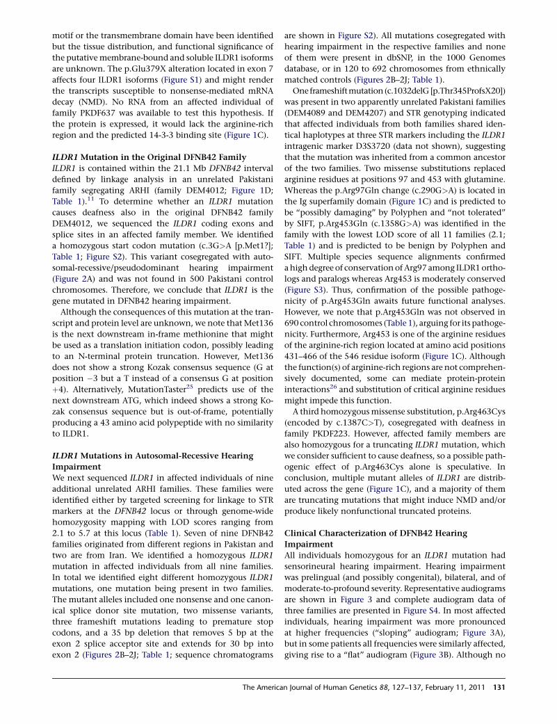
ILDR1 Mutation in the Original DFNB42 Family
ILDR1 is contained within the 21.1 Mb DFNB42 interval
defined by linkage analysis in an unrelated Pakistani
family segregating ARHI (family DEM4012; Figure 1D;
Table 1).11 To determine whether an ILDR1 mutation
causes deafness also in the original DFNB42 family
DEM4012, we sequenced the ILDR1 coding exons and
splice sites in an affected family member. We identified
a homozygous start codon mutation (c.3G>A [p.Met1?];
Table 1; Figure S2). This variant cosegregated with auto-
somal-recessive/pseudodominant hearing impairment
(Figure 2A) and was not found in 500 Pakistani control
chromosomes. Therefore, we conclude that ILDR1 is the
gene mutated in DFNB42 hearing impairment.
Although the consequences of this mutation at the tran-
script and protein level are unknown, we note that Met136
is the next downstream in-frame methionine that might
be used as a translation initiation codon, possibly leading
to an N-terminal protein truncation. However, Met136
does not show a strong Kozak consensus sequence (G at
position �3 but a T instead of a consensus G at position
þ4). Alternatively, MutationTaster25 predicts use of the
next downstream ATG, which indeed shows a strong Ko-
zak consensus sequence but is out-of-frame, potentially
producing a 43 amino acid polypeptide with no similarity
to ILDR1.
ILDR1 Mutations in Autosomal-Recessive Hearing
Impairment
We next sequenced ILDR1 in affected individuals of nine
additional unrelated ARHI families. These families were
identified either by targeted screening for linkage to STR
markers at the DFNB42 locus or through genome-wide
homozygosity mapping with LOD scores ranging from
2.1 to 5.7 at this locus (Table 1). Seven of nine DFNB42
families originated from different regions in Pakistan and
two are from Iran. We identified a homozygous ILDR1
mutation in affected individuals from all nine families.
In total we identified eight different homozygous ILDR1
mutations, one mutation being present in two families.
The mutant alleles included one nonsense and one canon-
ical splice donor site mutation, two missense variants,
three frameshift mutations leading to premature stop
codons, and a 35 bp deletion that removes 5 bp at the
exon 2 splice acceptor site and extends for 30 bp into
exon 2 (Figures 2B–2J; Table 1; sequence chromatograms
The America
are shown in Figure S2). All mutations cosegregated with
hearing impairment in the respective families and none
of them were present in dbSNP, in the 1000 Genomes
database, or in 120 to 692 chromosomes from ethnically
matched controls (Figures 2B–2J; Table 1).
One frameshiftmutation(c.1032delG[p.Thr345ProfsX20])
was present in two apparently unrelated Pakistani families
(DEM4089 and DEM4207) and STR genotyping indicated
that affected individuals from both families shared iden-
tical haplotypes at three STR markers including the ILDR1
intragenic marker D3S3720 (data not shown), suggesting
that the mutation was inherited from a common ancestor
of the two families. Two missense substitutions replaced
arginine residues at positions 97 and 453 with glutamine.
Whereas the p.Arg97Gln change (c.290G>A) is located in
the Ig superfamily domain (Figure 1C) and is predicted to
be ‘‘possibly damaging’’ by Polyphen and ‘‘not tolerated’’
by SIFT, p.Arg453Gln (c.1358G>A) was identified in the
family with the lowest LOD score of all 11 families (2.1;
Table 1) and is predicted to be benign by Polyphen and
SIFT. Multiple species sequence alignments confirmed
ahighdegree of conservationof Arg97 among ILDR1ortho-
logs and paralogs whereas Arg453 is moderately conserved
(Figure S3). Thus, confirmation of the possible pathoge-
nicity of p.Arg453Gln awaits future functional analyses.
However, we note that p.Arg453Gln was not observed in
690 control chromosomes (Table 1), arguing for its pathoge-
nicity. Furthermore, Arg453 is one of the arginine residues
of the arginine-rich region located at amino acid positions
431–466 of the 546 residue isoform (Figure 1C). Although
the function(s) of arginine-rich regions are not comprehen-
sively documented, some can mediate protein-protein
interactions26 and substitution of critical arginine residues
might impede this function.
A third homozygousmissense substitution, p.Arg463Cys
(encoded by c.1387C>T), cosegregated with deafness in
family PKDF223. However, affected family members are
also homozygous for a truncating ILDR1 mutation, which
we consider sufficient to cause deafness, so a possible path-
ogenic effect of p.Arg463Cys alone is speculative. In
conclusion, multiple mutant alleles of ILDR1 are distrib-
uted across the gene (Figure 1C), and a majority of them
are truncating mutations that might induce NMD and/or
produce likely nonfunctional truncated proteins.
Clinical Characterization of DFNB42 Hearing
Impairment
All individuals homozygous for an ILDR1 mutation had
sensorineural hearing impairment. Hearing impairment
was prelingual (and possibly congenital), bilateral, and of
moderate-to-profound severity. Representative audiograms
are shown in Figure 3 and complete audiogram data of
three families are presented in Figure S4. In most affected
individuals, hearing impairment was more pronounced
at higher frequencies (‘‘sloping’’ audiogram; Figure 3A),
but in some patients all frequencies were similarly affected,
giving rise to a ‘‘flat’’ audiogram (Figure 3B). Although no
n Journal of Human Genetics 88, 127–137, February 11, 2011 131
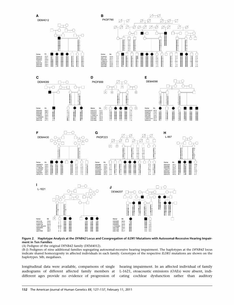
Figure 2. Haplotype Analysis at the DFNB42 Locus and Cosegregation of ILDR1Mutations with Autosomal-Recessive Hearing Impair-ment in Ten Families(A) Pedigree of the original DFNB42 family (DEM4012).(B–J) Pedigrees of nine additional families segregating autosomal-recessive hearing impairment. The haplotypes at the DFNB42 locusindicate shared homozygosity in affected individuals in each family. Genotypes of the respective ILDR1 mutations are shown on thehaplotypes. Mb, megabases.
longitudinal data were available, comparisons of single
audiograms of different affected family members at
different ages provide no evidence of progression of
132 The American Journal of Human Genetics 88, 127–137, February
hearing impairment. In an affected individual of family
L-1621, otoacoustic emissions (OAEs) were absent, indi-
cating cochlear dysfunction rather than auditory
11, 2011
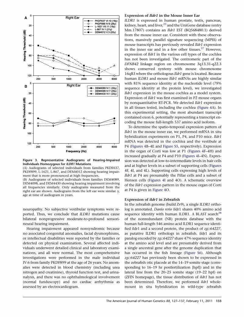
Figure 3. Representative Audiograms of Hearing-ImpairedIndividuals Homozygous for ILDR1 Mutations(A) Audiograms of selected individuals from families PKDF637,PKDF899, L-1621, L-867, and DEM4012 showing hearing impair-ment that is more pronounced at high frequencies.(B) Audiograms of selected individuals from families DEM4089,DEM4098, and DEM4430 showing hearing impairment involvingall frequencies similarly. Only audiograms measured from theright ear are shown. Audiograms from the left ear were similar. y,age at time of audiogram in years.
neuropathy. No subjective vestibular symptoms were re-
ported. Thus, we conclude that ILDR1 mutations cause
bilateral nonprogressive moderate-to-profound sensori-
neural hearing impairment.
Hearing impairment appeared nonsyndromic because
no associated congenital anomalies, facial dysmorphisms,
or intellectual disabilities were reported by the families or
detected on physical examination. Several affected indi-
viduals underwent detailed clinical and laboratory exami-
nations, and all were normal. The most comprehensive
investigations were performed in the male individual
IV:6 from family PKDF899 at the age of 26 years. No anom-
alies were detected in blood chemistry (including urea
nitrogen and creatinine), thyroid function test, and urina-
nalysis, and there was no ophthalmological involvement
(normal funduscopy) and no cardiac arrhythmia as
assessed by an electrocardiogram.
The America
Expression of Ildr1 in the Mouse Inner Ear
ILDR1 is expressed in human prostate, testis, pancreas,
kidney, heart, and liver,23 and the UniGene database (entry
Mm.17807) contains an Ildr1 EST (BQ568480.1) derived
from the mouse inner ear. Consistent with these observa-
tions, massively parallel signature sequencing (MPSS) of
mouse transcripts has previously revealed Ildr1 expression
in the inner ear and in a few other tissues.27 However,
expression of Ildr1 in the various cell types of the cochlea
has not been investigated. The centromeric part of the
DFNB42 linkage region on chromosome 3q13.31-q22.3
shows conserved synteny with mouse chromosome
16qB3 where the orthologous Ildr1 gene is located. Because
human ILDR1 and mouse Ildr1 mRNAs are highly similar
with 81% sequence identity at the nucleotide level (79%
sequence identity at the protein level), we investigated
Ildr1 expression in the mouse cochlea as a model system.
Expression of Ildr1 was first examined in P2 mouse organs
by nonquantitative RT-PCR. We detected Ildr1 expression
in all tissues tested, including the cochlea (Figure 4A). In
this experimental setting, the most abundant transcript
contained exon 6, potentially representing a transcript en-
coding the mouse full-length 537 amino acid isoform.
To determine the spatio-temporal expression pattern of
Ildr1 in the mouse inner ear, we performed mRNA in situ
hybridization experiments on P1, P4, and P10 mice. Ildr1
mRNA was detected in the cochlea and the vestibule at
P4 (Figures 4B–4E and Figure S5, respectively). Expression
in the organ of Corti was low at P1 (Figures 4F–4H) and
increased gradually at P4 and P10 (Figures 4I–4N). Expres-
sion was detected at low-to-intermediate levels in hair cells
and at higher levels in a subset of supporting cells (Figures
4F, 4I, and 4L). Supporting cells expressing high levels of
Ildr1 at P4 are presumably the Pillar cells and a subset of
Hensen cells (Figures 4I and 4O). A schematic overview
of the Ildr1 expression pattern in the mouse organ of Corti
at P4 is given in Figure 4O.
Expression of ildr1 in Zebrafish
In the zebrafish genome (build Zv9), a single ILDR1 ortho-
log is annotated. Danio rerio ildr1 shares 40% amino acid
sequence identity with human ILDR1. A BLAST search28
of the nonredundant (NR) protein database with the
human full-length 546 amino acid ILDR1 sequence identi-
fied ildr1 and a second protein, the product of zgc:64227,
as putative ILDR1 orthologs in zebrafish. ildr1 and its
paralog encoded by zgc:64227 share 47% sequence identity
at the amino acid level and are presumably derived from
a single ancestral gene after the genome duplication that
has occurred in the fish lineage (Figure S6). Although
zgc:64227 has previously been shown to be expressed in
the zebrafish otic placode at the 14–19 somite stage (corre-
sponding to 16–19 hr postfertilization [hpf]) and in the
lateral line from the 20–25 somite stage (19–22 hpf) on
(ZFIN homepage), the tissue distribution of ildr1 has not
been determined. Therefore, we performed ildr1 whole-
mount in situ hybridization in wild-type zebrafish
n Journal of Human Genetics 88, 127–137, February 11, 2011 133
Figure 4. Expression of Ildr1 in the Mouse Inner Ear(A) Nonquantitative RT-PCR with exonic Ildr1 primers on cDNAderived from P2 mouse organs. gDNA, genomic DNA; H2O,control PCR with water as a template.(B–E) In situ hybridization of Ildr1 in the mouse cochlea at P4.Antisense (AS) probe (B and D); control sense (S) probe (C andE). Arrowheads in (B) point at the organ of Corti.(F–N) In situ hybridization of Ildr1 in the organ of Corti at highmagnification at P1 (F–H), P4 (I–K), and P10 (L–N) with Ildr1 anti-sense (F, I, L) and sense (G, J, M) probes. Yellow dotted lines in (F),(I), and (L) indicate the positions of inner and outer hair cells. Ildr1is expressed at low-to-intermediate levels in hair cells and at higherlevels in the Pillar and Hensen cells adjacent to hair cells. In (H),(K), and (N), the hair cells are stained with a Loxhd1 antisenseprobe, which is used as a positive control (i.e., Loxhd1 is highlyexpressed in hair cells).18
(O) Diagram of Ildr1 expression in the mouse organ of Corti at P4.IHC, inner hair cell; OHC, outer hair cells.Scale bars represent 200 mm in (B) and (C), 100 mm in (D) and (E),and 20 mm in (F)–(N).
Figure 5. Expression of ildr1 in ZebrafishWhole-mount in situhybridizationwithprobes for ildr1 transcriptsin wild-type zebrafish embryos and larvae at ages indicated in thebottom left corners. (A, C, D) Lateral views with anterior to theleft. (B)Cross section at the level of the posterior lateral line primor-dium. The inset in (C) depicts a magnification of the ildr1-positiveanterior pituitary gland of the 48 hpf larvae. Arrowheads in (D)point to neuromasts, the sensory organs of the lateral line system.ap, anterior pituitary; e, ear; en, endoderm; n, nose; nc, notochord;oc, oral cavity; oe, olfactory epithelium; ov, otic vesicle; pllp, poste-rior lateral line primordium.
embryos and larvae between early segmentation stages (11
hpf) and day 6 of development. No distinct signal was
obtained with the control sense probe (data not shown).
We did not detect ildr1 expression at 11 hpf. At 15 hpf,
however, a weak signal was present in the developing
134 The American Journal of Human Genetics 88, 127–137, February
endoderm (data not shown), and a broader expression
pattern can be seen from 24 hpf onward (Figure 5). At this
stage, ildr1 is expressed in several derivatives of the prepla-
codal ectoderm including the adenohypophyseal placode,
the olfactory epithelium and, notably, the otic vesicle and
the primordium of the posterior lateral line, which later
gives rise to the neuromast-containing lateral line organ
(Figures 5A and 5B). In addition, transcripts can be detected
in the developing gastrointestinal duct (Figures 5A and 5B).
ildr1 expression persists at least up to day 6 (144 hpf) in the
endoderm, thenose, theneuromasts (which are the sensory
organs of the lateral line), and in the ear where expression is
high at 48 hpf (Figures 5C and 5D). Notably, the otic vesicle
from approximately 20 hpf onward, the ear, and the lateral
line primordium contain hair cells.8
Discussion
We have identified mutations of ILDR1 underlying auto-
somal-recessive nonsyndromic hearing impairment at
the DFNB42 locus. Seven of ten distinct ILDR1 mutations
are nonsense, frameshift, or splice site mutations predicted
to introduce premature stop codons that may lead to NMD
and/or protein truncation. Thus, complete loss of ILDR1
function appears to underlie hearing impairment in most
if not all cases. The DFNB42 locus was originally mapped
in a single Pakistani family.11 Our identification of ILDR1
mutations in nine Pakistani and two Iranian families raises
the possibility that mutations of ILDR1 may be among the
more prevalent causes of ARHI, a genetically highly hetero-
geneous condition. On the other hand, the large allelic
heterogeneity at the DFNB42 locus in families from
11, 2011
Pakistan alone is similar to allelic heterogeneity for other
genes associated with hearing impairment that have
been extensively studied in this population.21,22
ILDR1belongs to an evolutionarily conserved family of Ig
domain-containing proteins of unknown function. Mouse
Ildr2 (also known as Lisch-like) has been reported as a type
2 diabetes susceptibility gene in DBA mice.29 Another
homolog, LSR, encodes the lipolysis-stimulated lipoprotein
receptor that binds free fatty acids and contributes to the
control of plasma triglyceride and cholesterol levels.30,31
Notably, the zebrafish ildr2 ortholog is highly expressed in
the otic vesicle at 24 hpf and the ear at 48 hpf.29 We note
that human ILDR2 maps to the DFNA7 locus on 1q24.132
and is therefore an interesting candidate gene for auto-
somal-dominant deafness. Although the function of
ILDR1 remains elusive,23 its expression in the mouse
cochlea and vestibule as well as in the zebrafish ear and in
lateral line neuromasts supports an essential role in hearing
in vertebrates. A necessary function of ILDR1 only for
hearing is supported by the lack of obvious additional clin-
ical signsoutside the auditory system inaffected individuals
despite the wide tissue distribution of ILDR1. This is not an
unusual observation; there are mutant alleles of several
ubiquitously expressed genes associated with nonsyn-
dromic deafness such as TPRN (MIM 613354), MARVELD2
(also known as TRIC, MIM 610572), HGF (MIM 142409),
and ACTG1 (MIM 102560).17,33–36
Many genes that are mutated in DFNB hearing impair-
ment are highly expressed in cochlear hair cells and
predominant expression in supporting cells is an excep-
tion.37 Our in situ hybridization experiments in the mouse
inner ear show that Ildr1 is weakly expressed in hair cells
and at higher levels in a subset of supporting cells.
Whether DFNB42-linked hearing impairment is due to
disruption of ILDR1 function in hair cells, supporting cells,
or both remains to be addressed in future studies.
In conclusion, mutations of ILDR1 constitute a previ-
ously unreported cause of DFNB hearing impairment.
ILDR1 is a gene of unknown function but with a consistent
expression pattern in auditory and vestibular tissues in
mouse and zebrafish. Future studies will address the spec-
trum and prevalence of ILDR1 mutations in ARHI families
of different ethnic and geographical origins, the localiza-
tion of the ILDR1 protein in the mammalian cochlea, the
tissue distribution and functions of the different ILDR1 iso-
forms andpossible ligands, interaction partners, anddown-
stream effectors of this putative transmembrane receptor.
Supplemental Data
Supplemental Data include six figures and two tables and can be
found with this article online at http://www.cell.com/AJHG/.
Acknowledgments
We wish to thank the family members for their invaluable partic-
ipation and cooperation. We thank Dennis Drayna and Changsoo
The America
Kang for critically reading the manuscript. This work was funded
by the Deutsche Forschungsgemeinschaft (DFG; BO2985/3-1; to
G.B.), the European Commission FP6 Integrated Project EURO-
HEAR, Grant LSHG-CT-20054-512063 (to B.W. and C.K.), NIH
RO1 DC002842 (to R.J.H.S), NHMRC Career Development Award
(to M.B.), NHMRC Overseas Biomedical Fellowship (to M.S.H.),
Doris Duke Fellowship (to A.E.S), NIH DC007704 and Skaggs Insti-
tute for Chemical Biology (to U.M.), NIH intramural funds from
NIDCD DC000039-14 (to T.B.F.), Higher Education Commission
(HEC), Government of Pakistan (to W.A.), NIH-NIDCD grant
DC03594 (to S.M.L.), and the ICGEB, Trieste, Italy (to Sheikh R.).
J.A. thanks Dost Muhammad Baloch and Ahmed Farooq Bazai,
VC, BUITEMS, for faculty funds. Genotyping services were
partially provided by the Center for Inherited Disease Research
(CIDR). CIDR is fully funded through a federal contract from the
NIH to The Johns Hopkins University, Contract Number N01-
HG-65403 (to S.M.L.).
Received: November 17, 2010
Revised: December 16, 2010
Accepted: December 20, 2010
Published online: January 20, 2011
Web Resources
The URLs for data presented herein are as follows:
1000 Genomes browser, http://browser.1000genomes.org/index.
html
dbSNP, http://www.ncbi.nlm.nih.gov/projects/SNP
GenBank, http://www.ncbi.nlm.nih.gov/genbank
Hereditary Hearing Loss home page, http://hereditaryhearingloss.
org
MutationTaster, http://www.mutationtaster.org/
Online Mendelian Inheritance in Man (OMIM), http://www.ncbi.
nlm.nih.gov/Omim/
Polyphen, http://genetics.bwh.harvard.edu/pph
Sorting Intolerant from Tolerant (SIFT), http://sift.jcvi.org
TMHMM (prediction of transmembrane helices in proteins),
http://www.cbs.dtu.dk/services/TMHMM
UCSC Genome Bioinformatics, http://www.genome.ucsc.edu
UniGene, http://www.ncbi.nlm.nih.gov/unigene
ZFIN, http://zfin.org/cgi-bin/webdriver?MIval¼aa-ZDB_home.apg
References
1. Fritzsch, B., Beisel, K.W., and Bermingham, N.A. (2000).
Developmental evolutionary biology of the vertebrate ear:
Conserving mechanoelectric transduction and developmental
pathways indivergingmorphologies.Neuroreport11, R35–R44.
2. Dror, A.A., and Avraham, K.B. (2010). Hearing impairment: A
panoply of genes and functions. Neuron 68, 293–308.
3. Gillespie, P.G., andMuller, U. (2009). Mechanotransduction by
hair cells:Models,molecules, andmechanisms.Cell139, 33–44.
4. Frolenkov, G.I., Belyantseva, I.A., Friedman, T.B., and Griffith,
A.J. (2004). Genetic insights into the morphogenesis of inner
ear hair cells. Nat. Rev. Genet. 5, 489–498.
5. Petersen, M.B., and Willems, P.J. (2006). Non-syndromic,
autosomal-recessive deafness. Clin. Genet. 69, 371–392.
6. Brown, S.D., Hardisty-Hughes, R.E., and Mburu, P. (2008).
Quiet as a mouse: Dissecting the molecular and genetic basis
of hearing. Nat. Rev. Genet. 9, 277–290.
n Journal of Human Genetics 88, 127–137, February 11, 2011 135
7. Leibovici, M., Safieddine, S., and Petit, C. (2008). Mouse
models for human hereditary deafness. Curr. Top. Dev. Biol.
84, 385–429.
8. Nicolson, T. (2005). The genetics of hearing and balance in
zebrafish. Annu. Rev. Genet. 39, 9–22.
9. Nicolson, T., Rusch, A., Friedrich, R.W., Granato, M., Ruppers-
berg, J.P., and Nusslein-Volhard, C. (1998). Genetic analysis of
vertebrate sensory hair cell mechanosensation: The zebrafish
circler mutants. Neuron 20, 271–283.
10. Vrijens, K., Van Laer, L., and Van Camp, G. (2008). Human
hereditary hearing impairment: Mouse models can help to
solve the puzzle. Hum. Genet. 124, 325–348.
11. Aslam, M., Wajid, M., Chahrour, M.H., Ansar, M., Haque, S.,
Pham, T.L., Santos, R.P., Yan, K., Ahmad, W., and Leal, S.M.
(2005). A novel autosomal recessive nonsyndromic hearing
impairment locus (DFNB42) maps to chromosome 3q13.31-
q22.3. Am. J. Med. Genet. A. 133A, 18–22.
12. Abecasis, G.R., Cherny, S.S., Cookson, W.O., and Cardon, L.R.
(2001). GRR: Graphical representation of relationship errors.
Bioinformatics 17, 742–743.
13. O’Connell, J.R., andWeeks, D.E. (1998). PedCheck: A program
for identification of genotype incompatibilities in linkage
analysis. Am. J. Hum. Genet. 63, 259–266.
14. Abecasis, G.R., Cherny, S.S., Cookson, W.O., and Cardon, L.R.
(2002). Merlin—Rapid analysis of dense genetic maps using
sparse gene flow trees. Nat. Genet. 30, 97–101.
15. Gudbjartsson, D.F., Jonasson, K., Frigge, M.L., and Kong, A.
(2000). Allegro, a new computer program for multipoint
linkage analysis. Nat. Genet. 25, 12–13.
16. Ruschendorf, F., and Nurnberg, P. (2005). ALOHOMORA: A
tool for linkage analysis using 10K SNP array data. Bioinfor-
matics 21, 2123–2125.
17. Li, Y., Pohl, E., Boulouiz, R., Schraders, M., Nurnberg, G.,
Charif, M., Admiraal, R.J., von Ameln, S., Baessmann, I.,
Kandil, M., et al. (2010). Mutations in TPRN cause a progres-
sive form of autosomal-recessive nonsyndromic hearing loss.
Am. J. Hum. Genet. 86, 479–484.
18. Grillet, N., Schwander, M., Hildebrand, M.S., Sczaniecka, A.,
Kolatkar, A., Velasco, J., Webster, J.A., Kahrizi, K., Najmabadi,
H., Kimberling, W.J., et al. (2009). Mutations in LOXHD1, an
evolutionarily conserved stereociliary protein, disrupt hair cell
function in mice and cause progressive hearing loss in hu-
mans. Am. J. Hum. Genet. 85, 328–337.
19. Schwander, M., Sczaniecka, A., Grillet, N., Bailey, J.S., Avenar-
ius, M., Najmabadi, H., Steffy, B.M., Federe, G.C., Lagler, E.A.,
Banan, R., et al. (2007). A forward genetics screen in mice
identifies recessive deafness traits and reveals that pejvakin
is essential for outer hair cell function. J. Neurosci. 27,
2163–2175.
20. Hammerschmidt, M., Pelegri, F., Mullins, M.C., Kane, D.A.,
van Eeden, F.J., Granato, M., Brand, M., Furutani-Seiki, M.,
Haffter, P., Heisenberg, C.P., et al. (1996). dino and mercedes,
two genes regulating dorsal development in the zebrafish
embryo. Development 123, 95–102.
21. Santos, R.L., Wajid, M., Pham, T.L., Hussan, J., Ali, G., Ahmad,
W., and Leal, S.M. (2005). Low prevalence of Connexin
26 (GJB2) variants in Pakistani families with autosomal reces-
sive non-syndromic hearing impairment. Clin. Genet. 67,
61–68.
22. Anwar, S., Riazuddin, S., Ahmed, Z.M., Tasneem, S., Jaleel,
A.u., Khan, S.Y., Griffith, A.J., Friedman, T.B., and Riazuddin,
S. (2009). SLC26A4 mutation spectrum associated with
136 The American Journal of Human Genetics 88, 127–137, February
DFNB4 deafness and Pendred’s syndrome in Pakistanis.
J. Hum. Genet. 54, 266–270.
23. Hauge, H., Patzke, S., Delabie, J., and Aasheim, H.C. (2004).
Characterization of a novel immunoglobulin-like domain
containing receptor. Biochem. Biophys. Res. Commun. 323,
970–978.
24. Strausberg, R.L., Feingold, E.A., Grouse, L.H., Derge, J.G.,
Klausner, R.D., Collins, F.S., Wagner, L., Shenmen, C.M., Schu-
ler, G.D., Altschul, S.F., et al. (2002). Generation and initial
analysis of more than 15,000 full-length human and mouse
cDNA sequences. Proc. Natl. Acad. Sci. USA 99, 16899–16903.
25. Schwarz, J.M., Rodelsperger, C., Schuelke, M., and Seelow, D.
(2010). MutationTaster evaluates disease-causing potential of
sequence alterations. Nat. Methods 7, 575–576.
26. Brodeur, J., Larkin, H., Boucher, R., Theriault, C., St-Louis,
S.C., Gagnon, H., and Lavoie, C. (2009). Calnuc binds
to LRP9 and affects its endosomal sorting. Traffic 10,
1098–1114.
27. Peters, L.M., Belyantseva, I.A., Lagziel, A., Battey, J.F., Fried-
man, T.B., and Morell, R.J. (2007). Signatures from tissue-
specific MPSS libraries identify transcripts preferentially
expressed in the mouse inner ear. Genomics 89, 197–206.
28. McGinnis, S., and Madden, T.L. (2004). BLAST: At the core of
a powerful and diverse set of sequence analysis tools. Nucleic
Acids Res. 32, W20–W25.
29. Dokmanovic-Chouinard, M., Chung, W.K., Chevre, J.C.,
Watson, E., Yonan, J., Wiegand, B., Bromberg, Y., Wakae, N.,
Wright, C.V., Overton, J., et al. (2008). Positional cloning of
‘‘Lisch-Like’’, a candidate modifier of susceptibility to type 2
diabetes in mice. PLoS Genet. 4, e1000137.
30. Narvekar, P., Berriel Diaz, M., Krones-Herzig, A., Hardeland,
U., Strzoda, D., Stohr, S., Frohme, M., and Herzig, S. (2009).
Liver-specific loss of lipolysis-stimulated lipoprotein receptor
triggers systemic hyperlipidemia in mice. Diabetes 58,
1040–1049.
31. Yen, F.T., Roitel, O., Bonnard, L., Notet, V., Pratte, D., Stenger,
C., Magueur, E., and Bihain, B.E. (2008). Lipolysis stimulated
lipoprotein receptor: A novel molecular link between hyper-
lipidemia, weight gain, and atherosclerosis in mice. J. Biol.
Chem. 283, 25650–25659.
32. Fagerheim, T., Nilssen, O., Raeymaekers, P., Brox, V., Moum,
T., Elverland, H.H., Teig, E., Omland, H.H., Fostad, G.K., and
Tranebjaerg, L. (1996). Identification of a new locus for auto-
somal dominant non-syndromic hearing impairment
(DFNA7) in a large Norwegian family. Hum. Mol. Genet. 5,
1187–1191.
33. Rehman, A.U., Morell, R.J., Belyantseva, I.A., Khan, S.Y.,
Boger, E.T., Shahzad, M., Ahmed, Z.M., Riazuddin, S., Khan,
S.N., Riazuddin, S., and Friedman, T.B. (2010). Targeted
capture and next-generation sequencing identifies C9orf75,
encoding taperin, as the mutated gene in nonsyndromic deaf-
ness DFNB79. Am. J. Hum. Genet. 86, 378–388.
34. Riazuddin, S., Ahmed, Z.M., Fanning, A.S., Lagziel, A., Kitajiri,
S., Ramzan, K., Khan, S.N., Chattaraj, P., Friedman, P.L.,
Anderson, J.M., et al. (2006). Tricellulin is a tight-junction
protein necessary for hearing. Am. J. Hum. Genet. 79,
1040–1051.
35. Schultz, J.M., Khan, S.N., Ahmed, Z.M., Riazuddin, S., War-
yah, A.M., Chhatre, D., Starost, M.F., Ploplis, B., Buckley, S.,
Velasquez, D., et al. (2009). Noncoding mutations of HGF
are associated with nonsyndromic hearing loss, DFNB39.
Am. J. Hum. Genet. 85, 25–39.
11, 2011
36. Zhu, M., Yang, T., Wei, S., DeWan, A.T., Morell, R.J., Elfenbein,
J.L., Fisher, R.A., Leal, S.M., Smith, R.J., and Friderici, K.H.
(2003). Mutations in the gamma-actin gene (ACTG1) are
associated with dominant progressive deafness (DFNA20/26).
Am. J. Hum. Genet. 73, 1082–1091.
The America
37. Wilcox, E.R., Burton, Q.L., Naz, S., Riazuddin, S., Smith, T.N.,
Ploplis, B., Belyantseva, I., Ben-Yosef, T., Liburd, N.A., Morell,
R.J., et al. (2001). Mutations in the gene encoding tight
junction claudin-14 cause autosomal recessive deafness
DFNB29. Cell 104, 165–172.
n Journal of Human Genetics 88, 127–137, February 11, 2011 137


![Mutations in zinc finger 407 [ZNF407] cause a unique autosomal recessive cognitive impairment syndrome](https://img.pdfslide.net/doc/110x75/634e894d6b2f6dfefb0aee57/mutations-in-zinc-finger-407-znf407-cause-a-unique-autosomal-recessive-cognitive.jpg)


