Embed Size (px)
Citation preview
1581
The Canadian MineralogistVol. 43, pp. 1581-1588 (2005)
MULTIFREQUENCY EPR STUDY OF RADIATION-INDUCED DEFECTS IN CHLORAPATITE
SERGIY M. NOKHRIN AND YUANMING PAN§
Department of Geological Sciences, University of Saskatchewan, Saskatoon, Saskatchewan S7N 5E2, Canada
JOHN A. WEIL
Department of Chemistry, University of Saskatchewan, Saskatoon, Saskatchewan S7N 1C6, Canada
MARK J. NILGES
Illinois EPR Research Center, University of Illinois at Urbana-Champaign, Illinois 61801, U.S.A.
ABSTRACT
Gamma-irradiated chlorapatite, synthesized from a CaCl2 fl ux, has been investigated by powder and single-crystal electron paramagnetic resonance (EPR) spectroscopy at X- and W-band frequencies, including in situ high-T X-band EPR. The powder EPR spectra, particularly high-T X-band spectra and high-resolution W-band spectra, reveal a new hole-like center, H(III), in addition to two previously reported hole-like centers, H(I) and H(II). Center H(III) is characterized by an electron spin ½ and hyperfi ne interaction with one 35Cl nucleus, suggesting a structural model consisting of a hole trapped by a substitutional oxygen ion adjacent to a Cl– ion vacancy in the anion column. This discovery of center H(III) also lends support to the structural model already proposed by other authors for center H(II). Single-crystal X-band EPR spectra also disclose a new electronic center, E(I). The structure model for center E(I) includes an electron trapped at an isolated Cl– ion vacancy in the anion column, corresponding to center FE(II) in fl uorapatite and similar to the well-known F center in alkali halides.
Keywords: chlorapatite, electron-spin resonance spectra, radiation-induced defects.
SOMMAIRE
Nous avons irradié la chlorapatite, synthétisée à partir d’un fondant CaCl2, avec des rayons gamma, et nous en avons étudié les spectres de résonance des électrons paramagnétiques (EPR) prélevés sur poudre et sur monocristal aux fréquences des bandes X et W, y inclus la bande X à température élevée et in situ. Les spectres prélevés sur poudre, en particulier les spectres de la bande X à haute température et les spectres de haute résolution de la bande W, révèlent un nouveau centre semblable à un trou, H(III), en plus de deux centres semblables déjà connus, H(I) et H(II). Le centre H(III) possède un spin électronique ½ et une interaction hyperfi ne avec un nucléus 35Cl, ce qui concorde avec un modèle structural impliquant un trou piégé par un atome d’oxygène substitutionnel voisin d’une lacune dans la colonne d’anions, site autrement occupé par l’ion Cl–. Cette découverte du centre H(III) concorde aussi avec le modèle structural proposé antérieurement pour le centre H(II) par d’autres auteurs. Les spectres de la bande X prélevés sur monocristaux dévoilent aussi un nouveau centre électronique, E(I). Le modèle structural pour ce centre propose un électron piégé dans une lacune Cl– isolée dans la colonne d’anions, correspondant au centre FE(II) de la fl uorapatite et semblable au centre F bien connu dans les halogénures alcalins.
(Traduit par la Rédaction)
Mots-clés: chlorapatite, résonance des électrons paramagnétiques, défauts dus à l’irradiation.
§ E-mail address: [email protected]
1582 THE CANADIAN MINERALOGIST
defect centers and to re-evaluate the structural models of Knottnerus et al. (1972) and Roufosse et al. (1974) for the two known centers.
BACKGROUND INFORMATION
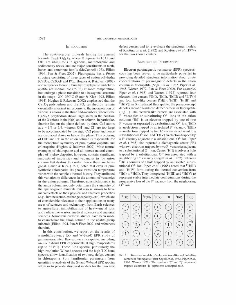
Electron paramagnetic resonance (EPR) spectros-copy has been proven to be particularly powerful in providing detailed structural information about dilute concentrations of paramagnetic defects in the anion column in fl uorapatite (Segall et al. 1962, Piper et al. 1965, Warren 1972, Pan & Fleet 2002). For example, Piper et al. (1965) and Warren (1972) reported four electron-like centers [FE(I), FE(II), FE(III) and FE(IV)] and four hole-like centers [FH(I), FH(II), FH(III) and FH(IV)] in X-irradiated fl uorapatite; the presuperscript denotes radiation-induced defect centers in fl uorapatite (Fig. 1). The electron-like centers are associated with F– vacancies or substituting O2– ions in the anion column: FE(I) is an electron trapped by one of two F– vacancies separated by a substitutional O2– ion, FE(II) is an electron trapped by an isolated F– vacancy, FE(III) is an electron trapped by two F– vacancies adjacent to a substitutional O2– ion, and FE(IV) an electron trapped by a F– vacancy adjacent to a substitutional O2– ion. Piper et al. (1965) also reported a diamagnetic center (FB) with two electrons trapped by two F– vacancies adjacent to a substitutional O2– ion. Center FH(I) involves a hole trapped by a substitutional O2– ion associated with a neighboring F– vacancy (Segall et al. 1962), whereas FH(II) consists of a hole trapped by an isolated substi-tutional O2– ion. Piper et al. (1965) noted that FH(III) and FH(IV) form during the thermal conversion from FH(I) to FH(II). They interpreted FH(III) and FH(IV) to represent stable intermediate confi gurations during the progressive loss of the F– vacancy from the neighboring O2– ion.
INTRODUCTION
The apatite-group minerals having the general formula Ca10(PO4)6X2, where X represents F, Cl and OH, are ubiquitous in igneous, metamorphic and sedimentary rocks, and are major constituents in teeth, bones and vertebrate fossils (McConnell 1973, Elliott 1994, Pan & Fleet 2002). Fluorapatite has a P63/m structure consisting of three types of cation polyhedra [Ca1O9, Ca2O6F and PO4; Hughes & Rakovan (2002) and references therein]. Pure hydroxylapatite and chlor-apatite are monoclinic (P21/b) at room temperature, but undergo a phase transition to a hexagonal structure in the range ~200–350°C (Bauer & Klee 1993, Elliott 1994). Hughes & Rakovan (2002) emphasized that the Ca1O9 polyhedron and the PO4 tetrahedron remain essentially invariant in response to the incorporation of various X anions in the three end-members, whereas the Ca2O6X polyhedron shows large shifts in the position of the X anions in the [00z] anion column. In particular, fl uorine lies on the plane defi ned by three Ca2 atoms at z = 1/4 or 3/4, whereas OH– and Cl– are too large to be accommodated by the rigid Ca2 plane and hence are displaced above or below the plane. This ordering of OH– and Cl– in the anion column is responsible for the monoclinic symmetry of pure hydroxylapatite and chlorapatite (Hughes & Rakovan 2002). Most natural examples of chlorapatite and all known natural exam-ples of hydroxylapatite, however, contain signifi cant amounts of impurities and vacancies in the anion column that destroy this order; hence these are hexa-gonal. Bauer & Klee (1993) noted that even in pure synthetic chlorapatite, its phase-transition temperature varies with the sample’s thermal history. They attributed this variation to differences in the amount of vacancies in the anion column. Therefore, nonstoichiometry in the anion column not only determines the symmetry of the apatite-group minerals, but also is known to have marked effects on their physical and chemical properties (e.g., luminescence, exchange capacity, etc.), which are of considerable relevance to their applications in many areas of sciences and technology, from Earth sciences to agriculture, immobilization of heavy-metal ions and radioactive wastes, medical sciences and material sciences. Numerous previous studies have been made to characterize the anion column in the apatite-group minerals (Elliott 1994, Pan & Fleet 2002, and references therein).
In this contribution, we report on the results of a multifrequency (X- and W-band) EPR study of gamma-irradiated fl ux-grown chlorapatite, including in-situ X-band EPR experiments at high temperatures (up to 323°C). These EPR spectra, particularly the high-resolution W-band spectra and the high-T X-band spectra, allow identifi cation of two new defect centers in chlorapatite. Spin-hamiltonian parameters from quantitative analysis of the X- and W-band EPR spectra allow us to provide structural models for the two new
FIG. 1. Structural models of color electron-like and hole-like centers in fl uorapatite (after Segall et al. 1962, Piper et al. 1965, Warren 1972). The symbols “↑” and “↓” represent trapped electrons; “h” represents a trapped hole.
RADIATION-INDUCED DEFECTS IN CHLORAPATITE 1583
Similarly, defect centers in the anion columns of hydroxylapatite and chlorapatite have been investigated by EPR studies (Knottnerus et al. 1972, Roufosse et al. 1974, Mengeot et al. 1975, Pan & Fleet 2002, and references herein). For example, Mengeot et al. (1975) reported a spin-½ hole-like center exhibiting hyperfi ne interaction with a hydrogen nucleus (confirmed by deuteration) in X-irradiated hydroxylapatite. They attri-buted this center to an O– ion arising from the removal of hydrogen from an OH– ion, and suggested that the hyperfi ne interaction is with the hydrogen of an adjacent OH– ion. Knottnerus et al. (1972) and Roufosse et al. (1974) both reported two oxygen-associated hole-like centers in X-irradiated fl ux-grown crystals of chlo-rapatite. It is noteworthy that the single-crystal EPR spectra in Roufosse et al. (1974) are similar to those of their respective centers in Knottnerus et al. (1972). However, these two groups of investigators reported different spin-hamiltonian parameters and disagreed in the proposed structural models.
EXPERIMENTAL PROCEDURES
Synthesis experiment
Chlorapatite, Ca10(PO4)6Cl2, was prepared as part of a program of syntheses for U-doped apatite-group mine-rals or analogues, following the fl ux method of Prener (1967). The starting materials were CaO (obtained from the decomposition of CaCO3 at 900°C), P2O5 and CaCl2 from the Sigma-Aldrich Chemical Company, and UO2 from Alfa Aesar, were weighed and mixed thoroughly to form the composition of chlorapatite with approxi-mately 10 ppm U. This mixture (11.75 g), together with fl ux materials (30 g CaCl2), was then placed in a tightly covered 50 mL platinum crucible. The synthesis was carried out under atmospheric pressures in a Ther-molyne muffl e furnace equipped with a programmable controller. The mixture was fi rst heated to 1280°C and held there for 24 hours to ensure complete melting and homogenization, and was then cooled down to 1060°C at a rate of 2°C/h and quenched in water. The resulting products contained several large crystals (up to 1 cm in length and ≥2 mm in diameter) and numerous smaller grains of chlorapatite, in a quenched melt of CaCl2. The CaCl2 was removed readily by washing in hot water. Chlorapatite crystals grown from this fl ux method are known to have 3–6% defi ciency in CaCl2, but remain monoclinic in symmetry (Prener 1967, Elliott et al. 1975).
Approximately ~100 mg powder and three selected crystals of synthetic chlorapatite were annealed at 1300°C and atmospheric pressure for two hours.
The EPR experiments
All X-band EPR experiments were performed on a Bruker ESP300E spectrometer at the Department
of Chemistry, University of Saskatchewan. Powder samples were prepared by grinding hand-picked grains of chlorapatite and then were subjected to 4 hours gamma irradiation at room temperature on a 60Co cell at a rate of 2260 Rad/min. Powder X-band EPR spectra at room temperature were collected by use of a Bruker T110 rectangular cavity with a fi eld-modulation frequency of 100 kHz and a microwave power of 15 dB. Powder X-band EPR spectra at elevated tempera-tures (from 31 to 323°C with intervals of 30 to 40°C) were obtained by use of a Bruker high-T cavity at the same modulation frequency and microwave power. Calibration of the magnetic fi eld was made by use of 2,2-Diphenyl-picrylhydrazyl (DPPH).
Single crystals of chlorapatite, selected on the basis of clarity, size and morphology, were irradiated for up to 24 hours on the 60Co source at room temperature. Crystal alignment of chlorapatite was made possible by its well-developed prismatic form {100}. Single-crystal X-band EPR spectra at room temperature were obtained on a Bruker cylindrical cavity for two orthogonal rota-tion planes (// and ⊥ the crystallographic c axis).
Powder and single-crystal W-band EPR spectra of gamma-irradiated chlorapatite were obtained at room temperature on a MARK II spectrometer at the Illinois EPR Research Center, University of Illinois at Urbana-Champaign (Nilges et al. 1999). A calibra-tion of the magnetic fi eld for W-band measurements was carried out by use of Metrolab NMR Teslameter PT2025 (Nilges et al. 1999). Simulations of the EPR spectra were performed by use of the software package EPR–NMR (Mombourquette et al. 1996).
RESULTS
Powder EPR spectra
The selected grains of synthetic chlorapatite for powder EPR are colorless, transparent and free of melt inclusions. The powder sample of synthetic chlorapatite before irradiation is EPR-silent at the X-band frequency. After 4 hours of gamma irradiation, the powder sample changes to a bluish color. Figure 2 shows that the room-temperature X-band EPR spectrum of the gamma-irra-diated chlorapatite reveals at least two hole-like centers, similar to those reported by Knottnerus et al. (1972) and Roufosse et al. (1974). The EPR spectra of these centers were described (Roufosse et al. 1974) by means of the axially symmetrical spin-hamiltonian:
Ĥ = �e[g||SzBz + g⊥(SxBx + SyBy)] + �i [A||
(i)SzIz(i) + A⊥(i)(SxIx
(i) + SyBy(i))] (1)
where g|| and g⊥ are the axially symmetrical g values, A|| and A⊥ are the hyperfi ne interaction parameters parallel and perpendicular to the crystal’s c axis; this axis was found to be an axis of symmetry for both
1584 THE CANADIAN MINERALOGIST
defect centers. The index “i” runs over the relevant spin-bearing nuclei.
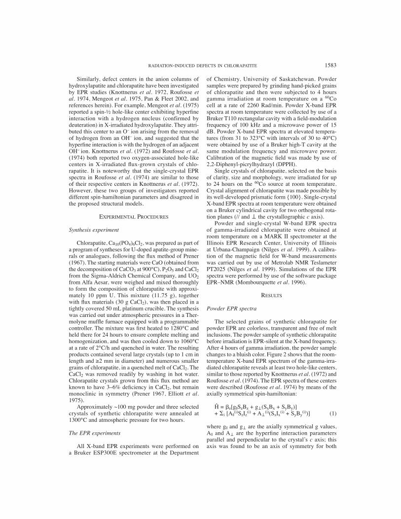
Roufosse et al. (1974) noted that center H(I) is characterized by hyperfi ne interaction with three non-equivalent chlorine nuclei. However, the principal values of the hyperfi ne-interaction matrix for one Cl nucleus are much larger than those for the other two. The single-crystal work of Roufosse et al. (1974) showed that the EPR spectrum at magnetic fi eld B perpendicular to the c axis consists of four major lines with approximately equal spacing and equal intensity, characteristic of hyperfi ne interaction with a single spin-3/2 nucleus. Closer examination indicated a second set of four equally spaced lines having intensity about one-third that of the fi rst set and constant splittings of about 0.8 G. These two sets of lines are attributable to hyperfi ne interaction with the nearest-neighbor chlo-rine nucleus, which has two magnetic isotopes 35Cl (75.77% natural abundance) and 37Cl (24.23%). Both 35Cl and 37Cl have nuclear spins of 3/2, but their nuclear magnetic g-factors are 0.5479198 and 0.4560854, respectively (Weil et al. 1994). For B parallel to the c axis, additional splittings are observed and have been attributed to hyperfi ne interactions with two non-equi-valent chlorine nuclei (Roufosse et al. 1974).
Spectral simulations using spin-hamiltonian para-meters (Table 1) from Roufosse et al. (1974) confi rmed the presence of centers H(I) and H(II). However, these two centers cannot explain all experimentally observed lines, which point to the presence of an additional para-magnetic defect center (Fig. 2).
Figure 3 illustrates that center H(I) persists to at least 155°C but is not observed at 199°C or higher temperatures. Center H(II) is present up to at least
254°C. The disappearance of center H(I) at elevated temperatures discloses the presence of a new hole-like defect center [hereafter referred to as center H(III)]. This center is characterized by an electron spin ½ and hyperfi ne interaction with a nuclear spin 3/ 2, and has uniaxial symmetry. This center is responsible for the extra lines observed in the room-temperature X-band EPR spectrum, but is partly overlapped with center H(I) (Fig. 2).
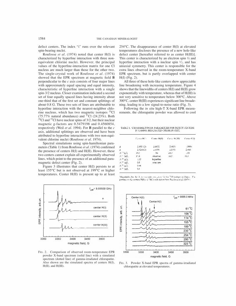
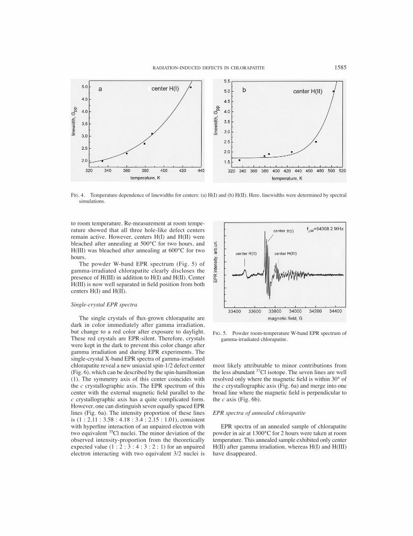
All three of these hole-like centers show appreciable line broadening with increasing temperature. Figure 4 shows that the linewidths of centers H(I) and H(II) grow exponentially with temperature, whereas that of H(III) is not very sensitive to temperature below 300°C. Above 300°C, center H(III) experiences signifi cant line broade-ning, leading to a low signal-to-noise ratio (Fig. 3).
Following the in situ high-T X-band EPR measu-rements, the chlorapatite powder was allowed to cool
FIG. 2. Comparison of observed room-temperature EPR powder X-band spectrum (solid line) with a simulated spectrum (dotted line) of gamma-irradiated chlorapatite. Also shown are the simulated spectra of centers H(I), H(II), and H(III).
FIG. 3. Powder X-band EPR spectra of gamma-irradiated chlorapatite at elevated temperatures.
RADIATION-INDUCED DEFECTS IN CHLORAPATITE 1585
to room temperature. Re-measurement at room tempe-rature showed that all three hole-like defect centers remain active. However, centers H(I) and H(II) were bleached after annealing at 500°C for two hours, and H(III) was bleached after annealing at 600°C for two hours.
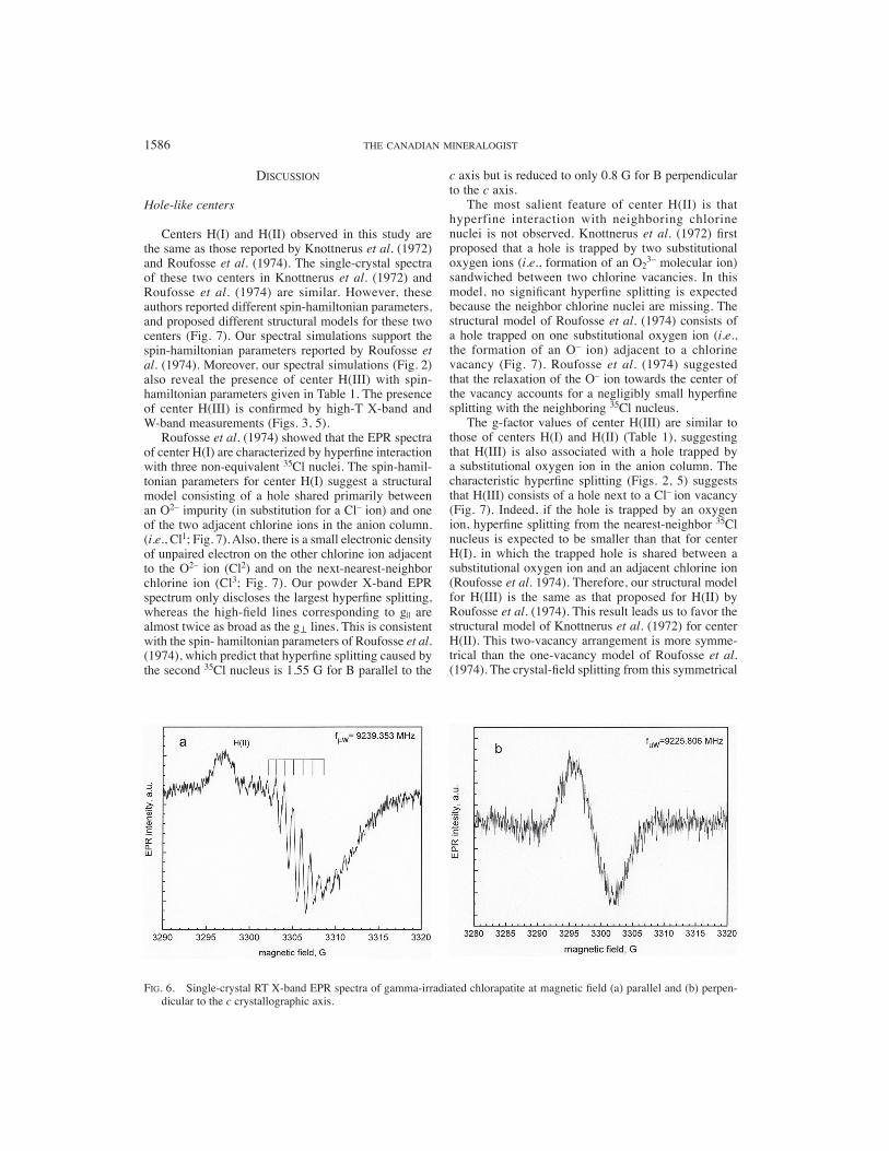
The powder W-band EPR spectrum (Fig. 5) of gamma-irradiated chlorapatite clearly discloses the presence of H(III) in addition to H(I) and H(II). Center H(III) is now well separated in fi eld position from both centers H(I) and H(II).
Single-crystal EPR spectra
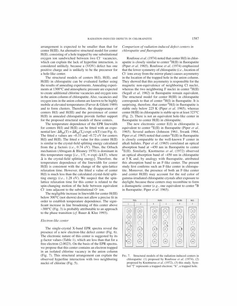
The single crystals of fl ux-grown chlorapatite are dark in color immediately after gamma irradiation, but change to a red color after exposure to daylight. These red crystals are EPR-silent. Therefore, crystals were kept in the dark to prevent this color change after gamma irradiation and during EPR experiments. The single-crystal X-band EPR spectra of gamma-irradiated chlorapatite reveal a new uniaxial spin-1/2 defect center (Fig. 6), which can be described by the spin-hamiltonian (1). The symmetry axis of this center coincides with the c crystallographic axis. The EPR spectrum of this center with the external magnetic fi eld parallel to the c crystallographic axis has a quite complicated form. However, one can distinguish seven equally spaced EPR lines (Fig. 6a). The intensity proportion of these lines is (1 : 2.11 : 3.58 : 4.18 : 3.4 : 2.15 : 1.01), consistent with hyperfi ne interaction of an unpaired electron with two equivalent 35Cl nuclei. The minor deviation of the observed intensity-proportion from the theoretically expected value (1 : 2 : 3 : 4 : 3 : 2 : 1) for an unpaired electron interacting with two equivalent 3/2 nuclei is
most likely attributable to minor contributions from the less abundant 37Cl isotope. The seven lines are well resolved only where the magnetic fi eld is within 30° of the c crystallographic axis (Fig. 6a) and merge into one broad line where the magnetic fi eld is perpendicular to the c axis (Fig. 6b).
EPR spectra of annealed chlorapatite
EPR spectra of an annealed sample of chlorapatite powder in air at 1300°C for 2 hours were taken at room temperature. This annealed sample exhibited only center H(II) after gamma irradiation, whereas H(I) and H(III) have disappeared.
FIG. 4. Temperature dependence of linewidths for centers: (a) H(I) and (b) H(II). Here, linewidths were determined by spectral simulations.
FIG. 5. Powder room-temperature W-band EPR spectrum of gamma-irradiated chlorapatite.
1586 THE CANADIAN MINERALOGIST
DISCUSSION
Hole-like centers
Centers H(I) and H(II) observed in this study are the same as those reported by Knottnerus et al. (1972) and Roufosse et al. (1974). The single-crystal spectra of these two centers in Knottnerus et al. (1972) and Roufosse et al. (1974) are similar. However, these authors reported different spin-hamiltonian parameters, and proposed different structural models for these two centers (Fig. 7). Our spectral simulations support the spin-hamiltonian parameters reported by Roufosse et al. (1974). Moreover, our spectral simulations (Fig. 2) also reveal the presence of center H(III) with spin-hamiltonian parameters given in Table 1. The presence of center H(III) is confi rmed by high-T X-band and W-band measurements (Figs. 3, 5).
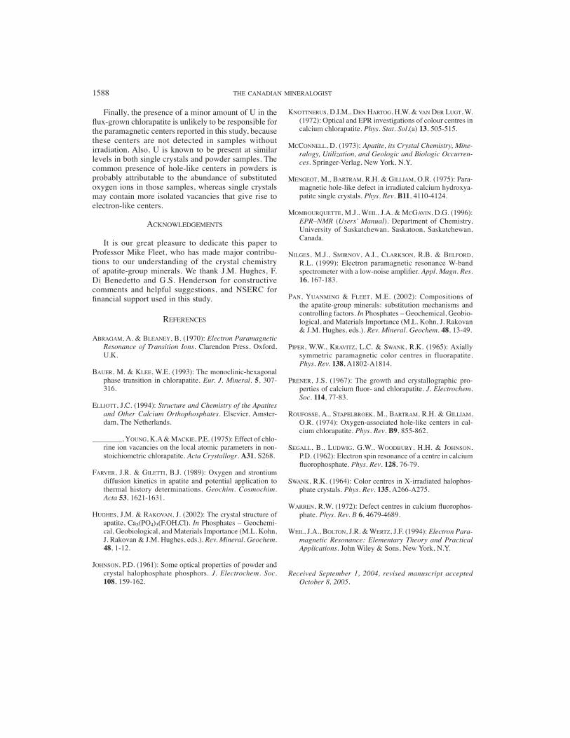
Roufosse et al. (1974) showed that the EPR spectra of center H(I) are characterized by hyperfi ne interaction with three non-equivalent 35Cl nuclei. The spin-hamil-tonian parameters for center H(I) suggest a structural model consisting of a hole shared primarily between an O2– impurity (in substitution for a Cl– ion) and one of the two adjacent chlorine ions in the anion column. (i.e., Cl1; Fig. 7). Also, there is a small electronic density of unpaired electron on the other chlorine ion adjacent to the O2– ion (Cl2) and on the next-nearest-neighbor chlorine ion (Cl3; Fig. 7). Our powder X-band EPR spectrum only discloses the largest hyperfi ne splitting, whereas the high-fi eld lines corresponding to g|| are almost twice as broad as the g⊥ lines. This is consistent with the spin- hamiltonian parameters of Roufosse et al. (1974), which predict that hyperfi ne splitting caused by the second 35Cl nucleus is 1.55 G for B parallel to the
c axis but is reduced to only 0.8 G for B perpendicular to the c axis.
The most salient feature of center H(II) is that hyperfine interaction with neighboring chlorine nuclei is not observed. Knottnerus et al. (1972) fi rst proposed that a hole is trapped by two substitutional oxygen ions (i.e., formation of an O2
3– molecular ion) sandwiched between two chlorine vacancies. In this model, no signifi cant hyperfi ne splitting is expected because the neighbor chlorine nuclei are missing. The structural model of Roufosse et al. (1974) consists of a hole trapped on one substitutional oxygen ion (i.e., the formation of an O– ion) adjacent to a chlorine vacancy (Fig. 7). Roufosse et al. (1974) suggested that the relaxation of the O– ion towards the center of the vacancy accounts for a negligibly small hyperfi ne splitting with the neighboring 35Cl nucleus.
The g-factor values of center H(III) are similar to those of centers H(I) and H(II) (Table 1), suggesting that H(III) is also associated with a hole trapped by a substitutional oxygen ion in the anion column. The characteristic hyperfi ne splitting (Figs. 2, 5) suggests that H(III) consists of a hole next to a Cl– ion vacancy (Fig. 7). Indeed, if the hole is trapped by an oxygen ion, hyperfi ne splitting from the nearest-neighbor 35Cl nucleus is expected to be smaller than that for center H(I), in which the trapped hole is shared between a substitutional oxygen ion and an adjacent chlorine ion (Roufosse et al. 1974). Therefore, our structural model for H(III) is the same as that proposed for H(II) by Roufosse et al. (1974). This result leads us to favor the structural model of Knottnerus et al. (1972) for center H(II). This two-vacancy arrangement is more symme-trical than the one-vacancy model of Roufosse et al. (1974). The crystal-fi eld splitting from this symmetrical
FIG. 6. Single-crystal RT X-band EPR spectra of gamma-irradiated chlorapatite at magnetic fi eld (a) parallel and (b) perpen-dicular to the c crystallographic axis.
RADIATION-INDUCED DEFECTS IN CHLORAPATITE 1587
arrangement is expected to be smaller than that for center H(III). An alternative structural model for center H(II), consisting of a hole trapped by one substitutional oxygen ion sandwiched between two Cl– vacancies, which can explain the lack of hyperfi ne interaction, is considered unlikely, because a (VOV) defect has one positive charge and is unlikely to be the precursor for a hole-like center.
The structural models of centers H(I), H(II), and H(III) in chlorapatite can be evaluated further using the results of annealing experiments. Annealing experi-ments at 1300°C and atmospheric pressure are expected to create additional chlorine vacancies and oxygen ions in the anion column of chlorapatite. Also, vacancies and oxygen ions in the anion column are known to be highly mobile at elevated temperatures (Farver & Giletti 1989) and to form clusters. Therefore, the disappearance of centers H(I) and H(III) and the persistence of center H(II) in annealed chlorapatite provide further support for the proposed structural models of these centers.
The temperature dependence of the EPR linewidth for centers H(I) and H(II) can be fi tted with an expo-nential law: �Bpp(T) = �Bpp(To) exp(–�/kT) (see Fig. 4). The fi tted � values are ~0.33 and ~0.72 eV for centers H(I) and H(II). The fi tted � value for this center H(II) is similar to the crystal-fi eld splitting energy calculated from the g factors (i.e., 0.74 eV). Thus, the Orbach mechanism (Abragam & Bleaney 1970) is dominant in this temperature range [i.e., 1/T1 � exp(–�/kT), where � is the crystal-fi eld splitting energy]. Therefore, the temperature dependence of the linewidth for center H(II) is consistent with the change of the spin-lattice relaxation time. However, the fi tted � value of center H(I) is much less than the calculated crystal-fi eld split-ting energy (i.e., 1.28 eV). We suspect that the spin-lattice relaxation time for this center is related to the spin-changing motion of the hole between equivalent Cl– ions adjacent to the substitutional O– ion.
The negligible increase in linewidth for center H(III) below 300°C (not shown) does not allow a precise fi t in order to establish temperature dependence. The signi-fi cant increase in line broadening of this center above ~300°C (Fig. 3) is probably attributable to an approach to the phase transition (cf. Bauer & Klee 1993).
Electron-like center
The single-crystal X-band EPR spectra reveal the presence of a new electron-like defect center (Fig. 6). The electronic nature of this center is suggested by its g-factor values (Table 1), which are less than that for a free electron (2.0023). On the basis of the EPR spectra, we propose that this center contains an electron trapped in an isolated chlorine vacancy in the anion column (Fig. 7). This structural arrangement can explain the observed hyperfi ne interaction with two neighboring nuclei of chlorine (Fig. 5).
Comparison of radiation-induced defect centers in chlorapatite and fl uorapatite
Roufosse et al. (1974) noted that center H(I) in chlor-apatite is closely similar to center FH(II) in fl uorapatite (Piper et al. 1965). Roufosse et al. (1974) emphasized that the lower symmetry of chlorapatite (i.e., location of Cl– ions away from the mirror plane) causes asymmetry in the location of the trapped hole in the anion column. They showed that this asymmetry is responsible for the magnetic non-equivalence of neighboring Cl nuclei, whereas the two neighboring F nuclei in center FH(II) (Segall et al. 1962) in fl uorapatite remain equivalent. The structural model for center H(III) in chlorapatite corresponds to that of center FH(I) in fl uorapatite. It is surprising, therefore, that center FH(I) in fl uorapatite is stable only below 225 K (Piper et al. 1965), whereas center H(III) in chlorapatite is stable up to at least 323°C (Fig. 2). There is not an equivalent hole-like center in fl uorapatite to center H(II) in chlorapatite.
The new electronic center E(I) in chlorapatite is equivalent to center FE(II) in fl uorapatite (Piper et al. 1965). Several authors (Johnson 1961, Swank 1964, Piper et al. 1965) noted that center FE(II) in fl uorapatite is closely comparable to the well-known F center in alkali halides. Piper et al. (1965) correlated an optical absorption band at ~450 nm in fl uorapatite to center FE(II). Similarly, Knottnerus et al. (1972) observed an optical absorption band of ~450 nm in chlorapatite at 5 K and, by analogy with fl uorapatite, attributed this absorption band to an F-like center. The present study fi rst confi rms such an F-like center in chlorapa-tite. Moreover, the presence of both an F-like center and center H(III) may account for the red color of gamma-irradiated chlorapatite crystals after exposure to daylight, because these centers may recombine to form a diamagnetic center (e.g., one equivalent to center FB in fl uorapatite; Piper et al. 1965).
FIG. 7. Structural models of the radiation-induced centers in chlorapatite: (1) proposed by Roufosse et al. (1974), (2) proposed by Knottnerus et al. (1972), (3) this study. Sym-bol “↑” represents a trapped electron; “h”, a trapped hole.
1588 THE CANADIAN MINERALOGIST
Finally, the presence of a minor amount of U in the fl ux-grown chlorapatite is unlikely to be responsible for the paramagnetic centers reported in this study, because these centers are not detected in samples without irradiation. Also, U is known to be present at similar levels in both single crystals and powder samples. The common presence of hole-like centers in powders is probably attributable to the abundance of substituted oxygen ions in those samples, whereas single crystals may contain more isolated vacancies that give rise to electron-like centers.
ACKNOWLEDGEMENTS
It is our great pleasure to dedicate this paper to Professor Mike Fleet, who has made major contribu-tions to our understanding of the crystal chemistry of apatite-group minerals. We thank J.M. Hughes, F. Di Benedetto and G.S. Henderson for constructive comments and helpful suggestions, and NSERC for fi nancial support used in this study.
REFERENCES
ABRAGAM, A. & BLEANEY, B. (1970): Electron Paramagnetic Resonance of Transition Ions. Clarendon Press, Oxford, U.K.
BAUER, M. & KLEE, W.E. (1993): The monoclinic-hexagonal phase transition in chlorapatite. Eur. J. Mineral. 5, 307-316.
ELLIOTT, J.C. (1994): Structure and Chemistry of the Apatites and Other Calcium Orthophosphates. Elsevier, Amster-dam, The Netherlands.
________, YOUNG, K.A & MACKIE, P.E. (1975): Effect of chlo-rine ion vacancies on the local atomic parameters in non-stoichiometric chlorapatite. Acta Crystallogr. A31, S268.
FARVER, J.R. & GILETTI, B.J. (1989): Oxygen and strontium diffusion kinetics in apatite and potential application to thermal history determinations. Geochim. Cosmochim. Acta 53, 1621-1631.
HUGHES, J.M. & RAKOVAN, J. (2002): The crystal structure of apatite, Ca5(PO4)3(F,OH,Cl). In Phosphates – Geochemi-cal, Geobiological, and Materials Importance (M.L. Kohn, J. Rakovan & J.M. Hughes, eds.). Rev. Mineral. Geochem. 48, 1-12.
JOHNSON, P.D. (1961): Some optical properties of powder and crystal halophosphate phosphors. J. Electrochem. Soc. 108, 159-162.
KNOTTNERUS, D.I.M., DEN HARTOG, H.W. & VAN DER LUGT, W. (1972): Optical and EPR investigations of colour centres in calcium chlorapatite. Phys. Stat. Sol.(a) 13, 505-515.
MCCONNELL, D. (1973): Apatite, its Crystal Chemistry, Mine-ralogy, Utilization, and Geologic and Biologic Occurren-ces. Springer-Verlag, New York, N.Y.
MENGEOT, M., BARTRAM, R.H. & GILLIAM, O.R. (1975): Para-magnetic hole-like defect in irradiated calcium hydroxya-patite single crystals. Phys. Rev. B11, 4110-4124.
MOMBOURQUETTE, M.J., WEIL, J.A. & MCGAVIN, D.G. (1996): EPR–NMR (Users’ Manual). Department of Chemistry, University of Saskatchewan, Saskatoon, Saskatchewan, Canada.
NILGES, M.J., SMIRNOV, A.I., CLARKSON, R.B. & BELFORD, R.L. (1999): Electron paramagnetic resonance W-band spectrometer with a low-noise amplifi er. Appl. Magn. Res. 16, 167-183.
PAN, YUANMING & FLEET, M.E. (2002): Compositions of the apatite-group minerals: substitution mechanisms and controlling factors. In Phosphates – Geochemical, Geobio-logical, and Materials Importance (M.L. Kohn, J. Rakovan & J.M. Hughes, eds.). Rev. Mineral. Geochem. 48, 13-49.
PIPER, W.W., KRAVITZ, L.C. & SWANK, R.K. (1965): Axially symmetric paramagnetic color centres in fluorapatite. Phys. Rev. 138, A1802-A1814.
PRENER, J.S. (1967): The growth and crystallographic pro-perties of calcium fl uor- and chlorapatite. J. Electrochem. Soc. 114, 77-83.
ROUFOSSE, A., STAPELBROEK, M., BARTRAM, R.H. & GILLIAM, O.R. (1974): Oxygen-associated hole-like centers in cal-cium chlorapatite. Phys. Rev. B9, 855-862.
SEGALL, B., LUDWIG, G.W., WOODBURY, H.H. & JOHNSON, P.D. (1962): Electron spin resonance of a centre in calcium fl uorophosphate. Phys. Rev. 128, 76-79.
SWANK, R.K. (1964): Color centres in X-irradiated halophos-phate crystals. Phys. Rev. 135, A266-A275.
WARREN, R.W. (1972): Defect centres in calcium fl uorophos-phate. Phys. Rev. B 6, 4679-4689.
WEIL, J.A., BOLTON, J.R. & WERTZ, J.F. (1994): Electron Para-magnetic Resonance: Elementary Theory and Practical Applications. John Wiley & Sons, New York, N.Y.
Received September 1, 2004, revised manuscript accepted October 8, 2005.


