Embed Size (px)
Citation preview
PROGRAMME & ABSTRACT BOOK
Main Partner:
Wednesday 24th & Thursday 25th February 2021Brought to you online
Page 2
ContentsWelcome
General Info
BSG Update
About the BSG
Main Conference Programme
NSF Programme
Posters
Sponsors
Abstracts - Oral Papers
Abstracts - NSF
Abstracts - Posters
3
4 - 5
5
6 - 7
8 - 11
12
14 - 18
20 - 21
22 - 28
29 - 30
31 - 58
Contents
Page 3
Welcome to the BSG Conference 2021
On behalf of the BSG board I would like to welcome you all to this year’s Annual Conference. Even though we won’t be able to all meet in person I still hope that you will find the programme interesting and exciting. We actually received more abstracts this year either for posters or oral presentations, which shows that sarcoma research is alive and well despite the pandemic. We have aimed to provide a broad range of updates across many areas of sarcoma care and have assembled an outstanding team of sarcoma experts from the UK and abroad to speak. We are delighted
to be able to invite three international Speakers, Dr Javier Martin-Broto from the Institute of Biomedicine of Seville University Sevilla, Dr Maude Toulmonde, from l’Institut Bergonié, Bordeaux and Dr Sylvie Bonvalot, Institute Curie, Paris who will be delivering plenary lectures that will be the high points of the Conference.
The subject matter in this year’s conference is broad covering updates in radiology, pathology, sarcoma care during COVID, clinical trial updates, as well as our usual sessions on cutting edge research in medical clinical and surgical oncology. On day two we will be hosting the National Sarcoma Forum which is the main nursing and allied health forum, and we have sessions on Personalised Cancer Care, Psychological Support as well as updates from the UK regions and research papers from nurses and AHPs.
I hope that you will be able to attend some or all of the sessions live as there will be plenty of opportunity to ask questions and interact with the plenary speakers virtually. If you cannot join us in real time, all of the sessions will be available to access online at a time of your convenience. Similarly, you will be able to access all the content from all of the sessions that run in parallel, so you will not miss an opportunity to hear all of our speakers.
I hope you all enjoy this year’s virtual congress and I look forward to talking with many of you online this year and in person next year.
Mr Andrew Hayes, BSG President
Page 4
General Info
PROGRAMMEA full conference programme can be found on pages 8 - 11. It can also be viewed on the conference platform.
The conference starts at 9am on Wednesday 24th February 2021.
ACCESSING THE CONFERENCEAll registered delegates will have been sent a link prior to the conference with the link to access the conference.
If you have not received this link please email [email protected]
CONFERENCE SESSIONSPresentations will take place throughout the day with live Q&A’s. Attendees are encouraged to submit questions via the online platform which will be read out by the chair, any questions not shared will be sent to the speaker after the conference.
NATIONAL SARCOMA FORUM The NSF will be running a dedicated stream for sarcoma nurses and allied health professionals from 12:50 - 16:00 on Day Two. View the full programme on page 12.
EXHIBITORS We encourage you to view our virtual exhibition booths and spend time viewing the products and services on display. You can contact any of our exhibitors directly through the conference system.
More information on our sponsors and exhibitors can be found on pages 20 - 21
PRESENTERSIf you are presenting you will receive a seperate link one hour before your session to log into the Green Room. If you have any questions please contact [email protected]
POSTER DISPLAYS Posters are available to view in the exhibition section of platform. These are available throughout both days and you can contact the authors directly through the conference system.
NETWORKINGThere are a number of ways to network through the conference platform. You can send direct messages to other delegates, speakers and sponsors and organise one to one live video chats.
We also have a speed networking function. In ‘Networking’ click the ‘Ready’ button, the system will then search for someone else who has also clicked the Ready button. If someone else is available, you will be matched instantly in a video chat for 2 mins. When the time expires, the meeting ends.
To get the most from all of the networking functions we reccommend you update your profile with a photo and some details about you.
Page 5
ABSTRACTSYou can read the full abstract submissions for oral talks and poster displays on pages 22 - 58.
CPD CREDITSThe Royal College of Surgeons of England has awarded up to 11.25 CPD points for this conference.
Attendance certificates will be emailed after the event.
PRIZES We’ll be presenting a prize to the best poster and best oral paper in the closing session on Day Two.
EVALUATIONAn online feedback form will be sent after the event. We appreciate your time in completing this.
British Sarcoma Group Update
After a very successful conference in Glasgow at the end of February 2020 within a matter of weeks the pandemic had resulted in a national lockdown, and a medical crisis the like of which none of us had ever experienced. The impact on sarcoma services nationally was huge, ranging from delays in presentation, cancellations of operations and curtailment of chemotherapy and clinical trials. I know from personal communications with many of you what a difficult time this year has been for patients carers and sarcoma professionals alike. However I also heard of amazing examples of innovative care delivered virtually and collaboration between units. Many of these initiatives could improve the care of the patients with sarcoma long after COVID 19 is a bad memory.
The BSG had planned to run our 2021 conference in Cardiff. We had identified a conference venue and the leaders of local scientific committee lead by Owen Tilsey and Tom Bragg had made provisional plans for a pro-gramme. Liverpool was the planned location for 2022. However it became clear in the summer that the likeli-hood of running a face to face conference this year was diminishing and we made an early decision to cancel Cardiff and convene this years conference virtually. I am grateful to all my colleagues on the BSG board in helping to pull together this programme, inviting speakers and reviewing the many abstracts we received. We very much hope that in 2022 we will be able to return to our face to face conferences in Liverpool.
Alongside developing the scientific programme for this years conference, the Board has started the process of reviewing the BSG guidelines on Bone, Soft Tissue and GIST which were last updated in 2015. We received over 30 expressions of interest from members of the BSG to contribute to the writing of these guidelines. We have now assembled the core writing committees for each of the three guideline documents. Those writing committees will be approaching many other people with specialist knowledge to help with writing these updates which will replace the 2015 guidelines on the website and hopefully be published in due course.
On a final note all of the Board Members have been trustees of the BSG for many years, most since we adopted charitable status 7 years ago and some since the inception of the BSG back in the early 2000s. The whole board feel that we should be continually refreshing the membership of the Board and more longstanding mem-bers will therefore obviously demit to allow in some fresh blood. In 2021 we will write to you all asking who would like to be involved with BSG at board level to shape the organisation for the next decade.
With all my very best wishes
Mr Andrew Hayes, BSG President
Page 6
About the British Sarcoma Group
THE AIMS OF THE BRITISH SARCOMA GROUP ARE:
1. To promote the education of medical, nursing and healthcare professionals in the UK concerned with the treatment and care of patients with bone and soft tissue sarcomas through the provision of information, training, and the identification of best practice
2. To promote excellence in the healthcare and treatment of patients with bone and soft tissue sarcoma, through enabling specialist healthcare professionals to identify and share best practice
3. To promote and support research, including the publication of results to support the development of more effective treatment and care of patients with bone and soft tissue sarcomas
4. To improve the knowledge and understanding of sarcoma amongst the medical profession and general public.
MEMBERSHIPAll clinicians, nurses and supportingprofessionals who treat patients withsarcoma in England, Wales, Scotland andNorthern Ireland are eligible to join theBritish Sarcoma Group – membership isfree and gives the following benefits:
Membership Benefits
• Become part of an engaged community of fellow specialists seeking to improve sarcoma treatment
• Access member surveys and research
• Help influence national guidelines on best practice
• Join a national network of those working to treat sarcoma
• Benefit from educational opportunities
• Receive regular newsletters and updates from us
The British Sarcoma Group is registered as a charitable incorporated organisation (CIO)by the Charity Commission. Our registration number is 1154928. A CIO has the legalprivileges of a full charity together with aspects of legal protection and limited liabilitywhich apply to companies.
HOW TO JOINBecoming a member of the British Sarcoma Group is quick and easy – simply fill in the BSG Membership Form and return to [email protected] and we will set up your membership.
Already a member? You can update your details using the BSG membership form available online at www.britishsarcomagroup.org.uk
Page 7
TRUSTEESThe BSG is managed by a Board of Trustees with support from advisory groups relating to specific projects.
The Trustees are:• President - Mr Andrew Hayes, Consultant Surgeon, Royal Marsden Hospital, London• Mr Craig Gerrand, Consultant Orthopaedic Surgeon, Royal National Orthopaedic
Hospital, Stanmore• Dr Adam Dangoor, Medical Oncologist, University Hospitals Bristol NHS Trust• Professor Ian Judson, Medical Oncologist, Royal Marsden Hospital, London• Anita Pabla, Macmillan Sarcoma Nurse Specialist, Leicester Royal Infirmary• Dr Beatrice Seddon, Clinical Oncologist, University College London Hospitals• Dr Owen Tilsley, Clinical Oncologist, Velindre Hospital, Wales• Dr. Paul Cool, Consultant Orthopaedic & Oncological Surgeon, The Robert Jones and
Agnes Hunt Orthopaedic Hospital• Dr Ioanna Nixon, Consultant Clinical Oncologist, Head and Neck / Sarcoma, The
Beatson West of Scotland Cancer Center, Glasgow, UK• Abby McCarthy, Orthopaedic Oncology Physiotherapist, Royal National Orthopaedic
Hospital, Stanmore
There are opportunities for BSG members to get involved in advisory and working groups that contribute to particular areas of the BSG’s work. If you are interested, please talk to one of the Trustees or email [email protected]
NSF MEMBERSHIPMembership of the NSF is open to nurse specialists/AHPs/key workers in academic orresearch roles related to sarcoma. NSF Membership has been formalised and now sitsas a strand of British Sarcoma Group Membership. To find out more about the NSFplease visit their dedicated page: www.britishsarcomagroup.org.uk/nsf
FUNDINGThe BSG’s income comes via the annual conference. Sponsorship and delegateregistrations fund the cost of running the conference, with any small surplus allocatedto support other BSG projects such as production of the BSG guidelines and to securethe conference venue for the following year.
Administration of the British Sarcoma GroupRoutine administration of the charity and conference management is undertaken bythe Secretariat who can be contacted at [email protected] or on0191 244 2581.
Page 8
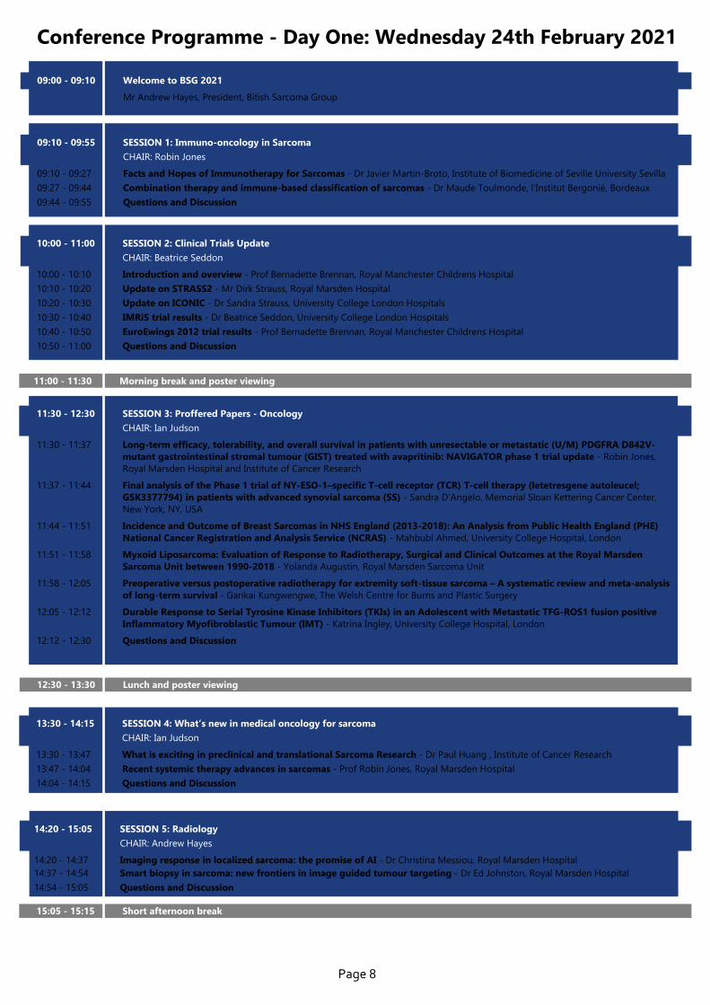
Conference Programme - Day One: Wednesday 24th February 2021
09:00 - 09:10 Welcome to BSG 2021
Mr Andrew Hayes, President, Bitish Sarcoma Group
09:10 - 09:55 SESSION 1: Immuno-oncology in Sarcoma
CHAIR: Robin Jones
09:10 - 09:27 Facts and Hopes of Immunotherapy for Sarcomas - Dr Javier Martin-Broto, Institute of Biomedicine of Seville University Sevilla
09:27 - 09:44 Combination therapy and immune-based classification of sarcomas - Dr Maude Toulmonde, l’Institut Bergonié, Bordeaux
09:44 - 09:55 Questions and Discussion
11:00 - 11:30 Morning break and poster viewing
11:30 - 12:30 SESSION 3: Proffered Papers - Oncology
CHAIR: Ian Judson
11:30 - 11:37 Long-term efficacy, tolerability, and overall survival in patients with unresectable or metastatic (U/M) PDGFRA D842V- mutant gastrointestinal stromal tumour (GIST) treated with avapritinib: NAVIGATOR phase 1 trial update - Robin Jones, Royal Marsden Hospital and Institute of Cancer Research
11:37 - 11:44 Final analysis of the Phase 1 trial of NY-ESO-1–specific T-cell receptor (TCR) T-cell therapy (letetresgene autoleucel; GSK3377794) in patients with advanced synovial sarcoma (SS) - Sandra D’Angelo, Memorial Sloan Kettering Cancer Center, New York, NY, USA
11:44 - 11:51 Incidence and Outcome of Breast Sarcomas in NHS England (2013-2018): An Analysis from Public Health England (PHE) National Cancer Registration and Analysis Service (NCRAS) - Mahbubl Ahmed, University College Hospital, London
11:51 - 11:58 Myxoid Liposarcoma: Evaluation of Response to Radiotherapy, Surgical and Clinical Outcomes at the Royal Marsden Sarcoma Unit between 1990-2018 - Yolanda Augustin, Royal Marsden Sarcoma Unit
11:58 - 12:05 Preoperative versus postoperative radiotherapy for extremity soft-tissue sarcoma – A systematic review and meta-analysis of long-term survival - Garikai Kungwengwe, The Welsh Centre for Burns and Plastic Surgery
12:05 - 12:12 Durable Response to Serial Tyrosine Kinase Inhibitors (TKIs) in an Adolescent with Metastatic TFG-ROS1 fusion positive Inflammatory Myofibroblastic Tumour (IMT) - Katrina Ingley, University College Hospital, London
12:12 - 12:30 Questions and Discussion
10:00 - 11:00 SESSION 2: Clinical Trials Update CHAIR: Beatrice Seddon
10:00 - 10:10 Introduction and overview - Prof Bernadette Brennan, Royal Manchester Childrens Hospital
10:10 - 10:20 Update on STRASS2 - Mr Dirk Strauss, Royal Marsden Hospital
10:20 - 10:30 Update on ICONIC - Dr Sandra Strauss, University College London Hospitals
10:30 - 10:40 IMRiS trial results - Dr Beatrice Seddon, University College London Hospitals
10:40 - 10:50 EuroEwings 2012 trial results - Prof Bernadette Brennan, Royal Manchester Childrens Hospital
10:50 - 11:00 Questions and Discussion
12:30 - 13:30 Lunch and poster viewing
13:30 - 14:15 SESSION 4: What’s new in medical oncology for sarcoma
CHAIR: Ian Judson
13:30 - 13:47 What is exciting in preclinical and translational Sarcoma Research - Dr Paul Huang , Institute of Cancer Research
13:47 - 14:04 Recent systemic therapy advances in sarcomas - Prof Robin Jones, Royal Marsden Hospital
14:04 - 14:15 Questions and Discussion
14:20 - 15:05 SESSION 5: Radiology
CHAIR: Andrew Hayes
14:20 - 14:37 Imaging response in localized sarcoma: the promise of AI - Dr Christina Messiou, Royal Marsden Hospital14:37 - 14:54 Smart biopsy in sarcoma: new frontiers in image guided tumour targeting - Dr Ed Johnston, Royal Marsden Hospital
14:54 - 15:05 Questions and Discussion
15:05 - 15:15 Short afternoon break
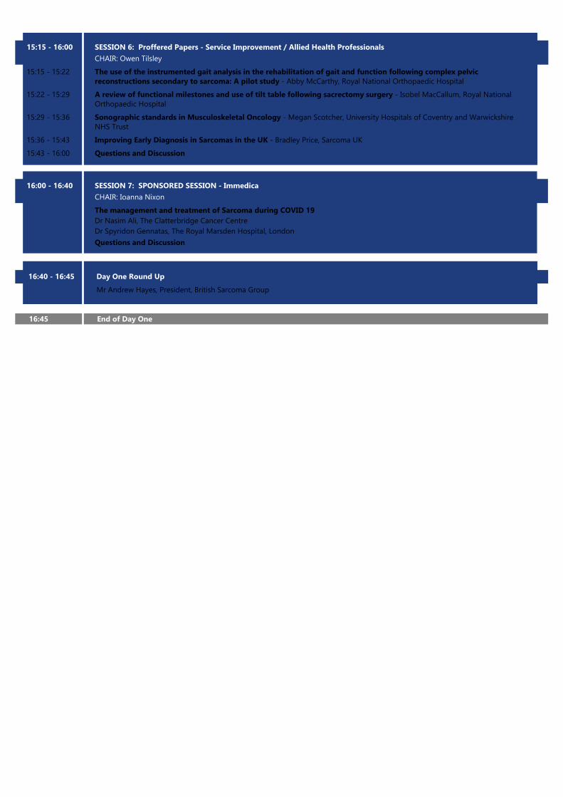
16:00 - 16:40 SESSION 7: SPONSORED SESSION - Immedica
CHAIR: Ioanna Nixon
The management and treatment of Sarcoma during COVID 19 Dr Nasim Ali, The Clatterbridge Cancer Centre Dr Spyridon Gennatas, The Royal Marsden Hospital, London
Questions and Discussion
16:40 - 16:45 Day One Round Up
Mr Andrew Hayes, President, British Sarcoma Group
16:45 End of Day One
15:15 - 16:00 SESSION 6: Proffered Papers - Service Improvement / Allied Health Professionals
CHAIR: Owen Tilsley
15:15 - 15:22 The use of the instrumented gait analysis in the rehabilitation of gait and function following complex pelvic reconstructions secondary to sarcoma: A pilot study - Abby McCarthy, Royal National Orthopaedic Hospital
15:22 - 15:29 A review of functional milestones and use of tilt table following sacrectomy surgery - Isobel MacCallum, Royal National Orthopaedic Hospital
15:29 - 15:36 Sonographic standards in Musculoskeletal Oncology - Megan Scotcher, University Hospitals of Coventry and Warwickshire NHS Trust
15:36 - 15:43 Improving Early Diagnosis in Sarcomas in the UK - Bradley Price, Sarcoma UK
15:43 - 16:00 Questions and Discussion
Conference Programme - Day Two: Thursday 25th February 2021
09:00 - 09:10 Welcome to Day Two
Mr Andrew Hayes, President, BSG
09:10 - 09:55 SESSION 8: Local therapies: Radiotherapy
CHAIR: Andrew Hayes
09:10 - 09:35 The Final results of the STRASS1 Trial - Dr Sylvie Bonvalot, Institute Curie, Paris
09:35 - 09:50 Relevance of STRASS1 for UK Practice - Dr Aisha Miah, Royal Marsden Hospital
09:50 - 09:55 Questions and Discussion
10:45 - 11:15 Morning break and poster viewing
11:15 - 12:15 SESSION 10: Proffered Papers - Surgery
CHAIR: Andrew Hayes and Paul Cool
11:15 - 11:22 Incidence and survival of patients with retroperitoneal Sarcoma (RPS) diagnosed in England between 2013-2018: An analysis from PHE NCRAS demonstrating the impact of specialist services on outcome - Shane Collins, University College London/NCRAS (PHE)
11:22 - 11:29 Impact of Cumulative Burden of Postoperative Complications on Oncological Outcomes in Patients Undergoing Surgery for Primary Retroperitoneal Sarcoma - Fabio Tirotta, University Hospital Birmingham NHS Foundation Trust
11:29 - 11:36 Prognostic implications of histological organ involvement in retroperitoneal sarcoma - Robert Tyler, University Hospital Birmingham
11:36 - 11:43 Follow-up protocol after Marginal resection of non-coelomic Atypical Lipomatous Tumours/Well-differentiated Liposarcomas - Gausihi Sivarajah, The Royal Marsden NHS Foundation Trust
11:43 - 11:50 Objective analysis of functional muscle transfers in lower limb sarcoma - Steven Lo, Canniesburn Plastic Surgery Unit Glasgow
11:50 - 11:57 Outcomes of limb-threatening sarcomas compromising major vessels: a 15 year single-centre review - Alex Dearden, Newcastle Hospitals Trust
11:57 - 12:15 Questions and Discussion
10:00 - 10:45 SESSION 9: Local therapies: Surgery CHAIR: Craig Gerrand
10:00 - 10:17 Multidisciplinary surgery for complex tumours at challenging sites - Mr Anant Desai, Queen Elizabeth University Hospital Birmingham
10:17 - 10:34 Patterns of treatment for Sarcoma in the UK. What we can learn from NCRAS - Dr Sandra Strauss, University College London Hospitals
10:34 - 10:45 Questions and Discussion
12:30 -13:30 Lunch and poster viewing
12:15 - 12:30 Update on the BSG: the BSG AGM
The BSG in the next decade - Mr Andrew Hayes, Consultant Surgeon, Royal Marsden Hospital and President, BSG
15:10 - 16:00 SESSION 13: SPONSORED SESSION - Bayer
CHAIR: Adam Dangoor
Targeting NTRK fusions
Prof Robin Jones, Royal Marsden Hospital
Dr Adam Dangoor, University Hospital Bristol
Questions and Discussion
16:00 - 16:10 Closing remarks & prizes
Mr Andrew Hayes, President, BSG
16:10 End of Conference
13:30 - 14:15 SESSION 11: COVID 19 : New Models of Care in the wake of
Coronavirus
CHAIR: Adam Dangoor
13:30 - 13:40 Virtual consultations and reducing risk during Covid – Dr Adam Dangoor, University Hospital Bristol
13:40 - 13:47 Remote consultations – Scotland – Dr Ioanna Nixon, Beatson West of Scotland Cancer Centre & Holly McCabe, The University of Strathclyde
13:47 - 13:54 Remote consultations - Marsden - Allanah Smrke & Eugenie Younger
13:54 - 14:01 Remote Prehab – Jane Masters, North Bristol Trust
14:01 - 14:08 Virtual Home Visits – Danielle Birch, The Robert Jones and Agnes Hunt Orthopaedic Hospital Nhs Foundation Trust
14:08 - 14:15 Questions and Discussion
14:15 - 15:00 SESSION 12: Pathology
CHAIR: Ian Judson
14:15 - 14:32 Update on 5th edition of the WHO classification of STS
Prof Adrienne Flanagan, Royal National Orthopaedic Hospital
14:32 - 14:49 Interpretation and implementation of WGS for Sarcoma
Dr Fernanda Amary, Royal National Orthopaedic Hospital
14:49 - 15:00 Questions and Discussion
15:00 - 15:10 Short afternoon break
13:00 - 14:00 SESSION 10: Free Talks
CHAIR: To be announced13:00 - 16:00 National Sarcoma Forum
See NSF programme on the
Conference website
Page 12
National Sarcoma Forum: Programme
12:50 - 12:55 Welcome to National Sarcoma Forum
Christine Millman, Chair, National Sarcoma Forum
12:55 - 13:45 SESSION 1: Regional updates
CHAIR: Christine Millman
Past, Present and future - Regional represenatives
EHNA, Health and Well-being Clinics updates - Regional represenatives
2WW triage clinic Bristol - Christine Millman, Sarcoma Clinical Nurse Specialist, North Bristol NHS Trust
Desmoid Fibromatosis patient conference - Anita Pabla, Macmillan Sarcoma Nurse Specialist, Leicester Royal Infirmary
Questions and Discussion
14:20 - 14:35 Afternoon Break
13:50 - 14:20 SESSION 2: Personalised Cancer Care CHAIR: Abby McCarthy
Personalised Care & Support Planning for People Living with Cancer: improving patient experience and
outcomes - Joanna Fairhurst, Macmillan Professional (Education) and Development Lead for Workforce
Embedding Personalised Cancer Care into the Sarcoma Patient Pathway - Suzy Hudson, Royal National Orthopaedic Hospital
Questions and Discussion
15:25 - 15:55 SESSION 4: Proffered Papers
CHAIR: Christine Millman & Abby McCarthy
Evolving role of a sarcoma radiographer and the benefits to patient care - Charlotte Harvey-Wright, Cambridge University Hospitals NHS Foundation Trust
Walking Balance and Compensatory Gait Mechanisms in Surgically treated Patients for Pelvic Musculoskeletal tumours – A Presentation of Preliminary Findings - Sherron Furtado, Royal National Orthopaedic Hospital
A multidisciplinary approach to the care of sarcoma patients within the proton beam therapy centre - Kirsten Ogden, The Christie NHS Foundation Trust
Questions and Discussion
15:55 - 16:00 Closing remarks
Christine Millman, Chair, National Sarcoma Forum
16:00 End of NSF
14:35 - 15:20 SESSION 3: Psychological Support
CHAIR: Christine Millman
Staff Well-being - Natalie Stott, North Bristol NHS Trust
Compassion fatigue - Julia Cordey, North Bristol NHS Trust
Sarcoma UK Patient Support line - Helen Stradling and Sam Hackett, Sarcoma UK
Questions and Discussion
Page 13
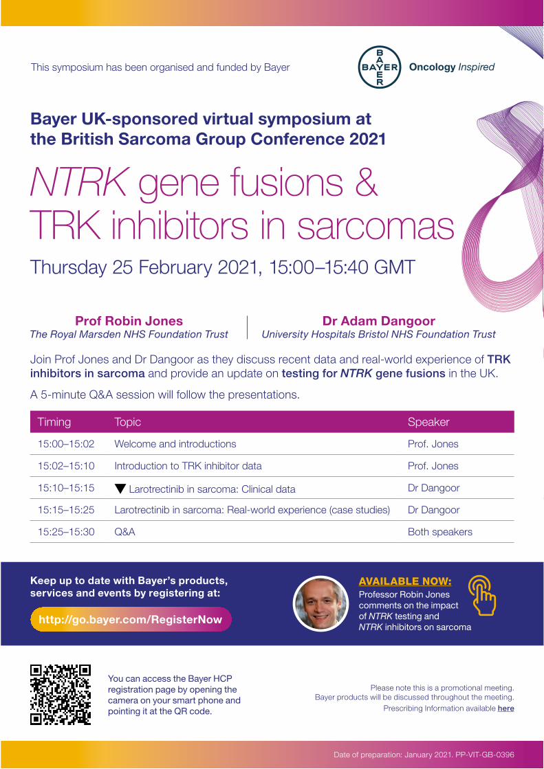
This symposium has been organised and funded by Bayer
NTRK gene fusions & TRK inhibitors in sarcomas
Bayer UK-sponsored virtual symposium at the British Sarcoma Group Conference 2021
Thursday 25 February 2021, 15:00–15:40 GMT
Join Prof Jones and Dr Dangoor as they discuss recent data and real-world experience of TRK inhibitors in sarcoma and provide an update on testing for NTRK gene fusions in the UK.
A 5-minute Q&A session will follow the presentations.
Please note this is a promotional meeting. Bayer products will be discussed throughout the meeting.
Prescribing Information available here
Timing Topic Speaker
15:00–15:02 Welcome and introductions Prof. Jones
15:02–15:10 Introduction to TRK inhibitor data Prof. Jones
15:10–15:15 Larotrectinib in sarcoma: Clinical data Dr Dangoor
15:15–15:25 Larotrectinib in sarcoma: Real-world experience (case studies) Dr Dangoor
15:25–15:30 Q&A Both speakers
Date of preparation: January 2021. PP-VIT-GB-0396
Prof Robin Jones The Royal Marsden NHS Foundation Trust
Dr Adam Dangoor University Hospitals Bristol NHS Foundation Trust
Keep up to date with Bayer’s products, services and events by registering at:
http://go.bayer.com/RegisterNow
AVAILABLE NOW:Professor Robin Jones comments on the impact of NTRK testing and NTRK inhibitors on sarcoma
You can access the Bayer HCP registration page by opening the camera on your smart phone and pointing it at the QR code.
Page 14
Posters1. Timing of free flap reconstruction in sarcoma patients Adam Misky, Royal Free NHS Foundation Trust
2. Perivascular epithelioid cell tumour (PEComa): the London Sarcoma Service experience (LSS). Alessandra Maleddu, University College Hospital London
3. Impact of the Covid-19 pandemic on patients receiving palliative sarcoma chemotherapy: the London Sarcoma Service experience. Alessandra Maleddu, University College Hospital London
4. Synovial sarcoma; a time sensitive tumour. Alex Dearden, Newcastle Hospitals NHS Foundation Trust
5. Modeling the efficacy of NY-ESO-1 TCR T cells (letetresgene autoleucel; GSK3377794) in patients with synovial sarcoma (SS): correlations of response with transduced cell kinetics and biomarkers Alexandra Gyurdieva, GlaxoSmithKline
6. Improving our Understanding of a Rare Diagnosis: A review of 13 cases of leiomyomatosis treated at The Royal Marsden Hospital Alexandra Ostler, The Royal Marsden NHS Foundation Trust
7. Real-world efficacy and safety of eribulin in advanced liposarcoma patients Andrea Napolitano, Sarcoma Unit, Royal Marsden Hospital, London
8. Pleomorphic dermal sarcoma and atypical fibroxanthoma of the scalp: Perspectives on treatment from a tertiary centre Anissa McClelland, Royal Marsden Sarcoma Unit
9. Impact of delayed sarcoma-surveillance imaging during the COVID-19 Pandemic. A
prospective audit Anne McTiernan, University College London Hospitals NHS Trust
10. Impact of delayed surveillance imaging for patients with desmoid tumour during the COVID-19 Pandemic. A prospective audit Anne McTiernan, University College London Hospitals NHS Trust
11. What is the value of routine chest x-ray in patients with lower risk sarcoma sub-types: A retrospective audit from the London Sarcoma Service (LSS) Anne McTiernan, University College London Hospitals NHS Trust
12. Cryoablation for desmoid-type fibromatosis Anneke Alves, The Royal Marsden Hospital
13. IMRiS: A phase II study of intensity modulated radiotherapy (IMRT) in extremity soft tissue sarcoma (STS) Beatrice Seddon, University College Hospital, London
14. Sarcoma Patient Experience in the UK: Results of the National Sarcoma Survey 2020 Bradley Price, Sarcoma UK
15. Advanced GIST tumours; A New Zealand Institutional Experience with Avapritinib and Ripretinib Catherine Bennett, Auckland City Hospital, New Zealand
16. Is CT chest re-staging necessary before surgery following preoperative radiotherapy for limb sarcoma? Cecilia McCormick, Addenbrooke’s Hospital, Cambridge
17. The Sarcoma Specialist Radiographer: A novel but necessary keyworker in radiotherapy delivery Clare David, University College Hospital
Page 15
18. Changes in a sarcoma Occupational Therapy pre-operative service during the Covid-19 pandemic: a lasting change Clare McKenzie Oxford Sarcoma service, Oxford University Hospitals NHS Foundation Trust
19. Functional outcomes in patients with custom shoulder prosthesis Clare McKenzie, Oxford sarcoma service
20. Extrauterine Smooth Muscle Tumours of Uncertain Malignant Potential: A review of 28 patients treated at a tertiary centre Edward Phillips, The Royal Marsden Hospital
21. Clinical examination is important in detecting tumour recurrence but not metastasis Elizabeth Ridgway, University Hospitals Plymouth NHS Trust / SW Peninsula Sarcoma Service
22. An Undiagnosed Case of Tenosynovial Giant Cell Tumour that Deteriorates During Chemotherapy for an Unrelated Leiomyosarcoma Fay Tough, Beatson West of Scotland Cancer Centre
23. A large cancer centre’s audit of over 100 cases of pleomorphic dermal sarcoma of the scalp Fei Sun, Leeds Teaching Hospitals
24. Preoperative versus postoperative radiotherapy for extremity soft-tissue sarcoma – A systematic review and meta-analysis of long-term survival Garikai Kungwengwe, The Welsh Centre for Burns and Plastic Surgery
25. Prodromic presentation of Retroperitoneal Sarcoma determines Prognosis Gausihi Sivarajah, The Royal Marsden NHS Foundation Trust
26. Primary Mesenteric Sarcomas: Collaborative experience from the Trans-Atlantic Australasian Retroperitoneal Sarcoma
Working Group (TARPSWG) Hannah L Tattersall, Midlands Abdominal and Retroperitoneal Sarcoma Unit, Queen Elizabeth Hospital Birmingham
27. Case report: Sinonasal tumour - a diagnostic conundrum Haran Devakumar, Imperial College Healthcare Trust
28. A Review Of The Role Of Intensity Modulated Radiotherapy (IMRT) For Sarcoma At One UK Institution Helen Clarke, University Hospitals Birmingham Foundation Trust
29. Physical function following bone reconstruction versus no bone reconstruction for pelvic primary bone sarcoma – a systematic review Helen Seaman, University College London
30. How COVID-19 has affected the lives of people contacting the Sarcoma UK support line Helen Stradling, Sarcoma UK
31. An 11 year evaluation of MPNST in Neurofibromatosis Type 1 (NF1); The Manchester Experience Judith Eelloo, Complex NF1 Service, Manchester University NHS Foundation Trust
32. Forequarter amputation reconstruction by free upper limb fillet flap – investigation of heparinised saline infusion to mitigate warm ischaemia effect & technical tips. Karen Lindsay, Canniesburn Plastic Surgery Unit
33. Sarcoma of the distal fibula; presentation of an anatomical reconstruction and literature review Katherine Wensley, Birmingham Women’s and Children’s NHS Foundation Trust
34. Giant Cell Tumour of Tendon Sheath (GCTTS) of the foot and ankle – a case series Katriona Pierce, Liverpool University Hospitals NHS Foundation Trust
Page 16
35. Chondrosarcoma of Bone: Analysis of prognostic factors and outcomes from a tertiary care centre in India Keerthi MSS, Cancer Institute (WIA), Adyar, Chennai, Tamil Nadu, India
36. Value of chest X-ray surveillance in extremity soft tissue sarcomas Kiran Ramesh, Manchester Hospitals NHS Foundation Trust
37. Do Stanmore Steppers interventions improve outcomes after major surgery for musculoskeletal tumours? Laura Mitham, Royal National Orthopaedic Hospital
38. An evaluation of the feasibility of a nurse led Giant Cell Tumour clinic for the administration of neo-adjuvant Denosumab. Laura Morrison, University College London Hospital
39. Novel systemic anti-cancer therapy (SACT) for metastatic Epitheliod Sarcoma (ES) during the CoVid pandemic Leena Mukherjee, Beatson West of Scotland Cancer Centre
40. Case Study: Ewing’s like sarcoma, a change to the current paradigm? Lotte Hardman, Southmead Hospital
41. Our COVID-19 response to a dedicated primary bone cancer Support and Information Service Louise Kirby, Bone Cancer Research Trust
42. Outcomes of Ewing Sarcoma/ Ewing’s-Like Sarcoma in patients aged over 25 Mabe Teng, University hospital Birmingham
43. Proton Beam Radiotherapy (PBT) in Adult/TYA sarcomas- initial toxicity and outcomes. Maeve Keys, The Christie NHS Foundation Trust
44. Promoting Excellence in Multidisciplinary
Team Meetings: A Sarcoma Service Review of Practice Two Years On Mandeep Kang, East Midlands Sarcoma Service
45. Patient satisfaction with Virtual Care in a specialist sarcoma centre in Ireland during the Covid-19 pandemic Mary O Reilly, St Vincents University Hospital Dublin
46. The Value of a virtual Sarcoma support group during a global pandemic Maxine Cumbo, The Christie
47. Incidence of Chondrosarcoma in the Hand – a 13 Year Experience Michael Khoo, Royal National Orthopaedic Hospital
48. The Implementation of a Nurse Led SACT clinic to reduce waiting times for patients with soft tissue sarcoma`s (STS) attending for systemic anti-cancer treatment (SACT). A service improvement project between the Beatson West of Scotland Cancer Centre (BWO) Miriam Brady, Beatson West of Scotland Cancer Centre
49. Salvage radiotherapy for an unresectable aorta recurrence of an angiosarcoma. Nicolas Jullian, MD Institut Jules Bordet-Université Libre de Bruxelles, Department of Radiation-Oncology, Brussels, Belgium
50. Tibial transfer following high femoral resection in paediatric sarcoma patients Oliver Bassett, Newcastle Hospitals NHS Foundation Trust
51. Update of Newcastle Head and Neck Sarcoma Experience Oliver Bassett, Newcastle Hospitals NHS Foundation Trust
52. Mohs micrographic surgery for cutaneous sarcoma – a review of current evidence. Oliver Bassett, St Helens and Knowsley Hospitals NHS Trust
Page 17
53. A 10-year experience of primary angiosarcomas in a regional bone and soft tissue service Oliver Bassett, Newcastle Hospitals NHS Foundation Trust
54. A VERY LARGE EXTRASKELETAL MESENCHAYMAL CHONDROSARCOMA: CASE REPORT Qi Yin, Royal Liverpool University Hospital, Liverpool University Hospital NHS Fundation Trust, Liverpool
55. Development of a sarcoma-specific fear of recurrence intervention: REASSURE_ME feasibility protocol Rachel Taylor, University College London Hospitals NHS Foundation Trust
56. “Am I going to be told it’s come back?”: Experiences of fear of recurrence (FoR) in sarcoma patients Rachel Taylor, University College London Hospitals NHS Foundation Trust
57. External Validation of the Toronto Sarcoma Flap Score Rebecca Rollett, Nottingham University Hospitals
58. Functional Gait following Lower Limb Amputation for Soft Tissue Sarcoma (STS) Rob Turner, Leeds Cancer Centre
59. 5-year review of DFSP cases with 3cm excision margins Robert Slade, Welsh Centre for Burns and Plastic Surgery
60. The role of the SDHA fusion gene in tumourigenesis and tumour metabolism in retroperitoneal liposarcoma Robert Tyler, University of Birmingham
61. Use of Whole Body MRI for staging myxoid liposarcoma. To CT or not to CT? Saiidy Hasham, East midlands Sarcoma Service
62. The Welsh Experience of Lymphatic Metastasis in Soft Tissue Sarcoma Sam Leong, Morriston Hospital, Swansea
63. Plastic Surgery Trainee Perspective on Soft Tissue Sarcoma Care and Training Sam Leong, The Welsh Centre for Burns and Plastic Surgery, Swansea
64. Safety and Activity of Autologous T Cells With Enhanced NY-ESO-1–Specific T-Cell Receptor (letetresgene autoleucel; GSK3377794) in HLA-A*02+ Previously-Treated and -Untreated Patients With Advanced Metastatic/Unresectable Synovial Sarcoma (SS): A Master P Sandra Strauss, University College London Hospitals NHS Foundation Trust, London, UK
65. The East of England Sarcoma Task and Finish Group: Implementation and standardisation of an agreed radiotherapy protocol in the network in a rare tumour Sarah Prewett, Cambridge University Hospitals NHS Trust
66. Expectations of mobility following lower limb massive endo-prosthetic replacement (EPR) for primary bone sarcoma in children Sherron Furtado, Royal National Orthopaedic Hospital NHS Trust
67. What Do We Know about the Lived experience of Pain and Fatigue after Treatment for Extremity Sarcoma? - A Systematic Review Sherron Furtado, Royal National Orthopaedic Hospital NHS Trust
68. Limb salvage with radical radiotherapy for a high-grade sarcoma in an elderly patient who refused amputation. Sofian Benkhaled, MD Institut Jules Bordet-Université Libre de Bruxelles, Department of Radiation-Oncology, Brussels, Belgium
69. Patterns of referral for extracranial spinal sarcomas for Proton Beam Therapy ( PBT) Srijith Sashidharan, The Christie NHS
70. Impact of the SARS-CoV-2 pandemic on trial patients: the research nurse contribution Steven Edmunds, Royal Marsden NHS Foundation Trust
Page 18
71. SUSPECTED SARCOMA REFERRALS FROM PCT DURING THE COVID-19 PANDEMIC Thatayaone Marotsi, University of Liverpool
72. Targeted treatment for neurotrophic tyrosine receptor kinase (NTRK) fusion-positive sarcomas: Two case reports Timothy Spencer, University Hospitals Bristol and Weston NHS Foundation Trust
73. Bone Cancer Awareness Initiative- Saving Lives Through Earlier Diagnosis Victoria Vinader Bone Cancer Research Trust
74. Do mainstream prognostic scores reliably predict complication rates in patients undergoing pelvic/femoral resection & reconstruction for bone tumours? Viswanath Jayasankar, Glasgow Royal Infirmary
75. Gastrointestinal Stromal Tumours: Radiotherapy Treatment and Clinical Outcomes at The Royal Marsden Sarcoma Unit 2010-2019 Yolanda Augustin, Royal Marsden Sarcoma Unit
Page 19C-APROM/UKI/MEP/0004 | February 2021
This promotional meeting is organised and fully funded by Takeda Oncology and is intended for UK and Ireland healthcare professionals only. Takeda medicines will be discussed at this meeting. Prescribing Information will be available during the meeting.
Save the Date: Webinar
The Osteosarcoma Treatment Story: Past, Present and Future
If you are interested in attending this meeting, please contact Steve Meredith ([email protected]) or Tory Griffiths ([email protected]). Further details on how to register for this meeting, along with a full agenda, will follow.
Chaired by Professor Bruce Morland (Birmingham Children’s Hospital), with a panel of national experts in sarcoma and orthopaedic surgery, plus a patient’s perspective
13th May 2021 13:30-16:00
Page 20
Thank you to our Sponsors
Immedica is a fast-growing private niche pharma group. Its headquarter is based in Stockholm, Sweden, and it has commercial coverage across Europe and the Middle East. Immedica provides significant know-how and experience from commercialization of niche/specialty care products across Europe and the Middle East, and the company’s management team has an outstanding track record of operating niche pharma products internationally. Immedica has capabilities to provide optimal access of specialty care medicines to patients with significant medical needs, including key areas such as regulatory affairs, pharmacovigilance, medical affairs, pricing & reimbursement, quality and product distribution
Website: https://immedica.com/
Bayer plc aims to develop products that improve people’s quality of life. We concentrate on the research and development of innovative drugs and novel therapeutic approaches to prioritise targets and pathways with the potential to impact not just the treatment of common cancers, but rarer cancers as well.
UK healthcare professionals and other relevant UK decision makers can register here to subscribe to electronic communications about Bayer’s products, services and events: https://hcpregistration.bayer.co.uk/
Visit https://oncology.bayer.co.uk/ for more information.
Takeda Pharmaceutical Company Limited is headquartered in Japan and is a global, values-based, R&D-driven biopharmaceutical leader committed to translating science into highly innovative medicines. Takeda focuses its R&D efforts on four therapeutic areas: Oncology, Gastroenterology (GI), Neuroscience and Rare Diseases. We also make targeted R&D investments in Plasma-Derived Therapies and Vaccines. Our employees are committed to improving quality of life for patients and to working with our partners in health care in approximately 80 countries and regions.
Additional information about Takeda UK Ltd. is available through its corporate website www.takeda.com/en-gb
Virtual Conference Partner:
Patinum Sponsor:
Gold Sponsor:
Page 21
Silver Sponsor:
Silver Sponsor:
Silver Sponsor:
Blueprint Medicines is a global precision therapy company striving to improve human health. With a focus on genomically defined cancers, rare diseases, and cancer immunotherapy, we combine our leading expertise in protein kinases with a uniquely targeted, scalable approach to empower the rapid design and development of transformational medicines.
Website: www.blueprintmedicines.com
Karyopharm Therapeutics is an innovation-driven pharmaceutical company focused on the discovery, development, and commercialization of medicines with the goal of improving the lives of patients with cancer. Our company was founded in 2008 with a vision of pioneering a potentially new approach to treating patients with cancer and other serious diseases. Our novel approach to cancer treatment involves targeting the export of specific proteins in a cancer cell’s nucleus with SINE technology (Selective Inhibition of Nuclear Export).
Our primary focus is on developing novel drugs which we hope will help treat patients with certain blood cancers or solid tumor malignancies.
Website: www.karyopharm.com/
We have three global businesses that research, develop and manufacture innovative pharmaceutical medicines, vaccines and consumer healthcare products. Every day, we help improve the health of millions of people around the world.
Our goal is to be one of the world’s most innovative, best performing and trusted healthcare companies. Our values and expectations are at the heart of everything we do - so that together we can deliver extraordinary impact for our patients and consumers and make GSK a brilliant place to work.
Our values are Patient focus, Transparency, Respect, IntegrityOur expectations are Courage, Accountability, Development, Teamwork
Website: www.gsk.com/en-gb/about-us/
Abstracts - Proffered Papers
Page 22
DAY ONE - SESSION 3: 11:30 - 12:30 Long-term efficacy, tolerability, and overall survival in patients with unresectable or metastatic (U/M) PDGFRA D842V-mutant gastrointestinal stromal tumour (GIST) treated with avapritinib: NAVIGATOR phase 1 trial update Robin Jones, Royal Marsden Hospital and Institute of Cancer Research; César Serrano, Vall d’ Hebron Institute of Oncology; Margaret von Mehren, Fox Chase Cancer Center; Maria Roche, Blueprint Medicines Corporation; Sebastian Bauer, Sarcoma Center, West German Cancer Center, University of Duisburg-Essen Introduction: PDGFRA D842V-mutant GIST is highly resistant to approved kinase inhibitors. Avapritinib, a novel KIT/PDGFRA kinase inhibitor, potently inhibits D842V-mutant PDGFRA. Methods: In NAVIGATOR (NCT02508532), patients with U/M PDGFRA D842V-mutant GIST (regardless of prior therapy) received oral, once-daily avapritinib (dose escalation, 30–600 mg; dose expansion, 300 mg [recommended phase 2 dose]/400 mg [maximum tolerated dose]). Here, we report long-term analyses for 38 patients with PDGFRA D842V-mutant GIST treated at 300/400 mg. Results: At 9 March 2020 (median follow-up, 26 months), overall response rate (modified Response Evaluation Criteria in Solid Tumours version 1.1) was 95% (36/38; 5 [13%] complete responses [CR], 31 [82%] partial responses [PR]). Median duration of response was 22 months (95% confidence interval [CI] 14–not reached [NR]). Median progression-free survival (PFS) was 24 months (95% CI 18–NR), and median overall survival (OS) was NR. At 36 months, PFS and OS were 34% and 71%. Of 5 tyrosine kinase inhibitor–naïve patients, 2 had a CR and 3 a PR. Most common adverse events (AEs) in patients treated at 300/400 mg (?30%) were nausea (74%), anaemia (68%), diarrhoea (66%), fatigue (58%), memory impairment (47%), periorbital oedema (45%), decreased appetite (39%), increased lacrimation (34%), and vomiting, abdominal pain, hypokalaemia, increased blood bilirubin and peripheral oedema (all 32%). Overall, 21% of patients discontinued treatment due to drug-related AEs. No treatment-related deaths were reported. Conclusion: In patients with U/M PDGFRA D842V-mutant GIST, avapritinib demonstrated robust, durable responses and a tolerable safety profile with no new safety signals.
DAY ONE - SESSION 3: 11:30 - 12:30 Final analysis of the Phase 1 trial of NY-ESO-1–specific T-cell receptor (TCR) T-cell therapy (letetresgene autoleucel; GSK3377794) in patients with advanced synovial sarcoma (SS)
Sandra P. D’Angelo, Memorial Sloan Kettering Cancer Center, New York, NY, USA; George D. Demetri, Dana-Farber Cancer Institute and Ludwig Center at Harvard, Boston, MA, USA; Brian A. Van Tine, Washington University in St. Louis, St. Louis, MO, USA; Mihaela Druta, H. Lee Moffitt Cancer Center, Tampa, FL, USA; John Glod, National Cancer Institute, Bethesda, MD, USA; Warren Chow, City of Hope Comprehensive Cancer Center, Duarte, CA, USA; Aisha Hasan, GlaxoSmithKline, Collegeville, PA, USA; Victoria L. Chiou, GlaxoSmithKline, Collegeville, PA, USA; Jenna Tress, GlaxoSmithKline, Collegeville, PA, USA; Julie Edwards, GlaxoSmithKline, Stockley Park, Middlesex, UK; Tim Young, GlaxoSmithKline, Collegeville, PA, USA; Mary Woessner, GlaxoSmithKline, Collegeville, PA, USA; Alexandra Gyurdieva, GlaxoSmithKline, Collegeville, PA, USA; Stefan Zajic, GlaxoSmithKline, Collegeville, PA, USA; Sophia Goodison, GlaxoSmithKline, Collegeville, PA, USA; Dejka Araujo, University of Texas/MD Anderson Cancer Center, Houston, TX, USA Introduction: Letetresgene autoleucel (lete-cel) is a genetically-modified autologous T-cell immunotherapy that recognizes tumors expressing NY-ESO-1 and HLA-A*02. This Phase I trial (NCT01343043) assessed safety, efficacy, and pharmacokinetics of lete-cel in patients with SS. Methods: Patients with unresectable/metastatic/recurrent SS enrolled in Cohort-1 (high NY-ESO-1 expression/high lymphodepleting regimen [LDR]), Cohort-2 (low NY-ESO-1/high LDR), Cohort-3 (high NY-ESO-1/high single-agent LDR), or Cohort-4 (high NY-ESO-1/low LDR) before lete-cel infusion. Primary endpoint was investigator-assessed ORR per RECIST v1.1; secondary endpoints included DoR, PFS, OS, and safety. Persistence was measured by qPCR. Results: Forty-five patients received lete-cel. Cohort demographics were similar. ORR was 50%/31%/20%/27% in Cohorts 1/2/3/4, respectively; with 1 complete (lasting 34-weeks) and 14 partial responses. In Cohorts 1/2/3/4, median DoR was 31.0/8.6/32.1/16.4 weeks; median PFS was 15.4/13.1/8.6/22.4 weeks, respectively. As of 27Jan2020, median OS for Cohorts 1/2/3 was 24.3/9.9/19.9 months, (Cohort-4 immature; data from long-term follow-up study). Grade ?3 AEs were mostly hematologic; Grade ?3 SAEs were most frequently febrile neutropenia, dyspnea, and neutropenia. AEs of special interest included cytokine release syndrome (n=20; maximum Grade 1/2/3/4 in 9/7/3/1 patients, respectively; SAEs [n=5; Grade ?3 n=2]; all resolved); Guillain-Barré syndrome (n=2; Grade 3 SAEs; resolved with sequalae); and multilineage cytopenias (n=43; maximum Grade 5 n=1, Grade 3/4 n=42). Median peak persistence was generally higher in responders vs non-responders; time to peak persistence was similar. Replication-competent lentivirus was not detected. Conclusions: Lete-cel had a manageable
Abstracts - Proffered Papers
Abstracts - Proffered Papers
Page 23
safety profile. Responses occurred in all cohorts; patients in Cohort-1 received greatest benefit. Previously presented at SITC 2020 (P298). Funding: GSK (208466). DAY ONE - SESSION 3: 11:30 - 12:30 Incidence and Outcome of Breast Sarcomas in NHS England (2013-2018): An Analysis from Public Health England (PHE) National Cancer Registration and Analysis Service (NCRAS) Mahbubl Ahmed, The London Sarcoma Service, UCLH; Sandra Strauss, The London Sarcoma Service, UCLH; Shane Collins, Public Health England – Wellington House, 133-155 Waterloo Road, London, SE1 8UG, United Kingdom; Andrew Bacon -Public Health England – Wellington House, 133-155 Waterloo Road, London, SE1 8UG, United Kingdom Lizz Paley -Public Health England – Wellington House, 133-155 Waterloo Road, London, SE1 8UG, United Kingdom Background: Breast sarcomas (BS) are rare cancers originating from mesenchymal breast tissue with a paucity of national population level data detailing their incidence and outcomes. Methods: We performed an analysis of data collected by NCRAS for patients diagnosed with BS 2013-2018. Chi-square test was used to compare groups. Overall survival was calculated by Kaplan-Meier. Specialist sarcoma centres (SSC) were defined as centres with a sarcoma MDT. Results: There were 688 patients with BS (357 malignant phyllodes (MP), 238 vascular tumours, 93 other morphology) with a median age of 64 (range 14-96); 187(27%) had received breast radiotherapy for a prior malignancy; 633(92%) had resection of the tumour within 12 months of diagnosis. When a biopsy was not performed prior to surgery, patients were more likely to undergo multiple operations (58%v29%, P<0.05). Biopsies were more frequently performed at SSC than non-specialist sarcoma centres (NSSC) (83%v72%, P<0.05) with 26% of patients undergoing multiple operations in SSC compared to 41% of patients undergoing first surgery at NSSC. Five-year overall survival was 81%, 50% and 45% in patients with MP, vascular tumours and other sarcomas, respectively and 60% for those with radiation-induced BS. Conclusion: This is the first population series evaluating incidence and outcomes for BS. Patients treated at NSSC are less likely to have a biopsy prior to surgery and more likely to require multiple operations. Based on this observational data we would recommend all BS are discussed at a sarcoma MDT early in their pathway and surgery to be performed at SSC where possible.
DAY ONE - SESSION 3: 11:30 - 12:30 Myxoid Liposarcoma: Evaluation of Response to Radiotherapy, Surgical and Clinical Outcomes at the Royal Marsden Sarcoma Unit between 1990-2018 Yolanda Augustin, Royal Marsden Sarcoma Unit; Aisha B Miah (Senior Author), Royal Marsden Sarcoma Unit; ,
; Yolanda Augustin, Shane Zaidi, Andrew J Hayes, Khin Thway, Christina Messiou, Aisha B Miah Background Myxoid liposarcoma is a rare soft tissue sarcoma (STS) predominantly affecting limb extremities. MLS is radiosensitive and surgery with pre-operative radiotherapy is preferred for intermediate and high-grade tumours. Materials and Methods Retrospective case notes review of MLS patients treated with surgery +/- radiotherapy at the Royal Marsden Sarcoma Unit between 1990-2018. We report radiotherapy response, local control rates and patterns of relapse. Results 243 patients were included. Tumours were located in the lower limb 211/243 (87%), upper limb 10/243 (4%) and trunk 23/243 (9%). 147/243 patients (61%) had surgery alone, 54/243 (22%) PRE-OP RT and 42/243 (17%) PORT. Median radiotherapy dose PRE-OP was 50Gy (range 36Gy-50Gy) and PORT was 60Gy (range 60-66Gy). Radiotherapy was well tolerated with mainly G1/G2 toxicity. Overall 20/243 (8%) patients developed local relapse. None treated with RT developed local relapse. All patients with isolated local relapse proceeded to salvage surgery. 14/20 (70%) of patients undergoing salvage surgery received PRE-OP RT/PORT. 45/243 (19%) patients developed distant metastases. Patterns of distant relapse were multifocal and included lung (21/45 cases), intra-abdominal (28/45 cases), bone (20/45 cases), nodal (5/45 cases) and distant limb (5/45 cases). Survival outcomes will be reported. Conclusion Combined modality treatment results in excellent local control and good functional outcome. Following the results of DOREMY, dose de-escalation PREOP RT may be considered but warrants further studies to predict for responders to RT. Patterns of relapse are different to other histological subtypes confirming a need to adapt surveillance imaging for MLS.
DAY ONE - SESSION 3: 11:30 - 12:30 Preoperative versus postoperative radiotherapy for extremity soft-tissue sarcoma – A systematic review and meta-analysis of long-term survival Garikai Kungwengwe, The Welsh Centre for Burns and Plastic Surgery; Rachel Clancy, Southmead Hospital; Joanne Vass, The Welsh Centre for Burns and Plastic Surgery; Robert Slade - Southmead Hospital Simarjit Sandhar - Queen Charlotte’s & Chelsea Hospital Thomas Dobbs - The Welsh Centre for Burns and Plastic Surgery Thomas Bragg - The Welsh Centre for Burns and Plastic Surgery Introduction The implications of radiotherapy for resectable extremity soft tissue sarcoma (ESTS) have been widely documented for local tumour control, wound complications, and long-term function. However, debate continues regarding pre- versus postoperative radiotherapy for long-term survival. This study aimed to determine whether the timing of perioperative radiotherapy affects long-term survival outcomes in adult patients with ESTS. Methods A systematic literature search of MEDLINE, EMBASE,
Abstracts - Proffered Papers
Page 24
Web of Science, and Cochrane was performed. The primary outcome measure was the pooled hazard ratio (HR) at 95% confidence intervals. Secondary outcomes and subgroup analyses were presented as cumulative odds ratios (OR). A random-effects, generic inverse variance method, and sensitivity analysis were performed to minimise and detect heterogeneity. Results Six studies (n=4192 patients) were identified. Meta-analysis demonstrated a statistically significant advantage in postoperative radiotherapy for overall survival (OS) (HR 1.15, p=0.05). Additionally, there was a non-significant trend supporting postoperative radiotherapy for disease-free (DFS) (HR 1.25, p=0.22) and cause-specific (CSS) (HR 1.06, p=0.43) survival. Postoperative radiotherapy was also shown to confer an OS (OR 1.19, p=0.01), DFS (OR 1.19, p=0.01), and CSS (OR 1.19, p=0.01) advantage on subgroup analysis. This survival advantage was best observed at three years in the DFS comparison (OR 1.55, p=0.003). Preoperative radiotherapy was associated with more wound complications (OR 2.74, p<0.00001). Conclusions Pooled analysis of published literature suggests postoperative radiotherapy confers a significant long-term survival advantage with fewer wound complications. Further large multi-centre randomised controlled trials with long-term follow-up are required to determine the optimal perioperative radiotherapy regime in adult ESTS.
DAY ONE - SESSION 3: 11:30 - 12:30 Durable Response to Serial Tyrosine Kinase Inhibitors (TKIs) in an Adolescent with Metastatic TFG-ROS1 fusion positive Inflammatory Myofibroblastic Tumour (IMT) Dr Katrina Ingley, Sarcoma Department, University College London Hospitals Foundation Trusts, United Kingdom; Debbie Hughes, Clinical Genomics Translational Research, Centre for Molecular Pathology, The Institute of Cancer Research Sutton, United Kingdom; Dr Michael Hubank, Molecular Diagnostics Department, The Institute of Cancer Research and Clinical Genomics, The Royal Marsden NHS Foundation, London, United Kingdom; 4. Dr Daniel, Lindsay, Department of Histopathology, Royal National Orthopaedic Hospital NHS Trust, Stanmore, United Kingdom. 5. Prof Louis, Chesler, Paediatric Tumour Biology, Division of Clinical Studies, The Institute of Cancer Research, London, United Kingdom. 6. Dr Sandra Strauss, Sarcoma Department, University College London Hospitals Foundation Trusts, United Kingdom. Introduction IMTs are rare mesenchymal tumours with a variable natural history. Genomic analysis can identify oncogenic kinase fusions, including anaplastic lymphoma kinase ALK and ROS1.1 We describe a patient with metastatic IMT harbouring a ROS1 fusion detected on tumour progression following benefit from multiple TKIs and retrospectively in the primary tumour after RNA sequencing. Methods Clinical data was acquired from patient records. Pathological
analysis included immunohistochemistry (IHC) for ALK, Fluorescent in situ hybridisation (FISH) for ALK and ROS1 fusions on diagnostic tissue and targeted fusion sequencing (TruSight Pan Cancer), targeted DNA capture panel sequencing (Paeds panel v2) on primary and relapsed tissue. Results A 15-year old girl presenting with a large thoracic mass and brain metastases was diagnosed with an IMT on biopsy of both disease sites. Diagnostic tumour was ALK negative by IHC with no evidence of ALK or ROS1 rearrangement on FISH. Following minimal benefit from anti-inflammatories and cytotoxic chemotherapy the patient experienced clinical and radiological response to crizotinib and brigatinib before progression of the primary tumour after 15 and 8 months, respectively. Targeted next generation panel sequencing from thoracic tumour biopsied at progression (CRUK-Stratified Medicine Paediatrics study, SMPaeds) detected a TFG-ROS1 fusion, also retrospectively identified on the initial biopsy but with lower confidence using clinical diagnostic, RNA-Seq assays. Treatment with third-generation ALK/ROS1 inhibitor, lorlatinib for 12 months resulted in resection of the primary tumour. Conclusion Serial TKIs may benefit IMT patients with ALK/ ROS1 alterations. Repeat biopsy and detailed molecular analysis is recommended to optimise ongoing therapy. References: 1. Lovly CM, Gupta A, Lipson D, et al: Inflammatory myofibroblastic tumors harbor multiple potentially actionable kinase fusions. Cancer Discov 4:889-895, 2014
DAY ONE - SESSION 6: 15:15 - 16:00 The use of the instrumented gait analysis in the rehabilitation of gait and function following complex pelvic reconstructions secondary to sarcoma: A pilot study Abby McCarthy, Royal National Orthopaedic Hospital Stanmore; Roisin Delany, Royal National Orthopaedic Hospital Stanmore; Prof Deborah Eastwood, Royal National Orthopaedic Hospital Stanmore; Dr Sherron Furtardo Royal National Orthopaedic Hospital Stanmore Introduction: Massive pelvic tumour excision with/without reconstruction requires prolonged rehabilitation for optimal outcomes. Recent literature describes a lack of objective outcome measures. Aim – To review the effect of instrumented gait analysis in targeting rehabilitation and improving outcome in patients who underwent tumour excision/reconstruction. Methods: Standard rehabilitation was supplemented with a 3D gait analysis collecting kinematic and kinetic data at 3 months (3m) to identify rehabilitation goals before the 9 months (9m) assessment which included the Toronto Extremity Salvage Score (TESS) as a patient reported functional outcome measure and the Quality of Life – Cancer Survivors QoL-CS measure. Results: Four male patients, mean age 42.5yrs (range 34-52yrs): 1 had no reconstruction, 3 underwent surgical resections with hemipelvic cone. 1 patient required postoperative
Abstracts - Proffered Papers
Page 25
plastic surgical intervention. In all cases, the 3m gait analysis exposed compensation strategies which improved by 9m. Common themes focussed rehabilitation on normalising pelvic movement patterns, improving symmetry and pelvic stabilisation to enable coupling of hip/knee/ankle joints. The gait data (objective parameters of speed, cadence, symmetry) all improved from 3m to 9m. Gait profile scores improved from the first to the second assessments from a mean of 8.5 (7-9.35) to 7.02 (6.76 – 7.4) At 9m, the mean TESS score was 74.2 (69.5–79.69). The mean QoL-C5 demonstrated good physical scores (mean 8.6) but low scores (mean 5.8) in the psychological domain. Conclusion: Preliminary results support the use of instrumented gait analysis, TESS and QOL-CS in reflecting outcomes following complex hip reconstruction and encouraging a tailored physical and psychological programme.
DAY ONE - SESSION 6: 15:15 - 16:00 A review of functional milestones and use of tilt table following sacrectomy surgery Isobel MacCallum, Royal National Orthopaedic Hospital; Abby McCarthy, Royal National Orthopaedic Hospital; Sherron Furtado, Royal National Orthopaedic Hospital Introduction: Post-operative protocol following sacrectomy with plastic surgery limits hip flexion and avoids pressure on the wound. This leads to extended bed rest affecting function, strength, range of movement, blood pressure and mood. This service evaluation reviewed the early use of tilt table following surgery to establish whether patients stand and mobilise independently sooner. Methods: 10 sets of data collected; 5 pre and 5 post change in practice. During Plastics Ward Round post-operative restrictions were discussed with consideration of whether tilt tabling could commence. Data collection recorded number of days post-operatively patients used the tilt table, mobilised, transferred from prone to standing, perched and discharged. Results: Mean age 60±15.8 years. 9 patients had a sacrococcyx chordoma and 1 a malignant peripheral nerve sheath tumour. 90% of patients were male. All were treated with a partial or sub-total sacrectomy to remove tumour. Number of days post sacrectomy surgery patients stood on the tilt table significantly reduced from a mean of 13±5.1 days to 4.8±2.8 days (p=0.013); the number of days patients mobilised significantly reduced from 28±13 to 10.6±2.7 days (p=0.019); days to prone transfer off the bed reduced from 22.8±8 to 16.5±2.1 days (p=0.344). Number of days post op to perch significantly decreased from 39.5±5.8 to 23.6±1.34 days (p= 0.01) and discharge from hospital decreased from 58.2±21.8 to 37.4±11.9 days (p=0.097). There were several post-operative complications which affected rehabilitation. The plastic reconstruction used varied. Conclusion: Results support the use of tilt table following sacrectomy in hastening the achievement of physical
milestones.
DAY ONE - SESSION 6: 15:15 - 16:00 Sonographic standards in Musculoskeletal Oncology Megan Scotcher, University Hospitals of Coventry and Warwickshire NHS Trust; Nicholas C Eastley, University Hospitals of Leicester NHS Trust; British Orthopaedic Oncology Society Collaborative, British Sarcoma Services; Introduction: To audit the quality of the current ultrasound (USS) assessment of soft tissue tumours referred to sarcoma 2 week wait clinics across the United Kingdom (UK). Method: A total of 194 US scan reports of soft tissue tumours were collated from seven sarcoma units. The quality of these reports were assessed against nine standards devised by the British Sarcoma Group in 2019. The quality of tumour characterisation, and necessity for subsequent referral onto 2WW clinic was reported. Results: Four of the nine standards were met by over 80% of the scans that were assessed. Tumours were generally well characterised with 5/9 standards met in 75% of USS reports. However, two standards were met by less than 60% of reports: 1) ‘Scan performed or supervised by clinician with FRCR or RCR accreditation’, 2) ‘Documentation that US performed with high resolution (up to 15/18mHz)’. Only 32.7% of 180 patients warranted urgent referral to a 2WW service based on their USS findings and BSG recommendations. Conclusions: Our results suggest there is scope to improve US reporting of soft tissue tumours across the United Kingdom. This may reflect variation in the interpretation of certain standards, a lack of local awareness of BSG guidelines and/or limited resources in MSK radiology departments. A large proportion of patients were not referred to 2WW clinics in conjunction with current BSG recommendations. This may again reflect a lack of awareness of BSG guidelines, or alternatively limited access to Magnetic Resonance Imaging in primary care.
DAY ONE - SESSION 6: 15:15 - 16:00 Improving Early Diagnosis in Sarcomas in the UK Bradley Price, Sarcoma UK; Dr Sorrel Bickley, Sarcoma UK Background Early diagnosis is one of the biggest issues facing patients. For many sarcomas, the earlier a patient is diagnosed, the better their chance of getting curative treatment. Methods This project from Sarcoma UK brings together quantitative and qualitative evidence, drawing on academic literature, the National Sarcoma Survey 2020, and discussions with experts from across the early diagnosis pathway in both a patient and medical professional context. There were more than 1,100 responses to the survey, including patients over 16, parents of patients
Abstracts - Proffered Papers
Page 26
under 16, and family and carers of sarcoma patients. We undertook a rapid literature review of existing literature, and interviewed patients, clinicians, and family members of patients across a mixture of one-to-one interviews and focus groups. Clinicians were also surveyed. Results Delays in diagnosis were shown to be common. Around a third of respondents (30%) took more than 6 months to receive an accurate diagnosis, with 17% waiting more than a year. 35% of respondents (including 50% of bone sarcoma patients) saw a healthcare professional more than 3 times before they were referred for further tests. Analysis of free text comments around diagnosis showed that the time before being referred was the most talked-about theme for the 16+ group. 13% of patients were not seen in sarcoma specialist centres. The full paper also delivers policy recommendations that ask government, public and professional bodies to take action to help improve a patient’s journey to a sarcoma diagnosis, primarily through education, awareness and accessibility.
DAY TWO - SESSION 10: 11:15 - 12:15 Incidence and survival of patients with retroperitoneal Sarcoma (RPS) diagnosed in England between 2013-2018: An analysis from PHE NCRAS demonstrating the impact of specialist services on outcome. Shane Collins, 1. Research Department of Oncology, University College London Cancer institute, London, UK 2. National Cancer Registration and Analysis Service, Public Health England – Wellington House, 133-155; Andrew Bacon, 2. National Cancer Registration and Analysis Service, Public Health England; Dirk Strauss, 3. Sarcoma Unit, Department of Surgery, Royal Marsden Hospital; Shane Collins1,2, Andrew Bacon2, Dirk Strauss3, Lizz Paley2, Sandra Strauss1,2,4 1. Research Department of Oncology, University College London Cancer institute. 2. National Cancer Registration and Analysis Service, Public Health England 3. Sarcoma Unit, Department of Surgery, Royal Marsden Hospital. 4. The London Sarcoma Service, University College London Hospitals, Introduction: Primary retroperitoneal soft tissue sarcomas (RPS) are rare tumours comprised of well-defined histologic subtypes (1), with consensus guidelines outlining that patients be managed within specialist sarcoma centres (SSC) (2-4). The aim of this study was to interrogate NCRAS data to evaluate patients undergoing radical surgery for RPS, and the impact of surgical centre on outcome. Methods: RPS was defined using 24 ICD-03 specified anatomical site codes, with 12 morphological restrictions, aligning the cohort definition with the TransAtlantic Retroperitoneal Sarcoma Working Group inclusion & exclusion criteria. Linked Hospital Episode Statistics (HES) data was used to assess the proportion undergoing surgery using specified surgical coding. RPS SSCs were defined as NHS trusts with a retroperitoneal soft tissue sarcoma MDT in collaboration with regional Sarcoma Advisory
Groups. Results: In all, 1,879 RPS tumours were diagnosed between 2013-2018, with 60% undergoing radical surgery within 12 months of diagnosis. Another 7% underwent chemotherapy, 4% underwent radiotherapy, and 2% combination chemotherapy & radiotherapy. Five-year survival for those undergoing radical surgery was 54% (CI: 50-57%) versus 15% (CI: 12-18%) in those who did not (p<0.05). Seventy six percent of patients underwent their first surgery at a SSC with a five-year survival of 57% (CI: 53-61%) compared to 43% (CI: 36-50%) in those undergoing first surgery in a non-specialist trust (p < 0.05). Overall survival varied by morphology and decreased with increasing age and comorbidity score. Conclusions: Patients with RPS that undergo surgery at SSC have superior survival outcomes, highlighting the importance of referral to these centres. References; Gronchi A, Strauss DC, Miceli R, Bonvalot S, Swallow CJ, Hohenberger P, et al. Variability in Patterns of Recurrence After Resection of Primary Retroperitoneal Sarcoma (RPS): A Report on 1007 Patients From the Multi-institutional Collaborative RPS Working Group. Ann Surg. 2016;263(5):1002-9. Bonvalot S, Raut CP, Pollock RE, Rutkowski P, Strauss DC, Hayes AJ, et al. Technical considerations in surgery for retroperitoneal sarcomas: position paper from E-Surge, a master class in sarcoma surgery, and EORTC-STBSG. Ann Surg Oncol. 2012;19(9):2981-91. Trans-Atlantic Retroperitoneal Sarcoma Working Group (TARPSWG). Management of metastatic retroperitoneal sarcoma: a consensus approach from the Trans-Atlantic Retroperitoneal Sarcoma Working Group (TARPSWG). Ann Oncol. 2018;29(4):857-71. Trans-Atlantic RPS Working Group. Management of primary retroperitoneal sarcoma (RPS) in the adult: a consensus approach from the Trans-Atlantic RPS Working Group. Ann Surg Oncol. 2015;22(1):256-63.
DAY TWO - SESSION 10: 11:15 - 12:15 Impact of Cumulative Burden of Postoperative Complications on Oncological Outcomes in Patients Undergoing Surgery for Primary Retroperitoneal Sarcoma Fabio Tirotta, University Hospital Birmingham NHS Foundation Trust; , ; , ; Alessandro Parente, James Hodson, Anant Desai, L. Max Almond, Samuel J. Ford University Hospital Birmingham NHS Foundation Trust Background: Associations between postoperative complications and oncological outcomes have been reported in several malignancies, but their impact in retroperitoneal sarcoma (RPS) is unclear. Our study aimed to evaluate the effect of postoperative complications on oncological outcomes in patients with RPS. Methods: Patients undergoing surgery for primary RPS from 2008 to 2019 at a sarcoma centre were evaluated. The cumulative burden of postoperative complications was quantified using the Comprehensive Complication Index (CCI), and associations with local recurrence (LR), distant metastases (DM) and overall survival (OS) were assessed. Results: Data were available for 191
Abstracts - Proffered Papers
Page 27
patients, of whom 160 (82.9%) developed at least one postoperative complication (mean: 1.8 per patient), with a median CCI of 20.9 (interquartile range: 8.7-33.5). After postoperative deaths were excluded (N=3, 1.6%), the remaining patients were divided into those with a CCI of 0-20.9 (N=97) and >20.9 (N=91). Patients with CCI>20.9 had significantly shorter OS than those with a CCI of 0-20.9 (43.3% vs. 69.5% at five years, p=0.005), and this difference remained significant after multivariable adjustment for patient- and treatment-related factors (hazard ratio [HR]: 2.31, 95% CI: 1.30 – 4.09, p=0.004). However, CCI>20.9 was not found to be a significant independent predictor of either LR (HR: 1.30, 95% CI: 0.76 – 2.23, p=0.333) or DM (1.08, 0.61 – 1.93, p=0.786). Conclusion: Increasing complication burden, as quantified by the CCI, is a significant independent predictor of OS. There is however no evidence of a significant association with LR or DM, which may be more related to tumour biological factors.
DAY TWO - SESSION 10: 11:15 - 12:15 Prognostic implications of histological organ involvement in retroperitoneal sarcoma Robert Tyler, University Hospital Birmingham; Miss. Megan Lee, Mr. James Hodson, Dr. Phillipe Taniere, Mr. Max Almond, Mr. Samuel Ford, Mr. Anant Desai Background: The prognostic significance of histological organ involvement by retroperitoneal sarcoma (RPS) subtype is unknown. The present study aimed to describe organ involvement across these subtypes, and the implications for survival. Methods: Patients undergoing surgery for primary retroperitoneal sarcoma at The Queen Elizabeth Hospital, Birmingham from April 2005 – September 2018 were retrospectively identified. Histological organ involvement was classed as pushing, infiltrative or neither. Cox regression models were produced, to analyse the association between histological organ involvement and both overall (OS) or recurrence-free (RFS) survival. Results: Data were available for a total of N=197 patients of whom 171 (86.8%) patients had at least one organ removed. Infiltrative organ behaviour was seen in 37 (19%) patients, whilst pushing behaviour was observed in 67 (34%) patients. For the cohort as a whole, comparisons to the ‘neither’ group found infiltration (HR: 4.32, 95% CI: 2.35-7.93, p<0.001), but not pushing (1.62, 0.90-2.92, p=0.106) to be associated with significantly shorter OS. For patients with dedifferentiated liposarcoma, no significant association between tumour behaviour and either OS (p=0.508) or RFS (p=0.313) was observed. However, in leiomyosarcoma, infiltrative behaviour was associated with shorter OS (p=0.002), and both infiltrative (p<0.001) and pushing (p=0.010) behaviours were associated with shorter RFS. Multivariable analyses of both OS and RFS returned similar results. Conclusion: The prognostic implications of organ involvement in RPS vary by histological subtype. This study shows for the first
time that pushing behaviour in certain tumour types carries an oncological penalty. This data adds new discussion points to the ongoing debate on extent of resection in RPS.
DAY TWO - SESSION 10: 11:15 - 12:15 Follow-up protocol after Marginal resection of non-coelomic Atypical Lipomatous Tumours/Well-differentiated Liposarcomas Gausihi Sivarajah, The Royal Marsden NHS Foundation Trust; Mr Andrew Hayes, The Royal Marsden NHS Foundation Trust; , ; Mr Myles Smith (The Royal Marsden NHS Foundation Trust) Mr Dirk Strauss (The Royal Marsden NHS Foundation Trust) Introduction: Historically, local recurrence (LR) following resection of extremity Atypical lipomatous tumours/Well-differentiated liposarcomas (ALT) have been reported as high as 43%. A retrospective study of patients who underwent function-preserving marginal resection rather than a radical wide resection of primary deep extremity and trunk ALTs at a specialist sarcoma centre was performed to determine recurrence rates and whether patients could be offered supported discharge. Methods: To review a homogeneous histological cohort, tumours with sclerosing/inflammatory features; myxoid, pleomorphic, and spindle cell sub-types; and de-differentiated components; were excluded. Prognostic variables – radiological diagnosis, size, margin status, and histological findings – were collected. Endpoints were LR, local DFS and distant DFS. Results: 127 patients were identified, with median follow-up of 54 months (0-235). Median maximal tumour size was 17.5 cm (5-36) with 85.0% in the lower limb. 93.7% underwent marginal resection. Median hospital length of stay was 3 days (0-16) with 7.9% returned to theatre for haematoma or infected seroma. 18.1% had outpatient seroma aspiration. No patients had radiotherapy. 12.6% had LRR, occurring at median time of 54.5 months. 6/19 of these patients had no intervention. The remaining underwent surgery. 1- and 5-year LDFS was 100% and 87.2%, respectively. Only one patient with pleomorphic liposarcomatous transformation on recurrence developed distant metastases. No patients died of this disease. Conclusion: Resection of non-coelomic ALTs by function-preserving marginal resection has a relatively low rate of LR and extremely low rate of distant relapse. Whether patients should be offered supported discharge will be discussed.
DAY TWO - SESSION 10: 11:15 - 12:15 Objective analysis of functional muscle transfers in lower limb sarcoma Steven Lo, Canniesburn Plastic Surgery Unit Glasgow; Bruce Carse, Westmarc Centre, Glasgow; Ashish Mahendra, Orthopaedic Surgery, Glasgow Royal Infirmary; Craig Childs, Strathclyde University
Abstracts - Proffered Papers
Page 28
Functional muscle transfers for quadriceps reconstruction, such as hamstring or sartorius transfers, have historically been described in the management of poliomyelitis over a century ago. Here we present the first objective data assessing patient outcomes following functional reconstruction in sarcoma. Methods and Results West of Scotland sarcoma service (2009-2019), 23 patients underwent functional quadriceps reconstruction patients, with a comparison group consisting of quadriceps resections without functional reconstruction (n =11). Outcomes analyses consisted of 3D Gait analysis, environmental simulator, and Patient Reported Outcome Measures (PROMs). Functional reconstruction patients had a significantly higher overall gait profile score (GPS) than non-functional reconstruction (mean 8.04 versus 10.2, p=0.0019). The CAREN Motek simulator was used for simulation of activities of daily living, included shopping, collision avoidance, and walking with weighted bags. The data indicated that patients with functional reconstructions were able to complete tasks as normal, without having to stop or slow down. Patients who had not undergone functional reconstruction were unable to complete the weighted bags task (Functional completed task n=7, non-functional n=0, p=0.028). TESS score was significantly higher for functional reconstruction patients 84.5, versus 67.9 for non-functional patients (p=0.028). Conclusions These are the first objective data using 3D gait analysis to show evidence of the benefits of functional reconstruction muscle transfers in sarcoma. This provides evidence to patients that after major surgery, patients with a functional transfer will have significantly better functional outcomes than those without, and that environmental simulators indicate that they will be able to return to activities of daily living.
DAY TWO - SESSION 10: 11:15 - 12:15 Outcomes of limb-threatening sarcomas compromising major vessels: a 15 year single-centre review Maniram Ragbir, Newcastle Hospitals NHS Foundation Trust; Alex Dearden, Newcastle Hospitals NHS Foundation Trust; Mr Juan Berner, Newcastle Hospitals NHS Foundation Trust; Mr Kenneth Rankin, Royal Victoria Infirmary Mr Ashish Magdum, Northern General Hospital Mr Timothy Crowley, Newcastle Hospitals NHS Foundation Trust Aims: Limb threatening sarcomas that invade major vessels present an oncological and reconstructive challenge. We review our 15 year case series of patients undergoing limb-salvage with resection and reconstruction of major vessels at the North of England Bone and Soft Tissue Tumour Service. Methods: Our prospectively collected database was reviewed to identify patients undergoing major vessel resection and reconstruction following sarcoma excision from 2003 to 2018. Patient demographics, oncological details, surgical details and outcomes were recorded and analysed. Results: 19 major vessel
reconstruction cases were identified. The median age of patient was 63 years. Vessel resection and reconstruction was most common in tumours of the thigh and groin and 26% (n=5) involved recurrent tumours. Clear margins were obtained in 79% (n=15). Arterial reconstruction was performed with autologous vein grafts in 17 of 19 cases. Soft tissue coverage with a pedicled or free flap was required in 31% of cases. All cases (n=2) where synthetic grafts were used lost patency and required postoperative revision. One patient required an amputation due to complications related to graft patency (5%). Local recurrence rate in the cohort was 53% (n=10). The estimated disease specific survival at 5 years was 58%. Conclusion: Limb preserving surgery requiring major vessel resection and reconstruction is safe with no perioperative mortality in our series and equivalent oncological outcomes. We believe that careful preoperative planning and multidisciplinary management can avoid amputation and obtain positive outcomes for this group of patients.
Abstracts - N
SF Papers
Page 29
Abstracts - NSFDAY TWO: NSF STREAM 13:50 – 14:20 Embedding Personalised Cancer Care into the Sarcoma Patient Pathway Suzy Hudson, Royal National Orthopaedic Hospital Trust Embedding Personalised Cancer Care into the Sarcoma Patient Pathway Suzy Hudson BSc MCSP, Macmillan Personalised Cancer Care Project Manager Royal National Orthopaedic Hospital Trust, Stanmore, UK Introduction: Personalised Cancer Care (PCC) was a joint initiative between Macmillan Cancer Support (MCS) and National Cancer Survivorship Initiative. The NHS Long Term Plan for Cancer states “by 2021, where appropriate, every person diagnosed with cancer will have access to personalised care, including holistic needs assessment (HNA), care plan (CP) and health and wellbeing information and support (HWBIS).” In January 2020 MCS provided funding to support PCC implementation within the sarcoma service at Royal National Orthopaedic Hospital (RNOH) until July 2022. Method: RNOH aims to ensure patients have access to all PCC interventions to support their physical, psychological/emotional, functional, social and spiritual needs/concerns and also enable patients to self-manage and lead a healthier more fulfilling life. RNOHT has successfully commenced embedding PCC into sarcoma patient pathway, enhancing support offered. Results: 4 interventions of PCC: HNA/CP – offered at initial diagnosis. Also established with in-patients. Expanding offer at end of treatment. 142 HNA/CP offered (March-Nov): 84 accepted (59%) 15 pending (11%) 39 Expired/declined (27%) 4 unable to complete (3%) Treatment Summaries - commencing (Cancer Care Review – Primary Care complete) HWBIS - increased specific HWBIS to patients, expanded website HWBIS, virtual support sessions, consider HOPE sessions. Expansion of Allied Health Professional/Nurse led clinics - holistically support and stratify patients’ longer term follow-up. Conclusion: PCC well received to date. Embedding additional aspects will improve patient experience and impact quality of life. Team working and support given is having a positive impact. All interventions introduced by end of project (July 2022).
DAY TWO: NSF STREAM 15:25 – 15:55 Evolving role of a sarcoma radiographer and the benefits to patient care Charlotte Harvey-Wright, Cambridge University Hospitals NHS Foundation Trust Introduction At Addenbrooke’s Hospital, the need for a dedicated sarcoma advanced practice
[AP] therapy radiographer was identified and a new position created. Case report In addition to advocating for patients and MDT participation, the initial focus was on Facilitating same day radiotherapy CT planning, including advising on individual radiotherapy immobilisation Completing competency for radiographer-led on treatment review (a core element of AP practice locally). Includes delivery of vaginal dilator information for women undergoing radiotherapy to the pelvis Updating sarcoma Patient information leaflets Evolution of role led to Same day superficial radiotherapy treatment delivered by author New holistically focused radiographer-led patient review during week 1 of radiotherapy. Focus on recall and adherence to radiotherapy side effect advice, general diet/exercise/smoking cessation advice, ensures awareness of charities/ available psychological support Establishing radiographer-led telephone clinic for radiotherapy follow up/routine long term follow up Establishing MDT links particularly with the local physiotherapy team Collective benefits to patient care after introducing AP radiographer Optimisation of sarcoma radiotherapy pathway Reduction in waiting times Reduction of unnecessary travel Benefits for professional development Tricia Moate Award winner Sarcoma UK Allied Health Professional Guide input Task and Finish Group participation standardising sarcoma practice in East of England Conclusions Despite the low number of radical sarcoma patients, a personalised approach is often required. Therefore, having an AP radiographer as key worker during radiotherapy allows for a focused and tailored service incorporating an individualised and supportive relationship with patients.
DAY TWO: NSF STREAM 15:25 – 15:55 Walking Balance and Compensatory Gait Mechanisms in Surgically treated Patients for Pelvic Musculoskeletal tumours – A Presentation of Preliminary Findings Sherron Furtado, Royal National Orthopaedic Hospital NHS Trust; Matt Thornton, Royal National Orthopaedic Hospital NHS Trust; Roisin Delaney, Royal National Orthopaedic Hospital NHS Trust; Abby McCarthy - Royal National Orthopaedic Hospital NHS Trust Deborah Eastwood - Royal National Orthopaedic Hospital NHS Trust Will Aston - Royal National Orthopaedic Hospital NHS Trust Craig Gerrand - Royal National Orthopaedic Hospital NHS Trust Introduction: People undergoing surgery for pelvic tumours present with impaired balance and gait capacity, which reduces daily functioning. Established sarcoma scales cannot quantify such impairments, but a 3D motion capture system can. Our objective was
Abstracts - N
SF Papers
Page 30
therefore to describe balance and gait after pelvic tumour surgery using MOTEK-GRAIL (Gait Real-time Analysis Interactive Lab) comprising an instrumented treadmill and VICON marker-based assessment. Methods: Patients were referred between 3-9 months after surgical treatment for pelvic tumours. Patients underwent gait measurements in the Motor learning laboratory at RNOH. Balance and gait for patients were characterised and compared to healthy controls. Results: 8 patients (6 males, 2 females) of mean age 48±10 years were enrolled. Patients underwent resection (5), reconstruction with endoprosthesis (2) and cone (1) for a chondrosarcoma (3), osteosarcoma (1), pleomorphic bone sarcoma (1), benign tumour (2) and metastatic renal cancer (1). During walking, patients compensated with altered kinematics including pelvic tilt, hip rotation, thorax rotation, ankle movement. Other outcomes were also affected in patients compared to healthy people. For instance, Centre of Mass (COM) forward velocity indicative of dynamic balance 0.0001± 0.0009 vs 0.0014±0.0056 m/s; single-support time (time-based gait outcome) 0.4787±0.1028 vs 1.2756± 0.0221 seconds and left ankle power during push off (kinetic outcome) 0.8119± 0.9501 vs 2.9996 ± 0.5866 W/kg. Conclusion: These preliminary results highlight altered postural control and gait after pelvic tumour surgery. There is a need to target impairments and intervene with innovative targeted rehabilitation including virtual reality and rehabilitation robotics at multiple clinic follow-ups to optimise patient outcomes.
DAY TWO: NSF STREAM 15:25 – 15:55 A multidisciplinary approach to the care of sarcoma patients within the proton beam therapy centre Kirsten Ogden, The Christie NHS Foundation Trust; Rachel Campsey, The Christie NHS Foundation Trust; Maja Belancic, The Christie NHS Foundation Trust; Lorna Wilson The Christie NHS Foundation Trust Mark Reed The Christie NHS Foundation Trust Introduction The first NHS Proton Beam Therapy (PBT) service in the UK opened at The Christie NHS Foundation Trust in Autumn 2018. PBT is an advanced form of radiotherapy; using high energy proton beams and is currently commissioned for the treatment of specific cancers. Diagnoses such Chordoma, Chondrosarcoma, Ewings and Rhabdomyosarcoma are commonly seen within the service. Methods: Patients are referred nationally from their local cancer centres to the proton panel. Once funding has been approved by NHS England each patient is discussed in the site specific proton multidisciplinary team (MDT) meeting. The patient attends an assessment visit which includes treatment planning scans and baseline MDT assessments. Once the treatment plan has been confirmed, the patient commences PBT at The Christie. Results To date, the service has supported 102 patients aged 16 and above with a sarcoma diagnosis. The challenges experienced during treatment can be multifactorial. Factors include
disease locality, length and dose of treatment and side effects. These combined with a prolonged time away from home and usual support networks, can have a significant impact on the person’s physical and psychological well-being, quality of life and their resilience to tolerate treatment. To reflect the diverse range of needs, a unique specialist MDT is available to support the patients. Conclusion: The development of the specialist MDT enables a high level of communication and inter-professional working. This ensures a consistent approach in order to support each patient to achieve the best possible outcome.
Abstracts - Poster Papers
Page 31
Abstracts - Posters1. Timing of free flap reconstruction in sarcoma patients Adam Misky, Royal Free NHS Foundation Trust; Sarah Williams, Royal Free NHS Foundation Trust; Alexander Woollard, Royal Free NHS Foundation Trust; Introduction In 1986 Godina reported that microsurgical reconstruction of traumatic wounds in the extremities undertaken with >72 hour delay caused significant tissue damage, hampering the microsurgical reconstruction and significantly increasing complication rates. It is our opinion that surgical treatment of sarcoma in the extremity is comparable to trauma surgery, given the extent of tissue destruction. The nature of sarcoma management often means that performing definitive reconstruction within 72 hours is not possible. We analyse the outcomes of our ‘delayed’ extremity reconstruction. Method We performed a retrospective analysis of 52 consecutive cases of free flap extremity reconstruction following sarcoma excision. Data was analysed for time from resection to reconstruction, significant patient demographics, details of the flap and post-operative complications. Results Between March 2017 and December 2020, we performed 52 free flaps for sarcoma reconstruction in the extremities. Mean time to reconstruction was 28 days (Median 15 days, Range: 0-316 days), with two reconstructions within 72 hours of excision. 9 patients had neoadjuvant chemo- or radiotherapy. Three patients had significant post-operative complications; two flaps failed intra-operatively (3.8%) and one patient (1.9%) experienced wound infection. Conclusion Godina’s study demonstrated the negative impact of delaying surgical reconstruction in the traumatised extremity, with a 12% failure and 17.5% infection rate in delayed reconstruction (72 hours to 3 weeks). Our results show that with advancements in microsurgical techniques and physiological optimization, it is possible to safely and successfully undertake delayed (>72 hours) microsurgical extremity reconstruction in sarcoma patients and achieve low flap failure and infection rates.
2. Perivascular epithelioid cell tumour (PEComa): the London Sarcoma Service experience (LSS). Alessandra Maleddu, University College Hospital London; Mahbubl Ahmed, University College Hospital London; Laura Morrison, University College Hospital London; Beatrice Seddon Sandra Strauss Vasilios Karavasilis Introduction Perivascular epithelioid cell tumours (PEComa) are mesenchymal soft tissue neoplasms associated with mTOR pathway upregulation and
unpredictable clinical behaviour. There is limited evidence regarding the role of systemic treatment in the advanced setting. We are reporting our experience in the management of patients with PEComa. Method All cases of adult patients diagnosed with PEComa referred to the LSS between 2011 and November 2020 were reviewed. Demographic data, treatment and outcomes were collected from hospital records. Median overall survival (mOS) was calculated for all patients from the time of diagnosis to death or last follow-up. Results Eighteen patients (12females) with PEComa were identified. The diagnosis was malignant PEComa in 15 cases, non-malignant PEComa in 2 and angiomyolipoma in 1; median age at diagnosis was 48 years (range 24-86), performance status (PS) was 2 for two patients, 0 or 1 for the rest of them. Primary site of disease was uterus(n=4), soft tissue(n=4), retroperitoneum(n=4), abdomen(n=2), gastrointestinal(n=2), pelvis(n=2). Four patients had metastases at presentation. 12 patients underwent primary surgical resection, 6 were deemed inoperable. Seven patients with metastatic disease received first line systemic treatment; mTOR inhibitors (5 sirolimus, 1 everolimus) in 6 cases, chemotherapy in 1. mOS for all 18 patients was 9 months (range 1-45), mOS for patients treated with mTOR inhibitors was 10 months (range 2-45). Conclusion Our data confirm the aggressive behaviour of malignant PEComa and highlights the potential role of mTOR inhibitors in the metastatic setting. Further prospective research is needed to define optimal treatment strategies.
3. Impact of the Covid-19 pandemic on patients receiving palliative sarcoma chemotherapy: the London Sarcoma Service experience. Alessandra Maleddu, University College Hospital London; Laura Morrison, University College Hospital London; Vasilios Karavasilis, University College Hospital London; Palma Dileo Beatrice Seddon Sandra Strauss Introduction At the height of Covid-19, due to potential risk to patients and concern about resources, trusts were asked to attenuate use of palliative chemotherapy for incurable patients (priority 6). A prospective audit of deviations from standard of care was undertaken to understand impact on sarcoma patients. Method All patients aged >25 years diagnosed with sarcoma scheduled to receive chemotherapy between March and May 2020 were audited. Demographic data, treatment and outcomes were collected from hospital records. Results Twenty-five priority 6 patients with sarcoma were identified. The median age was 54 years, performance status was 0-1; 22 had soft tissue sarcoma and 3 bone sarcoma. Median time from
Abstracts - Poster Papers
Page 32
diagnosis was 20 months. Nine, 10 and 6 patients were receiving 1st,2nd and 3rd line treatment respectively. Treatment was stopped for 12 patients, after a median of 3 cycles (range 2-5). Seven patients had stable disease (SD) or response on imaging, 5 had progressive disease (PD). Ten patients continued chemotherapy for clinical benefit; with no unexpected toxicity; 5 remain on treatment, 3 on surveillance, 2 have died of PD. Three patients with PD commenced treatment and died within two months. Once restrictions were eased, patients with SD resumed treatment, all remain alive. Patients with PD died after a median of 3 months, we believe 2 of them may have benefited from additional treatment. Conclusion Palliative sarcoma chemotherapy at the height of Covid-19 was not associated with additional toxicity; temporary treatment discontinuation may have had a negative impact on a minority of patients.
4. Synovial sarcoma; a time sensitive tumour. M Ragbir, Newcastle Hospitals NHS Foundation Trust; A Dearden, Newcastle Hospitals NHS Foundation Trust; O Bassett, Newcastle Hospitals NHS Foundation Trust; C Whitehead Newcastle Hospitals NHS Foundation Trust J Berner Newcastle Hospitals NHS Foundation Trust Introduction Synovial sarcoma is a rare and aggressive malignancy usually associated with a poor prognosis. We reviewed our experience managing patients as part of the North of England’s bone and soft tissue tumour service. Methods A 10-year retrospective review between 2009-2018 of a prospectively kept database was performed. Patient and tumour demographics, treatment modalities and outcomes were assessed for 5-year survival and overall recurrence. Results 47 patients were identified. The extremities were the most common site for the primary tumour (n=27, 57%) followed by trunk (n=16, 34%) and head and neck (n=4, 9%). Surgical resection was the primary treatment in 89% (n=40), of which 35 were treated with a combination of chemoradiotherapy. There was no presence of stage 1 tumours and advancing stage correlated with a worse overall 5 year survival (P<0.001). Clear surgical margins resulted in the best 5-year survival rate of 75% compared to incomplete margins (45%) and no surgery (15%) (p<0.001). 21/47 (45%) of patients developed either local or regional recurrence. Overall 5 year survival was 58%. Conclusions A delay in presentation of synovial sarcoma is proven to be associated with poorer outcomes. Our findings suggest that correct diagnosis and prompt referral to an MDT is important as early surgical management with adjuvant therapies has the potential for long term survival. We believe that aggressive surgical excision to achieve clear margins remains the most important factor for improved overall survival.
5. Modeling the efficacy of NY-ESO-1 TCR T cells (letetresgene autoleucel; GSK3377794) in
patients with synovial sarcoma (SS): correlations of response with transduced cell kinetics and biomarkers Alexandra Gyurdieva, GlaxoSmithKline; Stefan Zajic, GlaxoSmithKline; Ya-Fang Chang, GlaxoSmithKline; E. Andres Houseman, GlaxoSmithKline, Collegeville, PA, USA; Shan Zhong, GlaxoSmithKline, Collegeville, PA, USA; Jaegil Kim, GlaxoSmithKline, Collegeville, PA, USA; David C. Turner, GlaxoSmithKline, Collegeville, PA, USA; Laura A. Johnson, GlaxoSmithKline, Collegeville, PA, USA; Ioanna Eleftheriadou, GlaxoSmithKline, Collegeville, PA, USA; Jenna Tress, GlaxoSmithKline, Collegeville, PA, USA; Aisha Hasan, GlaxoSmithKline, Collegeville, PA, USA; Victoria L. Chiou, GlaxoSmithKline, Collegeville, PA, USA; John Glod, National Cancer Institute, Bethesda, MD, USA; Dejka Araujo, University of Texas/MD Anderson Cancer Center, Houston, TX, USA; Warren Chow, City of Hope Comprehensive Cancer Center, Duarte, CA, USA; Mihaela Druta, H. Lee Moffitt Cancer Center, Tampa, FL, USA; George D. Demetri, Dana-Farber Cancer Institute and Ludwig Center at Harvard, Boston, MA, USA; Brian A. Van Tine, Washington University in St. Louis, MO, USA; Sandra P. D’Angelo, Memorial Sloan Kettering Cancer Center, New York, NY, USA Introduction: Letetresgene autoleucel (lete-cel) is a genetically-modified autologous T-cell immunotherapy recognizing tumors expressing NY-ESO-1 and HLA-A*02. We present correlations of transduced cell kinetics and biomarkers with response from the Phase I study (NCT01343043) of lete-cel in synovial sarcoma (SS). Methods: Patients with unresectable/metastatic/recurrent SS (N=45) enrolled in Cohort-1 (high NY-ESO-1 expression/high lymphodepleting regimen [LDR]), Cohort-2 (low NY-ESO-1/high LDR), Cohort-3 (high NY-ESO-1/high single-agent LDR), or Cohort-4 (high NY-ESO-1/low LDR), before lete-cel infusion. ORR was assessed per RECIST v1.1. Persistence, serum cytokines, and tumor gene expression were measured by quantitative-PCR, immunoassay, and Nanostring, respectively. Hypothesis-driven post-hoc analyses used logistic and linear regression. Generalized linear models tested determinants of peak persistence and response. Results: ORR ranged from 20% in Cohort 3 to 50% in Cohort 1. Higher peak persistence (Pmax) was associated (p=0.012) with response. Higher weight-normalized cell dose (p=0.00421) and LDR (p=0.000910) were associated with Pmax according to the generalized linear model: Pmax ~ (cell dose+LDR). Low LDR yielded higher endogenous lymphocyte counts at dosing, trending with lack of response. Pre-infusion IL-15 correlated positively with response (p=0.0332). IFNγ, IL-6, and IL-2RA were increased in responders vs non-responders one week post-lete-cel infusion. Peak IL-2RA expression showed linear correlation to Pmax. Tumor biopsies showed good correlation between NY-ESO-1 mRNA and protein expression. Conclusions: Efficacy was driven by weight-normalized cell dose and LDR via Pmax. LDR impacted pre-infusion IL-15 levels, correlating
Abstracts - Poster Papers
Page 33
with response. IFNγ, IL-6, and IL-2RA are promising pharmacodynamic markers. Previously presented at SITC 2020 (P297). Funding: GSK (208466).
6. Improving our Understanding of a Rare Diagnosis: A review of 13 cases of leiomyomatosis treated at The Royal Marsden Hospital Charlotte Benson, The Royal Marsden NHS Foundation Trust; Alexandra Ostler, The Royal Marsden NHS Foundation Trust; Alannah Smrke, The Royal Marsden NHS Foundation Trust; Siraj Yusuf Aisha B Miah Khin Thway Ian Judson Robin L Jones Background Leiomyomatosis is a rare intermediate malignancy tumour of uterine origin. There is limited understanding of its natural history and response to systemic therapy. Methods A retrospective chart review was performed from 1 January 2000 – 11 November 2020 for patients greater than 18 years of age with leiomyomatosis at The Royal Marsden Hospital. Descriptive statistics were performed. Results Thirteen female patients with a median age of 45 years (range 35-49) were identified. Patients were ethnically diverse (n=3 Black,n=1 Asian,n=7 Caucasian,n=1 Hispanic, n=1 Unknown). All patients with available information (n=8) had symptoms prompting diagnosis; commonly abnormal uterine bleeding (n=4/8,50%) or abdominal pain (n=4/8,50%). Most patients with baseline staging imaging (n=10/11,91%) presented with advanced disease; common sites being abdomen (n=7/10,70%) and abdominal veins (n=6/10,60%), with most (n=4/10,40%) presenting with IVC involvement. Two patients (20%) had cardiac extension from the IVC. Hormone receptors were nearly universally positive in available cases (oestrogen-receptor; n=12/12,100%, progesterone-receptor; n=12/13,92%). Initially, patients underwent surgery (n=10/13,77%), active surveillance (n=2/13,15%) or hormonal therapy (n=1/13, 8%). Most who underwent surgery had residual disease (n=7/10, 70%). Seven patients received systemic therapy (median 1 treatment, range 1-5) and all patients (n=7/7,100%) were treated with hormonal therapy. One patient each was treated with sirolimus, cyclophosphamide and nintedanib. No patients had confirmed malignant transformation; one had sarcomatous change on imaging, with pathologic confirmation awaited. Response analysis is ongoing. Conclusion Leiomyomatosis is a rare, benign, but morbid condition. The mainstay of treatment is hormonal therapy, the choice of initial approach requires careful consideration within a multi-disciplinary team.
7. Real-world efficacy and safety of eribulin in advanced liposarcoma patients Robin Jones, Sarcoma Unit, Royal Marsden Hospital, London SW3 6JJ, UK; Andrea Napolitano, Sarcoma Unit, Royal Marsden Hospital, London SW3 6JJ, UK; Spyridon Gennatas, Sarcoma Unit, Royal Marsden
Hospital, London SW3 6JJ, UK; - Charlotte Benson, Royal Marsden Hospital, London SW3 6JJ, UK - Nasim Ali, Clatterbridge Cancer Centre, Wirral CH63 4JY, UK - Robin Young, Sheffield Teaching Hospitals, Sheffield S10 2JF, UK - Jenny Sherriff, University Hospitals Birmingham, Birmingham B15 2TH, UK - Mariam Jafri, University Hospitals Birmingham, Birmingham B15 2TH, UK - Bruno Vincenzi, Università Campus Bio-Medico, Rome 00128, Italy Introduction. Eribulin is a unique inhibitor of microtubule dynamics. In a prospective clinical trial, eribulin nearly doubled the median overall survival of liposarcoma (LPS) patients compared to dacarbazine. Real-world data evaluating the activity of eribulin are scarse. Methods. We retrospectively collected data on LPS patients treated with eribulin between November 2017 and November 2020 in four UK hospitals and one in Italy. The primary objectives were best overall response (BOR), progression-free survival (PFS) and overall survival (OS). We also analysed PFS as individual-level surrogate for OS and the toxicity profile of eribulin. Continuous data are presented as median (minimum-maximum). Survival endpoints were described using the Kaplan–Meier method. Analyses were carried out with SPSS 26.0 and GraphPad Prism 8.0. Results. Of the patients included, nine were male and the median age was 50.7 years. Eight patients had primary unresectable LPS and twelve recurrent LPS. In fourteen patients, the disease originated in the retroperitoneum. The commonest histological subtype was dedifferentiated LP (14/20). Eribulin was more commonly used as third line (range 1-5) treatment (8/20 patients). The median number of cycles of eribulin administered was 3 (1-16). The BOR was stable disease (9/20). The median PFS was 2.25 months and the median OS was 6.8 months. We observed a significant correlation between PFS and OS (R2 0.50). The toxicity profile of eribulin was consistent with previous reports. Conclusion. Eribulin showed antitumor activity against LPS in a real-world population, without unexpected toxicities. PFS should be explored as a potential surrogate for OS in eribulin-treated LPS patients.
8. Pleomorphic dermal sarcoma and atypical fibroxanthoma of the scalp: Perspectives on treatment from a tertiary centre Anissa McClelland, Royal Marsden Sarcoma Unit; Gausihi Sivarajah, Royal Marsden Sarcoma Unit; , ; Anissa McClelland, Gausihi Sivarajah, Yolanda Augustin, Aisha B Miah, Shane Zaidi, Andrew J Hayes Introduction: Surgical resection +/- adjuvant radiotherapy is the mainstay of treatment in pleomorphic dermal sarcomas (PDS) and atypical fibroxanthomas (AFX). PDS and AFX present a treatment challenge due to the difficulty of achieving clear surgical resection margins. Methods: Retrospective review of patients referred to RMH with PDS or AFX between 2010-2019, treated with surgery +/- radiotherapy. Endpoints were local recurrence
Abstracts - Poster Papers
Page 34
LR, Distant relapse and OS. Results: Fifty-six patients with PDS(48cases)/AFX(8 cases) were identified (median age 83(57-94). 53/56(94.6%) underwent primary excision, at a non-sarcoma specialist unit. Median tumour size 30 mm(range 9-65) and 86% were on the central scalp. Pre-treatment biopsies were done in 6/56 (11%) cases, and a definitive initial cancer resection performed in 37/56 patients. Reconstruction included split skin graft (17/56) or transposition flap (5/56). Following MDT review, 5/56 (9%) had a re-excision due to positive margins, 5/56 (9%) received adjuvant radiotherapy (median dose 60 Gy). In 19/56 cases with close or positive deep margins observation was advised as taking down a reconstruction or radiotherapy to a skin graft would introduce significant morbidity. Overall 10/56(17.9%) developed local recurrence (all initial surgery outside RMH) and 9/10 (90%) were treated with salvage surgery. 5/56(8.9%) developed metastatic disease (60% lung metastases). Survival outcomes and patterns of relapse will be reported. Conclusion: Primary treatment of scalp PDS and AFX is surgery.Patients ideally require a pre-treatment biopsy and reconstruction should use a flap that will tolerate adjuvant radiotherapy if close margins are likely.
9. Impact of delayed sarcoma-surveillance imaging during the COVID-19 Pandemic. A prospective audit Anne McTiernan, University College London Hospitals NHS Trust; Franel Le Grange, University College London Hospitals NHS Trust; Beatrice Seddon, University College London Hospitals NHS Trust; Vasilios Karavasilis, University College London Hospitals NHS Trust Sandra Strauss, University College London Hospitals NHS Trust Palma Dileo, University College London Hospitals NHS Trust Introduction: During the COVID-19 pandemic, reprioritisation and restrictions in travel resulted in delays in routine surveillance imaging for sarcoma patients on follow-up. Method: A prospective audit was undertaken to ascertain the impact of this delay. Results: Between 16rd March and 12th June 2020, 249 patients had delayed imaging; 64 with bone sarcoma, 175 soft tissue sarcoma, and 10 GIST. Median time from last treatment was 2.35 years. One hundred and eighty patients were in first complete remission, (CR1), 35 in second or subsequent remission (CR2), and 34 had locally advanced or metastatic disease. Imaging delayed was chest x-ray in 199 patients; CT/MRI/PET scan in 50. As of 1st December, 206 patients (83%) had their delayed imaging; 3 had died of advanced sarcoma; 2 were lost to follow-up; and 38 (15%) were still awaiting imaging. The median delay was 81 days. Two of 150 patients in CR1 (1.3%) had recurrences in this period; one local and one lung metastasis. Both subsequently underwent complete resection. Four of 26 patients in CR2 (15.4%) had metastatic relapse. Two underwent surgery, two were inoperable. Sixteen of 30 patients with disease in situ (53.3%) had progressive disease at next imaging. Seven
proceeded to subsequent treatment. Conclusion: Six of the 176 patients in complete remission had a possible delay in detection of relapse, which was more common in those with previous recurrence. Delayed imaging can result in delayed detection of relapsed disease, which may affect treatment options in certain patients, and should be avoided where possible.
10. Impact of delayed surveillance imaging for patients with desmoid tumour during the COVID-19 Pandemic. A prospective audit Anne McTiernan, University College London Hospitals NHS Trust; Franel Le Grange, University College London Hospitals NHS Trust; Vasilios Karavasilis, University College London Hospitals NHS Trust; Alessandra Maleddu, University College London Hospitals NHS Trust Palma Dileo, University College London Hospitals NHS Trust Introduction: At the outset of the COVID-19 pandemic, all but essential travel was discouraged. For patients with desmoid tumour (DT), routine imaging was postponed on an individual basis and/or patient choice. Method: A prospective audit of the first 13 weeks was undertaken to ascertain the impact of this delay Results: Between 16rd March and 12th June 2020, 21 patients with DT had their imaging delayed. The median time from diagnosis was 2.1 years (range, 0.3–9.2 years). Five patients were disease free. Sixteen had disease in situ; 9 on surveillance, 5 on treatment with sulindac, and two on sulindac + hormone treatment. As of 1st December 2020, 16/21 (76%) had their delayed imaging, with a median delay of 87 days (range, 32–151 days); 4 patients (19%) chose to continue to delay their imaging, 3 with disease in situ; and one patient relocated. No patient had recurrent disease. Two of the 13 with disease in situ (15%) had progressive disease. The first had prior symptomatic progression on sulindac, and upon confirmation of progression on delayed imaging commenced a clinical trial. The second remained asymptomatic despite progression and continued on active surveillance. Conclusion Two patients had progressive disease detected on delayed imaging, which was confirmatory in one. The first commenced treatment while the other remained on active surveillance. In this small cohort, treatment was not compromised due to delayed imaging in DT, and a small number have felt safer in continuing to delay their imaging, due to the risk of COVID-19.
11. What is the value of routine chest x-ray in patients with lower risk sarcoma sub-types: A retrospective audit from the London Sarcoma Service (LSS) Anne McTiernan, University College London Hospitals NHS Trust; Farzana Aslam, University College London Hospitals NHS Trust; Franel Le Grange, University College London Hospitals NHS Trust; Beatrice Seddon, Palma Dileo, Sandra Strauss. University College London Hospitals NHS Trust
Abstracts - Poster Papers
Page 35
Introduction: During the COVID-19 pandemic patients were advised against all but essential travel. The value of attending for routine surveillance chest x-rays (CXR) was assessed in common sarcoma subtypes with a lower risk of metastases to ascertain if CXRs could be avoided in this population. Method: A retrospective audit of patients with malignant phyllodes tumours, pleomorphic dermal sarcoma, low grade endometrial stromal sarcoma (ESS) and conventional chordoma, diagnosed between 2010 and 2019, was undertaken to quantify the incidence of metastases, and method of detection. Results: There were 250 patients; 49 with malignant phyllodes tumour, 50 pleomorphic dermal sarcoma, 38 with ESS, and 113 chordoma. Median age was 58 years; median follow-up, 3.50 years. Five patients had metastases at diagnosis; and 36 developed metastases at a median interval of 3.94 years; 75% with prior or concurrent locoregional-recurrence (LR). Overall incidence of metastases was 7/49 (14%) in malignant phyllodes tumours; 4/50 (8%) in pleomorphic dermal sarcoma; 7/38 (18%) in ESS; and 23/113 (20%) in chordoma. The most common site was lung (71%). For those on follow-up, metastases were detected by restaging scans for LR in 7 patients (3%); symptomatic presentation in 12 (5%); routine CT/MRI in 15 (6%); unknown in 1; and routine CXR in 1 patient (<1%) where CXR detected a metastasis at T2. No new lung metastases were detected on routine CXR. Conclusion: Metastases occur in these cohorts, but routine CXR may have more limited value in their detection. These data can aide decision making where attending hospital is challenging.
12. Cryoablation for desmoid-type fibromatosis Anneke Alves, The Royal Marsden Hospital; Spyridon Gennatas, The Royal Marsden Hospital; Edward Johnston, The Royal Marsden Hospital; Robin Jones The Royal Marsden Hospital Nicos Fotiadis The Royal Marsden Hospital Charlotte Benson The Royal Marsden Hospital Alannah Smrke The Royal Marsden Hospital Background: The management of desmoid-type fibromatosis (DF) has evolved with active surveillance being the initial management for most patients. Local and systemic treatment options are available. Percutaneous image-guided cryoablation has demonstrated potential as an effective treatment for patients with progressive disease in a prospective Phase 2 trial. We assessed the outcomes of all patients treated with cryoablation at The Royal Marsden Hospital so far to determine its safety and effectiveness. Method: A retrospective search of a prospectively-maintained database was performed to identify patients treated with CT-guided cryoablation. Results: Of 8 patients treated, 2 experienced both symptomatic and radiological responses following cryoablation. Two other patients had radiological changes to the component of the tumour that was treated; one with symptomatic benefit for 6 months,
the other had no clinical benefit. One patient did not experience any symptomatic or radiological benefit. Three patients have had their procedures performed recently and their response has not yet been assessed. The most common toxicity post-procedure was pain that resolved with analgesia and time. One patient developed a transient radial nerve palsy as a consequence of the procedure. Discussion: The majority of DF patients treated with cryoablation demonstrated a radiological response and most had accompanying symptomatic benefit. Post-procedure complications were minimal except for one patient who developed a transient nerve palsy. Cryoablation is a potential treatment option for DF and further work is required to define the precise role of this approach.
13. IMRiS: A phase II study of intensity modulated radiotherapy (IMRT) in extremity soft tissue sarcoma (STS) Beatrice Seddon, University College Hospital, London; Franel Le Grange, University College Hospital, London; Andre Lopes, ; Chris Stacey, University College Hospital, London; Syed Moinuddin, University College Hospital, London; Rita Simoes, National Radiotherapy Trials Quality Assurance Group, Mount Vernon Hospital, London; Shumona Shelly, UCL Cancer Trials Centre, London; Sharon Forsyth, UCL Cancer Trials Centre, London; Laura White, UCL Cancer Trials Centre, London; Charles Candish, Cheltenham General Hospital, Cheltenham; Peter Dickinson, Leeds Teaching Hospitals, Leeds; Aisha Miah, Royal Marsden Hospital, London; James Wylie, Christie Hospital, Manchester Background: Primary STS is rare, with many occurring in extremities. Local management is limb-sparing surgery and neoadjuvant/adjuvant radiotherapy (RT) for patients at high risk of local recurrence. We investigated if the use of IMRT in extremity STS reduces late normal tissue toxicity compared with historical use of 3D-conformal RT (3D-CRT). Methods: Patients with STS of upper or lower limb/limb girdle, age ≥16 years. Two treatment cohorts: IMRT 50 Gy in 25 fractions (neoadjuvant) or 60/66 Gy in 30/33 fractions (adjuvant). Primary endpoint was rate of ³grade 2 late soft tissue fibrosis at 2 years after IMRT using RTOG late radiation morbidity scoring. Results: 168 patients registered March 2016-July 2017; 18 UK/1 Irish sites. 62% male; median age 58 years. Median follow-up 34.8 months. Of 102 patients assessable for primary endpoint, 12 (11.8%, 90%CI: 6.9%-18.4%) had ³grade 2 soft tissue fibrosis. Overall survival at 2 years 84.6%, and disease-free survival 65.3%. Acute skin toxicity ³grade 2 observed in 45 (28.3%) patients, more frequently in adjuvant (23, 42.6%) than neoadjuvant RT (23, 21.7%). More surgical adverse reactions observed in neoadjuvant than adjuvant RT (56, 53% vs 20, 38%), and more wound complications observed with neoadjuvant than adjuvant RT (23, 22% vs 5, 9%). Local recurrence event-rate at 2 years was 10% for adjuvant RT and
Abstracts - Poster Papers
Page 36
8% for neoadjuvant RT. Conclusion: Rate of ³grade 2 soft tissue fibrosis at 2 years after IMRT (11.8%) significantly reduced compared with 30% historical rate after 3D-CRT, providing further evidence for routine use in this patient population.
14. Sarcoma Patient Experience in the UK: Results of the National Sarcoma Survey 2020 Bradley Price, Sarcoma UK; Dr Sorrel Bickley, Sarcoma UK Background Sarcoma UK hears regularly about issues faced by patients and their families. However, data collection on sarcoma is incredibly poor, which makes it difficult to persuade the policymakers that changes need to be made. To address this, Sarcoma UK and Quality Health created a survey on the experiences of people diagnosed with sarcoma in the UK. Data was collected around patient experience from their first concerns that something was wrong, through their diagnosis, treatment, and follow-up. Methods Participants were recruited through Sarcoma UK’s supporter network and social media channels. Results There were 1,117 responses to the survey: 661 patients over 16; 56 parents of patients under 16; and 400 family and carers of sarcoma patients. A lack of awareness among patients and primary healthcare professionals was reported. Sarcoma patients were unlikely to know about sarcoma or its signs and symptoms before being diagnosed. 22% of respondents were told at their first appointment with a primary care professional that their symptom(s) wasn’t serious, showing problems in primary care awareness. 35% of respondents saw a healthcare professional more than 3 times before being referred for further tests. Emotional impact for both patients and carers was clear. 90% of respondents said that the diagnosis and treatment of sarcoma negatively affected their overall mental health/emotional wellbeing. Despite this, 40% said they were not given enough emotional support by hospital staff (formal support, such as counselling). Almost a quarter (24%) of carers and families said they spent more than 50 hours a week providing care and support, with 81% saying it had a temporary or permanent impact on their ability to work or complete education.
15. Advanced GIST tumours; A New Zealand Institutional Experience with Avapritinib and Ripretinib Catherine Bennett, Auckland City Hospital, New Zealand; Dr Joanna Connor, Auckland City Hospital, New Zealand; , ; Prof. Ian Judson, The Institute of Cancer Research Dr George Laking, Auckland City Hospital Background In the treatment of advanced GIST, the development of resistance to approved inhibitors is challenging and paves the way for new drug development to overcome resistance mechanisms. Avapritinib, a small molecule broad inhibitor of KIT
and PDGFRA mutations, was introduced on an access scheme in 2019.In 2020 this scheme was adjusted to include patients only with PDGFRA D842V mutations following results of the Voyager trial. Ripretinib, a switch control kinase inhibitor, was introduced in 2020 for advanced GIST patients following the Invictus trial. Method Electronic records were used to identify patients in the Auckland and Northland regions of New Zealand treated with Avapritinib and Ripretinib between August 2019 and December 2020. Results 5 patients have been treated with Avapritinib and 2 patients with Ripretinib. Of the 5 patients treated with Avapritinib, 1 patient with a D842V mutation continues to have ongoing response after 17 months of treatment. The remaining 4 patients progressed at first radiological assessment and were either KIT/PDGFRA wild type or had a KIT ex.11 mutation. Of the 2 patients treated with Ripretinib, 1 had a partial response. Avapritinib was reasonably well tolerated with fatigue, periorbital oedema and hair changes being commonly reported. One patient developed erythema multiforme major. Ripretinib has been well tolerated to date with no significant skin toxicity. Conclusion These drugs have been well tolerated in our small cohort and provide encouraging results for pre-treated patients. It highlights the importance of mutational analysis to help select appropriate drugs for patients beyond progression.
16. Is CT chest re-staging necessary before surgery following preoperative radiotherapy for limb sarcoma? Cecilia McCormick, Addenbrooke’s Hospital, Cambridge; Sarah Prewett, Addenbrooke’s Hospital, Cambridge; Gail Horan, Addenbrooke’s Hospital, Cambridge; Charlotte Harvey-Wright, Addenbrooke’s Hospital, Cambridge Introduction: All UK patients with soft tissue sarcoma are staged with a CT chest to exclude pulmonary metastases prior to definitive treatment. Patients with limb sarcoma who have pre-operative radiotherapy may also have CT chest restaging prior to surgery. There is no definitive guideline regarding the necessity for a restaging chest CT prior to surgery. Method: We compared the pre- and post-radiotherapy CT chest results of 24 patients with limb sarcomas who received pre-operative radiotherapy in Addenbrooke’s Hospital between 2013 and 2020. Results: 23 patients received pre-operative radiotherapy for limb sarcoma. 18 patients (78%) had a clear CT chest at presentation. 17 patients (95% of those with a clear initial CT) had a clear CT after pre-operative radiotherapy, 1 patient had a new small indeterminate lung nodule on CT post RT but subsequently (at follow up after surgery) was found to have no lung metastases. 5 patients had indeterminate lung nodules on their initial staging chest CT. 4 of these patients had unchanged CT findings after pre-operative RT. 1 patient did not have a restaging CT scan before surgery and was found to have metastatic disease 6 months after his
Abstracts - Poster Papers
Page 37
surgery. Conclusion: No patients with a clear CT chest at diagnosis for limb sarcoma were found to have lung metastases at restaging after pre-operative radiotherapy. Based on these findings we propose that restaging with CT chest is not required prior to surgery in those with a normal scan. This has an impact upon CT resources, and saves unnecessary radiation dose.
17. The Sarcoma Specialist Radiographer: A novel but necessary keyworker in radiotherapy delivery. Clare David, University College Hospital Introduction Since June 2019, the London Sarcoma Service at University College Hospital has had a Macmillan Sarcoma Specialist Radiographer in post. This new and unusual role has developed to include a wide variety of different clinical, technical and research aspects. The role has increased sarcoma awareness amongst junior radiographers, improved patient immobilisation and ensured superior treatment setup troubleshooting which has resulted in fewer re-planned patients during the last year; a positive technical and clinical outcome. To evaluate the direct impact of this role on patient experience, a detailed audit was conducted to assess the recent changes to the radiotherapy pathway and gauge patient satisfaction. Methods An open-ended anonymous questionnaire survey was sent via post to 32 sarcoma patients who were treated with radiotherapy in the 12 months preceding the commencement of the specialist role. Identical surveys were then sent via post to 28 sarcoma patients who were treated in the 9 months after the role commenced. Results Response rates for the pre-role audit and post-role audit were 62.5% (20/32) and 85.7% (24/28) respectively. The post-role audit results demonstrate a distinct improvement in patient satisfaction across the entire radiotherapy pathway with notable positive impacts seen in relation to information and support and psychological wellbeing. Conclusion This audit validates the importance and necessity of the Macmillan Sarcoma Specialist Radiographer post and shows an enhanced treatment pathway and improved patient experience since its commencement. This role will also expand to include NHS England Proton Beam Therapy patients from 2021.
18. Changes in a sarcoma Occupational Therapy pre-operative service during the Covid-19 pandemic: a lasting change Clare McKenzie, Oxford Sarcoma service, Oxford university hospitals NHS Foundation Trust Introduction The Oxford Sarcoma Service provides occupational therapy (OT) input in the multidisciplinary pre-operative assessment clinic. During the Covid-19 pandemic, the OT service continued to meet the needs of our patients, through telephone contact and video calls. This revised service
was found to be a more efficient use of therapist time and had benefits for patients. Methods An audit of the sarcoma pre-operative OT service was carried out for the period April - November 2020. Sarcoma patients were assessed pre-operatively through; face to face contact, telephone, video call, patient self-reported questionnaires and triaging were also used. Electronic questionnaires using functional outcome data were developed and no paper questionnaires were used. Results A total of 273 patients were seen by OT (compared to 253 in the same period 2019), 43 received a telephone assessment, 34 were seen face to face in clinic and 193 were triaged with no further action taken pre-operatively other than a documented plan for post operative intervention were indicated. 3 home assessments were carried out, 2 virtual and 1 in the patients home. Conclusion Virtual assessment for pre-operative sarcoma patients provides a supportive service for patients enabling OTs to begin early assessment of discharge needs and manage rehabilitation expectations. Due to the success of the revised service these changes will be incorporated into our future practice, with video calls having the potential to be used for multidisciplinary assessments.
19. Functional outcomes in patients with custom shoulder prosthesis Clare McKenzie, Oxford sarcoma service; Tom Cosker, Oxford sarcoma service; Varun Sethurajah, Oxford Sarcoma service Introduction There has been extensive evolution of the design of proximal humeral implants over the last 20 years with the advent of patient specific prostheses with varying degrees of success. Custom made shoulder prosthesis is expected to provide good clinical and functional improvements. An evaluation of patients with sarcoma and metastatic lesions who had a custom made proximal humeral prosthesis was carried out to measure their post operative function. Functional outcome data was collected and analysed. Method 15 patients with primary bone sarcoma or metastatic lesions were identified as having surgery with a custom implant within a 12 month period. Patient reported functional outcome measure questionnaires were sent to the patients who were who were 6 months or more post surgery. Functional outcomes were measured using the Oxford shoulder score (OSS) and the Quick disabilities of arm, shoulder and hand outcome measure (DASH). Results The response rate was 50% with a range of scores, the OSS was 22-46 DASH 6-68. At 12 months patients described little difficulty with most activities of daily living and were managing tasks such as dressing, bilateral use of cutlery, activities overhead such as combing hair and washing. Tasks involving greater range such as reaching to hang clothing remain moderately difficult and tasks involving weight bearing/carrying are impossible to manage. Most patients were pain free at 12 months. Conclusion Results support the need for intensive rehabilitation during the 12 month post operative period and the
Abstracts - Poster Papers
Page 38
need for follow up to determine long term functional outcomes.
20. Extrauterine Smooth Muscle Tumours of Uncertain Malignant Potential: A review of 28 patients treated at a tertiary centre Edward Phillips, The Royal Marsden Hospital; Alannah Smrke, The Royal Marsden Hospital; Khin Thway, The Royal Marsden Hospital; Myles Smith (The Royal Marsden Hospital) Dirk Strauss (The Royal Marsden Hospital) Andrew Hayes (The Royal Marsden Hospital) Charlotte Benson (The Royal Marsden Hospital) Robin L Jones (The Royal Marsden Hospital) Background: Extrauterine smooth muscle tumours of uncertain malignant potential (STUMPs) are a rare subtype of smooth muscle tumours with limited understanding of their clinical characteristics. Methods: Retrospective review of patients with a diagnosis of extrauterine STUMP on expert pathology review between June 1997 - August 2020 at The Royal Marsden Hospital. Results: Twenty-eight patients were identified, with most presenting with symptoms (palpable mass: n=7, 21% or pain: n=5, 18%) or incidentally (n=7, 25%). The most common primary site was retroperitoneal (n=5, 18%). Mean age at diagnosis was 55 years (26-84) and most patients were female (n=22, 79%). Median STUMP size was 6.8cm (0.4-26). The most common abnormal histological feature was atypia (n=23, 82%). Mean mitotic count per 10 HPF was 1.0 (range 0-8). Four patients (14%) had some evidence of tumour cell necrosis. In fifteen cases, tumour cells were positive for oestrogen (n=15/21, 71%) and progesterone receptors (n=12/20, 60%). Two patients underwent neoadjuvant endocrine therapy, with one showing reduction in tumour size and the other stable disease. Most patients (n=21/28,75%) underwent surgery. One (n=1/21, 5%) patient had a local recurrence following surgery and is currently under active surveillance. No patients presented with or developed metastatic disease. Conclusion: This represents the largest reported cohort of extrauterine STUMPs. These tumours have low rates of recurrent disease following resection.
21. Clinical examination is important in detecting tumour recurrence but not metastasis Elizabeth Ridgway, University Hospitals Plymouth NHS Trust / SW Peninsula Sarcoma Service; Cathy Dennis, University Hospitals Plymouth NHS Trust / SW Peninsula Sarcoma Service; Christopher Taylor, University Hospitals Plymouth NHS Trust / SW Peninsula Sarcoma Service; Rory Rickard; University Hospitals Plymouth NHS Trust / SW Peninsula Sarcoma Service; not presenting but need ability to support the presenter. Introduction. The 2019 NHS Long Term Plan includes the ambition of stratified follow-up for cancer
patients, digitally-enabled where feasible. The Covid-19 pandemic catapulted remote consultation into clinical practice. The resource benefits of these paradigms are clear, but the safety of following patients without clinical examination is unclear. Any change of practice from the current standard should be adopted nationally, to avoid the risk inherent in treatment variance. We sought to inform discussion by examining how patients within our Soft Tissue Sarcoma service had presented with local recurrence and tumour metastasis. Method: Retrospective observational study of patients presenting with recurrent and/or metastatic disease during the five years to Dec 2020. Cases were identified from the cancer register (SCR) and cross-checked with histopathology reports. Route of presentation was determined from clinic letters and SCR records. Recurrences were categorised by site (H&N/Skin; Extremity and Trunk; Retroperitoneal) and route of presentation (Patient; Clinical Examination; Scheduled Imaging). Results: 67 patients presented with local recurrence, 35 with metastasis and seven with synchronous recurrence and metastasis. Overall, approximately two fifths of recurrences were identified by patients, one fifth by clinical examination and two fifths through scheduled imaging. No retroperitoneal recurrences were identified in clinic. In contrast, 5/14 (36%) of H&N/Skin recurrences were identified in clinic and 8/37 (22%) of Extremity and Truncal recurrences. No metastases were identified by clinical examination, unless synchronous with tumour recurrence. Conclusion: Clinical examination of patients is important for detecting H&N/Skin, and Extremity and Truncal recurrence, but not retroperitoneal recurrence or tumour metastasis.
22. An Undiagnosed Case of Tenosynovial Giant Cell Tumour that Deteriorates During Chemotherapy for an Unrelated Leiomyosarcoma Fay Tough, Beatson West of Scotland Cancer Centre; Dr Umber Khan, Beatson West of Scotland Cancer Centre; Dr Elaine Macduff, NHS Greater Glasgow and Clyde; Dr Mark McCleery - NHS GGC Mr Peter Chong - NHS GGC Mr Steven Low - NHS GGC Mr Ashish Mahendra - NHS GGC Dr Jeff White - BWOSCC Dr Fiona Cowie - BWOSCC Tenosynovial giant cell tumours - TGCT (previously known as pigmented nodulovillar synovitis - PVNS) are benign tumours that affect the synovial lining of joints and tendons, most commonly the knee. We would like to present a case of undiagnosed TGCT with symptomatic deterioration during chemotherapy treatment for an unrelated sarcoma. The patient was diagnosed with a high grade leiomyosarcoma of the left groin in 2019 after noticing a lump. Primary site was unclear hence PET scan was done, which showed avid uptake in the right knee showing possible primary. However MRI of the knee was consistent with a TGCT. He started doxorubicin and ifosfamide chemotherapy. Knee symptoms deteriorated after each chemotherapy with marginal resolution prior
Abstracts - Poster Papers
Page 39
to next cycle. Further MRI and then biopsy to clarify the diagnosis were then performed, both confirming diagnosis of TGCT. It was proposed that perhaps chemotherapy or ancillary medications could be the cause of the rapid deterioration during chemotherapy. All patients receiving this regime receive gCSF (such as filgrastim) in the days following each cycle to prevent neutropenia. The aetiology of TGCT is related to CSF-1 (colony stimulating factor-1) overexpression, and translocations of chromosome 1p13 are common. CSF – 1 is a growth factor linked to the haematopetic pathway. It is hypothesised that gCSF as a stimulating growth factor in this pathway could have played a role in the deterioration of the knee symptoms during chemotherapy. Literature search has revealed no other cases of this in the literature.
23. A large cancer centre’s audit of over 100 cases of pleomorphic dermal sarcoma of the scalp. Fei Sun, Leeds teaching hospitals; Dr Kieran Zucker, Leeds teaching hospitals; Dr Rob Turner, Leeds teaching hospitals Background Pleomorphic dermal sarcoma of the scalp (PDSS), previously known as malignant fibrous histocytoma is a rare form of sarcoma. Management is primarily with surgery, however there is no consensus over definition of safe margin or use of adjuvant radiotherapy . Published literature is scarce in this area. Here we present the management and outcome of over 100 cases of PDSS. Methods Patients who were diagnosed with PDSS between 01/01/2010 and 01/01/2020 at Leeds cancer centre were included in the study. Electronic health records of eligible patients were reviewed for tumour and patient characteristics, surgery and margin status, and use of adjuvant radiotherapy. Tumour recurrence, including date and type(local or regional/distal) were documented. Results 105 patients were identified and included in the study. Median follow up was 17 months. Median age at presentation was 81. 90% of patients were male. Median tumour size was 19mm (SD 16.9mm). Surgery was performed on all patients. Re-excision was performed in 14% of patients to obtain optimal margin. The mean peripheral margin was 7mm (SD 4.7mm). The mean deep margin was 1.7mm(SD 1.4mm). Adjuvant radiotherapy was used in 2 patients who had margin less than 0.1mm. Local recurrence occurred in 8 patients (7.6%). There was one case of metastatic disease to neck lymph nodes. Deep margin status correlates with local recurrence (figure 1). Conclusion Recurrence rates are generally low for PDSS. Increased risk of recurrence if deep margin is <0.5mm. For those patients a regime of close surveillance or adjuvant radiotherapy may be considered.
24. Preoperative versus postoperative radiotherapy for extremity soft-tissue sarcoma – A systematic review and meta-analysis of long-term survival Garikai Kungwengwe, The Welsh Centre for Burns and
Plastic Surgery; Rachel Clancy, Southmead Hospital; Joanne Vass, The Welsh Centre for Burns and Plastic Surgery; Robert Slade - Southmead Hospital Simarjit Sandhar - Queen Charlotte’s & Chelsea Hospital Thomas Dobbs - The Welsh Centre for Burns and Plastic Surgery Thomas Bragg - The Welsh Centre for Burns and Plastic Surgery Introduction The implications of radiotherapy for resectable extremity soft tissue sarcoma (ESTS) have been widely documented for local tumour control, wound complications, and long-term function. However, debate continues regarding pre- versus postoperative radiotherapy for long-term survival. This study aimed to determine whether the timing of perioperative radiotherapy affects long-term survival outcomes in adult patients with ESTS. Methods A systematic literature search of MEDLINE, EMBASE, Web of Science, and Cochrane was performed. The primary outcome measure was the pooled hazard ratio (HR) at 95% confidence intervals. Secondary outcomes and subgroup analyses were presented as cumulative odds ratios (OR). A random-effects, generic inverse variance method, and sensitivity analysis were performed to minimise and detect heterogeneity. Results Six studies (n=4192 patients) were identified. Meta-analysis demonstrated a statistically significant advantage in postoperative radiotherapy for overall survival (OS) (HR 1.15, p=0.05). Additionally, there was a non-significant trend supporting postoperative radiotherapy for disease-free (DFS) (HR 1.25, p=0.22) and cause-specific (CSS) (HR 1.06, p=0.43) survival. Postoperative radiotherapy was also shown to confer an OS (OR 1.19, p=0.01), DFS (OR 1.19, p=0.01), and CSS (OR 1.19, p=0.01) advantage on subgroup analysis. This survival advantage was best observed at three years in the DFS comparison (OR 1.55, p=0.003). Preoperative radiotherapy was associated with more wound complications (OR 2.74, p<0.00001). Conclusions Pooled analysis of published literature suggests postoperative radiotherapy confers a significant long-term survival advantage with fewer wound complications. Further large multi-centre randomised controlled trials with long-term follow-up are required to determine the optimal perioperative radiotherapy regime in adult ESTS. 25. Prodromic presentation of Retroperitoneal Sarcoma determines Prognosis Gausihi Sivarajah, The Royal Marsden NHS Foundation Trust; Dr Marina Likos-Corbett, The Royal Marsden NHS Foundation Trust; Mr Myles Smith, The Royal Marsden NHS Foundation Trust; Mr Dirk Strauss (The Royal Marsden NHS Foundation Trust) Mr Andrew Hayes (The Royal Marsden NHS Foundation Trust) Introduction: Retroperitoneal sarcomas (RPS) are rare tumours that are typically present as an asymptomatic abdominal mass or patients have symptoms secondary to mass effect of the tumour. While solitary fibrous tumours are known to be associated with paraneoplastic syndromes, we have observed a subset
Abstracts - Poster Papers
Page 40
of patients with other RPS histologies presenting with constitutional-type symptoms, which subside on surgical resection. We identified this cohort of patients, to determine its effect on prognosis, and for whom we aim to give a syndromic classification. Methods: A retrospective review was performed, from a pre-existing database, for patients that underwent surgery for a retroperitoneal sarcoma between 2002 and 2020. Included were those who presented with fever, night sweats, weight loss, fatigue, anaemia without an overt source of bleeding, and electrolyte disturbances. Oncological endpoints were operative morbidity, locoregional (LRR) and distant recurrence (DR), and disease-specific (DSS) and overall survival (OS). Results: 114 patients were identified with a median age of 63 (19-85) and median follow-up of 27.5 months (0-200). 69.3% of these RPS were histologically liposarcoma. The median tumour weight was 4 kg (124 g-21.9 kg), with 66.7% high grade (2-3) and 72.8% demonstrating evidence of necrosis. 21.9% of patients required pre-operative blood transfusion, however 51.8% were clinically anaemic with haemoglobin level <100 g/dL. 30.7% patients had symptoms of fever and/or night sweats. 48.2% developed locoregional recurrence and 58.8% developed distant metastases. Conclusion: Patients with retroperitoneal sarcoma, who present with this constellation of systemic symptoms, should be considered in a defined syndrome, which may be predictive of outcome.
26. Primary Mesenteric Sarcomas: Collaborative experience from the Trans-Atlantic Australasian Retroperitoneal Sarcoma Working Group (TARPSWG) Hannah L Tattersall, Midlands Abdominal and Retroperitoneal Sarcoma Unit, Queen Elizabeth Hospital Birmingham; James Hodson, Department of Medical Statistics, Institute of Translational Medicine, University Hospitals Birmingham NHS Foundation Trust; Samuel J Ford, Midlands Abdominal and Retroperitoneal Sarcoma Unit, Queen Elizabeth Hospital Birmingham; Alessandro Gronchi - Department of Surgery, Fondazione IRCSS Istituto Nazionale dei Tumori, Milan, Italy Abstract Background: Primary mesenteric soft tissue sarcomas (STS) are rare and limited evidence is available to inform management. Surgical resection is challenging due to the proximity of vital structures and a need to preserve enteric function. Objectives: To determine overall survival (OS) and recurrence-free survival (RFS) for patients undergoing primary resection for mesenteric STS. Methods: The Trans-Atlantic Australasian Retroperitoneal Sarcoma Working Group (TARPSWG) is an intercontinental collaborative comprising specialist sarcoma centres. Data were collected retrospectively for all patients with mesenteric STS undergoing primary resection between 2000-2019. Results: Fifty-six cases from 15 institutions were included. The spectrum of pathology was similar to the retroperitoneum, although of
higher grade. R0/R1 resection was achieved in 87%. Median OS was 56 months. OS was significantly shorter in higher grade tumours (p=0.018) and extensive resection (p<0.001). No significant association between OS and resection margin or tumour size was detected. Rates of local recurrence (LR) and distant metastases (DM) at five years were 60% and 41%, respectively. Liver metastases were common (60%), reflecting portal drainage of the mesentery. Conclusion: Primary mesenteric sarcoma is rare, with a modest survival rate. LR and DM are frequent events. Liver metastases are common, highlighting the need for surveillance imaging.
27. Case report: Sinonasal tumour - a diagnostic conundrum Haran Devakumar, Imperial College Healthcare Trust; Nicholas Cereceda-Monteoliva, Imperial College Healthcare Trust; Mark Ferguson, Imperial College Healthcare Trust Background: Myoepithelioma is a rare benign neoplasm, most commonly derived from salivary glands but there are limited cases of extra salivary gland involvement too. There is little knowledge on typical investigative findings and instead, diagnosis relies on immunohistochemistry analysis. To the best of our knowledge, we report the thirteenth case of sinonasal myoepithelioma in the English literature. Case: We present a 25 year old gentleman who complained of chronic nasal obstruction. A sinonasal mass was noted on examination that appeared benign on imaging. Biopsy revealed a grade 2 chondrosarcoma that was endoscopically resected; however excisional margins were positive. On MDT histopathological review, the lesion was more in keeping with chondromyxoid fibroma but immunohistochemistry analysis confirmed a myoepithelioma lesion. In light of this revised diagnosis quorate opinion was for follow up with active monitoring. Conclusion: Sinonasal tumours require a thorough history, examination and investigation before a treatment plan can be formulated. If there is diagnostic uncertainty, it is important to keep a wide differential list and seek a second specialist opinion where possible.
28. A Review Of The Role Of Intensity Modulated Radiotherapy (IMRT) For Sarcoma At One UK Institution Helen Clarke, University Hospitals Birmingham Foundation Trust; Jennifer Sherriff, University Hospitals Birmingham Foundation Trust; Mariam Jafri, University Hospitals Birmingham Foundation Trust; David Peake (University Hospitals Birmingham Foundation Trust) Peter Nightingale (University Hospitals Birmingham Foundation Trust) A Review Of The Role Of Intensity Modulated Radiotherapy (IMRT) For Sarcoma At One UK
Abstracts - Poster Papers
Page 41
Institution Helen Clarke, David Peake, Mariam Jafri, Peter Nightingale, Jennifer Sherriff Introduction Intensity modulated radiotherapy (IMRT) has become the standard radiation technique used to treat sarcomas and is now widely available within the UK. IMRT allows improved target volume coverage and sparing of organs at risk. The aim of this study was to review adult patients treated with IMRT over a two-year period at one UK institution. Methods A retrospective review of all patients with bone or soft tissue sarcoma (STS) who received IMRT at one UK institution over two years was carried out. This included both radical and palliative settings, using techniques including volume modulated arc therapy (VMAT) and TomoTherapy. The primary outcome was local recurrence-free survival (RFS) rate at 12 months. In addition, basic demographics, indication (pre-operative, definitive, post-operative or palliative), distant RFS rates and toxicity were reviewed. Results Seventy-five patients received IMRT between April 2017 and December 2019. The median age at diagnosis was 65 (range 16-87) years and the median follow-up 15 (4-87) months. Seventy-one (95%) were STS. Twenty-three patients were pre-operative, 4 definitive, 46 post-operative and 2 palliative. The local RFS rate was 98.5% at 12 months (95% CI 89.7-99.8%) and the distant RFS was 87.4% at 12 months (95% CI 76.3-93.5%). Acute toxicity was acceptable. Conclusion IMRT is safe, achieving high local and distant recurrence free rates for sarcomas and should remain the standard radiotherapy technique for treating both bone and soft tissue sarcomas within the UK. Further follow up is needed to assess late toxicity.
29. Physical function following bone reconstruction versus no bone reconstruction for pelvic primary bone sarcoma – a systematic review Helen Seaman, University College London; Dr. Sherron Furtado, Royal National Orthopaedic Hospital, Stanmore; Mr Craig Gerrand, Royal National Orthopaedic Hospital, Stanmore; Mr W Aston, Royal National Orthopaedic Hospital, Stanmore Mrs A. McCarthy, Royal National Orthopaedic Hospital, Stanmore Background: Primary pelvic bone sarcoma is usually treated with limb sparing surgery (LSS). After resection, bone reconstruction (BR) may be considered to optimise physical function and leg length but at the risk of significant complications. Aim: To assess the difference in physical function and quality of life (QoL) between BR and no bone reconstruction (NBR) following LSS for primary pelvic bone sarcoma. Design: A systematic review and meta-analysis of cohort studies assessing physical function of BR and NBR following LSS for primary pelvic bone sarcoma. Method: A search of four databases up to May 2020 retrieved 18 eligible studies with 699 participants. Quality assessment was conducted for cohort studies and a meta-analysis was performed using inverse variance method. Results: Meta-analysis showed no significant effects
(95% confidence intervals) for physical function or non-oncological complications. However, results showed trends towards higher physical function and non-oncological complications in patients with BR compared to NBR. QoL was reported in one study (QLQ-30: 70%). Majority of included studies were of low to moderate quality. Conclusion: BR may provide better physical function, but higher non-oncological complications compared to NBR. There was a lack of objective outcome measures used to assess physical function and minimal QoL reporting. The heterogeneity of studies makes it difficult to generalise results. Prospective multi-centered clinical trials assessing quantitative physical function and QoL are required to identify ideal approaches for individual patients.
30. How COVID-19 has affected the lives of people contacting the Sarcoma UK support line Helen Stradling, Sarcoma UK; Sam Hackett, Sarcoma UK Since Sarcoma UK opened their support line on February 2016, the team have had over 9000 contacts with over 2300 individuals. 2020 bought about an unprecedented year with the COVID-19 pandemic, and as the severity of the outbreak began to take hold, we observed many changes to the numbers and nature of contacts to the support line In March alone the team had 387 contacts from 125 individuals. The week leading up to lockdown saw an 83.5 % increase in the number of contacts compared to the previous week, and an increase of 55% in the number of individuals. This lead to the team doubling up on staff from late March until the end of May. Initially people were unsure of how the new shielding regulations related to them and their sarcoma diagnosis. As time went on, many people receiving palliative chemotherapy had their treatment stopped, raising much concern around the impact on prognosis. As hospitals adapted and people received news via tele messaging, many people felt that this was better for them, and had a positive effect. We also heard how people who sadly lost their lives to Sarcoma during this time were able to spend quality time with the families during the lockdown. For many this was a very positive experience that would not have happened prior to COVID-19.
31. An 11 year evaluation of MPNST in Neurofibromatosis Type 1 (NF1); The Manchester Experience Judith Eelloo, Complex NF1 Service, Manchester University NHS Foundation Trust; Alex Lee, The Christie NHS Foundation Trust; David Mowatt, The Christie NHS Foundation Trust; John Ealing -Complex NF1 Service, Manchester University NHS Foundation Trust Emma Burkitt-Wright-Complex NF1 Service, Manchester University NHS Foundation Trust Gareth Evans- Complex NF1 Service, Manchester University NHS Foundation Trust
Abstracts - Poster Papers
Page 42
NF1 is an autosomal dominant inherited condition with a predisposition to neural tumours. It carries a 8-13% lifetime risk of developing a malignant peripheral nerve sheath tumour (MPNST) with the highest risk in those with large internal burden or specific pathogenic NF1 variants. MPNSTs in NF1 are usually high grade and associated with a poor prognosis partly due to delayed detection and limited treatment options. Tumour location, sometimes within a larger plexiform, and the morbidity associated with nerve resection can make complete surgical excision difficult. In 2009, a two centre national highly specialised service was commissioned by NHS England to provide holistic multi-disciplinary care for Complex NF1. One of the primary outcomes being to improve mortality and morbidity in NF1 associated MPNST. We will present data on the 75 cases of MPNST attending the service, evaluating their pathway from presenting symptom, diagnosis, management and outcomes including 5 and 10 year survival. This will provide a unique set of data with which to inform future pathways for patients with NF1 and MPNST.
32. Forequarter amputation reconstruction by free upper limb fillet flap – investigation of heparinised saline infusion to mitigate warm ischaemia effect & technical tips. Karen Lindsay, Canniesburn Plastic Surgery Unit; James Paget, Canniesburn Plastic Surgery Unit; Steven Lo, Canniesburn Plastic Surgery Unit; Mr Ashish Mahendra - Department of Trauma and Orthopaedics, Glasgow Royal Infirmary Prof Andy Hart - Canniesburn Plastic Surgery Unit Forequarter amputation morbidity can be reduced by contour restoration using a free fillet flap. Challenges includes ischaemia management, size discrepancy at anastomoses, and neurogenic pain. Methods: A 74-year old underwent forequarter amputation for axillary recurrence after previous sarcoma resection, scapulectomy, and cemented custom glenohumeral prosthesis. While raising the fillet flap on a side table the amputate was infused with heparanised saline (ischaemic parameters measured serially). The humerus was plated onto the medial clavicle after excising medullary cement. Revascularisation involved end-side subclavian anastomoses, and end-end cephalic onto turned down external jugular vein. Brachial plexus neurosyntheses to major nerves sought to mitigate neurogenic pain. Radius and ulna sutured onto ribs. Results: Flap weight 1940g pre-perfusion, 1968g after. Blood gases from filleted arm at 26 minutes: p02=1.94kPa, ctHb=116g/L, cLac=1.8mmol/L; at 54 minutes: p02=1.45kPa, ctHb=32g/L, cLac=3.6mmol/L; at 75 minutes: p02=3.07kPa, ctHb=3.9g/L, cLac=3.5mmol/L. Reperfusion induced a transient, mild, hypotensive event. Acute kidney injury did not arise. Secondary haemorrhage required a return to theatre, with no complication thereafter. Flap survival was
100%, shoulder contour restoration is good. After 1-month phantom awareness is significant but pain minimal. Discussion: Here, an established technique is refined using perfusion and neurotisation to mitigate warm ischaemia, and neurogenic pain. Saline perfusion could be continued during insetting and plating to further mitigate warm ischaemic effects, and a protocol is proposed for elective fillet flaps incorporating significant muscle mass. Conclusion: Technical modifications, including saline perfusion to mitigate ischaemic effects, can improve the profile of complex fillet flap reconstruction in sarcoma surgery.
33. Sarcoma of the distal fibula; presentation of an anatomical reconstruction and literature review Katherine Wensley, Birmingham Women’s and Children’s NHS Foundation Trust; Robert McCulloch, Oxford University Hospitals NHS Foundation Trust; Thomas Cosker, Max Gibbons, Henk Giele Introduction Musculoskeletal tumours affecting the lower limb are rare; in only 2.4% of cases is the distal fibula thought to be the primary site. Substantial variation exists in both the reconstructive techniques employed in the management of such tumours and reported results; a consequence of their low incidence and the specific challenges presented by the location. Advances in imaging and neo-adjuvant chemotherapy have sparked a paradigm shift in the management of these cases. Where amputation was once the mainstay, limb salvage surgery is now a viable option and multiple techniques exist, but outcomes are poorly studied. Methods We performed a literature review and report a three-patient case series. All three patients underwent our novel reconstruction technique, utilising a vascularised ipsilateral fibula graft. Results 102 patient cases of distal fibula resection were identified, 57 had malignant disease. Evidence that limb salvage surgery produces good functional outcomes for sarcoma patients is limited; 25% of patients with malignant disease were reported as having poor outcomes or disease progression. All three patients undergoing our novel reconstructive technique have had good functional and oncological outcomes. Conclusion This is the largest literature review of patients undergoing distal fibula resection. Reconstruction is complex and no single solution exists. We present the use of a vascular proximal ipsilateral fibula graft, of which no case series currently exist. The reconstructive method we present appears a durable technique offering an excellent functional outcome and is an appealing alternative to arthrodesis or amputation in appropriate patients.
34. Giant Cell Tumour of Tendon Sheath (GCTTS) of the foot and ankle – a case series Katriona Pierce, Liverpool University Hospitals NHS Foundation Trust; Dr Susha Varghese, Liverpool University Hospitals NHS Foundation Trust; , ; Mr Coonoor Chandrasekar Liverpool University Hospitals NHS Foundation Trust
Abstracts - Poster Papers
Page 43
Giant cell tumour of Tendon Sheath (GCTTS), previously known as Pigmented Villonodular Synovitis [PVNS] is a rare, benign tumour. It can be locally aggressive, destroying surrounding tissue including bone. Management usually involves surgical excision, with a low but notable risk of recurrence. The objective of this study was to evaluate patient characteristics, pre-operative investigations, treatment and outcomes in cases of GCTTS of the foot and ankle. Pathology database review (2010-20) yielded 20 diagnoses of GCTTS (PVNS). Further information was collected from radiology, pathology reports and clinic letters. Of the 20 specimens (6 from biopsy, 14 followed surgical resection), from 15 patients. One patient had a biopsy without subsequent operative excision so was excluded. 7 patients had ultrasound-guided biopsy pre-resection, 12 underwent MRI scan. The average age at diagnosis was 39 years (+/-16), with 7 male and 7 female patients. Average symptom duration was 26 months (+/- 23). Pathological reports classified 2/14 patients with diffuse GCTTS. Average follow up was 21 months, and 7 patients were discharged, at an average of 16 months. There was one recurrence of localised GCTTS. Minor complications were experienced by 4/14 patients either occasional discomfort or scar sensitivity. 2 had further MRI, neither showing any evidence of recurrence. GCTTS of the foot and ankle is rare. It can be surgically treated with good clinical outcomes with a low recurrence rate and minor complications.
35. Chondrosarcoma of Bone: Analysis of prognostic factors and outcomes from a tertiary care centre in India Keerthi MSS, Cancer Institute (WIA), Adyar, Chennai, Tamil Nadu, India; Anand Raja, Cancer Institute (WIA), Adyar, Chennai, Tamil Nadu, India; Vishnu Ramanujam, Cancer Institute (WIA), Adyar, Chennai, Tamil Nadu, India; Chandrakumar K Cancer Institute (WIA), Adyar, Chennai, Tamil Nadu, India Introduction Chondrosarcomas are a heterogeneous group of tumours accounting for 20% of primary malignant tumours of bone. Though studies focussed on natural history and prognostic indicators, there is paucity of literature from developing countries where socioeconomic conditions, availability of quality medical care vary leading to large tumours, late presentation, and prior unplanned interventions. This study evaluates oncologic outcomes and prognostic factors that determine overall survival in chondrosarcoma patients treated at tertiary cancer centre in India. Materials and Methods This is a retrospective study including patients surgically treated for chondrosarcoma of bone between March 1995 and 2017. Patients with chest wall, head and neck sites, those with post-operative biopsy inconsistent with chondrosarcoma, metastatic chondrosarcoma, inoperable lesions, and extra-skeletal mesenchymal chondrosarcoma were excluded. Survival rates were calculated using Kaplan-Meier
analysis using SPSS, p<0.05 considered significant. Results Fifty-one patients underwent curative intent surgery (M:F=38:13) at median age of 45 years (16-62 years). 39 cases were primary while 12 were secondary chondrosarcomas. Femur was the most common site (n=22). 35 patients underwent limb salvage surgery while 16 underwent amputation. Surgical margins were negative in 47 patients. Median follow-up period was 39 months. Twenty four patients had recurrences. Median survival was 84 months. Five and ten year survival rates were 64.2±7.7 and 48.6±11.6 respectively. Median post-recurrence survival was 14±4.6 months. Multivariate analysis revealed post-operative recurrence (HR=14.9, p=0.014) significantly influences overall survival. Conclusion Overall survival rates for patients with chondrosarcoma in Indian patients is comparable to that quoted in western literature. Post-operative recurrence indicates poor prognosis.
36. Value of chest X-ray surveillance in extremity soft tissue sarcomas Kiran Ramesh, Manchester Hospitals NHS Foundation Trust; George Butaliu, Manchester Hospitals NHS Foundation Trust; Omar El Omar, Manchester Hospitals NHS Foundation Trust; Helen Murray (Manchester Hospitals NHS Foundation Trust) Ismail Omar (Manchester Hospitals NHS Foundation Trust) Ashok Paul (Manchester Hospitals NHS Foundation Trust) Aims To ascertain the value of chest X-rays in screening for pulmonary metastases during routine follow-up following excision of limb soft tissue sarcomas. Materials and Methods A retrospective analysis of 111 patients undergoing surgery for limb soft tissue sarcomas from January 2009 to August 2017 was performed. Patients were followed up as per British Sarcoma Group (BSG) guidelines which included chest X-rays to screen for pulmonary metastases. Parameters included incidence of pulmonary metastasis. Patients without confirmed metastases or suspicious pulmonary nodules as per the staging CT scan were included in the study. A robust cost analysis was also performed. Results 27 patients (24.32%) had pulmonary metastases. 17 of these were diagnosed on staging CT scans and were therefore excluded from the study. The remaining 10 patients with metastasis were reliably suspected or diagnosed on chest X-ray at an average of 10 months follow-up and confirmed on CT imaging subsequently. Median survival in these patients was 9 months following diagnosis on chest X-ray. X-rays are 3.5 times cheaper than CTs with 400-fold less potent radiation. Conclusion This study has shown a variable time of onset (1 to 40 months) of pulmonary metastasis from diagnosis of the primary tumour and supports the BSG recommendations regarding follow-up with routine chest X-rays. Our data demonstrate that the chest X-ray is a reliable, cost-effective, and valuable modality with good diagnostic efficacy as a screening test to detect pulmonary metastasis
Abstracts - Poster Papers
Page 44
in follow-up of patients with extremity soft tissue sarcomas.
37. Do Stanmore Steppers interventions improve outcomes after major surgery for musculoskeletal tumours? Laura Mitham, Royal National Orthopaedic Hospital; Abby McCarthy, Royal National Orthopaedic Hospital; Sherron Furtado, Royal National Orthopaedic Hospital Aim: To evaluate Stanmore Steppers interventions through review of timed up and go (TUG) (1), ULS-8 (2), Physical activity in inpatient rehabilitation (PAIR) (3) and qualitative feedback at the Royal National Orthopaedic Hospital (RNOH) Background: Anecdotally patients following surgery for bone and complex soft tissue tumours reported a feeling of isolation and reduced motivation to mobilise. A new hospital building with single rooms provides further challenges for social integration on the ward. A daily walking group introduced “Stanmore Steppers” may enable extra mobility and opportunities to meet other people. This concept fed into the national initiative of #PJparalysis which is a campaign aiming to get patients up and moving. Method: 12 Stanmore Stepper attendees’ datasets were analysed. The TUG, PAIR, ULS-8 were completed on initial attendance and discharge and feedback forms on discharge. A Wilcoxon signed-rank test investigated pre-post intervention differences in scores. Statistical significance was set at p<0.05* Results: Stanmore Steppers - 10 males and 2 females. 2 massive replacements, 3 spinal surgeries, 6 soft tissues and 1 other. Statistically improvements were seen in TUG (p=0.005*) and PAIR (0.011*) after Stanmore Steppers and although ULS-8 didn’t show a statistical difference (p=0.482), qualitative data indicated improvements in confidence, motivation and a value of peer support. Conclusion: Stanmore Steppers significantly improved physical activity and performance outcomes after major surgery for musculoskeletal tumours, yet some improvements maybe due to natural recovery. These interventions are valuable additions to inpatient rehabilitation. Word Count: 250 References: 1. Barry E., et al (2014). Is the Timed Up and Go test a useful predictor of risk of falls in community dwelling older adults: a systematic review and meta- analysis. 1.1 Verweij N., et al (2016). Physical Performance Measures for Predicting Outcome in Cancer Patients: A Systematic Review. Acta Oncologica [online] 55 (12): 1386-1391. [viewed March 2020]. Available from: https://www.tandfonline.com/doi/full/10.1080/0284186X.2016.1219047 2. Wua C, Yao G., (2008). Psychometric analysis of the short-form UCLA Loneliness Scale (ULS-8) in Taiwanese undergraduate students.Science Direct [online]. 44 (8): 1762-1771. [viewed 20 March 2020] Available from: https://doi.org/10.1016/j.paid.2008.02.003 3. Denkinger M D, et al., (2011). Assessing Physical Activity in Inpatient Rehabilitation: Validity, Practicality, and Sensitivity to Change in the Physical Activity in Inpatient Rehabilitation Assessment. Elsevier [online]. 92 (12):
2012-12017. [viewed 20 March 2020] Available from: https://doi.org/10.1016/j.apmr.2011.06.032
38. An evaluation of the feasibility of a nurse led Giant Cell Tumour clinic for the administration of neo-adjuvant Denosumab. Laura Morrison, University College London Hospital; Palma Dileo, University College London Hospital; , ; Nicola Willis, Royal National Orthopaedic Hospital, UK Louise Gillings, Royal National Orthopaedic Hospital, UK Introduction: Giant cell tumour of bone (GCT) is a relatively rare, benign skeletal tumour. They can be locally aggressive and although classified as benign, up to 5% of GCT metastasize to the lungs. Denosumab, a monoclonal antibody to RANKL, has been associated with tumour response and can reduce the need for morbid surgery. We aimed to establish a pathway for a nurse led GCT clinic, evaluate the feasibility of this pathway and assess the number of patients referred. Method: The pathway proposes an MDT discussion with imaging followed by an outpatient appointment with the oncology team and the surgical team. Those deemed appropriate and consenting for denosumab will attend a monthly nurse led denosumab clinic for treatment and review. Results: Between October 2018 and October 2020, sixteen patients with GCT were referred to the clinic and treated with neoadjuvant Denosumab. Twelve were male. The mean age at treatment initiation was 39.6 years. Eight patients had a lower limb GCT, six an upper limb location and two central lesions. Thirteen patients were referred following their initial diagnosis. Three patients were referred for recurrent GCT. Conclusion: To our knowledge this is the first example of a nurse led clinic to be established for the treatment of treatment naïve or recurrent GCT. We have shown that it is feasible to establish a pathway for a nurse led clinic which is straightforward to implement. We suggest that this pathway could be exported to other centres to facilitate the streamlining and timely treatment of patients.
39. Novel systemic anti-cancer therapy (SACT) for metastatic Epitheliod Sarcoma (ES) during the CoVid pandemic Leena Mukherjee, Beatson West of Scotland Cancer Centre; Jeff White, Beatson West of Scotland Cancer Centre; Fiona Cowie, Beatson West of Scotland Cancer Centre; Elaine MacDuff, Consultant Pathologist, Queen Elizabeth University Hospital, Glasgow Sanjay Gupta, Consultant Orthopaedic Surgeon, Glasgow Royal Infirmary Ashish Mahendra, Consultant Orthopaedic Surgeon, Glasgow Royal Infirmary Steven Lo, Consultant Plastic Surgeon, Glasgow Royal Infirmary Alan Abraham, Sarcoma CNS, Beatson West of Scotland Cancer Centre Grant Martin, Senior Clinical Pharmacist, Beatson West of Scotland Cancer Centre Introduction ES are insensitive to cytotoxic SACT,
Abstracts - Poster Papers
Page 45
thus offering treatments requiring hospitalisation, with a risk of myelosuppression during a pandemic is sub-optimal. We present a case of novel SACT use during the pandemic. Case Report In 2016 a 24-year old female presented with a 40 mm intramuscular lump on the right (R) forearm & palpable nodules in the axilla, biopsy showed ES (INI1, ER & PR negative). PET raised the possibility of lung metastases & FDG avid nodes. The patient declined doxorubicin & ifosphamide and thus underwent resection of the primary & axillary dissection; 2 / 11 nodes involved (T1N1MX) followed by radiotherapy to the forearm. In December 2019 imaging revealed recurrence in the axilla/chest wall, confirmed on biopsy. Restaging showed no evidence of metastases. Clearance was not feasible, even with extended fore-quarter amputation. The NHS issued (NHS 001559 Clinical Guide for The Management of Cancer Patients During the Coronavirus Pandemic, 17 March 2020 Version 1) “General approach to prioritising patients on SACT: Consider alternative and less resource-intensive regimes”. In January 2020, the FDA approved Tazemetostat, (selective small molecule inhibitor of the histone-lysine methyltransferase EZH2 gene) based on Study EZH-202, NCT02601950. The patient commenced Tazemetostat (oral 800 mg BD) in May 2020. Within 4 weeks a clinical reduction in both the palpable nodules & neuropathic pain occurred. In November 2020 imaging showed no evidence of dissemination & disease stabilisation. Toxicity of Tazemetostat has been mild; nausea, hyperbilrubinaemia & fatigue. Conclusion The pandemic has accelerated access to effective treatment for ES.
40. Case Study: Ewing’s like sarcoma, a change to the current paradigm? Lotte Hardman, Southmead Hospital; Roba Khundkar, Southmead Hospital; Paul Wilson, Southmead Hospital; Mina Ibrahim Introduction The classification of soft tissue and bone neoplasms is experiencing a transformation as methods to detect gene fusions expand. Next-generation sequencing panels, anchored multiplex polymerase chain reaction systems and comprehensive RNA sequencing have all contributed to new histological categorisation. The EWSR1-NFATC2 is one such novel translocation found in a round cell sarcomas. These sarcoma sit within the WHO group of undifferentiated round and spindle cell sarcomas. They presents as either a primary bone or soft tissue tumour and exhibits distinctive histopathologic features. Methods This is a case study of a soft tissue mesenchymal tumour with the recently described gene fusion EWSR1-NFATC2. The case is described, with 3-month outcomes and considered in the most recent literature on this sarcoma subset (26 cases). Results This case study prompted a look at the current NHS paradigm for these emerging histopathologically distinctive sarcoma subgroups. At present the expertise with the sarcoma MDT, along
with second opinion from other national MDTs is the format for subtype specific sarcoma management, informed by recent literature. Conclusion The EWSR1-NFATC2 fusion round cell sarcoma tumours are an example of a recently emerged sarcoma subset which display oncological variance to the wider Ewing’s family. The novel literature guided the Southmead and Royal Marsden MDTs to proceed straight to surgical excision. The Authors recommend a national prospective database for rare sarcoma subsets, particularly those that appear to exhibit distinctive clinicopathology characteristics. The format could be through the RSTN collaborative, secure anonymised data collection can be collected through REDCap.
41. Our COVID-19 response to a dedicated primary bone cancer Support and Information Service Louise Kirby, Bone Cancer Research Trust; Joanne Wright, Bone Cancer Research Trust Our COVID-19 response to a dedicated primary bone cancer Support and Information Service Louise Kirby and Joanne Wright, Bone Cancer Research Trust Introduction: In response to COVID-19, our Support & Information Service had to rapidly adapt to the changing regulations around group gatherings and hospital footfall. We assessed the need for different types of support and here we present our achievements: Results: Virtual Cuppa & Evening Cuppa Our Virtual Cuppa and Evening Cuppa, both hosted via Zoom, are meet-ups open to current and former primary bone cancer/ bone tumour patients and their loved ones. We have met both weekly and monthly since Spring and continue to welcome new members globally. 16-30s Virtual Social Specifically designed for our younger patients, our Virtual Social is a space for teenagers and young adults. Our monthly meet-up is attended by current/ former patients, their family members, and friends. Children’s Virtual Storytime For the younger children in our community, we have collaborated with Henry Dancer Days to bring children exciting tales and magical worlds via video told by actress and storyteller, Tara Ellis. #Bitesize Research and Support and Information Webinar Series In response to the postponement of our two planned 2020 UK conferences, we organised informative, passionate webinars, covering current primary bone cancer research and other topics pertinent to bone cancer. Conclusion and further developments: We are astounded by the feedback from our work this year and hope to continue to support as many patients and families affected by primary bone cancer as possible worldwide.
42. Outcomes of Ewing Sarcoma/ Ewing’s-Like Sarcoma in patients aged over 25 Sudha Karanam, University hospital Birmingham; Mabe Teng, University hospital Birmingham; Jenny Sherriff, University hospital Birmingham; Hassan Douis University hospital Birmingham David Peake University hospital Birmingham Mariam Jafri University hospital
Abstracts - Poster Papers
Page 46
Birmingham Introduction: Ewing sarcoma (ES) is the second most common bone malignancy in children but is very rare in adults. Treatment approaches for adults have been extrapolated from evidence from children and historically, many case series observed worse outcome in adult as compared to children [1-3]. But in recent years, there are increasing evidence suggesting that comparable outcome can be achieved through multi-modality treatment especially in the localized disease [4, 5]. However, data regarding age-adapted therapeutic strategies remained very limited. Method: analysis of the management and outcomes of 17 patients with ES, with a median age of 33 (26-52), treated between 2012 and 2019 in one tertiary referral centre. Result: 14 patients had localised disease while 3 had metastatic disease at presentation. 15 out of 17 patients were positive for EWSR1-FL1/ EWSR1-ERG. 2 showed Ewing’s like small round blue cell tumour without translocation. All patients received chemotherapy, 16 were commenced on VIDE/VAI (Vincristine, Ifosfamide, Doxorubicin, Etoposide, Actinomycin D and cyclophosphamide), of which 4 were given as adjuvant and 2 in metastatic setting. 1 had doxorubicin and ifosfamide in metastatic setting due to limited performance status. 7% achieved completed response with chemotherapy while 84% had partial response. All patients with localized disease received local treatments: surgery (1), radiotherapy (5) or a combination of both (8). Median follow-up is 31 months. Outcome: 8 patients remain alive. Median survival for localised disease was 90 months and for metastatic disease was 13.3 months.
43. Proton Beam Radiotherapy (PBT) in Adult/TYA sarcomas- initial toxicity and outcomes. Maeve Keys, The Christie NHS Foundation Trust; Daniel Saunders, The Christie NHS Foundation Trust; Stephen Kennedy, The Christie NHS Foundation Trust; James Wylie, The Christie NHS Foundation Trust Srijith Sashidharan, The Christie NHS Foundation Trust Catherine Coyle, The Christie NHS Foundation Trust Report on prospectively gathered toxicity data on the first 40 TYA and adult patients receiving PBT for sarcoma at the Christie PBT Centre. Methods December 2018 onwards 40 consecutive patients with at least 6 months of follow up from end of treatment and a diagnosis of extra cranial sarcoma were identified. Toxicity data was prospectively gathered with CTCAE V4 and RTOG skin toxicity grading plus clinical annotation. Recorded at baseline, completion of treatment, 6 weeks and 6 months post treatment. Results Population characteristics : Median age 37 years ( 16-80years), Gender evenly split (female 52.5%). Histology: 18 Chordoma (15)/Chondrosarcoma (3), 16 Ewing, 2 Embryonal Rhabdomyosarcoma, 1 fibromyxoid, fibrosarcoma, myofibroroblastic, osteosarcoma. Commonest anatomic sites: sacrum (13), lumbar spine, pelvis 13. Surgery: 15 biopsy only. PBT indications: 22 post-
operative, 14 definitive, 4 pre-operative. 50% received chemoradiotherapy. Median prescribed dose: 70.2Gy/CGE for Chordoma/chondrosarcoma; 54Gy/CGE for Ewings. Acute skin toxicity: 9 patients chordoma/chondrosarcoma RTOG G1 . 9 Ewing patients G1. One patient definitive treatment for Chordoma G3 . 7 patients G1 late skin toxicity (5 chordoma/chondrosarcoma, 2 Ewing) 17 patients had missing data. Acute fatigue: more common in Ewings. Late fatigue: similar across groups. Acute and late pain: more common in Chordoma/Chondrosarcoma than Ewings. 9 non chemoradiotherapy patients had acute lymphopenia, 3 G2 and 1 G3 (9 undocumented). No local recurrences documented. Conclusion PBT is well tolerated and effective in this early analysis of the first 40 patients.
44. Promoting Excellence in Multidisciplinary Team Meetings: A Sarcoma Service Review of Practice Two Years On. Mandeep Kang, East Midlands Sarcoma Service; Anna Raurell, East Midlands Sarcoma Service; Kathryn Steele, East Midlands Sarcoma Service; Robert Ashford, East Midlands Sarcoma Service; Muhammad Tamimy, East Midlands Sarcoma Service. Introduction Over recent years, the number of patients referred to the East Midlands Sarcoma service has increased. This placed pressures among the service and more specifically upon the Sarcoma Multidisciplinary Team Meetings [MDTMs] case management discussions. Therefore, in 2018 an audit was conducted to review practices within our Sarcoma MDTMs. Changes were implemented including review of protocols for referral and Sarcoma triage, an assigned formal MDTM chair and streamlining of outcomes. We aim to re-audit our MDTM practices to assess the effect of applied changes. Method Retrospective review of the East Midlands Sarcoma MDTM agendas and outcomes over an 8-week period. Member contribution to MDTM discussion was recorded prospectively over a 2-week period. Results The 8-week retrospective period encompassed 8 MDTMs, with 208 patient discussions. During a 150minute Sarcoma MDTM, the mean number of patient discussions were 26, allowing 5.4minutes per discussion (2018 = 3minutes per discussion). Overall, 52% were new patient discussions (71% in 2018); triage MDT has reduced this burden. Of all case discussions, 80% had an MDTM defined plan at conclusion (51% in 2018). 19% had discussions deferred (11% in 2018). Performance status was known in 74% of discussions (20% in 2018). The 2-week prospective period encompassed 2 MDTMs with 50 patient discussions. The total average time per case discussion was 4.6minutes (2.7minutes in 2018). The median number of MDT members contributing per discussion was 4 (3 in 2018). Conclusion The results from this re-audit reveal that the efficiency and effectiveness of the East Midlands Sarcoma MDTM has improved from 2018 to present, following the implementation of structured protocols, triage MDTM
Abstracts - Poster Papers
Page 47
and focused discussion led by a chair.
45. Patient satisfaction with Virtual Care in a specialist sarcoma centre in Ireland during the Covid-19 pandemic Mary O Reilly, St Vincents University Hospital Dublin; Dr Mark Doherty, St Vincents University Hospital Dublin Introduction: The COVID-19 pandemic has caused huge disruption to cancer care globally. In an effort to limit physical contacts, there has been rapid implementation of virtual cancer care clinics(using messaging, audio, and video communication for patient consultations). The aim of this study was to analyse as to whether patients attending St Vincent’s University Hospital, the largest specialist sarcoma centre in Ireland, were satisfied with the new consultation methods and if they believed there was any changes that could be made to improve the experience, as part of a larger survey on all patients who used virtual cancer care clinics. Methods: Patients attending St Vincent’s University Hospital Oncology department who had received a virtual clinic appointment fromApril to October 2020 were sent a text message inviting them to participate in a study analysing attitudes towards virtual oncology clinics. Results: Between April and October 2020, 206 patients (of 600 invited) who had at least one virtual clinic consultation responded to the survey. Among these were 20 patients with a diagnosis of sarcoma. 55% of sarcoma respondents were between the age of 18-55, 45% were male, 20% of patients were on active treatment with the rest attending for surveillance. 90% had their consultation via telephone, and 10% by email. 80% of the patients reported being satisfied with the experience. Patients travelling from outside Dublin found virtual clinics more convenient.50% of patients want to continue virtual consultations post Covid-19; the main criticism of the service was that patients want to receive the call at the time they are expecting the call. Conclusions: Irish patients attending a specialist sarcoma centre were mostly satisfied with the telephone consultations they had with their oncology team. The opportunity to provide excellent virtual care exists and is attainable if small adjustments are made to our practice.
46. The Value of a virtual Sarcoma support group during a global pandemic Maxine Cumbo, The Christie; Wendy Winn, The Christie; Ann Buchan, The Christie; Jill Dickenson Introduction In response to COVID- 19 the sarcoma nursing and AHP team felt it was more important than ever to still deliver our support group to patients and carers, due to the restrictions we needed to move away from face to face and swiftly move to virtual. We were able to deliver our support group virtually with support from the Maggie’s centre starting in March 2020 on a monthly basis. Due to the change
in practice we felt it was important to evaluate the effectiveness of this new way of working and how we move forward, this was done with input from the service users who were given the opportunity to offer feedback and suggestions for the future. Method We devised a series of questions which we then asked the attendees at one of our groups to gain some feedback. These questions were emailed to patients prior to the meeting to read over. Results The numbers attending the group was relatively small and on this occasion were 5. We received some useful feedback which demonstrated how ‘invaluable’ the attendees found the support group. Unfortunately the signal and audio during the meeting was not working as efficiently as we hoped therefore we were unable to hear all the feedback. This demonstrates the reality of the issues of providing virtual groups as opposed to face to face. Conclusion The feedback we received was helpful and reassuring that the attendees were gaining something positive from attending the virtual group. Moving forward we would like to complete a more formal questionnaire, with a larger group of attendees and try to promote the support group more effectively in order to increase attendance.
47. Incidence of Chondrosarcoma in the Hand – a 13 Year Experience Michael Khoo, Royal National Orthopaedic Hospital Introduction: Chondral lesions of the hand are frequently encountered and the vast majority will represent enchondromas or a low grade lesion. High grade chondrosarcoma of the hands and feet are considered rare. We aim to establish the incidence of high grade chondrosarcoma in our tertiary centre where the lesion has reached a level of clinical concern to require surgical intervention. Methods: All histopathologically proven chondral tumours of the hand from curettage, resection or amputation during a 13 year period were included. Assessment of anatomical location, grade and patient demographics were collated. Results: 164 patients (70 M; 94 F) with an age range of 3 – 81 years. Lesion location: 1 carpal (trapezium) 33 metacarpal 130 phalanx Lesion grade: 151 low grade chondral tumours (92.1%). 13 high grade chondral tumours (7.9%). 4x low grade with grade 2; 5x grade 2; 3x grade 2 with grade 3; 1x grade 3. High grade chondral tumour location and demographics: Grade Metacarpal Phalanx LG with G2 0 4 G2 1 4 G2 with G3 2 1 G3 1 0 Age range = 23 -81 (6M; 7F) Conclusion: The vast majority of chondral lesions of the hand clinically requiring surgical intervention are low grade in nature and
Abstracts - Poster Papers
Page 48
predominantly involve the phalanges. 13 cases (7.9%) represented high grade chondrosarcomas. Chondrosarcoma of the hand is rare with the overall incidence likely to be far lower when numerous small enchondroma not requiring any intervention are also taken into account.
48. The Implementation of a Nurse Led SACT clinic to reduce waiting times for patients with soft tissue sarcoma`s (STS) attending for systemic anti-cancer treatment (SACT). A service improvement project between the Beatson West of Scotland Cancer Centre (BWO) Miriam Brady, Beatson West of Scotland Cancer Centre; Dr Fay Tough, Beatson West of Scotland Cancer Centre; Stephanie Harvey, Lilly UK; Dr Ioanna Nixon, Dr Fiona Cowie, Dr Jordon Kelly, Beatson West of Scotland Cancer Centre Introduction: The principal aim of the project was to reduce the amount of time spent in hospital when attending for Day Case SACT treatment for soft tissue sarcoma. Additional aims included measuring patient satisfaction and assessing efficiency of the new service. Methods: The project was run following the Lean Six Sigma DMAIC framework; defining and measuring the problem of patients spending longer than necessary in hospital during day case appointments, with patient diaries and mapping current clinic flow. Points in the pathway were identified for improvement and optimum time was calculated. A new process map was then developed, and patient satisfaction questionnaires completed. One year review looked at reduction of time in clinic, efficiency of the nurse led service and patient satisfaction. Results Total time in hospital was reduced from 8 hours 31 minutes on average, to 3 hours 57 minutes. This is a reduction of 4 hours 34 minutes (53.6%). If treatment was authorized the day beforehand, time in hospital was reduced further, to 3 hours 18 minutes, reducing pre project time in hospital by 61.3% Patient questionnaires were completed by 12 patients and showed high rates of satisfaction. The majority of answers were scored (out of 5) 5 excellent, or 4 very good. Conclusion: The implementation of a nurse led SACT clinic led to a significant reduction in the amount of time patients spent in hospital on the day of their treatment. The nurse led service was successful in delivering SACT more efficiently and patients were highly satisfied.
49. Salvage radiotherapy for an unresectable aorta recurrence of an angiosarcoma. Nicolas Jullian, Institut Jules Bordet-Université Libre de Bruxelles, Department of Radiation-Oncology, Brussels, Belgium; Sofian Benkhaled, MD, Institut Jules Bordet-Université Libre de Bruxelles, Department of Radiation-Oncology, Brussels, Belgium; Paul Van Houtte, MD, PhD, Institut Jules Bordet-Université Libre de Bruxelles, Department of Radiation-Oncology, Brussels, Belgium; Dirk Van Gestel, MD, PhD Institut
Jules Bordet-Université Libre de Bruxelles, Department of Radiation-Oncology, Brussels, Belgium Background: Primary tumors of the aorta are extremely rare and sarcoma remains the most common histology subtype. Due to the disease rarity, treatments are very different across the world. Case report: We report on a 50-year-old man with a grade III angiosarcoma of the thoracic aorta intima with a right thigh metastasis at presentation. An aorta resection (R0) was performed without adjuvant radiotherapy (RT) followed by three cycles of doxorubicine-ifosfamide. After thigh metastasectomy (R0), the same chemotherapy was pursued for three additional cycles. Three months later, a nodular recurrence on the right limb was treated by six cycles of taxol. Two years after the initial presentation, a first aorta recurrence was resected but for a second aortic relapse 6 months later surgery was not possible hence a definitive RT was proposed. Discussion: A total dose of 66 Gray was administered in 30 fractions using a rotational RT technique. The RT was well tolerated. Unfortunately, 18 months later the patient developed multiple retroperitoneal lymph nodes which progressed despite two different systemic therapies. The primary site remained controlled until his death in a palliative unit 23 months after RT. Conclusion: Angiosarcoma is a highly invasive tumor with a high rate of loco-regional and distant recurrences, hence with poor prognosis. RT is an effective treatment to prevent recurrence after surgery, but can also be used when surgery is not possible.
50. Tibial transfer following high femoral resection in paediatric sarcoma patients M Ragbir, Newcastle Hospitals NHS Foundation Trust; O Bassett, Newcastle Hospitals NHS Foundation Trust; T P Crowley, Newcastle Hospitals NHS Foundation Trust; T Beckingsale Newcastle Hospitals NHS Foundation Trust RH Milner Newcastle Hospitals NHS Foundation Trust Introduction Extensive malignant bone tumours of the femur with significant soft tissue involvement in children present a difficult problem in limb salvage surgery. High transfemoral amputation is associated with increased functional morbidity due to difficulty in prosthetic attachment. We present our early experience with augmenting high femoral resections with tibial transfer in 3 paediatric patients in order to maximise stump length Results The patients were aged 8, 13 and 17 at time of diagnosis. 2 patients presented with a pathological fracture. Diagnosis was Ewing’s sarcoma in two patients and osteosarcoma in the other patient. All patients received pre and post-operative chemotherapy. All proximal osteotomies were intra-trochanteric. Clear resection margins were achieved in all cases. Two patients required free tibial transfer due to the tumour abutting or invading femoral vessels. The popliteal vessels were anastomoses to femoral vessels in both cases. Ischaemia time was less than two hours in both
Abstracts - Poster Papers
Page 49
cases. The third patient underwent a turnup-plasty as the femoral vessels could be preserved during resection. We present our technique and results for both free tibial transfer and turnup-plasty. Tumour necrosis rates were <80% in all patients. 1 patient had lung metastasis at time of diagnosis and following a response to chemotherapy underwent a chest metastectomy. 1 patient relapsed with lung metastasis at 30 months post operatively. Following further chemotherapy and bilateral chest metastectomy are current disease free. The third patient remains disease free on follow up. Summary Augmentation of high femoral resection in paediatric sarcoma patients with vascularised tibia is a safe procedure with important functional outcomes for patients. All patients in our series were able to achieve ambulation with custom made prostheses. We present our technique and early experience.
51. Update of Newcastle Head and Neck Sarcoma Experience M Ragbir, Newcastle Hospitals NHS Foundation Trust; O Bassett, Newcastle Hospitals NHS Foundation Trust; I Anderson, Newcastle Hospitals NHS Foundation Trust; R W F Breakey Newcastle Hospitals NHS Foundation Trust T P Crowley I Newcastle Hospitals NHS Foundation Trust Introduction Head and neck sarcomas are a rare and challenging subgroup of sarcoma patients. We previously published our early experience of the surgical management of head and neck sarcomas. We present an updated experience including longer term follow up and a larger patient series. Methods Using our prospectively managed sarcoma database we identified all patients with a diagnosis of head and neck sarcoma that underwent surgical management between 2000 and 2020. Patient who had primary treatment with chemoradiotherapy were excluded. We excluded patients with a diagnosis of pleomorphic dermal sarcoma which we will present as a separate cohort. We collected patient demographics, management and outcomes. Results 63 patients underwent surgical management of a sarcoma of the head and neck. There were 45 males. The average age was 53.5 (range 0.5-92). There were 9 paediatric patients. The commonest diagnosis seen were angiosarcoma (13), leiomyosarcoma (6) and rhabdomyosarcoma (6). All patients underwent surgery with curative intent except for one patient who had debulking surgery prior to radial radiotherapy. Resection margins were clear in 87%. Reconstruction required free tissue transfer in 27 patients. 14 patients had neoadjuvant or adjuvant chemotherapy and 19 had radiotherapy. Mean duration of follow up was 102months (Range 201-20) 17 patients developed local recurrence. Average time to local recurrence was 20 months (range 1-54) 13 patients developed distant metastases. Summary Head and Neck sarcoma are challenging case to manage with more complex anatomical constraints to wide excision margins. We believe our experience
shows early aggressive surgical management and reconstruction improves patient outcomes. Head and neck sarcoma should be managed as part of the sarcoma MDT.
52. Mohs micrographic surgery for cutaneous sarcoma – a review of current evidence. Ollie Bassett, St Helens and Knowsley Hospitals NHS Trust Introduction Mohs micrographic surgery (MMS) is a technique which allows for examination of entire resection margin. It is seen as the gold standard for some cutaneous malignancies. Cutaneous sarcomas are traditionally managed with wide surgical margins. Despite this there is a significant local recurrence rate. This study examines the current evidence for Mohs micrographic surgery in the management of cutaneous sarcoma. Methods A comprehensive search of Medline, Embase and Google Scholar was conducted for relevant articles published between 1990 and 2020. Search terms included Mohs Surgery, Sarcoma, Dermatofibrosarcoma protuberans (DFSP), Atypical Fibroxanthoma (AFX) Pleomorphic Dermal Sarcoma (PDS), Angiosarcoma and Leiomyosarcoma. Results 289 papers were identified. All published results were in the form of case series or cohort studies. There were no RTCs. Moderate level evidence for the use of MMS in DFSP exists showing local recurrence rates of 1 vs 6%. Comparative studies in AFX have shown promising local control rates but the literature on PDS is limited to small case series or case reports. Evidence for the use in leiomyosarcoma is limited to around 50 cases but shows low recurrence rates in comparison to historical data for wide excision. Data for angiosarcoma is even less but appears to show no difference in overall survival. Summary MMS appears to have an advantage over standard excision for DFSP but no prospective studies exist. Evidence in other cutaneous sarcomas is more limited. Further studies are needed to establish the role of MMS in cutaneous sarcoma particularly in PDS where it appears promising.
53. A 10-year experience of primary angiosarcomas in a regional bone and soft tissue service M Ragbir, Newcastle Hospitals NHS Foundation Trust; Ollie Bassett, Newcastle Hospitals NHS Foundation Trust; I Anderson, Newcastle Hospitals NHS Foundation Trust; D Kyle Newcastle Hospitals NHS Foundation Trust T P Crowley Newcastle Hospitals NHS Foundation Trust Introduction Angiosarcoma is a rare and aggressive malignancy that originates from vascular endothelial cells. It can occur spontaneously, primary angiosarcoma, or as a result of an external insult, most commonly radiotherapy, termed secondary angiosarcoma. We present our experience in the management of primary angiosarcomas in a
Abstracts - Poster Papers
Page 50
regional bone and soft tissue service over a 10 year period. Methods 61 patients were identified from a prospectively populated database, 19 of whom were excluded, having received prior radiotherapy. The remaining 42 patients were managed with a varying combination of surgery, chemotherapy and radiotherapy as appropriate following MDT discussion. Results 27 patients were managed primarily with surgery, with or without adjuvant treatment. There was an equal incidence of males and females with the head and neck and lower limb most commonly affected. Four patients had a history of lymphoedema. Primary excision was complete in 78% of patients. 52% of patients developed a local recurrence. Of the 42 patients included in the study 8 remain alive at the time of writing with a median follow up of 4.4 years (2.1-9.8). In the patients that died the median survival time from diagnosis was 14.5 months. Surgery complimented with adjuvant chemotherapy and/or radiotherapy seems to convey a survival benefit. Summary Primary angiosarcoma is an aggressive disease with a poor prognosis. Where possible it is best managed with wide surgical resection plus adjuvant therapies.
54. A VERY LARGE EXTRASKELETAL MESENCHAYMAL CHONDROSARCOMA: CASE REPORT Qi Yin, Royal Liverpool University Hospital, Liverpool University Hospital NHS Fundation Trust, Liverpool; Thatayaone Marotsi, University of Liverpool; Tim Helliwell, University of Liverpool INTRODUCTION Extraskeletal mesenchymal chondrosarcoma (EMCS) is an exceedingly rare and aggressive form of chondrosarcoma. It accounts for < 1% of all chondrosarcomas. Its common primary sites are the axial skeleton, femur and meninges. Histologically, it consists of differentiated hyaline cartilage admixed with solid highly cellular areas that are composed of undifferentiated small round cells. MATERIAL AND METHOD A 37-year old male presented four months history of a rapidly enlarging and painful mass in the right buttock, no history of injury. PMH: heavy smoker including cannabis for 10 years, alcohol related liver cirrhosis (5 years). PE: Very large, warm and tender mass in the right buttock measuring at least 25x25cm. The patient was unable to bear weight, the right leg looks grossly swollen, but no evidence of DVT. RESULTS MRI was abandoned due to severe pain. CT scan show a very large, well circumscribed and irregularly calcified mass centred in the right gluteal region which extends through the sciatic notch, no evidence of bony involvement. CT C/A/P shows multiple metastatic disease involving bilateral lungs, ribs, T8, T10, L1 and L5. Ultrasound guided biopsy confirmed mesenchymal chondrosarcom. In view of no bony involvement, this very large chondroid malignancy is regarded as EMCS. Following our Liverpool sarcoma MDT, the patient was referred for palliative chemo- and radiotherapy (on-going). CONCLUSION We
present an extremely rare, very large extraskeletal mesenchymal chondrosarcoma in a 37 years old male. Extraskeletal mesenchymal chondrosarcoma is a very aggressive, rapidly enlarging and developing multiple metastasis at the presentation, which is different from the relatively slow progression, pain free subtypes of extraskeletal chondrosarcoma.
55. Development of a sarcoma-specific fear of recurrence intervention: REASSURE_ME feasibility protocol Rachel Taylor, University College London Hospitals NHS Foundation Trust; Lesley Storey, Birmingham City University; Rachael Windsor, University College London Hospitals NHS Foundation Trust; Anika Petrella, University College London Hospitals NHS Foundation Trust Maria Onasanya, University College London Hospitals NHS Foundation Trust Mary Wells, Imperial College Healthcare NHS Trust Cecilia Vindrola, University College London Lorna Fern, University College London Hospitals NHS Foundation Trust Craig Gerrand, Royal National Orthopaedic Hospital Jeremy Whelan, University College London Hospitals NHS Foundation Trust Julie Woodford, Royal National Orthopaedic Hospital Background: The negative impact of fear of recurrence (FoR) among cancer survivors has been acknowledged, though sarcoma patients have been significantly underrepresented within this area of research. As identified by the James Lind Alliance ‘Living with and beyond cancer’ research priorities, interventions to best support cancer patients cope with FoR are needed. In order to develop and test interventions, the investigation of the unique experience of sarcoma patients is essential. Aim: To explore the nature of FoR in patients with sarcoma to inform the development of an intervention to help sarcoma patients cope. Methods: This is a mixed methods study based on the Medical Research Council guidance for developing a complex intervention: Analysis of an existing dataset including over 1000 patients aged 13 onward to identify factors associated with FoR to determine the theory on which to base an intervention. A survey will examine the prevalence and predictors of FoR in the sarcoma population (n≈500). Review of the literature on interventions to manage FoR to examine relevance and applicability to the sarcoma population. Stakeholder workshops including patients and clinicians to interpret findings to determine how interventions could be implemented in clinical practice and to develop a trial protocol. Anticipated outcome: A FoR intervention for sarcoma patients, as well as a trial protocol to evaluate it.
56. “Am I going to be told it’s come back?”: Experiences of fear of recurrence (FoR) in sarcoma patients Cecilia Vindrola, University College London; Rachel Taylor, University College London Hospitals NHS Foundation Trust; Lorna Fern, University College
Abstracts - Poster Papers
Page 51
London Hospitals NHS Foundation Trust; Julie Woodford, Royal National Orthopaedic Hospital Craig Gerrand, Royal National Orthopaedic Hospital Maria Onasanya, University College London Hospitals NHS Foundation Trust Lesley Storey, Birmingham City University Rachael Windsor, University College London Hospitals NHS Foundation Trust Anika Petrella, University College London Hospitals NHS Foundation Trust Mary Wells, Imperial College Healthcare NHS Trust Jeremy Whelan, University College London Hospitals NHS Foundation Trust Introduction: Fear of recurrence (FoR) is considered one of the most distressing consequences of cancer, having a negative impact on quality of life. Existing FoR research focuses predominantly on patients with breast, colorectal and prostate cancer. The aim of this presentation is to explore experiences of FoR in sarcoma patients. Methods: The study was based on secondary analysis of qualitative data informed by the common sense model of self-regulation of health and illness (CSM). It was based on audio-recorded telephone and face-to-face interviews with patients with a sarcoma diagnosis (n=121). The data were transcribed and analysed using framework analysis. Results: We found several instances of scanxiety (an increase in distress associated with scheduled imaging appointments). Physical symptoms also acted as triggers of FoR and patients reported being hypervigilant in relation to their bodies. FoR had an impact on daily activities and shaped patient-healthcare professional interactions. Patients developed strategies to distract themselves from fears. Healthcare professionals helped manage patients’ fears by providing the results of their scan before appointments and directing them to support groups. Conclusions: Cases of FoR were identified in the interviews of sarcoma patients from all age groups. Future research will need to explore sarcoma patients’ experiences with FoR in greater depth, identifying the factors that need to be taken into consideration in the design and implementation of interventions to help manage these fears.
57. External Validation of the Toronto Sarcoma Flap Score Rebecca Rollett, Nottingham University Hospitals; Rini Vyas, Nottingham University Hospitals; , ; 1Muhammad S. Tamimy, 1Kathryn Steele, 1,2Robert U. Ashford, 1Anna Raurell, 1A. Graeme B. Perks 1Nottingham University Hospitals NHS Trust, East Midlands Sarcoma Service, Nottingham, UK 2Leicester Cancer Research Centre, University of Leicester, Leicester, UK Introduction The Toronto Sarcoma Flap Score (TSFS) aims to standardise the reporting of wound- and flap-related complications after extremity soft tissue sarcoma (ESTS) flap reconstruction, to facilitate data comparison across studies. The authors invited external validation of the TSFS; we therefore present our experience using the tool at the East Midlands
Sarcoma Service. Methods We retrospectively reviewed the medical notes of all consecutive sarcoma ESTS flap reconstructions undertaken at our regional sarcoma unit between November 2018 and October 2020. Two raters (RV and RR) independently assigned each patient a TSFS score based on the documented evidence. Results Overall, we performed 31 ESTS flap reconstructions over 24 months. We can confirm the excellent inter-rater reliability of the tool, 97% (30) scores in agreement. We were unable to assign a score to patient 7 as they had non-operative partial flap failure, which is not currently represented in the TSFS system. Overall, the tool was easy-to-use and organised in a logical fashion of increasing intervention. However, the 0 to 10 ordinal ranking of the TSFS assumes increasing severity of complications, but this did not always translate to the clinical setting. Conclusions The TSFS has good potential to standardise outcomes in ESTS flap reconstruction literature. In a surgical field with relatively low volumes, a uniform approach to analysing complications across multiple centres is essential for the development of benchmarking and guidelines to improve the quality of care in ESTS.
58. Functional Gait following Lower Limb Amputation for Soft Tissue Sarcoma (STS) Rob Turner, Leeds Cancer Centre; Debbie Artis, Leeds Cancer Centre Abstract Since the pivotal study of Rosenberg et. Al. in 1982 showed that radiotherapy facilitates limb preservation in STS there is a general sense in the STS community that amputation represents functional failure. Amputation rates remain a metric against which STS Multidisciplinary Teams (MDTs) report. Locally advanced tumour shares risk factors for metastatic disease and a key driver for limb preservation is the observed difficulty in post-amputation functioning in a group with guarded survival outlook. We were interested in understanding functional and survival endpoints for patients treated with lower limb amputation (LLA) at our STS centre. Bone sarcoma (BS) amputations served as a comparator group. Methods 32 Patients under the supervision of our MDT who had undergone LLA for STS and bone sarcoma since 2014 were identified from an in-house database. Patients had at least 6 months post-amputation follow-up. Baseline demographic, tumour and treatment details were extracted along with date of amputation, amputation type and SIGAM (Special Interest Group for Amputee Medicine) ambulatory status. SIGAM status was further split, dichotomously, into (I) for limited to indoor functioning versus (O) for outdoor gait function, without or without walking aids. Findings 10/32 LLA were performed for STS. 3/12 were primary excisions, 1/12 was for fracture malunion (a late complication of treatment) and 6/12 were for recurrent sarcoma. Only 2/10 patients were category (O). 3/8 (I) patients did not progress beyond transfers only. 17/22 BS had evaluable function data.
Abstracts - Poster Papers
Page 52
In contrast to the STS group only 4/17 were limited to (I) functioning, with similar median survival (30 months, p=0.94). Conclusions Our data show that amputation for STS is associated with limited gait function in all but a small proportion of patients. Patient age, the presence of metastases and level of amputation are all factors in gait function outcome. Gait function, importantly, may not be the key goal of amputation in STS. Our findings are useful in informing MDT decision-making and, more critically, in informing patient discussion and expectation.
59. 5-year review of DFSP cases with 3cm excision margins Robert Slade, Welsh Centre for Burns and Plastic Surgery; Rachel Clancey, Welsh Centre for Burns and Plastic Surgery; Brian Gurdas, Welsh Centre for Burns and Plastic Surgery; Jo Vass, Welsh Centre for Burns and Plastic Surgery Thomas Bragg, Welsh Centre for Burns and Plastic Surgery Aim This project aims to review the management of dermatofibrosarcoma (DFSP) in a regional centre for soft tissue sarcomas. DFSP is an aggressive soft tissue sarcoma with high reported rates of incomplete excision and local recurrence. There is controversy in the literature regarding excision margins. Methods All patients with a histological diagnosis of DFSP who underwent operative management were included. Patients had excision with 3cm peripheral margin including deep fascia. For DFSP on the face a 1cm margin was used. Demographics were collected including incomplete excision rate, local and distant recurrence rate, metastasis, and deaths. Results 19 DFSP excisions were performed in a 5-year period, all of which were completely excised. One excision was for a recurrence of a lesion excised 6 years earlier but no other recurrence was noted. Surgical defects were reconstructed with skin grafts (six) and local flaps (five). The remaining patients underwent direct closure. Only one biopsy proven case of DFSP was on the face, with a 1cm margin used. In the 19 cases of DFSP, 16 had a biopsy that was in keeping with the diagnosis. Three cases were initially identified as other subtypes of sarcoma; two spindle cell sarcomas and one as dermatofibroma on biopsy. Conclusions There were three patients’ first pathological diagnosis was not DFSP despite being reviewed by sarcoma specialist pathologists and this is in keeping with the literature of DFSP. This shows the complexities in subtyping soft tissue sarcomas. DFSP represents a highly aggressive local disease, and as such we recommend that 3cm margins for DFSP be used in all cases as no local recurrence was found in lesions excised with a 3cm margin.
60. The role of the SDHA fusion gene in tumourigenesis and tumour metabolism in retroperitoneal liposarcoma Robert Tyler, University of Birmingham; Dr. Paul Walker, Mr. Max Almond, Mr. Samuel Ford, Mr. Anant
Desai, Dr. Dan Tennant, Dr. Andrew Beggs Introduction Retroperitoneal liposarcoma (RPL) is a rare tumour, with a poor response to neo-adjuvant chemotherapy and a significant five-year mortality. Preliminary results identified a gene fusion within SDHA, potentially causing a loss of function, which required further interrogation due to the therapeutic implications. Methods Transcriptomic evidence of gene fusions was examined in 32 samples (19 normal, 13 tumour) using an SDHA fusion assay. Western blotting was then undertaken in 3 paired and 5 tumour only samples to assess protein expression. To explore the in vivo function of SDHA and assess metabolic flux, four patients undergoing surgery to remove a RPL were given a carbon 13 metabolic tracer intraoperatively. Gas Chromatography-Mass Spectrometry (GC-MS) and total RNA sequencing was then performed comparing the metabolomes and transcriptomes of tumour versus normal tissue. Results Significant fusions between SDHA and other fusion targets were identified in 5/13 tumour samples using an SDHA fusion assay. Elevated levels of SDHA expression were observed in normal tissue compared to tumour in both Western blotting and RNA sequencing. Metabolic tracing experiments revealed elevated levels of succinate compared to fumarate in tumours. These findings were in keeping with an impairment of SDHA function in RPL. No gene fusions were identified in these four patients. Conclusion There is evidence for a downregulation of SDHA both in the transcriptome and metabolome of RPL. Gene fusions do not appear to be driving this, so other mechanisms must be explored. Impairment of SDHA expression reduces tumoural metabolic plasticity which represents a therapeutic target.
61. Use of Whole Body MRI for staging myxoid liposarcoma. To CT or not to CT? Saiidy Hasham, East midlands Sarcoma Service; Kathryn Steele, East midlands Sarcoma Service; Anna Raurell, East midlands Sarcoma Service; 4.Rob Ashford - East midlands Sarcoma Service 5. EMSS Radiologists - East midlands Sarcoma Service 6. Muhammad Tamimy - East midlands Sarcoma Service (Senior Author) Introduction Whole body magnetic resonance imaging (WBMRI) is considered a useful adjunct to computed-tomography of the thorax, abdomen and pelvis (CT) in detecting occult metastatic disease associated with myxoid liposarcoma (MLS). We analyse data from our institution to assess its effectiveness of both the modalities, in identifying metastasis. Method A retrospective review of all patients with MLS who underwent WBMRI for initial staging at our institution between December 2014 and November 2020 was undertaken. Patient demographics, clinical presentation, imaging findings, tumour histology, occurrence and location of metastases using WBMRI and CT were analysed. Results 28 patients were identified as having MLS. 23 patients had both
Abstracts - Poster Papers
Page 53
CT and WBMRI. 22 patients had presented with known primary soft tissue lesion and one patient had presented with liver lesion showing MLS, with unknown primary. No metastases were identified in 22 cases. The only patient who had presented with unknown primary, was found to have the primary in the thigh with multiple visceral & a bone metastasis on WB-MRI. In our series, at the time of staging, CT did not convey any additional information as compared to WBMRI when performed at the same time. In one case WB MRI was able to pick up more additional spinal metastasis as compared to CT. Conclusion We conclude that CT CAP does not add any additional benefit as compared to WBMRI, while staging the MLS. WBMRI on its own provides more accurate information to assess the disease spread while avoiding the CT associated radiation exposure.
62. The Welsh Experience of Lymphatic Metastasis in Soft Tissue Sarcoma Sam Leong, Morriston Hospital, Swansea; Rachel Clancy Harry Warren Brian Gurdas Robert Slade Thomas Bragg Introduction Soft tissue sarcomas (STS) typically spread through haematogenous routes. Although lymphatic spread is rare, evidence suggests lymphadenectomy can improve prognosis in select groups. COVID-19 continues to restrict theatre access and there is increased mortality associated with general anaesthesia. It is vital to determine the rate and sequelae of spread with associated STS subtypes to assess the true benefit of lymphadenectomies in the COVID-19 pandemic for these patients. Methodology: A retrospective review was performed of Welsh STS patients from 2013 to 2019. A literature review was undertaken to compare the incidence of lymph node metastasis in our patient cohort to patients in the United Kingdom and globally. The presence of lymphatic metastasis was identified via imaging and histological confirmation. Patients with reactive lymph nodes were excluded. Results: We identified 29 STS patients with lymphatic metastases. Five patients underwent lymphadenectomy. Common subtypes for lymphatic metastasis were leiomyosarcoma (4/32), angiosarcoma (5/32) and undifferentiated pleomorphic sarcoma (3/32). This is the first study to report the increased risk of lymphatic infiltration with leiomyosarcoma and undifferentiated pleomorphic sarcoma. Angiosarcoma, clear cell sarcoma and epitheloid sarcoma were the riskiest STS subtypes for lymphatic metastases worldwide. Patients with lymphatic spread had poorer prognosis but lymphadenectomy improved survival and function in patients with isolated lymphatic metastasis. These findings were consistent with multiple large-scale retrospective reviews in USA and Japan. Conclusions This study is the first to highlight the potential of leiomyosarcoma for lymphatic infiltration. Despite the impact of COVID-19 on reducing in-person patient reviews, we must remain vigilant in monitoring STS patients for lymphatic spread as lymphadenectomies
can improve survival in patients with isolated lymphatic metastatic disease. Results: We identified 32 STS patients with lymphatic metastases. 29 patients underwent chemoradiotherapy. Five patients underwent lymphadenectomy. Common subtypes for lymphatic metastasis were leiomyosarcoma (4/32), angiosarcoma (5/32) and undifferentiated pleomorphic sarcoma (3/32). This is the first study to report the increased risk of lymphatic infiltration with leiomyosarcoma and undifferentiated pleomorphic sarcoma. Patients with lymphatic spread had poorer prognosis but lymphadenectomy improved long-term survival in 80% of patients. Conclusions This study is the first to highlight the potential of leiomyosarcoma for lymphatic infiltration. Despite the impact of COVID-19 on reducing in-person patient reviews, we must remain vigilant in monitoring STS patients for lymphatic spread as lymphadenectomies can improve long-term survival.
63. Plastic Surgery Trainee Perspective on Soft Tissue Sarcoma Care and Training Sam Leong, The Welsh Centre for Burns and Plastic Surgery, Swansea; Rachel Clancy Zita Jessop Thomas Bragg Aims Soft tissue sarcomas (STS) are rare and aggressive tumours. Appropriate multidisciplinary management requires robust knowledge of best practice and understanding of current guidelines. Plastic surgery trainees must therefore be proficient in the assessment, investigation and treatment of patients with STS. We aimed to evaluate trainee education, confidence and competence in managing patients with STS. Methods All plastic surgery trainees in Wales completed an online questionnaire and written assessment. This was designed by the lead for STS and TPD. Formal training, knowledge of national guidelines and excision margins for different types of STS and skin cancer was assessed using short answer questions. A five-point Likert scale was used to assess trainee confidence in STS and skin cancer management and follow-up. Volume and type of teaching in sarcoma and skin cancer was also quantified. Results Trainees received less sarcoma training and were less confident in STS assessment and follow-up, in comparison to skin cancer. More senior trainees or those with formal sarcoma training were more confident in skin cancer management (mean confidence rating 4.4 vs 2.2) and performed better in the written assessment of excision margins (mean score 100% vs 61.45%) Conclusion Trainees receive less training in sarcoma with a significant impact on their competence and confidence in sarcoma care. We believe this is consistent across the United Kingdom as evidenced by just two sarcoma webinars held during the COVID-19 pandemic. Formal national evaluation of sarcoma teaching should be carried out to inform ongoing efforts to improve educational resources on this important topic for trainees.
Abstracts - Poster Papers
Page 54
64. Safety and Activity of Autologous T Cells With Enhanced NY-ESO-1–Specific T-Cell Receptor (letetresgene autoleucel; GSK3377794) in HLA-A*02+ Previously-Treated and -Untreated Patients With Advanced Metastatic/Unresectable Synovial Sarcoma (SS): A Master P Sandra Strauss, University College London Hospitals NHS Foundation Trust, London, UK; S.P. D’Angelo, Memorial Sloan Kettering Cancer Center, New York, NY, USA; J-Y. Blay, Département de Cancérologie Médicale, Centre Léon Bérard, Lyon, France; W. Chow, City of Hope Comprehensive Cancer Center, Duarte, CA, USA; G. Demetri, Dana-Farber Cancer Institute and Ludwig Center at Harvard Medical School, Boston, MA, USA; F. Thistlethwaite, The Christie NHS Foundation Trust and University of Manchester, Manchester, UK; S. Sen, Sarah Cannon Research Institute, Denver, CO, USA; A. Razak, Princess Margaret Cancer Centre and Mount Sinai Hospital, Toronto, Ontario, Canada; J. Haanen, Plesmanlaan 121, Antoni van Leeuwenhoek Ziekenhuis, Amsterdam, Netherlands; J. Noujaim, Institut D’Hématologie-Oncologie, Hôpital Maisonneuve-Rosemont, Montreal, Canada; M.L. Johnson, Sarah Cannon Research Institute, Nashville, TN, USA; T.W. Laetsch, University of Texas Southwestern Medical Center, Dallas, TX, USA; V.L. Chiou, GlaxoSmithKline, Philadelphia, PA, USA; L. Pearce, GlaxoSmithKline, Philadelphia, PA, USA; T.H. Faitg, GlaxoSmithKline, Philadelphia, PA, USA; R. Ji, GlaxoSmithKline, Philadelphia, PA, USA; L.A. Johnson, GlaxoSmithKline, Philadelphia, PA, USA; A. Shalabi, GlaxoSmithKline, Philadelphia, PA, USA; K. Thornton, Dana-Farber Cancer Institute and Ludwig Center at Harvard Medical School, Boston, MA, USA; C. Mackall, Stanford University, Palo Alto, CA, USA; B.A. Van Tine, Washington University in St. Louis, St. Louis, MO, USA Introduction: T cells targeting NY-ESO-1 show activity in HLA-A*02+ patients with NY-ESO-1–positive SS. NY-ESO-1 is a cancer/testis antigen expressed across multiple cancers, including SS. Letetresegene autoleucel (lete-cel) are genetically modified autologous polyclonal TCR T cells transduced to express a high-affinity TCR recognizing an NY-ESO-1/HLA-A*02 complex. Methods: NCT03967223 uses a Master Protocol design that allows investigation of lete-cel in multiple cancers in separate substudies, including two single-arm substudies in patients with advanced metastatic or unresectable SS: treatment-naïve (1st line [1L], substudy 1; n=10 planned) and progressing after anthracycline-based chemotherapy (2L+, substudy 2; n=55 planned). Eligibility criteria include age ≥10y, adequate organ function, ECOG performance status 0–1, measurable disease, and no central nervous system metastases. Excluded prior treatments include gene therapy with integrating vector or NY-ESO-1–specific T cells, vaccine or targeting antibody, or allogeneic stem cell transplant. Patients will undergo leukapheresis and manufacture of lete-cel; lymphodepletion; lete-cel infusion; safety/disease assessments; and 15-year follow-up (separate protocol). The primary objective of substudy 2 is overall response rate per RECIST v1.1 by central
independent review. Secondary objectives include time to response, duration of response, disease control rate, progression-free survival, overall survival, safety and tolerability. Exploratory objectives include correlation of T cell persistence with safety, clinical responses, and infused T cell phenotype. Quality of life and daily functioning will also be assessed. Enrollment began December 2019. ©2020 American Society of Clinical Oncology, Inc. Reused with permission. Abstract was accepted and previously presented (P459) at 2020 ASCO Annual Meeting. All rights reserved. Funding: GSK (208467).
65. The East of England Sarcoma Task and Finish Group: Implementation and standardisation of an agreed radiotherapy protocol in the network in a rare tumour Sarah Prewett, Cambridge University Hospitals NHS Trust; Dr Gail Horan, Cambridge University Hospitals NHS Trust; Michelle Bates, Norfolk and Norwich University Hospital; Dr Daniel Holyoake - Norfolk and Norwich University Hospital Charlotte Harvey-Wright - Cambridge University Hospitals NHS Trust Nicola Day - Cambridge University Hospitals NHS Trust Hannah Chantler - Cambridge University Hospitals NHS Trust Sarah Bull - Southend University Hospital Dr Zina Aladili - Southend University Hospital Angharad Baker - Southend University Hospital Dr Krishnaswamy Madhavan - Southend University Hospital Dr Sunil Skaria - Ipswich NHS Trust Dr Suat Loo - Colchester Hospitals Sarah Betts - Norfolk and Norwich University Hospital Dr Beatrice Seddon - UCLH Introduction The East of England radiotherapy operational delivery network has implemented a sarcoma task and finish group, with the aim of standardising treatment protocols for sarcoma across the East of England. Aims included the following: standardisation of indications for treatment, standardising radiotherapy doses & fractionations, rehab protocols and the implementation of the peer review process of clinician contouring with a view to improved consistency across the network. Method A group of staff were selected, to include representation from each site in the network. These staff groups included Clinical Oncology Consultants, Therapy Radiographers, Physicists, Physiotherapist & the network Programme Manager. Network centres included Cambridge University Hospitals NHS Trust, Norfolk and Norwich University Hospitals, Southend, Colchester and Ipswich NHS Trusts. The group has met virtually on line, on various occasions since April 2020 using Microsoft teams. An agenda has been submitted prior to each meeting, and protocols have been discussed and agreed as a group. An external Clinical Oncologist sarcoma specialist from UCLH, London was involved to peer review the agreed protocols. Results A radiotherapy protocol has been agreed and implemented across the region. Note was made of varying level of rehab/physio input across the region. The process of auditing adherence to the guidelines has commenced December 2020
Abstracts - Poster Papers
Page 55
and is ongoing. Conclusion In a rare tumour site, the sarcoma task and finish group has implemented an agreed protocol. Audit is in process to determine if the guidelines have been met.
66. Expectations of mobility following lower limb massive endo-prosthetic replacement (EPR) for primary bone sarcoma in children Cecilia Vindrola-Padros, UCL; Natalie Morgan, Royal National Orthopaedic Hospital NHS Trust; Craig Gerrand, Royal National Orthopaedic Hospital NHS Trust; Sherron Furtado, Royal National Orthopaedic Hospital NHS Trust Sherron will be the presenting author Introduction: The majority of children with bone sarcoma will have surgery and/or chemotherapy and radiotherapy. Massive endo-prosthetic replacements (EPR) are the most common form of surgical reconstruction, yet there is limited evidence of the adverse effects of EPR on children’s physical activity, social functioning and quality of life. The aim of this presentation is to explore the expectations and mobility experiences of child sarcoma patients who have undergone EPR from the point of view of healthcare professionals, parents and the children themselves. Methods: The study was designed as a qualitative study based on 19 interviews with patients, parents/carers, healthcare professionals and prosthesis manufacturers. The interviews were audio recorded and transcribed. The data were analysed using framework analysis. Results: Healthcare professionals expressed a desire for children to be able to return to a normal life post EPR, but we found variation in their perceptions of the physical activities children could/could not do. Healthcare professionals also mentioned that they had to prepare and advise both children and their parents, and this often involved different processes and levels of information. Children highlighted concerns about being able to engage in the same types of activities as before the surgery and mentioned feeling pain post-surgery. Conclusions: Most participants agreed that children’s capacity for physical activity post EPR needed to be considered as an individualised process (shaped by children’s interests, physical limitations and EPR precautions), but they also highlighted the need to develop standardised and evidence-based guidelines to advise children and their parents.
67. What Do We Know about the Lived experience of Pain and Fatigue after Treatment for Extremity Sarcoma? - A Systematic Review Sherron Furtado, Royal National Orthopaedic Hospital NHS Trust; Lvufang Lui, University College London (UCL), MSc in Physical Therapy in Musculoskeletal Healthcare and Rehabilitation projects, Stanmore, Greater London, United Kingdom. Current role in Seventh Affiliated Hospital of Sun Yat-sen University.; Craig Gerrand, Royal National Orthopaedic Hospital NHS Trust
Introduction: Pain and fatigue adversely affect the living experience and quality of life of people surgically treated for extremity sarcomas. An in-depth understanding these issues could support delivery of high quality clinical care. Our aim is to investigate what is currently known about pain and fatigue after sarcoma. Methods: This systematic review searched five databases ‘Medline, Embase, Scopus, CINAHL and Web of Science’ upto May 2020. Search terms used were 1) Bone sarcoma OR Soft tissue sarcoma 2) Pain and fatigue 3) Extremities 4) Treatment. An eligibility criteria was set for papers and a quality assessment completed. Results were compiled in sections of incidence, factors predicting poor outcomes, outcomes in different sarcoma groups, and common ways of assessing and treating pain and fatigue. Results: Of 182 studies identified, 21 met the inclusion criteria. Of the 21 articles, 14 were considered high-quality studies and 7 medium-quality studies. Sarcoma survivors demonstrated mild to moderate fatigue and pain, causing interference in quality of life. Females were more affected than males. A wide range of subjective pain instruments were used to assess pain. Gabapentin and mirror therapy were beneficial in managing post-operative pain. Conclusions: Pain and fatigue are commonly reported after treatments for extremity sarcoma. The focus of clinic follow-ups must therefore expand to the patient’s lived experience which includes such issues. Common assessments are subjective and limited, and more objective assessments are required to capture these issues holistically. Insufficient research is available on the treatment of pain and fatigue in patients with sarcoma.
68. Limb salvage with radical radiotherapy for a high-grade sarcoma in an elderly patient who refused amputation. Sofian Benkhaled, MD, Institut Jules Bordet-Université Libre de Bruxelles, Department of Radiation-Oncology, Brussels, Belgium.; Nicolas Jullian, MD, Institut Jules Bordet-Université Libre de Bruxelles, Department of Radiation-Oncology, Brussels, Belgium.; Felix Shumelinsky, MD, Institut Jules Bordet-Université Libre de Bruxelles, Departement of Surgery, Brussels, Belgium; Dirk Van Gestel, MD, PhD. Institut Jules Bordet-Université Libre de Bruxelles, Department of Radiation-Oncology, Brussels, Belgium. Background: Sarcoma often present as a mass in the extremities. Surgery +/- (neo)-adjuvant radiotherapy is the standard treatment. In myxofibrosarcoma, negative margins are more difficult to obtain with organ sparing surgery due to fascial spreading. Moreover, those patients are usually in their sixth to eighth decade, which makes them more vulnerable. Few studies describe outcomes of radical radiotherapy in (myxofibro)-sarcoma. Case report: We report on a 84-year-old patient who refused a transtibial amputation for an ulcerated >10 cm, grade III myxofibrosarcoma of his left lower leg, stage
Abstracts - Poster Papers
Page 56
cT3N0M0. The mass had increased in volume over 2 years and started bleeding. As such, he was sent for palliative radiotherapy. In view of his good general condition and after collegiate discussion, a radical radiotherapy was performed. Discussion: Seventy Gray in 35 fractions was initially prescribed. At the 32nd sessions, in view of the significant volumetric reduction of the tumor [406 vs. 260 cm³; -64%], we decided to give a boost of 6 Gray in 3 fractions. In total, 76 Gray was administered over 52 days using a volumetric arc therapy technique with bolus. Radiotherapy was well tolerated, with exception of a local grade IV dermatitis. Two years later, the patient remains radiologically disease free, with only two minor ulcers. Conclusion: Older sarcoma patients are vulnerable to be undertreated, and this is even more true in situations where surgery is not an option. Definitive radiotherapy should be considered when surgery is not an option as it may lead to encouraging results.
69. Patterns of referral for extracranial spinal sarcomas for Proton Beam Therapy ( PBT) Srijith Sashidharan, The Christie NHS; Dr Maeve Keys, The Christie NHS; Anna France, The Christie NHS; Dr Anand Kirwadi Royal Manchester NHS , Dr Stephen Kennedy , Dr Daniel Saunders , Dr James Wylie , Dr Catherine Coyle (all co authors from The Christie NHS ) Aim- To evaluate the surgical trends for primary spinal sarcomas referred for (PBT) in UK. Methods – Retrospective review of patients treated at the Christie Proton Centre. Inclusion criteria: non metastatic primary bone tumors( excluding base of skull tumors and Ewings sarcoma) . Results – 27 patients were eligible. 23/27 cases were chordomas . Commonest anatomical location was pelvis – 15 /27 ( 55.5 %) then cervical spine 4/27 ( 18.5%) . Primary tumour volume : 3.8cc to 580.3 cc ( Median 97.7cc) 17 /27 patients were postoperative. 7 had stabilisation hardware. 9/27 patients ( 33.3%) underwent definitive treatment Discussion - Patients were divided into mobile spine and sacrum. For the definitive cases in the mobile spine some cases were considered technically inoperable ie unable to obtain R0. In the sacrum fewer cases were considered inoperable by the surgeon but referred on patient choice for functional outcomes. Post operative patients were reviewed as to number and indication for procedures. The Clinical Target Volume ( CTV) took into account whether the surgery was undertaken by surgeons in specialist sarcoma centres, the reason and number of operations and the type of hardware. The prescribed dose depended on the indication, location, histology, dose distribution, spinal cord constraint, the presence and constituent of hardware. Size of tumour ( GTV) influenced the CTV and correlated with acute toxicity. Tumour volumes should be compared with other proton centres before further dose escalation.
70. Impact of the SARS-CoV-2 pandemic on trial
patients: the research nurse contribution Steven Edmunds, Royal Marsden NHS Foundation Trust; Elizabeth Barquin, Royal Marsden NHS Foundation Trust; Diego Bottero, Royal Marsden NHS Foundation Trust; Jone Vicente-Urrutia Royal Marsden NHS Foundation Trust Bader Al-Badri Royal Marsden NHS Foundation Trust The impact of the SARS-CoV-2 pandemic on sarcoma clinical trials at a national referral centre was significant. This poster describes the challenges and novel responses taken to ensure safe and continued access to treatment. Trial patients attending our unit tend to be treated with oral therapies. The average return trip is around 4 hours. Trials involve substantial regulation, independent oversight and additional safety tests. This has presented significant challenges in safely caring for a vulnerable group of patients in the pandemic. The perspectives of patients with sarcoma and their clinicians during the pandemic are documented1,2. In this poster the measures implemented by the research nursing team at the start of the UK lockdown are described. All patients (n= 48) continued trial treatment during the pandemic. Telephone support and nurse prescribing significantly increased. Collaboration with local care providers allowed 58% (n= 28/48) of patients to have safety blood tests and when required CT scans without attending our hospital. These measures facilitated the introduction of telephone clinics and home delivery of treatment. Our experience shows that despite the pandemic we have been able to ensure safe and continued access to treatment. These creative and novel approaches question the traditional way trials are conducted. Moving forward, trial designs particularly those for patients with rare cancers such as sarcomas, may incorporate greater partnership with primary care and use of telemedicine. This would improve patient experience, by avoiding long and expensive journeys to a tertiary centre.
71. SUSPECTED SARCOMA REFERRALS FROM PCT DURING THE COVID-19 PANDEMIC Qi Yin, Royal Liverpool University Hospital, Liverpool University Hospitals NHS Foundation Trust; Thatayaone Marotsi, Liverpool University; Sarah Massey, Royal Liverpool University Hospital, Liverpool University Hospitals NHS Foundation Trust INTRODUCTION The COVID-19 pandemic has significant impact on healthcare and a major switch towards telephone consultation was adopted by primary care trusts. We performed prospective study to review the effectiveness of all referrals from PCT to Liverpool Sarcoma Service during the pandemic. METHODS All new GP’s referrals to Liverpool Sarcoma Service were reviewed from the 14th September to the 9th October 2020. All referrals were analysed in conjunction with clinical assessments and further investigations to assess the urgency and effectiveness. RESULTS There were 49 patients identified, all of them
Abstracts - Poster Papers
Page 57
(100%) were referred under the 2ww rule by GPs. Eight patients were excluded from the study due to absence (telephone or face-to-face consultation). Of 41 cases, seven patients (17%) were eligible for the 2ww referrals under the 2015 updated NICE guidance, the rest (83%) were not eligible. Among these, only one patient turned to be a sarcoma. Any sarcomas referred from other sources during the same period were not included. CONCLUSION Our data show that the number of referrals have slightly reduced compared with non-pandemic, but the 2ww referrals with no investigation had increased substantially. Due to changes in practice, many GPs have not been able to see patients and as such without clinical assessment and investigation decided to just refer their patients for urgent sarcoma specialist to be on the side of caution. Therefore rapid diagnostic service (one stop shop clinic) in Liverpool is currently underway to speed up diagnosis and ease the huge pressure on scans, in particular MRI and CT scans.
72. Targeted treatment for neurotrophic tyrosine receptor kinase (NTRK) fusion-positive sarcomas: Two case reports Timothy Spencer, University Hospitals Bristol and Weston NHS Foundation Trust; Zoe Hudson, University Hospitals Bristol and Weston NHS Foundation Trust; Gareth Ayre, University Hospitals Bristol and Weston NHS Foundation Trust; Adam Dangoor, University Hospitals Bristol and Weston NHS Foundation Trust Introduction: The impact of targeted agents for the treatment of metastatic sarcoma has until recently been limited. The advent of next generation sequencing (NGS) has led to increasing use of novel therapies in these patients. Recently, NTRK inhibitors have been approved for treatment of any tumours demonstrating an NTRK-fusion, as seen in around 1% of sarcomas. Methods: We report on two unusual NTRK fusion-positive sarcoma cases that were treated with larotrectinib, an NTRK inhibitor, after progressing on conventional chemotherapy. Case 1 is a 29 year old male with a high-grade cardiac sarcoma of the right ventricle. He developed progressive disease at the primary site, new brain metastases and lung nodules following first-line palliative chemotherapy. NGS of the tumour identified a TPR-NTRK1 fusion and he was subsequently commenced on larotrectinib. Case 2 is a 65 year old male with a metastatic dedifferentiated tenosynovial giant cell tumour (TGCT) who progressed on first-line chemotherapy and second-line imatinib. NGS identified a TPM3-NTRK1 fusion and he was started on larotrectinib. Results: Case 1 achieved an excellent partial response of the right ventricular mass and lung nodules after 3 and 6 months of treatment, without any reported side-effects. Case 2 has an ongoing partial response after 6 months of treatment, again with no reported toxicity. Conclusion: These cases confirm an early and ongoing benefit of larotrectinib as a well-tolerated treatment of advanced NTRK fusion-positive sarcomas. This highlights the increasing importance
of NGS in identifying actionable mutations in sarcomas, as new therapeutic options are introduced.
73. Bone Cancer Awareness Initiative- Saving Lives Through Earlier Diagnosis Victoria Vinader, Bone Cancer Research Trust; Zoe Davison, Bone Cancer Research Trust Introduction: The Bone Cancer Awareness Initiative - saving lives through earlier diagnosis, by the Bone Cancer Research Trust and Children with Cancer UK, aimed to provide healthcare professionals with the tools to recognise the symptoms of primary bone cancer and to emphasise the referral pathways for diagnosis. We supported the campaign with the most comprehensive Patient Survey to date on symptoms and diagnostic experiences for primary bone cancer patients. Here we report our key findings. Results: We aimed to gain a deeper understanding of the symptoms of primary bone cancer and the anatomical sites at which they occur. The second objective was to obtain a comprehensive summary of the length and routes of diagnosis for our patients. The survey received 739 responses (42% UK, 58% abroad); 29% were children, 30% TYAs, and 41% adults at the time of diagnosis. The larger numbers of responses were for Osteosarcoma (31%), Ewing sarcoma (24%) and Chondrosarcoma (13%). We mapped the specific and general symptoms and correlated them to age, location, and type of tumour. On average, patients wait 6 months before receiving a primary bone cancer diagnosis, 76% first reported symptoms to a GP and 11% attended A&E; they visit approximately 4 and 8 times a GP or other healthcare professionals, respectively. Conclusion and further developments: Our findings substantiate a nationwide awareness and educational campaign, targeting all key healthcare professionals involved in the diagnostic pathway.
74. Do mainstream prognostic scores reliably predict complication rates in patients undergoing pelvic/femoral resection & reconstruction for bone tumours? Viswanath Jayasankar, Glasgow Royal Infirmary; Ashish Mahendra, Glasgow Royal Infirmary; Ermias Hailemeskel, Glasgow Royal Infirmary; Sanjay Gupta Robert Silverwood. Excision and reconstruction for primary and metastatic bone tumours are associated with higher than usual complication rates. Preoperative risk assessment in this group of patients will aid in shared decision-making and to individualise management plan. This study evaluates the reliability of three mainstream prognostic scoring systems viz. POSSUM and ACS-NSQIP in predicting complications in patients undergoing major surgical reconstruction for primary and metastatic bone tumours. Clinical data of 75 patients who underwent resection and reconstruction for primary and metastatic bone tumours were
Abstracts - Poster Papers
Page 58
recorded. These details were used to calculate the predicted rates of complications using the ACS-NSQIP and POSSUM risk calculators, which are available for open access online. The predicted rates of complications were compared to the observed rates. The mean predicted rate of morbidity was 66.8±2.1% with the POSSUM score, the mean predicated rate for any complication was 15.7±6.1% using the ACS-NSQIP calculator. These rates differed significantly from the observed complication rate of 42.6% (P=0.0015, P=0.001, respectively). Brier’s score to assess accuracy of prediction was 0.270 and 0.289 for the POSSUM and ACS-NSQIP scores respectively. Neither of the mainstream prognostic risk indices was reliable in predicting post-operative morbidity in the cohort of patients undergoing major excision and reconstructive surgery for bone tumours. This study highlights the need for developing a reliable prognostic scoring system for patients undergoing major bone tumour surgery.
75. Gastrointestinal Stromal Tumours: Radiotherapy Treatment and Clinical Outcomes at The Royal Marsden Sarcoma Unit 2010-2019 Yolanda Augustin, Royal Marsden Sarcoma Unit; Shane Zaidi, Robin L Jones, Charlotte Benson, Aisha B Miah (Senior Author) Royal Marsden Sarcoma Unit Note to editorial team: Apologies if this appears twice as I was not sure if my first submission was successful Introduction Gastrointestinal stromal tumours (GIST) are rare mesenchymal tumours of the gastrointestinal tract for which primary treatment is surgery +/- tyrosine kinase inhibitor (TKI) systemic therapy for local disease and TKI therapy for metastatic disease. Traditionally considered radioresistant, radiotherapy can be effective for both palliation and local control of advanced disease. Method Retrospective review of GIST patients treated with radiotherapy between 2010-2019. Primary objective was to evaluate local disease control at 1 year. Secondary objectives included: local progression (in field relapse) free survival, overall survival and factors that may influence response to radiotherapy. Results Seventeen patients were included, primary sites were stomach (4), small bowel (3), large bowel (2), rectum (3), pelvis (5). Palliative dose radiotherapy was delivered in 11/17 (65%) patients (most frequently 20Gy/5f; median dose BED3 46.7 Gy; range 9.32-72) and radical/ablative dose radiotherapy in 6/17 (35%) patients (most frequently 30Gy/3f; median dose BED3 130Gy; range 80.6-171.2). In the palliative dose symptomatic cohort, 8/10 (80%) patients reported symptomatic benefit. Local control (LC) and overall survival (OS) at 1 year in the palliative radiotherapy cohort was 38% and 55% respectively (median survival 12.6 months;range 1-38 months). In the radical radiotherapy cohort LC and OS at 1 year were 83% and 100% respectively (median survival 41 months;range 15-50 months). Conclusion Our RMH experience showed that palliative radiotherapy is effective for haemostasis and pain control. In carefully selected patients with
oligometastatic or oligo-progressive disease, radical dose/ablative dose radiotherapy can provide effective local control with minimal toxicity.
Yondelis prescribing information can be found overleaf
*Yondelis (trabectedin) 0.25mg/ 1mg powder for concentrate for solution for infusion. After failure of anthracyclines and ifosfamide, or who are unsuited to receive these agents.
Referencees: 1. Martín-Broto J, et al. Future Oncol. 2017;13(Suppl. 1):23-31, 2. Martín-Broto J, et al. Future Oncol.2017;13(16s):11-28, 3. Casali PG, et al. Ann Oncol. 2018 Oct 1;29(Supplement_4):iv51-i 4. Jones R & Le Cesne A. Future Oncol. 2018; 14(10 Supl):51-62 5. Le Cesne A, et al. Invest New Drugs. 2012;30:1193-1202 6. Blay JY. Future Oncol. 2018;14(10 Supl):3-13 7. Le Cesne A, et al. Eur J Cancer. 2015;51(6):742-50 8. Buonadonna et al Anticancer Drugs. 2017 Nov; 28 (10): 1157-65 9. Dangoor et al.UK Guidelines for the Management of Soft Tissue Sarcomas. Clin Sarcoma Res 2016 6:20 10. Scottish Medicine Consortium Guidance 2020
Date of preparation: November 2020, COM-000498
Goal of treatment for most sarcoma patients in the 2nd line setting1
Preserved QoL1-2
Long-Term Tumour
Control1-2