Embed Size (px)
Citation preview

STUDY PROTOCOL Open Access
Lung cancer risk test trial: study design,participant baseline characteristics,bronchoscopy safety, and establishment ofa biospecimen repositoryE. L. Crawford1, A. Levin2, F. Safi1, M. Lu2, A. Baugh1, X. Zhang1, J. Yeo1, S. A. Khuder1, A. M. Boulos1,P. Nana-Sinkam3, P. P. Massion4, D. A. Arenberg5, D. Midthun6, P. J. Mazzone7, S. D. Nathan8, R. Wainz9, G. Silvestri10,J. Tita11 and J. C. Willey1*
Abstract
Background: The Lung Cancer Risk Test (LCRT) trial is a prospective cohort study comparing lung cancer incidenceamong persons with a positive or negative value for the LCRT, a 15 gene test measured in normal bronchialepithelial cells (NBEC). The purpose of this article is to describe the study design, primary endpoint, and safety;baseline characteristics of enrolled individuals; and establishment of a bio-specimen repository.
Methods/Design : Eligible participants were aged 50–90 years, current or former smokers with 20 pack-years ormore cigarette smoking history, free of lung cancer, and willing to undergo bronchoscopic brush biopsy for NBECsample collection. NBEC, peripheral blood samples, baseline CT, and medical and demographic data were collectedfrom each subject.
Discussion: Over a two-year span (2010–2012), 403 subjects were enrolled at 12 sites. At baseline 384 subjectsremained in study and mean age and smoking history were 62.9 years and 50.4 pack-years respectively, with 34 %current smokers. Obstructive lung disease (FEV1/FVC <0.7) was present in 157 (54 %). No severe adverse eventswere associated with bronchoscopic brushing. An NBEC and matched peripheral blood bio-specimen repositorywas established.The demographic composition of the enrolled group is representative of the population for which the LCRT isintended. Specifically, based on baseline population characteristics we expect lung cancer incidence in this cohortto be representative of the population eligible for low-dose Computed Tomography (LDCT) lung cancerscreening. Collection of NBEC by bronchial brush biopsy/bronchoscopy was safe and well-tolerated in thispopulation. These findings support the feasibility of testing LCRT clinical utility in this prospective study. Ifvalidated, the LCRT has the potential to significantly narrow the population of individuals requiring annuallow-dose helical CT screening for early detection of lung cancer and delay the onset of screening forindividuals with results indicating low lung cancer risk. For these individuals, the small risk incurred byundergoing once in a lifetime bronchoscopic sample collection for LCRT may be offset by a reduction intheir CT-related risks. The LCRT biospecimen repository will enable additional studies of genetic basis forCOPD and/or lung cancer risk.(Continued on next page)
* Correspondence: [email protected] of Pulmonary and Critical Care, The University of ToledoMedical Center, Toledo, OH, USAFull list of author information is available at the end of the article
© 2016 Crawford et al. Open Access This article is distributed under the terms of the Creative Commons Attribution 4.0International License (http://creativecommons.org/licenses/by/4.0/), which permits unrestricted use, distribution, andreproduction in any medium, provided you give appropriate credit to the original author(s) and the source, provide a link tothe Creative Commons license, and indicate if changes were made. The Creative Commons Public Domain Dedication waiver(http://creativecommons.org/publicdomain/zero/1.0/) applies to the data made available in this article, unless otherwise stated.
Crawford et al. BMC Pulmonary Medicine (2016) 16:16 DOI 10.1186/s12890-016-0178-4

(Continued from previous page)
Trial registration: The LCRT Study, NCT 01130285, was registered with Clinicaltrials.gov on May 24, 2010.
Keywords: Lung cancer risk test, Hereditary lung cancer risk, Normal bronchial epithelial cells, Lung cancerscreening, Bronchoscopy safety, Bronchial brush safety
BackgroundLung cancer claimed nearly 160,000 lives in 2014 in theUnited States alone [1]. Prevention efforts have reducedcigarette smoking prevalence from about 50 % in 1960to less than 20 % today but, due to past and continuedcigarette smoking and the lack of effective treatment foradvanced disease, lung cancer kills more than the nextthree most deadly cancers (breast, colon, prostate) com-bined and is expected to do so for decades to come [1].Because prognosis is related to stage, there has longbeen interest in detecting lung cancer in early stagewhen it is amenable to potentially curative treatment.Thus, it is notable that the US Preventive Services TaskForce (USPSTF) now recommends lung cancer screen-ing with LDCT for healthy individuals at high risk forlung cancer on the basis of evidence that it will detectthe majority of lung cancers in early stage and therebyreduce lung cancer mortality by > 20 % [2, 3]. However,the overall benefit of screening is associated with adverseconsequences, including identification of large numbersof nodules, most of which will be nonmalignant, and thecomplications, costs, and anxiety associated with diag-nostic tests [4]. These adverse consequences could be re-duced by restricting screening eligibility to only those atgreatest risk. Among the approximately 8 million subjectseligible for screening according to current criteria, whichinclude smoking history ≥30 pack-years and age 55–80years [3, 5], risk varies widely from less than 0.08 % peryear to over 1 % per year [6–13]. As such, a large majorityof screened individuals will not develop lung cancer intheir lifetime and the overall benefit of screening is re-duced by the adverse events and large cost associated withscreening subjects who will not benefit due to low risk.For these reasons, there is increasing interest in the devel-opment of an accurate diagnostic molecular test for lungcancer risk that will more accurately stratify subjects forscreening. It is expected that limiting screening to thosewith a positive risk test will reduce the high cost and sideeffects of screening programs.Different approaches are currently in progress to de-
velop a molecular diagnostic test for lung cancer risk inthe group eligible for annual CT screening based on demo-graphic criteria. These approaches may be divided intotwo broad categories, early diagnosis and hereditary risk.The early diagnosis strategy is to detect lung cancers in
early stage before symptoms occur so that they can betreated with high chance for cure. This category includes
approaches to identify pre-clinical early lung cancer basedon blood tests for circulating proteins, antibodies, and/ormicroRNA [14–21], or gene expression tests measured innon-cancer bronchial or nasal airway epithelium that re-flect presence of lung cancer due to a field effect [22–24].Because these tests are for early detection they will needto be repeated periodically. A positive test will inform adecision regarding more conservative or more rigorous as-sessment for presence of lung cancer, including chest CTand/or PET-CT, followed by biopsy. If the intended use isto serve as the primary screening method, an early diagno-sis test will need to demonstrate non-inferiority relative tothe screening test currently recommended by the USPSTF,annual low dose helical CT.The hereditary risk test strategy is to identify individuals
who have a genetic predisposition to lung cancer so thatthey can be prioritized for annual chest CT screening. Ap-proaches to identify hereditary risk include a) genomewide association studies (GWAS) to discover DNA poly-morphisms associated with lung cancer [25, 26] and b)studies to identify risk-associated proximate phenotypicmarkers [5]. The Lung Cancer Risk Test (LCRT) falls intothis latter category. The LCRT is a 15 gene test measuredin grossly normal bronchial epithelial cells (NBEC) ob-tained through bronchial brush biopsy [5]. The proximatephenotypic markers of hereditary risk comprised by theLCRT are key protective antioxidant, DNA repair, and cellcycle control genes that are sub-optimally regulated in nor-mal bronchial epithelial cells (NBEC). The rationale for thisapproach is that sub-optimal NBEC regulation of a protect-ive gene has greater effect on risk than an individual singlenucleotide polymorphism (SNP). This conclusion is basedon results of previous studies in which we identified cis-regulatory SNPs associated with sub-optimal regulation ofgenes comprised by the LCRT, including ERCC5 [27];[Zhang, submitted] and CEBPG [28]. For example, weidentified two cis-regulatory SNPs that independently con-tribute to regulation of ERCC5 transcript abundance [27];[Zhang, submitted]. Thus, a proximate phenotype based onsub-optimal NBEC regulation of a protective gene enrichesfor cis-regulatory SNPs that may contribute to risk.The clinical setting for LCRT biomarker intended use
is individuals who are approaching annual CT screeningeligibility according to USPSTF criteria [2]. In order tohave clinical utility it is important that the test be bothaccurate and safe to perform in this intended population.In an effort to assess the accuracy and safety of the
Crawford et al. BMC Pulmonary Medicine (2016) 16:16 Page 2 of 12

LCRT we initiated a multi-site prospective cohort trial.The purpose of this report is to describe 1) the LCRTtrial study design and primary endpoint, 2) baselinecharacteristics of enrolled individuals including demo-graphic and lung function data, and 3) secondary end-points reached thus far, including a) analysis of safety forthe bronchoscopic brush method used to obtain samplesfor LCRT testing, and b) establishment of a biospecimenrepository containing NBEC and peripheral blood sam-ples collected from the LCRT cohort.
MethodsStudy designThis LCRT study (Clinicaltrials.gov, NCT 01130285) wasconducted after approval by an institutional review boardat each participating institution (University of ToledoMedical Center, Mayo Clinic, University of Michigan, TheToledo Hospital, Ohio State University, Vanderbilt Univer-sity Medical Center/Tennessee Valley VA Medical Center,Henry Ford Health System, National Jewish Health, Med-ical University of South Carolina, Inova Fairfax Hospital,Cleveland Clinic Foundation and Mercy St. Vincent Med-ical Center, see Additional file 1: Table S1) and under aFederal Drug Administration (FDA) approved Investiga-tional Device Exemption (IDE G090273). The original de-sign to assess the clinical utility of the LCRT biomarkerwas a prospective, blinded, nested case–control study.The original primary endpoint was prediction of risk fordevelopment of lung cancer with an odds ratio of at least5.0. It was estimated that there would have been sufficientpower to test this endpoint by enrolling approximately800 subjects and following them for 3 years, resulting inidentification of at least 15 prospective lung cancer cases.LCRT analysis would then be conducted in NBEC of the15 cases and 120 matched controls. However, the studywas revised to a prospective cohort design due to a) ad-vances in technology that enable cost-effective measure-ment of LCRT in all subjects, and b) the greater powerassociated with this design. The new design and primaryendpoints are described below.The secondary endpoints and analyses are unchanged
and include: 1) determination of study safety at day 30, 2)establishment and maintenance of a biospecimen reposi-tory of biological specimens derived from NBEC [RNAand cytology slides] and corresponding blood samples [per-ipheral blood leukocyte Buffy Coat and frozen plasma]from the subjects enrolled, 3) analysis of the predictiveability of LCRT positive for lung cancer including sensitiv-ity, specificity, positive predictive value, and negative pre-dictive value, 4) calculation of absolute risk of LCRTpositive for lung cancer and, 5) measurement of the inci-dence of lung cancer in the study cohort every two yearsuntil the end of study. Additionally, we will explore the
influence of demographic or clinical variables for lung can-cer on the predictive ability of LCRT.
Revised study designAfter development of a novel targeted NGS platform [29],we implemented LCRT measurement on this platform.The higher throughput of the NGS method enables cost-effective analysis of samples from all 384 subjects and con-version to a prospective cohort study with greater powercompared to the original nested case–control design. Weplan to assess association of the LCRT value with develop-ment of lung cancer in this cohort through follow-up everyone to two years for up to 20 years. We will estimatedisease-free probabilities for different measured LCRTvalues at six and eight years of follow up. The primaryendpoint will be the prediction of risk for development oflung cancer with a risk ratio of at least 5.0 and we expectto reach this endpoint at the six year follow-up.Assuming a 20 % rate of failure to re-contact (due to
death or other factors), approximately 300 individualsfrom the cohort will be available for analysis. Based onthe demographic characteristics of the LCRT cohort, theexpected cumulative incidence at six years following enroll-ment (which will be reached for all subjects between 2016and 2018) is >5 %. Assuming a two-tailed test of signifi-cance and a type-1 error rate of 0.05, there will be >80 %power to detect a risk ratio associated with LCRT positivityof ≥ 2.45, 1.82, 1.65, 1.57, 1.49, and 1.42 for cumulative inci-dence rates of lung cancer of 1 %, 2 %, 3 %, 4 %, 5 %, and6 %, respectively, in the cohort at the six year follow-up.Thus, this proposed study is more than adequately pow-ered to detect even modest LCRT effects at the nextplanned follow-up. In addition to the risk ratio associatedwith a positive LCRT, we will also calculate the concord-ance index of the test based on the estimated Cox propor-tional hazards model. The concordance index in the Coxmodel is the correlate to the area under the receiver oper-ator characteristic curve for a logistic regression model.We will use it to measure LCRT biomarker accuracy in thefull cohort analysis.
ParticipantsTo participate in the study, subjects had to be willingand able to provide and sign both written InformedConsent and Health Insurance Portability and Account-ability Act Authorization (HIPAA) forms for this study,undergo bronchoscopy and phlebotomy procedures forthe collection of biological specimens and follow up in-terviews and CT scans. Entry criteria required subjectsto be at high demographic risk for lung cancer based onage 50–90 years, and a minimum of 20 pack-years ofcigarette smoking history, but to have low likelihood forlung cancer at the time of bronchoscopy. Both current(defined as self-reported regular use of cigarettes) and
Crawford et al. BMC Pulmonary Medicine (2016) 16:16 Page 3 of 12

former cigarette smokers were eligible. Consent includedbronchial brush biopsy to obtain NBEC samples at timeof either a) standard of care (SOC) bronchoscopy for aclinical indication for bronchoscopy, b) a study-driven(SD) bronchoscopy, or c) bronchoscopy done for an-other research study to which they had consented (alsoconsidered to be SD). Subjects had to be without a diag-nosis of lung cancer prior to or at enrollment. Womenwith the potential for pregnancy had to have a negativeresult on a pregnancy test. Subjects were excluded ifthey were previously diagnosed or treated for lung can-cer or had a high pretest likelihood of lung cancer, ifthey were positive for hepatitis B, C, HIV, or had activeTB or if the physician deemed them to be medically in-appropriate due to safety concerns. Also excluded werechildren, pregnant women, prisoners, mentally disabled,those that had received a double lung transplantation,radiation or chemotherapy of any kind within the lastmonth and those scheduled to receive either radiation orchemotherapy.
Recruitment strategiesTwelve medical institutions participated in the LCRT(Clinicaltrials.gov, NCT 01130285, Additional file 1:Table S1).Participants were recruited through physician referral
as well as by advertisements in local newspapers, on in-stitutional web sites and through Clinical Trial.gov. Thegoal was to enroll a sample representative of the U.S.population at high risk of lung cancer death based ondemographic criteria.
EnrollmentSubjects were considered enrolled in the LCRT studywhen they underwent the study procedure (bronchial brushbiopsy with NBEC sample collection). All enrolled subjectshad a CT of the chest performed within 3 months prior tostudy entry or a research driven CT scan within two weeksafter study entry to rule out prevalent lung cancer. Studyeligibility, including smoking history, was assessed throughinitial contact interview by a trained clinical coordinator ateach site. The initial Contact Report Form (CRF) was de-signed to allow for computation of number of pack-years ofcigarettes smoked as well as a detailed smoking history thatincluded information on periods of smoking cessation anduse of other forms of tobacco such as pipes and cigars. TheCRF also contained questions on personal history of se-lected diseases, stroke, and diabetes, family history of lungcancer, occupational history (jobs and industries either pre-viously demonstrated or thought to be associated with in-creased risk for lung disease or lung cancer), education, andmarital status.
Sample collectionStandardized sample collection kits were provided toeach site. Kits contained supplies for the collection andlabeling of biological samples including a disposablebronchial cytology brush (ConMed Corporation, Utica,NY ref.#149) for the collection of NBEC, a 10 ml K2-EDTA vacutainer tube (Becton, Dickinson and Com-pany, Franklin Lakes, NJ ref.#366643) for the collectionof whole blood and barcoded stickers. Following posi-tioning of the bronchoscope, the cytology brush wasinserted and NBEC were collected from a grossly normalregion of either main stem bronchus. For SOC bron-choscopies, this occurred immediately after the diagnos-tic procedures on the opposite side or in a separate areafrom the lung region under clinical investigation. If thepatient had received a lung transplant, the specimen wasobtained from the recipient native mainstem bronchus.The brush was withdrawn, shaken into a tube of normalsaline chilled on ice and re-inserted into the bronchoscopefor collection of additional NBEC. This procedure was re-peated a total of 5–10 times. After the last brushing, thecytology brush was shaken in the saline and then dabbedonto a glass slide to enable assessment by a pathologist.Immediately prior to or immediately following bronchos-copy, approximately 10 ml of whole blood was obtainedusing standard phlebotomy techniques into a K2-EDTAvacutainer tube. Blood and NBEC samples were trans-ferred to the lab within 10 min. for processing andstabilization, which was initiated within 1 hour post-collection.
Follow upSubjects enrolled into the study were followed at 30 daysfor adverse events (AE) and serious adverse events(SAE) possibly related to the study procedure and thenevery 3 months throughout the first two years followingenrollment. A research driven CT was done at the oneand two year anniversaries of enrollment if a standard ofcare CT was not done within three months of the anniver-sary. The next follow-up is planned for 2016 with anotherin 2018. At each follow-up subjects will receive medicalrecord review and phone interview. Those who meetUSPSTF guidelines will be encouraged to enter the closestCT screening program for early detection of lung cancer.Those who do not meet current reimbursement criteriafor CT screening will receive a study driven chest CT.
Safety analysis: adverse events and serious adverseeventsSubjects were monitored for all adverse events (AE) im-mediately following bronchoscopy until deemed medic-ally stable and ready for discharge and again at 30 daysafter study enrollment by way of a phone call with the
Crawford et al. BMC Pulmonary Medicine (2016) 16:16 Page 4 of 12
subject. Subjects were monitored for serious adverseevents (SAE) for two years following enrollment.Possible AEs included, but were not limited to, fatigue,
muscle aches, bitter taste in mouth, dry or sore throat,hoarseness, fever [greater than 100 °F for more than24 hours], bronchospasm, arrhythmia, pneumothorax,hemoptysis, shortness of breath and infections. A SAEwas defined as any serious effect on the health or safetyor any life-threatening problem or death caused by, or as-sociated with the study procedure if that effect, problemor death was not previously identified in the investiga-tional plan or application. These included hospitalization[≥24 hours], death, disability, or any event that requireintervention to prevent damage.AEs and SAEs were documented and classified in
terms of severity [mild, moderate, severe], expectedness[expected or unexpected] and relatedness [unlikely, pos-sibly, probably or unknown]. A medical monitor at thedata coordinating center (Dr. Paul Kvale at Henry FordHealth System) worked closely with each site PI and ul-timately was responsible for the final determination ofSAE relatedness. Treatments or interventions and out-comes also were documented.
Statistical analysisStatistical significance was determined using an F-test ofequality of variances following by a Student’s t-test forcomparison of groups on continuous variables and Chisquare or Fisher exact test for categorical variables. Dif-ferences were considered significant if p < 0.05. Poweranalysis was conducted as described above in the Re-vised Study Design section.
ResultsHere we present the baseline characteristics of the en-rolled LCRT cohort, and results for secondary endpointsthat have been reached including safety analysis and es-tablishment of the NBEC and peripheral blood samplebiospecimen repository.
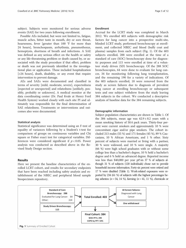
EnrollmentAccrual for the LCRT study was completed in March2012. We enrolled 403 subjects with demographic riskfactors for lung cancer into a prospective multi-site,blinded LCRT study, performed bronchoscopy at enroll-ment, and collected NBEC and blood (buffy coat andplasma) samples from each subject (Fig. 1). Of the 403subjects enrolled, 288 were enrolled at the time of astandard of care (SOC) bronchoscopy done for diagnos-tic purposes and 115 were enrolled at time of a volun-teer study driven (SD) bronchoscopy. Of the 288 SOCbronchoscopies, 64 were done to evaluate for lung can-cer, 34 for monitoring following lung transplantation,and the remaining 190 for a variety of indications. Ofthe 403 subjects enrolled, 18 were removed from thestudy as screen failures due to diagnosis of prevalentlung cancer at enrolling bronchoscopy or subsequenttests and one subject withdrew from the study leaving384 subjects in the cohort. We conducted a descriptiveanalysis of baseline data for the 384 remaining subjects.
Demographic informationSubject population characteristics are shown in Table 1. Ofthe 384 subjects, mean age was 62.9 ± 8.2 years with amean smoking history of 50.4 pack years. Thirty-four per-cent were current smokers and approximately 10 % wereconcomitant cigar and/or pipe smokers. The cohort in-cluded 213 males (55 %) and 171 females (45 %), 89 % Cau-casians, 10 % African Americans, and 1 % other. Sixtypercent of subjects were married or living with a partner,30 % were widowed, and 10 % were single. A majority(66 %) were high school graduates with or without somecollege less than a bachelor’s degree, 10 % held a bachelor’sdegree and 6 % held an advanced degree. Reported incomewas less than $40,000 per year pt?>in 37 % of subjects al-though 31 % of subjects (120 individuals) chose not to providehousehold income information. Forty-six percent were retired and17 % were disabled (Table 1). Work-related exposures were re-ported by 234 (61 %) of subjects with the highest percentages be-ing asbestos (n=54, 14 %), farming (n=41, 11 %), chemicals or
Fig. 1 Summary of Enrolled Cohort
Crawford et al. BMC Pulmonary Medicine (2016) 16:16 Page 5 of 12

plastics (10 %), welding (10 %), foundry or steel milling (9 %), andpainting (9 %) (Additional file 2: Table S2). Each subject had achest CT scan at the time of enrollment; 242 subjects (63 %) hada clinically indicated (standard of care) CT scan within threemonths prior to enrollment and the remaining 142 (37 %) had aresearch driven CT scan within 2 weeks of enrollment. Twelvepercent of subjects were undergoing evaluation for lung cancer attime of enrollment and were negative for cancer (Additional file 3:Table S3). Based on responses to baseline questionnaire, self-reported prevalence of chronic obstructive pulmonary disease(COPD) was 41 % (n=156), chronic bronchitis 18 % (n=68),and emphysema 28 % (n=106) (Additional file 3: Table S3).
Because Pulmonary Function Test (PFT) data were available formost subjects, it was possible to compare self-reported COPDprevalence to test data (see below). Prevalence of other self-reported lung diseases were: interstitial lung disease9 % (n = 35), and sarcoidosis 3 % (n = 10) (Additionalfile 3: Table S3).
SOC vs SD bronchoscopy characteristicsThe intended population for the LCRT includes bothsubjects for whom diagnostic bronchoscopy is indicatedwho also will benefit from LCRT measurement and sub-jects who will have bronchoscopy only to obtain NBECsamples for LCRT measurement. Therefore, we com-pared baseline characteristics between the SOC and SDbronchoscopy subject groups, which represent each ofthese respective intended population categories. Of 384subjects enrolled, bronchoscopy was SOC in 269 (70 %)and SD in 115 (30 %). There were no significant differ-ences in in pack years smoked (Additional file 4: Table S4).SD subjects were slightly younger (mean age of 61.5 com-pared to 63.6, p = 0.021), more likely to be current smokers(55 % vs. 25 %, p < 0.001), and less likely to have COPD(41 % vs. 60 %, p = 0.002) (Additional file 4: Table S4).
Lung cancer screening eligible sub-groupThe USPSTF age and smoking pack year eligibility cri-teria for lung cancer screening by annual low-dose hel-ical chest CT are 55–80 years and a minimum of 30pack-years, respectively. Among subjects enrolled intothe LCRT study, 253/384 (65.9 %) were eligible for an-nual screening at enrollment, according to these criteria.Seventy subjects did not meet the minimum age criter-ion at time of enrollment. By the 2016 follow up timepoint, 45 of these 70 will be eligible for screening and69/70 will be eligible by the 2018 follow up.
Chronic obstructive pulmonary diseaseWe assessed COPD status in the enrolled cohort be-cause COPD is an independent risk factor for lung can-cer [30–38]. COPD was defined using GOLD criteriabased on pulmonary function test (PFT) data [39].Demographic information relative to COPD status is dis-played in Table 2. PFT information was available for 290subjects. Fifty-four percent of these (157 subjects) hadCOPD based on PFT. Among the 157 subjects with COPDbased on PFT, COPD severity was GOLD stage 2 or worsein more than 70 % based on established criteria [39]. MeanFEV1/FVC was 0.52 for the 157 subjects with COPD (allstages) compared to 0.78 for the 133 without COPD.Those with COPD were more likely to be male (62 % vs.38 % female, p = 0.027) and have a higher mean pack yearsmoking history (56 vs. 45 for non-COPD, p < 0.001). Nodifferences were noted in age, race or smoking status(current vs. former smokers) (Table 2.)
Table 1 LCRT Subject Characteristics
Baseline characteristics n = 384
Age in years [mean (SDa)] 62.9 (8.2)
Age in years [median] 62
Male 213 (55 %)
Female 171 (45 %)
Caucasian 343 (89 %)
African American 37 (10 %)
Other or not reported 4 (1 %)
Cigarette pack years [mean (SD)] 50.4 (25.5)
Cigarette pack years [median] 43
Age in years at smoking inception [mean (SD)] 16.1, 3.8
Age in years at smoking inception [median] 16
Total years of smoking [mean (SD)] 37.4 (10)
Total years of smoking [median] 38
History of cigar use 35 (9 %)
History of pipe use 29 (8 %)
Married or living as married 231 (60 %)
Widowed 116 (30 %)
Single 37 (10 %)
Less than high school education 52 (14 %)
High school diploma or GEDb 118 (31 %)
Associate degree or some college 136 (35 %)
Bachelor's degree 40 (10 %)
Graduate degree 24 (6 %)
Other or not reported 14 (4 %)
Employed 109 (28 %)
Unemployed 30 (8 %)
Retired 175 (46 %)
Disabled 64 (17 %)
Other or not reported 6 (2 %)
Income < $40,000/year 141 (37 %)
Income > $40,000/year 123 (32 %)
Other or not reported 120 (31 %)aSD = standard deviationbGED = Graduate Educational Development
Crawford et al. BMC Pulmonary Medicine (2016) 16:16 Page 6 of 12

Of the 157 subjects with COPD based on PFT criteria,clinical history of COPD based on self-report or chartreview was available for 150. Overall, self-reported status
matched the diagnosis by PFT in 67 % (Additional file 5:Table S5.).
Lung transplantNine percent (34 subjects) of our cohort had received a(single) lung transplant prior to enrollment. We evalu-ated differences between lung transplant and non-lungtransplant subjects to determine if there were compar-able demographic risk factors for lung cancer. Age (62.9vs. 63.9 years, p = 0.202), gender, race and smoking his-tory (51.7 vs. 50.0 pack years, p = 0.681) were statisticallysimilar, but 100 % of transplant subjects were formersmokers compared to only 63 % of non-transplant sub-jects (p < 0.001). Prevalence of COPD was comparable,62 % vs. 53 %, p = 0.648. Interstitial lung disease, how-ever, was more prevalent among transplant subjects29 % vs. 7 %, p < 0.001 (Additional file 6: Table S6).
Adverse eventsSerious Adverse Events (SAEs) included any serious ef-fects on the health or safety or any life-threatening prob-lems or death caused by, or associated with the studyprocedures. There were no SAEs attributable to thisstudy for either the 241 SOC bronchoscopy subjects or142 SD bronchoscopy subjects. Adverse Events (AEs)were collected immediately post-procedure and again atthe 30 day follow up. AEs classified as possibly orprobably attributable to the study were those associ-ated with bronchoscopy and bronchial brush biopsysuch as sore throat, hoarseness, cough, throat swell-ing, chest soreness, bleeding, fever, fatigue and upperrespiratory infection. Since the SOC group receivedthe bronchoscopy as part of their standard-of-care,study related AEs were those associated with thebronchial brushing only. There were no AEs classifiedas study related among the SOC group.
Table 2 Chronic Obstructive Pulmonary Disease by PFT
Classification n M/Fa Mean age Race Smoking status Pack years FEV1%d FEV1/FVCe
in years C/AA/Otherb current/former smokedc
No COPD 133 65 / 68 62 113 / 16 / 4 46 / 87 45 80 0.78
COPD (all) 157 97 / 60 63 145 / 12 / 0 50 / 107 56 58 0.52
COPD (stage 1) 45 32 / 12 62 41 / 4 / 0 22 / 23 54 76 0.59
COPD (stage 2) 77 48 / 29 64 73 / 4 / 0 22 / 55 57 57 0.55
COPD (stage 3) 26 15 / 11 64 23 / 3 / 0 5 / 21 53 35 0.39
COPD (stage 4) 7 2 / 5 63 7 / 0 / 0 1 / 6 65 22 0.27
COPD (stage unknown) 2 1 / 1 66 1 / 1 / 0 0 / 2 66 - 0.57
Unknown 94 50 / 44 63 85 / 9 / 0 35 / 60
50 - -
aM =male, F = femalebC = Caucasian, AA = African-American, Other = other race or race not reportedcPack years = packs of cigarettes smoked per day x years of smokingdFEV1% = forced expiratory volume in 1 second, percent of expectedeFEV1/FVC = FEV1/Forced Vital Capacity
Table 3 Adverse Events (AE)
Subject # AE Description Severity Relatedness Treatment Notes
1035 Felt poorly(like he hada fever) for3 days
Mild Possible Did not seektreatment or notifystudy personneluntil 30 dayfollow-up
1044 Upperrespiratorytract infection
Mild Possible Treated withantibiotics andsteroids, infectionresolved
1048 Hoarsenessfor 2 days
Mild Probable
1050 Bruisingaround eyes
Mild Unlikely
1057 Bleeding fromears postbronchoscopy
Mild Possible
1057 Petechiaearound eyes
Mild Possible
1059 Cough Mild Possible
1059 Difficultyswallowing
Mild Possible
1060 Felt sorenessin lung
Mild Possible
1061 Dry scratchyarea in throat,feels need tocough
Mild Possible
1076 Slight cough Mild Possible
1077 Back of throatswollen
Mild Probable
1078 Cough Mild Unlikely
Crawford et al. BMC Pulmonary Medicine (2016) 16:16 Page 7 of 12

Among the SD group, there were 11 AEs noted in 9subjects that were possibly (n = 9) or probably (n = 2) at-tributable to study procedures. Additionally, AEs weredocumented in two additional subjects that weredeemed unlikely to be related (Table 3). All AEs wereclassified as mild.
Establishment of NBEC and peripheral blood samplebiospecimen repositoryMatched blood and NBEC were collected for 361/384(94 %) subjects and banked in multiple aliquots. Bloodsamples were processed at each site at the time of collec-tion to generate 2 aliquots of buffy coat and 2–5 aliquotsof plasma from each subject. These aliquots were frozenand stored at −80 °C until shipment to the Early Detec-tion Research Network (EDRN) Biorepository in Fre-drick, MD. One aliquot of buffy coat was transferred tothe University of Toledo for analysis and the other re-mains in storage. NBEC were stabilized at each site inRNA Later (Ambion, Austin, TX) and shipped alongwith matching slides to ResearchDx, Irvine, CA. RNAwas extracted from NBEC within 24–48 hours of receipt,assessed for quality and quantity and stored in aliquotsat −80 °C. One NBEC RNA aliquot was shipped to theUniversity of Toledo for analysis for those samples witha minimum yield of 1 microgram and aliquots for eachsubject remain in storage at ResearchDx.At the University of Toledo, genomic DNA (gDNA)
was extracted from one aliquot of buffy coat derivedfrom the blood sample from approximately 80 % of sub-jects and 100 % of these yielded gDNA of sufficientquality and quantity for proposed molecular studies. Thequality and quantity of NBEC RNA from approximately40 % of subjects has been assessed to date. RNA fromeach sample was treated with DNase I, tested via PCR toensure removal of contaminating gDNA from the RNAand then reverse transcribed into cDNA. For 90 % ofsubjects the cDNA generated from these purified NBECRNA samples was PCR amplifiable and of sufficientquantity to perform LCRT testing. Additional aliquots ofRNA remain for the roughly 10 % of samples that didnot pass this quality control. Samples from over 120subjects were used successfully in preliminary targetednext generation sequencing (NGS) RNA sequencinganalysis studies.
Lung cancer incidenceTwo years following initiation of the study, 5 subjects(1.3 %) without prevalent lung cancer developed bron-chogenic carcinoma. Due to the blinded status of theLCRT study, no further details are available regardingthese subjects.
DiscussionEnrolled cohort is representative of LCRT targetpopulationThe target population of the LCRT biomarker is individ-uals who meet USPSTF eligibility criteria for annual lowdose helical CT screening [2]. The enrollment criteriafor the LCRT study included both current and formersmokers, individuals with and without concurrent pul-monary disease and/or respiratory exposures as well asboth subjects undergoing medically recommended bron-choscopy (SOC group) and volunteers (SD group). Atthe time of enrollment into the LCRT study, most sub-jects (66 %) met USPSTF age and smoking pack-year eli-gibility criteria (55–80 years of age, ≥ 30 pack years).Additionally, most of those not eligible at enrollmentwill be eligible for screening by the 2016 follow up timepoint due to increased age, and this fraction is expectedto further increase at the 2018 follow up. Therefore, thisgroup is highly representative of the LCRT biomarkertarget population.
Feasibility to reach LCRT study endpoint based on cohortcharacteristicsBased on demographic characteristics of the enrolledpopulation (Table 1), we expect lung cancer incidence inthe LCRT study to be similar to the 3.1 % incidence over3.9 years reported by Bach et al. [40] in which mean agewas 60.1 and smoking history of 52 pack-years. The fiveincidental lung cancers observed two years after initi-ation of the study are consistent with this rate. Takinginto account that some of the 384 study subjects willhave died from causes other than bronchogenic carcin-oma prior to these time points and that some will be lostto follow up we estimated incidental lung cancers in thecohort based on 300 subjects. As such we expect to ob-serve approximately 12 incidental lung cancers by the2016 follow up (mean time since enrollment approxi-mately 5 years) and 17 by the 2018 follow up point(mean time since enrollment approximately 7 years),which will be more than sufficient to reach the proposedendpoint of a risk ratio of ≥ 5.0.
Feasibility of LCRT implementation (safety andacceptance by subjects)The LCRT biomarker requires a one-time acquisition ofNBEC through bronchial brush biopsy at the time ofbronchoscopy. In addition to the LCRT study, Departmentof Defense Lung Cancer Research Program, and NIH re-cently funded other large studies assessing utility of bio-markers measured in NBEC obtained at bronchoscopyintended to more accurately determine lung cancer riskand/or to enable early lung cancer diagnosis (Massion,Clinicaltrials.gov NCT01475500 CA152662 and CA102353;Spira, Clinicaltrials.gov NCT02504697 DECAMP-2 and
Crawford et al. BMC Pulmonary Medicine (2016) 16:16 Page 8 of 12

CA164783-04; Dubinett, CA152751-05S2). Therefore, it isimportant to carefully evaluate the safety and comfort ofthis procedure, which will impact general acceptance by pa-tients and clinicians. Based on published studies, bronchos-copy with or without biopsy is considered a safe procedureand it is used not only for medical purposes but also toconduct research [5, 41–49]. Reported complication rates(also known as serious adverse event/SAE rates) for allbronchoscopy procedures range from 0.08-1.93 % and mor-tality rates range from 0.004-0.045 % [50–52]. One largeJapanese study of almost 50,000 patients who underwentbronchoscopy with brush biopsy in either central or periph-eral airways reported a complication (SAE) rate of 0.46 %.This risk of complication is similar to the 0.28-0.32 % com-plication (SAE) rate reported for colonoscopy [53, 54]which is routinely used and repeated for colorectal cancerscreening. Importantly, a bronchoscopy with brush biopsylimited to the central airways for collection of NBEC,the procedure used here, virtually eliminates risk forthe primary complications reported to be associatedwith bronchoscopy, including pneumothorax or signifi-cant hemorrhage. Consistent with this, we observed noSAE associated with bronchoscopic brush biopsy in thesubjects enrolled based on SD bronchoscopy.It is particularly important to assess safety and comfort
in the subjects meeting accepted criteria for lung cancerscreening, a group that has increased prevalence for nu-merous comorbidities. Results from at least one previousreport have suggested that research bronchoscopy andbrush biopsy can be safely performed in subjects withheavy smoking history and those with obstructive lungdisease [45]. Previous guidelines have suggested that anFEV1 less than 60 % is considered a contraindication toperforming research driven bronchoscopy. However,bronchoscopy in adults with stable asthma and COPDhas been performed safely at lower values of FEV1 [55].Pulmonary function test data were available for morethan 75 % of the subjects enrolled here (290 of 384 sub-jects). One hundred fifty-seven had clinical COPD andmore than 70 % had GOLD stage 2 or worse (Table 2).Additionally, 9 % of enrolled subjects had a history ofinterstitial lung disease, 9 % were single-lung transplantrecipients and a small percentage had other pulmonarydisease (Additional file 3: Table S3) and bronchoscopywas safely performed on all of them. Specifically, nocomplications (SAEs) were associated with broncho-scopic brushing and sample collection in either standardof care (SOC) or study driven (SD) group.In summary, bronchoscopic brush of the central air-
ways to collect NBEC for lung cancer risk analysis wassafe and well-tolerated in this study of subjects demo-graphically at risk for lung cancer, including those withsignificant co-morbid conditions. Because the AE ratewas much lower than that reported for routinely used
screening colonoscopy [53] we expect that this proced-ure will be acceptable to patients and clinicians if theLCRT or other tests in development are validated toidentify subjects with increased risk for lung cancer and/or early stage lung cancer.
COPD characteristics of LCRT CohortThe enrolled cohort had a high fraction of COPD basedon PFT criteria. This is important because COPD is anindependent risk factor for lung cancer [30–38]. Not-ably, using PFT data (FEV1/FVC <0.7) as the diagnosticcriterion, one-third of individuals in this study misclassi-fied their COPD status on the enrollment survey self-report. This is consistent with multiple reports of data ac-quisition through self-report leading to either misclassifi-cation or under-diagnosis of COPD [56–60]. Some of thismisclassification could be due to patient being told theyhave COPD on the basis of radiographic imaging whilethe PFT data do not meet criteria for COPD diagnosis.Additionally, a portion of the subjects here underwentPFT at the time of enrollment that revealed COPD for thefirst time because the subject had not been tested prior toenrollment in the study. Given the importance of accurateCOPD diagnosis, we plan to obtain both chest CT andPFT data from each subject at each subsequent follow-up.We will then evaluate COPD based on CT (presence ofemphysema and/or bronchial thickening) or PFT criteriaalone, or in combination as a risk factor for lung cancer.
LCRT cohort and biospecimen repository as a resource forsubsequent studiesAs presented here, the LCRT cohort is well characterizedwith respect to demographic characteristics. In addition,NBEC and matching blood samples were collected fromeach subject. Each subject had a baseline CT scan andpulmonary function test (PFT) data are available for 76 %of individuals. It is planned to obtain a repeat PFT and CTscan on all subjects at each subsequent follow-up. This in-formation will enable longitudinal assessment for rate ofdecline in pulmonary function by both physiologic andradiographic measures and to assess for presence or ab-sence of lung cancer. More than 90 % of samples assessedso far passed QC quality and quantity criteria for reliableLCRT measurement. The NBEC and matching bloodsamples collected in this study are archived and the major-ity of subjects have given consent for use of samplesremaining after LCRT analysis for future IRB approvedstudies. Currently, we are using NBEC gene expressiondata and genotyping data from matched peripheral bloodcell gDNA to identify proximate phenotypic biomarkersfor COPD risk and additional biomarkers for hereditarylung cancer risk. We are integrating these data withCOPD genome wide association study (GWAS) data from
Crawford et al. BMC Pulmonary Medicine (2016) 16:16 Page 9 of 12

the Lung Health Study and the COPDgene study availableonline at NCI dbGAP.
ConclusionsThe demographic composition of the enrolled group isrepresentative of the population for which the LCRT isintended. Specifically, based on baseline population char-acteristics we expect lung cancer incidence in this cohortto be representative of the population eligible for LDCTlung cancer screening. Collection of NBEC by bronchialbrush biopsy/bronchoscopy was safe and well-tolerated inthis population. These findings support the feasibility oftesting LCRT clinical utility in this prospective study. Ifvalidated, the LCRT has the potential to significantly nar-row the population of individuals requiring annual low-dose helical CT screening for early detection of lung can-cer and to enable safe delay the onset of screening for in-dividuals with results indicating low lung cancer risk. Forthese individuals, the small risk incurred by undergoingonce in a lifetime bronchoscopic sample collection forLCRT may be offset by a reduction in their CT-relatedrisks. The LCRT biospecimen repository will enable add-itional studies of genetic basis for COPD and/or lung can-cer risk.
Additional files
Additional file 1: Lung Cancer Risk Test Study Enrollment by StudySite. (DOCX 15 kb)
Additional file 2: Work Type and Exposures. (DOCX 15 kb)
Additional file 3: Medical History. (DOCX 15 kb)
Additional file 4: Standard of Care (SOC) vs. Study Driven (SD)Bronchoscopies. (DOCX 16 kb)
Additional file 5: Self-reported vs. Clinical COPD. (DOCX 14 kb)
Additional file 6: Transplant vs Non-Transplant Subjects. (DOCX 16 kb)
AbbreviationsAE: Adverse event; COPD: Chronic obstructive pulmonary disease;FEV1: Forced expiratory volume in 1 second; FVC: Forced vital capacity;GWAS: Genome wide association study; LCRT: Lung Cancer Risk Test;LDCT: Low dose computed tomography; NBEC: Normal bronchial epithelialcells; PFT: Pulmonary function test; SAE: Serious adverse event; SNP: Singlenucleotide polymorphism; SD: Study driven; SOC: Standard of care;USPSTF: United States Preventive Services Task Force.
Competing interestsJCW has equity interest in and serves as a consultant to Accugenomics, Inc.which licenses technology utilized here. JCW and ELC are inventors on U.Sand international patents related to the technology and biomarkerspresented here.
Authors' contributionsELC contributed to the design of the study, interpretation of results, draftingof the manuscript and performed quality assessment of gDNA and RNA. AL,ML and SAK participated in data analysis and statistical interpretation andcontributed to the study design. FS and AMB participated in the preparationof the manuscript, XZ, JY and AB participated in the interpretation of data,PN, PPM, DAA, DM, PJM, SDN, RW, GS and JT served as site directors for theLCRT study and participated in the study design and coordination, JCW
conceived of the study, participated in its design and coordination andcontributed to the preparation of the manuscript.
AcknowledgementsThis work was funded by grants from the NIH National Cancer InstituteCA148572 and National Heart Lung and Blood Institute HL108016, and theGeorge Isaac Cancer Research Fund. The NIH did not participate in the studydesign, data interpretation or manuscript preparation. We thank Dr. PaulKvale for his professional review and assessment of adverse and seriousadverse events in relation to the study, we thank Dr.James Jett forcontributions in planning of the study, and Dr. Ali Musani for supportingenrollment at National Jewish Hospital.
Author details1Department of Pulmonary and Critical Care, The University of ToledoMedical Center, Toledo, OH, USA. 2Department of Biostatistics, Henry FordHospital System, Detroit, MI, USA. 3Ohio State University JamesComprehensive Cancer Center and Solove Research Institute, Columbus, OH,USA. 4Thoracic Program, Vanderbilt Ingram Cancer Center, Nashville, TN, USA.5University of Michigan, Ann Arbor, MI, USA. 6Mayo Clinic, Rochester, MN,USA. 7Cleveland Clinic, Cleveland, OH, USA. 8Inova Fairfax Hospital, FallsChurch, VA, USA. 9The Toledo Hospital, Toledo, OH, USA. 10Medical Universityof South Carolina, Charleston, SC, USA. 11Mercy/St. Vincent’s Hospital, Toledo,OH, USA.
Received: 2 November 2015 Accepted: 12 January 2016
References1. (2014) Cancer Facts & Figures. American Cancer Society. www.cancer.org/
acs/groups/content/@research/documents/webcontent/acspc-042151.pdf2. Humphrey LL, Deffebach M, Pappas M, Baumann C, Artis K, Mitchell JP, et al.
Screening for lung cancer with low-dose computed tomography: asystematic review to update the US Preventive services task forcerecommendation. Ann Intern Med. 2013;159:411–20.
3. Aberle DR, Adams AM, Berg CD, Black WC, Clapp JD, Fagerstrom RM, et al.Reduced lung-cancer mortality with low-dose computed tomographicscreening. N Engl J Med. 2011;365:395–409.
4. Bach PB, Mirkin JN, Oliver TK, Azzoli CG, Berry DA, Brawley OW, et al.Benefits and harms of CT screening for lung cancer: a systematic review.JAMA. 2012;307:2418–29.
5. Blomquist T, Crawford EL, Mullins D, Yoon Y, Hernandez DA, Khuder S, et al.Pattern of Antioxidant and DNA Repair Gene Expression in Normal AirwayEpithelium Associated with Lung Cancer Diagnosis. Cancer Res. 2009;69:8629–35.
6. Bach PB, Kattan MW, Thornquist MD, Kris MG, Tate RC, Barnett MJ, et al.Variations in lung cancer risk among smokers. J Natl Cancer Inst. 2003;95:470–8.
7. Raji OY, Duffy SW, Agbaje OF, Baker SG, Christiani DC, Cassidy A, et al.Predictive accuracy of the Liverpool Lung Project risk model for stratifyingpatients for computed tomography screening for lung cancer: a case–controland cohort validation study. Ann Intern Med. 2012;157:242–50.
8. Tammemagi CM, Pinsky PF, Caporaso NE, Kvale PA, Hocking WG, Church TR,et al. Lung Cancer Risk Prediction: Prostate, Lung, Colorectal and OvarianCancer Screening Trial Models and Validation. J Natl Cancer Inst. 2011;103:1058–68.
9. Field JK, Baldwin D, Brain K, Devaraj A, Eisen T, Duffy SW, et al. CT screeningfor lung cancer in the UK: position statement by UKLS investigatorsfollowing the NLST report. Thorax. 2011;66:736–7.
10. Cassidy A, Myles JP, van Tongeren M, Page RD, Liloglou T, Duffy SW, et al.The LLP risk model: an individual risk prediction model for lung cancer. Br JCancer. 2008;98:270–6.
11. Spitz MR, Hong WK, Amos CI, Wu XF, Schabath MB, Dong Q, et al. A riskmodel for prediction of lung cancer. J Natl Cancer Inst. 2007;99:715–26.
12. van Klaveren RJ, Habbema JDF, Pedersen JH, de Koning HJ, Oudkerk M,Hoogsteden HC. Lung cancer screening by low-dose spiral computedtomography. Eur Respir J. 2001;18:857–66.
13. Kovalchik SA, Tammemagi M, Berg CD, Caporaso NE, Riley TL, Korch M, et al.Targeting of low-dose CT screening according to the risk of lung-cancer death.N Engl J Med. 2013;369:245–54.
Crawford et al. BMC Pulmonary Medicine (2016) 16:16 Page 10 of 12

14. Birse CE, Lagier RJ, FitzHugh W, Pass HI, Rom WN, Edell ES, et al. Blood-basedlung cancer biomarkers identified through proteomic discovery in cancertissues, cell lines and conditioned medium. Clin Proteomics. 2015;12:18.
15. Pecot CV, Li M, Zhang XJ, Rajanbabu R, Calitri C, Bungum A, et al. Addedvalue of a serum proteomic signature in the diagnostic evaluation of lungnodules. Cancer Epidemiol Biomarkers Prev. 2012;21:786–92.
16. Daly S, Rinewalt D, Fhied C, Basu S, Mahon B, Liptay MJ, et al. Development andvalidation of a plasma biomarker panel for discerning clinical significance ofindeterminate pulmonary nodules. J Thorac Oncol. 2013;8:31–6.
17. Mehan MR, Williams SA, Siegfried JM, Bigbee WL, Weissfeld JL,Weissfeld JL, et al. Validation of a blood protein signature for non-smallcell lung cancer. Clin Proteomics. 2014;11:32.
18. Higgins G, Roper KM, Watson IJ, Blackhall FH, Rom WN, Pass HI, et al. VariantCiz1 is a circulating biomarker for early-stage lung cancer. Proc Natl AcadSci U S A. 2012;109:E3128–3135.
19. Vachani A, Pass HI, Rom WN, Midthun DE, Edell ES, Laviolette M, et al.Validation of a multiprotein plasma classifier to identify benign lungnodules. J Thorac Oncol. 2015;10:629–37.
20. Cazzoli R, Buttitta F, Di Nicola M, Malatesta S, Marchetti A, Rom WN, et al.microRNAs derived from circulating exosomes as noninvasive biomarkers forscreening and diagnosing lung cancer. J Thorac Oncol. 2013;8:1156–62.
21. Boeri M, Verri C, Conte D, Roz L, Modena P, Facchinetti F, et al. MicroRNAsignatures in tissues and plasma predict development and prognosis of computedtomography detected lung cancer. Proc Nat Acad Sci. 2011;108:3713–8.
22. Spira A, Beane JE, Shah V, Steiling K, Liu G, Schembri F, et al. Airwayepithelial gene expression in the diagnostic evaluation of smokers withsuspect lung cancer. Nat Med. 2007;13:361–6.
23. Gower AC, Steiling K, Brothers 2nd JF, Lenburg ME, Spira A. Transcriptomicstudies of the airway field of injury associated with smoking-related lungdisease. Proc Am Thorac Soc. 2011;8:173–9.
24. Silvestri GA, Vachani A, Whitney D, Elashoff M, Porta Smith K, Ferguson JS,et al. A Bronchial Genomic Classifier for the Diagnostic Evaluation of LungCancer. N Engl J Med. 2015;373:243–51.
25. Wang Z, Zhu B, Zhang M, Parikh H, Jia J, Chung CC, et al. Imputation andsubset-based association analysis across different cancer types identifiesmultiple independent risk loci in the TERT-CLPTM1L region onchromosome 5p15.33. Hum Mol Genet. 2014;23:6616–33.
26. Wang Y, McKay JD, Rafnar T, Wang Z, Timofeeva MN, Broderick P, et al. Rarevariants of large effect in BRCA2 and CHEK2 affect risk of lung cancer.Nature genetics. 2014;46:736–41.
27. Blomquist TM, Crawford EL, Willey JC. Cis-acting genetic variation at anE2F1/YY1 response site and putative p53 site is associated with alteredallele-specific expression of ERCC5 (XPG) transcript in normal humanbronchial epithelium. Carcinogenesis. 2010;31:1242–50.
28. Blomquist TM, Brown RD, Crawford EL, de la Serna I, Williams K, Yoon Y,et al. CEBPG Exhibits Allele-Specific Expression in Human Bronchial EpithelialCells. Gene Regul Syst Bio. 2013;7:125–38.
29. Blomquist TM, Crawford EL, Lovett JL, Yeo J, Stanoszek LM, Levin A, et al.Targeted RNA-sequencing with competitive multiplex-PCR ampliconlibraries. PLoS One. 2013;8, e79120.
30. Mannino DM, Aguayo SM, Petty TL, Redd SC. Low lung function andincident lung cancer in the United States: data From the First NationalHealth and Nutrition Examination Survey follow-up. Arch Intern Med.2003;163:1475–80.
31. Schwartz AG. Genetic epidemiology of cigarette smoke-induced lungdisease. Proc Am Thorac Soc. 2012;9:22–6.
32. de-Torres JP, Wilson DO, Sanchez-Salcedo P, Weissfeld JL, Berto J, Campo A,et al. Lung cancer in patients with chronic obstructive pulmonary disease.Development and validation of the COPD Lung Cancer Screening Score.Am J Respir Crit Care Med. 2015;191:285–91.
33. Young RP, Hopkins RJ, Christmas T, Black PN, Metcalf P, Gamble GD.COPD prevalence is increased in lung cancer, independent of age, sexand smoking history. Eur Respir J. 2009;34:380–6.
34. Mayne ST, Buenconsejo J, Janerich DT. Previous lung disease and risk oflung cancer among men and women nonsmokers. Am J Epidemiol.1999;149:13–20.
35. Tockman MS, Anthonisen NR, Wright EC, Donithan MG. Airways obstructionand the risk for lung cancer. Ann Intern Med. 1987;106:512–8.
36. Skillrud DM, Offord KP, Miller RD. Higher risk of lung cancer in chronicobstructive pulmonary disease. A prospective, matched, controlled study.Ann Intern Med. 1986;105:503–7.
37. Purdue MP, Gold L, Jarvholm B, Alavanja MC, Ward MH, Vermeulen R.Impaired lung function and lung cancer incidence in a cohort of Swedishconstruction workers. Thorax. 2007;62:51–6.
38. Wasswa-Kintu S, Gan WQ, Man SFP, Pare PD, Sin DD. Relationship betweenreduced forced expiratory volume in one second and the risk of lungcancer: a systematic review and meta-analysis. Thorax. 2005;60:570–5.
39. Global strategy for the diagnosis, management, and prevention of chronicobstructive pulmonary disease: Revised 2015. Global Initiative for ChronicObstructive Lung Disease (GOLD). www.goldcopd.org.
40. Bach PB, Jett JR, Pastorino U, Tockman MS, Swensen SJ, Begg CB.COmputed tomography screening and lung cancer outcomes. JAMA. 2007;297:953–61.
41. Crawford EL, Khuder SA, Durham SJ, Frampton M, Utell M, Thilly WG,et al. Normal bronchial epithelial cell expression of glutathionetransferase P1, glutathione transferase M3, and glutathione peroxidaseis low in subjects with bronchogenic carcinoma. Cancer Res. 2000;60:1609–18.
42. Willey JC, Coy E, Brolly C, Utell MJ, Frampton MW, Hammersley J, et al.Xenobiotic metabolism enzyme gene expression in human bronchial epithelialand alveolar macrophage cells. Am J Respir Cell Mol Biol. 1996;14:262–71.
43. Crawford EL, Blomquist T, Mullins DN, Yoon Y, Hernandez DR, Al-BagdhadiM, et al. CEBPG regulates ERCC5/XPG expression in human bronchialepithelial cells and this regulation is modified by E2F1/YY1 interactions.Carcinogenesis.2007;28:2552–9.
44. Willey JC, Coy EL, Frampton MW, Torres A, Apostolakos MJ, Hoehn G, et al.Quantitative RT-PCR measurement of cytochromes p450 1A1, 1B1, and2B7, microsomal epoxide hydrolase, and NADPH oxidoreductaseexpression in lung cells of smokers and nonsmokers. Am J Respir Cell MolBiol. 1997;17:114–24.
45. Romagnoli M, Vachier I, Vignola AM, Godard P, Bousquet J, Chanez P. Safetyand cellular assessment of bronchial brushing in airway diseases. RespirMed. 1999;93:461–6.
46. Lo Tam Loi AT, Hoonhorst SJM, Franciosi L, Bischoff R, Hoffmann RF, HeijinkI, et al. Acute and chronic inflammatory responses induced by smoking inindividuals susceptible and non-susceptible to development of COPD: fromspecific disease phenotyping towards novel therapy. Protocol of a cross-sectional study. BMJ Open. 2013;3.
47. Kim V, Oros M, Durra H, Kelsen S, Aksoy M, Cornwell WD, et al. ChronicBronchitis and Current Smoking Are Associated with More Goblet Cells inModerate to Severe COPD and Smokers without Airflow Obstruction. PLoSONE.2015;10, e0116108.
48. Barnes NC, Saetta M, Rabe KF. Implementing lessons learned from previousbronchial biopsy trials in a new randomized controlled COPD biopsy trialwith roflumilast. BMC Pulm Med. 2014;14:9.
49. Eissa NT, Erzurum SC. Flexible bronchoscopy in molecular biology.Clin Chest Med. 2001;22(343–353):ix.
50. Asano F, Aoe M, Ohsaki Y, Okada Y, Sasada S, Sato S, et al. Deaths andcomplications associated with respiratory endoscopy: A survey by the JapanSociety for Respiratory Endoscopy in 2010. Respirology. 2012;17:478–85.
51. Jin F, Mu D, Chu D, Fu E, Xie Y, Liu T. Severe complications ofbronchoscopy. Respiration. 2008;76:429–33.
52. Facciolongo N, Patelli M, Gasparini S, Lazzari Agli L, Salio M, Simonassi C,et al. Incidence of complications in bronchoscopy. Multicentre prospectivestudy of 20,986 bronchoscopies. Monaldi Arch Chest Dis. 2009;71:8–14.
53. Fisher DA, Maple JT, Ben-Menachem T, Cash BD, Decker GA, Early DS, et al.Complications of colonoscopy. Gastrointest Endosc. 2011;74:745–52.
54. Ko CW, Riffle S, Michaels L, Morris C, Holub J, Shapiro JA, et al. Seriouscomplications within 30 days of screening and surveillance colonoscopy areuncommon. Clin Gastroenterol Hepatol. 2010;8:166–73.
55. Hattotuwa K, Gamble EA, O’Shaughnessy T, Jeffery PK, Barnes NC. Safety ofbronchoscopy, biopsy, and bal in research patients with COPD. Chest. 2002;122:1909–12.
56. Aldrich MC, Munro HM, Mumma M, Grogan EL, Massion PP, BlackwellTS, et al. Chronic obstructive pulmonary disease and subsequent overalland lung cancer mortality in low-income adults. PLoS One. 2015;10,e0121805.
57. Barr RG, Herbstman J, Speizer FE, Camargo Jr CA. Validation of self-reportedchronic obstructive pulmonary disease in a cohort study of nurses. Am JEpidemiol. 2002;155:965–71.
Crawford et al. BMC Pulmonary Medicine (2016) 16:16 Page 11 of 12

58. Eisner MD, Trupin L, Katz PP, Yelin EH, Earnest G, Balmes J, et al.Development and validation of a survey-based COPD severity score. Chest.2005;127:1890–7.
59. Straus SE, McAlister FA, Sackett DL, Deeks JJ. Accuracy of history, wheezing,and forced expiratory time in the diagnosis of chronic obstructivepulmonary disease. J Gen Intern Med. 2002;17:684–8.
60. Zhai R, Yu X, Shafer A, Wain JC, Christiani DC. The impact of coexistingCOPD on survival of patients with early-stage non-small cell lung cancerundergoing surgical resection. Chest. 2014;145:346–53.
• We accept pre-submission inquiries
• Our selector tool helps you to find the most relevant journal
• We provide round the clock customer support
• Convenient online submission
• Thorough peer review
• Inclusion in PubMed and all major indexing services
• Maximum visibility for your research
Submit your manuscript atwww.biomedcentral.com/submit
Submit your next manuscript to BioMed Central and we will help you at every step:
Crawford et al. BMC Pulmonary Medicine (2016) 16:16 Page 12 of 12