Embed Size (px)
Citation preview
87
Chapter
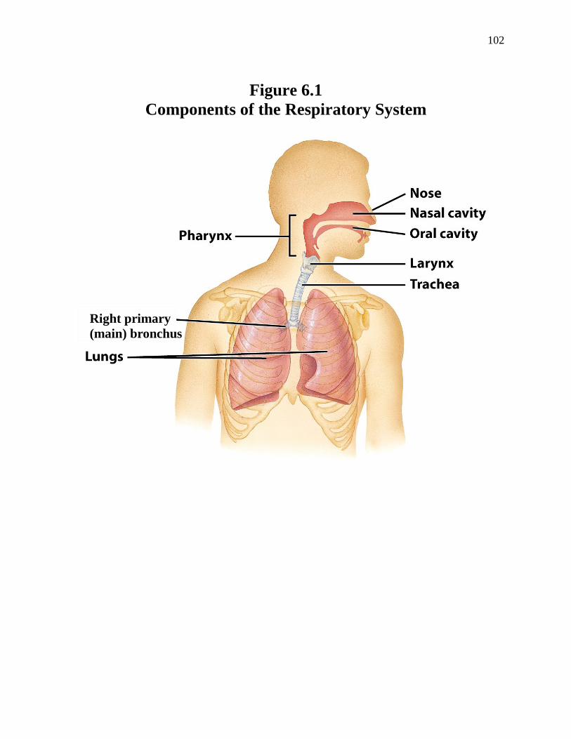
6 Respiratory System ♦Overview -The respiratory system consists of the nose, pharynx, larynx, trachea, bronchi, and the lungs (Figure 6.1, Derrickson). -The purpose of the respiratory system is to bring O2 into the body and to take CO2 out of the body. O2 enters the body when you inhale. The O2 then diffuses into the blood, which transports it to the cells of the body. The body cells use the O2 to produce ATP (energy) through a process called cellular (aerobic) respiration: glucose + O2 → CO2 + H2O + 32 ATP (energy) CO2 is released from this reaction. Body cells cannot tolerate CO2 since it is a toxic waste product; therefore, CO2 diffuses into the blood and is removed from the body when you exhale. otorhinolaryngology -the study of the structure, function, and disorders of the ears, nose, and throat pulmonology -the study of structure, function, and disorders of the lungs ♦Components of the Respiratory System 1. nose -The nose is divided into 2 major regions (Figures 23.3a, Tortora): a. external nose -consists of bone and cartilage covered with skin b. nasal cavity -the space within the nose -surrounded by bones that are lined by a mucosa
88
-Note that a mucosa consists of an epithelial layer and an underlying layer of connective tissue. -The epithelium of the mucosa that lines the nasal cavity consists of ciliated cells and scattered goblet cells. -Goblet cells are cells that secrete mucus. -The anterior region of the nasal cavity opens up to the outside environment as the external nares (singular is naris) or nostrils. -The posterior region of the nasal cavity joins the pharynx. -The superior region of the nasal cavity contains the olfactory epithelium, which consists of sensory receptors that are involved in the detection of smell. 2. pharynx -also called the throat -a tube that joins the nose and mouth to the larynx and esophagus -Consequently, the pharynx is considered to be part of the respiratory system and part of the digestive system. -The pharynx consists of 3 regions (Figure 23.3b, Tortora): a. nasopharynx -the upper region of the pharynx -lies posterior to and is continuous with the nasal cavity b. oropharynx -the middle region of the pharynx -lies posterior to and is continuous with the oral cavity c. laryngopharynx -the lower region of the pharynx -continuous with the larynx anteriorly and the esophagus inferiorly -The wall of the pharynx consists of 2 layers: a. skeletal muscle -forms the outer layer b. mucosa -inner layer that lines the lumen of the pharynx -The epithelium of the mucosa that lines the pharynx consists of ciliated cells and scattered goblet cells that secrete mucus. 3. larynx -also called the voice box -The larynx consists of the following (Figure 23.5, Tortora) a. cartilage -Several pieces of cartilage form the outer layer of the larynx; the most important ones include the following: 1. thyroid cartilage -also called the Adam’s apple -forms the anterior wall of the larynx -It is larger in males than in females because testosterone stimulates its growth during puberty.
89
2. cricoid cartilage -a ring of cartilage that is inferior to the thyroid cartilage 3. epiglottis -a spoon-shaped piece of cartilage that is attached to the thyroid cartilage at one end, while the other end is free to move up and down -When only air is moving into the nose and pharynx, the free end of the epiglottis is pointed upward and the larynx is open, which allows air to travel into the rest of the respiratory system. -During swallowing, the free end of the epiglottis moves down to cover the larynx like a lid. This prevents food and liquids from entering the larynx, thereby forcing these substances into the esophagus. - If anything besides air finds its way into the larynx by mistake, a vigorous coughing reflex occurs to expel the material. -Recall that the coughing reflex is coordinated by the coughing center in the medulla oblongata. -2 pairs of folds (connective tissue that is string-like) extend between the cartilages of the larynx: a superior pair of vestibular folds (false vocal cords) and an inferior pair of vocal folds (true vocal cords). 1. vestibular folds -not involved in vocalization 2. vocal folds -involved in vocalization (sound production) -When air strikes the vocal folds, the vocal folds vibrate and a sound is produced. -The loudness (volume) of the sound is determined by how vigorously air strikes the vocal folds. -When air strikes the vocal folds forcefully, a loud sound is produced. –When air strikes the vocal folds mildly, a soft sound is produced. -The pitch of the sound is determined by the tautness (tightness) of the vocal folds. - Tight vocal folds produce a high pitched sound, while loose vocal folds produce a low pitched sound. -Testosterone causes the vocal folds in men to be thicker, longer, and looser than the vocal folds in women. -Consequently, men typically have lower pitched (deeper) voices than women.
90
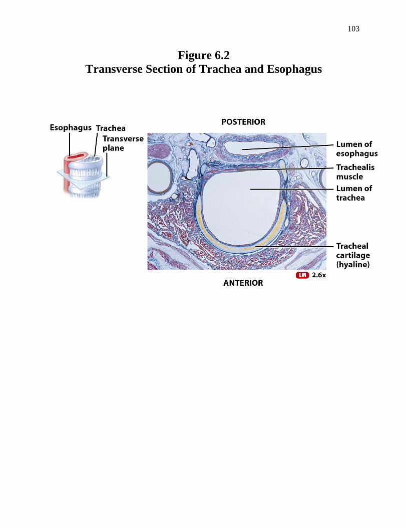
b. skeletal muscle -forms the middle layer of larynx c. mucosa -inner layer that lines the lumen of the larynx -The epithelium of the mucosa that lines the larynx consists of ciliated cells and scattered goblet cells that secrete mucus. 4. trachea -also called the windpipe -The trachea consists of the following components (Figures 23.8a, Tortora and Figure 6.2, Derrickson): a. cartilage -Several “C-shaped” rings of cartilage form the anterior and lateral walls of the trachea -These cartilages provide support to the trachea and prevent it from collapsing. b. smooth muscle -A region of smooth muscle called the trachealis forms the outer posterior wall of the trachea. -Contraction or relaxation of the trachealis changes the diameter of the trachea, which alters airflow. c. mucosa -inner layer that lines the lumen of the trachea -The epithelium of the mucosa that lines the trachea consists of ciliated cells and scattered goblet cells that secrete mucus. 5. bronchi -The trachea gives rise to the left primary (main) bronchus, which goes to the left lung and the right primary (main) bronchus, which goes to the right lung (Figure 23.8a, Tortora). -The histological structure of each primary bronchus is similar to that of the trachea: “C-shaped” rings of cartilage form the outer anterior and lateral walls, smooth muscle forms the outer posterior wall, and a mucosa lines the lumen. -The epithelium of the mucosa consists of ciliated cells and scattered goblet cells that secrete mucus. 6. lungs -There are 2 cone-shaped lungs located in the thoracic cavity. -Pleura -a connective tissue sac that surrounds each lung (Figure 23.8a, Tortora) -organized into 2 layers a. parietal layer -outer layer -fused to the wall of the thoracic cavity b. visceral layer -inner layer -attached to the lung surface
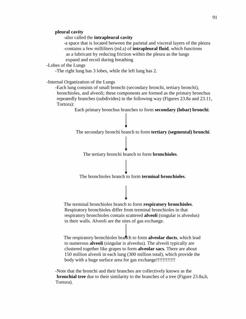
91
pleural cavity -also called the intrapleural cavity -a space that is located between the parietal and visceral layers of the pleura -contains a few milliliters (mLs) of intrapleural fluid, which functions as a lubricant by reducing friction within the pleura as the lungs expand and recoil during breathing -Lobes of the Lungs -The right lung has 3 lobes, while the left lung has 2. -Internal Organization of the Lungs -Each lung consists of small bronchi (secondary bronchi, tertiary bronchi), bronchioles, and alveoli; these components are formed as the primary bronchus repeatedly branches (subdivides) in the following way (Figures 23.8a and 23.11, Tortora): Each primary bronchus branches to form secondary (lobar) bronchi. The secondary bronchi branch to form tertiary (segmental) bronchi. The tertiary bronchi branch to form bronchioles. The bronchioles branch to form terminal bronchioles. The terminal bronchioles branch to form respiratory bronchioles. Respiratory bronchioles differ from terminal bronchioles in that respiratory bronchioles contain scattered alveoli (singular is alveolus) in their walls. Alveoli are the sites of gas exchange. The respiratory bronchioles branch to form alveolar ducts, which lead to numerous alveoli (singular is alveolus). The alveoli typically are clustered together like grapes to form alveolar sacs. There are about 150 million alveoli in each lung (300 million total), which provide the body with a huge surface area for gas exchange!!!!!!!!!!!! -Note that the bronchi and their branches are collectively known as the bronchial tree due to their similarity to the branches of a tree (Figure 23.8a,b, Tortora).
92
). - Structural Changes in the Bronchi, Bronchioles, and Alveoli -Each secondary bronchus consists of an outer layer of cartilage, a middle layer of smooth muscle, and an inner layer of mucosa. –The epithelium of the mucosa that lines the secondary bronchus consists of ciliated cells and scattered goblet cells that secrete mucus. -As the secondary bronchi branch to form the smaller components of the bronchial tree, several structural changes occur in order to facilitate gas exchange in the respiratory bronchioles and alveoli: • The amount of cartilage becomes smaller and smaller such that by the time the bronchioles are formed, the cartilage has disappeared. • Smooth muscle is present from the secondary bronchi up to and including the terminal bronchioles. It is not present in subsequent branches. • The epithelium of the mucosa gradually changes from ciliated epithelium in the secondary bronchi, tertiary bronchi, and bronchioles to nonciliated epithelial cells that lack goblet cells altogether in the terminal bronchioles, respiratory bronchioles, and alveoli. -The Alveolus: A Closer Look -The gradual disappearance of the layers of the bronchial tree results in alveoli that consist of only a mucosa composed of epithelium surrounded by a thin layer of connective tissue (Figure 23.12, Tortora). –The thinness of the alveolar wall is vital to the process of gas exchange. -Each alveolus consists of 2 types of nonciliated epithelial cells: a. type I alveolar cells -form the wall of the alveolus b. type II alveolar cells -also called septal cells -secrete surfactant -Surfactant is a chemical that helps to keep the alveoli open; without surfactant, the alveoli would collapse (close). -In addition, there are alveolar macrophages present in the alveoli; these cells remove debris and microbes via phagocytosis. -Note that each alveolar sac is surrounded by an extensive capillary bed.
93
♦The Airway -The components of the respiratory system are considered to be one continuous airway. -The components of the airway that are located outside the lung include the nose, pharynx, larynx, trachea, and right and left primary bronchi. -The components of the airway that are within the lung include all the subsequent branches of the primary bronchi. -Based on function, the airway can be divided into 2 zones: 1. conducting zone -the part of the airway from the nose to the terminal bronchioles -has 2 important functions: a. It conducts (brings) air into the lungs. -There is no gas exchange in the conducting zone. b. It removes debris and microbes from the lungs. -This occurs due to the cilia of the epithelial cells that line the lumen of the conducting zone. –In this way, the conducting zone functions as mucociliary escalator. 2. respiratory zone -consists of the respiratory bronchioles and the alveoli -The major function of the respiratory zone is gas exchange, which involves the following: • During inspiration, O2 diffuses from the alveoli into the blood of the pulmonary capillaries. • CO2 diffuses from the blood of the pulmonary capillaries into the alveoli and is then removed from the body via expiration. ♦Autonomic Innervation of Respiratory System -The smooth muscle in the walls of the bronchi and larger bronchioles are innervated by both divisions of the autonomic nervous system. -Parasympathetic activity (via the Vagus nerves) constricts (contracts) the smooth muscle of the bronchi and larger bronchioles (bronchoconstriction) . –The body requires less air during the parasympathetic mode. -Sympathetic activity dilates (relaxes) the smooth muscle of the bronchi and larger bronchioles (bronchodilation). –The body requires more air during the sympathetic mode.
94
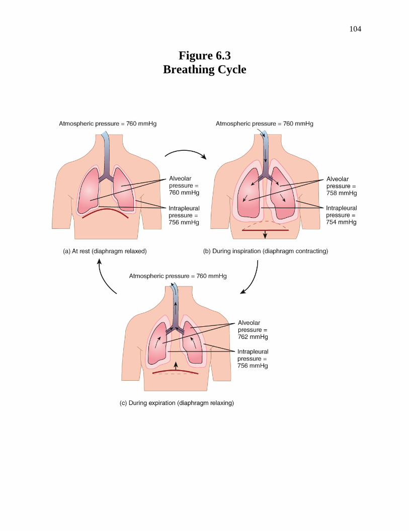
♦The Mechanics of Breathing -Breathing: A Definition -Breathing (pulmonary ventilation) is the process by which air flows into and out of the lungs in order to undergo gas exchange in the alveoli. -Air flows between the atmosphere and lungs based on a pressure gradient. • Air flows from the atmosphere into the lungs when the alveolar pressure is less than atmospheric pressure. -Alveolar pressure is the pressure exerted on the walls of the alveoli by gases (like O2, CO2, N2, etc.) in air within the lungs. -Atmospheric pressure is the pressure exerted on our surroundings by gases in the air of the atmosphere. ⇒At sea level, atmospheric pressure is 760 mm Hg. • Air flows from the lungs into the atmosphere when the alveolar pressure is greater than atmospheric pressure. -Lung-Chest Wall System -Recall that each lung is surrounded by a connective tissue sac called the pleura: -The parietal pleura is fused to the thoracic cavity wall, while the visceral pleura lines the surface of the lungs. -Between the parietal and visceral layers of the pleura is the pleural cavity, which contains a few milliliters of intrapleural fluid. -The intrapleural fluid causes the lungs and chest wall to be coupled together like two pieces of film that are held together by water. -Consequently, if the thoracic cavity increases in size, the lungs also expand; if the thoracic cavity decreases in size, the lungs recoil (become smaller). -There is a certain amount of pressure associated with the intrapleural fluid; this pressure is called intrapleural pressure. -Intrapleural pressure is normally subatmospheric, ranging from 754 to 756 mm Hg at any time during the breathing cycle (Figure 6.3, Derrickson). -Since intrapleural pressure is less than atmospheric pressure, it is said to be a negative pressure (vacuum). -It is the negative pressure of the intrapleural fluid that causes the suction that holds the lungs to the thoracic cavity wall via the pleura. -Breathing Cycle -Each breathing cycle consists of 3 phases: a period of rest, an inspiration, and an expiration (Figure 6.3, Derrickson): 1. rest -During the resting phase of the breathing cycle, alveolar pressure is equal to atmospheric pressure, which is 760 mm Hg (Figure 6.3, Derrickson). -Therefore, no air flows into or out of the lungs due to the lack of a pressure gradient.
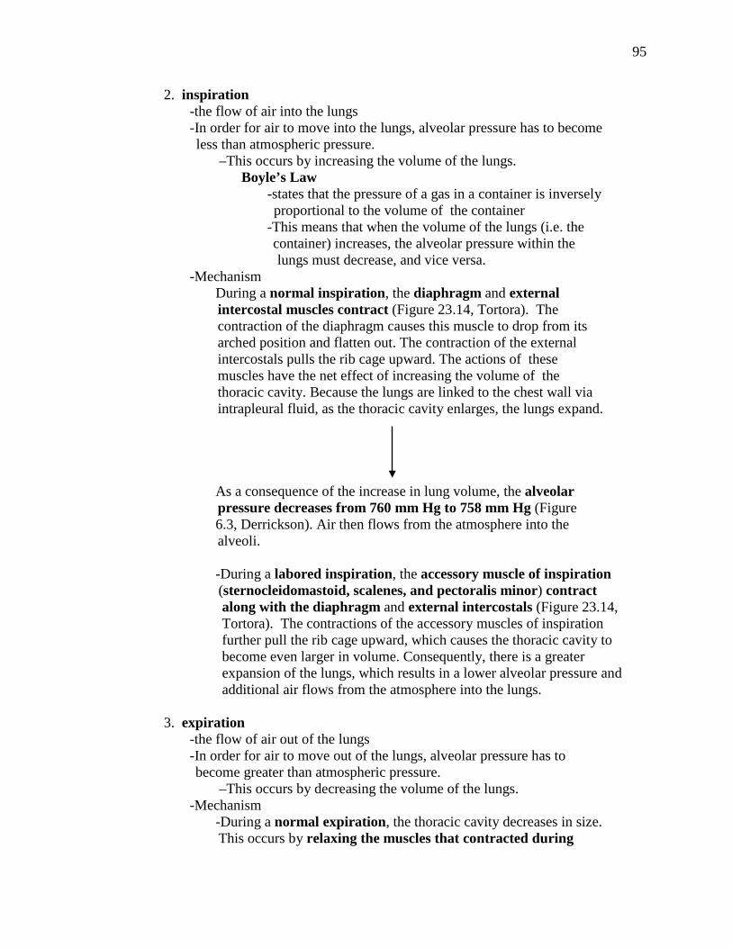
95
2. inspiration -the flow of air into the lungs -In order for air to move into the lungs, alveolar pressure has to become less than atmospheric pressure. –This occurs by increasing the volume of the lungs. Boyle’s Law -states that the pressure of a gas in a container is inversely proportional to the volume of the container -This means that when the volume of the lungs (i.e. the container) increases, the alveolar pressure within the lungs must decrease, and vice versa. -Mechanism During a normal inspiration, the diaphragm and external intercostal muscles contract (Figure 23.14, Tortora). The contraction of the diaphragm causes this muscle to drop from its arched position and flatten out. The contraction of the external intercostals pulls the rib cage upward. The actions of these muscles have the net effect of increasing the volume of the thoracic cavity. Because the lungs are linked to the chest wall via intrapleural fluid, as the thoracic cavity enlarges, the lungs expand. As a consequence of the increase in lung volume, the alveolar pressure decreases from 760 mm Hg to 758 mm Hg (Figure 6.3, Derrickson). Air then flows from the atmosphere into the alveoli. -During a labored inspiration, the accessory muscle of inspiration (sternocleidomastoid, scalenes, and pectoralis minor) contract along with the diaphragm and external intercostals (Figure 23.14, Tortora). The contractions of the accessory muscles of inspiration further pull the rib cage upward, which causes the thoracic cavity to become even larger in volume. Consequently, there is a greater expansion of the lungs, which results in a lower alveolar pressure and additional air flows from the atmosphere into the lungs. 3. expiration -the flow of air out of the lungs -In order for air to move out of the lungs, alveolar pressure has to become greater than atmospheric pressure. –This occurs by decreasing the volume of the lungs. -Mechanism -During a normal expiration, the thoracic cavity decreases in size. This occurs by relaxing the muscles that contracted during
96
inspiration. The diaphragm relaxes and moves back to its arched position. The external intercostals relax, which lowers the rib cage. Because the lungs are linked to the chest wall via intrapleural fluid, as the thoracic cavity decreases its size, the lungs recoil (become smaller). As a consequence of the decrease in lung volume, the alveolar pressure increases to 762 mm Hg and air moves from the alveoli into the atmosphere (Figure 6.3, Derrickson). -A labored expiration is caused by the contraction of the internal intercostals and the abdominal muscles (rectus abdominis, tranversus abdominis, external oblique, and internal oblique) (Figure 23.14, Tortora). The contractions of these muscles further pull the rib cage downward, which causes the thoracic cavity to become even smaller in volume. Consequently, the lungs become smaller, which results in a greater increase in alveolar pressure and additional air flows from the alveoli into the atmosphere. -Neural Regulation of the Breathing Cycle -Breathing is regulated by the respiratory center. respiratory center -located in the medulla and the pons of the brain stem (Figure 23.24, Tortora) -involuntarily causes the different phases of the breathing cycle (rest, inspiration, and expiration) -Various nerves connect the respiratory center with the muscles of respiration; this allows the respiratory center to activate these muscles (i.e. to cause their contraction) or to inhibit these muscles (i.e. to cause their relaxation). -Examples • The respiratory center has neural connections with the phrenic nerve, which innervates the diaphragm. • The respiratory center also has neural connections with the intercostal nerves, which innervate the external intercostals and the internal intercostals. -In addition, neural connections between the cerebral cortex and the respiratory center allow for voluntary (conscious) control of breathing (such as during talking, singing, whistling, etc.)
97
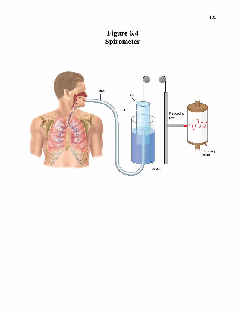
♦ Lung Volumes and Capacities -The amount of air that flows into or out of the lungs can be measured by an apparatus called a spirometer (Figure 6.4, Derrickson) -A spirometer consists of a breathing tube, a floating bell (which is a cylindrical device that floats in water) and a pen recorder. -The spirometer is used in the following way: A patient breathes into the breathing tube. The floating bell moves up and down in the water as the patient inhales and exhales. Changes in the position of the floating bell reflect changes in lung volume. A pen recorder then graphs the changes in lung volume on a chart called a spirogram. -There are 4 types of lungs volumes (Figure 23.16, Tortora): 1. tidal volume (VT) -the amount of air inspired and then expired during a normal breathing cycle -equals 500 mL 2. inspiratory reserve volume (IRV) -the maximum amount of air that can be inspired after a normal inspiration -equals 3100 mL in men and 1900 mL in women 3. expiratory reserve volume (ERV) -the maximum amount of air that can be expired after a normal expiration -equals 1200 mL in men and 700 mL in women 4. residual volume (RV) -the amount of air that remains in the lungs after a maximum expiration -equals 1200 mL in men and 1100 mL in women -cannot be determined using a spirometer
98
-There are 4 types of lung capacities (Figure 23.16, Tortora): -The term capacity refers to combinations of the various lungs volumes. 1. functional residual capacity (FRC) -the amount of air in the lungs at the end of a normal expiration -the sum of the ERV and the RV -equals 2400 mL in men and 1800 mL in women -cannot be determined using a spirometer 2. inspiratory capacity (IC) -the maximum amount of air that can be inspired after a normal expiration -the sum of the VT and IRV -equals 3600 mL in men and 2400 mL in women 3. vital capacity (VC) -the maximum volume of air that can be inspired after a maximum expiration -the sum of the ERV, VT, and IRV -equals 4800 mL in men and 3100 mL in women 4. total lung capacity (TLC) -the total amount of air in the lungs after a maximum inspiration -the sum of all lung volumes -equals 6000 mL in men and 4200 mL in women ♦Respiratory Minute Volume respiratory minute volume (V) -the amount of air that flows into and out of the entire respiratory system per minute -equals the product of the tidal volume (VT) and the respiratory rate (RR): V = VT X RR -Under normal, resting conditions, VT = 500 mL/ breath and the RR = 12 breaths/min. Therefore, the normal V = 6000 mL/min: 500 mL 12 breaths V = ________ X _________ = 6000 mL/min breath min
99
♦Alveolar Ventilation -Not all of the air that flows into the respiratory system actually enters the alveoli where gas exchange takes place; instead, some of the air remains in the anatomical dead space: anatomical dead space (VD) -the volume of air that is located in the conduction zone (i.e. the nose, pharynx, trachea, bronchi, and nonrespiratory bronchioles), which contains air that does not undergo gas exchange -equals 150 mL/breath alveolar minute volume (VA) -the amount of air that actually flows into and out of the respiratory zone (respiratory bronchioles and alveoli) to undergo gas exchange -can be calculated from the following equation: VA = (VT -VD) x RR -Under normal, resting circumstances, VT = 500 mL/breath, VD = 150 mL/breath, and the RR = 12 breaths/min. Therefore, a normal VA = 4200 mL/min. 500 mL 150 mL 12 breaths 4200 mL VA = ______ - ______ X ________ = _______ breath breath min min ♦Clinical Applications and Disorders -Look up the following clinical applications and disorders in Tortora: 1. rhinoplasty p. 853 2. laryngitis and cancer of the larynx p. 860 3. tracheostomy and intubation p. 862 4. pneumothorax, hemothorax, and atelectasis p. 863 5. coryza, seasonal influenza, and H1N1 influenza p. 866
100
6. respiratory distress syndrome p. 872 7. hyperbaric oxygenation p. 876 8. carbon monoxide poisoning p. 882 9. hypoxia p. 887 10. effects of smoking on respiratory efficiency p. 889 11. asthma p. 890, 892 12. chronic obstructive pulmonary disease, emphysema, and bronchitis p. 892 13. lung cancer p. 892 14. pneumonia p. 893 15. tuberculosis p. 893 16. pulmonary edema p. 893 17. sudden infant death syndrome p. 893-894 18. abdominal thrust (Heimlich) maneuver p. 894 19. asphyxia p. 894 20. aspiration p. 894
101
21. bronchoscopy p. 894 22. Cheyne-Stokes respiration p. 894 23. dyspnea p. 894 24. epistaxis p. 894 25. rales p. 894 26. respirator p. 894 27. respiratory failure p. 894 28. rhinitis p. 894 29. sleep apnea p. 894 30. sputum p. 894 31. strep throat p. 894 32. tachypnea p. 894 33. wheeze p. 894
102
Figure 6.1
Components of the Respiratory System
Right primary (main) bronchus
103
Figure 6.2 Transverse Section of Trachea and Esophagus
104
Figure 6.3 Breathing Cycle
105
Figure 6.4 Spirometer


![Respiratory system roadmap.pptx [Repaired] - Loginanatomical-sciences.health.wits.ac.za/roadmaps/Respiratory system... · DIVISION OF THE RESPIRATORY SYSTEM CONDUCTING PORTION Nasal](https://img.pdfslide.net/doc/110x75/5a78c3d87f8b9ae6228c9db0/respiratory-system-repaired-loginanatomical-scienceshealthwitsaczaroadmapsrespiratory.jpg)


![Respiratory System [โหมดความเข้ากันได้] · PATHOLOGY OF RESPIRATORY SYSTEM นพ. อรรณพ นาคะป ท Respiratory system U it](https://img.pdfslide.net/doc/110x75/5fa578efd4e80f055f6b3401/respiratory-system-aaaaaaaaaaaaaaaaaa-pathology.jpg)


![Anatomy and Physiology Respiratory System [Tab 2] Respiratory System](https://img.pdfslide.net/doc/110x75/56649ebd5503460f94bc631f/anatomy-and-physiology-respiratory-system-tab-2-respiratory-system.jpg)