Embed Size (px)
Citation preview
1
Applied Clinical Neuroimaging
in Cerebral Amyloid Angiopathy
and Spontaneous Intracerebral
Haemorrhage
by
Dr Andreas Charidimou MD MSc
Thesis submitted for degree of Doctor of Philosophy in Clinical Neurology
of University College London (UCL)
UCL Institute for Neurology, National Hospital for Neurology and Neurosurgery
London, UK
2015
2
DECLARATIONS
I, Andreas Charidimou confirm that the work presented in this thesis entitled “Applied
Clinical Neuroimaging in Cerebral Amyloid Angiopathy and Spontaneous Intracerebral
Haemorrhage” is my own. Where information has been derived from other sources, I
confirm that this has been indicated in the thesis.
I collected, reviewed, cleaned and analysed (including imaging and statistical analysis) the
data included in this thesis. I personally performed the searches, the collection of papers
and the data extraction for the systematic reviews incorporated in this thesis. Dr Simone
Gregoire significantly contributed in collecting data for the multicentre cohort of UCLH,
Belgium and Cambridge (UK). Dr Young T. Hong collected the PET and MRI data included
in Chapter 7 from the Wolfson Brain Imaging Centre, Department of Clinical
Neurosciences, University of Cambridge, UK. Dr Sergi Martinez-Ramirez, Dr Anand
Viswanathan, Prof Steve Greenberg and other members of the Hemorrhagic Stroke
Research Program in Massachusetts General Hospital Stroke Research Center, Harvard
Medical School, Boston, MA, USA, contributed in setting up the cerebral amyloid
angiopathy pathology cohort which appears in Chapter 10.
I was supported in the statistical approaches and analyses by Dr Zoe Fox, the statistician
associated with the UCL Institute of Neurology. Finally, this thesis was drafted in its
entirety by myself, but with critical revisions by my principal supervisor, Dr David Werring
and my subsidiary supervisor, Dr Hans R. Jäger. Sections of this thesis have been published
in peer-reviewed scientific journals as stated below. Finally, Prof. Jean-Claude Baron
provided critical revisions for the study presented in Chapter 7.
Signature:
3
ABSTRACT
Sporadic cerebral amyloid angiopathy is a common small vessel disease that preferentially
involves small cortical and leptomeningeal arteries due to progressive amyloid-β deposition
in their walls. Cerebral amyloid angiopathy occurs frequently in elderly people, and is a
common and important cause of symptomatic lobar intracerebral haemorrhage and
cognitive impairment. There is currently a growing interest in cerebral amyloid angiopathy,
at least partly thanks neuroimaging, which now allows an unprecedented ability to
investigate the disease dynamics in vivo using MRI to reveal complex patterns of cerebral
bleeding and ischaemia. The detection of CAA during life is becoming an increasingly
important challenge, since approaches of prevention or treatment (disease-modification)
are now emerging as realistic possibilities. Determining the most promising treatments
requires development of reliable biomarkers, the goal of my research. The main objective
of this PhD thesis is to provide new insights into potential clinical and applied clinical
neuroimaging biomarkers in patients with cerebral amyloid angiopathy. This is accomplished
by a portfolio of research studies investigating: (a) the clinical and radiological spectrum of
transient focal neurological episodes as a potential clinical clue for cerebral amyloid
angiopathy; (b) cortical superficial siderosis, a distinct pattern on bleeding in the brain, as
both a diagnostic and a prognostic marker of cerebral amyloid angiopathy; (c) MRI-visible
perivascular spaces topography, as a new marker of small vessel disease and cerebral
amyloid angiopathy; (d) potential pathological, neuroimaging and genetic differences in
patients with pathology-proven CAA with and without intracerebral haemorrhage and
presents evidence for different disease phenotypes; (e) the evidence whether the presence
and burden of cerebral microbleeds on MRI scans is associated with an increased risk of
recurrent spontaneous ICH, and if this risk is different according to MRI-defined
microangiopathy subtype, in a meta-analysis.
4
Table of contents
Chapter 1 General Introduction ..................................................................................................... 17
Chapter 2 Clinical-radiological spectrum of transient focal neurological episodes in
cerebral amyloid angiopathy: multicentre MRI cohort study and meta-analysis ...................... 51
Chapter 3 Prevalence and mechanisms of cortical superficial siderosis in sporadic
cerebral amyloid angiopathy ................................................................................................................. 65
Chapter 4 Cortical superficial siderosis and intracerebral haemorrhage risk in patients
with sporadic cerebral amyloid angiopathy: multicentre MRI cohort study ............................. 77
Chapter 5 MRI-visible perivascular spaces as a marker of underlying arteriopathy in
intracerebral haemorrhage: Multicentre MRI cohort study .......................................................... 90
Chapter 6 MRI-visible perivascular spaces as a neuroimaging marker of cerebral amyloid
angiopathy: MRI-histopathological study .......................................................................................... 103
Chapter 7 MRI-visible white matter perivascular spaces: a marker of cerebrovascular
amyloid burden? ..................................................................................................................................... 114
Chapter 8 White matter perivascular spaces on MRI are related to cortical superficial
siderosis in sporadic cerebral amyloid angiopathy ........................................................................ 126
Chapter 9 Cerebral amyloid angiopathy with and without intracerebral haemorrhage:
MRI-pathological evidence for different disease phenotypes ...................................................... 137
Chapter 10 Cerebral microbleeds, microangiopathy subtype and recurrent spontaneous
intracerebral haemorrhage risk: systematic review and meta-analysis ..................................... 150
Chapter 11 General discussion, future perspectives and challenges ................................. 166
Appendices .............................................................................................................................................. 189
Appendix A: Supplementary material for Chapter 2 .................................................................... 190
Appendix B: Supplementary material for Chapter 3 ..................................................................... 192
Appendix C: Supplementary material for Chapter 4 .................................................................... 196
Appendix D: Supplementary material for Chapter 5 .................................................................... 201
Appendix E: Supplementary material for Chapter 6 ..................................................................... 202
References ............................................................................................................................................... 204
5
LIST OF TABLES
Table 1.1 Research definitions of commonly used terms in the field of cerebral small vessel
disease31 as well as basic structural MRI techniques.35 ......................................................... 23
Table 1.2 Classic and modified Boston criteria for diagnosis of cerebral amyloid angiopathy
(CAA). (*Modifications compared to the classic Boston criteria based on Linn et al.) 44
Table 2.1 Demographic, clinical and MRI characteristics of multicentre cohort. .................... 57
Table 2.2 Characteristics of patients with cerebral amyloid angiopathy (CAA) and transient
focal neurological episodes (TFNE) vs. CAA patients without TFNE. ............................. 61
Table 3.1 Characteristics of patients with probable cerebral amyloid angiopathy (CAA) and
our comparison patients groups with spontaneous symptomatic ICH not fulfilling the
Boston criteria for probable CAA: “single lobar ICH and mixed haemorrhages” and
“strictly deep haemorrhages”. .................................................................................................... 70
Table 3.2 Characteristics and comparison of CAA patients with and without superficial
cortical siderosis. P-values refer to differences between CAA patients with vs. without
cortical superficial siderosis (cSS), using chi-square tests and the Fisher’s exact test for
categorical variables, and two-sample t-tests or Mann-Whitney U-tests depending on
the distribution of continuous variables. .................................................................................. 71
Table 3.3 Multivariate regression analysis showing the factors associated with cortical
superficial siderosis in patients with probable cerebral amyloid angiopathy. The model
remains consistent when history of hypertension and acute cSAH (%) are included. . 73
Table 4.1 Characteristics and comparison of CAA patients according to symptomatic lobar
intracerebral haemorrhage at follow-up. The p-values refer to differences between
patients with vs. without symptomatic lobar intracerebral haemorrhage at follow-up.
The median time between all baseline symptomatic ICH and first MRI was 17 days
(IQR: 3-75.5 days). ........................................................................................................................ 83
Table 4.2 Prespecified multivariate analyses of predictors of symptomatic lobar
intracerebral haemorrhage during follow-up in patients with cerebral amyloid
angiopathy. The models remain consistent if number of cerebral microbleeds (CMBs),
presence of CMBs or ≥5 CMBs are included. ........................................................................ 86
Table 6.1 Patient and imaging characteristics. ................................................................................ 108
Table 6.2 Diagnosis of cerebral angiopathy (CAA) using the original Boston criteria
(including strictly lobar microbleeds) with and without the inclusion of severe centrum
semiovale PVS (>20 PVS). Only cases with T2*-GRE were included in this analysis
6
(n=21). Inclusion of severe centrum semiovale PVS resulted in the diagnostic
upgrading of three patients: two from possible to probable CAA and one from non-
CAA to possible CAA. ............................................................................................................... 111
Table 7.1 Basic demographic and imaging characteristics of the study population. The p-
values refer to the comparison between probable cerebral amyloid angiopathy cases
(CAA) and age-matched elderly healthy subjects ................................................................ 120
Table 7.2 Univariable and multivariable linear regression analysis of the association between
whole cortex PiB retention (DVR) and CSO-PVS grade (as continuous variable) in the
whole study population and in sensitivity analyses including only probable CAA
patients and the age-matched healthy elderly (≥60 years) subgroup. ............................ 122
Table 7.3 Univariable and multivariable linear regression analysis of the association between
whole cortex PiB retention (DVR) and high CSO-PVS degree (>40) in the whole study
population and in sensitivity analyses including only probable CAA patients and the
age-matched healthy elderly (≥60 years) subgroup. ........................................................... 122
Table 8.1 Characteristics and comparison of CAA patients with and without severe
centrum semiovale perivascular spaces (CSO-PVS). P-values refer to differences
between CAA patients with vs. without CSO-PVS, using chi-square tests and the
Fisher’s exact test for categorical variables, and two-sample t-tests or Mann-Whitney
U-tests depending on the distribution of continuous variables........................................ 132
Table 8.2 Univariate (unadjusted) and multivariable (adjusted) logistic regression analysis of
associations with high degree centum semiovale perivascular spaces (CSO-PVS), in the
whole cohort of patients with cerebral amyloid angiopathy (CAA) and in patients with
probable CAA. Both models remained consistent when multiple (≥2) CMBs or CMBs
categories (0, 2-4, 5-10 and >10 CMBs) were included. ................................................... 133
Table 9.1 Cerebral amyloid angiopathy with and without intracerebral haemorrhage:
severity and associations with neuritic plaques (NP) and tangles (NT)*. ...................... 144
Table 9.2 Comparison of clinical, imaging and genetic characteristics between
pathologically-proven CAA with vs. without ICH cohorts. .............................................. 144
Table 9.3 APOE allele prevalences according to cortical superficial siderosis (cSS) burden in
cerebral amyloid angiopathy (CAA) patients with or without intracerebral
haemorrhage (ICH). .................................................................................................................... 146
Table 10.1 Basic study design and patients characteristics of included studies. ..................... 156

7
Table 11.1 Recommended criteria for identification of CAA-related cortical superficial
siderosis and acute convexity subarachnoid haemorrhage (acute cSAH) ..................... 180
Table 11.2 Advised standards for evaluating cortical superficial siderosis (cSS) in research
studies of small vessel disease and cerebral amyloid angiopathy ..................................... 180
8
LIST OF FIGURES
Figure 1.1 The topography of sporadic cerebral small vessel disease. The small vessels of
the brain can mainly be affected by two types of pathological process: (1) hypertensive
arteriopathy – which typically affects small deep arterial perforators; or (2) cerebral
amyloid angiopathy (CAA) – which preferentially affects the small arteries and
arterioles of the cerebral cortex and gray–white-matter junction by the deposition of
amyloid-β in the vessel walls. ..................................................................................................... 19
Figure 1.2 The first published photograph of cerebral amyloid angiopathy (left panel),
accompanied by the author’s illustration (right panel), from Fischer, 1910. Note the
“fur-like” staining of the walls of the 3 small arteries. .......................................................... 25
Figure 1.3 The entrance of the University Psychiatric Clinic in Bel-Air at Geneve, where the
University of Geneva brain collection was founded at the beginning of the 20th
century. The panel on the right shows the brain of one of Stefanos Pantelakis original
brains included in his study on congophilic angiopathy,55 along with the patients’
cognitive evaluation of executive skills (lower panel). .......................................................... 26
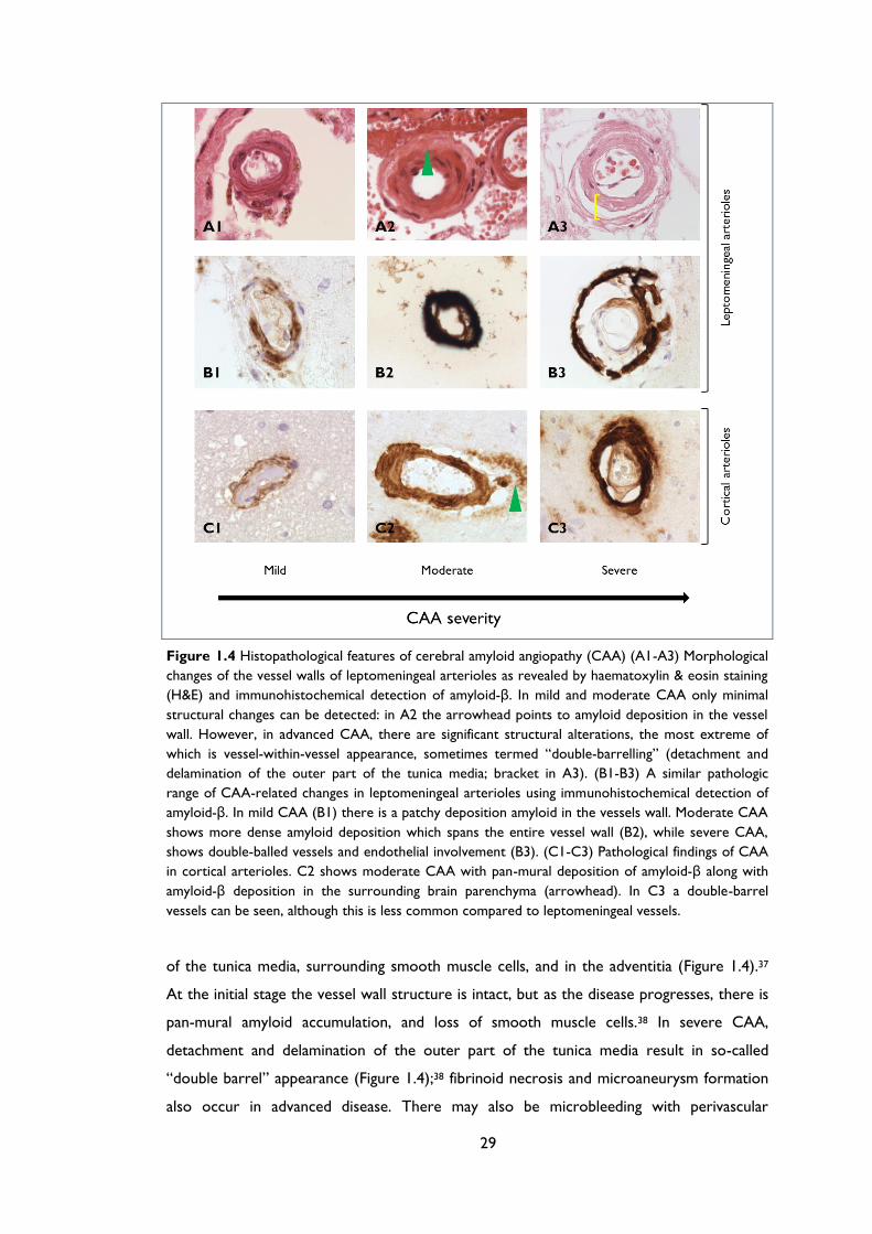
Figure 1.4 Histopathological features of cerebral amyloid angiopathy (CAA) (A1-A3)
Morphological changes of the vessel walls of leptomeningeal arterioles as revealed by
haematoxylin & eosin staining (H&E) and immunohistochemical detection of amyloid-
β. In mild and moderate CAA only minimal structural changes can be detected: in A2
the arrowhead points to amyloid deposition in the vessel wall. However, in advanced
CAA, there are significant structural alterations, the most extreme of which is vessel-
within-vessel appearance, sometimes termed “double-barrelling” (detachment and
delamination of the outer part of the tunica media; bracket in A3). (B1-B3) A similar
pathologic range of CAA-related changes in leptomeningeal arterioles using
immunohistochemical detection of amyloid-β. In mild CAA (B1) there is a patchy
deposition amyloid in the vessels wall. Moderate CAA shows more dense amyloid
deposition which spans the entire vessel wall (B2), while severe CAA, shows double-
balled vessels and endothelial involvement (B3). (C1-C3) Pathological findings of CAA
in cortical arterioles. C2 shows moderate CAA with pan-mural deposition of amyloid-
β along with amyloid-β deposition in the surrounding brain parenchyma (arrowhead).
In C3 a double-barrel vessels can be seen, although this is less common compared to
leptomeningeal vessels. ................................................................................................................ 29
Figure 1.5 (A) Amyloid-β production, elimination and deposition in cerebral amyloid
angiopathy (CAA). Converging evidence indicates that the major source of Aβ is
9
neuronal. It is generated by sequential cleavage of amyloid precursor protein (APP) by
β- and γ-secretases, in proportion to neuronal activity. Aβ is eliminated from the
brain by four major pathways: (a) proteolytic degradation by endopeptidases [such as
neprilysin and insulin‑degrading enzyme (IDE)]; (b) receptor-mediated clearance by
cells in the brain parenchyma (microglia, astrocytes and to a lesser extent neurones;
(c) active transport into the blood through the blood-brain barrier (BBB); (d)
elimination along the perivascular pathways by which interstitial fluid drains from the
brain.[86, 87] Specialized carriers (e.g. ApoE) and/or receptor transport mechanisms
(e.g. the low-density lipoprotein receptor [LDLR] and LDLR‑related protein [LRP1])
are involved in all major cellular clearance pathways. Vascular deposition is facilitated
by factors that increase the Aβ40:Aβ42 (while increased Aβ42 leads to
oligomerisation and amyloid plaques) and impede perivascular passage. As the
clearance mechanisms fail with age, Αβ is increasingly entrapped from the perivascular
drainage pathways into the basement membranes of capillaries and arterioles of the
brain leading to CAA. ................................................................................................................... 32
Figure 1.6 APOE alleles have a differential effect on different molecular and cellular
processes of Aβ production, elimination and deposition in a way that they either
increase or decrease the risk of developing CAA (B). The roles of different ApoE
alleles in various pathways in the brain which might contribute in the pathogenesis and
pathogenicity of CAA are summarised in (B). ........................................................................ 33
Figure 1.7 The spectrum of imaging manifestations of sporadic cerebral amyloid angiopathy
(CAA) . (A) An acute lobar haematoma on computer tomography (CT) scan. Some
extension of the bleeding in the posterior horn of the left ventricle can be seen. (B)
CT scan of a patient with a small posterior cortical haematoma. Acute cortical
subarachnoid haemorrhage (cSAH) is evident in two adjacent sulci (arrowheads). (C)
A T2-weighted MRI of a patient with probable CAA showing two lobar foci of
recent/subacute intracerebral haemorrhage (ICH): in the medial aspect of the left
occipital lobe, and in the right inferior frontal gyrus. There is also a large old lobar
haemorrhage involving the right occipital lobe, some scattered cortical-subcortical
cerebral microbleeds (CMBs) in posterior brain regions, as well as confluent white
matter hyperintensities (leukoaraiosis: arrow). (D) SWI and T2*-GRE (inset) done on
the same day in a patient with a large lobar bleed. The detection of strictly lobar
CMBs (better demonstrated on SWI) allowed the diagnosis of probable CAA. (E)
cSAH (linear hypointensities in the subarachnoid space on T2*-GRE/SWI) and focal
cortical siderosis (hyperintense on T2*-GRE/SWI). The inset in (E) demonstrates the
10
co-existence of cSAH (arrowhead), focal cortical siderosis in an adjacent sulcus
(arrow) and some CMBs (circles). Focal cortical siderosis represents the chronic
lesion following an acute cSAH. (F) The use of SWI, in a patient presenting with
progressive cognitive impairment let to the detection of multiple strictly lobar
microbleeds, characteristic of CAA. Confluent white matter changes (arrow) are also
visible. (G) Restricted diffusion (diffusion-weighted image on left, absolute diffusion-
weighted imaging (DWI) on the right) consistent with a small acute ischaemic lesion in
a patient with probable CAA. ..................................................................................................... 39
Figure 2.1 (A) Patient 7 had recurrent brief episodes of smoothly migrating tingling, starting
in the fingers of the left hand: T2*-GRE MRI shows extensive superficial cortical
siderosis in the right hemisphere and lobar cerebral microbleeds (CMBs). (B) Patient 2
had recurrent episodes of tingling in the right thumb, index and middle finger: T2*-
GRE shows cortical superficial siderosis (arrows) and lobar CMBs (arrowheads). (C-
D) Patient 12 had episodes of visual disturbances in the right hemifield: T2*-GRE
shows superficial cortical siderosis in the parietal and occipital lobes. ............................ 58
Figure 2.2 Patient 20 experienced two episodes of dysphasia and lightheadedness. (A)
Diffusion-weighted imaging one week after the first episode shows two small
ischaemic lesions in the left parietal lobe and a lesion in the right cerebellar
hemisphere (inset). (B) Susceptibility-weighted imaging indicates cortical superficial
siderosis in the sulci very close the ischaemic lesions. (C) Cortical superficial siderosis
extends to many sulci, especially in the left hemisphere (dotted arrows), associated
with lobar microbleeds (arrowheads) [not visible on T2-weighted MRI (D)]. ............... 58
Figure 2.3 Kaplan-Meier analyses for event-free rates of symptomatic intracerebral
haemorrhage after transient focal neurological episodes (TFNE) in our multicentre
cohort (n=24; A), all published studies (n=43, B), and combined (n=67; C). ................. 60
Figure 2.4 The prevalence of different types of transient focal neurological symptoms in
published cases (n=54) and in our multicentre cohort of CAA patients. Percentages
were compared using Pearson Chi-squared tests. (Percentages do not add up to 100
as some patients experienced multiple episode types). ....................................................... 62
Figure 3.1 T2*-GRE and FLAIR MRI of two patients with probable cerebral amyloid
angiopathy and a patient with mixed deep and lobar haemorrhages. (A) A patient with
probable CAA and an acute right frontal intracerebral haemorrhage (ICH) and
multifocal cortical superficial siderosis (cSS) in the left hemisphere. Multiple lobar
cerebral microbleeds (CMBs) are also present, sometimes close to cSS. (B) A patient
with probable CAA with focal, bilateral cSS and lobar CMBs, in whom no ICHs were
11
present. (C) A patient with a mixed pattern of bleeds (ICH and CMBs) not fulfilling
the Boston criteria for CAA: note the focal cSS in left occipital lobe and the lobar
CMBs in close vicinity to cSS. ..................................................................................................... 72
Figure 4.1 Subsequent intracerebral haemorrhage (ICH) at the site of pre-existing siderosis
in two patients with cSS. 85-year-old female patient; time interval between baseline
imaging (A) and incident ICH (B): 19 months. 80-year-old male patient, time interval
between baseline imaging (C) and incident ICH (D): 35 months. A, C Baseline
magnetic resonance imaging, T2*-weighted images; B, D Follow-up unenhanced
computed tomography scans. ..................................................................................................... 84
Figure 4.2 Kaplan-Meier estimates of progression to symptomatic lobar intracerebral
haemorrhage (ICH) in the presence of: (A) cortical superficial siderosis (cSS); (B) focal
cSS; and (C) disseminated (>3 sulci) cortical superficial siderosis in all patients with
cerebral amyloid angiopathy (CAA). Testing of significance is by the log-rank test. .... 85
Figure 6.1 MRI-visible white matter perivascular spaces in CAA and non-CAA ICH.
Perivascular spaces (PVS) are defined as small, sharply delineated structures of (or
close to) cerebrospinal fluid intensity, measuring <3mm following the course of
perforating or medullary vessels, in basal ganglia and centrum semiovale. In (A) and (B)
the left panels show dot-like hyperintensities (florid PVS) on representative axial T2-
weighted MRI at 1.5 Tesla in the centrum semiovale, in patients with
histopathologically confirmed cerebral amyloid angiopathy (CAA); the right panels
show the corresponding pathology (immunostaining for Aβ) with dense amyloid
deposition spanning the entire vessel wall of a cortical (A, right panel) and a
leptomeningeal arteriole (B, right panel). (C) and (D) show axial T2-weighted MRI
from two of the non-CAA cases, showing a paucity of centrum semiovale PVS. ....... 110
Figure 7.1 Scatterplot of whole cortex PiB retention (i.e. DVR) and centrum semiovale
perivascular spaces (CSO-PVS) severity categories in cerebral amyloid angiopathy
(CAA) healthy subjects. There was a positive correlation between PiB retention and
increasing CSO-PVS categories across the whole group (Kendall's tau-b=0.552;
p=0.0001). ..................................................................................................................................... 121
Figure 8.1 T2 and T2*-GRE MRI of a patient with probable cerebral amyloid angiopathy
showing (A) High degree of CSO-PVS on MRI (small, sharply delineated structures of
CSF intensity (or close to CSF intensity), following the course of cortical vessels),
associated with (B) multifocal cortical superficial siderosis (cSS) especially in the right
hemisphere. ................................................................................................................................... 132
12
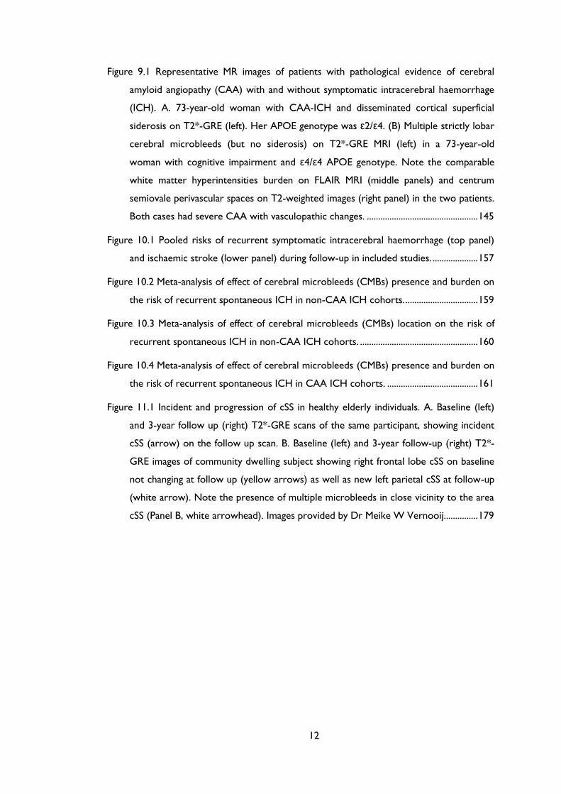
Figure 9.1 Representative MR images of patients with pathological evidence of cerebral
amyloid angiopathy (CAA) with and without symptomatic intracerebral haemorrhage
(ICH). A. 73-year-old woman with CAA-ICH and disseminated cortical superficial
siderosis on T2*-GRE (left). Her APOE genotype was ε2/ε4. (B) Multiple strictly lobar
cerebral microbleeds (but no siderosis) on T2*-GRE MRI (left) in a 73-year-old
woman with cognitive impairment and ε4/ε4 APOE genotype. Note the comparable
white matter hyperintensities burden on FLAIR MRI (middle panels) and centrum
semiovale perivascular spaces on T2-weighted images (right panel) in the two patients.
Both cases had severe CAA with vasculopathic changes. ................................................. 145
Figure 10.1 Pooled risks of recurrent symptomatic intracerebral haemorrhage (top panel)
and ischaemic stroke (lower panel) during follow-up in included studies. .................... 157
Figure 10.2 Meta-analysis of effect of cerebral microbleeds (CMBs) presence and burden on
the risk of recurrent spontaneous ICH in non-CAA ICH cohorts. ................................ 159
Figure 10.3 Meta-analysis of effect of cerebral microbleeds (CMBs) location on the risk of
recurrent spontaneous ICH in non-CAA ICH cohorts. .................................................... 160
Figure 10.4 Meta-analysis of effect of cerebral microbleeds (CMBs) presence and burden on
the risk of recurrent spontaneous ICH in CAA ICH cohorts. ........................................ 161
Figure 11.1 Incident and progression of cSS in healthy elderly individuals. A. Baseline (left)
and 3-year follow up (right) T2*-GRE scans of the same participant, showing incident
cSS (arrow) on the follow up scan. B. Baseline (left) and 3-year follow-up (right) T2*-
GRE images of community dwelling subject showing right frontal lobe cSS on baseline
not changing at follow up (yellow arrows) as well as new left parietal cSS at follow-up
(white arrow). Note the presence of multiple microbleeds in close vicinity to the area
cSS (Panel B, white arrowhead). Images provided by Dr Meike W Vernooij............... 179
13
MANUSCRIPTS BASED ON THE MATERIAL AND STUDIES
PRESENTED IN THIS THESIS
Chapter 1 Charidimou A, Gang Q, Werring DJ. Sporadic cerebral amyloid angiopathy
revisited: recent insights into pathophysiology and clinical spectrum J Neurol
Neurosurg Psychiatry. 2012; 83(2): 124-37.
Chapter 2
Charidimou A, Peeters A, Fox Z, Gregoire SM, Vandermeeren Y, Laloux P, et
al. Spectrum of transient focal neurological episodes in cerebral amyloid
angiopathy: multicentre magnetic resonance imaging cohort study and meta-
analysis. Stroke. 2012; 43(9): 2324-30.
Chapter 3 Charidimou A, Jager RH, Fox Z, Peeters A, Vandermeeren Y, Laloux P, et al.
Prevalence and mechanisms of cortical superficial siderosis in cerebral amyloid
angiopathy. Neurology. 2013; 81(7): 626-32.
Chapter 4 Charidimou A, Peeters AP, Jager R, Fox Z, Vandermeeren Y, Laloux P, et al.
Cortical superficial siderosis and intracerebral haemorrhage risk in cerebral
amyloid angiopathy. Neurology. 2013; 81(19): 1666-73.
Chapter 5
Charidimou A, Meegahage R, Fox Z, Peeters A, Vandermeeren Y, Laloux P, et
al. Enlarged perivascular spaces as a marker of underlying arteriopathy in
intracerebral haemorrhage: a multicentre MRI cohort study. J Neurol
Neurosurg Psychiatry. 2013; 84(6): 624-9.
Chapter 6 Charidimou A, Jaunmuktane Z, Baron JC, Burnell M, Varlet P, Peeters A, et al.
White matter perivascular spaces: An MRI marker in pathology-proven
cerebral amyloid angiopathy? Neurology. 2014; 82(1): 57-62.
Chapter 7 Submitted
Chapter 8 Charidimou A, Peeters AP, Jager R, Vandermeeren Y, Laloux P, et al. White
matter perivascular spaces are related to cortical superficial siderosis in
cerebral amyloid angiopathy. Stroke. 2014; accepted.
Chapter 9 In preparation
Chapter 10 Submitted
Chapter 11
Charidimou A, Baron JC, Werring DJ. Transient focal neurological episodes,
cerebral amyloid angiopathy, and intracerebral haemorrhage risk: looking
beyond TIAs. International journal of stroke. 2013; 8(2): 105-8.
Charidimou A, Jager HR. Developing biomarkers for cerebral amyloid
angiopathy trials: do potential disease phenotypes hold promise? Lancet
Neurol. 2014; 13(6): 538-40.
Charidimou A, Werring DJ. Cerebral microbleeds as a predictor of
macrobleeds: what is the evidence? International journal of stroke. 2014; 9(4):
457-9.
14
ACKNOWLEDGEMENTS
I would like to acknowledge and extend my heartfelt gratitude to the following persons
who made this project possible. Firstly, I would like to thank Dr David J Werring, Honorary
Consultant Neurologist at the National and Reader at the UCL Institute of Neurology, for
his guidance throughout my studies. His tireless encouragement and continuous support as
my principal supervisor, and above all as my mentor, have been invaluable and shaped me as
a researcher. He supervised the writing and editing of our manuscripts and was principally
responsible for the supervision of this thesis, for which I am very grateful.
Sincere thanks are also due to my other supervisor, Dr Rolf H Jäger, for his training
in neuroimaging, valuable feedback, insightful comments, amazing ideas and his support in
general.
I am also grateful to Prof. Martin M Brown for his support throughout my studies
(including his extremely useful feedback on my presentations given to national and
international conferences).
My special thanks also go to Clare Shakeshaft, who is the CROMIS-2 study
coordinator, and Juliet Solomon. Dr Duncan Wilson, new clinical research fellow in our
group, contributed in extending the current research portfolio and I am sure we will soon
have some exciting results. I would like to thank my colleague and friend from Japan, Dr
Yusuke Yakushiji, with whom we worked very closely on some exciting studies comparing
aspects of small vessel disease in East and West.
None of the studies on cerebral amyloid angiopathy presented here would have been
possible without the contribution of Prof. Jean-Claude Baron, an amazing mentor
generously sharing his ideas, experience and passion for cerebrovascular disease.
I would like to address very special thanks to my colleagues in Belgium, Dr Andre
Peeters, Prof. Patrice Laloux and Prof. Yves Vandermeeren for their enthusiasm and hard
work. Together, we set up a multicentre collaboration which still carries on today.
I am very grateful to Simone Gregoire, my predecessor in the Stroke Research
Group, for her generous help during the early stages of my research studies and continue
support and enthusiasm.
Special thanks to Zoe Fox, statistician at the Education Unit, for her support and
statistical advice on numerous occasions and on short notice.
I would like to extend my thanks to Prof Steve Greenberg, Dr Anand Viswanathan,
Dr Edip Gurol and all my friends in Boston, who kindly hosted me in their group for two
amazing months during the last period of my PhD.
Finally, I would like to thank my colleagues and friends for contributing in four fruitful
years at Queen Square and London.

15
FUNDING
I receive research support from the Greek State Scholarship Foundation (IKY), the Stroke
Association and the British Heart Foundation.

16
For my family, for the love and opportunities you have given me,
and, for Maria,
who have been there for me through everything,
with love

17
Chapter 1 General Introduction
18
Sporadic cerebral small vessel disease: key definitions and
current concepts
Many of the modern advances and effective interventions in cerebrovascular disorders
currently target only disease of large arteries. Until recently, small cerebral arteries have
received little attention and clinicians have much less to offer for the prevention and
treatment of small vessel disease.1 This is partly because small vessels are technically
inaccessible, hard to study directly and hence the underlying mechanisms of small vessel
disease remain relatively poorly understood.2 Yet, these diseases are considered to be
among the most prevalent known neurologic processes and contribute substantially to
stroke, cognitive impairment or other disabilities commonly seen in elderly persons (e.g.
depression, motor and gait disturbances, urinary symptoms, functional impairment etc.).2-4
In addition, small vessel disease can increase mortality.5, 6
Although the term sporadic cerebral small vessel disease is used with various meanings
in different contexts (e.g. clinical, neuroimaging, pathological etc.), in its most basic form it
encompasses a group of age-related neuropathological processes affecting the small
arteries, arterioles, capillaries, and rarely venules in the brain.1-3 Before defining these
pathological processes, it is important to define what a small vessel is and specifically, “how
small a small vessel is”. Interestingly, a survey performed among leading neuropathological
centres, revealed that the definition of a small vessel is not consistent: less than 50% of the
participants agreed on a size limit of less than 500 μm in transverse diameter or all vessels
located deeper than in the cortex.7 Others have arbitrarily suggested a transverse diameter
of ≤300 μm, predominantly referring to arterioles – likely illustrating that pathological
processes of the arteriolar tree are more well know that those of other small vascular
components (e.g. capillaries).2 The current definition of small vessels is more inclusive,
referring to all vascular structures (ranging from around 5μm up to 2mm), small arteries,
arterioles, capillaries, venules and small veins located in the brain parenchyma (i.e.
intraparenchymal) or in the subarachnoid space (i.e. leptomeningeal).2 These small vessels
can either: (a) penetrate the brain cortex superficially, supplying the gray matter with short
branches of three lengths (reaching cortical layer III, V and the gray–white matter junction),
and the subcortical white matter with longer branches; or (b) stem from arterial
perforators deeper at the base of the brain and supplying the basal ganglia, thalami and
brainstem structures.2 Specific small vessel pathologies can differentially affect these two
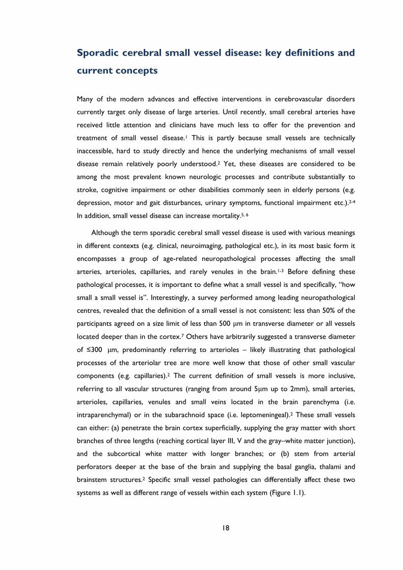
systems as well as different range of vessels within each system (Figure 1.1).
19
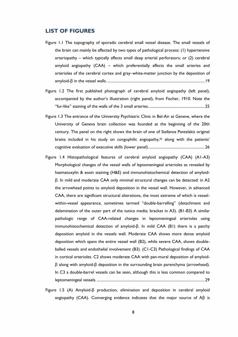
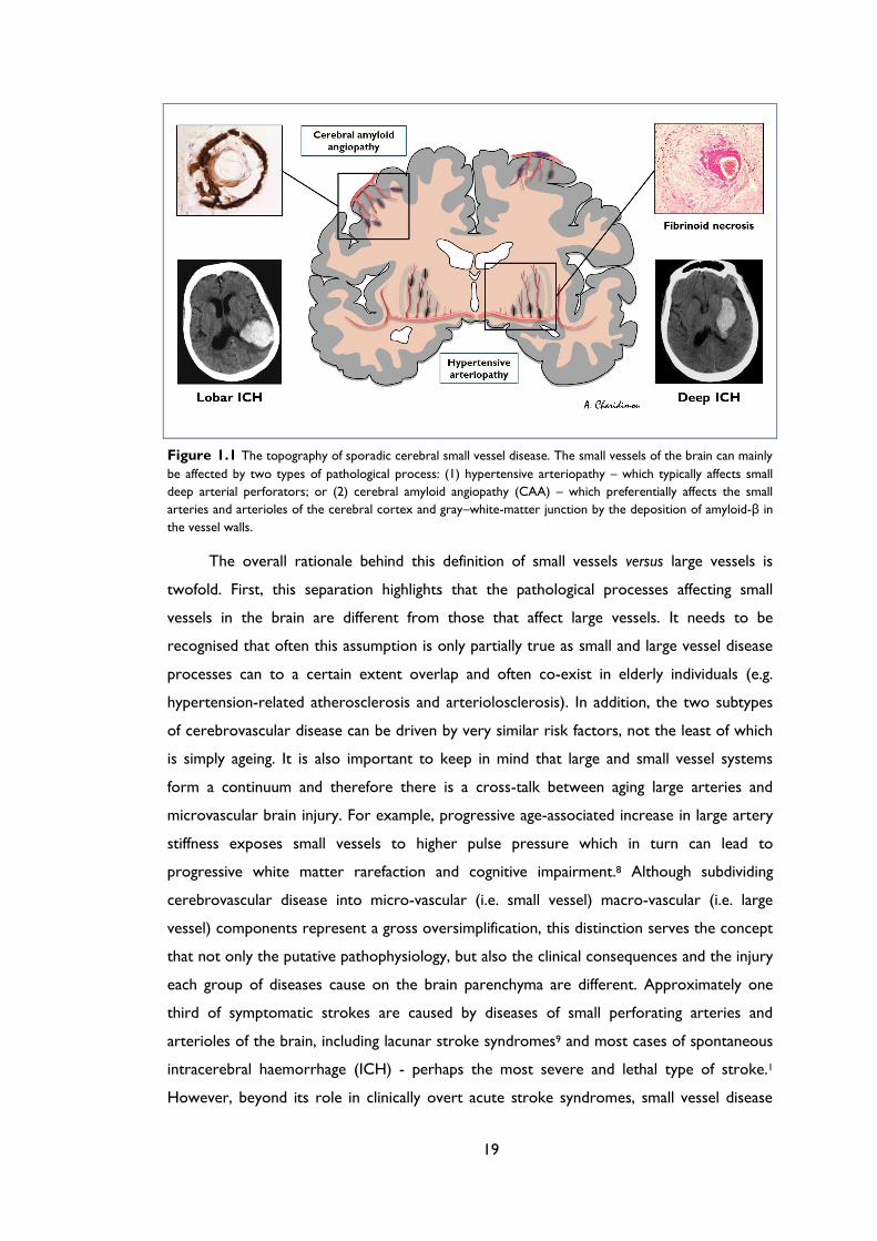
Figure 1.1 The topography of sporadic cerebral small vessel disease. The small vessels of the brain can mainly
be affected by two types of pathological process: (1) hypertensive arteriopathy – which typically affects small
deep arterial perforators; or (2) cerebral amyloid angiopathy (CAA) – which preferentially affects the small
arteries and arterioles of the cerebral cortex and gray–white-matter junction by the deposition of amyloid-β in
the vessel walls.
The overall rationale behind this definition of small vessels versus large vessels is
twofold. First, this separation highlights that the pathological processes affecting small
vessels in the brain are different from those that affect large vessels. It needs to be
recognised that often this assumption is only partially true as small and large vessel disease
processes can to a certain extent overlap and often co-exist in elderly individuals (e.g.
hypertension-related atherosclerosis and arteriolosclerosis). In addition, the two subtypes
of cerebrovascular disease can be driven by very similar risk factors, not the least of which
is simply ageing. It is also important to keep in mind that large and small vessel systems
form a continuum and therefore there is a cross-talk between aging large arteries and
microvascular brain injury. For example, progressive age-associated increase in large artery
stiffness exposes small vessels to higher pulse pressure which in turn can lead to
progressive white matter rarefaction and cognitive impairment.8 Although subdividing
cerebrovascular disease into micro-vascular (i.e. small vessel) macro-vascular (i.e. large
vessel) components represent a gross oversimplification, this distinction serves the concept
that not only the putative pathophysiology, but also the clinical consequences and the injury
each group of diseases cause on the brain parenchyma are different. Approximately one
third of symptomatic strokes are caused by diseases of small perforating arteries and
arterioles of the brain, including lacunar stroke syndromes9 and most cases of spontaneous
intracerebral haemorrhage (ICH) - perhaps the most severe and lethal type of stroke.1
However, beyond its role in clinically overt acute stroke syndromes, small vessel disease
20
causes widespread microvascular damage (seen on neuroimaging or at autopsy) which is
not symptomatic itself but has important cumulative effects on cognition. Hence, cerebral
small vessel disease is one of the most important contributors to cognitive impairment in
the elderly.10, 11 As people live longer, the burden of these diseases will rapidly grow over
the coming years becoming an increasing healthcare challenge facing all societies
worldwide.12
There are two main sporadic forms of small vessel disease (Figure 1.1). The first one
is sporadic cerebral amyloid angiopathy (CAA) (extensively covered in the next section of
the Introduction), a chronic degenerative disease characterised with progressive deposition
of amyloid-β in the media and adventitia of small arteries, arterioles and capillaries in the
cerebral cortex, overlying leptomininges and grey–white matter junction.10, 11 In contrast to
CAA which is relatively easy to define, the remaining sporadic small vessel disease is more
difficult to define and name. To this end, the term “hypertensive arteriopathy” is widely
used to describe a non-amyloid process often related to advanced age (but not clearly age-
driven), hypertension, diabetes mellitus and other common vascular risk factors, typically
affecting the small perforating end-arteries of the deep grey nuclei and deep white matter.2
It is characterised pathologically by lipohyalinosis, arteriolosclerosis or fibrinoid necrosis.
This very common sporadic form of small vessel disease has been variously known as
arteriolosclerosis, age-related or vascular risk-factor-related small vessel disease, or
degenerative microangiopathy in the literature.2, 13, 14 The term “hypertensive arteriopathy”
is not ideal (or even misleading) as it probably groups together a variety of sporadic small
vessel disease pathologies not accounted by sporadic CAA, but not necessarily related
specifically to hypertension.15 In other words, hypertension might not be a specific cause of
these small vessel changes collectively called “hypertensive arteriopathy” (at least not in all
cases), but can to a certain extent influence their evolution. Furthermore, there is not a
consensus on the microscopic small vessel lesions best described under the term
“hypertensive arteriopathy”, meaning that its severity might be difficult to evaluate in any
given case. From a histopathologic perspective, hypertensive arteriopathy is mainly
characterised by vessel wall thickening, narrowing of the lumen, loss of smooth muscle cells
from the tunica media and deposits of an amorphous fibro-hyaline material. Other possible
pathological features can include distal manifestations of atherosclerosis (microatheroma)
and the presence of microaneurysms (i.e. elongated and dilated vessels).2 However, many
researchers tend to subdivide hypertensive arteriopathy based on the most pronounced
structural histopathological features found, e.g. atherosclerosis, arteriolosclerosis,
lipohyalinosis, fibrinoid necrosis (the proposed acute from of lipohyalinosis),
microaneurysms etc.2 These subtypes tend to predominantly affect different small vessel
21
sizes and can exist separately or in various combinations in any given case. Of these
histopathological features, perhaps the one most strongly associated with hypertension-
related injury is fibrinoid necrosis, which is much more common in hypertensive patients’
brains than in those without hypertension,16-18 as well as in arterioles adjacent to deep
ICH.17-21 Complicating matters further, the more effective treatments for hypertension in
recent years is likely to have modified the specific pathological features, natural history and
disease spectrum of hypertensive arteriopathy.15 Despite the limitations in definitions and
given the lack of an alternative widely accepted term, for simplicity and consistency, the
term “hypertensive arteriopathy” has been adopted in this thesis as one of convenience to
avoid unnecessarily long and complex terms being repeated. Whatever term one prefers
for this sporadic small vessel disease type, the microvascular changes associated with
hypertensive arteriopathy presumably lead to both occlusion (including reduced vessel
compliance and impaired vasoreactivity) and haemorrhage in the brain territories of the
affected deep perforating arteries. Unsurprisingly, hypertensive arteriopathy has historically
been associated with two types of cerebrovascular disease: lacunar infarcts22 and
intracerebral haemorrhage.23
Since small vessels (and hence the structural alterations of small vessel disease)
cannot be easily visualised in vivo with the current neuroimaging techniques used in clinical
practice, the brain parenchymal magnetic resonance imaging (MRI) lesions which they are
thought to cause have been adopted as markers of small vessel damage.2 As a result, the
term cerebral small vessel disease is frequently used indiscriminately to describe both the
underlying small vessel pathologies and the neuroimaging correlates of their consequences
on the brain parenchyma.2 Of note, these consequences are heterogeneous in nature
including both ischaemic and haemorrhagic manifestations.2, 24 However, historically the
term small vessel disease has been often (and still is) used misleadingly to describe only the
ischaemic consequences on imaging.3 Despite this, sporadic small vessel disease is the
leading cause of ICH, which in fact parallels the topography of the underlying microvascular
pathology, so that spontaneous deep ICH (in the basal ganglia, thalami etc.) is
predominantly caused by hypertensive arteriopathy, whereas lobar (cortical-subcortical)
ICH is frequently caused by CAA. Lacunes and white matter hyperintensities (leukoaraiosis)
are well known imaging markers of cerebral small vessel disease that have been extensively
studied with MRI since the early 90s.25, 26 Advances in neuroimaging, now allows an
unprecedented ability to investigate more complex dynamics (both haemorrhagic and non-
haemorrhagic) of small vessel disease in vivo: new imaging manifestations of small vessel
disease include cerebral microbleeds,27 cortical superficial siderosis (cSS) and convexity
subarachnoid haemorrhage,28 cerebral microinfarcts29 and perivascular spaces.30
22
A further concept to bear in mind when approaching neuroimaging markers of small
vessel disease (and hence markers of pathologic consequences on the brain parenchyma), is
that their pathogenic interpretation is not uniform, and given marker may be found in
different types of small vessel disease.24 For example, in view of different topographical
distribution of hypertensive arteriopathy and CAA, it is hypothesized that cerebral
microbleeds have a preferential location depending on the underlying small vessel
pathology: hypertensive arteriopathy is commonly associated with cerebral microbleeds in
deep brain regions (e.g. basal ganglia, thalamus and brainstem), whereas CAA is
characterised by cerebral microbleeds in a lobar distribution (cortical-subcortical).27 Hence,
each marker or lesion on neuroimaging should not be taken in isolation.
These advances in neuroimaging were recently illustrated in an international working
group position paper from the Centres of Excellence in Neurodegeneration under the
acronym STandards for ReportIng Vascular changes on nEuroimaging (STRIVE v1).31 This
consensus paper presents a unified approach to small vessel disease at present as a
neuroimaging-defined concept, which is changing rapidly paralleling the continued advances
in neuroimaging techniques. Table 1.1 provides a glossary and definitions of the most
commonly used research terms in the field of small vessel disease (a defined in the STRIVE
consensus paper), including MRI modalities.
In the next section of the introduction a closer look at CAA is provided, since this
type of small vessel disease is the main focus of the research presented in this PhD thesis.
Given that hypertension is theoretically treatable, whereas CAA is currently not, and
considering that CAA is strongly associated with brain aging and is also directly linked to
Alzheimer's disease, is not surprising that CAA is becoming the most common sporadic
small vessel disease, at least in Western populations.32 This is partly illustrated by the
changing profiles of spontaneous ICH in recent epidemiological studies, showing an increase
in ICH incidence among patients aged 75 years or older, especially in relation to
antithrombotic use.33, 34 Also, while the incidence of deep (probably hypertensive
arteriopathy-related) ICH has fallen, the proportion of non-hypertensive lobar bleeds in
those aged 75 years or over increased. It is likely that CAA is implicated in the majority of
these lobar haemorrhages and might also account for the increased incidence of
anticoagulation-associated ICH.28
23
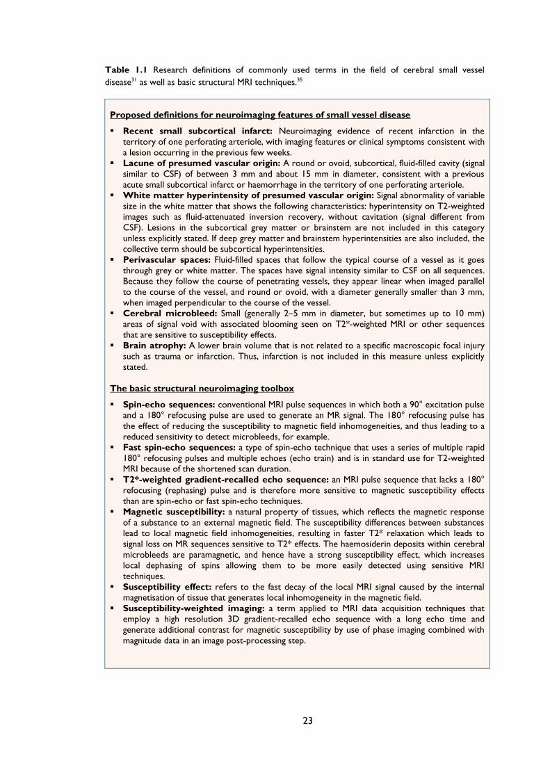
Table 1.1 Research definitions of commonly used terms in the field of cerebral small vessel
disease31 as well as basic structural MRI techniques.35
Proposed definitions for neuroimaging features of small vessel disease
Recent small subcortical infarct: Neuroimaging evidence of recent infarction in the
territory of one perforating arteriole, with imaging features or clinical symptoms consistent with
a lesion occurring in the previous few weeks.
Lacune of presumed vascular origin: A round or ovoid, subcortical, fluid-filled cavity (signal
similar to CSF) of between 3 mm and about 15 mm in diameter, consistent with a previous
acute small subcortical infarct or haemorrhage in the territory of one perforating arteriole.
White matter hyperintensity of presumed vascular origin: Signal abnormality of variable
size in the white matter that shows the following characteristics: hyperintensity on T2-weighted
images such as fluid-attenuated inversion recovery, without cavitation (signal different from
CSF). Lesions in the subcortical grey matter or brainstem are not included in this category
unless explicitly stated. If deep grey matter and brainstem hyperintensities are also included, the
collective term should be subcortical hyperintensities.
Perivascular spaces: Fluid-filled spaces that follow the typical course of a vessel as it goes
through grey or white matter. The spaces have signal intensity similar to CSF on all sequences.
Because they follow the course of penetrating vessels, they appear linear when imaged parallel
to the course of the vessel, and round or ovoid, with a diameter generally smaller than 3 mm,
when imaged perpendicular to the course of the vessel.
Cerebral microbleed: Small (generally 2–5 mm in diameter, but sometimes up to 10 mm)
areas of signal void with associated blooming seen on T2*-weighted MRI or other sequences
that are sensitive to susceptibility effects.
Brain atrophy: A lower brain volume that is not related to a specific macroscopic focal injury
such as trauma or infarction. Thus, infarction is not included in this measure unless explicitly
stated.
The basic structural neuroimaging toolbox
Spin-echo sequences: conventional MRI pulse sequences in which both a 90° excitation pulse
and a 180° refocusing pulse are used to generate an MR signal. The 180° refocusing pulse has
the effect of reducing the susceptibility to magnetic field inhomogeneities, and thus leading to a
reduced sensitivity to detect microbleeds, for example.
Fast spin-echo sequences: a type of spin-echo technique that uses a series of multiple rapid
180° refocusing pulses and multiple echoes (echo train) and is in standard use for T2-weighted
MRI because of the shortened scan duration.
T2*-weighted gradient-recalled echo sequence: an MRI pulse sequence that lacks a 180°
refocusing (rephasing) pulse and is therefore more sensitive to magnetic susceptibility effects
than are spin-echo or fast spin-echo techniques.
Magnetic susceptibility: a natural property of tissues, which reflects the magnetic response
of a substance to an external magnetic field. The susceptibility differences between substances
lead to local magnetic field inhomogeneities, resulting in faster T2* relaxation which leads to
signal loss on MR sequences sensitive to T2* effects. The haemosiderin deposits within cerebral
microbleeds are paramagnetic, and hence have a strong susceptibility effect, which increases
local dephasing of spins allowing them to be more easily detected using sensitive MRI
techniques.
Susceptibility effect: refers to the fast decay of the local MRI signal caused by the internal
magnetisation of tissue that generates local inhomogeneity in the magnetic field.
Susceptibility-weighted imaging: a term applied to MRI data acquisition techniques that
employ a high resolution 3D gradient-recalled echo sequence with a long echo time and
generate additional contrast for magnetic susceptibility by use of phase imaging combined with
magnitude data in an image post-processing step.
24
Sporadic cerebral amyloid angiopathy: pathophysiology
and clinical spectrum
Sporadic cerebral amyloid angiopathy (CAA) is a common small vessel disease of the brain,
characterised by the progressive deposition of amyloid–β protein in the walls of small-to-
medium sized arteries (up to about 2mm in diameter36), arterioles and capillaries in the
cerebral cortex and overlying leptomininges.37, 38 CAA can also affect cerebellar vessels, but
only rarely those in the brainstem or basal ganglia. Although known to pathologists for over
a century,39, 40 CAA was not linked to clinical disease until as late as the 1960’s, when it was
suggested to be a rare cause of intracerebral haemorrhage (ICH).41-43 In recent years, CAA
has been “rediscovered” as a common and important cause of spontaneous ICH, which
remains the most devastating form of stroke, with a fatality rate approaching 50% in
contrast to improved outcomes from ischaemic stroke.44, 45 An increased understanding of
CAA thus holds promise for improved prevention and treatment of ICH.
The growing interest in CAA is at least partly thanks to two fields of research, which
have been important in defining the expanding clinical-radiological phenotype and the
underlying pathophysiology of the disease: (1) neuroimaging, which now allows an
unprecedented ability to investigate CAA dynamics in vivo using MRI to reveal complex
patterns of cerebral bleeding (including lobar microbleeds27) and ischaemia, and an
increasing repertoire of specific amyloid-binding ligands;38, 46-50 and (2) transgenic mouse
studies, which have allowed the experimental alteration of amyloid peptide expression and
molecular structure, providing significant mechanistic insights. Despite these advances, CAA
remains under-recognized by neurologists and stroke physicians, making a fresh look
especially timely (see box for search strategy).
The entity of CAA encompasses a number of highly diverse sporadic and genetic
disorders that share the same pathological hallmark of amyloid-β fibril deposition in small
leptomeningeal and cortical vessels. Here, we focus only on sporadic CAA.
Search strategy and selection criteria
References were identified through PubMed with the search terms: “cerebral amyloid angiopathy”;
“microbleed(s) or microh(a)emorrhage(s) and cerebral amyloid angiopathy”; “intracerebral
h(a)emorrhage”; and “vascular cognitive impairment” between January 1970 and August 2014. The
references from identified articles and the authors’ own files were also searched for relevant
publications. Only papers published in English were reviewed. The final reference list was chosen on
the basis of relevance to the topics covered in this thesis.
25
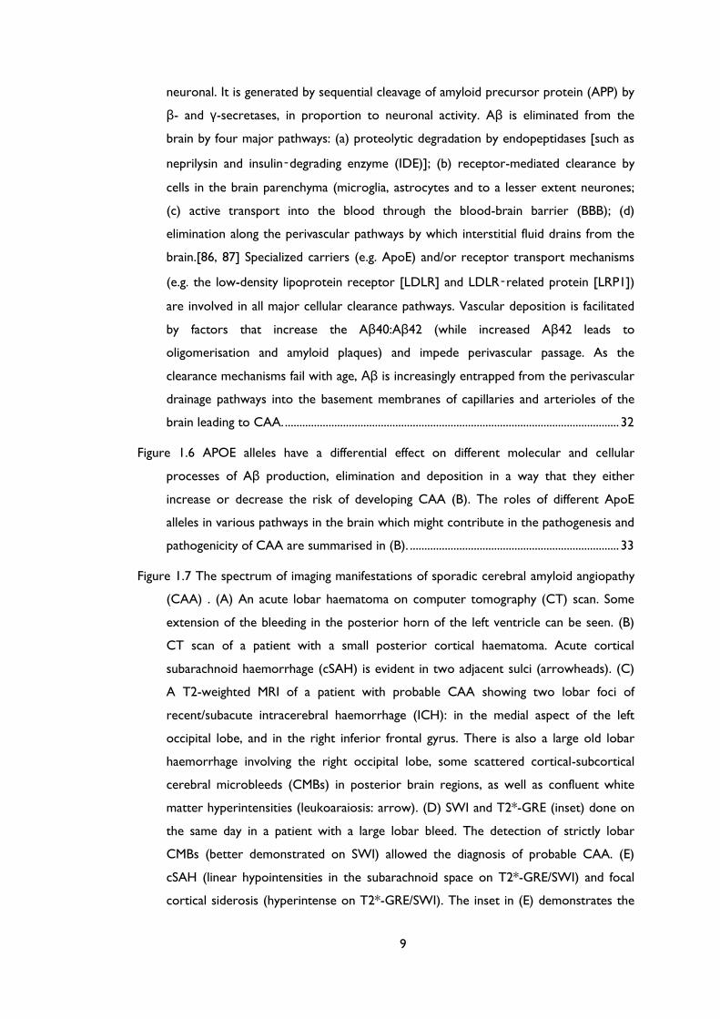
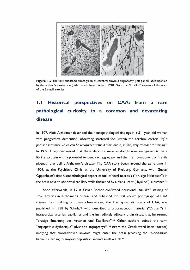
Figure 1.2 The first published photograph of cerebral amyloid angiopathy (left panel), accompanied
by the author’s illustration (right panel), from Fischer, 1910. Note the “fur-like” staining of the walls
of the 3 small arteries.
1.1 Historical perspectives on CAA: from a rare
pathological curiosity to a common and devastating
disease
In 1907, Alois Alzheimer described the neuropathological findings in a 51- year-old woman
with progressive dementia,51 observing scattered foci, within the cerebral cortex, “of a
peculiar substance which can be recognized without stain and is, in fact, very resistant to staining.”
In 1927, Divry discovered that these deposits were amyloid,52 now recognized to be a
fibrillar protein with a powerful tendency to aggregate, and the main component of “senile
plaques” that define Alzheimer’s disease. The CAA story began around the same time, in
1909, at the Psychiatry Clinic at the University of Freiburg, Germany, with Gustav
Oppenheim’s first histopathological report of foci of focal necrosis (“drusige Nekrosen”) in
the brain next to abnormal capillary walls thickened by a translucent (“hyaline”) substance.39
Soon afterwards, in 1910, Oskar Fischer confirmed occasional “fur-like” staining of
small arteries in Alzheimer’s disease, and published the first known photograph of CAA
(Figure 1.2). Building on these observations, the first systematic study of CAA, was
published in 1938 by Scholz,40 who described a proteinaceous material (“Drusen”) in
intracortical arteries, capillaries and the immediately adjacent brain tissue, that he termed
“drusige Entartung der Arterien und Kapillaren’’.40 Other authors coined the term
“angiopathie dyshorique” (dyshoric angiopathy)53, 54 (from the Greek word horos=border)
implying that blood-derived amyloid might enter the brain (crossing the “blood-brain
barrier”) leading to amyloid deposition around small vessels.54
26
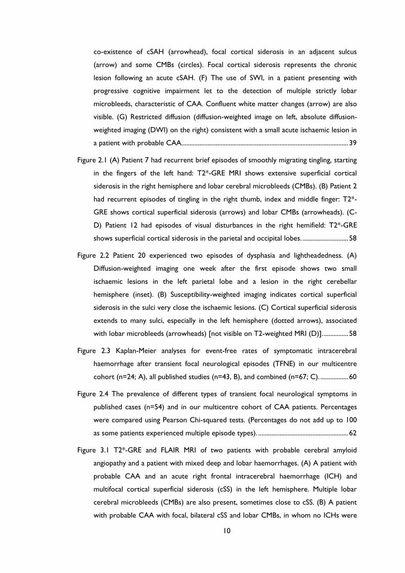
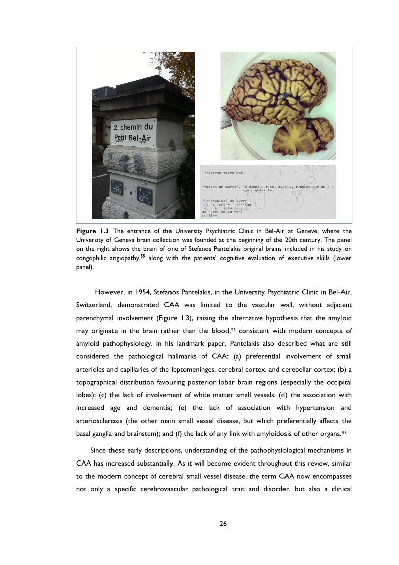
Figure 1.3 The entrance of the University Psychiatric Clinic in Bel-Air at Geneve, where the
University of Geneva brain collection was founded at the beginning of the 20th century. The panel
on the right shows the brain of one of Stefanos Pantelakis original brains included in his study on
congophilic angiopathy,55 along with the patients’ cognitive evaluation of executive skills (lower
panel).
However, in 1954, Stefanos Pantelakis, in the University Psychiatric Clinic in Bel-Air,
Switzerland, demonstrated CAA was limited to the vascular wall, without adjacent
parenchymal involvement (Figure 1.3), raising the alternative hypothesis that the amyloid
may originate in the brain rather than the blood,55 consistent with modern concepts of
amyloid pathophysiology. In his landmark paper, Pantelakis also described what are still
considered the pathological hallmarks of CAA: (a) preferential involvement of small
arterioles and capillaries of the leptomeninges, cerebral cortex, and cerebellar cortex; (b) a
topographical distribution favouring posterior lobar brain regions (especially the occipital
lobes); (c) the lack of involvement of white matter small vessels; (d) the association with
increased age and dementia; (e) the lack of association with hypertension and
arteriosclerosis (the other main small vessel disease, but which preferentially affects the
basal ganglia and brainstem); and (f) the lack of any link with amyloidosis of other organs.55
Since these early descriptions, understanding of the pathophysiological mechanisms in
CAA has increased substantially. As it will become evident throughout this review, similar
to the modern concept of cerebral small vessel disease, the term CAA now encompasses
not only a specific cerebrovascular pathological trait and disorder, but also a clinical
27
syndrome (or syndromes) and brain parenchymal lesions seen on neuroimaging (including a
set of imaging criteria).
1.2 Epidemiology and risk factors
Pathologically-defined CAA is common in the elderly.56-59 Population-based autopsy studies
indicate a CAA prevalence of 20-40% in non-demented, and 50-60% in demented elderly
populations.58, 60-63 Furthermore, CAA pathology may be severe in older individuals: in the
Honolulu-Asia aging autopsy study (HAAS), severe CAA was found in 43% of demented and
24% of non-demented elderly individuals (mean age at death: 85 years).62 In Alzheimer’s
disease, CAA is almost invariable, being found at autopsy in more than 90% of cases.56, 64
However, most of these patients have mild CAA; severe CAA is found in about 25% of
Alzheimer’s disease brains.65
Advancing age is the strongest known clinical risk factor for developing CAA.37 In a
community-based sample of 100 individuals, the prevalence of cortical vascular amyloid-β
deposition progressively increased from the 7th to the 9th decades,66 a pattern also
observed in 784 consecutive autopsies, corrected for over-representation of Alzheimer’s
disease.67 Moreover, patients with CAA-related ICH (suggesting advanced disease) in large
autopsy series, were all older than 60 years (and most over age 70).42, 68, 69 CAA is seldom
reported before the sixth decade of life; occasional patients presenting in their 50s have
been described.70
By contrast with hypertensive arteriopathy-the other main form of small vessel disease
and cause of ICH,2 the risk of CAA is not accounted for by conventional cardiovascular risk
factors other than age.37 Hypertension is not considered a risk factor for developing CAA,
but may increase the risk of CAA-related ICH. Vinters37 – in a clinicopathological series of
107 pathologically-proven CAA cases – found the prevalence of hypertension to be around
32%, similar to community-dwelling elderly populations,71 while another pathological study
reported that CAA patients with ICH were more frequently hypertensive (50%) than those
without ICH (23%), suggesting that hypertension may contribute to CAA-related cerebral
bleeding. 72 In a recent multicentre cohort of patients with spontaneous ICH, we found that
the prevalence of hypertension in CAA-related ICH was 62% - significantly less than in non-
CAA-related ICH (85%).73 Whether hypertension in association with CAA confers a
greater risk for ICH, compared to CAA alone is an important clinical question.74-76
Apolipoprotein E (APOE) genotype is the only known genetic risk factor for sporadic
CAA.77 APOE is a protein with crucial roles in lipoprotein complexes, which regulate lipid
28
metabolism by binding to cell-surface APOE receptors and proteins associated with lipid
transfer and lipolysis.77 There are three major polymorphisms in the APOE gene, namely
ε4, ε2 and ε3, resulting in a single amino-acid change78 which dramatically alters the
functional properties of APOE isoforms.79 These alleles have a strong dose-dependent
effect on the risk of developing CAA and its clinical severity. Thus, APOE ε4 in both post-
mortem and clinical series increases the risk of sporadic CAA-related lobar ICH; moreover,
the number of ε4 alleles relate to clinical severity.77, 80-82 Individuals carrying the APOE ε2
allele also have an increased risk of CAA-related lobar ICH.82, 83 Both of these risk alleles
are also associated with a younger age of first ICH,84 greater likelihood of haematoma
expansion, poorer clinical outcome,85, 86 and a higher risk of recurrence.87 Furthermore, the
two allelic variants interact: patients with both APOE ε2 and ε4 alleles have the earliest
disease onset and highest risk of early ICH recurrence.87, 88 The ε2 and ε4 alleles might
promote CAA-related haemorrhage through distinct mechanisms: ε4 by promoting
amyloid-β deposition; and ε2 by inducing structural changes in amyloid-laden vessels,
making them prone to rupture.85, 86, 88-90 Other as yet unidentified genetic polymorphisms
relating to amyloid metabolic pathways may also play a role in sporadic CAA, (for example
presenilin-1, neprilysin and transforming growth factor beta-1),91-93 and are a topic of
ongoing investigation.
1.3 Neuropathology
1.3.1 Morphological characteristics, natural history and
severity grading
CAA primarily involves neocortical and leptomeningeal arterioles, to a lesser extent
capillaries and, very rarely, venules.38 In contrast to amyloid plaques found in Alzheimer’s
disease - which are predominantly composed of the 42 amino-acid residue fragment
(amyloid-β42) - the vascular amyloid in CAA is mostly composed of the more soluble, 40
amino-acid fragment (amyloid-β40), suggesting different pathophysiological mechanisms for
pathological deposition (see below).94-97 Cerebral vessels with moderate to severe CAA
show an acellular wall thickening with a strongly eosinophilic smudgy appearance on
haematoxylin-eosin stained sections.98 Congo-red staining, under polarized light, reveals
amyloid deposits as “apple-green” birefringence (hence the term congophilic angiopathy)37, 99
although immunological stains for amyloid-β are highly specific and now widely used. The
development of CAA is progressive, with amyloid-β first appearing in the abluminal aspect
29
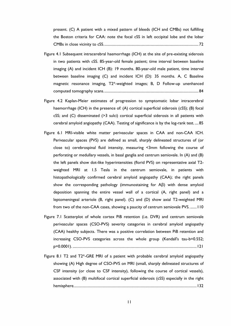
Figure 1.4 Histopathological features of cerebral amyloid angiopathy (CAA) (A1-A3) Morphological
changes of the vessel walls of leptomeningeal arterioles as revealed by haematoxylin & eosin staining
(H&E) and immunohistochemical detection of amyloid-β. In mild and moderate CAA only minimal
structural changes can be detected: in A2 the arrowhead points to amyloid deposition in the vessel
wall. However, in advanced CAA, there are significant structural alterations, the most extreme of
which is vessel-within-vessel appearance, sometimes termed “double-barrelling” (detachment and
delamination of the outer part of the tunica media; bracket in A3). (B1-B3) A similar pathologic
range of CAA-related changes in leptomeningeal arterioles using immunohistochemical detection of
amyloid-β. In mild CAA (B1) there is a patchy deposition amyloid in the vessels wall. Moderate CAA
shows more dense amyloid deposition which spans the entire vessel wall (B2), while severe CAA,
shows double-balled vessels and endothelial involvement (B3). (C1-C3) Pathological findings of CAA
in cortical arterioles. C2 shows moderate CAA with pan-mural deposition of amyloid-β along with
amyloid-β deposition in the surrounding brain parenchyma (arrowhead). In C3 a double-barrel
vessels can be seen, although this is less common compared to leptomeningeal vessels.
of the tunica media, surrounding smooth muscle cells, and in the adventitia (Figure 1.4).37
At the initial stage the vessel wall structure is intact, but as the disease progresses, there is
pan-mural amyloid accumulation, and loss of smooth muscle cells.38 In severe CAA,
detachment and delamination of the outer part of the tunica media result in so-called
“double barrel” appearance (Figure 1.4);38 fibrinoid necrosis and microaneurysm formation
also occur in advanced disease. There may also be microbleeding with perivascular
30
deposition of erythrocytes and blood-breakdown products.98 Endothelial cells are usually
preserved even in vessels severely affected by CAA.100 Occasionally amyloid-β is deposited
in the surrounding brain parenchyma immediately adjacent to an affected vessel (sometimes
called “dyshoric CAA”).
CAA is also associated with cerebral ischaemic damage,56, 65, 101, 102 including cortical
microinfarcts,103 and white matter pathology (demyelination and gliosis).43, 56, 96 Microinfarcts
are predominantly lobar (cortical-subcortical), usually in patients with severe CAA. One
possible mechanism for these ischaemic lesions is occlusion or reduced perfusion in
amyloid-laden cortical vessels affected by CAA.
The changes described above provide the basis of neuropathological scoring systems
for CAA 72, 101, 104 each with strengths and limitations.105 No standardized consensus
neuropathological criteria for rating CAA are available,106 but are desirable to allow
comparison of CAA pathological studies between centres. A more detailed discussion of
CAA severity grading can be found in a recent review by Attems and colleagues.38
1.3.2 Pathological subtypes of sporadic CAA
At least two distinct pathological subtypes of CAA have been described: CAA-type 1,
characterised by amyloid-β in cortical capillaries (with or without involvement of other
vessels)38; and CAA-type 2, where amyloid-β deposits are restricted to leptomeningeal and
cortical arteries, arterioles and, rarely, veins.107 Amyloid-β deposition in the wall of
capillaries (capillary CAA, also used to be termed “dyshoric angiopathy”) may cause luminal
obstruction in the most severe stages.36 The APOE ε4 allele is most strongly associated
with CAA-type 1, while APOE ε2 is more associated with CAA-type 2.107 CAA-type 1
appears to be more closely associated with parenchymal amyloid deposition in Alzheimer’s
disease.108
1.4 Topographical distribution
Sporadic CAA favours posterior cortical regions; the occipital lobe is most frequently
affected, followed by the frontal, temporal and parietal lobes.37, 38 The occipital lobe is also
most severely affected.109, 110 The cerebellum can be affected in advanced stages, while the
basal ganglia, thalami, white matter and brainstem are typically spared.105 The distribution of
CAA pathology shows a characteristic patchy pattern,37 so that foci of vessels severely
31
affected by CAA may be adjacent to others with mild or absent amyloid-β deposition.37, 38
The practical consequence of this is that cerebral biopsy may miss patchy CAA pathology.
1.5 Pathophysiological pathways
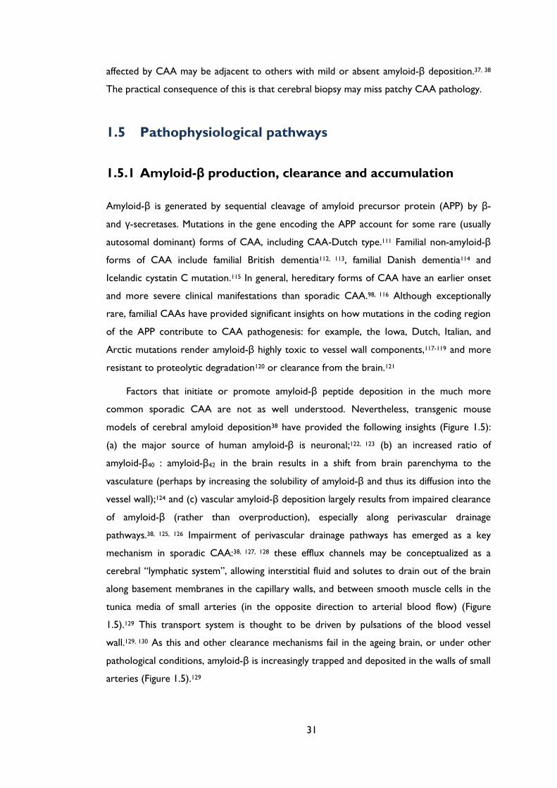
1.5.1 Amyloid-β production, clearance and accumulation
Amyloid-β is generated by sequential cleavage of amyloid precursor protein (APP) by β-
and γ-secretases. Mutations in the gene encoding the APP account for some rare (usually
autosomal dominant) forms of CAA, including CAA-Dutch type.111 Familial non-amyloid-β
forms of CAA include familial British dementia112, 113, familial Danish dementia114 and
Icelandic cystatin C mutation.115 In general, hereditary forms of CAA have an earlier onset
and more severe clinical manifestations than sporadic CAA.98, 116 Although exceptionally
rare, familial CAAs have provided significant insights on how mutations in the coding region
of the APP contribute to CAA pathogenesis: for example, the Iowa, Dutch, Italian, and
Arctic mutations render amyloid-β highly toxic to vessel wall components,117-119 and more
resistant to proteolytic degradation120 or clearance from the brain.121
Factors that initiate or promote amyloid-β peptide deposition in the much more
common sporadic CAA are not as well understood. Nevertheless, transgenic mouse
models of cerebral amyloid deposition38 have provided the following insights (Figure 1.5):
(a) the major source of human amyloid-β is neuronal;122, 123 (b) an increased ratio of
amyloid-β40 : amyloid-β42 in the brain results in a shift from brain parenchyma to the
vasculature (perhaps by increasing the solubility of amyloid-β and thus its diffusion into the
vessel wall);124 and (c) vascular amyloid-β deposition largely results from impaired clearance
of amyloid-β (rather than overproduction), especially along perivascular drainage
pathways.38, 125, 126 Impairment of perivascular drainage pathways has emerged as a key
mechanism in sporadic CAA:38, 127, 128 these efflux channels may be conceptualized as a
cerebral “lymphatic system”, allowing interstitial fluid and solutes to drain out of the brain
along basement membranes in the capillary walls, and between smooth muscle cells in the
tunica media of small arteries (in the opposite direction to arterial blood flow) (Figure
1.5).129 This transport system is thought to be driven by pulsations of the blood vessel
wall.129, 130 As this and other clearance mechanisms fail in the ageing brain, or under other
pathological conditions, amyloid-β is increasingly trapped and deposited in the walls of small
arteries (Figure 1.5).129
32
Figure 1.5 (A) Amyloid-β production, elimination and deposition in cerebral amyloid angiopathy
(CAA). Converging evidence indicates that the major source of Aβ is neuronal. It is generated by
sequential cleavage of amyloid precursor protein (APP) by β- and γ-secretases, in proportion to
neuronal activity. Aβ is eliminated from the brain by four major pathways: (a) proteolytic
degradation by endopeptidases [such as neprilysin and insulin‑degrading enzyme (IDE)]; (b) receptor-
mediated clearance by cells in the brain parenchyma (microglia, astrocytes and to a lesser extent
neurones; (c) active transport into the blood through the blood-brain barrier (BBB); (d) elimination
along the perivascular pathways by which interstitial fluid drains from the brain.[86, 87] Specialized
carriers (e.g. ApoE) and/or receptor transport mechanisms (e.g. the low-density lipoprotein receptor
[LDLR] and LDLR‑related protein [LRP1]) are involved in all major cellular clearance pathways.
Vascular deposition is facilitated by factors that increase the Aβ40:Aβ42 (while increased Aβ42 leads
to oligomerisation and amyloid plaques) and impede perivascular passage. As the clearance
mechanisms fail with age, Αβ is increasingly entrapped from the perivascular drainage pathways into
the basement membranes of capillaries and arterioles of the brain leading to CAA.
Evidence is emerging that cerebrovascular disease may impede the drainage along the
perivascular pathways, contributing to CAA pathogenesis.127, 131 It has been suggested that
amyloid-β deposition could further impair/block the perivascular drainage, leading to
dilation of perivascular spaces (also known as Virchow-Robin spaces) not only within lobar
regions, but also in the underlying white matter, that itself shows is unaffected by CAA.132,
133 These enlarged perivascular spaces can reach several millimetres in diameter and may be
visible on appropriate brain imaging; this requires further investigation as a potential useful
neuroimaging marker of CAA.30, 133
33
Figure 1.6 APOE alleles have a differential effect on different molecular and cellular processes of
Aβ production, elimination and deposition in a way that they either increase or decrease the risk of
developing CAA (B). The roles of different ApoE alleles in various pathways in the brain which might
contribute in the pathogenesis and pathogenicity of CAA are summarised in (B).
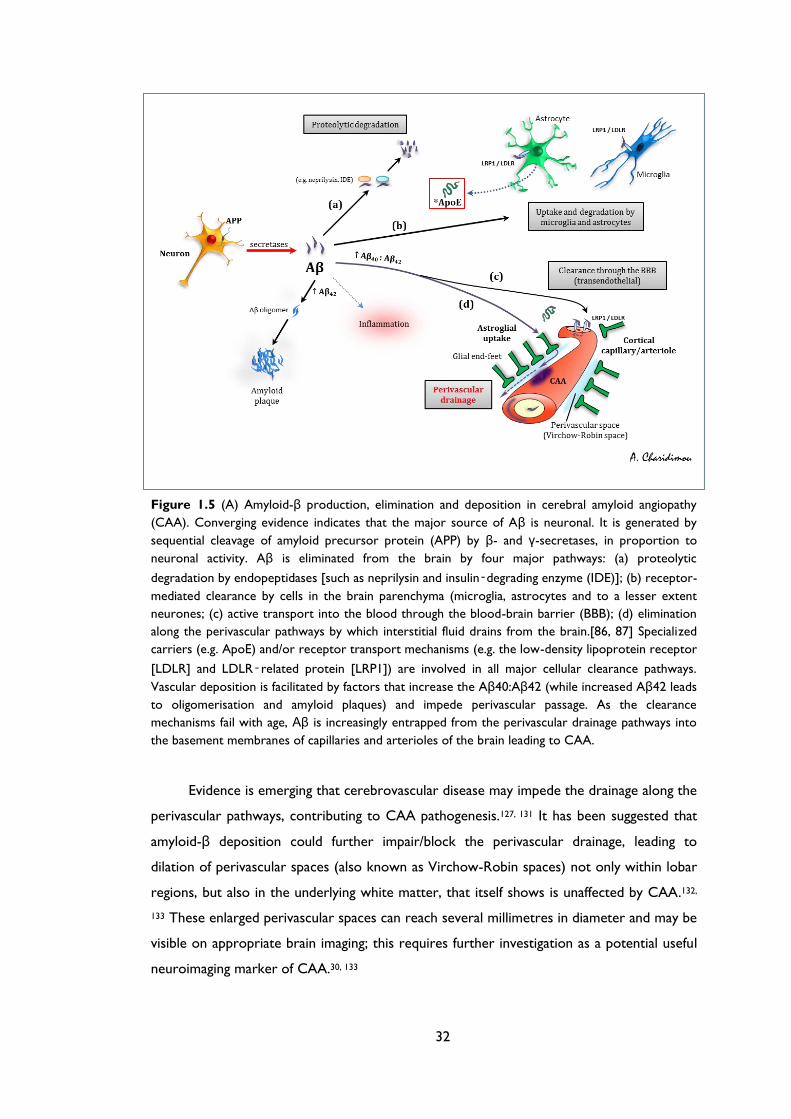
As we have seen, APOE is a strong genetic risk factor for CAA, an effect mediated by
its important role in amyloid-β metabolism, aggregation and clearance (Figure 1.6).77, 125, 126.
APOE ε4 increases the amyloid-β40:amyloid-β42 ratio shifting amyloid deposition to the
vessels instead of brain parenchyma,124 and may reduce the efficiency of efflux of amyloid-β
along perivascular channels,38, 134 influencing CAA risk and age of onset.77, 135 APOE
genotype may also interact with other small vessel disease changes: hypertensive
arteriopathy, which leads to stiffening of the vessel wall, may reduce the pulsatile driving
movements required for efficient perivascular drainage, and thus contribute to the risk of
CAA.38
1.5.2 From amyloid-β deposition to CAA pathogenesis
Amyloid-β deposition has complex effects on vascular structure and function, which can
result in brain injury.2, 136 Important morphological changes include: loss of smooth muscle
cells;137 vessel wall thickening and lumen restriction;101 endothelial dysfunction; and a loss of
compliance leading to brittle, fragile vessels prone to microaneurysm formation and
34
leakage.38 Acute trigger factors, for example sudden increases in blood pressure, or minor
trauma (regularly encountered in clinical practice, but not to our knowledge formally
studied), may cause the rupture of these abnormally weak, amyloid-laden vessels. Amyloid-
β deposition may also impair local regulation of cerebral blood flow,136 neurovascular unit
function138 and general homeostatic mechanisms in the ageing brain.73 Other effects of
vascular amyloid-β, including BBB disruption and active inflammation, could also
contribute.38, 136 Moreover, even without vascular deposition, soluble amyloid-β can cause
abnormal vascular reactivity,136 and induce the activation of inflammatory mediators
including matrix metalloproteinase-9 and -2.38, 139, 140
1.6 The expanding clinical spectrum of sporadic CAA
There are at least four important clinical presentations associated with CAA:
Symptomatic intracerebral haemorrhage
Cognitive impairment and dementia
Rapidly progressive cognitive and neurological decline (inflammatory CAA)
Transient neurological symptoms
1.6.1 Intracerebral haemorrhage
1.6.1.1 The association between CAA and ICH
CAA is most often recognized in life by symptomatic, spontaneous, lobar ICH in elderly
patients. The majority of ICHs (>75%) in the elderly are classified as spontaneous
(sometimes also termed primary or non-traumatic), resulting from rupture of small arteries
affected by two main processes: hypertensive arteriopathy or CAA. Hypertensive
arteriopathy - characterized by lipohyalinosis and fibrinoid necrosis of small lenticulostriate
arterial perforators - is considered an important cause of spontaneous ICH in deep or
infratentorial locations (basal ganglia, thalamus, and pons). By contrast, CAA-related ICHs
preferentially affect cortical-subcortical (lobar) regions (especially the occipital and
temporal lobes141), less commonly the cerebellum, and rarely deep or brainstem structures,
reflecting the distribution of the underlying microangiopathy.37, 69, 109 The predilection for
the occipital lobes is not well understood, but one hypothesis is that greater tortuosity of
occipital small arteries impairs perivascular drainage.38
Clinicopathological studies suggest that CAA-related ICH accounts for at least 5-20%
of all spontaneous ICH,37, 56, 65, 72, 142, 143 and that the link is strongest for lobar ICH.
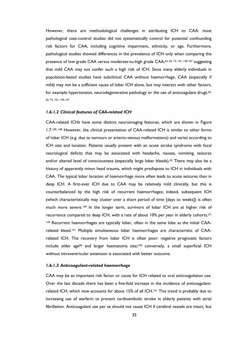
35
However, there are methodological challenges in attributing ICH to CAA: most
pathological case-control studies did not systematically control for potential confounding
risk factors for CAA, including cognitive impairment, ethnicity, or age. Furthermore,
pathological studies showed differences in the prevalence of ICH only when comparing the
presence of low grade CAA versus moderate-to-high grade CAA,62, 65, 72, 101, 144-147 suggesting
that mild CAA may not confer such a high risk of ICH. Since many elderly individuals in
population-based studies have subclinical CAA without haemorrhage, CAA (especially if
mild) may not be a sufficient cause of lobar ICH alone, but may interact with other factors,
for example hypertension, neurodegenerative pathology or the use of anticoagulant drugs.62,
65, 72, 101, 144-147
1.6.1.2 Clinical features of CAA-related ICH
CAA-related ICHs have some distinct neuroimaging features, which are shown in Figure
1.7.109, 148 However, the clinical presentation of CAA-related ICH is similar to other forms
of lobar ICH (e.g. due to tumours or arterio-venous malformations) and varies according to
ICH size and location. Patients usually present with an acute stroke syndrome with focal
neurological deficits that may be associated with headache, nausea, vomiting, seizures
and/or altered level of consciousness (especially large lobar bleeds).45 There may also be a
history of apparently minor head trauma, which might predispose to ICH in individuals with
CAA. The typical lobar location of haemorrhage more often leads to acute seizures than in
deep ICH. A first-ever ICH due to CAA may be relatively mild clinically, but this is
counterbalanced by the high risk of recurrent haemorrhages; indeed, subsequent ICH
(which characteristically may cluster over a short period of time [days to weeks]) is often
much more severe.149 In the longer term, survivors of lobar ICH are at higher risk of
recurrence compared to deep ICH, with a rate of about 10% per year in elderly cohorts.37,
149 Recurrent haemorrhages are typically lobar, often in the same lobe as the initial CAA-
related bleed.141 Multiple simultaneous lobar haemorrhages are characteristic of CAA-
related ICH. The recovery from lobar ICH is often poor: negative prognostic factors
include older age84 and larger haematoma size;150 conversely, a small superficial ICH
without intraventricular extension is associated with better outcome.
1.6.1.3 Anticoagulant-related haemorrhage
CAA may be an important risk factor or cause for ICH related to oral anticoagulation use.
Over the last decade there has been a five-fold increase in the incidence of anticoagulant-
related ICH, which now accounts for about 15% of all ICH.151 This trend is probably due to
increasing use of warfarin to prevent cardioembolic stroke in elderly patients with atrial
fibrillation. Anticoagulant use per se should not cause ICH if cerebral vessels are intact, but
36
the presence of CAA, rendering vessels brittle and fragile, is a plausible aggravating factor
for such haemorrhage; an otherwise innocuous minor and self-limiting vessel leak (e.g a
cerebral microbleed [CMB], see below) could form a life-threatening haematoma if the
leaking vessel is damaged by advanced CAA. Evidence supporting a link between CAA and
anticoagulation-related ICH includes the following observations: first, most such ICH occur
with international normalised ratios within the therapeutic range152 suggesting that an
intrinsic disorder of cerebral small vessels could be important; and second, the APOE ε2
allele is more common in warfarin-related ICH than in patients on warfarin without ICH,
supporting a role for CAA.152 Although CAA may underlie a substantial proportion of
anticoagulation-related haemorrhages, prospective studies with reliable diagnosis of CAA in
life (e.g. by MRI evidence of lobar cerebral microbleeds or molecular imaging) in cohorts of
patients treated with anticoagulants are urgently needed to answer this question (one large
prospective MRI study is currently underway in the UK: www.ucl.ac.uk/cromis-2).
CAA may also be a risk factor for ICH after thrombolysis: spontaneous CAA-related
and thrombolysis-related haemorrhages share some features including a predilection of
lobar brain regions, multiplicity of haemorrhages, age-dependency and an association with
dementia and leukoaraiosis.153 In one small study, 2 of 5 cases of ICH after thrombolysis for
acute myocardial infarction had severe CAA identified.154
1.6.2 Cognitive impairment and dementia
There is now increasing evidence that CAA is an important contributor to cognitive
impairment,10, 106 although dissecting its independent cognitive impact is confounded by the
presence of co-existing Alzheimer’s disease and other age-related pathologies (e.g.
hypertensive arteriopathy). Nevertheless, in population-based clinical-pathological studies,
the prevalence of CAA is consistently higher in demented compared to non-demented
patients.58 In the population-based Medical Research Council Cognitive Function and Aging
Study (MRC CFAS) CAA was significantly associated with dementia (OR 9.3, 95% CI 2.7–
41.0) even after controlling for age and dementia-related neuropathologies (e.g. neuritic and
diffuse plaques).61 Similarly, the HAAS autopsy study revealed a significantly higher
prevalence of severe CAA in demented versus non-demented patients (43% vs. 24%).62
CAA may worsen the severity of cognitive dysfunction in Alzheimer’s disease: CAA
together with Alzheimer’s disease pathology has been associated with significantly worse
cognitive performance during life, compared to Alzheimer’s disease alone, even after
controlling for age, neurofibrillary tangles and amyloid plaques number, infarctions and
APOE genotype.62 There are few studies of the specific pattern of cognitive impairment
37
associated with CAA; a recent autopsy series found that moderate-to-severe CAA (present
in 19% of the study population) was associated with lower performance in specific cognitive
domains, notably perceptual speed and episodic memory, after accounting for Alzheimer’s
disease pathology and other potential covariates.155 The pathophysiological mechanisms by
which CAA could cause cognitive impairment have not been well established,10 but relevant
lesions on brain imaging could include cerebral microbleeds,156 microinfarcts,73, 157 and white
matter changes.158
CAA is thus emerging as a potentially important link between neurodegenerative and
cerebrovascular pathology.159 Vascular cognitive impairment and Alzheimer’s disease are
now conceptualised as a continuum10, 160, 161 with complex interactions and shared risk
factors.136, 159 CAA seems likely to exacerbate the deleterious effect of neurodegenerative
pathology on the brain, lowering the threshold for overt dementia.10, 136 Unravelling the
independent contribution of CAA to cognitive function is particularly important as it could
lead to new therapeutic strategies.
1.6.3 Rapidly progressive cognitive and neurological decline:
CAA-related inflammation
CAA is clearly a direct cause of cognitive impairment in the uncommon but clinically
striking presentation of CAA-related inflammation (also termed cerebral amyloid angiitis,
amyloid beta related angiitis and cerebral amyloid inflammatory vasculopathy).162 CAA-
related inflammation typically affects older adults, who present with acute to subacute
cognitive decline, headache, behavioural change, seizures and focal neurological deficits.162
Typical MRI findings include patchy or confluent, asymmetric T2-weighted or FLAIR white
matter hyperintensities (with or without mass effect and leptomeningeal or parenchymal
enhancement).162 T2*-weighted gradient recalled echo (T2*-GRE) or susceptibility weighted
imaging (SWI) may reveal previous lobar haemorrhage and/or multiple cortical and
subcortical microbleeds.162 The major differential diagnoses include infections (in particular
progressive multifocal leucoencephalopathy), neuro-sarcoidosis, immune-related conditions
(e.g. acute disseminated encephalomyelitis)163 and malignancies.162 Definite diagnosis
requires brain and leptomeningeal biopsy showing perivascular inflammation with
mononuclear or multinucleated giant cells associated with amyloid-β-laden vessels and/or
frank vasculitis.162 Although the clinical course of CAA-related inflammation is varied, it is
important to recognize because it may respond well to immunosuppressive treatment (e.g.
high dose corticosteroids or cyclophosphamide).162, 164 This distinct syndrome has parallels
with that observed in patients with Alzheimer’s disease who developed meningo-
38
encephalitis after immunization against human Amyloid-β, where post mortem examination
revealed inflammation and/or vasculitis associated with CAA.165, 166
1.6.4 Transient focal neurological episodes
After ICH, the next most commonly described clinical presentation of sporadic CAA is
with transient neurologic episodes,167-169 sometimes termed “amyloid spells”. The most
common type of attack involves recurrent, stereotyped episodes of “positive” spreading
sensory symptoms (paraesthesias).167, 168 Although there are a number of small case reports
and series,167, 168, 170, 171 no large systematic studies have investigated the prevalence or
semiology of these phenomena. At least two other types of transient events have been
described: partial motor seizure-like episodes (e.g. limb shaking); and visual disturbances
(usually positive visual symptoms similar to migrainous auras). Spells are typically brief,
almost always less than about 30 minutes, and usually less than a few minutes. The attacks
seem likely to be related to haemorrhagic components of CAA: associated neuroimaging
findings reported include cerebral microbleeds and convexity subarachnoid haemorrhage
(cSAH) in the cortical region corresponding to the spell.167, 171 The diagnosis of these CAA-
related attacks is of clinical relevance, since they seem to precede serious symptomatic ICH
in some patients; antiplatelet or anticoagulant use following such an attack misdiagnosed as
a transient ischaemic attack (TIA) could therefore cause potentially avoidable intracranial
bleeding. The underlying mechanisms of CAA transient spells remain unclear, but could
include seizure-like activity (perhaps related to small areas of bleeding, e.g. microbleeding,
cSAH or superficial siderosis); a direct effect of amyloid or bleeding on local cortical
function; or spreading cortical depression.167 The responsiveness of these attacks to anti-
epileptic drugs as well as their spreading nature in many of the reported cases are in favor
of a seizure-like mechanism for their pathophysiology. In a case series by Roth and co-
workers,168 four out of six patients with these transient attacks responded to
anticonvulsants, while the other two patients showed improvement after the cessation of
antiplatelet therapy. Typical TIA-like episodes have also been reported in CAA,169 but
whether these are genuinely due to ischaemia and should be treated with antithrombotic
agents requires further study.
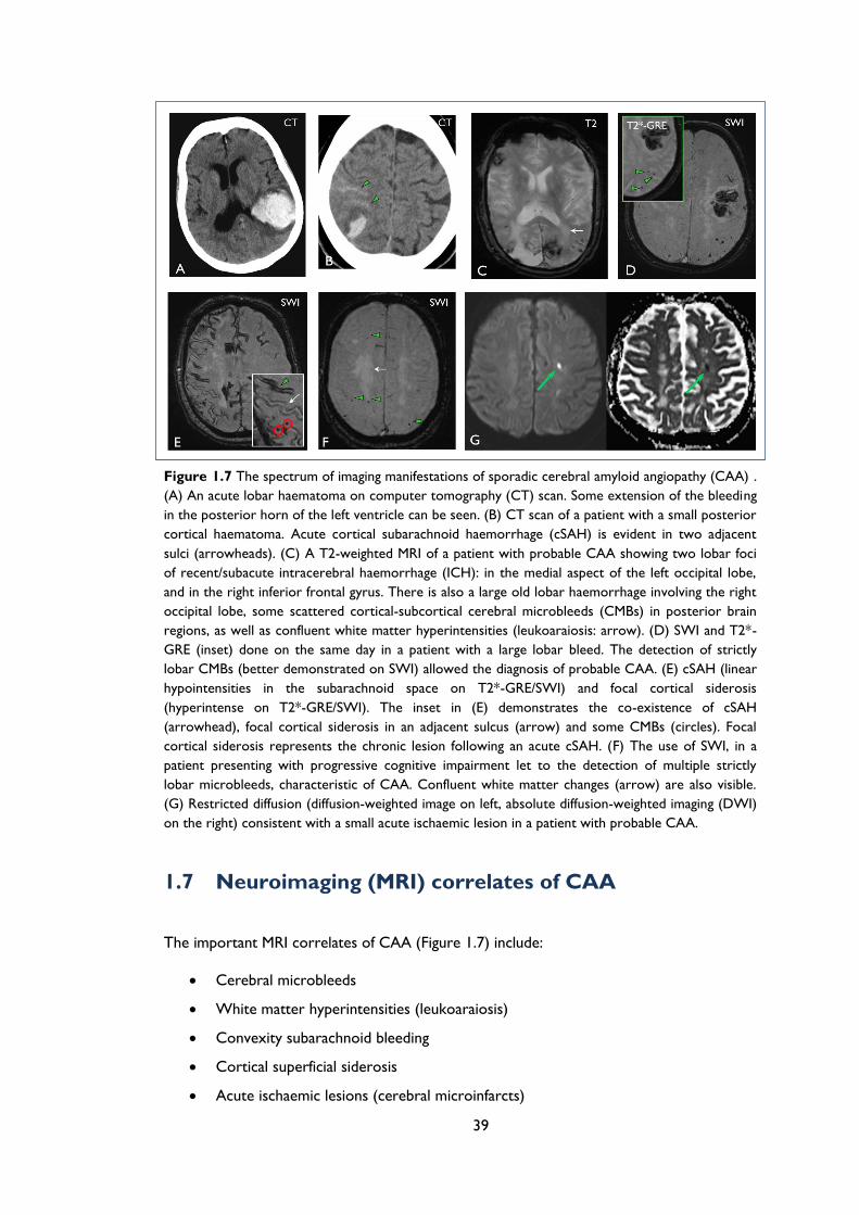
39
Figure 1.7 The spectrum of imaging manifestations of sporadic cerebral amyloid angiopathy (CAA) .
(A) An acute lobar haematoma on computer tomography (CT) scan. Some extension of the bleeding
in the posterior horn of the left ventricle can be seen. (B) CT scan of a patient with a small posterior
cortical haematoma. Acute cortical subarachnoid haemorrhage (cSAH) is evident in two adjacent
sulci (arrowheads). (C) A T2-weighted MRI of a patient with probable CAA showing two lobar foci
of recent/subacute intracerebral haemorrhage (ICH): in the medial aspect of the left occipital lobe,
and in the right inferior frontal gyrus. There is also a large old lobar haemorrhage involving the right
occipital lobe, some scattered cortical-subcortical cerebral microbleeds (CMBs) in posterior brain
regions, as well as confluent white matter hyperintensities (leukoaraiosis: arrow). (D) SWI and T2*-
GRE (inset) done on the same day in a patient with a large lobar bleed. The detection of strictly
lobar CMBs (better demonstrated on SWI) allowed the diagnosis of probable CAA. (E) cSAH (linear
hypointensities in the subarachnoid space on T2*-GRE/SWI) and focal cortical siderosis
(hyperintense on T2*-GRE/SWI). The inset in (E) demonstrates the co-existence of cSAH
(arrowhead), focal cortical siderosis in an adjacent sulcus (arrow) and some CMBs (circles). Focal
cortical siderosis represents the chronic lesion following an acute cSAH. (F) The use of SWI, in a
patient presenting with progressive cognitive impairment let to the detection of multiple strictly
lobar microbleeds, characteristic of CAA. Confluent white matter changes (arrow) are also visible.
(G) Restricted diffusion (diffusion-weighted image on left, absolute diffusion-weighted imaging (DWI)
on the right) consistent with a small acute ischaemic lesion in a patient with probable CAA.
1.7 Neuroimaging (MRI) correlates of CAA
The important MRI correlates of CAA (Figure 1.7) include:
Cerebral microbleeds
White matter hyperintensities (leukoaraiosis)
Convexity subarachnoid bleeding
Cortical superficial siderosis
Acute ischaemic lesions (cerebral microinfarcts)
40
1.7.1 Cerebral microbleeds
The widespread use of T2*-weighted MRI sequences in the last decade or so has led to the
increasing detection of cerebral microbleeds: small, well-demarcated, hypointense, rounded
lesions, not detected on conventional MRI (Figure 1.7 D-f).35. Histopathological studies
show that cerebral microbleeds correspond to focal accumulations of haemosiderin-laded
macrophages (a blood-breakdown product) adjacent to abnormal small vessels affected by
hypertensive angiopathy or CAA.172, 173 There is increasing evidence that hypertensive
vasculopathy is associated with cerebral microbleeds in deep brain regions (basal ganglia,
thalamus and brainstem), whereas CAA is characterised by cerebral microbleeds in a lobar
distribution35, 173 with a predilection for the parietal lobes.141 The Rotterdam scan study174,
175 showed a strong association between strictly lobar (but not deep) cerebral microbleeds
and APOE ε4, consistent with the well-known relation of this allele with CAA.87 A recent
imaging study in clinically probable CAA, using non-invasive amyloid imaging with Pittsburgh
Compound B (PiB), found that cerebral microbleeds correspond to areas with a high
concentration of amyloid.176 Further evidence derives from neuroimaging studies of
cerebral microbleeds in patients presenting with symptomatic ICH: patients with deep ICH,
predominantly caused by hypertensive arteriopathy, are more likely to have deep cerebral
microbleeds; whereas those patients with multiple strictly lobar ICH, frequently caused by
CAA (although the exact proportion of lobar ICH which are actually due to CAA in not
known), are more likely to have multiple strictly lobar cerebral microbleeds.177-179
Moreover, cerebral microbleeds correlate with the risk of lobar ICH recurrence180
suggesting an important role in prognosis (as well as diagnosis) in CAA.27
Recent population-based studies have revealed a high percentage of community-
dwelling elderly people with strictly lobar microbleeds, (particularly in the posterior brain
regions) suggesting sub-clinical CAA.174, 175, 181, 182 This may have important implications: if
strictly lobar cerebral microbleeds are validated as a diagnostic marker of CAA, such
asymptomatic individuals could benefit from new therapeutic agents to reduce the
progression of the disease.
Neuroimaging studies have revealed lobar cerebral microbleeds in more than 20% of
patients with Alzheimer’s disease,183 probably reflecting advanced CAA (in keeping with
neuropathological findings). Patients with autosomal dominant forms of familial Alzheimer’s
disease (who have a younger age at symptom onset), also seem to have a prevalence of
lobar cerebral microbleeds similar to sporadic Alzheimer’s disease184 – a striking recent
observation since these patients are much more likely to have ‘pure’ neurodegenerative
Alzheimer’s disease without co-existing sporadic small vessel disease. It has been suggested
41
that the presence of multiple lobar cerebral microbleeds in patients with Alzheimer’s
disease, may identify a specific subgroup of patients with a different clinical phenotype with
therapeutic implications which need to be explored in future studies.183
1.7.2 White matter hyperintensities
White matter hyperintensities (also known as leukoaraiosis) is a radiological term which
describes imaging changes (often confluent) in deep cerebral white matter. Leukoaraiosis
appears as low attenuation on computer tomography (CT) scans or hyperintensity on T2-
weighted or FLAIR MRI, typically sparing subcortical U-fibres (Figure 1.7 C).5 Pathological
substrates include demyelination, axon loss and mild gliosis. The pathogenesis of
leukoaraiosis in CAA probably involves chronic hypoperfusion of the vulnerable peri-
ventricular white matter and disruption of the BBB due to amyloid in the overlying cortical
small vessels.2, 24, 133 Another possible mechanism of leukoaraiosis in CAA is as a result of
the accumulation of silent ischaemic lesions (microinfarcts).46, 73, 185 Indeed, leukoaraiosis is
very common in CAA, preferentially involving posterior regions,186 although some studies
suggest no major difference in the topography of leukoaraiosis in CAA compared to
hypertensive arteriopathy.158, 186 A recent investigation suggested that subjects with CAA-
related lobar ICH have a higher prevalence of occipital dominant leukoaraiosis compared to
normal elderly controls;187 this interesting finding requires further clinical attention and
investigation. Leukoaraiosis may be an important contributor to overall disease burden,
especially progressive cognitive impairment,188 given its tendency to accumulate over
time.189 A recent study found that leukoaraiosis volume was greater in patients with CAA
and hypertension than those without, suggesting that strict control of hypertension might
reduce leukoaraiosis-related disability in CAA.188
1.7.3 Acute convexity subarachnoid haemorrhage (cSAH) and
cortical superficial siderosis
Atraumatic convexity subarachnoid haemorrhage (cSAH) and cortical superficial siderosis
are recently recognised imaging correlates of sporadic CAA190 which seem to be quite
characteristic of the disorder (Figure 1.7 E). cSAH is localized bleeding, usually in up to
several adjacent sulci, without other subarachnoid bleeding at the base of the brain in the
pattern typically associated with saccular aneurysm rupture.191 Although rare in isolation, in
CAA, cSAH often results from lobar ICH extending to the cortical surface.170, 190, 192 The
largest cohort of patients with isolated cSAH published (n=29), found that CAA was a
42
frequent apparent cause in patients over 60 years old.191, 193 A recent retrospective analysis
of consecutive patients admitted to a tertiary stroke unit with cSAH suggested that CAA
could be a common cause in the elderly, with a characteristic clinical presentation of single
or recurrent transient focal neurological attacks.171 Another recent study of a cohort of
patients presenting with cSAH, reported similar findings.194
Superficial siderosis describes haemosiderin deposition in the superficial layers of the
cerebral cortex, and may follow repeated episodes of bleeding in the subarachnoid space.190
On T2*-GRE MRI sequences superficial siderosis shows a characteristic “gyriform” pattern
of hypointense signal (Figure 1.7 E).190 Linn et al. have recently detected superficial siderosis
in 47.4% (n=38) of patients with a clinical diagnosis of CAA, compared to no controls
(mean age, 54 years), suggesting that it might be helpful for the clinical diagnosis of CAA
(see below).195 Compared to the well-known syndrome of central nervous system
superficial siderosis, which typically affects the brainstem and posterior fossa (associated
with cerebellar and brainstem signs), CAA-related siderosis has a predilection for the
cerebral convexity196 and may be associated with transient neurological manifestations.168
1.7.4 Small acute ischaemic lesions on diffusion-weighted
imaging (DWI)
Neuropathological evidence of asymptomatic ischaemic infarction (cerebral
microinfarcts197) is an established finding in the brains of patients with advanced CAA.43, 101-
103 Recent studies using magnetic resonance DWI, -which is extremely sensitive to even
small areas of acute ischaemia - have shed light on the dynamics of this phenomenon in vivo
(Figure 1.7 G). A case report47 and a recent case-control study46 found a high prevalence of
DWI-positive lesions, in patients with advanced CAA. These lesions were associated with
CMB burden, suggesting shared pathophysiological pathways.198 Gregoire et al recently
established that acute, subclinical ischaemic brain lesions are frequent after recent acute
ICH, and are three times more common in CAA-related ICH than other spontaneous
bleeds;73 the lesions were associated with the severity of leukoaraiosis and lobar cerebral
microbleeds, suggesting that that they were due to a CAA-related occlusive arteriopathy.73
However, a more recent larger study did not appear to show this higher prevalence of
DWI lesions in CAA-related intracerebral haemorrhage, suggesting that both CAA and
hypertensive arteriopathy trigger high rates of infarction.199 These data suggest a dynamic
interplay between the haemorrhagic (“microbleeding”) and ischaemic (“microinfarction”)
components in CAA and small vessel disease in general,198 though the therapeutic
implications and prognostic significance of these findings require further study. The topic of
43
microinfarcts and DWI lesions in CAA as well as current uncertainties in the terminology
and pathophysiology of these lesions, are further covered in Discussion (Chapter 11).
1.8 Molecular imaging of vascular amyloid in vivo
MRI indirectly detects the consequences of CAA (e.g. cerebral microbleeds, cSAH, and
siderosis) rather than vascular amyloid itself. Consequently, a large proportion of “silent”
CAA may be as yet undetectable. Positron emission tomography (PET) methods allow the
in vivo imaging of amyloid in the brain, using several radioligands, of which the most widely
studied is 11C Pittsburgh Compound B (PiB).200 Ly and colleagues demonstrated that CAA
subjects had increased global PiB uptake relative to a healthy elderly control group, and
found an occipital predominance of PiB retention in CAA compared with Alzheimer’s
disease.50 PiB PET might therefore ultimately detect CAA in vivo, even before it causes
symptomatic ICH or the known radiological sequelae including cerebral microbleeds.48, 49,
176, 201
1.9 Diagnostic approach to CAA: the critical role of
neuroimaging
A common clinical scenario where sporadic CAA should be suspected is in elderly patients
presenting with lobar ICH. The most commonly used criteria for CAA diagnosis are the
Boston criteria.202 In the absence of direct neuropathological examination, CAA is
diagnosed based on characteristic neuroimaging findings.202 The diagnosis of probable CAA
requires (Table 1.2):
Age >55 years
the detection of multiple haemorrhagic cerebral lesions
haemorrhages confined to cortical or cortical-subcortical (lobar) brain regions
exclusion of secondary causes of ICH, such as arteriovenous malformations, head
trauma, brain tumour, vasculitis and excessive anticoagulation
The specificity of the Boston criteria has been validated against the established gold
standard of neuropathological diagnosis from autopsy, haematoma evacuation, or cortical
biopsy.202 In this study, the criteria showed excellent specificity: all cases identified as
“probable CAA” (n=13) had pathological evidence of severe CAA. However, the sensitivity
of the probable category was 44%, so that it failed to identify over 50% of those with
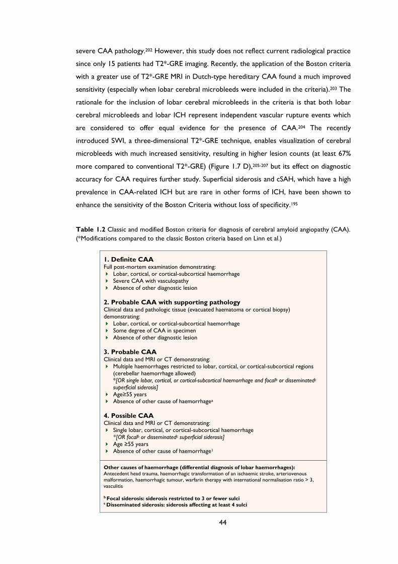
44
severe CAA pathology.202 However, this study does not reflect current radiological practice
since only 15 patients had T2*-GRE imaging. Recently, the application of the Boston criteria
with a greater use of T2*-GRE MRI in Dutch-type hereditary CAA found a much improved
sensitivity (especially when lobar cerebral microbleeds were included in the criteria).203 The
rationale for the inclusion of lobar cerebral microbleeds in the criteria is that both lobar
cerebral microbleeds and lobar ICH represent independent vascular rupture events which
are considered to offer equal evidence for the presence of CAA.204 The recently
introduced SWI, a three-dimensional T2*-GRE technique, enables visualization of cerebral
microbleeds with much increased sensitivity, resulting in higher lesion counts (at least 67%
more compared to conventional T2*-GRE) (Figure 1.7 D),205-207 but its effect on diagnostic
accuracy for CAA requires further study. Superficial siderosis and cSAH, which have a high
prevalence in CAA-related ICH but are rare in other forms of ICH, have been shown to
enhance the sensitivity of the Boston Criteria without loss of specificity.195
Table 1.2 Classic and modified Boston criteria for diagnosis of cerebral amyloid angiopathy (CAA).
(*Modifications compared to the classic Boston criteria based on Linn et al.)
1. Definite CAA Full post-mortem examination demonstrating:
Lobar, cortical, or cortical-subcortical haemorrhage
Severe CAA with vasculopathy
Absence of other diagnostic lesion
2. Probable CAA with supporting pathology Clinical data and pathologic tissue (evacuated haematoma or cortical biopsy)
demonstrating:
Lobar, cortical, or cortical-subcortical haemorrhage
Some degree of CAA in specimen
Absence of other diagnostic lesion
3. Probable CAA Clinical data and MRI or CT demonstrating:
Multiple haemorrhages restricted to lobar, cortical, or cortical-subcortical regions
(cerebellar haemorrhage allowed)
*[OR single lobar, cortical, or cortical-subcortical haemorrhage and focalb or disseminatedc
superficial siderosis]
Age≥55 years Absence of other cause of haemorrhagea
4. Possible CAA Clinical data and MRI or CT demonstrating:
Single lobar, cortical, or cortical-subcortical haemorrhage
*[OR focalb or disseminatedc superficial siderosis]
Age ≥55 years
Absence of other cause of haemorrhage1
Other causes of haemorrhage (differential diagnosis of lobar haemorrhages): Antecedent head trauma, haemorrhagic transformation of an ischaemic stroke, arteriovenous malformation, haemorrhagic tumour, warfarin therapy with international normalisation ratio > 3,
vasculitis b Focal siderosis: siderosis restricted to 3 or fewer sulci c Disseminated siderosis: siderosis affecting at least 4 sulci
45
Although the value of T2*-weighted MRI and SWI to detect cerebral microbleeds, cSAH
and siderosis has mainly been validated in cohorts of patients who presented with
symptomatic ICH, such imaging may also have a role in the diagnosis of patients presenting
without major haemorrhage, but with other syndromes raising suspicion of CAA; for
example, elderly patients with progressive cognitive impairment.10, 171, 206, 208, 209 In addition,
although at present T2* MRI or SWI sequences are not part of the routine investigation of
TIA-like attacks, there might be useful in patients with CAA-related transient focal
neurological episodes (“amyloid spells”) – rather atypical of TIAs.168, 171 However, current
data are insufficient to make evidence-based recommendations.
Other biomarkers might also prove useful in the non-invasive diagnosis of CAA, in
particular the assessment of amyloid-β concentrations in the cerebrospinal fluid (CSF).
Decreased levels of CSF amyloid-β42 but not amyloid-β40 are found in Alzheimer’s
disease,210 while it has been reported that both amyloid-β42 and amyloid-β40 concentrations
are decreased in CAA, relative to control and Alzheimer’s disease patients.211 It has also
been suggested that the combination of low amyloid-β42 with increased total tau levels in
the CSF, can discriminate CAA patients from normal controls with high accuracy.211
Another potentially promising marker of CAA might include retinal changes
(microaneurysms and dot and blot haemorrhages212). A critical goal of all of these potential
approaches is to reliably identify CAA at the early (asymptomatic) stages of the disease, to
allow the best chance for disease modifying or preventive treatments to be effective.
1.10 Management of CAA and its consequences
1.10.1 Acute treatment
No treatment is specific for symptomatic management of CAA or CAA-related ICH. As in
all forms of spontaneous ICH, CAA-related haematomas enlarge in the first few hours after
onset, providing a potential target for treatment. One of the most promising available
treatments in acute ICH is intensive lowering blood pressure, which has been shown to
reduce haematoma expansion in a randomized trial,213 presumably by reducing pressure
into the ICH in the critical hyperacute phase. A further large study (INTERACT-2) showed
that intensive blood pressure lowering in acute ICH patients did not result in a reduction in
the rate of death or severe disability (a score of 3 to 6 on the modified Rankin scale).
However, an ordinal analysis of modified Rankin scores indicated improved functional
outcomes in the patient group with intensive lowering of blood pressure.214 The role of
46
neurosurgery in ICH is a topic of ongoing investigation. Although there have previously
been concerns regarding surgery in CAA due the risk of bleeding from fragile amyloid-laden
vessels, the available evidence does not suggest a particularly high operative risk.215-217
Neurosurgery for haematoma evacuation appears relatively safe in at least some patients
with CAA-related ICH, particularly in patients under 75-year old without intraventricular
extension.217 This topic was recently explored in the STICH-2 trial (early surgery versus
initial conservative treatment in patients with spontaneous supratentorial lobar
intracerebral haematomas). STICH-2 confirmed that early surgery does not seem to
increase the rate of death or disability at 6 months and might actually have a small but
clinically relevant role in patients with spontaneous superficial ICH without intraventricular
haemorrhage.218 Minimally invasive surgery plus recombinant tissue-type plasminogen
activator (MISTIE trial) is also an area under investigation.219 Until further evidence for
specific acute treatments is available, it is reasonable to follow the American Heart
Association Stroke Council guidelines for acute management of ICH, without modification
for individuals with suspected CAA.220 Active research into new approaches for acute ICH
treatment is expected to benefit patients with CAA-related bleeds. Novel approaches
including neuroprotective drugs221, 222 which target the multitude of processes that occur
after ICH (e.g. cerebral edema, thrombin release, red blood cell lysis and haemoglobin-
induced neurotoxicity),223, 224 and iron-chelating agents (such as deferoxamine)225 are all
being studied in early-phase trials.44, 220, 226
1.10.2 Prevention of recurrent ICH
1.10.2.1 Withholding anticoagulants and antiplatelets
It is a paradox that many elderly patients at highest risk of occlusive vascular events are also
at the highest risk of haemorrhage complications including ICH. Judging the balance of risk
and benefit of antithrombotic treatment after ICH in those patients with an indication for
vascular secondary prevention is thus a major clinical challenge. The available evidence on
this topic is limited, consisting of generally small case-control and prospective observational
studies. In a recent prospective cohort of patients with spontaneous lobar ICH, an
association was found between aspirin use and ICH recurrence after adjusting for other
potential ICH risk factors (HR 3.95, 95% CI 1.6–8.3, p <0.021).227 Re-bleeding risk was
associated with the number of lobar cerebral microbleeds, and the presence of
leukoaraiosis in posterior brain regions-possible markers of underlying CAA and its
severity.227 Gregoire et al. in a small case-control study found that lobar cerebral
microbleeds were associated with antiplatelet-related ICH, also supporting a link between
47
CAA and antiplatelet-related ICH.228 Another small case-control study of warfarin-related
ICH and matched ICH-free warfarin users showed an association of warfarin with cerebral
microbleeds, but with large confidence intervals around the odds ratios for the
association.212 There are no randomized trial data, but a decision analysis suggested that in
patients with CAA-related ICH, the use of anticoagulants to prevent future cardioembolic
(atrial fibrillation-related) stroke would lead to an ICH rate that outweighs any benefit from
the treatment.229
For the moment, anticoagulation should usually be avoided in patients with a diagnosis
of CAA and symptomatic lobar ICH, unless there is a very compelling need to treat that
could outweigh the very high risk of recurrent ICH (e.g. life threatening pulmonary
embolism or a mechanical heart valve). Although antiplatelet drugs probably also increase
future ICH risk in CAA, it may be reasonable to consider them in selected patients with
CAA for secondary prevention in whom the risk of intracerebral bleeding is judged to be
low and the risk of occlusive vascular events high, based in their clinical and imaging
characteristics. In primary prevention the risk/benefit ratio may favour withholding
treatment in patients with multiple lobar cerebral microbleeds. Further randomized clinical
trials are urgently needed to help clarify the optimum antithrombotic treatment in these
different CAA patient groups.
1.10.2.2 Blood pressure control
A recent subgroup analysis of the PROGRESS trial reported that lowering blood pressure
with the antihypertensive drug perindopril (with or without indapamide) reduced the risk
of probable CAA-related ICH by 77% (95% CI, 19%–93%) over a follow-up period of 3.9
years.76 Despite a small total number of CAA-related ICH events, this is the first trial to
show that blood pressure-lowering treatment protects against CAA-related ICH,
regardless of the presence of hypertension.76 Blood pressure lowering may also be
associated with a more general benefit in cardiovascular risk and mortality in patients over
the age of 80 years.230 Thus, most patients with CAA and a history of symptomatic ICH
should be offered antihypertensive treatment, but further trials are clearly needed.
1.10.2.3 Statins
Recently, concerns have been raised over statins as a risk factor for ICH, in light of the
results of the SPARCL trial of atorvastatin in patients with stroke, which showed a small
increase in the incidence of ICH among patients receiving high doses of the drug;231 the
hazard was higher for patients with baseline haemorrhagic compared to ischaemic stroke
(HR 4.1 versus 1.6).232 A decision analysis showed that the risk of statin therapy likely
48
outweighs any potential benefit in patients with recent lobar ICH.233 Thus, although there
are inadequate data and a hot debate for clear recommendations on statin use,220, 234, 235
they should perhaps be avoided in the setting of a recent CAA-related ICH.236 For
individuals with suspected CAA based on the presence of multiple lobar cerebral
microbleeds (without any associated macro-bleeding) the risks and benefit of statin therapy
are uncertain27 and randomised trials are needed.
1.11 Conclusions
During the last decade, there have been tremendous advancements in our understanding of
CAA, relating to its pathophysiology, clinical spectrum, imaging manifestations and
diagnosis.
Sporadic CAA is a common disease of the elderly and will become an increasingly
important healthcare challenge as populations age further.
Sporadic CAA is an important contributor to cognitive decline and spontaneous or
anticoagulant-related lobar ICH.152
Transient neurological spells in CAA may be misdiagnosed as TIAs, but seem to
have characteristic clinical features; they need to be recognized since treating them
with antithrombotic drugs may increase the risk of future ICH.
Recent advances in neuroimaging have provided a new imaging window into the
dynamic haemorrhagic and ischaemic features of CAA.
Lobar cerebral microbleeds, cSAH and cortical superficial siderosis show promise
to reliably diagnose CAA in life, though validation of these findings against their
histopathological correlates requires further study.
Molecular imaging of amyloid-β may further improve our ability to detect this
condition in vivo and define its true prevalence and burden.48-50, 176
The rapidly developing field of transgenic mouse modelling has provided significant
insights into the pathophysiology of human CAA, including the key pathogenetic
role of the perivascular drainage pathway and the differential effects of different
APOE genotype.38
Despite our improved understanding of CAA, there are still many questions to
be answered in order to identify targets for therapeutic and preventive interventions.
Exciting diagnostic and therapeutic developments are on the horizon for this fascinating
small vessel disorder.
49
Aims and rationale of this PhD thesis
The detection of CAA during life is becoming an increasingly important challenge, since
approaches of prevention or treatment (disease-modification) are now emerging as realistic
possibilities, including reducing amyloid-β production, clearing amyloid-β using immune-
based therapies or protecting small vessels from its toxic effects (covered in Chapter 11).1
Determining the most promising treatments requires development of reliable biomarkers,
the goal of my research. A biomarker is a measurable characteristic that reflects the
presence or severity of a disease. Biomarkers may predict susceptibility to disease, improve
understanding of mechanisms, improve the accuracy of diagnosis, or track progression,
including treatment effects (when they are often termed “surrogate” markers); they may be
based on clinical factors (e.g. clinical events), structural neuroimaging lesions, biological
samples (blood, cerebrospinal fluid, genetic markers), or in the case of CAA, markers of
vascular pathophysiology and direct amyloid-β radioligands.
The main objective of this PhD thesis is to provide new insights into potential clinical
and applied clinical neuroimaging biomarkers in patients with CAA. This is accomplished by
a portfolio of research studies.
Chapter 2 explores the clinical and radiological spectrum of transient focal
neurological episodes as a potential clinical clue for CAA. This is a poorly characterised
phenotype of CAA with important clinical relevance.
Chapters 3 and 4 focus on cortical superficial siderosis as both a diagnostic and a
prognostic marker of CAA respectively. Implications for mechanims are also explored.
Chapters 5 to 7 are dedicated to MRI-visible perivascular spaces, a recently
recognized (or rediscovered) potential neuroimaging marker of small vessel disease,30, 133
which has often been ignored. Chapter 5 focuses on the imaging characteristics of
perivascular spaces in relation to spontaneous ICH with the aim to determine the
prevalence, distribution and severity of perivascular spaces in ICH, and investigate
associations with other imaging markers of small vessel disease, and with ICH aetiology. In
Chapter 6, results from an MRI-neuropathological study exploring MRI-visible perivascular
spaces in the centrum semiovale (i.e. cerebral white matter) as a diagnostic marker of
pathology-confirmed CAA are presented. Chapter 7 describes the relationship between
centrum semiovale perivascular spaces and cerebrovascular amyloid-β burden in a PiB-PET-
MRI study across a range of CAA severity.
50
Chapter 8 discusses a novel association between white matter perivascular spaces
and cortical superficial siderosis in CAA patients, and its potential pathophysiological
importance.
Chapter 9 explores potential pathological, neuroimaging and genetic differences in
patients with pathology-proven CAA with and without intracerebral haemorrhage and
presents evidence for different disease phenotypes.
Chapter 10 is a systematic review and meta-analysis which assess the evidence
whether the presence and burden of cerebral microbleeds on MRI scans is associated with
an increased risk of recurrent spontaneous ICH, and if this risk is different according to
MRI-defined microangiopathy subtype.
This thesis concludes in Chapter 11 with a very brief review of the main findings, a
discussion in the context of current knowledge and the description of further research
plans in the field, including prospects for disease modifications in CAA.
Although overlap has been minimized as far as possible, each chapter is designed to
stand alone, so some repetition between chapters is inevitable and necessary.
51
Chapter 2 Clinical-radiological spectrum of
transient focal neurological episodes in
cerebral amyloid angiopathy: multicentre
MRI cohort study and meta-analysis
52
ABSTRACT
Objective: Transient focal neurological episodes (TFNE) are recognized in cerebral
amyloid angiopathy (CAA) and may herald a high risk of intracerebral haemorrhage (ICH).
We aimed to determine their prevalence, clinical-neuroimaging spectrum and association
with future ICH risk.
Methods: Multicentre retrospective cohort study of 172 CAA cases. Clinical, imaging and
follow-up data were collected. We classified TFNE into: (1) predominantly positive
symptoms (“aura-like” spreading paraesthesias/positive visual phenomena; or limb jerking);
and (2) predominantly negative symptoms (“TIA-like” sudden-onset limb weakness,
dysphasia, or visual loss). We pooled our results with all published cases identified in a
systematic review.
Results: In our multicentre cohort, 25 patients (14.5%; 95% CI: 9.6%-20.7%) had TFNE.
Positive and negative symptoms were equally common (52% vs. 48% respectively). The
commonest neuroimaging features were: leukoaraiosis (84%); lobar ICH (76%); multiple
lobar cerebral microbleeds (58%); and superficial cortical siderosis/convexity subarachnoid
haemorrhage (54%). CAA patients with TFNE had a significantly higher prevalence of
superficial cortical siderosis compared to those without TFNE (50% vs. 19%; p=0.001.).
Over a median period of 14 months, 50% of TFNE patients had a symptomatic lobar ICH.
The meta-analysis showed a risk of symptomatic ICH of 25.3% (95% CI: 16.3%-38.0%) at 8
weeks, related neither to clinical features, nor previous symptomatic ICH.
Conclusions: TFNE are common in CAA, and include both positive and negative
neurological symptoms. TFNE are associated with superficial cortical siderosis and predict a
high early risk of symptomatic ICH (which may be amenable to prevention). Blood-sensitive
MRI sequences are important in the investigation of such episodes.
53
2.1 Introduction
Sporadic cerebral amyloid angiopathy (CAA) is a common age-related cerebral small vessel
disease characterised by the progressive deposition of amyloid-β in the wall of cortical and
leptomeningeal small arteries.28 CAA is a common cause of spontaneous lobar intracerebral
haemorrhage (ICH) and cognitive impairment in the elderly.28
Another characteristic clinical presentation associated with CAA is with transient
focal neurologic episodes (TFNE), sometimes termed “amyloid spells”.167-169 Most published
cases describe recurrent, stereotyped, spreading paraesthesias, usually lasting several
minutes.167, 168 The recognition of these episodes is of clinical importance because: (a) they
may have diagnostic value as the most common clinical presentation of CAA other than
ICH; and (b) they may precede symptomatic ICH,237 a risk which could be reduced, for
example by avoiding antithrombotic use following misdiagnosis of such an episode as a
typical transient ischaemic attack (TIA). The available evidence on TFNE associated with
CAA consists only of case reports and small case-series (<10 patients),167, 168, 170, 171, 194
which may be subject to publication bias, limiting their generalizability.
In the present study our aims were to determine the prevalence, clinical features,
neuroimaging correlates and future ICH risk associated with CAA-related TFNE, using data
from a multicentre CAA cohort. We hypothesised that TFNE are common in CAA and
that they signify a high risk of future symptomatic ICH. We pooled our results with all
previously published studies identified in a systematic review.
2.2 Methods
2.2.1 Participants
We included patients diagnosed with CAA at four stroke centres in the United Kingdom
and Belgium over defined time periods. The hospitals were: University College London
Hospitals NHS Foundation Trust (London) (03/2003–09/2011), Addenbrooke’s Hospital
(Cambridge) (07/2002–03/2010), Cliniques Universitaires Saint Luc (Brussels) (12/2003–
04/2010) and Université Catholique de Louvain (08/2005–03/2009). At all participating
centres, MRI scanning is a routine investigation for all cases of suspected CAA, unless there
are contra-indications. Our inclusion criteria were: (1) possible, probable or definite CAA,
defined according to the Boston criteria;202 and (2) a clearly documented history of
transient (≤24 hours), fully resolving, focal neurological episodes with no known alternative
54
explanation other than CAA (e.g. structural brain lesion, atrial fibrillation, extracranial or
intracranial stenosis). We classified TFNE into two categories: (a) predominantly positive
focal symptoms (“aura-like” spreading paraesthesias, positive visual phenomena or limb
jerking); and (b) predominantly negative focal symptoms (“TIA-like” sudden-onset limb
weakness, dysphasia, or visual loss). We excluded patients without an adequate medical
history or imaging, those not meeting the criteria for CAA, and those with non-focal
transient symptoms (e.g. generalized seizures, confusion, and disorientation).
2.2.2 Data collection
Cases were ascertained using multiple overlapping methods from prospective clinical
databases and radiological reports; 172 patients with possible (n=54), probable (n=115),
probable with supportive pathology (n=2) or definite (n=1) CAA were included. Two
patients were excluded because of an alternative explanation of TFNE (one with significant
carotid stenosis, one with sepsis), two because an adequate history was not available, and
11 because episodes were not focal.
Demographic and clinical information were collected using standardised data
collection forms. Follow-up information on recurrent cerebrovascular events (including
ICH) was obtained from prospective databases and medical records.
2.2.3 Magnetic resonance imaging acquisition and analysis
The MRI protocol was standardized in each hospital. Imaging was at 1.5T field strength for
all patients and included T1-weighted, T2-weighted, fluid attenuated inversion recovery
(FLAIR), and T2*-weighted gradient-recalled echo (T2*-GRE) sequences. For some patients
susceptibility-weighted imaging (SWI) and diffusion-weighted imaging (DWI) were also
available. All MRI scans were performed after the TFNE. MR images were reviewed blinded
to clinical data. Haemorrhagic lesions, ischaemic lesions (chronic or acute) and white
matter changes (leukoaraiosis) were recorded according to predefined standardized
criteria. The presence and distribution of cerebral microbleeds (CMBs) were evaluated on
T2*-GRE images using the Microbleed Anatomical Rating Scale.238 Prior symptomatic ICH
was defined as a symptomatic stroke syndrome associated with imaging evidence of a
corresponding ICH (>5 mm in diameter).198 Asymptomatic prior ICH (>5 mm in diameter)
was also noted. Cortical subarachnoid haemorrhage (cSAH) was defined as linear
hypointensity in the subarachnoid space affecting one or more cortical sulci of the cerebral
convexities on T2*-GRE/SWI sequences with corresponding hyperintensity in the
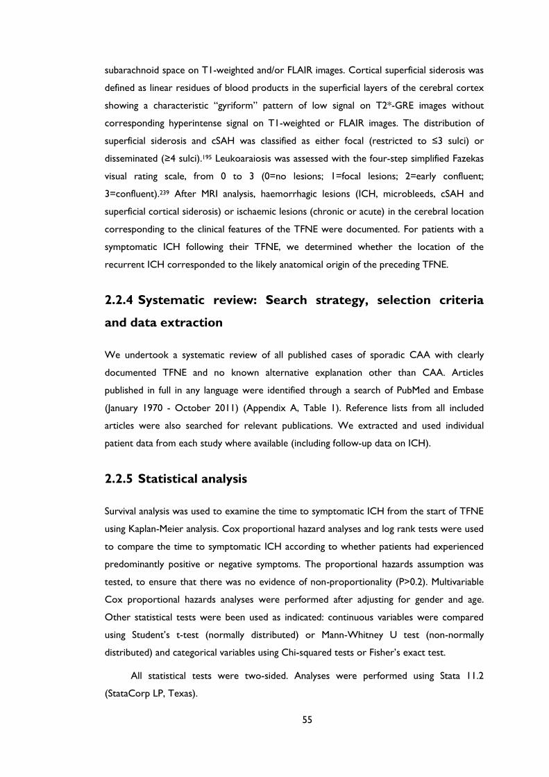
55
subarachnoid space on T1-weighted and/or FLAIR images. Cortical superficial siderosis was
defined as linear residues of blood products in the superficial layers of the cerebral cortex
showing a characteristic “gyriform” pattern of low signal on T2*-GRE images without
corresponding hyperintense signal on T1-weighted or FLAIR images. The distribution of
superficial siderosis and cSAH was classified as either focal (restricted to ≤3 sulci) or
disseminated (≥4 sulci).195 Leukoaraiosis was assessed with the four-step simplified Fazekas
visual rating scale, from 0 to 3 (0=no lesions; 1=focal lesions; 2=early confluent;
3=confluent).239 After MRI analysis, haemorrhagic lesions (ICH, microbleeds, cSAH and
superficial cortical siderosis) or ischaemic lesions (chronic or acute) in the cerebral location
corresponding to the clinical features of the TFNE were documented. For patients with a
symptomatic ICH following their TFNE, we determined whether the location of the
recurrent ICH corresponded to the likely anatomical origin of the preceding TFNE.
2.2.4 Systematic review: Search strategy, selection criteria
and data extraction
We undertook a systematic review of all published cases of sporadic CAA with clearly
documented TFNE and no known alternative explanation other than CAA. Articles
published in full in any language were identified through a search of PubMed and Embase
(January 1970 - October 2011) (Appendix A, Table 1). Reference lists from all included
articles were also searched for relevant publications. We extracted and used individual
patient data from each study where available (including follow-up data on ICH).
2.2.5 Statistical analysis
Survival analysis was used to examine the time to symptomatic ICH from the start of TFNE
using Kaplan-Meier analysis. Cox proportional hazard analyses and log rank tests were used
to compare the time to symptomatic ICH according to whether patients had experienced
predominantly positive or negative symptoms. The proportional hazards assumption was
tested, to ensure that there was no evidence of non-proportionality (P>0.2). Multivariable
Cox proportional hazards analyses were performed after adjusting for gender and age.
Other statistical tests were been used as indicated: continuous variables were compared
using Student’s t-test (normally distributed) or Mann-Whitney U test (non-normally
distributed) and categorical variables using Chi-squared tests or Fisher’s exact test.
All statistical tests were two-sided. Analyses were performed using Stata 11.2
(StataCorp LP, Texas).
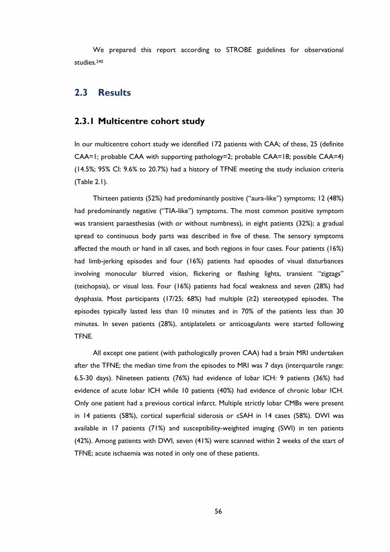
56
We prepared this report according to STROBE guidelines for observational
studies.240
2.3 Results
2.3.1 Multicentre cohort study
In our multicentre cohort study we identified 172 patients with CAA; of these, 25 (definite
CAA=1; probable CAA with supporting pathology=2; probable CAA=18; possible CAA=4)
(14.5%; 95% CI: 9.6% to 20.7%) had a history of TFNE meeting the study inclusion criteria
(Table 2.1).
Thirteen patients (52%) had predominantly positive (“aura-like”) symptoms; 12 (48%)
had predominantly negative (“TIA-like”) symptoms. The most common positive symptom
was transient paraesthesias (with or without numbness), in eight patients (32%); a gradual
spread to continuous body parts was described in five of these. The sensory symptoms
affected the mouth or hand in all cases, and both regions in four cases. Four patients (16%)
had limb-jerking episodes and four (16%) patients had episodes of visual disturbances
involving monocular blurred vision, flickering or flashing lights, transient “zigzags”
(teichopsia), or visual loss. Four (16%) patients had focal weakness and seven (28%) had
dysphasia. Most participants (17/25; 68%) had multiple (≥2) stereotyped episodes. The
episodes typically lasted less than 10 minutes and in 70% of the patients less than 30
minutes. In seven patients (28%), antiplatelets or anticoagulants were started following
TFNE.
All except one patient (with pathologically proven CAA) had a brain MRI undertaken
after the TFNE; the median time from the episodes to MRI was 7 days (interquartile range:
6.5-30 days). Nineteen patients (76%) had evidence of lobar ICH: 9 patients (36%) had
evidence of acute lobar ICH while 10 patients (40%) had evidence of chronic lobar ICH.
Only one patient had a previous cortical infarct. Multiple strictly lobar CMBs were present
in 14 patients (58%), cortical superficial siderosis or cSAH in 14 cases (58%). DWI was
available in 17 patients (71%) and susceptibility-weighted imaging (SWI) in ten patients
(42%). Among patients with DWI, seven (41%) were scanned within 2 weeks of the start of
TFNE; acute ischaemia was noted in only one of these patients.
57
Table 2.1 Demographic, clinical and MRI characteristics of multicentre cohort.
Characteristics Values
(n=25)
Clinical demographics
Age, mean (95% CI:), years 69.7 (66.9 to 72.5)
Sex, male (%) 14 (56)
Hypertension (%) 13 (52)
Smoking history (%) 3 (13)
On antithrombotics at onset of TFNE(%) 8 (32)
Previous symptomatic ICH (%) 8 (32)
Clinical features of TFNE
Predominantly positive symptoms (%) 13 (52)
Predominantly negative symptoms (“TIA-like”) (%) 17 (68)
Associated headache (%) 3 (12)
Patients with >1 type of attack (%) 4 (16)
Patients with multiple (≥2) episodes (%) 17 (68)
Episodes’ duration
<6 minutes (%) 11 (44)
6-19 minutes (%) 5 (20)
>20 minutes (%) 9 (36)
MRI findings (n=24)
Median time (days) from episodes to MRI
(interquartile range) 7 (6.5-30)
Superficial cortical siderosis (%) 12 (50)
Focal (≤3 sulci) (%) 4 (17)
Disseminated (≥4 sulci) (%) 8 (33)
cSAH (%) 4 (17)
cSAH or superficial cortical siderosis (%) 14 (58)
Acute ischaemic lesions 1
Evidence of lobar ICH (%) 19 (76)
Acute lobar ICH evidence (%) 9 (36)
Chronic lobar ICH evidence (%) 10 (40)
Multiple lobar cerebral microbleeds (≥2) (%) 14 (58)
Leukoaraiosis: Fazekas category (%) 0
1
2
3
4 (16)
9 (36)
8 (32)
4 (16)
Antiplatelets/anticoagulants given after TFNE (%) 7 (28)
Aspirin 6 (24)
Aspirin + Clopidogrel 1 (4)
Follow-up information (n=24)
Median follow-up duration (interquartile range) 14 months (4-35 months)
Occurrence of symptomatic ICH (%) 12 (50)
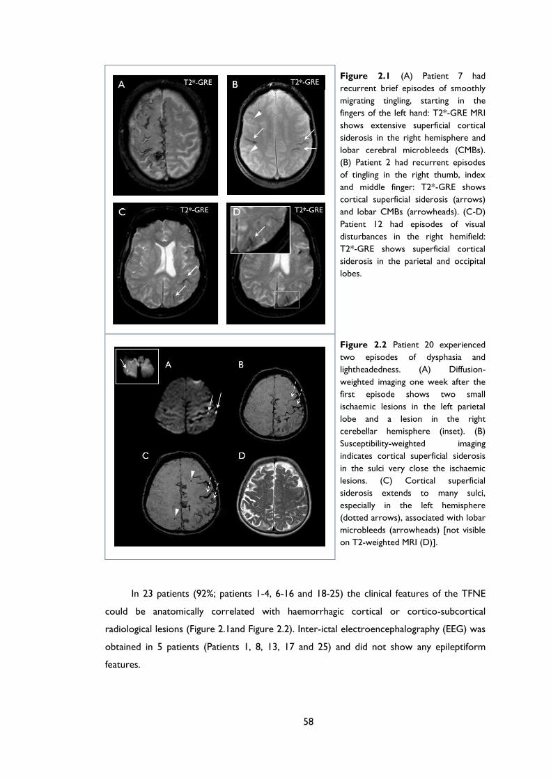
58
Figure 2.1 (A) Patient 7 had
recurrent brief episodes of smoothly
migrating tingling, starting in the
fingers of the left hand: T2*-GRE MRI
shows extensive superficial cortical
siderosis in the right hemisphere and
lobar cerebral microbleeds (CMBs).
(B) Patient 2 had recurrent episodes
of tingling in the right thumb, index
and middle finger: T2*-GRE shows
cortical superficial siderosis (arrows)
and lobar CMBs (arrowheads). (C-D)
Patient 12 had episodes of visual
disturbances in the right hemifield:
T2*-GRE shows superficial cortical
siderosis in the parietal and occipital
lobes.
Figure 2.2 Patient 20 experienced
two episodes of dysphasia and
lightheadedness. (A) Diffusion-
weighted imaging one week after the
first episode shows two small
ischaemic lesions in the left parietal
lobe and a lesion in the right
cerebellar hemisphere (inset). (B)
Susceptibility-weighted imaging
indicates cortical superficial siderosis
in the sulci very close the ischaemic
lesions. (C) Cortical superficial
siderosis extends to many sulci,
especially in the left hemisphere
(dotted arrows), associated with lobar
microbleeds (arrowheads) [not visible
on T2-weighted MRI (D)].
In 23 patients (92%; patients 1-4, 6-16 and 18-25) the clinical features of the TFNE
could be anatomically correlated with haemorrhagic cortical or cortico-subcortical
radiological lesions (Figure 2.1and Figure 2.2). Inter-ictal electroencephalography (EEG) was
obtained in 5 patients (Patients 1, 8, 13, 17 and 25) and did not show any epileptiform
features.
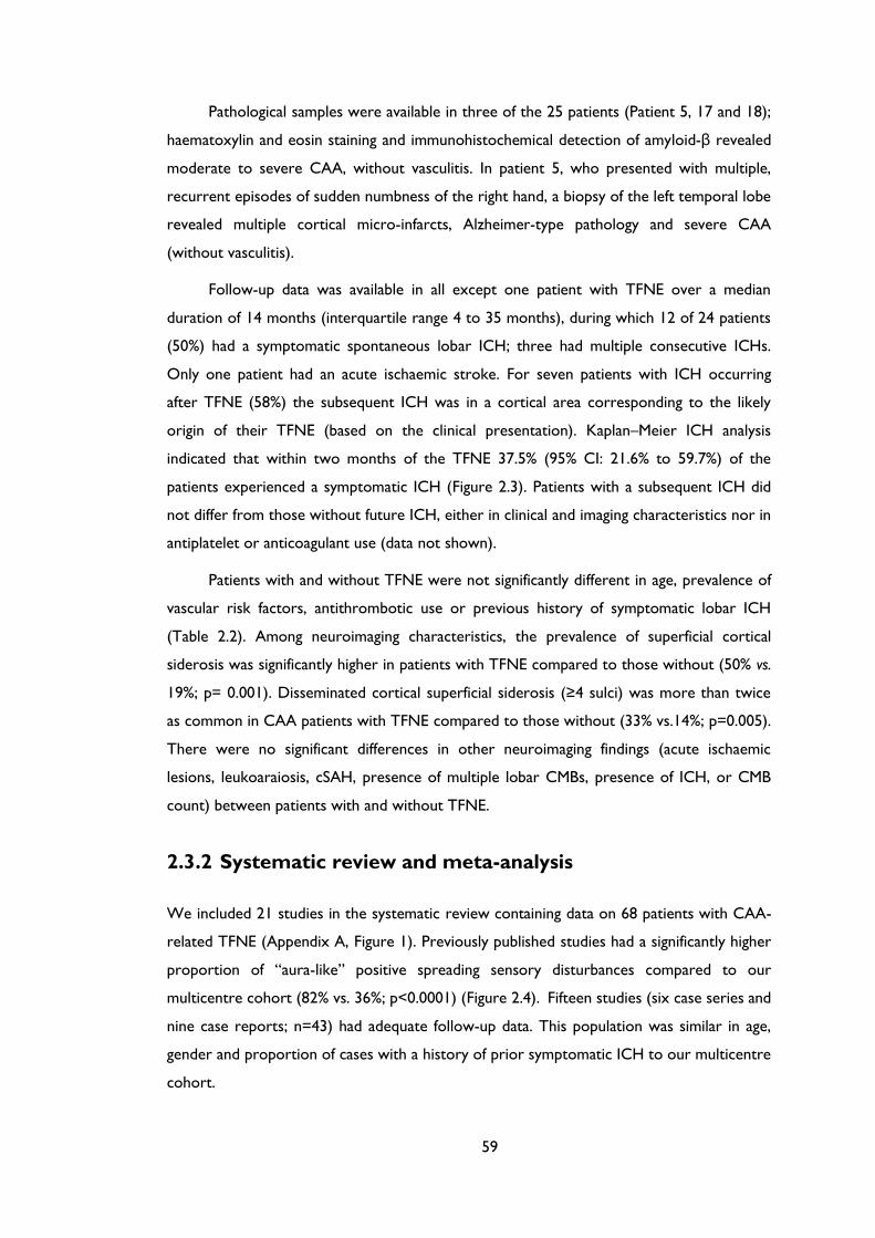
59
Pathological samples were available in three of the 25 patients (Patient 5, 17 and 18);
haematoxylin and eosin staining and immunohistochemical detection of amyloid-β revealed
moderate to severe CAA, without vasculitis. In patient 5, who presented with multiple,
recurrent episodes of sudden numbness of the right hand, a biopsy of the left temporal lobe
revealed multiple cortical micro-infarcts, Alzheimer-type pathology and severe CAA
(without vasculitis).
Follow-up data was available in all except one patient with TFNE over a median
duration of 14 months (interquartile range 4 to 35 months), during which 12 of 24 patients
(50%) had a symptomatic spontaneous lobar ICH; three had multiple consecutive ICHs.
Only one patient had an acute ischaemic stroke. For seven patients with ICH occurring
after TFNE (58%) the subsequent ICH was in a cortical area corresponding to the likely
origin of their TFNE (based on the clinical presentation). Kaplan–Meier ICH analysis
indicated that within two months of the TFNE 37.5% (95% CI: 21.6% to 59.7%) of the
patients experienced a symptomatic ICH (Figure 2.3). Patients with a subsequent ICH did
not differ from those without future ICH, either in clinical and imaging characteristics nor in
antiplatelet or anticoagulant use (data not shown).
Patients with and without TFNE were not significantly different in age, prevalence of
vascular risk factors, antithrombotic use or previous history of symptomatic lobar ICH
(Table 2.2). Among neuroimaging characteristics, the prevalence of superficial cortical
siderosis was significantly higher in patients with TFNE compared to those without (50% vs.
19%; p= 0.001). Disseminated cortical superficial siderosis (≥4 sulci) was more than twice
as common in CAA patients with TFNE compared to those without (33% vs.14%; p=0.005).
There were no significant differences in other neuroimaging findings (acute ischaemic
lesions, leukoaraiosis, cSAH, presence of multiple lobar CMBs, presence of ICH, or CMB
count) between patients with and without TFNE.
2.3.2 Systematic review and meta-analysis
We included 21 studies in the systematic review containing data on 68 patients with CAA-
related TFNE (Appendix A, Figure 1). Previously published studies had a significantly higher
proportion of “aura-like” positive spreading sensory disturbances compared to our
multicentre cohort (82% vs. 36%; p<0.0001) (Figure 2.4). Fifteen studies (six case series and
nine case reports; n=43) had adequate follow-up data. This population was similar in age,
gender and proportion of cases with a history of prior symptomatic ICH to our multicentre
cohort.
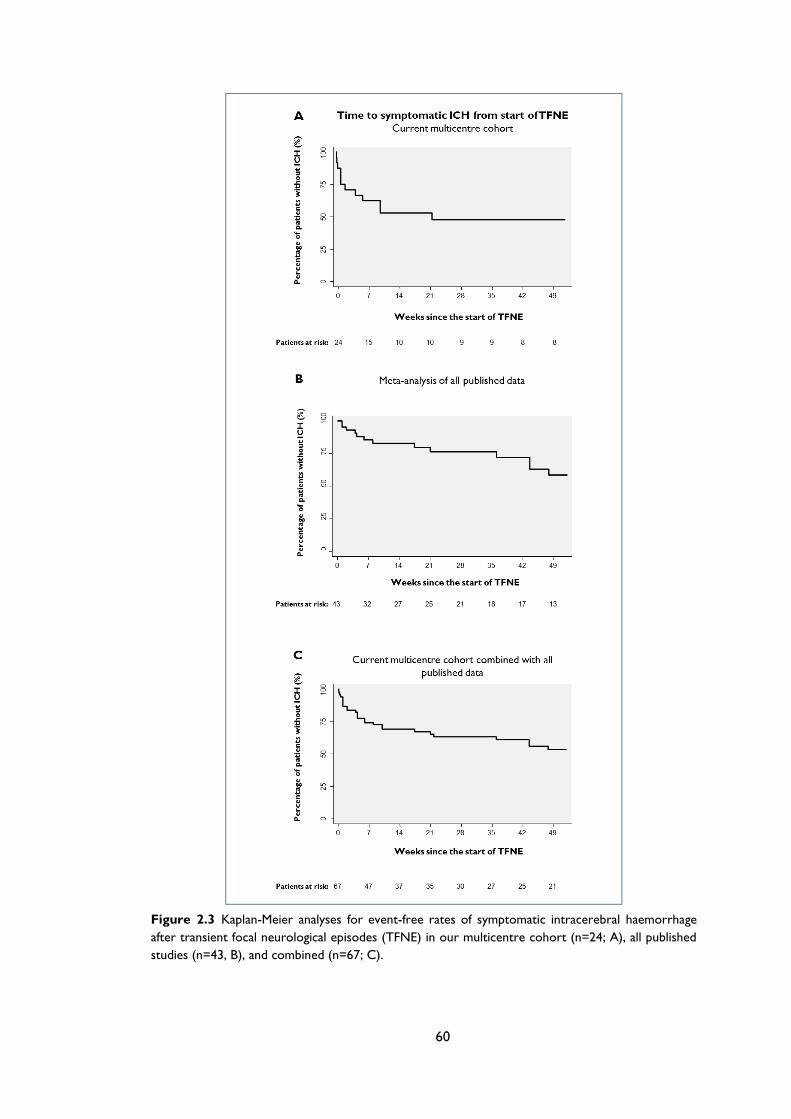
60
Figure 2.3 Kaplan-Meier analyses for event-free rates of symptomatic intracerebral haemorrhage
after transient focal neurological episodes (TFNE) in our multicentre cohort (n=24; A), all published
studies (n=43, B), and combined (n=67; C).
61
Table 2.2 Characteristics of patients with cerebral amyloid angiopathy (CAA) and transient focal
neurological episodes (TFNE) vs. CAA patients without TFNE.
Characteristics
CAA patients
with TFNE
(n=25)
CAA patients
without TFNE
(n=147)
P-value
Age, mean (95% CI:), years 69.7
(66.9-72.5)
73.1
(71.4-74.9) 0.08
Sex, male (%) 14 (56) 73 (50) 0.558
Hypertension (%) 13 (52) 92 (67) 0.145
On antithrombotics (%) 8 (32) 41 (30) 0.801
Previous symptomatic ICH (%) 8 (32) 54 (39) 0.532
Superficial cortical siderosis (%) 12 (50) 24 (19) 0.001
Focal (≤3 sulci) (%) 4 (17) 10 (8) 0.187
Disseminated (≥4 sulci) (%) 8 (33) 14 (11) 0.005
cSAH (%) 4 (17) 8 (7) 0.106
cSAH or superficial cortical siderosis (%) 14 (58) 30 (25) 0.001
Acute ischaemic lesions (%) 1 (6) 12 (11) 0.512
Chronic lobar ICH evidence (%) 10 (40) 72 (53) 0.248
Acute lobar ICH evidence (%) 9 (36) 65 (48) 0.250
Multiple lobar CMBs (≥2) (%) 14 (58) 71 (56) 0.975
Number of CMBs, median
(IQR range) 3 (0-16) 2.5 (0-8) 0.552
Leukoaraiosis: Fazekas category (%)
0
1
2 3
4 (16)
9 (36)
8 (32) 4 (16)
25 (19)
43 (32)
43 (32) 22 (17)
0.981
A meta-analysis combining these 15 studies with our cohort showed a future risk of
symptomatic ICH of 27.6% (95% CI: 18.3% to 40.2%) at two months (Figure 2.3). Patients
with negative symptoms had a similar risk of future ICH to patients with positive symptoms
(hazard ratio: 0.91; 95% CI: 0.40-2.08; p=0.83). There was a borderline significant lower risk
of future ICH among patients with prior symptomatic ICH compared to those without
(hazard ratio: 0.47; 95% CI: 0.22-1.01).
2.4 Discussion
In our multicentre cohort study we found TFNE in 14% of patients with CAA. Our study
confirms previous reports that CAA-related TFNE are mostly recurrent, stereotyped and
brief (usually >30 minutes).167, 168 However, unlike published studies, in which “aura-like”
sensory episodes seem to be most frequent, in our cohort, negative symptoms were just as
common. This difference might reflect previous publication bias in favour of CAA cases
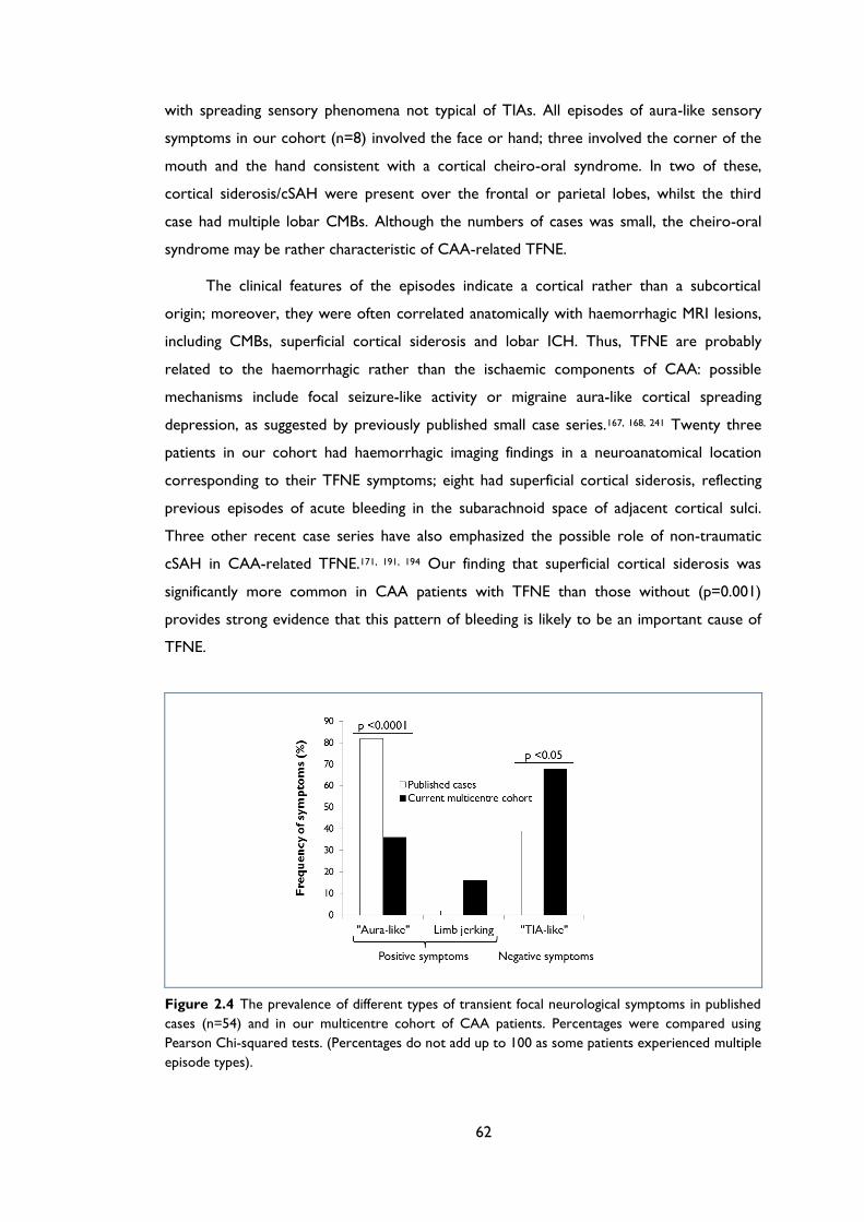
62
with spreading sensory phenomena not typical of TIAs. All episodes of aura-like sensory
symptoms in our cohort (n=8) involved the face or hand; three involved the corner of the
mouth and the hand consistent with a cortical cheiro-oral syndrome. In two of these,
cortical siderosis/cSAH were present over the frontal or parietal lobes, whilst the third
case had multiple lobar CMBs. Although the numbers of cases was small, the cheiro-oral
syndrome may be rather characteristic of CAA-related TFNE.
The clinical features of the episodes indicate a cortical rather than a subcortical
origin; moreover, they were often correlated anatomically with haemorrhagic MRI lesions,
including CMBs, superficial cortical siderosis and lobar ICH. Thus, TFNE are probably
related to the haemorrhagic rather than the ischaemic components of CAA: possible
mechanisms include focal seizure-like activity or migraine aura-like cortical spreading
depression, as suggested by previously published small case series.167, 168, 241 Twenty three
patients in our cohort had haemorrhagic imaging findings in a neuroanatomical location
corresponding to their TFNE symptoms; eight had superficial cortical siderosis, reflecting
previous episodes of acute bleeding in the subarachnoid space of adjacent cortical sulci.
Three other recent case series have also emphasized the possible role of non-traumatic
cSAH in CAA-related TFNE.171, 191, 194 Our finding that superficial cortical siderosis was
significantly more common in CAA patients with TFNE than those without (p=0.001)
provides strong evidence that this pattern of bleeding is likely to be an important cause of
TFNE.
Figure 2.4 The prevalence of different types of transient focal neurological symptoms in published
cases (n=54) and in our multicentre cohort of CAA patients. Percentages were compared using
Pearson Chi-squared tests. (Percentages do not add up to 100 as some patients experienced multiple
episode types).
63
Nevertheless, a role for ischaemic lesions in CAA-related TFNE cannot be ruled out
by our study: small (apparently asymptomatic) ischaemic lesions have been detected in vivo
in clinically probable CAA,73 and “microinfarcts” are a frequent neuropathological finding in
the brains of patients with CAA.28 Since not all cases in our study had DWI soon after the
onset of TFNE, we may have underestimated the contribution from small acute ischaemic
lesions.
We report a strikingly high early risk of symptomatic lobar ICH (37.5% at two
months) following CAA-related TFNE, which we confirmed in a meta-analysis of our data
with all published studies. We found only a borderline difference in the risk of ICH among
patients with previous symptomatic ICH compared to those without, suggesting that TFNE,
rather than simply the presence of CAA with previous symptomatic ICH (with a known
recurrent ICH risk of up to about 10% per year28, 37), are an independent marker of high
early future ICH risk. TFNE in CAA may thus be a clinical marker for cerebral areas of
focally-active and severe CAA pathology, with more vascular leakage leading to an
increased risk of future ICH. This is supported by our finding of a significantly higher
prevalence of superficial cortical siderosis in CAA patients with TFNE, since superficial
siderosis is hypothesized to result from recurrent bleeding into the subarachnoid space,
leading to subpial accumulation of blood-breakdown products. However, the role of
superficial siderosis as a prognostic imaging marker of increased ICH risk in CAA requires
further study.
Although in 58% of patients with ICH following TFNE the new haematoma was
located in a cortical area corresponding to the likely origin of their TFNE (based on the
clinical presentation), further work is also needed to establish whether this apparent
clustering of ICH in brain regions implicated by TFNE symptoms is greater than predicted
by chance.
Our study has several potential limitations. We may have underestimated the true
prevalence of CAA-related TFNE because of the retrospective study design; further large
prospective studies with systematic enquiry about previous TFNE are needed. Some of our
CAA cohort may have been misdiagnosed due to the imperfect specificity of the Boston
criteria (particularly the “possible CAA” category).202 MRI was performed at different times
from TFNE onset; this, combined with the lack of availability of acute DWI sequences in all
cases may have influenced the detection of haemorrhagic over ischaemic lesions. We also
acknowledge the potential for referral, selection and publication bias in the cases identified
in the systematic review. Our multicentre cohort results might not be generalizable to all
CAA patients, but only to those presenting to vascular neurology services in whom other
64
possible causes of transient focal neurological symptoms have been excluded. Although this
study includes the largest number of CAA-related TFNE cases to date, we did not have
sufficient statistical power to definitively determine potential predictors for ICH.
We have shown that TFNE are common in CAA and signify a very high early future
risk of ICH. Hence, their diagnosis has important clinical implications. Our findings clearly
suggest a key role for T2*-GRE MRI (or other blood-sensitive sequences) in the
investigation of patients with unexplained TFNE-especially in individuals without known risk
factors for TIA. The very high early risk of lobar ICH following TFNE in CAA may be an
opportunity to commence preventive strategies: we suggest clinicians should discontinue
and avoid giving antiplatelets or anticoagulants in cases of TFNE with imaging evidence of
CAA, even if the episodes seem clinically likely to be ischaemic. Since TFNE were observed
in patients without a previous history of symptomatic ICH, they may also prove to be a
useful diagnostic marker of CAA, potentially allowing diagnosis earlier in its disease
course.28
65
Chapter 3 Prevalence and mechanisms of
cortical superficial siderosis in sporadic
cerebral amyloid angiopathy
66
ABSTRACT
Objective: We investigated the prevalence and clinical-radiological associations of cortical
superficial siderosis (cSS) in patients with probable cerebral amyloid angiopathy (CAA)
compared to those with intracerebral haemorrhage (ICH) not attributed to CAA.
Methods: Retrospective multicentre cohort study of 120 patients with probable CAA and
two comparison groups: 67 patients with either single lobar ICH or mixed (deep and lobar)
haemorrhages; and 22 patients with strictly deep haemorrhages. We rated cSS, ICH, white
matter changes (WMC) and cerebral microbleeds (CMBs).
Results: cSS was detected in 48/120 (40%; 95%CI: 31.2%-49.3%) patients with probable
CAA, 10/67 (14.9%; 95%CI: 7.4-25.7%) with single lobar ICH or mixed haemorrhages, and
1/22 (4.6%; 95%CI: 0.1%-22.8%) patients with strictly deep haemorrhages (p<0.001 for
trend). Disseminated cSS was present in 29/120 (24%: 95%CI: 16.8-32.8%) probable CAA
patients, but none of the other ICH patients (p<0.001). In probable CAA, age (OR: 1.09;
95%CI: 1.03-1.15; p=0.002), chronic lobar ICH (OR: 3.94, 95%CI: 1.54-10.08; p=0.004) and
transient focal neurological episodes (OR: 11.08; 95%CI: 3.49-35.19; p<0.001) were
independently associated with cSS. However, cSS occurred in 17 of 48 probable CAA
patients (35.4%, 95%CI: 22.2-50.5%) without chronic ICH.
Conclusions: cSS (particularly if disseminated) is a common and characteristic feature of
CAA. Chronic lobar ICH is an independent risk factor for cSS, but the causal direction and
mechanism of association is uncertain. Haemorrhage into the subarachnoid space,
independent of previous (chronic) lobar ICH, must also contribute to cSS in CAA.
Transient focal neurological episodes are the strongest clinical marker of cSS.
67
3.1 Introduction
Sporadic cerebral amyloid angiopathy (CAA) is a common age-related small vessel disease
due to progressive deposition of amyloid–β in the walls of small arteries, arterioles and
capillaries in the cerebral cortex and overlying leptomeninges.28 CAA is most often
recognised in life by symptomatic, lobar intracerebral haemorrhage (ICH) in elderly
patients.28, 37, 242 CAA is also associated with characteristic magnetic resonance imaging
(MRI) findings including lobar cerebral microbleeds (CMBs) and white matter
hyperintensities (leukoaraiosis).28, 35
Recent studies have identified cortical superficial siderosis (cSS) as another
manifestation of CAA.171, 191, 194, 195 In CAA, cSS has a characteristic predilection for the
cerebral convexities, reflecting linear blood residues in the superficial layers of the cerebral
cortex or in the subarachnoid space.190, 195, 243 cSS may have clinical relevance as an
important cause of transient focal neurological episodes (sometimes called “amyloid
spells”),244, 245 and potential “warning sign” for future symptomatic ICH.246
Although cSS is a promising diagnostic neuroimaging marker of CAA,8 the strength of
the association and underlying mechanisms have not been systematically studied. In the
present study we therefore sought to determine the prevalence and extent of cSS in a
European multicentre cohort of patients with clinical-radiological probable CAA, and
investigate its associations with other imaging findings including CMBs, white matter
changes and ICH. We hypothesized that cSS: (1) is common in subjects with probable CAA,
and more prevalent compared to comparison subjects with other spontaneous ICH not
attributed to probable CAA; and (2) is associated with other hemorrhagic manifestations of
CAA (lobar ICH and lobar CMBs).
3.2 Methods
3.2.1 Study population
We included consecutive patients diagnosed with probable CAA (according to the original
Boston criteria202 – i.e. not including cSS as a criterion, see below) at four stroke centres
over defined time periods (Appendix B Figure 1). At participating centres, MRI scanning is a
routine investigation for cases of suspected CAA, unless there are contra-indications.
Essential inclusion criteria for the main CAA case group were: (1) probable or
pathologically verified CAA, defined according to the Boston criteria202 – including lobar
68
CMBs, but not cSS; and (2) available MRI sequences of adequate quality including T2*-
weighted gradient-recalled echo (T2*-GRE) and FLAIR MRI sequences. Cases defined as
possible CAA according to the Boston criteria202 (i.e. single lobar ICH without lobar
microbleeds), were included in one of the comparison groups (see next paragraph).
As comparison groups we included patients with other symptomatic, spontaneous ICH
not fulfilling the Boston criteria for probable CAA, seen in the four stroke centres over the
same time period, with available MRI scans of adequate quality including T2*-GRE and
FLAIR sequences. Cases were systematically and consecutively ascertained using multiple
overlapping methods from prospective clinical and radiological databases. The two non-
CAA comparison groups were as follows: (a) single lobar ICH (without any CMBs) and
mixed (deep and lobar) haemorrhages (including any combination of ICH or CMBs) which
we subsequently refer to as “single lobar ICH and mixed haemorrhages”; and (b) strictly
deep haemorrhages (including ICH and CMBs in the basal ganglia and brainstem).
In total, 149 subjects were excluded from our study because MRI scans were
unavailable (Appendix B Figure 1); those excluded were not significantly different from
those included in gender, age or measures of stroke severity (all p values>0.10).
3.2.2 Standard Protocol Approvals, Registrations, and Patient
Consents
The study received ethical approval by the National Hospital for Neurology and
Neurosurgery and Institute of Neurology Joint Research Ethics Committee, the
Commission d’Ethique Biomedicale Hospitalo Facultaire of the Faculte de Medicine
(Universite Catholique de Louvain), and the Comite d’ethique medicale of the CHU Mont
Godinne UCL.
3.2.3 Data collection
Demographic and clinical information was obtained from prospective databases and by
medical records review using standardised data collection forms. Hypertension was defined
as a history of hypertension, taking antihypertensive treatment or documented elevated
blood pressure (systolic >150 or diastolic >95mmHg) before admission, diabetes as ongoing
use of a hypoglycaemic agent and smoking as history of tobacco use before admission. In
patients with probable CAA, a clearly documented history of transient (≤24 hours), fully
resolving, focal neurological episodes with no known alternative explanation other than
69
CAA (e.g. structural brain lesion, atrial fibrillation, extracranial or intracranial stenosis) was
ascertained by review of medical records.
3.2.4 Magnetic resonance imaging acquisition and analysis
The MRI protocol was similar in each hospital. Imaging was at 1.5T field strength for all
patients and included T1-weighted, T2-weighted, fluid attenuated inversion recovery
(FLAIR), T2*-GRE (slice thickness 5 mm, repetition time 500–1,000 ms, echo time
40/26/15/50-70 ms) and diffusion-weighted imaging (DWI) sequences. Images were
reviewed blinded to clinical data. Hemorrhagic lesions,35, 195, 198, 238 ischaemic lesions
(chronic or acute)73 and white matter hyperintensities (leukoaraiosis)239 were recorded
according to predefined standardized criteria. CMBs were evaluated on T2*-GRE images.35,
238 Prior symptomatic ICH was defined as a symptomatic stroke syndrome associated with
imaging evidence of a corresponding “macro” ICH (>5 mm in diameter on T2*-GRE).198
Asymptomatic prior ICH (>5 mm in diameter on T2*-GRE MRI) was also noted: chronic
ICH was defined on neuroimaging as ICH with no acute bleeding identified on either CT or
MRI scans. Acute convexity subarachnoid haemorrhage (cSAH) was defined as linear
hypointensity in the subarachnoid space affecting one or more cortical sulci of the cerebral
convexities on T2*-GRE sequences with corresponding hyperintensity in the subarachnoid
space on T1-weighted or FLAIR images. cSS was defined as linear residues of chronic blood
products in the superficial layers of the cerebral cortex showing a characteristic “gyriform”
pattern of low signal on T2*-GRE images, without corresponding hyperintense signal on T1-
weighted or FLAIR images. We did not include cSS if it was contiguous with any ICH. The
distribution of cSS and acute cSAH was classified as focal (restricted to ≤3 sulci) or
disseminated (≥4 sulci).195 We also noted whether cSS was contralateral or ipsilateral to
any chronic ICH. Using a sample of MRI scans from probable CAA patients (n=48) and
control non-CAA cases (n=27), the inter-rater agreement for the presence or absent of
cSS was 93.3% (Cohen’s kappa=0.85) and for cSS categories was 95.1% (weighted Cohen’s
kappa=0.86). White matter hyperintensities (leukoaraiosis) were assessed with the four-
step simplified Fazekas rating scale, from 0 to 3 (0=no lesions; 1=focal lesions; 2=early
confluent; 3=confluent).239
3.2.5 Statistical analysis
We compared demographic, clinical and imaging characteristics of probable CAA patients
with versus without cSS. Both binary and ordinal logistic regression analysis was used to
identify predictors of cSS (cSS presence and cSS extent respectively). Multivariable analysis
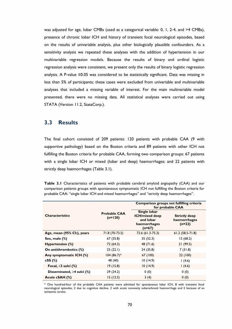
70
was adjusted for age, lobar CMBs (used as a categorical variable: 0, 1, 2-4, and >4 CMBs),
presence of chronic lobar ICH and history of transient focal neurological episodes, based
on the results of univariable analysis, plus other biologically plausible confounders. As a
sensitivity analysis we repeated these analyses with the addition of hypertension in our
multivariable regression models. Because the results of binary and ordinal logistic
regression analysis were consistent, we present only the results of binary logistic regression
analysis. A P-value ≤0.05 was considered to be statistically significant. Data was missing in
less than 5% of participants; these cases were excluded from univariable and multivariable
analyses that included a missing variable of interest. For the main multivariable model
presented, there were no missing data. All statistical analyses were carried out using
STATA (Version 11.2, StataCorp.).
3.3 Results
The final cohort consisted of 209 patients: 120 patients with probable CAA (9 with
supportive pathology) based on the Boston criteria and 89 patients with other ICH not
fulfilling the Boston criteria for probable CAA, forming two comparison groups: 67 patients
with a single lobar ICH or mixed (lobar and deep) haemorrhages; and 22 patients with
strictly deep haemorrhages (Table 3.1).
Table 3.1 Characteristics of patients with probable cerebral amyloid angiopathy (CAA) and our
comparison patients groups with spontaneous symptomatic ICH not fulfilling the Boston criteria for
probable CAA: “single lobar ICH and mixed haemorrhages” and “strictly deep haemorrhages”.
Characteristics Probable CAA
(n=120)
Comparison groups not fulfilling criteria
for probable CAA
Single lobar
ICH/mixed deep
and lobar
haemorrhages
(n=67)
Strictly deep
haemorrhages
(n=22)
Age, mean (95% CI:), years 71.8 (70-73.5) 72.6 (61.3-75.3) 61.2 (58.5-71.8)
Sex, male (%) 67 (55.8) 35 (52.3) 15 (68.2)
Hypertension (%) 72 (64.3) 48 (71.6) 21 (99.5)
On antithrombotics (%) 25 (22.1) 24 (35.8) 7 (31.8)
Any symptomatic ICH (%) 104 (86.7)* 67 (100) 22 (100)
cSS (%) 48 (40) 10 (14.9) 1 (4.6)
Focal, ≤3 sulci (%) 19 (15.8) 10 (14.9) 1 (4.6)
Disseminated, ≥4 sulci (%) 29 (24.2) 0 (0) 0 (0)
Acute cSAH (%) 15 (12.5) 3 (4) 0 (0)
* One hundred-four of the probable CAA patients were admitted for spontaneous lobar ICH, 8 with transient focal
neurological episodes, 2 due to cognitive decline, 2 with acute convexity subarachnoid haemorrhage and 2 because of an ischaemic stroke.
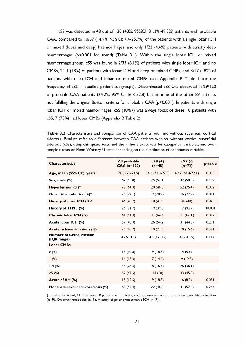
71
cSS was detected in 48 out of 120 (40%; 95%CI: 31.2%-49.3%) patients with probable
CAA, compared to 10/67 (14.9%; 95%CI: 7.4-25.7%) of the patients with a single lobar ICH
or mixed (lobar and deep) haemorrhages, and only 1/22 (4.6%) patients with strictly deep
haemorrhages (p<0.001 for trend) (Table 3.1). Within the single lobar ICH or mixed
haemorrhage group, cSS was found in 2/33 (6.1%) of patients with single lobar ICH and no
CMBs, 2/11 (18%) of patients with lobar ICH and deep or mixed CMBs, and 3/17 (18%) of
patients with deep ICH and lobar or mixed CMBs (see Appendix B Table 1 for the
frequency of cSS in detailed patient subgroups). Disseminated cSS was observed in 29/120
of probable CAA patients (24.2%; 95% CI 16.8-32.8) but in none of the other 89 patients
not fulfilling the original Boston criteria for probable CAA (p<0.001). In patients with single
lobar ICH or mixed haemorrhages, cSS (10/67) was always focal; of these 10 patients with
cSS, 7 (70%) had lobar CMBs (Appendix B Table 2).
Table 3.2 Characteristics and comparison of CAA patients with and without superficial cortical
siderosis. P-values refer to differences between CAA patients with vs. without cortical superficial
siderosis (cSS), using chi-square tests and the Fisher’s exact test for categorical variables, and two-
sample t-tests or Mann-Whitney U-tests depending on the distribution of continuous variables.
Characteristics All probable
CAA (n=120)
cSS (+)
(n=48)
cSS (-)
(n=72) p-value
Age, mean (95% CI:), years 71.8 (70-73.5) 74.8 (72.3-77.3) 69.7 (67.4-72.1) 0.005
Sex, male (%) 67 (55.8) 25 (52.1) 42 (58.3) 0.499
Hypertension (%)* 72 (64.3) 20 (46.5) 52 (75.4) 0.002
On antithrombotics (%)* 25 (22.1) 9 (20.9) 16 (22.9) 0.811
History of prior ICH (%)* 46 (40.7) 18 (41.9) 28 (40) 0.845
History of TFNE (%) 26 (21.7) 19 (39.6) 7 (9.7) <0.001
Chronic lobar ICH (%) 61 (51.3) 31 (64.6) 30 (42.3.) 0.017
Acute lobar ICH (%) 57 (48.3) 26 (54.2) 31 (44.3) 0.291
Acute ischaemic lesions (%) 20 (18.7) 10 (23.3) 10 (15.6) 0.321
Number of CMBs, median
(IQR range) 4 (2-13.5) 4.5 (1-10.5) 4 (2-15.5) 0.147
Lobar CMBs
0 (%) 13 (10.8) 9 (18.8) 4 (5.6)
1 (%) 16 (13.3) 7 (14.6) 9 (12.5)
2-4 (%) 34 (28.3) 8 (16.7) 26 (36.1)
≥5 (%) 57 (47.5) 24 (50) 33 (45.8)
Acute cSAH (%) 15 (12.5) 9 (18.8) 6 (8.3) 0.091
Moderate-severe leukoaraiosis (%) 63 (53.4) 22 (46.8) 41 (57.6) 0.244
† p-value for trend; *There were 10 patients with missing data for one or more of these variables: Hypertension
(n=9), On antithrombotics (n=8), History of prior symptomatic ICH (n=7).
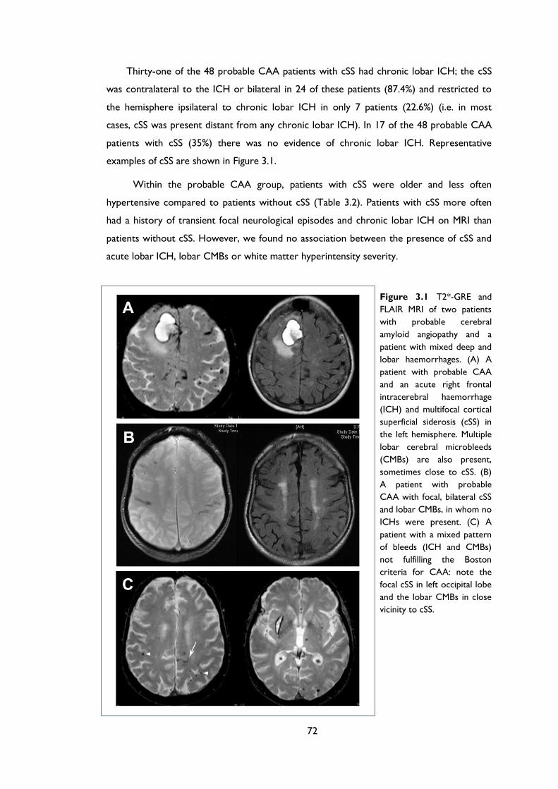
72
Thirty-one of the 48 probable CAA patients with cSS had chronic lobar ICH; the cSS
was contralateral to the ICH or bilateral in 24 of these patients (87.4%) and restricted to
the hemisphere ipsilateral to chronic lobar ICH in only 7 patients (22.6%) (i.e. in most
cases, cSS was present distant from any chronic lobar ICH). In 17 of the 48 probable CAA
patients with cSS (35%) there was no evidence of chronic lobar ICH. Representative
examples of cSS are shown in Figure 3.1.
Within the probable CAA group, patients with cSS were older and less often
hypertensive compared to patients without cSS (Table 3.2). Patients with cSS more often
had a history of transient focal neurological episodes and chronic lobar ICH on MRI than
patients without cSS. However, we found no association between the presence of cSS and
acute lobar ICH, lobar CMBs or white matter hyperintensity severity.
Figure 3.1 T2*-GRE and
FLAIR MRI of two patients
with probable cerebral
amyloid angiopathy and a
patient with mixed deep and
lobar haemorrhages. (A) A
patient with probable CAA
and an acute right frontal
intracerebral haemorrhage
(ICH) and multifocal cortical
superficial siderosis (cSS) in
the left hemisphere. Multiple
lobar cerebral microbleeds
(CMBs) are also present,
sometimes close to cSS. (B)
A patient with probable
CAA with focal, bilateral cSS
and lobar CMBs, in whom no
ICHs were present. (C) A
patient with a mixed pattern
of bleeds (ICH and CMBs)
not fulfilling the Boston
criteria for CAA: note the
focal cSS in left occipital lobe
and the lobar CMBs in close
vicinity to cSS.
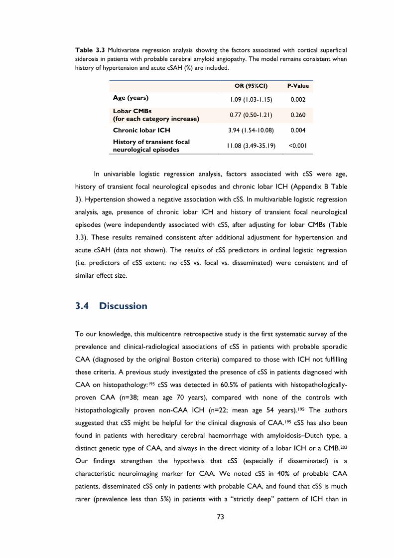
73
Table 3.3 Multivariate regression analysis showing the factors associated with cortical superficial
siderosis in patients with probable cerebral amyloid angiopathy. The model remains consistent when
history of hypertension and acute cSAH (%) are included.
OR (95%CI) P-Value
Age (years) 1.09 (1.03-1.15) 0.002
Lobar CMBs
(for each category increase) 0.77 (0.50-1.21) 0.260
Chronic lobar ICH 3.94 (1.54-10.08) 0.004
History of transient focal
neurological episodes 11.08 (3.49-35.19) <0.001
In univariable logistic regression analysis, factors associated with cSS were age,
history of transient focal neurological episodes and chronic lobar ICH (Appendix B Table
3). Hypertension showed a negative association with cSS. In multivariable logistic regression
analysis, age, presence of chronic lobar ICH and history of transient focal neurological
episodes (were independently associated with cSS, after adjusting for lobar CMBs (Table
3.3). These results remained consistent after additional adjustment for hypertension and
acute cSAH (data not shown). The results of cSS predictors in ordinal logistic regression
(i.e. predictors of cSS extent: no cSS vs. focal vs. disseminated) were consistent and of
similar effect size.
3.4 Discussion
To our knowledge, this multicentre retrospective study is the first systematic survey of the
prevalence and clinical-radiological associations of cSS in patients with probable sporadic
CAA (diagnosed by the original Boston criteria) compared to those with ICH not fulfilling
these criteria. A previous study investigated the presence of cSS in patients diagnosed with
CAA on histopathology:195 cSS was detected in 60.5% of patients with histopathologically-
proven CAA (n=38; mean age 70 years), compared with none of the controls with
histopathologically proven non-CAA ICH (n=22; mean age 54 years).195 The authors
suggested that cSS might be helpful for the clinical diagnosis of CAA.195 cSS has also been
found in patients with hereditary cerebral haemorrhage with amyloidosis–Dutch type, a
distinct genetic type of CAA, and always in the direct vicinity of a lobar ICH or a CMB.203
Our findings strengthen the hypothesis that cSS (especially if disseminated) is a
characteristic neuroimaging marker for CAA. We noted cSS in 40% of probable CAA
patients, disseminated cSS only in patients with probable CAA, and found that cSS is much
rarer (prevalence less than 5%) in patients with a “strictly deep” pattern of ICH than in
74
probable CAA. We also detected cSS in 15% of patients with a single lobar ICH or mixed
(lobar and deep) haemorrhages; however, in most of these mixed patients, lobar CMBs
were also present, suggesting that they might in fact harbour some degree of CAA
pathology.
Among patients with a single lobar ICH and no CMBs (i.e. “possible CAA” according
to the original Boston criteria202) the prevalence of cSS was also very low (6%). In a
validation of these criteria, 16 of 26 patients (62%) classified as possible CAA had
pathologically-confirmed CAA,202 but only 11 of the pathologically diagnosed patients had
T2*-GRE imaging, limiting the generalizability of the findings to current cohorts (including
our study), in which such imaging is now routine. In another validation study of the Boston
criteria, using T2*-weighted gradient echo MRI in a hereditary Dutch-type CAA population,
all patients with lobar ICH also had CMBs.203 Our data suggest that further pathological
correlation is required to determine the prevalence of pathologically-confirmed CAA in
patients with a single lobar ICH (and no CMBs or cSS) on appropriate blood-sensitive MRI
sequences. Further studies with histopathological confirmation or in vivo amyloid imaging176
will also help to determine whether cSS is a useful diagnostic feature of CAA in patients
with mixed patterns of haemorrhage.
In the healthy population-based Rotterdam Scan Study cSS was found in 0.7% (7/1062)
of elderly individuals (mean age 69.6 years), all of whom had lobar CMBs (6 had strictly
lobar CMBs, 5 of whom had multiple CMBs), in close vicinity to the cSS.243 In agreement
with our findings, the mean age of persons with cSS was higher than those without cSS
(mean age 79.9 vs. 69.6 years; p<0.001), and also compared to persons who had CMBs but
no cSS (mean age 71.8 years).243 Since the severity of CAA is age-related,28, 37 these finding,
together with our data showing an association of cSS with chronic ICH, suggest that cSS
might be a marker of more advanced CAA, but this also requires pathological confirmation,
with standardized grading of CAA severity.
Experimental studies confirm that repeated bleeding into the subarachnoid space leads
to subpial haemosiderin deposition.247 There are thus at least two possible
pathophysiological mechanisms which could lead to cSS deposition in CAA: (a) repeated
episodes of haemorrhage from brittle superficial cortical or leptomeningeal CAA-affected
vessels into the subarachnoid space (independent of lobar ICH); and (b) leakage from a
previous lobar ICH (or superficial lobar CMBs) into the subarachnoid space. Our
observation of cSS in 17 patients without chronic lobar ICH, and even in those with chronic
lobar ICH mostly distant from the ICH (in nearly 90% of cases in the contralateral
hemisphere or bilaterally), favours a contribution from direct haemorrhage into the
75
subarachnoid space, independent of lobar ICH. This implies that cSS may arise
independently of known characteristic imaging features of CAA (lobar ICH and CMBs),
supporting its role as an independent diagnostic marker; further studies are needed to
confirm the value of cSS in improving the sensitivity of in vivo CAA diagnosis.
Perhaps surprisingly, we did not find any association between lobar CMBs and cSS,
which may reflect selection bias towards generally advanced disease with high lobar CMBs
prevalence in our cohort. Nevertheless in some cases cSS was observed close to one or
more lobar CMBs (see example in Figure 2), suggesting that leakage from very superficial
CMBs may also be a mechanism of cSS.
Another interpretation of the association between cSS and lobar ICH is that cSS
precedes lobar ICH. Although our cross-sectional data cannot confirm this hypothesis, some
recent data support this explanation. A retrospective study of 51 patients with cSS and due
to possible or probable CAA found that after a median 35.3 months follow-up, 47.1% of the
patients had new radiological ICH or cSAH, often at the site of pre-existing siderosis,246
providing preliminary evidence that cSS heralds a high risk of future ICH. A small
neuropathological series of six autopsy cases of subcortical haematoma caused by CAA
showed that at least in some cases the primary haemorrhage appeared to originate from
the subarachnoid space.248 Further prospective studies are urgently required to determine
the risk of future intracranial bleeding associated with cSS in CAA.
Our study confirms a strong independent association of cSS with transient focal
neurological episodes (sometimes called “amyloid spells”249), which are increasingly
recognized in CAA and can resemble transient ischaemic attacks, migraine auras or focal
seizures.167, 244, 249 Such attacks could plausibly be caused by disruption of cortical function
due to cSS, for example by superficial cortical haemosiderin deposition inducing focal
seizures or cortical spreading depression.244, 249-251 CAA-related transient focal neurological
episodes are associated with a high early risk of symptomatic lobar ICH (24.5% [95% CI:
15.8%–36.9%] at 8 weeks);244 cSS may be one mechanism underpinning this increased
clinical risk, reflecting focally active CAA near the cortical surface.252
Our study has several strengths including the systematic evaluation of MRI scans by
trained raters using validated scales for a range of imaging markers of small vessel disease. A
limitation is the lack of pathological confirmation of the CAA pathology. In view of the
imperfect specificity of the Boston criteria, especially for the “possible CAA” category,202
we focussed on patients fulfilling the criteria for probable CAA, for which the specificity is
between 82-100%.202, 203 Limitations of our study include the retrospective design, the
variation in inception point of the disease at inclusion, and the potential of bias in our
76
sample as MRIs were done as part of routine clinical care, tending to exclude more severe
cases of CAA and ICH.
Our study indicates that cSS (particularly if disseminated) is a characteristic
neuroimaging feature of CAA. Chronic lobar ICH is an independent risk factor for cSS, but
the causal direction and mechanism of the association is uncertain: although leakage from
previous lobar ICH into the subarachnoid space may lead to cSS, it is also possible that cSS
heralds an increased risk of future lobar ICH. Further prospective studies are needed to
clarify how cSS relates to future ICH risk, which may have important clinical relevance, for
example regarding antithrombotic treatment. Our results also show that haemorrhage into
the subarachnoid space, independent of lobar ICH, must also contribute to cSS in CAA,
suggesting that cSS should be considered an additional neuroimaging marker of CAA-
related haemorrhage.
77
Chapter 4 Cortical superficial siderosis and
intracerebral haemorrhage risk in patients
with sporadic cerebral amyloid angiopathy:
multicentre MRI cohort study
78
ABSTRACT
Objective: To investigate whether cortical superficial siderosis (cSS) on MRI, especially if
disseminated (involving more than 3 sulci), increases the risk of future symptomatic lobar
intracerebral haemorrhage (ICH) in cerebral amyloid angiopathy (CAA).
Methods: European multicentre cohort study of 118 CAA patients (104 with baseline
symptomatic lobar ICH) diagnosed according to the Boston criteria. We obtained baseline
clinical, MRI and follow-up data on symptomatic lobar ICH. Using Kaplan-Meier and Cox
regression analyses, we investigated cSS and ICH risk, adjusting for known confounders.
Results: During a median follow-up time of 24 months (interquartile range 9–44 months),
23/118 (19.5%, 95%CI: 12.8-27.8) patients experienced symptomatic lobar ICH. Any cSS,
and disseminated cSS were predictors of time until first or recurrent ICH (log-rank test:
p=0.0045 and p=0.0009 respectively). ICH risk was 25% (95%CI: 7.6-28.3%) for patients
without siderosis; 28.9% (95%CI: 7.7-76.7%) for patients with focal siderosis; and 74%
(95%CI: 44.1-95.7) for patients with disseminated cSS (log-rank test: p=0.0031). In Cox
regression models, any cSS and disseminated cSS were both independently associated with
increased lobar ICH risk, after adjusting for ≥2 microbleeds and age (HR: 2.53; 95%CI: 1.05-
6.15; p=0.040 and HR: 3.16; 95%CI: 1.35-7.43; p=0.008, respectively). These results
remained consistent in sensitivity analyses including only patients with symptomatic lobar
ICH at baseline.
Conclusions: Our findings indicate that cSS, particularly if disseminated, is associated with
an increased risk of symptomatic lobar ICH in CAA. cSS may help stratify future bleeding
risk in CAA, with implications for prognosis and treatment.
79
4.1 Introduction
Sporadic cerebral amyloid angiopathy (CAA) is a highly prevalent age-related small vessel
disease61 caused by amyloid–β deposition in cortical and leptomeningeal vessel walls.28 CAA
is a major cause of lobar intracerebral haemorrhage (ICH), particularly in elderly patients.28,
33, 37, 242 Spontaneous ICH is one of the most catastrophic forms of stroke, with a high risk
of recurrence;220, 253, 254 CAA-related lobar ICH may carry a greater risk than deep ICH
presumed due to hypertensive arteriopathy,254, 255 but this is currently difficult to predict.
Predisposing factors for lobar ICH and lobar ICH recurrence in CAA include: APOE
ε4 and ε2 alleles;87 hemorrhagic neuroimaging markers of CAA such as lobar cerebral
microbleeds (CMBs);180 and anticoagulant or antiplatelet use.227 Little is known about
cortical superficial siderosis (cSS), a recently identified neuroimaging marker of CAA,195 and
the risk of subsequent ICH. cSS reflects linear blood residues in the superficial (subpial)
layers of the cerebral cortex.190 One likely mechanism leading to cSS is repeated episodes
of haemorrhage into the subarachnoid space from brittle superficial cortical or
leptomeningeal CAA-laden vessels, potentially heralding a high risk of future lobar ICH. A
recent study showed that nearly 50% of CAA patients with cSS experienced intracranial
haemorrhage over a period of 35 months,246 but this study did not include patients without
cSS as a control group.
We tested the hypothesis that in CAA patients, cSS, especially involving multiple sulci
(reflecting more widespread or active disease), is associated with an increased risk of future
symptomatic lobar ICH, in a European multicentre cohort study.
4.2 Patients and Methods
4.2.1 Study population and baseline data collection
We included consecutive patients diagnosed with CAA (according to the original Boston
criteria,202 – not including cSS as a criterion) at four stroke centres over defined time
periods. The centres were: University College London Hospitals NHS Foundation Trust
(London) (03/2003–09/2011), Addenbrooke’s Hospital (Cambridge) (07/2002–03/2010),
Cliniques Universitaires Saint Luc (Brussels) (12/2003–04/2010) and CHU Mont-Godinne
UCL (08/2005–03/2009). At participating centres, MRI scanning is a routine investigation for
cases of suspected CAA, unless there are contra-indications. Our inclusion criteria were:
(1) patients fulfilling the original Boston criteria for CAA;202 (2) available MRI sequences
80
including T2*-weighted gradient-recalled echo (T2*-GRE) and fluid attenuated inversion
recovery (FLAIR) MRI; and (3) available follow-up information on symptomatic ICH,
confirmed by neuroimaging. We included all patients with CAA, including survivors of
spontaneous lobar ICH and those who, during investigation for other symptoms, were
found to have strictly lobar CMBs (or asymptomatic lobar ICH). We excluded CAA
patients without adequate MR imaging (n=26), or reliable follow-up data (n=37). Excluded
subjects (n=63) did not differ significantly from those included in any baseline characteristics
(all p>0.05).
Clinical data at the time of presentation (age, gender, vascular risk factors including
hypertension, use of antithrombotics and previous symptomatic ICH) were obtained from
prospective databases and by medical records review using standardised data collection
forms. A clearly documented prior history of transient (≤24 hours), focal neurological
episodes with no known alternative explanation other than CAA (e.g. structural brain
lesion, atrial fibrillation, extracranial or intracranial stenosis) was ascertained by review of
medical records.
4.2.2 Standard Protocol Approvals, Registrations, and Patient
Consents
The study received ethical approval by the National Hospital for Neurology and
Neurosurgery and Institute of Neurology Joint Research Ethics Committee, the
Commission d’Ethique Biomedicale Hospitalo Facultaire of the Faculte de Medicine
(Universite Catholique de Louvain), and the Comite d’ethique medicale of the CHU Mont
Godinne UCL.
4.2.3 Magnetic resonance imaging acquisition and analysis
The MRI protocol was similar in each hospital. Imaging was at 1.5T field strength for
all patients and included T1-weighted, T2-weighted, FLAIR and axial T2*-GRE (slice
thickness 5 mm, repetition time 500–1,000 ms, echo time 15-70ms). Images were reviewed
by a trained clinical research fellow blinded to clinical and follow-up data. The presence and
distribution of CMBs were evaluated on T2*-GRE images using the Microbleed Anatomical
Rating Scale.238 Asymptomatic or symptomatic prior ICH (>5 mm in diameter on T2*-GRE
MRI) was also noted.198 cSS was defined as linear residues of chronic blood products in the
superficial layers of the cerebral cortex showing a characteristic “gyriform” pattern of low
signal on T2*-GRE images, without corresponding hyperintense signal on T1-weighted or

81
FLAIR images (i.e. without acute subarachnoid haemorrhage). We did not include cSS
contiguous with any ICH. The distribution of cSS was classified as focal (restricted to ≤3
sulci) or disseminated (>3 sulci).195 White matter changes were evaluated using the four-
step simplified Fazekas rating scale (0-3: 0=no lesions; 1=focal lesions; 2=early confluent;
3=confluent).239
4.2.4 Follow-up
Follow-up data were obtained from a systematic review of multiple overlapping sources
including prospective databases, medical records review (including discharge summaries,
follow-up outpatient and general practitioner letters) and radiological databases, using
standardized data collection forms. We collected information on clinically symptomatic
ICH, defined as a symptomatic stroke syndrome associated with neuroimaging evidence of
a corresponding ICH (>5 mm in diameter),198 and death of any cause. Outcome events
were assessed using all clinical, radiological, and pathological information available, blinded
to the presence of cSS at baseline MRI. We determined whether the location of
symptomatic ICH at follow-up corresponded to the anatomical distribution of cSS on
baseline MRI.
4.2.5 Statistical analysis
We compared clinical and imaging characteristics of CAA patients with symptomatic lobar
ICH during follow-up to patients without ICH, using chi-square tests and the Fisher’s exact
test for categorical variables, and two-sample t-tests or Mann-Whitney U-tests depending
on the distribution of continuous variables. The reliability of rating for presence and
category rating of cSS was assessed in a sample of MRI scans from probable CAA patients
(n=48) by calculating Cohen’s kappa and weighted kappa statistic respectively. We also
compared basic clinical and imaging characteristics of patients with vs. without cSS. We
determined the presence of cSS and disseminated cSS as univariate predictors of ICH risk
using Kaplan-Meier plots with significance testing by the log-rank test. Survival time was
calculated from date of baseline MRI scan until the date of symptomatic lobar ICH at
follow-up or the last known date without the outcome event of interest. For individuals
experiencing multiple lobar ICHs during follow-up, data were censored at time of first ICH.
Data were also censored at the time of death from causes other than documented
symptomatic ICH. Cox regression analysis was performed to calculate univariate hazard
ratio (HR) as a measure of the effect size.
82
We estimated that our study would have a power of 88% to detect a difference in
ICH risk between CAA patients with and without cSS, assuming 50% and 20% rates of ICH
over four years respectively180, 227, 246 (two-tailed test and with alpha values of 0.05).
According to the ‘rule of 10’ for developing proportional hazards models, we require
approximately 10 outcome events for each potential covariate in multivariable analyses.256,
257 Therefore, we investigated the effect the following prespecified potential predictors
using Cox proportional-hazards model: presence of cSS or disseminated cSS, the presence
of multiple CMBs (≥2), prespecified on the basis of the hypothesised effect on ICH risk
from previous published series in CAA,180, 227 and age, being an important potential
confounder. As sensitivity analyses, we repeated all statistical tests in CAA patients with
symptomatic lobar ICH at baseline, and those who had their MRI within six months of the
index ICH. We also undertook exploratory multivariable models incorporating previous
ICH as another potential confounding factor. The proportional hazard assumption was
tested using graphical checks and Schoenfeld residuals-based tests.
A P-value≤0.05 was considered to be statistically significant. All analyses were
performed using STATA (Version 11.2). The manuscript has been prepared with reference
to the STROBE guidelines.258
4.3 Results
The final cohort consisted of 118 patients fulfilling the Boston diagnostic criteria202 for
CAA: 8 with pathologically-proven CAA, 86 with probable CAA and 24 with possible CAA.
Of these, 104 (88%) patients presented with symptomatic lobar ICH at baseline, 8 with
transient focal neurological episodes, 3 with cognitive decline, 1 with acute convexity
subarachnoid haemorrhage and 2 with ischaemic stroke. The inter-rater agreement for the
presence or absent of cSS was 89.6% (Cohen’s kappa=0.79) and for cSS categories was
89.6% (weighted Cohen’s kappa=0.75). Forty-one patients (43.8%, 95% CI: 26.2%-44.6%)
had cSS at baseline. The presence of cSS was strongly associated with a prior history of
transient focal neurological episodes (48.8% vs. 8.8%; p<0.001 respectively) and the
presence of ≥5 CMBs (48.8% vs. 27.3%; p=0.019 respectively) (Appendix C Table 1). CAA
patients with cSS were slightly older compared to patients without siderosis (mean age;
95%CI: 73.2; 70.4-76 vs. 70.3; 68.2-72.5 respectively, p=0.111).
During a median follow-up time of 24 months (interquartile range 9–44 months), 23
of 118 patients (19.5%, 95%CI: 12.8-27.8) experienced a symptomatic lobar ICH; four of
these 23 patients experienced multiple (>1) sequential symptomatic lobar ICH during
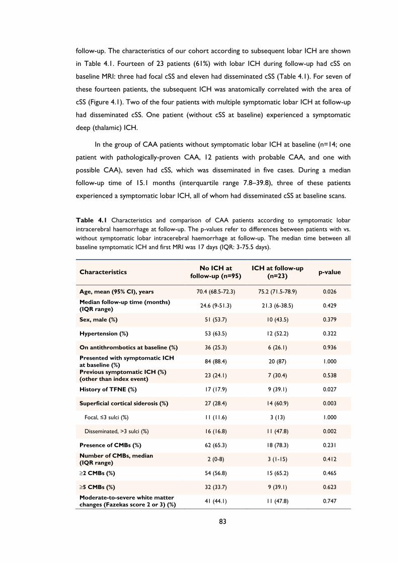
83
follow-up. The characteristics of our cohort according to subsequent lobar ICH are shown
in Table 4.1. Fourteen of 23 patients (61%) with lobar ICH during follow-up had cSS on
baseline MRI: three had focal cSS and eleven had disseminated cSS (Table 4.1). For seven of
these fourteen patients, the subsequent ICH was anatomically correlated with the area of
cSS (Figure 4.1). Two of the four patients with multiple symptomatic lobar ICH at follow-up
had disseminated cSS. One patient (without cSS at baseline) experienced a symptomatic
deep (thalamic) ICH.
In the group of CAA patients without symptomatic lobar ICH at baseline (n=14; one
patient with pathologically-proven CAA, 12 patients with probable CAA, and one with
possible CAA), seven had cSS, which was disseminated in five cases. During a median
follow-up time of 15.1 months (interquartile range 7.8–39.8), three of these patients
experienced a symptomatic lobar ICH, all of whom had disseminated cSS at baseline scans.
Table 4.1 Characteristics and comparison of CAA patients according to symptomatic lobar
intracerebral haemorrhage at follow-up. The p-values refer to differences between patients with vs.
without symptomatic lobar intracerebral haemorrhage at follow-up. The median time between all
baseline symptomatic ICH and first MRI was 17 days (IQR: 3-75.5 days).
Characteristics No ICH at
follow-up (n=95)
ICH at follow-up
(n=23) p-value
Age, mean (95% CI), years 70.4 (68.5-72.3) 75.2 (71.5-78.9) 0.026
Median follow-up time (months)
(IQR range) 24.6 (9-51.3) 21.3 (6-38.5) 0.429
Sex, male (%) 51 (53.7) 10 (43.5) 0.379
Hypertension (%) 53 (63.5) 12 (52.2) 0.322
On antithrombotics at baseline (%) 36 (25.3) 6 (26.1) 0.936
Presented with symptomatic ICH
at baseline (%) 84 (88.4) 20 (87) 1.000
Previous symptomatic ICH (%)
(other than index event) 23 (24.1) 7 (30.4) 0.538
History of TFNE (%) 17 (17.9) 9 (39.1) 0.027
Superficial cortical siderosis (%) 27 (28.4) 14 (60.9) 0.003
Focal, ≤3 sulci (%) 11 (11.6) 3 (13) 1.000
Disseminated, >3 sulci (%) 16 (16.8) 11 (47.8) 0.002
Presence of CMBs (%) 62 (65.3) 18 (78.3) 0.231
Number of CMBs, median
(IQR range) 2 (0-8) 3 (1-15) 0.412
≥2 CMBs (%) 54 (56.8) 15 (65.2) 0.465
≥5 CMBs (%) 32 (33.7) 9 (39.1) 0.623
Moderate-to-severe white matter
changes (Fazekas score 2 or 3) (%) 41 (44.1) 11 (47.8) 0.747
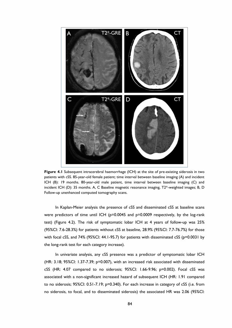
84
Figure 4.1 Subsequent intracerebral haemorrhage (ICH) at the site of pre-existing siderosis in two
patients with cSS. 85-year-old female patient; time interval between baseline imaging (A) and incident
ICH (B): 19 months. 80-year-old male patient, time interval between baseline imaging (C) and
incident ICH (D): 35 months. A, C Baseline magnetic resonance imaging, T2*-weighted images; B, D
Follow-up unenhanced computed tomography scans.
In Kaplan-Meier analysis the presence of cSS and disseminated cSS at baseline scans
were predictors of time until ICH (p=0.0045 and p=0.0009 respectively, by the log-rank
test) (Figure 4.2). The risk of symptomatic lobar ICH at 4 years of follow-up was 25%
(95%CI: 7.6-28.3%) for patients without cSS at baseline, 28.9% (95%CI: 7.7-76.7%) for those
with focal cSS, and 74% (95%CI: 44.1-95.7) for patients with disseminated cSS (p=0.0031 by
the long-rank test for each category increase).
In univariate analysis, any cSS presence was a predictor of symptomatic lobar ICH
(HR: 3.18; 95%CI: 1.37-7.39; p=0.007), with an increased risk associated with disseminated
cSS (HR: 4.07 compared to no siderosis; 95%CI: 1.66-9.96; p=0.002). Focal cSS was
associated with a non-significant increased hazard of subsequent ICH (HR: 1.91 compared
to no siderosis; 95%CI: 0.51-7.19; p=0.340). For each increase in category of cSS (i.e. from
no siderosis, to focal, and to disseminated siderosis) the associated HR was 2.06 (95%CI:
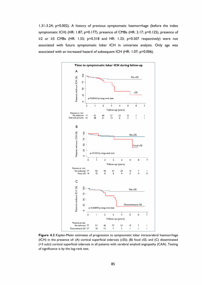
85
1.31-3.24; p=0.002). A history of previous symptomatic haemorrhage (before the index
symptomatic ICH) (HR: 1.87; p=0.177), presence of CMBs (HR: 2.17; p=0.125), presence of
≥2 or ≥5 CMBs (HR: 1.55; p=0.318 and HR: 1.33; p=0.507 respectively) were not
associated with future symptomatic lobar ICH in univariate analysis. Only age was
associated with an increased hazard of subsequent ICH (HR: 1.07; p=0.006).
Figure 4.2 Kaplan-Meier estimates of progression to symptomatic lobar intracerebral haemorrhage
(ICH) in the presence of: (A) cortical superficial siderosis (cSS); (B) focal cSS; and (C) disseminated
(>3 sulci) cortical superficial siderosis in all patients with cerebral amyloid angiopathy (CAA). Testing
of significance is by the log-rank test.

86
Table 4.2 Prespecified multivariate analyses of predictors of symptomatic lobar intracerebral
haemorrhage during follow-up in patients with cerebral amyloid angiopathy. The models remain
consistent if number of cerebral microbleeds (CMBs), presence of CMBs or ≥5 CMBs are included.
Model 1: HR (95%CI) p-value
Presence of cortical superficial siderosis 2.53 (1.05-6.15) 0.040
Presence of ≥2 CMBs 1.10 (0.45-2.71) 0.836
Age (for each year increase) 1.06 (1.01-1.11) 0.028
Model 2: HR (95%CI) p-value
Disseminated cortical superficial siderosis 3.16 (1.35-7.43) 0.008
Presence of ≥2 CMBs 1.07 (0.44-2.63) 0.876
Age (for each year increase) 1.07 (1.01-1.13) 0.017
Prespecified multivariable Cox regression models demonstrated that cSS and
disseminated cSS were independently associated with increased risk of symptomatic lobar
ICH at follow-up, after adjusting for the presence of ≥2 CMBs and age (Table 4.2). These
effect sizes remained consistent in similar multivariable models controlling for CMBs
number, CMBs presence and ≥5 CMBs. Our main results were also consistent in sensitivity
analyses including cSS, CMBs, previous history of symptomatic ICH and age in multivariable
models (Appendix C Table 2).
All the results of Kaplan-Meier and prespecified Cox regression analyses remained
consistent in sensitivity analyses which included only CAA patients with symptomatic lobar
ICH at baseline (Appendix C Figure 1) and patients who had their MRI within six months of
the index ICH (data not shown).
4.4 Discussion
In this multicentre retrospective cohort study we found that cSS on T2*-GRE MRI
(reflecting hemosiderin deposition in the subpial superficial cortical layers) significantly
increases the risk of future symptomatic lobar ICH in patients with CAA.202 The risk rate
for ICH was greatest for patients with disseminated cSS, involving multiple sulci on baseline
scans. These results remained consistent after adjusting for age, the presence of multiple
(≥2) lobar CMBs (a haemorrhagic marker of CAA previously shown to influence the risk of
ICH)227, and previous symptomatic ICH prior to the index inclusion event. In a subanalysis
87
of those patients who presented with symptomatic ICH at baseline, cSS remained a
predictor of ICH risk, consistent with the main analysis.
cSS is emerging as a common and characteristic feature of CAA.28, 243 One study
reported cSS in 60.5% (n=38; mean age 70±6.4 years) of patients with histopathologically
proven CAA, compared with no control subjects with histopathologically proven non-CAA
ICH (n=22; mean age 54±18 years).195 Another recent study found cSS in 40% of patients
with a clinic-radiological diagnosis of probable CAA-related ICH but only 5% of patients
with purely deep ICH, presumed due to hypertensive arteriopathy.259 Increasing data
support the hypothesis that cSS might precede lobar ICH in patients with CAA.191, 237, 260 In
a recent retrospective study, 51 patients with cSS and no apparent cause other than CAA
were identified through a single centre systematic database search and followed-up for a
median of 35.3 months.246 Over this period 24 patients (47.1%) experienced any new
“intracranial haemorrhage” (ICH or acute convexity subarachnoid haemorrhage): 18
patients (35.3%) had an ICH, of which 13 were at the site of pre-existing siderosis.246 This
study was limited by the incomplete ascertainment of outcome intracranial bleeding events
(without details of how many were symptomatic), and the lack of a comparison group
without cSS at baseline.
Our larger study confirms that cSS is a risk factor for future symptomatic ICH in
CAA, independent of multiple lobar CMBs. It is hypothesized that repeated episodes of
haemorrhage from brittle superficial cortical or leptomeningeal CAA-affected vessels into
the subarachnoid space leads to subpial haemosiderin deposition and cSS on MRI.247 The
finding of cSS without previous ICH, and its tendency to occur distantly from previous ICH,
favour this “primary” mechanism,195, 246 rather than leakage of blood into the subarachnoid
space secondary to previous lobar “macro” ICH.259 Consequently, cSS may be a marker of
increased cortical and leptomeningeal small vessel fragility, high CAA disease activity, and
vulnerability to subarachnoid bleeding, which in some circumstances may extend and
develop into a lobar ICH.248 Indeed, a neuropathological series of six autopsy cases of
subcortical haematoma caused by CAA, showed that multiple leptomeningeal arteries can
rupture into the subarachnoid space and the brain parenchyma.248 This hypothesis is
supported by the observation that symptomatic ICH has been noted at (or close to) the
site of previous siderosis,246 though we found that ICH only occurred at the site of cSS in
50% of cases. We found the highest ICH risk in patients with disseminated cSS, which may
indicate widespread and numerous leptomeningeal vessels damaged by advanced CAA,
providing multiple potential initiation sites for future ICH, increasing the probability of this
outcome.248 Further serial MRI studies will help to unravel the sequence of events and
mechanisms linking CAA, cSS and lobar ICH, including asymptomatic bleeding.252
88
Assessment of the associations between ApoE genotype (which was not available in our
cohort) and the extent or severity of CAA-related pathology in leptomeningeal vessels may
also be of interest.
Although our results suggest an increased risk of subsequent ICH in CAA patients
with cSS on MRI, antithrombotic drug use probably also plays a role in this risk by impairing
haemostatic mechanisms.28, 227, 255 In our retrospective cohort, it was not possible to
systematically collect data on the use of antithrombotic drugs. However, routine clinical
practice in all four centres in our study was to avoid of all antiplatelet agents (including
aspirin) and anticoagulants in patients with suspected CAA, unless there was a very strong
indication, so is unlikely to have contributed significantly to the outcome events in our
study.
Our findings might have important implications for patients presenting with transient
focal neurological episodes (sometimes called “amyloid spells”249) resembling transient
ischaemic attacks, migraine auras or focal seizures,167, 244, 249 which are increasingly
recognized in CAA. It was recently reported that such attacks are often related to cSS and
herald a high early risk of symptomatic lobar ICH (24.5% [95%CI: 15.8%–36.9%] at 8
weeks).244 Thus, these types of attacks (which are often recurrent and stereotyped) should
alert the clinician to the possibility of cSS; giving antithrombotic drugs to these patients due
to misdiagnosis as TIA, could significantly increase the risk of serious future ICH.252
Moreover, our current data suggest that disseminated siderosis may be a particular risk
factor for future ICH in this situation.
Perhaps surprisingly, we did not find a significant association between multiple lobar
CMBs and future ICH risk,180, 227 which may reflect selection bias towards generally
advanced disease with high prevalence of multiple lobar CMBs in our cohort. Although our
study had adequate power to detect an increase in the risk of ICH in the presence of cSS,
our sample size was not large enough to investigate additional potential baseline predictors
of ICH (e.g. index ICH volume, ApoE genotype) or to investigate ICH risk in the subset of
patients without lobar ICH at baseline. Other potential limitations include the retrospective
design, and the potential of bias in our sample since the requirement for MRI may exclude
more severe cases of ICH. Furthermore, a proportion of otherwise eligible patients did not
have reliable follow-up data. Finally, we did not have pathological confirmation of CAA
pathology; and acknowledge that the Boston criteria have imperfect specificity, especially
for the “possible CAA” category.202, 203
Our findings nevertheless suggest that cSS is a useful independent prognostic marker
of intracerebral bleeding risk in CAA. Larger cohorts are needed to confirm our results and

89
explore the potential implications for CAA treatment (e.g. avoiding antithrombotic agents
in patients with disseminated cSS). cSS could also have implications for future disease-
modifying treatments in CAA which may cause amyloid–β shifts between brain parenchyma
and blood vessels (e.g. immunotherapy); CMBs have been considered a possible caution for
such treatments,261 but the role of cSS in this setting remains to be determined.
90
Chapter 5 MRI-visible perivascular spaces
as a marker of underlying arteriopathy in
intracerebral haemorrhage: Multicentre
MRI cohort study
91
ABSTRACT
Objective: Small vessel disease (mainly hypertensive arteriopathy and cerebral amyloid
angiopathy [CAA]) is an important cause of spontaneous intracerebral haemorrhage (ICH),
a devastating and still poorly-understood stroke type. Enlarged perivascular spaces (PVS)
are a promising neuroimaging marker of small vessel disease. Based on the underlying
arteriopathy distributions, we hypothesised that severe centrum semiovale PVS are more
common in lobar ICH attributed to CAA than other ICH. We evaluated PVS prevalence,
severity and distribution, and their clinical-radiological associations.
Methods: Retrospective multicentre cohort study of 121 ICH patients. Clinical
information was obtained using standardised forms. Basal ganglia and centrum semiovale
PVS on T2-weighted MRI [graded 0-4 (>40 PVS)], white matter changes, cerebral
microbleeds (CMBs) and lacunes were rated using validated scales.
Results: Patients with probable or possible CAA (n=76) had a higher prevalence of severe
(>40) centrum semiovale PVS compared to other ICH patients (35.5% vs. 17.8%; p=0.041).
In logistic regression age (OR: 1.43; 95%CI: 1.01-2.02; p=0.045), deep CMBs (OR: 3.27,
95%CI: 1.27-8.45; p=0.014) and mean white matter changes score (OR: 1.29; 95%CI: 1.17-
1.43; p<0.0001) were independently associated with increased basal ganglia PVS severity;
only age was associated with increased centrum semiovale PVS severity (OR: 1.50; 95%CI:
1.08-2.10; p=0.017).
Conclusions: PVS are common in ICH. Different mechanisms may account for PVS
according to their anatomical distribution. Severe centrum semiovale PVS may be
secondary to, and indicative of, CAA with value as a new neuroimaging marker. By
contrast, basal ganglia PVS severity is associated with markers of hypertensive arteriopathy.
92
5.1 Introduction
Spontaneous (non-traumatic) intracerebral haemorrhage (ICH) is usually defined as ICH
not due to any structural, vascular or other primary cause (e.g. tumour, arteriovenous
malformation, aneurysm, cavernoma) and therefore attributed to intrinsic disease of the
cerebral small vessels. Spontaneous ICH accounts for more than 75% of ICH in adults,
leading to high rates of death and disability.45 It results from the rupture of small arteries
damaged by two main age-related cerebral small vessel diseases: hypertensive arteriopathy
(including arteriolosclerosis) and cerebral amyloid angiopathy (CAA). Hypertensive
arteriopathy typically affects the small perforating end-arteries of the deep grey nuclei and
deep white matter and is an important cause of ICH in deep structures. CAA is
characterised by the progressive deposition of amyloid-β in the walls of small vessels in the
cerebral cortex, overlying leptomeninges and grey–white matter junction and is a major
cause of lobar ICH in the elderly.28 There is an urgent need to better understand the
pathophysiology of cerebral small vessel disease and spontaneous ICH, to enhance
diagnosis, prevention and treatment.
Perivascular spaces (also known as Virchow-Robin spaces) are interstitial fluid-filled 262
cavities surrounding the small penetrating vessels which function as the brain drainage
system,263 and have been recognized pathologically for many years. Enlarged perivascular
spaces (PVS) can also be seen on MRI, and have attracted attention as a potential
neuroimaging marker of small vessel disease.30, 133 Although PVS are common finding in the
elderly,264, 265 and show consistent associations with other markers of small vessel damage
[6], they have received relatively little attention to date–usually being considered
unimportant or even normal. PVS are visible on T2-weighted cerebral MRI as
characteristically small, high-signal areas in the basal ganglia and centrum semiovale.30
ICH is perhaps the most devastating clinical consequence of small vessel disease, yet
PVS have not been systematically studied in this population. There is an urgent need to
develop more sensitive diagnostic markers of the arteriopathies underlying ICH, particularly
CAA.28 In the present study our aims were to determine the prevalence, distribution and
severity of PVS in ICH, and investigate their associations with other imaging markers of
small vessel disease, and with presumed ICH aetiology. We hypothesized that, in patients
with ICH: (1) PVS are common; (2) PVS severity is associated with other small vessel
disease MRI markers including white matter changes, lacunes and cerebral microbleeds
(CMBs); and (3) patients with strictly lobar ICH attributed to CAA have a higher
93
prevalence of severe PVS in the centrum semiovale compared to other ICH, due to the
expected distributions of the underlying arteriopathies.
5.2 Patients and Methods
5.2.1 Study participants and data collection
We performed a retrospective analysis of patients with spontaneous ICH (defined as ICH
without evidence for an underlying cause, except for small vessel disease) referred to four
specialist stroke centres over defined time periods in the United Kingdom and Belgium.
The hospitals were: University College London Hospitals NHS Foundation Trust (09/2004-
06/2011), Cliniques Universitaires Saint Luc (Brussels) (12/2004–04/2008), CHU Mont-
Godinne UCL (Belgium) (08/2005–06/2008) and Addenbrooke’s Hospital (Cambridge)
(07/2002–12/2009). At all participating centres, MRI scanning is a routine investigation for
ICH, unless there are contra-indications. Cases were systematically ascertained using
multiple overlapping methods from prospective clinical databases and radiological reports.
A total of 377 patients with ICH were screened, of whom 129 were excluded because of
diagnosis of non-spontaneous ICH (e.g. secondary to aneurysms, tumours, cavernomas
etc.), and 127 because MRI with the necessary sequences of adequate quality was not
available for analysis.
We categorised cases of spontaneous ICH as either strictly lobar, consistent with a
diagnosis of CAA (clinically possible or probable CAA according to the Boston criteria202)
or "other ICH" (i.e. deep ICH and mixed deep and lobar ICH). Demographic and clinical
information (e.g. age at time of MRI, gender, vascular risk factors, use of antithrombotics
etc.) was obtained from prospective databases and by medical records review using
standardised data collection forms. Hypertension was defined as a history of hypertension,
taking antihypertensive treatment or documented elevated blood pressure (systolic >150
or diastolic >95mmHg) prior to ICH, diabetes as ongoing use of a hypoglycaemic agent and
smoking as history of tobacco use before admission.
The study received ethical approval by the National Hospital for Neurology and
Neurosurgery and Institute of Neurology Joint Research Ethics Committee, the
Commission d’Ethique Biomedicale Hospitalo Facultaire of the Faculte de Medicine
(Universite Catholique de Louvain), and the Comite d’ethique medicale of the Cliniques
Universitaires UCL de Mont Godinne.
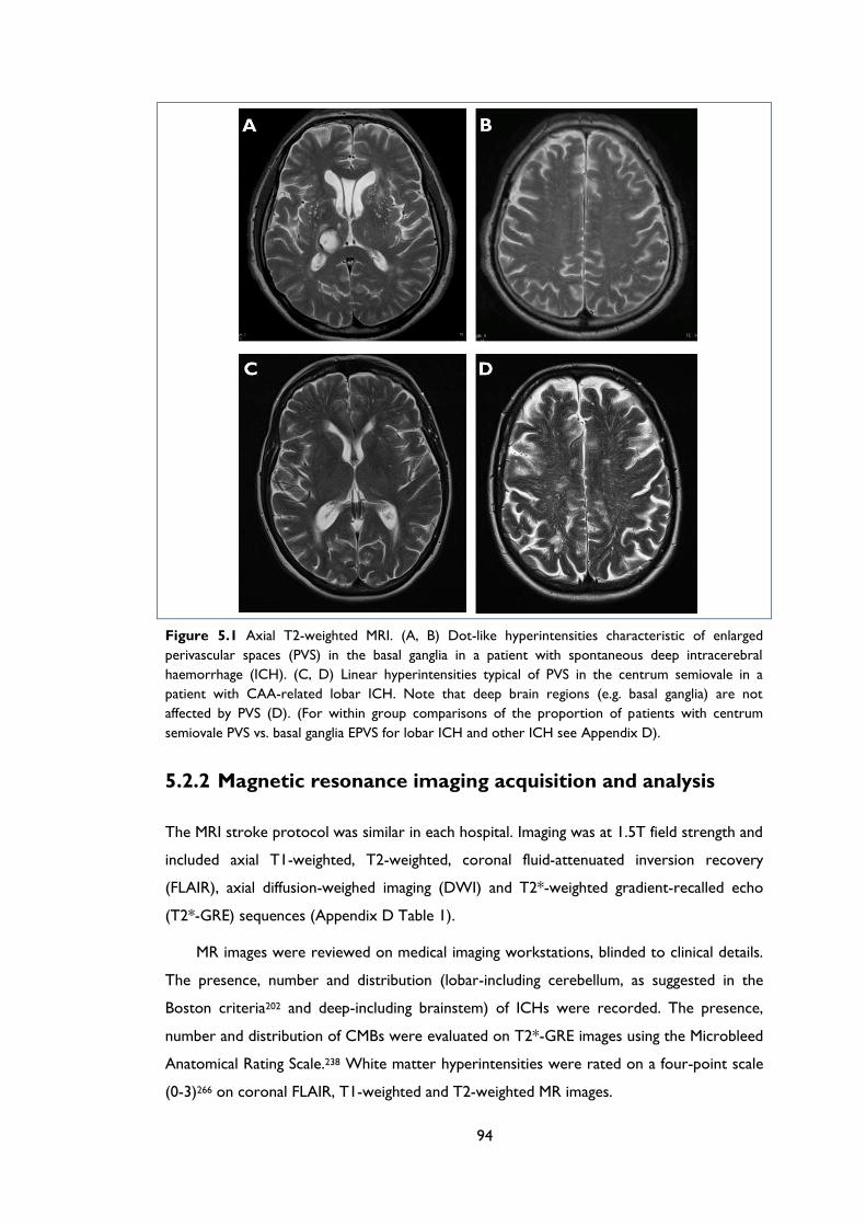
94
Figure 5.1 Axial T2-weighted MRI. (A, B) Dot-like hyperintensities characteristic of enlarged
perivascular spaces (PVS) in the basal ganglia in a patient with spontaneous deep intracerebral
haemorrhage (ICH). (C, D) Linear hyperintensities typical of PVS in the centrum semiovale in a
patient with CAA-related lobar ICH. Note that deep brain regions (e.g. basal ganglia) are not
affected by PVS (D). (For within group comparisons of the proportion of patients with centrum
semiovale PVS vs. basal ganglia EPVS for lobar ICH and other ICH see Appendix D).
5.2.2 Magnetic resonance imaging acquisition and analysis
The MRI stroke protocol was similar in each hospital. Imaging was at 1.5T field strength and
included axial T1-weighted, T2-weighted, coronal fluid-attenuated inversion recovery
(FLAIR), axial diffusion-weighed imaging (DWI) and T2*-weighted gradient-recalled echo
(T2*-GRE) sequences (Appendix D Table 1).
MR images were reviewed on medical imaging workstations, blinded to clinical details.
The presence, number and distribution (lobar-including cerebellum, as suggested in the
Boston criteria202 and deep-including brainstem) of ICHs were recorded. The presence,
number and distribution of CMBs were evaluated on T2*-GRE images using the Microbleed
Anatomical Rating Scale.238 White matter hyperintensities were rated on a four-point scale
(0-3)266 on coronal FLAIR, T1-weighted and T2-weighted MR images.
95
PVS were rated on axial T2-weighted MR images by a trained observer, using a
validated visual rating scale.30, 267 PVS were defined as small, sharply delineated structures of
CSF intensity (or close to CSF intensity), measuring <3mm following the course of
perforating or medullary vessels (Figure 5.1). Lacunes were distinguished form PVS by their
larger size (>3mm), spheroid shape and surrounding hyperintensity on FLAIR. PVS were
rated in basal ganglia and centrum semiovale regions. For both of these anatomical areas,
the PVS rating categories applied were: 0=no PVS, 1=<10 PVS, 2=11-20 PVS, 3=21-40 PVS
and 4=>40 PVS. The numbers refer to PVS on one side of the brain: after reviewing all
relevant slices for the anatomical area being assessed, the slice and side with the highest
number of PVS was recorded.30, 267 In patients with other ICH, basal ganglia PVS were
evaluated contralateral of the lesion if deep ICH was present. The assessment of PVS may
have been influenced by the presence of confluent white matter changes; in such cases, as
per guidance provided for the rating scale we used,30 estimation was made of the closest
PVS rating category, using the appearance of non-involved white matter, and cortical gray
matter. Similarly, in cases of large lobar ICH, centrum semiovale PVS were assessed
contralateral to the index ICH lesion; an estimation of the closest centrum semiovale PVS
rating category ipsilateral to the lesion was made, and the highest number of PVS was
recorded. Intra-rater reliability testing (n=30 scans) showed an excellent intra-rater
Cohen’s kappa value of 0.91 for basal ganglia PVS and 0.82 for centrum semiovale PVS.
5.2.3 Statistical analysis
We compared clinical and imaging characteristics of patients with strictly lobar ICH
(attributed to CAA according to the Boston criteria) to those with other ICH, using chi-
square tests and the Fisher’s exact test for categorical variables, and two-sample t-tests or
Mann-Whitney U-tests depending on the distribution of continuous variables. To test our
hypothesis, we sought to compare the prevalence of severe centrum semiovale and basal
ganglia PVS (>40 PVS) in the two ICH groups. However, since only one patient had severe
basal ganglia PVS, in a post-hoc analysis we compared frequent-to-severe basal ganglia PVS
(>20) in the two ICH groups. Both crude and age-adjusted severe PVS centrum semiovale
prevalence rates are reported. For the logistic regression analysis, severity of PVS was
trichotomized due to the small number of patients with no PVS or >40 PVS [1 (mild; 1 to
10), 2 (moderate; 11-20) and 3 (severe; over 21)]. Ordinal logistic regression analysis was
used to identify predictors of increasing PVS severity in the two anatomical areas (centrum
semiovale and basal ganglia) and for total brain PVS. Multivariable analysis was adjusted for
age, lobar microbleeds, deep microbleeds, white matter changes score and lacunes, based
96
on the results of univariable analysis, plus other biologically plausible confounders, especially
neuroimaging markers of small vessel disease. As a sensitivity analysis we repeated these
analyses with the addition of other variables in our model (including sex, hypertension,
CAA-group etc.). Using a likelihood ratio test, the assumption of proportional odds for the
ordinal logistic regression models was met. A p-value ≤0.05 was considered to be
statistically significant. All statistical analyses were carried out using STATA data analysis
and statistical software (Version 11.2, StataCorp.).
5.3 Results
Patients with spontaneous ICH excluded from the study were not significantly different
from those included in measures of ICH severity including: median (IQR range) ICH volume
(10.5 cm3; 2.93–18.81 cm3 versus 12.84 cm3; 4.05–25.31 cm3; p=0.288), median (IQR range)
Glasgow Coma Score on admission (14; 11–15 versus 15; 14–15; p=0.087) in patients
excluded versus patients included respectively. Patients excluded had a higher prevalence of
deep ICH (50.5% versus 27.4%, p=0.005). The final cohort consisted of 121 patients with
ICH [mean age 69.4 (95%CI: 67.2–69.6); 69 (57%) male]; 76 (63%) had strictly lobar ICH,
fulfilling the Boston criteria for probable (n=53) or possible (n=23). The clinical and
radiological characteristics of the ICH cohort overall and according to ICH location are
reported in Table 5.1. The median time from ICH to MRI was 10 days (interquartile range:
3-61 days).
5.3.1 Prevalence and distribution of PVS in ICH
Almost all patients had some degree of PVS (Table 5.1); the mean total score was 4.38
(95%CI: 4.18-4.58).
Patients with strictly lobar ICH had a significantly higher prevalence of severe PVS (>40
PVS) in the centrum semiovale compared to other ICH patients (35.5% vs. 17.8%,
respectively; p=0.041) (Figure 5.2). The age-adjusted prevalences of severe PVS in the
centrum semiovale are 29.3% (95% CI: 23.1% to 35.5%) in the strictly lobar ICH group vs.
13.5% (95% CI: 9.3% to 17.8%) in the “other ICH” group (p<0.05).
It was not possible to test for differences between the strictly lobar and other ICH
groups in the prevalence of severe basal ganglia PVS since only one patient had severe basal
ganglia PVS. However, in a post-hoc analysis the proportion of patients with frequent-to-
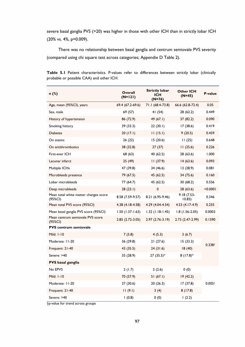
97
severe basal ganglia PVS (>20) was higher in those with other ICH than in strictly lobar ICH
(20% vs. 4%, p=0.009).
There was no relationship between basal ganglia and centrum semiovale PVS severity
(compared using chi square test across categories; Appendix D Table 2).
Table 5.1 Patient characteristics. P-values refer to differences between strictly lobar (clinically
probable or possible CAA) and other ICH.
n (%) Overall
(N=121)
Strictly lobar
ICH
(N=76)
Other ICH
(N=45) P-value
Age, mean (95%CI), years 69.4 (67.2-69.6) 71.1 (68.4-73.8) 66.6 (62.8-72.4) 0.05
Sex, male 69 (57) 41 (54) 28 (62.2) 0.449
History of hypertension 86 (72.9) 49 (67.1) 37 (82.2) 0.090
Smoking history 39 (33.3) 22 (30.1) 17 (38.6) 0.419
Diabetes 20 (17.1) 11 (15.1) 9 (20.5) 0.459
On statins 26 (22) 15 (20.6) 11 (25) 0.648
On antithrombotics 38 (32.8) 27 (37) 11 (25.6) 0.226
First-ever ICH 68 (63) 40 (62.5) 28 (63.6) 1.000
Lacunar infarct 25 (49) 11 (37.9) 14 (63.6) 0.093
Multiple ICHs 47 (39.8) 34 (46.6) 13 (28.9) 0.081
Microbleeds presence 79 (67.5) 45 (62.5) 34 (75.6) 0.160
Lobar microbleeds 77 (64.7) 45 (62.5) 30 (68.2) 0.556
Deep microbleeds 28 (23.1) 0 28 (63.6) <0.0001
Mean total white matter changes score (95%CI)
8.58 (7.59-9.57) 8.21 (6.95-9.46) 9.18 (7.53-
10.83) 0.346
Mean total PVS score (95%CI) 4.38 (4.18-4.58) 4.29 (4.04-4.54) 4.53 (4.17-4.9) 0.255
Mean basal ganglia PVS score (95%CI) 1.50 (1.37-1.63) 1.32 (1.18-1.45) 1.8 (1.56-2.05) 0.0003
Mean centrum semiovale PVS score
(95%CI) 2.85 (2.72-3.05) 2.97 (2.76-3.19) 2.73 (2.47-2.99) 0.1590
PVS centrum semiovale
Mild: 1-10 7 (5.8) 4 (5.3) 3 (6.7)
0.338† Moderate: 11-20 36 (29.8) 21 (27.6) 15 (33.3)
Frequent: 21-40 43 (35.5) 24 (31.6) 18 (40)
Severe: >40 35 (28.9) 27 (35.5)* 8 (17.8)*
PVS basal ganglia
No EPVS 2 (1.7) 2 (2.6) 0 (0)
0.005†
Mild: 1-10 70 (57.9) 51 (67.1) 19 (42.2)
Moderate: 11-20 37 (30.6) 20 (26.3) 17 (37.8)
Frequent: 21-40 11 (9.1) 3 (4) 8 (17.8)
Severe: >40 1 (0.8) 0 (0) 1 (2.2)
†p-value for trend across groups
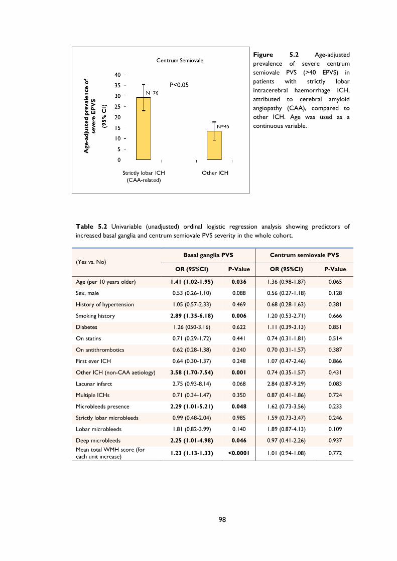
98
Figure 5.2 Age-adjusted
prevalence of severe centrum
semiovale PVS (>40 EPVS) in
patients with strictly lobar
intracerebral haemorrhage ICH,
attributed to cerebral amyloid
angiopathy (CAA), compared to
other ICH. Age was used as a
continuous variable.
Table 5.2 Univariable (unadjusted) ordinal logistic regression analysis showing predictors of
increased basal ganglia and centrum semiovale PVS severity in the whole cohort.
(Yes vs. No) Basal ganglia PVS Centrum semiovale PVS
OR (95%CI) P-Value OR (95%CI) P-Value
Age (per 10 years older) 1.41 (1.02-1.95) 0.036 1.36 (0.98-1.87) 0.065
Sex, male 0.53 (0.26-1.10) 0.088 0.56 (0.27-1.18) 0.128
History of hypertension 1.05 (0.57-2.33) 0.469 0.68 (0.28-1.63) 0.381
Smoking history 2.89 (1.35-6.18) 0.006 1.20 (0.53-2.71) 0.666
Diabetes 1.26 (050-3.16) 0.622 1.11 (0.39-3.13) 0.851
On statins 0.71 (0.29-1.72) 0.441 0.74 (0.31-1.81) 0.514
On antithrombotics 0.62 (0.28-1.38) 0.240 0.70 (0.31-1.57) 0.387
First ever ICH 0.64 (0.30-1.37) 0.248 1.07 (0.47-2.46) 0.866
Other ICH (non-CAA aetiology) 3.58 (1.70-7.54) 0.001 0.74 (0.35-1.57) 0.431
Lacunar infarct 2.75 (0.93-8.14) 0.068 2.84 (0.87-9.29) 0.083
Multiple ICHs 0.71 (0.34-1.47) 0.350 0.87 (0.41-1.86) 0.724
Microbleeds presence 2.29 (1.01-5.21) 0.048 1.62 (0.73-3.56) 0.233
Strictly lobar microbleeds 0.99 (0.48-2.04) 0.985 1.59 (0.73-3.47) 0.246
Lobar microbleeds 1.81 (0.82-3.99) 0.140 1.89 (0.87-4.13) 0.109
Deep microbleeds 2.25 (1.01-4.98) 0.046 0.97 (0.41-2.26) 0.937
Mean total WMH score (for
each unit increase) 1.23 (1.13-1.33) <0.0001 1.01 (0.94-1.08) 0.772
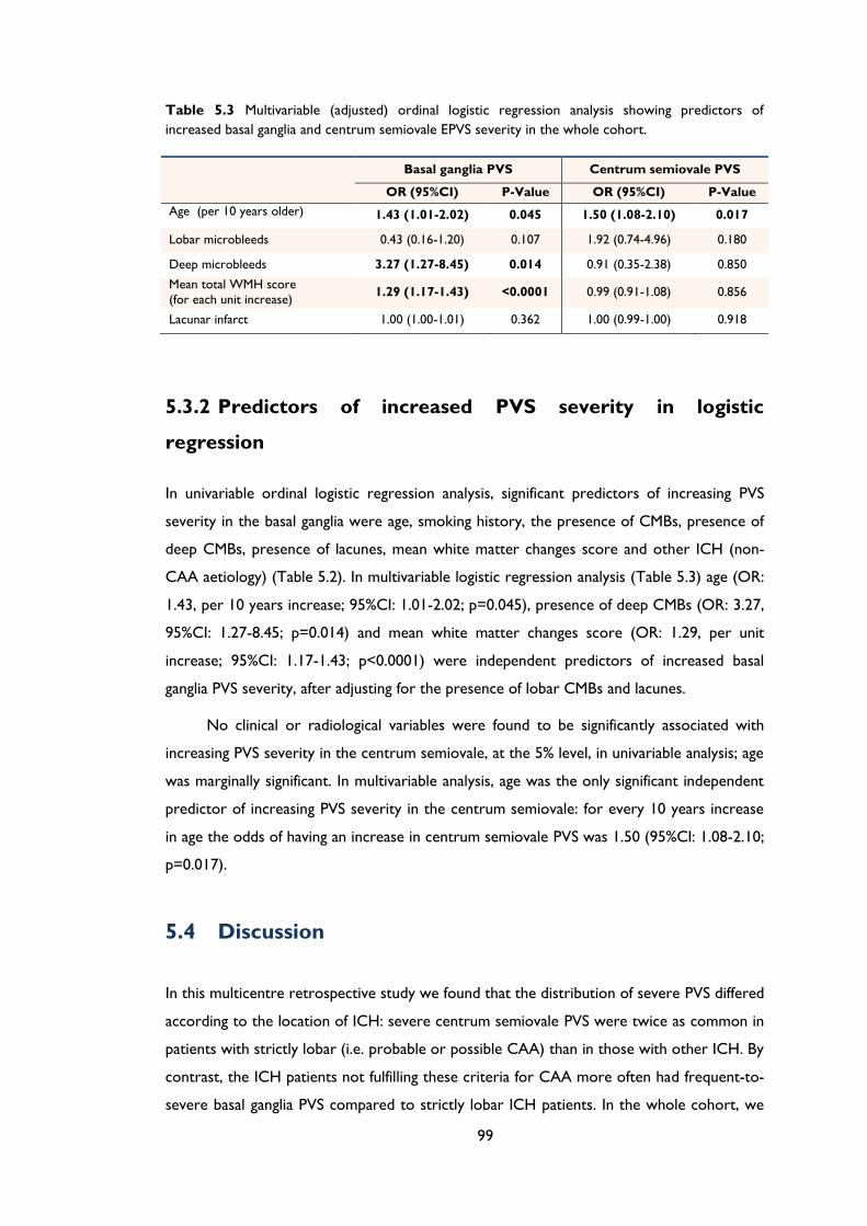
99
Table 5.3 Multivariable (adjusted) ordinal logistic regression analysis showing predictors of
increased basal ganglia and centrum semiovale EPVS severity in the whole cohort.
Basal ganglia PVS Centrum semiovale PVS
OR (95%CI) P-Value OR (95%CI) P-Value
Age (per 10 years older) 1.43 (1.01-2.02) 0.045 1.50 (1.08-2.10) 0.017
Lobar microbleeds 0.43 (0.16-1.20) 0.107 1.92 (0.74-4.96) 0.180
Deep microbleeds 3.27 (1.27-8.45) 0.014 0.91 (0.35-2.38) 0.850
Mean total WMH score (for each unit increase)
1.29 (1.17-1.43) <0.0001 0.99 (0.91-1.08) 0.856
Lacunar infarct 1.00 (1.00-1.01) 0.362 1.00 (0.99-1.00) 0.918
5.3.2 Predictors of increased PVS severity in logistic
regression
In univariable ordinal logistic regression analysis, significant predictors of increasing PVS
severity in the basal ganglia were age, smoking history, the presence of CMBs, presence of
deep CMBs, presence of lacunes, mean white matter changes score and other ICH (non-
CAA aetiology) (Table 5.2). In multivariable logistic regression analysis (Table 5.3) age (OR:
1.43, per 10 years increase; 95%CI: 1.01-2.02; p=0.045), presence of deep CMBs (OR: 3.27,
95%CI: 1.27-8.45; p=0.014) and mean white matter changes score (OR: 1.29, per unit
increase; 95%CI: 1.17-1.43; p<0.0001) were independent predictors of increased basal
ganglia PVS severity, after adjusting for the presence of lobar CMBs and lacunes.
No clinical or radiological variables were found to be significantly associated with
increasing PVS severity in the centrum semiovale, at the 5% level, in univariable analysis; age
was marginally significant. In multivariable analysis, age was the only significant independent
predictor of increasing PVS severity in the centrum semiovale: for every 10 years increase
in age the odds of having an increase in centrum semiovale PVS was 1.50 (95%CI: 1.08-2.10;
p=0.017).
5.4 Discussion
In this multicentre retrospective study we found that the distribution of severe PVS differed
according to the location of ICH: severe centrum semiovale PVS were twice as common in
patients with strictly lobar (i.e. probable or possible CAA) than in those with other ICH. By
contrast, the ICH patients not fulfilling these criteria for CAA more often had frequent-to-
severe basal ganglia PVS compared to strictly lobar ICH patients. In the whole cohort, we
100
found that age, white matter changes and deep CMBs were independently associated with
basal ganglia PVS severity, but only age was independently associated with increased
centrum semiovale PVS severity. Our study thus provides new evidence that the
distribution of severe PVS reflects the underlying arteriopathy type in spontaneous ICH.
We found PVS in almost every participant in our study, and moderate to severe
centrum semiovale and basal ganglia PVS in more than 90% and 40% of them respectively.
To our knowledge, no previous imaging study has investigated the prevalence and
distribution of PVS in spontaneous ICH. A recent prospective study which evaluated PVS in
patients with acute lacunar stroke (n=129), cortical stroke (n=124) and age-matched non-
stroke controls (n=97), found that PVS were more prevalent in small vessel (lacunar)
ischaemic stroke than large vessel stroke.30 In this study, Doubal and colleagues30 used the
same PVS rating scale, applied to T2-weighted images, as in our study, thus allowing
comparisons to be made. The mean total PVS score in our ICH cohort (4.38, 95% CI: 4.18-
4.58) is close to that of patients with lacunar stroke (3.81, SD: 1.76) but higher than non-
stroke controls (1.02, SD: 0.89).30 However, the absence of a non-ICH control group in our
study makes it difficult to directly compare PVS counts with other patient groups.
Although PVS have not been studied in ICH cohorts, previous pathological and
neuroimaging studies in other patient populations have shown a significant association
between hypertension, white matter changes, and PVS.24, 268 The associations we found
between the severity of basal ganglia and total PVS with the severity of white matter
changes further add to evidence that PVS are an imaging manifestation of cerebral small
vessel disease rather than an incidental finding. The association of PVS with white matter
changes seems consistent across different populations including vascular dementia,269
Alzheimer’s disease and mild cognitive impairment. A large population-based study also
reported an association between the degree of PVS and the volume of white matter
changes, as well as with the prevalence of lacunes.270
We also found that PVS associations vary according to their distribution, suggesting
different pathophysiological mechanisms relevant to the underlying arteriopathy in ICH.
Basal ganglia PVS were independently associated with age, white matter changes burden and
deep CMBs, suggesting hypertensive arteriopathy as a possible underlying mechanism. It is
likely that at least some white matter changes share a common pathophysiological
mechanism with PVS, perhaps related to blood-brain barrier disruption, a potentially key
pathogenic process in cerebral small vessel disease.133 Indeed, it has been suggested that
white matter changes tend to form around PVS133 and that PVS may be an early imaging
marker of small vessel disease and blood-brain barrier alteration. By contrast, in the
101
present study, PVS in the centrum semiovale were only associated with age, and not with
other imaging or clinical factors. In the study by Doubal et al30 in patients with minor
ischaemic stroke, total PVS were also associated with white matter changes after adjusting
for age, the presence of stroke and vascular risk factors (p<0.001); centrum semiovale PVS
were not associated with any explanatory variables, in keeping with our findings.30 It is thus
possible that a distinct pathophysiological process other than hypertensive arteriopathy
contributes to the development of centrum semiovale PVS: CAA is a likely candidate, a
hypothesis consistent with evidence that advancing age is the strongest clinical risk factor
for sporadic CAA.28 Although we did not find a significant association between PVS severity
in the centrum semiovale and the presence of lobar microbleeds (a putative neuroimaging
marker of CAA) at the 5% level, the odds ratio suggests a possible effect (OR: 1.92; 95%CI:
0.74-4.96; Table 3), our ability to detect any association may be limited by our sample size
and the high prevalence of hypertension, which can also contribute to lobar CMBs. In
addition, the absence of association between basal ganglia and centrum semiovale PVS
severity further supports the notion that PVS in these two anatomical compartments are
related to different pathophysiological processes.
Severe PVS in our ICH cohort showed a different anatomical pattern depending on the
location and the presumed small vessel arteriopathy underlying ICH. In patients with strictly
lobar, presumed CAA-related ICH the centrum semiovale was more often affected by
severe PVS compared to other ICH patients. A predilection of severe PVS for the centrum
semiovale most parallels the topography of CAA pathology, since amyloid-β is deposited
primarily in cortical and leptomeningeal small vessels, largely sparing small vessels in the
basal ganglia.28 Moreover, several lines of evidence suggest that perivascular spaces have a
key role in the pathophysiology of CAA.28 It is hypothesized that an important mechanism
underlying vascular amyloid-β deposition is impaired clearance of amyloid-β along
perivascular drainage pathways.28 As amyloid-β is deposited in the walls of small arteries it
could block perivascular drainage, leading to dilation of perivascular spaces downstream in
the underlying deep white matter, even in regions not directly affected by CAA pathology.
This regional preference for severe PVS further supports our hypothesis that severe
centrum semiovale PVS are at least partly related to CAA, whereas basal ganglia PVS are
most likely related to hypertensive arteriopathy affecting deep perforators. Severe centrum
semiovale PVS may therefore be a potential useful new neuroimaging marker of CAA, in an
appropriate clinical context. The diagnosis of CAA currently depends on the radiological
demonstration of multiple strictly lobar haemorrhages; existing criteria have high specificity
but limited sensitivity.202, 203 Whether severe PVS presence and topography might usefully
102
improve the sensitivity for current diagnostic criteria for CAA202 requires further study
with pathological verification of CAA.
Our multicentre study has several strengths including the systematic evaluation of MRI
scans by trained raters using validated scales for a range of imaging markers of small vessel
disease, and the use of a validated PVS rating scale.30 A potential limitation is the lack of
pathological confirmation of the CAA pathology as a cause of ICH, in view of the imperfect
sensitivity and specificity of the Boston criteria (particularly their overall sensitivity and the
specificity of the “possible CAA” category).202 We may thus have misclassified some
patients as CAA-related ICH when their ICH was related to hypertensive arteriopathy. It is
also likely that some of the patients with “other ICH” (e.g. spontaneous deep ICH), many
of whom had lobar CMBs, not classified as CAA-related might in fact have some degree of
CAA pathology as well as hypertensive arteriopathy. However, any misclassification of
patients would tend to bias toward a null result (no between-group differences in PVS
severity or distribution), suggesting that PVS may in fact be a more powerful marker for
CAA than we have been able to demonstrate here. It was not possible to blind the PVS
rater to ICH location, potentially introducing a bias towards the study hypothesis. We also
acknowledge the potential of bias in our sample as MRIs were done as part of routine
clinical care and many patients were excluded from this analysis because of inadequate MRI
data. Finally, due to the retrospective and cross-sectional nature of our study we were not
able to determine the prognostic significance of PVS in this cohort.
In summary, we found that PVS are present in almost all patients with spontaneous
ICH, but with evidence for different underlying mechanisms according to their distribution.
Basal ganglia PVS severity is associated with markers of hypertensive arteriopathy including
white matter changes and deep CMBs, whilst centrum semiovale PVS are associated with
age and are more often severe in patients with strictly lobar ICH attributed to CAA. A
predilection of severe PVS for the centrum semiovale rather than the basal ganglia may
therefore be secondary to, and indicative of, CAA. Further studies with pathological
verification are needed to determine the relevance of PVS for improving the sensitivity of
CAA diagnostic criteria. Longitudinal studies are required to explore whether PVS are an
early feature of the small vessel disease underlying ICH, their change over time, and
prognostic implications for clinical outcome.267, 271
103
Chapter 6 MRI-visible perivascular spaces
as a neuroimaging marker of cerebral
amyloid angiopathy: MRI-histopathological
study
104
ABSTRACT
Objective: We investigated whether severe MRI-visible Perivascular Spaces (PVS) in the
cerebral hemisphere white matter (centrum semiovale) are more common in patients with
pathology-proven CAA vs. those with pathology-proven non-CAA related intracerebral
haemorrhage (ICH).
Methods: Using a validated 4-point scale on axial T2-weighted MRI, we compared PVS in
patients with pathology-proven CAA to those with spontaneous ICH but no
histopathological evidence of CAA. In a preliminary analysis restricted to patients with T2*-
weighted gradient-recalled echo MRI, we also investigated whether including severe
centrum semiovale PVS increases the sensitivity of existing diagnostic criteria for probable
CAA.
Results: Fourteen CAA patients and 10 non-CAA related ICH patients were included.
Eight of the CAA patients were admitted for symptomatic spontaneous lobar ICH, one
because of ischaemic stroke, one with transient focal neurological episodes, and four due to
cognitive decline. Severe (>20) centrum semiovale PVS were more frequent in patients with
CAA compared to controls (12/14 [85.7%; 95%CI: 57.2-98.2%] vs. 0/10 [one-sided 95%CI:
0-30.8%], p<0.0005); this was robust to adjustment for age. The original Boston criteria for
probable CAA showed a sensitivity of 76.9% (95% CI: 46.2-95%), which increased to 92.3%
(95% CI: 64-99.8%), without loss of specificity, after including severe centrum semiovale
PVS.
Conclusions: Severe centrum semiovale PVS on MRI may be a promising new
neuroimaging marker for the in vivo diagnosis of CAA. However, our findings are
preliminary and require confirmation and external validation in larger cohorts of pathology-
proven CAA.
105
6.1 Introduction
Sporadic cerebral amyloid angiopathy (CAA),2 caused by progressive vascular amyloid-β
deposition in small cortical and leptomeningeal vessels, is a major and increasingly frequent
cause of lobar intracerebral haemorrhage (ICH) and cognitive impairment in older
patients.28, 37, 242 CAA is associated with neuroimaging markers including lobar ICH and
strictly lobar cerebral microbleeds, which allow its clinical-radiological diagnosis using the
Boston criteria.28, 202, 203 However, these criteria have limited sensitivity in some studies;
new non-invasive diagnostic markers of CAA are therefore needed, particularly to identify
early disease.28, 242
Recently, perivascular spaces (PVS) have emerged as a potential MRI marker of the
presence and severity of cerebral small vessel disease.30, 31, 272-275 Perivascular spaces are
interstitial fluid-filled cavities surrounding small penetrating vessels, which function to allow
interstitial fluid and solute efflux from the brain.129, 263, 276 CAA almost invariably
accompanies Alzheimer’s disease (AD), and in this setting vascular amyloid-β is associated
with enlargement of perivascular spaces.38, 128, 132, 277 However, whether PVS are associated
with sporadic CAA is not known. We hypothesized that progressive amyloid-β deposition
in CAA impairs interstitial fluid drainage, causing MRI-visible severe PVS in the cerebral
hemisphere white matter (centrum semiovale). To test this hypothesis, we compared the
prevalence of severe centrum semiovale PVS in patients with pathology-proven sporadic
CAA compared to patients with pathology-proven non-CAA related spontaneous ICH. In a
preliminary analysis, we also investigated whether including severe centrum semiovale PVS
increases the sensitivity of existing diagnostic criteria for CAA.202
6.2 Methods
6.2.1 Study participants and data collection
We included all eligible patients from four stroke centres identified retrospectively by a
systematic keyword search of pathology reports (Appendix E Figure 1). Cases were defined
as subjects with both pathology-proven CAA (from routinely collected brain biopsy, biopsy
at haematoma evacuation or autopsy) and adequate T2-weighted MRI sequences. Controls
were defined as subjects with spontaneous symptomatic ICH, no pathological evidence of
CAA, and adequate T2-weighted MRI.
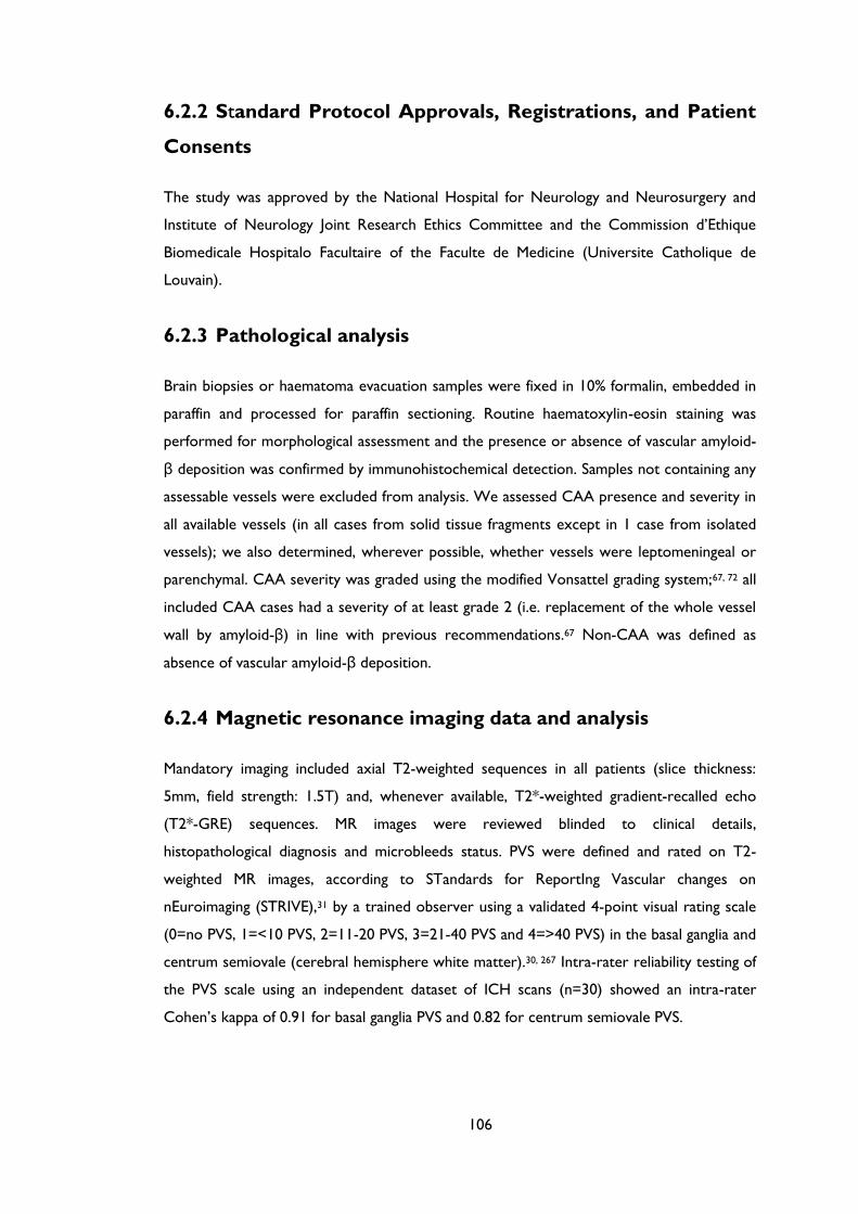
106
6.2.2 Standard Protocol Approvals, Registrations, and Patient
Consents
The study was approved by the National Hospital for Neurology and Neurosurgery and
Institute of Neurology Joint Research Ethics Committee and the Commission d’Ethique
Biomedicale Hospitalo Facultaire of the Faculte de Medicine (Universite Catholique de
Louvain).
6.2.3 Pathological analysis
Brain biopsies or haematoma evacuation samples were fixed in 10% formalin, embedded in
paraffin and processed for paraffin sectioning. Routine haematoxylin-eosin staining was
performed for morphological assessment and the presence or absence of vascular amyloid-
β deposition was confirmed by immunohistochemical detection. Samples not containing any
assessable vessels were excluded from analysis. We assessed CAA presence and severity in
all available vessels (in all cases from solid tissue fragments except in 1 case from isolated
vessels); we also determined, wherever possible, whether vessels were leptomeningeal or
parenchymal. CAA severity was graded using the modified Vonsattel grading system;67, 72 all
included CAA cases had a severity of at least grade 2 (i.e. replacement of the whole vessel
wall by amyloid-β) in line with previous recommendations.67 Non-CAA was defined as
absence of vascular amyloid-β deposition.
6.2.4 Magnetic resonance imaging data and analysis
Mandatory imaging included axial T2-weighted sequences in all patients (slice thickness:
5mm, field strength: 1.5T) and, whenever available, T2*-weighted gradient-recalled echo
(T2*-GRE) sequences. MR images were reviewed blinded to clinical details,
histopathological diagnosis and microbleeds status. PVS were defined and rated on T2-
weighted MR images, according to STandards for ReportIng Vascular changes on
nEuroimaging (STRIVE),31 by a trained observer using a validated 4-point visual rating scale
(0=no PVS, 1=<10 PVS, 2=11-20 PVS, 3=21-40 PVS and 4=>40 PVS) in the basal ganglia and
centrum semiovale (cerebral hemisphere white matter).30, 267 Intra-rater reliability testing of
the PVS scale using an independent dataset of ICH scans (n=30) showed an intra-rater
Cohen’s kappa of 0.91 for basal ganglia PVS and 0.82 for centrum semiovale PVS.
107
6.2.5 Statistics
We compared clinical characteristics between the patients with vs. without CAA on
neuropathology, using the Fisher’s exact test for categorical variables, and Mann-Whitney
U-test for age (not normally distributed continuous variable). We pre-specified a definition
of severe centrum semiovale PVS as >20 (Grade 3). Although arbitrary, this definition is in
line with the most severe category of white matter PVS used in a previous study (and found
to relate to vascular risk factors),270 and provided two numerically-balanced categories
consistent with our aim of increasing the sensitivity of CAA diagnosis. We compared both
the crude and age-standardized prevalence of severe PVS between groups using Fisher’s
exact test. We calculated the sensitivity and specificity after the addition of severe centrum
semiovale PVS to the original Boston diagnostic criteria for CAA (including strictly lobar
cerebral microbleeds)202 in line with Standards for Reporting of Diagnostic Accuracy
(STARD) guidelines.278 The histopathological information was not taken into account for
the classification. The analysis for clinically probable CAA was restricted to patients with
T2*-GRE MRI sequences available. Significance level was set at 0.05 for all analyses.
Statistical analyses were carried out using STATA (Version 11.2, StataCorp.).
6.3 Results
Fourteen CAA patients and 10 non-CAA ICH patients were included (Table 6.1) out of 56
screened; 30 patients were excluded because of no MRI available, 1 non-CAA patient
because of no evidence of ICH on MRI, and another non-CAA patient in whom the tissue
sample contained no assessable vessels. Eight of the CAA patients were admitted with
symptomatic spontaneous lobar ICH, one with ischaemic stroke, one with transient focal
neurological episodes, and four with cognitive decline (one of these also had radiological
evidence of a previous ICH). All non-CAA patients were admitted with symptomatic
spontaneous ICH. Twenty-three of the 24 available pathological samples contained at least
2 assessable parenchymal or leptomeningeal vessels. There were 23 solid tissue samples
including vessels, and one sample including only isolated vessels. All 14 CAA cases
contained >10 assessable vessels; 12 cases with both cortical and leptomeningeal, and 2
with only leptomeningeal vessels. Of the 10 non-CAA cases, 4 contained >10 assessable
vessels, while the other 6 cases contained a median number of 2 assessable vessels
(Interquartile Range: 2-3); 1 case contained both cortical and leptomeningeal, 5 only
cortical, and 3 only leptomeningeal vessels; in the case containing only isolated vessels, their
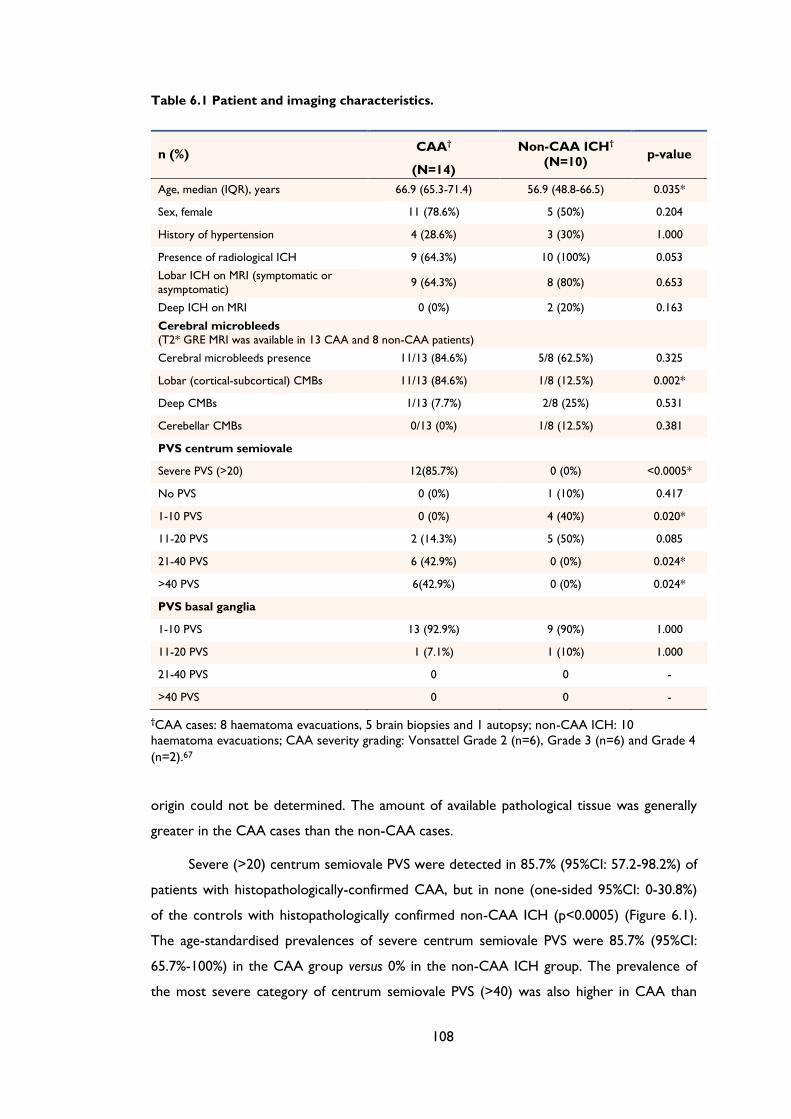
108
Table 6.1 Patient and imaging characteristics.
n (%) CAA†
(N=14)
Non-CAA ICH†
(N=10) p-value
Age, median (IQR), years 66.9 (65.3-71.4) 56.9 (48.8-66.5) 0.035*
Sex, female 11 (78.6%) 5 (50%) 0.204
History of hypertension 4 (28.6%) 3 (30%) 1.000
Presence of radiological ICH 9 (64.3%) 10 (100%) 0.053
Lobar ICH on MRI (symptomatic or
asymptomatic) 9 (64.3%) 8 (80%) 0.653
Deep ICH on MRI 0 (0%) 2 (20%) 0.163
Cerebral microbleeds
(T2* GRE MRI was available in 13 CAA and 8 non-CAA patients)
Cerebral microbleeds presence 11/13 (84.6%) 5/8 (62.5%) 0.325
Lobar (cortical-subcortical) CMBs 11/13 (84.6%) 1/8 (12.5%) 0.002*
Deep CMBs 1/13 (7.7%) 2/8 (25%) 0.531
Cerebellar CMBs 0/13 (0%) 1/8 (12.5%) 0.381
PVS centrum semiovale
Severe PVS (>20) 12(85.7%) 0 (0%) <0.0005*
No PVS 0 (0%) 1 (10%) 0.417
1-10 PVS 0 (0%) 4 (40%) 0.020*
11-20 PVS 2 (14.3%) 5 (50%) 0.085
21-40 PVS 6 (42.9%) 0 (0%) 0.024*
>40 PVS 6(42.9%) 0 (0%) 0.024*
PVS basal ganglia
1-10 PVS 13 (92.9%) 9 (90%) 1.000
11-20 PVS 1 (7.1%) 1 (10%) 1.000
21-40 PVS 0 0 -
>40 PVS 0 0 -
†CAA cases: 8 haematoma evacuations, 5 brain biopsies and 1 autopsy; non-CAA ICH: 10
haematoma evacuations; CAA severity grading: Vonsattel Grade 2 (n=6), Grade 3 (n=6) and Grade 4
(n=2).67
origin could not be determined. The amount of available pathological tissue was generally
greater in the CAA cases than the non-CAA cases.
Severe (>20) centrum semiovale PVS were detected in 85.7% (95%CI: 57.2-98.2%) of
patients with histopathologically-confirmed CAA, but in none (one-sided 95%CI: 0-30.8%)
of the controls with histopathologically confirmed non-CAA ICH (p<0.0005) (Figure 6.1).
The age-standardised prevalences of severe centrum semiovale PVS were 85.7% (95%CI:
65.7%-100%) in the CAA group versus 0% in the non-CAA ICH group. The prevalence of
the most severe category of centrum semiovale PVS (>40) was also higher in CAA than
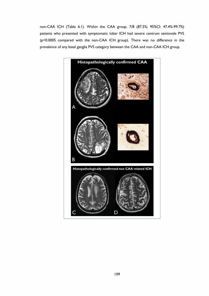
109
non-CAA ICH (Table 6.1). Within the CAA group, 7/8 (87.5%; 95%CI: 47.4%-99.7%)
patients who presented with symptomatic lobar ICH had severe centrum semiovale PVS
(p<0.0005 compared with the non-CAA ICH group). There was no difference in the
prevalence of any basal ganglia PVS category between the CAA and non-CAA ICH group.

110
Figure 6.1 MRI-visible white matter perivascular spaces in CAA and non-CAA ICH. Perivascular
spaces (PVS) are defined as small, sharply delineated structures of (or close to) cerebrospinal fluid
intensity, measuring <3mm following the course of perforating or medullary vessels, in basal ganglia
and centrum semiovale. In (A) and (B) the left panels show dot-like hyperintensities (florid PVS) on
representative axial T2-weighted MRI at 1.5 Tesla in the centrum semiovale, in patients with
histopathologically confirmed cerebral amyloid angiopathy (CAA); the right panels show the
corresponding pathology (immunostaining for Aβ) with dense amyloid deposition spanning the
entire vessel wall of a cortical (A, right panel) and a leptomeningeal arteriole (B, right panel). (C) and
(D) show axial T2-weighted MRI from two of the non-CAA cases, showing a paucity of centrum
semiovale PVS.

111
Table 6.2 Diagnosis of cerebral angiopathy (CAA) using the original Boston criteria (including
strictly lobar microbleeds) with and without the inclusion of severe centrum semiovale PVS (>20
PVS). Only cases with T2*-GRE were included in this analysis (n=21). Inclusion of severe centrum
semiovale PVS resulted in the diagnostic upgrading of three patients: two from possible to probable
CAA and one from non-CAA to possible CAA.
Clinical Diagnosis
Original Boston criteria With inclusion of severe
centrum semiovale PVS
n Pathologic diagnosis
n Pathologic diagnosis
CAA (+) CAA (-) CAA (+) CAA (-)
Probable CAA 11 10 1 13 12 1
Non-probable CAA 10 3 7 8 1 7
-Possible CAA 4 2 2 3 1 2
-Non CAA 6 1 5 5 0 5
Total 21 13 8 21 13 8
T2*-GRE MRI was available in 13 CAA and 8 non-CAA ICH patients. Adding severe
centrum semiovale PVS (>20 PVS) to the original Boston criteria for CAA resulted in the
diagnostic upgrading of three patients (Table 6.2). The original Boston criteria showed a
sensitivity of 92.3% (95% CI: 64-99.8%) for possible or probable CAA diagnosis, while
inclusion of severe centrum semiovale PVS increased their sensitivity to 100% (95% CI:
75.3-100%), without loss of specificity (62.5%; 95% CI: 24.5-91.5%). For probable CAA the
original Boston criteria had a sensitivity of 76.9% (95% CI: 46.2-95%); inclusion of severe
centrum semiovale PVS increased this to 92.3% (95% CI: 64-99.8%). The specificity for
probable CAA was 87.5% (95% CI 47.3–99.7%) for both criteria.
6.4 Discussion
We have shown that severe PVS in the centrum semiovale white matter are more common
in subjects with pathology-proven sporadic CAA than control non-CAA subjects. These
findings suggest that severe centrum semiovale PVS, in this patient population, may be a
promising new neuroimaging marker to improve the sensitivity of in vivo diagnosis of CAA.
However, the sensitivity and specificity of severe centrum semiovale PVS require external
validation, ideally in a larger independent cohort.
Perivascular spaces are a key route for the drainage of interstitial fluid and solutes
(including soluble amyloid-β) from the brain. Progressive amyloid-β deposition in small
cortical and leptomeningeal arteries in CAA could gradually impair perivascular drainage,
112
causing retrograde dilation of perivascular spaces in the underlying white matter of the
centrum semiovale, either by blocking bulk flow, or by diminishing the pulsatility of small
vessels (required for efficient interstitial fluid drainage) due to smooth muscle cell loss.263,
279 Impaired perivascular drainage could then further exacerbate vascular and perivascular
amyloid-β deposition creating a “feed-forward loop”.274, 276 Consistent with this hypothesis,
a post-mortem study of AD found that amyloid-β blocks perivascular drainage pathways in
CAA associated with AD, and the degree of enlargement of white matter perivascular
spaces correlates with CAA severity.132 Moreover, a recent MRI study in ICH patients
reported that very severe centrum semiovale PVS were associated with CAA defined
according to clinical-radiological criteria.272 However, more studies are needed to
definitively determine the pathological basis of MRI-visible PVS.
We found no increased prevalence of basal ganglia PVS in CAA, consistent with deep
perforating arteries being largely spared by amyloid-β deposition.37 Previous studies report
that neuroimaging markers of hypertensive arteriopathy (lacunes, deep cerebral
microbleeds and white matter changes) are more strongly linked to basal ganglia than
centrum semiovale PVS, also suggesting that the distribution of PVS may reflect the
underlying arteriopathy.30, 272, 274, 275 Since lobar ICHs are more likely to be surgically
evacuated, our sample of non-CAA ICH is biased towards lobar ICH, which may contribute
to the lack of severe basal ganglia PVS in the non-CAA group. However, non-CAA ICH
cases more often had milder (1-10) centrum semiovale PVS compared to CAA cases.
Limitations of our study include the small cohort and selection bias due to the
requirement for T2-weighted MRI. Because we included routinely collected haematoma
evacuation samples, which may be suboptimal in comparison to post-mortem examinations,
it was not possible to definitively assess in all patients whether vessels were parenchymal or
leptomeningeal. Due to limited tissue sampling it is possible that some non-CAA ICH
patients might have had undetected CAA. Moreover, the amount of available tissue (and
number of assessable cortical and leptomeningeal vessels) was generally higher in the CAA
group, which could have led to a higher chance of sampling error in the non-CAA group.
However, misclassification and small sample size would tend to bias toward a null result.
Although CAA patients were older than non-CAA patients, the difference in severe
centrum semiovale PVS was robust to adjustment for age; furthermore, the CAA group had
a higher prevalence of PVS only in the centrum semiovale, while confounding by age would
be expected to increase PVS severity in both the centrum semiovale and basal ganglia.270, 272,
274 Finally, although the PVS raters were blinded to clinical and histopathological information
as well as microbleeds status, it was impossible to blind them to ICH presence.

113
Our findings are preliminary and hypothesis-generating, requiring external validation
in larger independent cohorts with pathological verification of CAA. Nevertheless, if
confirmed, PVS in the centrum semiovale may help improve the sensitivity of MRI for in vivo
diagnosis of sporadic CAA in an appropriate clinical context. Further studies are also
needed to determine whether centrum semiovale PVS precede other imaging features of
CAA280, which might help identify CAA earlier in its natural history.

114
Chapter 7 MRI-visible white matter
perivascular spaces: a marker of
cerebrovascular amyloid burden?
115
ABSTRACT
Objective: To investigate the relationship between MRI-visible centrum semiovale
perivascular spaces (CSO-PVS), a biomarker of impaired interstitial fluid drainage, and PET-
based amyloid-β burden across a wide range of cerebrovascular amyloid deposition, we
analyzed a sample merging healthy subjects of variable age, a fraction of whom is expected
to harbor significant cerebrovascular amyloid, and patients diagnosed with probable
cerebral amyloid angiopathy (CAA).
Methods: Thirty-one non-demented subjects (11 patients with probable CAA, 10 age-
matched healthy subjects >60yrs, and 10 healthy subjects <60yrs), had brain MRI and PiB-
PET. CSO-PVS was evaluated on axial T2-weighted sequences using a validated 4-point
scale and further classified as high (score>3) or low (score≤3) degree for pre-specified
analyses. Linear regression was used to assess the association between whole cortex PiB
and CSO-PVS.
Results: In multivariable linear regression adjusting for age, white matter changes and
microbleeds count, whole cortex PiB binding was associated with CSO-PVS degree both as
continuous variable (coefficient: 0.11; CI: 0.01-0.22, P=0.040) and as dichotomous variable
(coefficient: 0.27; 95% CI: 0.11-0.44, p=0.002). In addition, the median PiB retention was
higher in high compared to low CSO-PVS degree (median1.42; IQR: 1.37-1.50 vs. 1.14; IQR:
1.12-1.23, p=0.0007), an association also present within the healthy subjects group
separately (p=0.023). All results remained consistent in sensitivity analyses excluding
healthy subjects <60 years.
Conclusion: These results suggest a strong association between PiB uptake and CSO-PVS,
which has important pathophysiological implications for both cerebrovascular amyloid
deposition and CSO-PVS, but also highlights the possible utility of the latter as a surrogate
for cerebrovascular amyloid burden.
116
7.1 Introduction
Perivascular (or Virchow-Robin) spaces are interstitial fluid-filled cavities surrounding small
penetrating vessels,281 which function to allow interstitial fluid and solute efflux from the
brain.129, 263, 276 Accumulating evidence suggests that MRI-visible perivascular spaces in the
centrum semiovale (CSO-PVS) and basal ganglia (BG-PVS) are potential neuroimaging
markers of cerebral small vessel disease.30, 31, 272-275 PVS are also very common in healthy
elderly subjects,264, 270 in whom CSO-PVS are a risk factor for dementia.271 CSO-PVS on
MRI are also attracting increasing attention as a promising indicator of cerebrovascular
amyloid deposition, a common age-related neuropathological process characterised by
progressive amyloid-β accumulation within the walls of leptomeningeal and cortical small
arteries, arterioles and occasionally capillaries.28, 37
Recent studies emphasize not only that cerebrovascular amyloid deposition is widely
prevalent in the aged population, affecting up to 30-40% of healthy elderly,58, 174 but also
that it has become a major cause of spontaneous lobar intracerebral haemorrhage (ICH)28,
37, can also cause or contribute significantly to cognitive impairment,58, 155 and is almost
invariably present in Alzheimer’s disease.65 CSO-PVS is important to consider in this
context as it might reflect impaired interstitial fluid drainage from the white matter to the
cortical surface and in turn contribute to cerebrovascular amyloid deposition.276 Indeed,
emerging evidence suggests that cerebrovascular amyloid deposition is a “protein
elimination failure angiopathy”:282 as perivascular drainage pathways fail with age (or are
overloaded by reduced capacity of other elimination mechanisms), amyloid-β is increasingly
trapped in the perivascular compartment and deposited in the walls of small arteries.276
However, the precise relationship and mechanisms between MRI-visible CSO-PVS and
cerebrovascular amyloid deposition has not been explored in vivo.
11C-Pittsburgh compound B (PiB) has been developed as a positron emission
tomography (PET) radioligand for imaging both parenchymal and vascular fibrillar amyloid-β
deposits, and has revolutionized research into potential pathomechanisms of
cerebrovascular amyloid deposition and Alzheimer’s disease.48-50, 283-285 If MRI-visible CSO-
PVS and perivascular amyloid-β deposition are indeed linked, one would expect cortical PiB
retention to be associated with the overall CSO-PVS burden. If confirmed, this would in
turn further strengthen the potential of CSO-PVS as a surrogate for vascular amyloid
burden. To investigate this relationship across as wide a range of cerebrovascular amyloid
burden and CSO-PVS as possible, we analysed in an MRI-PET pilot study a well-sized sample
purposely merging patients diagnosed with probable cerebral amyloid angiopathy (CAA)
117
and healthy subjects across the adult age span, a fraction of whom is expected based on
established neuropathological knowledge58 to harbour some degree of both
cerebrovascular amyloid and CSO-PVS.264, 270
7.2 Subjects and Methods
7.2.1 Study subjects
Non-demented lobar ICH patients fulfilling the original Boston criteria for probable CAA195
were recruited through the Addenbrooke’s Hospital Stroke Unit or ICH clinic.
Consecutive eligible patients were approached to take part in the study. Admission records
and clinic files were screened for discharged potentially eligible patients who were then
contacted. All included CAA patients lived independently and their Mini-mental State
Examination (MMSE) was >23 in all. Age at PiB PET, gender and presence of vascular risk
factors were ascertained by medical records review and by patient and caregiver interview.
Medication-free healthy subjects with no memory or cognitive complaints and normal
MMSE results (≥29) were also enrolled through advertisements posted at the University of
Third Age and hospital inboard. The absence of memory or cognitive complaint was
specifically verified, and to avoid any recruitment bias the participants were informed that
they would not be given any results from their scans (MR or PET) unless an unexpected
lesion was found. The healthy sample comprised both a group of healthy elderly individuals
≥60 years age-matched to the CAA group and a group of healthy adults <60 years of age.
All subjects underwent a brain 11C-PiB PET scan and structural MRI on the same day.
7.2.2 PET
The methodology used for 11C-PiB production and PET scanning in our centre has
previously been described.286 Briefly, PET scanning was performed on a GE Advance PET
scanner. Following a 68Ge/68Ga transmission scan (15 minutes) for attenuation correction,
~550 MBq of 11C-PiB was injected as a bolus through an antecubital vein, and the 90 min
dynamic 11C-PIB-PET scan was reconstructed using the PROMIS 3D filtered backprojection
algorithm.287
The PET data analysis was completely automated and carried out by a physicist blinded
to the results of the MRI (YTH). To facilitate segmentation, spatial normalization and
delineation of anatomical regions of interest (ROIs), participants also underwent a
118
structural T1-weighted MRI scan on a 3T Siemens Tim-Trio (Siemens Medical Solutions,
Erlangen, Germany), using a 3-dimensional MPRAGE scan (magnetization-prepared rapid
gradient-echo sequence; repetition time/echo time/inversion time=2300 milliseconds/2.98
milliseconds/900 milliseconds; flip angle, 9°; 1 average; 176 slices; 256256 matrix size;
111 mm voxel size). To allow the use of standard space ROIs, the MRI, and thence the
co-registered PET images, was spatially normalised to the MNI/ICBM152 T1-weighted
template using the symmetric image normalisation method (SyN).288
As previously described,289 ROIs were defined on these spatially normalized images as
the intersection between ≥65% thresholded grey matter segments and Automated
Anatomic Labeling (AAL) ROIs. ROIs overlapping with areas affected by the ICH were
excluded a priori. In order to mitigate the partial volume effects of cortical atrophy, the CSF
probability map for each subject, smoothed to match the PET spatial resolution, was used
to correct each voxel time-activity curve (TAC) for CSF fraction (fCSF) through division by
(1-fCSF).290, 291 For each ROI, distribution volume ratio (DVR) was estimated from the mean
CSF-corrected TAC using the reference tissue Logan graphical method fitted over the 35-
90min post-injection interval, with the cerebellum as the reference tissue.292 Whole cortex
DVR was obtained through volume-weighted averaging of the cortical ROI DVR values
across both hemispheres.
7.2.3 MRI data and analysis
On the same day as the PiB-PET study, each subject underwent detailed structural MRI at
3T (slice thickness: 4mm, slice gap: 5mm), which included T2-weighted, T1-weighted MP-
RAGE, FLAIR and T2*-GRE, using standard sequences. MR images were reviewed blinded
to clinical and PET data. PVS were defined and rated on axial T2-weighted MR images,
according to STandards for ReportIng Vascular changes on nEuroimaging (STRIVE),31 by a
trained observer using a validated 4-point visual rating scale (0=no PVS, 1=<10 PVS, 2=11-
20 PVS, 3=21-40 PVS and 4=>40 PVS) in the basal ganglia (BG) and CSO.30, 272, 293 The
numbers refer to PVS on one side of the brain: after reviewing all relevant slices for the
anatomical area being assessed, the slice and side with the highest number of PVS was
recorded. In cases of large lobar ICH, centrum semiovale PVS were assessed contralateral
to the index ICH lesion; an estimation of the closest category ipsilateral to the lesion was
made, and the highest severity was recorded. Intra-rater reliability testing of the PVS scale
using an independent dataset of ICH scans (n=30) showed an intra-rater Cohen’s kappa of
0.91 for BG-PVS and 0.82 for CSO-PVS. White matter hyperintensities of presumed
vascular origin were assessed using the age-related white matter changes (ARWMC)
119
scale.294 CMBs were evaluated on T2*-GRE (echo time=20 ms) images according to the
Microbleeds Anatomic Rating Scale (MARS) and published guidelines.35, 238
7.2.4 Standard Protocol Approvals, Registrations, and Patient
Consents
The study received ethical approval by the Cambridgeshire Regional Ethics Committee and
all participants provided signed informed consent.
7.2.5 Statistical analysis
We compared basic demographic, clinical and imaging characteristics of CAA patients
versus healthy subjects and age-matched elderly healthy subjects using appropriate
univariable tests: chi-square test or Fisher’s exact test for categorical variables, and Mann-
Whitney U test for continuous variables. The relationship between whole cortex DVR and
CSO-PVS degree, both included as continuous variables, was explored across the whole
sample using Kendall's non-parametric correlation analysis. Linear regression analyses were
then used to quantify the relationship between PiB DVR and CSO-PVS grade after
adjustment for other biologically plausible covariates found to be related to both PiB
retention and PVS in previous studies, including age, white matter hyperintensities total
score and lobar CMBs count.284, 295 We repeated the regression analysis using the CSO-PVS
degree dichotomised into high (score >3) or low (score ≤3) (i.e. >40 CSO-PVS vs. ≤40
CSO-PVS respectively). This definition was pre-specified and is in line with the most severe
category of CSO-PVS which might be more characteristic and sensitive for CAA.270 This
cut-off separated our data into two balanced groups. As a sensitivity analysis, we repeated
the above linear regression models excluding healthy subjects <60 years (i.e. using only
probable CAA patients and age-matched healthy elderly cases).
We also compared PiB retention (i.e. whole cortex DVR) between subjects with high
vs. low CSO-PVS degree within the whole study population, and then separately within the
CAA patients and the healthy group.
All tests of significance were 2-tailed, with values below the 10% level considered
indicative of a trend. Statistical analyses were carried out using STATA (Version 12.1,
StataCorp.).
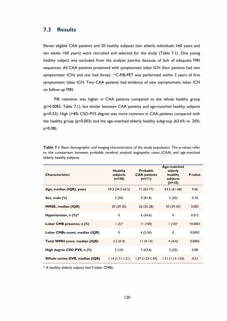
120
7.3 Results
Eleven eligible CAA patients and 20 healthy subjects (ten elderly individuals ≥60 years and
ten adults <60 years) were recruited and selected for the study (Table 7.1). One young
healthy subject was excluded from the analysis post-hoc because of lack of adequate MRI
sequences. All CAA patients presented with symptomatic lobar ICH (four patients had two
symptomatic ICHs and one had three). 11C-PiB-PET was performed within 3 years of first
symptomatic lobar ICH. Two CAA patients had evidence of new asymptomatic lobar ICH
on follow-up MRI.
PiB retention was higher in CAA patients compared to the whole healthy group
(p=0.0082; Table 7.1), but similar between CAA patients and age-matched healthy subjects
(p=0.53). High (>40) CSO-PVS degree was more common in CAA patients compared with
the healthy group (p=0.003) and the age-matched elderly healthy subgroup (63.6% vs. 20%;
p=0.08).
Table 7.1 Basic demographic and imaging characteristics of the study population. The p-values refer
to the comparison between probable cerebral amyloid angiopathy cases (CAA) and age-matched
elderly healthy subjects
Characteristics
Healthy
subjects
(n=20)
Probable
CAA patients
(n=11)
Age-matched
elderly
healthy
subjects
(n=10)
P-value
Age, median (IQR), years 59.5 (34.5-63.5) 71 (63-77) 63.5 (61-68) 0.06
Sex, male (%) 5 (50) 9 (81.8) 5 (50) 0.18
MMSE, median (IQR) 30 (29-30) 26 (25-28) 30 (29-30) 0.003
Hypertension, n (%)* 0 6 (54.6) 0 0.012
Lobar CMB presence, n (%) 1 (5)* 11 (100) 1 (10)* <0.0001
Lobar CMBs count, median (IQR) 0 4 (2-50) 0 0.0002
Total WMH score, median (IQR) 2.5 (0-4) 11 (9-14) 4 (4-6) 0.0002
High degree CSO-PVS, n (%) 2 (10) 7 (63.6) 2 (20) 0.08
Whole cortex DVR, median (IQR) 1.14 (1.11-1.21) 1.37 (1.23-1.43) 1.21 (1.13-1.65) 0.53
* A healthy elderly subject had 3 lobar CMBs.
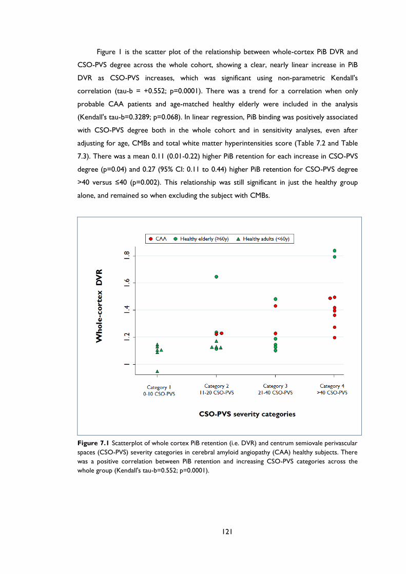
121
Figure 1 is the scatter plot of the relationship between whole-cortex PiB DVR and
CSO-PVS degree across the whole cohort, showing a clear, nearly linear increase in PiB
DVR as CSO-PVS increases, which was significant using non-parametric Kendall's
correlation (tau-b = +0.552; p=0.0001). There was a trend for a correlation when only
probable CAA patients and age-matched healthy elderly were included in the analysis
(Kendall's tau-b=0.3289; p=0.068). In linear regression, PiB binding was positively associated
with CSO-PVS degree both in the whole cohort and in sensitivity analyses, even after
adjusting for age, CMBs and total white matter hyperintensities score (Table 7.2 and Table
7.3). There was a mean 0.11 (0.01-0.22) higher PiB retention for each increase in CSO-PVS
degree (p=0.04) and 0.27 (95% CI: 0.11 to 0.44) higher PiB retention for CSO-PVS degree
>40 versus ≤40 (p=0.002). This relationship was still significant in just the healthy group
alone, and remained so when excluding the subject with CMBs.
Figure 7.1 Scatterplot of whole cortex PiB retention (i.e. DVR) and centrum semiovale perivascular
spaces (CSO-PVS) severity categories in cerebral amyloid angiopathy (CAA) healthy subjects. There
was a positive correlation between PiB retention and increasing CSO-PVS categories across the
whole group (Kendall's tau-b=0.552; p=0.0001).
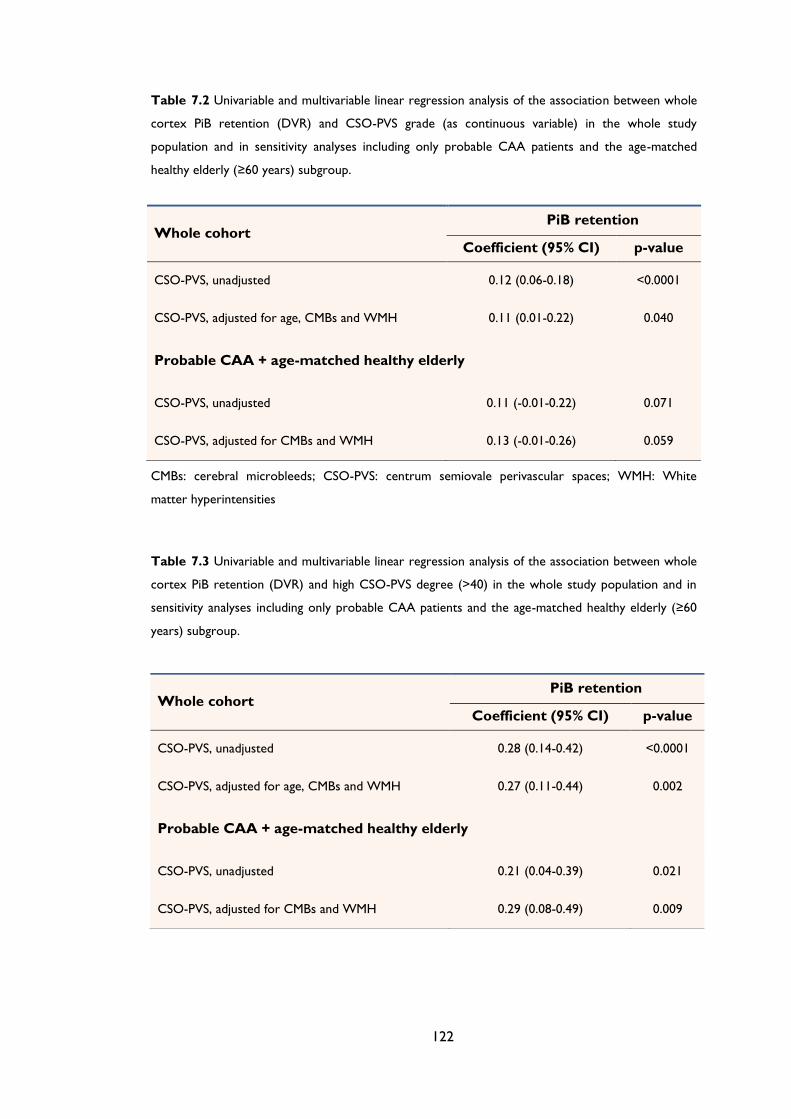
122
Table 7.2 Univariable and multivariable linear regression analysis of the association between whole
cortex PiB retention (DVR) and CSO-PVS grade (as continuous variable) in the whole study
population and in sensitivity analyses including only probable CAA patients and the age-matched
healthy elderly (≥60 years) subgroup.
CMBs: cerebral microbleeds; CSO-PVS: centrum semiovale perivascular spaces; WMH: White
matter hyperintensities
Table 7.3 Univariable and multivariable linear regression analysis of the association between whole
cortex PiB retention (DVR) and high CSO-PVS degree (>40) in the whole study population and in
sensitivity analyses including only probable CAA patients and the age-matched healthy elderly (≥60
years) subgroup.
Whole cohort PiB retention
Coefficient (95% CI) p-value
CSO-PVS, unadjusted 0.12 (0.06-0.18) <0.0001
CSO-PVS, adjusted for age, CMBs and WMH 0.11 (0.01-0.22) 0.040
Probable CAA + age-matched healthy elderly
CSO-PVS, unadjusted 0.11 (-0.01-0.22) 0.071
CSO-PVS, adjusted for CMBs and WMH 0.13 (-0.01-0.26) 0.059
Whole cohort PiB retention
Coefficient (95% CI) p-value
CSO-PVS, unadjusted 0.28 (0.14-0.42) <0.0001
CSO-PVS, adjusted for age, CMBs and WMH 0.27 (0.11-0.44) 0.002
Probable CAA + age-matched healthy elderly
CSO-PVS, unadjusted 0.21 (0.04-0.39) 0.021
CSO-PVS, adjusted for CMBs and WMH 0.29 (0.08-0.49) 0.009
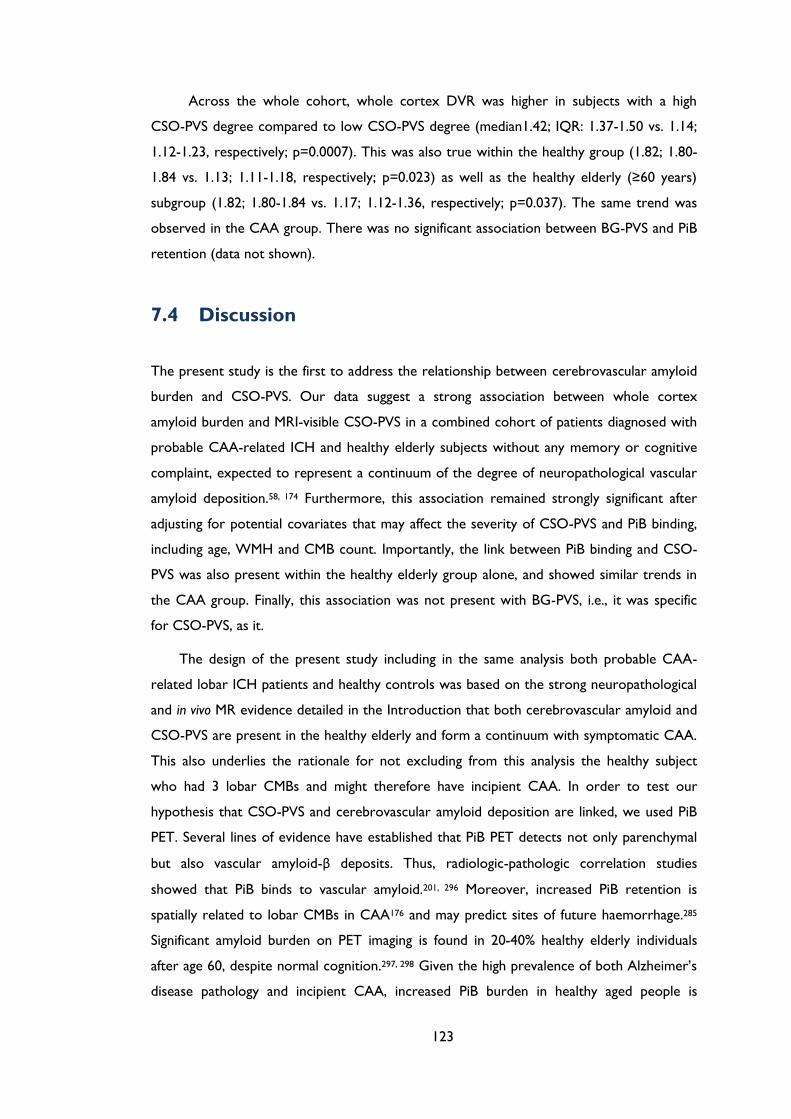
123
Across the whole cohort, whole cortex DVR was higher in subjects with a high
CSO-PVS degree compared to low CSO-PVS degree (median1.42; IQR: 1.37-1.50 vs. 1.14;
1.12-1.23, respectively; p=0.0007). This was also true within the healthy group (1.82; 1.80-
1.84 vs. 1.13; 1.11-1.18, respectively; p=0.023) as well as the healthy elderly (≥60 years)
subgroup (1.82; 1.80-1.84 vs. 1.17; 1.12-1.36, respectively; p=0.037). The same trend was
observed in the CAA group. There was no significant association between BG-PVS and PiB
retention (data not shown).
7.4 Discussion
The present study is the first to address the relationship between cerebrovascular amyloid
burden and CSO-PVS. Our data suggest a strong association between whole cortex
amyloid burden and MRI-visible CSO-PVS in a combined cohort of patients diagnosed with
probable CAA-related ICH and healthy elderly subjects without any memory or cognitive
complaint, expected to represent a continuum of the degree of neuropathological vascular
amyloid deposition.58, 174 Furthermore, this association remained strongly significant after
adjusting for potential covariates that may affect the severity of CSO-PVS and PiB binding,
including age, WMH and CMB count. Importantly, the link between PiB binding and CSO-
PVS was also present within the healthy elderly group alone, and showed similar trends in
the CAA group. Finally, this association was not present with BG-PVS, i.e., it was specific
for CSO-PVS, as it.
The design of the present study including in the same analysis both probable CAA-
related lobar ICH patients and healthy controls was based on the strong neuropathological
and in vivo MR evidence detailed in the Introduction that both cerebrovascular amyloid and
CSO-PVS are present in the healthy elderly and form a continuum with symptomatic CAA.
This also underlies the rationale for not excluding from this analysis the healthy subject
who had 3 lobar CMBs and might therefore have incipient CAA. In order to test our
hypothesis that CSO-PVS and cerebrovascular amyloid deposition are linked, we used PiB
PET. Several lines of evidence have established that PiB PET detects not only parenchymal
but also vascular amyloid-β deposits. Thus, radiologic-pathologic correlation studies
showed that PiB binds to vascular amyloid.201, 296 Moreover, increased PiB retention is
spatially related to lobar CMBs in CAA176 and may predict sites of future haemorrhage.285
Significant amyloid burden on PET imaging is found in 20-40% healthy elderly individuals
after age 60, despite normal cognition.297, 298 Given the high prevalence of both Alzheimer’s
disease pathology and incipient CAA, increased PiB burden in healthy aged people is
124
thought to reflect not only very early Alzheimer’s disease-related parenchymal
accumulation of amyloid-β but also underlying asymptomatic cerebrovascular amyloid.58, 174
Accordingly, in a recent study in asymptomatic older adults, PiB uptake was strongly
associated with lobar CMBs, a putative marker of CAA, independent from APOE ε4 status,
vascular risk factors, and antiplatelet therapy.295 All these factors may explain why in the
present sample there was no significant difference in whole cortex PiB retention or regional
ratios between CAA patients and healthy subjects (see Baron et al289 for further details and
a discussion on this). The rationale of the present study, namely a continuum in
cerebrovascular amyloid across healthy participants and probable CAA patients, was
strongly supported by the significant relationship between CSO-PVS degree and PiB uptake
found within the healthy control group alone. In other words, the significant findings in the
merged sample do not merely reflect the higher CSO-PVS burden in the CAA subset.
Although the mechanisms of MRI-visible CSO-PVS remain poorly understood, our
current findings highlight a potential pathophysiologic link between amyloid burden and
CSO-PVS. This link may reflect interstitial fluid drainage impairment within the perivascular
spaces by progressive vascular amyloid-β deposition, causing retrograde dilation of
perivascular spaces in the underlying white matter of the centrum semiovale.263, 279 Impaired
perivascular drainage could then further exacerbate vascular and perivascular amyloid-β
deposition in small cortical and leptomeningeal vessels, setting in motion a “feed-forward
loop”, by which cumulative amyloid deposition promotes further cerebrovascular amyloid
deposition.274, 276, 299 Consistent with this hypothesis, a post-mortem study of Alzheimer’s
disease found that amyloid-β blocks perivascular drainage pathways in cerebrovascular
amyloid deposition associated with Alzheimer’s disease pathology; the degree of white
matter perivascular spaces enlargement correlated with cerebrovascular amyloid burden.132
Moreover, a recent MRI study in spontaneous ICH patients reported that high grade CSO-
PVS were strongly associated with CAA defined according to clinical-radiological criteria.272
Similar associations were reported among subjects from a memory clinic, in which CSO-
PVS was associated with cerebrovascular amyloid markers, whereas BG-PVS were
indicative of hypertensive arteriopathy.274 Our findings are also consistent with the
hypothesis of BG-PVS have different pathophysiology to CSO-PVS and deep perforating
arteries being largely spared by amyloid-β deposition.37
Strong points of our study include the systematic MRI evaluation for a range of imaging
markers of cerebral small vessel disease, which allowed testing a pre-specified hypothesis,
and the fact that MR was obtained on the same day as PET. The main limitation, intrinsic to
amyloid imaging, is that incipient Alzheimer’s disease pathology might also be present in
non-demented ICH patients suspected of CAA, particularly given the frequent co-
125
occurrence and overlapping molecular mechanisms of these two amyloid-related
conditions, but also in healthy elderly. Another limitation is the relatively small sample size
limiting statistical power. For this reason our study should be considered preliminary and
the p-values should be interpreted cautiously, particularly within the subgroups. As our
study was cross-sectional, we were unable to determine the cause–effect relationship
between increased PiB retention and CSO-PVS.
Although preliminary, our results suggest that amyloid-β burden in associated with
CSO-PVS degree, and have implications for understanding cerebrovascular amyloid
pathophysiology. Furthermore, our work further supports the idea of CSO-PVS being a
useful marker of vascular amyloid burden in future studies of both clinically healthy subjects
and symptomatic CAA cohorts, as well as disease-modifying trials.300 Larger prospective
cohorts are needed to validate our findings and clarify exactly how exactly CSO-PVS are
related to amyloid deposition, impaired interstitial fluid drainage as well as other markers of
small vessels disease in CAA.
126
Chapter 8 White matter perivascular
spaces on MRI are related to cortical
superficial siderosis in sporadic cerebral
amyloid angiopathy
127
ABSTRACT
Objectives: We set out to investigate whether MRI-visible centrum semiovale perivascular
spaces (CSO-PVS) - a potential biomarker of impaired interstitial fluid drainage in sporadic
cerebral amyloid angiopathy (CAA) - is associated with cortical superficial siderosis (cSS),
reflecting recurrent haemorrhage from severe leptomeningeal and superficial cortical
vascular amyloid.
Methods: Retrospective multicentre cohort study of possible/probable CAA according to
the Boston criteria. PVS were rated in basal ganglia (BG-PVS) and CSO (CSO-PVS) on axial
T2-weighted sequences, using a validated 4-point visual rating scale, and were classified as
high (score >2) or low degree (score ≤2) for pre-specified analyses. Independent risk
factors for high CSO-PVS degree were investigated in logistic regression.
Results: The final cohort consisted of 138 CAA patients (mean age: 71.8; 95%CI: 70.2-73.4
years, 52.2% male). High CSO-PVS degree was present in 61.2% of cases. The prevalence of
any cSS, and disseminated cSS (involving >3 sulci), was higher in patients with high vs. low
CSO-PVS degree (for any cSS 45.9% vs. 13.5%; p<0.00005; for disseminated cSS 31.8% vs.
0%; p<0.00005). In multivariable logistic regression analysis, cSS presence (OR: 4.78; 95%CI:
1.64-13.87; p=0.004) was an independent predictors of high CSO-PVS degree. We found no
associations between BG-PVS and cSS.
Conclusions: High degree of CSO-PVS is highly prevalent in sporadic CAA, and is related
to cSS. Our findings suggest that severe leptomeningeal and cortical vascular amyloid
(causing cSS) is related to impaired interstitial fluid drainage from cerebral white matter,
although determining the causal direction of this relationship requires prospective studies.
128
8.1 Introduction
Sporadic cerebral amyloid angiopathy (CAA) is a common small vessel disease that results
from progressive amyloid–β deposition within the walls of small cortical and leptomeningeal
arteries.28, 242 CAA is most often recognised in life by symptomatic, lobar intracerebral
haemorrhage (ICH) and cognitive impairment in elderly patients.28, 37, 242 CAA is also
associated with characteristic magnetic resonance imaging (MRI) markers including strictly
lobar cerebral microbleeds (CMBs) and white matter hyperintensities.28, 35 These imaging
markers might be suggestive of CAA even in asymptomatic elderly individuals.
Accumulating evidence suggests that cortical superficial siderosis (cSS)171, 191, 194, 195 and
MRI-visible centrum semiovale (i.e. cerebral hemisphere white matter) perivascular spaces
(CSO-PVS) are new imaging markers of CAA, which may reflect distinct but related aspects
of pathophysiology. However, the precise relationship between CSO-PVS and other
imaging manifestations of CAA, including cSS and lobar CMBs has not been explored.
We hypothesised that progressive amyloid-β deposition in small leptomeningeal or
superficial cortical vessels (leading to fragility, repeated haemorrhage into the subarachnoid
space and cSS) blocks perivascular drainage pathways from white matter to the cortical
surface, resulting in CSO-PVS. To test this hypothesis and to gain insights into potential
mechanisms we investigated the relationship between MRI-visible CSO-PVS and cSS in
patients with possible or probable CAA according to the Boston criteria.
8.2 Methods
8.2.1 Study population and data collection
Consecutive CAA patients(according to the original Boston criteria202 – i.e. not including
cSS as a criterion) from an ongoing multicentre cohort study at four stroke centres over
defined time periods as previously described,301, 302
were evaluated. The centres were:
University College London Hospitals NHS Foundation Trust (London) (03/2003–09/2011),
Addenbrooke’s Hospital (Cambridge) (07/2002–03/2010), Cliniques Universitaires Saint Luc
(Brussels) (12/2003–04/2010) and CHU Mont-Godinne UCL (Brussels) (08/2005–03/2009).
At participating centres, MRI is a routine investigation for cases of suspected CAA, unless
there are contra-indications. Essential inclusion criteria for the multicentre CAA cohort301,
302 and this analysis were: (1) CAA, defined according to the Boston criteria202 – including
lobar CMBs, but not cSS; and (2) available MRI sequences of adequate quality including T2-
129
weighted, T2*-weighted gradient-recalled echo (T2*-GRE) and FLAIR MRI sequences. The
selection process of this multicentre cohort has been previously described:10, 11 in brief,
from a total of 358 patients with suspected CAA/spontaneous ICH screened, 144 were
initially excluded because of MRI was not performed or was not available/interpretable.
After reviewing available MRI and clinical data, 56 patients not fulfilling the Boston criteria
for CAA were further excluded, leaving a total of 158 CAA patients potentially eligible for
the current analysis:302 59 patients from University College London Hospitals, 49 from
Addenbrooke’s Hospital, 36 from Cliniques Universitaires Saint Luc and 14 patients from
CHU Mont-Godinne UCL. Patients with no T2 MRI (n=14), T2 MRI of insufficient quality
(n=1) and irretrievable sequences (n=5) where further excluded from this analysis.
Demographic and clinical information was obtained from prospective databases and by
medical records review using standardised data collection forms, as previously described.301,
302 Hypertension was defined as a history of hypertension, taking antihypertensive
treatment or documented elevated blood pressure (systolic >150 or diastolic >95mmHg)
before admission, diabetes as ongoing use of a hypoglycaemic agent and smoking as history
of tobacco use before admission.
The study received ethical approval by the National Hospital for Neurology and
Neurosurgery and Institute of Neurology Joint Research Ethics Committee, the
Commission d’Ethique Biomedicale Hospitalo Facultaire of the Faculte de Medicine
(Université Catholique de Louvain), and the Comite d’ethique medicale of the CHU Dinant-
Godinne (Université Catholique de Louvain).
8.2.2 Neuroimaging data and analysis
The MRI protocols were similar in each hospital. Imaging was performed at field strength of
1.5T for all patients and included T2-weighted (slice thickness: 4-6 mm, slice gap: 2-6 mm,
echo time: 89-90/92 ms, relaxation time: 4408.8/6040/5610/6630/4520 ms), FLAIR, T2*-
GRE (slice thickness 5 mm, slice gap: 1.5/5/6.5 mm, repetition time 500–1,000 ms, echo
time 40/26/15/50-70 ms) and diffusion-weighted imaging (DWI) sequences. MR images were
reviewed by a single trained rater blinded to clinical data.
PVS were assessed and rated on axial T2-weighted MR images, according to STandards
for ReportIng Vascular changes on nEuroimaging (STRIVE),31 by a trained observer using a
validated 4-point visual rating scale (0=no PVS, 1=<10 PVS, 2=11-20 PVS, 3=21-40 PVS and
4=>40 PVS) in the basal ganglia (BG) and CSO.30, 267, 293 The numbers refer to PVS on one
side of the brain: after reviewing all relevant slices for the anatomical area being assessed,
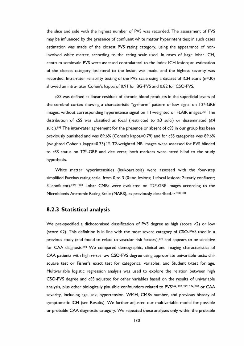
130
the slice and side with the highest number of PVS was recorded. The assessment of PVS
may be influenced by the presence of confluent white matter hyperintensities; in such cases
estimation was made of the closest PVS rating category, using the appearance of non-
involved white matter, according to the rating scale used. In cases of large lobar ICH,
centrum semiovale PVS were assessed contralateral to the index ICH lesion; an estimation
of the closest category ipsilateral to the lesion was made, and the highest severity was
recorded. Intra-rater reliability testing of the PVS scale using a dataset of ICH scans (n=30)
showed an intra-rater Cohen’s kappa of 0.91 for BG-PVS and 0.82 for CSO-PVS.
cSS was defined as linear residues of chronic blood products in the superficial layers of
the cerebral cortex showing a characteristic “gyriform” pattern of low signal on T2*-GRE
images, without corresponding hyperintense signal on T1-weighted or FLAIR images.301 The
distribution of cSS was classified as focal (restricted to ≤3 sulci) or disseminated (≥4
sulci).195 The inter-rater agreement for the presence or absent of cSS in our group has been
previously punished and was 89.6% (Cohen’s kappa=0.79) and for cSS categories was 89.6%
(weighted Cohen’s kappa=0.75).302 T2-weighted MR images were assessed for PVS blinded
to cSS status on T2*-GRE and vice versa; both markers were rated blind to the study
hypothesis.
White matter hyperintensities (leukoaraiosis) were assessed with the four-step
simplified Fazekas rating scale, from 0 to 3 (0=no lesions; 1=focal lesions; 2=early confluent;
3=confluent).239, 301 Lobar CMBs were evaluated on T2*-GRE images according to the
Microbleeds Anatomic Rating Scale (MARS), as previously described.35, 238, 301
8.2.3 Statistical analysis
We pre-specified a dichotomised classification of PVS degree as high (score >2) or low
(score ≤2). This definition is in line with the most severe category of CSO-PVS used in a
previous study (and found to relate to vascular risk factors),270 and appears to be sensitive
for CAA diagnosis.293 We compared demographic, clinical and imaging characteristics of
CAA patients with high versus low CSO-PVS degree using appropriate univariable tests: chi-
square test or Fisher’s exact test for categorical variables, and Student t-test for age.
Multivariable logistic regression analysis was used to explore the relation between high
CSO-PVS degree and cSS adjusted for other variables based on the results of univariable
analysis, plus other biologically plausible confounders related to PVS264, 270, 272, 274, 303 or CAA
severity, including age, sex, hypertension, WMH, CMBs number, and previous history of
symptomatic ICH (see Results). We further adjusted our multivariable model for possible
or probable CAA diagnostic category. We repeated these analyses only within the probable
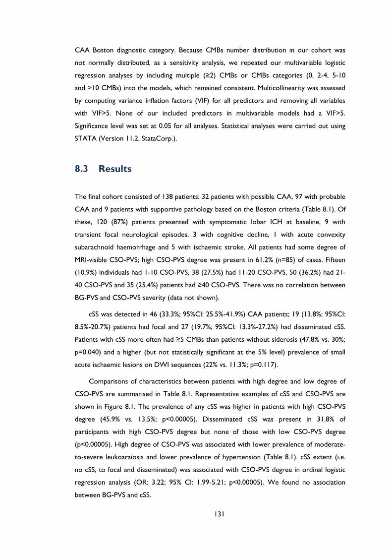
131
CAA Boston diagnostic category. Because CMBs number distribution in our cohort was
not normally distributed, as a sensitivity analysis, we repeated our multivariable logistic
regression analyses by including multiple (≥2) CMBs or CMBs categories (0, 2-4, 5-10
and >10 CMBs) into the models, which remained consistent. Multicollinearity was assessed
by computing variance inflation factors (VIF) for all predictors and removing all variables
with VIF>5. None of our included predictors in multivariable models had a VIF>5.
Significance level was set at 0.05 for all analyses. Statistical analyses were carried out using
STATA (Version 11.2, StataCorp.).
8.3 Results
The final cohort consisted of 138 patients: 32 patients with possible CAA, 97 with probable
CAA and 9 patients with supportive pathology based on the Boston criteria (Table 8.1). Of
these, 120 (87%) patients presented with symptomatic lobar ICH at baseline, 9 with
transient focal neurological episodes, 3 with cognitive decline, 1 with acute convexity
subarachnoid haemorrhage and 5 with ischaemic stroke. All patients had some degree of
MRI-visible CSO-PVS; high CSO-PVS degree was present in 61.2% (n=85) of cases. Fifteen
(10.9%) individuals had 1-10 CSO-PVS, 38 (27.5%) had 11-20 CSO-PVS, 50 (36.2%) had 21-
40 CSO-PVS and 35 (25.4%) patients had ≥40 CSO-PVS. There was no correlation between
BG-PVS and CSO-PVS severity (data not shown).
cSS was detected in 46 (33.3%; 95%CI: 25.5%-41.9%) CAA patients; 19 (13.8%; 95%CI:
8.5%-20.7%) patients had focal and 27 (19.7%; 95%CI: 13.3%-27.2%) had disseminated cSS.
Patients with cSS more often had ≥5 CMBs than patients without siderosis (47.8% vs. 30%;
p=0.040) and a higher (but not statistically significant at the 5% level) prevalence of small
acute ischaemic lesions on DWI sequences (22% vs. 11.3%; p=0.117).
Comparisons of characteristics between patients with high degree and low degree of
CSO-PVS are summarised in Table 8.1. Representative examples of cSS and CSO-PVS are
shown in Figure 8.1. The prevalence of any cSS was higher in patients with high CSO-PVS
degree (45.9% vs. 13.5%; p<0.00005). Disseminated cSS was present in 31.8% of
participants with high CSO-PVS degree but none of those with low CSO-PVS degree
(p<0.00005). High degree of CSO-PVS was associated with lower prevalence of moderate-
to-severe leukoaraiosis and lower prevalence of hypertension (Table 8.1). cSS extent (i.e.
no cSS, to focal and disseminated) was associated with CSO-PVS degree in ordinal logistic
regression analysis (OR: 3.22; 95% CI: 1.99-5.21; p<0.00005). We found no association
between BG-PVS and cSS.
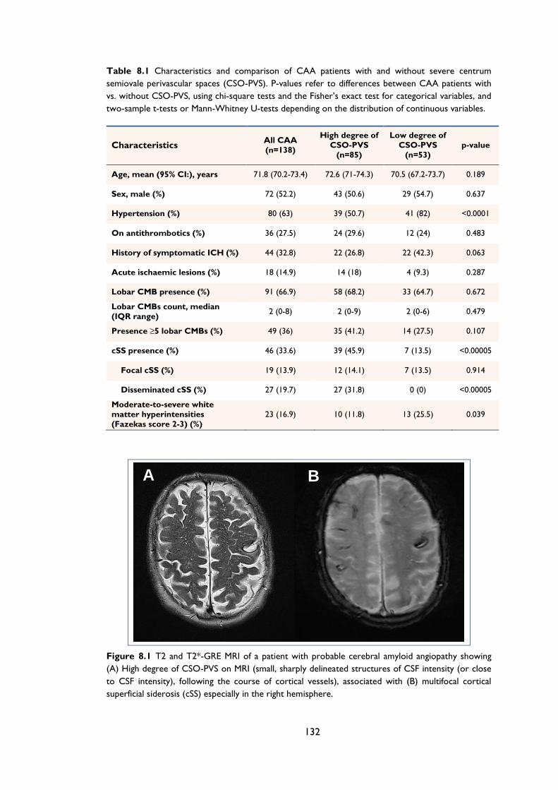
132
Table 8.1 Characteristics and comparison of CAA patients with and without severe centrum
semiovale perivascular spaces (CSO-PVS). P-values refer to differences between CAA patients with
vs. without CSO-PVS, using chi-square tests and the Fisher’s exact test for categorical variables, and
two-sample t-tests or Mann-Whitney U-tests depending on the distribution of continuous variables.
Characteristics All CAA
(n=138)
High degree of
CSO-PVS
(n=85)
Low degree of
CSO-PVS
(n=53)
p-value
Age, mean (95% CI:), years 71.8 (70.2-73.4) 72.6 (71-74.3) 70.5 (67.2-73.7) 0.189
Sex, male (%) 72 (52.2) 43 (50.6) 29 (54.7) 0.637
Hypertension (%) 80 (63) 39 (50.7) 41 (82) <0.0001
On antithrombotics (%) 36 (27.5) 24 (29.6) 12 (24) 0.483
History of symptomatic ICH (%) 44 (32.8) 22 (26.8) 22 (42.3) 0.063
Acute ischaemic lesions (%) 18 (14.9) 14 (18) 4 (9.3) 0.287
Lobar CMB presence (%) 91 (66.9) 58 (68.2) 33 (64.7) 0.672
Lobar CMBs count, median
(IQR range) 2 (0-8) 2 (0-9) 2 (0-6) 0.479
Presence ≥5 lobar CMBs (%) 49 (36) 35 (41.2) 14 (27.5) 0.107
cSS presence (%) 46 (33.6) 39 (45.9) 7 (13.5) <0.00005
Focal cSS (%) 19 (13.9) 12 (14.1) 7 (13.5) 0.914
Disseminated cSS (%) 27 (19.7) 27 (31.8) 0 (0) <0.00005
Moderate-to-severe white
matter hyperintensities
(Fazekas score 2-3) (%)
23 (16.9) 10 (11.8) 13 (25.5) 0.039
Figure 8.1 T2 and T2*-GRE MRI of a patient with probable cerebral amyloid angiopathy showing
(A) High degree of CSO-PVS on MRI (small, sharply delineated structures of CSF intensity (or close
to CSF intensity), following the course of cortical vessels), associated with (B) multifocal cortical
superficial siderosis (cSS) especially in the right hemisphere.
A B
133
In multivariable logistic regression models, high degree of PVS in the CSO was
independently associated with the presence of cSS (OR: 4.78; 95%CI: 1.64-13.87; p=0.004)
after adjusting for age, sex, hypertension, CMBs number, white matter hyperintensities,
previous history of symptomatic ICH and probable vs. possible CAA. All results were of
similar effect size in sensitivity analyses including only patients with probable CAA (Table
8.2). Both models remained consistent when multiple (≥2) CMBs or CMBs categories (0, 2-
4, 5-10 and >10 CMBs) were included instead of CMBs number.
Table 8.2 Univariate (unadjusted) and multivariable (adjusted) logistic regression analysis of
associations with high degree centum semiovale perivascular spaces (CSO-PVS), in the whole cohort
of patients with cerebral amyloid angiopathy (CAA) and in patients with probable CAA. Both models
remained consistent when multiple (≥2) CMBs or CMBs categories (0, 2-4, 5-10 and >10 CMBs)
were included.
Whole CAA cohort (n=138) Unadjusted Adjusted
OR (95% CI) p-value OR (95% CI) p-value
cSS presence 5.45 (2.21-13.45) <0.0001 4.78 (1.64-13.87) 0.004
Age (per year increase) 1.03 (0.99-1.06) 0.189 1.02 (0.97-1.06) 0.501
Hypertension 0.23 (0.10-0.53) 0.001 0.27 (0.10-0.74) 0.011
Sex (male vs. female) 0.85 (0.43-1.69) 0.637 0.85 (0.33-2.18) 0.735
CMBs count 1.01 (0.98-1.04) 0.449 1.15 (0.54-2.42) 0.718
History of symptomatic ICH 0.50 (0.24-1.04) 0.065 0.65 (0.26-1.61) 0.354
WMH (moderate-to-severe) 1.10 (0.55-2.20) 0.709 1.78 (0.66-4.73) 0.254
Probable vs. possible CAA 1.58 (0.71-3.51) 0.263 0.69 (0.17-2.82) 0.608
Probable CAA (n=106)
cSS presence 5.29 (2.05-13.65) 0.001 4.05 (1.36-12.08) 0.012
Age (per year increase) 1.04 (1.00-1.08) 0.073 1.03 (0.98-1.09) 0.235
Hypertension 0.24 (0.09-0.63) 0.004 0.28 (0.09-0.89) 0.032
Sex (male vs. female) 0.78 (0.35-7.74) 0.543 1.30 (0.43-4.00) 0.642
CMBs count (0.98-1.04) 0.582 1.00 (0.97-1.04) 0.824
History of prior symptomatic
ICH 0.63 (0.27-1.47) 0.288 0.83 (0.28-2.40) 0.724
WMH (moderate-to-severe) 0.90 (0.40-2.03) 0.799 1.18 (0.39-3.59) 0.768
134
8.4 Discussion
In this multicentre study we have shown that high degree of MRI-visible CSO-PVS are highly
prevalent in patients with CAA and are related to the presence and severity of cSS, a
characteristic neuroimaging marker of CAA.195, 301 This association further adds to the
accumulating evidence that CSO-PVS (but not BG-PVS) might be another potential MRI
marker of CAA with implications for improving the sensitivity of in vivo diagnosis.272, 274, 293
We found neither an association between BG-PVS and any CAA-specific imaging markers
(including cSS and lobar CMBs), nor any correlation between severe CSO-PVS and BG-PVS,
supporting the hypothesis that MRI-visible CSO-PVS compared to BG-PVS are due to
different pathophysiological processes.272, 274, 293 The structure of perivascular spaces in the
basal ganglia, however, differs from those in the cerebral cortex, in that they are
surrounded by two periarterial membranes, perhaps making them less vulnerable to
vascular amyloid accumulation.304 Our findings also have implications for understanding
CAA pathophysiology, and suggest that severe leptomeningeal and cortical vascular amyloid
(causing cSS) might be related to impaired interstitial fluid drainage from cerebral white
matter (causing severe CSO-PVS).
The link between CSO-PVS and CAA may reflect interstitial fluid drainage impairment
within the perivascular spaces caused by leptomeningeal and superficial cortical vascular
amyloid-β - a major feature in sporadic CAA and Alzheimer’s disease. Perivascular spaces
form important pathways (along the basement membranes of capillaries and arteries) for
the drainage of interstitial fluid and solutes, including soluble amyloid-β, from the brain.
One key hypothesis for the development of CAA is that as perivascular drainage pathways
fail with age (particularly in association with the ApoE e4 allele), or are overloaded by
reduced capacity of other elimination mechanisms, amyloid-β is increasingly trapped and
deposited in the walls of small arteries.276 Amyloid-β deposition in small cortical and
leptomeningeal arteries in CAA could disrupt drainage, leading to retrograde dilation of
perivascular spaces in the underlying white matter of the centrum semiovale (either by
blocking bulk flow, or by diminishing the pulsatility of small vessels required for efficient
interstitial fluid drainage),263, 279 leading to CSO-PVS visible on MRI. Cumulative superficial
amyloid deposition could then promote further amyloid deposition, creating a “feed-
forward loop”, further reducing drainage efficiency.274, 276, 299
Of note, histopathological studies have shown a gradient of reducing vascular amyloid-
β severity moving from the cortical surface into the cerebral white matter: CAA is
significantly more severe in leptomeningeal than in parenchymal vessels of the same brain
135
section,67 and superficial cortical layers have more extensive vascular amyloid-β deposition
than deeper layers.305 Repeated episodes of haemorrhage from these brittle, severely CAA-
affected leptomeningeal or very superficial cortical vessels into the subarachnoid space are
probably an important cause of cSS. Indeed, in CAA, cSS has a characteristic predilection
for the cerebral convexities, reflecting linear blood residues in the superficial layers of the
cerebral cortex or in the subarachnoid space.190, 195, 243
Our study has several strengths including the systematic evaluation of MRI scans by
trained raters using validated scales for a range of imaging markers of small vessel disease
and the testing of a prespecified hypothesis. The main limitation is the potential selection
bias due to the requirement for MRI done as part of routine clinical care; hence, our results
can only be extrapolated to patient populations in a similar clinical context with available
MRI and need to be validated using larger independed cohorts. Also, although the MRI
protocols were similar in each hospital, they varied across centres, introducing another
potential source of bias. However, our findings are unlikely to be accounted by differences
in MRI sequence parameters. Although we did our best to blind the MRI rating of cSS from
CSO-PVS severity and vice versa (e.g. by assessing T2-weighted MR images for PVS blinded
to cSS status on T2*-GRE and vice versa, by rating both markers blinded to the study main
hypothesis etc.), we do acknowledge that the blinding might not be complete. In addition,
the retrospective cross-sectional design prevents conclusions on the causal direction of the
reported associations. Although it is likely that cSS and severe CSO-PVS each reflect the
overall CAA severity in the brain, severe CSO-PVS was not associated with lobar CMBs
number, a putative marker of CAA, indicating that the two phenomena may be causally
related to each other, independed of CAA severity. In addition, we may have had
insufficient statistical power to confirm or refute an association between CSO-PVS and
small acute ischaemic lesions on DWI, due to our limited sample size and the relatively low
prevalence of these lesions in CAA. However, severe CAA is associated with numerous
cortical microinfarcts, which are difficult to resolve on 1.5T MRI, as used in this study. It
may be of interest to study the relation of CSO-PVS and microinfarcts on high field
strength in the future.197 The inverse association between high CSO-PVS degree and
presence of hypertension is also interesting and may reflect a protective effect related to
aggressive treatment of hypertension in these patients. Finally, we did not have pathological
confirmation of CAA, and acknowledge that the Boston criteria for CAA diagnosis have
imperfect specificity, especially for the “possible CAA” category, which in some cases may
include other cerebral small vessel diseases (e.g. hypertensive arteriopathy).202, 203Our study
should be considered hypothesis-generating. Larger prospective CAA cohorts are needed
to clarify how exactly CSO-PVS are linked to cSS (i.e. to determine the causal direction of
136
this relationship) as well as other specific MRI markers, and whether they might identify an
imaging phenotype of CAA with more superficial disease. A key question for future work
will be whether CSO-PVS are an early marker of a CAA-related disease process which
ultimately leads to severe cortical and leptomeningeal amyloid deposition, with important
implications for future haemorrhage risk.302 Both cSS and PVS have been shown to be more
prevalent in memory clinic patients (with Alzheimer’s disease or mild cognitive impairment)
compared to healthy controls;243, 306-308 it would be very interesting to investigate whether
patients with cSS and CSO-PVS differ from those without these findings with regard to
cognitive function. MRI-visible PVS are also quite common in the general population,
especially with increased age,264, 303, 308, 309 and might be related with worse cognitive
function267 and incident dementia.271
137
Chapter 9 Cerebral amyloid angiopathy
with and without intracerebral
haemorrhage: MRI-pathological evidence
for different disease phenotypes
138
ABSTRACT
Objective: To gain further insights into different cerebral amyloid angiopathy (CAA)
phenotypes and mechanisms, we investigated cortical superficial siderosis (cSS) - a new
imaging marker of the disease, and its relation with APOE genotype in patients with
pathologically-proven CAA, who presented with and without intracerebral haemorrhage
(ICH).
Methods: MRI scans of 105 consecutive patients with pathological confirmation of CAA
and MRI obtained during life were analysed for cSS (focal, ≤3 sulci; disseminates, ≥4 sulci),
and other small vessel disease markers. We compared pathological, imaging and APOE
genotype data between subjects with vs. without ICH, and investigated associations
between cSS and APOE genotype.
Results: Our cohort consisted of 54 CAA patients with symptomatic lobar ICH and 51
without ICH. APOE genotype was available in 53 patients. More than 90% of pathology
samples in both groups had neuritic plaques, whereas neurofibrillary tangles were more
commonly present in the non-ICH patients (87% vs. 42%, p<0.0001). On the other hand,
the combination of neuritic plaques and neurofibrillary tangles was more common in the
CAA group without ICH (84.8% vs. 41.7%, p=0.001). There was a trend for CAA patients
with ICH to more commonly have APOE ε2 (48.7% vs. 21.4%, p=0.075), whereas non-ICH
patients were more likely to be APOE ε4 carriers (85.7% vs. 53.9%, p=0.035). Disseminated
cSS was considerably commoner in ICH patients (33.3% vs. 5.9%, p<0.0001). In logistic
regression, disseminated cSS was associated with APOE ε2 (but not APOE ε4) (OR: 5.83;
95% CI: 1.49-22.82, p=0.011).
Conclusions: This neuropathological-defined cohort of CAA patients suggests that cSS
and APOE ε2 are related to the hemorrhagic expression of the disease. In addition, APOE
ε4 is enriched in non-hemorrhagic CAA; plaques and tangles pathology appears to be
different in the two groups. Our study emphasizes that different clinical presentations of
CAA have distinct features. This may suggest divergent pathophysiological mechanisms
underlie these different CAA phenotypes.
139
9.1 Introduction
Sporadic cerebral amyloid angiopathy (CAA) is a small vessel disease that preferentially
involves small cortical and leptomeningeal arteries due to progressive amyloid-β deposition
in their walls.28, 242 CAA occurs frequently in elderly people, and is now considered a
common and important cause of symptomatic lobar intracerebral haemorrhage (ICH).28, 37,
242 However, it has long been recognised that CAA might present without major lobar ICH
but instead with cognitive impairment (either chronic or rapidly progressive), a very
clinically salient manifestation of the disease, or transient focal neurological symptoms.167
CAA is also almost invariably found in Alzheimer's disease, but in most cases is relatively
mild.65
CAA is also associated with characteristic magnetic resonance imaging (MRI)
biomarkers including strictly lobar cerebral microbleeds (CMBs), cortical superficial
siderosis (cSS),171, 191, 194, 195 centrum semiovale perivascular spaces (CSO-PVS) and white
matter hyperintensities (WMH).28, 35 Lobar CMBs might be suggestive of CAA even in
asymptomatic elderly individuals.174 These neuroimaging markers probably reflect related
but distinct aspects of CAA pathophysiology. cSS in particular, is an interesting recently
recognized form of CAA-related haemorrhage, which likely reflects repeated episodes of
blood leaking into the subarachnoid space from brittle and fragile CAA-affected vessels.
Recently, cSS has been shown to carry a high risk of future symptomatic lobar ICH.195, 246,
302
Genetic factors, such as apolipoprotein E (APOE) genotype, are important in the
pathophysiology of CAA.80, 84 APOE ε4 appears to enhance vascular amyloid-β deposition in
a dose-dependent fashion,310 whilst APOE ε2 promotes vasculopathic changes which can
lead to vessel rupture.88 To gain further insights into different CAA phenotypes and
potential mechanisms,311 we investigated associations between neuroimaging markers of the
disease, APOE genotype, and pathologic findings in patients with CAA presenting with and
without symptomatic ICH. We hypothesized that: (1) hemorrhagic markers of CAA (cSS
and lobar CMBs) would be more strongly associated with APOE ε2 genotype; (2)
hemorrhagic markers of disease severity in CAA (cSS and lobar CMBs) and APOE ε2
genotype would be more common in patients presenting with symptomatic ICH than in
those presenting without ICH; and (3) neurofibrillary tangles will be more common in the
non-haemorrhagic compared to haemorrhagic CAA, whereas amyloid plaques might be
invariably present in the two groups due to their close molecular pathogenesis.
140
9.2 Methods
9.2.1 Case selection and clinical data collection
We included all eligible patients from the Massachusetts General Hospital (MGH) identified
retrospectively by a systematic keyword search of pathology reports and prospective
clinical databases. Cases were defined as subjects with both pathology-proven CAA (from
routinely collected brain biopsy, biopsy at haematoma evacuation or autopsy) and adequate
brain MRI sequences for the study. Data search covered patients seen at the hospital
between 1997 and 2012. An additional overlapping search through established prospective
datasets of patients with lobar haemorrhages (ICH and/or CMB) was performed to confirm
identification of all potential eligible cases for the study. Among more than 3200 retrieved
cases, we initially included those having: 1) a pathology report containing explicit
information regarding CAA assessment; and 2) available brain MRI sequences of adequate
quality including T2-weighted, T2*-weighted gradient-recalled echo (T2*-GRE) and/or SWI
and FLAIR sequences. After reviewing all neuroimaging, pathological and clinical data
available, we excluded subjects with: 1) small brain biopsy samples (greater diameter <1
cm) or samples from clot evacuations not containing any assessable vessels; 2) autopsy
studies not grading CAA; and 3) known alternative causes for lobar ICH. A total of 192
patients were eligible for analysis. Patients with no pathological evidence of CAA (n=54),
clinical/imaging presentations not characteristic of CAA (3 cases with deep ICH and 9 with
ischaemic stroke at baseline), no T2 or T2*-GRE MRI (n=18), T2/T2*-GRE MRI of
insufficient quality (n=2) and irretrievable sequences (n=1) were further excluded.
Demographic and clinical information was obtained from prospective databases and by
medical records review using standardised data collection forms. Variables of interest were
age, gender, history of hypertension, antithrombotic drug use and clinical presentation at
baseline. Hypertension was defined as a history of hypertension, taking antihypertensive
treatment or documented elevated blood pressure (systolic >150 or diastolic >95mmHg)
before admission. APOE genotype was determined in a subset of patients who provided
blood samples and consented to genetic testing as previously described,312 and without
knowledge of clinical or neuroimaging data.
The original clinical presentation of patients included in the study was ascertained from
all available neuroimaging, pathological and clinical data, and was determined as either
symptomatic lobar ICH confirmed on neuroimaging or non-haemorrhagic (including
cognitive impairment, transient focal neurological episodes, or other neurological
141
symptoms). Cases of inflammatory CAA were also included in the analysis when an MRI
outside the acute phase (≥1 month) was available. To investigate how stable these clinical
phenotypes are (e.g. CAA with ICH and CAA without ICH), for the patient group
presenting without ICH we extrapolated follow-up information from prospective databases
and medical records on incident symptomatic ICH.
9.2.2 Standard Protocol Approvals, Registrations, and Patient
Consents
The study received ethical approval by the Institutional Review Board of MGH.
9.2.3 Pathological data collection
Morphological assessment was performed in routine haematoxylin-eosin staining and the
presence or absence and severity of vascular amyloid-β deposition was confirmed by
immunohistochemical detection and/or congo red staining. CAA presence and severity was
assessed in all available vessels. Cases were considered positive for CAA when they had at
least one leptomeningeal or cortical vessel with amyloid-β reported, providing enough
information to reliably classify CAA severity using the Vonsattel grading system67, 72 and
were classified as mild (Vonsattel grade 1) or moderate to severe (Vonsattel grades 2-4).
Where available from neuropathology reports, we also systematically extracted information
on: 1) neuritic plaques and neurofibrillary tangles assessed in routinely immunostained
sections for amyloid-β and phosphorylated tau and recorded as present or absent; and 2)
the presence or absence of moderate to severe arteriolosclerosis features (including thick
arteriolar walls, lipohyalinosis, perivascular haemosiderin-laden macrophages etc.) mainly in
the basal ganglia and deep cerebral white matter.2
9.2.4 Neuroimaging data and analysis
Imaging for all patients included T2-weighted, FLAIR, T2*-GRE and or susceptibility-
weighted imaging (SWI). MR images were reviewed blinded to clinical, histopathological and
genetic data by trained observers, according to STandards for ReportIng Vascular changes
on nEuroimaging (STRIVE).31
CMBs presence and number were evaluated on axial T2*-GRE or SWI images
according to current consensus criteria35 and categorised as lobar (i.e. cortical-subcortical),
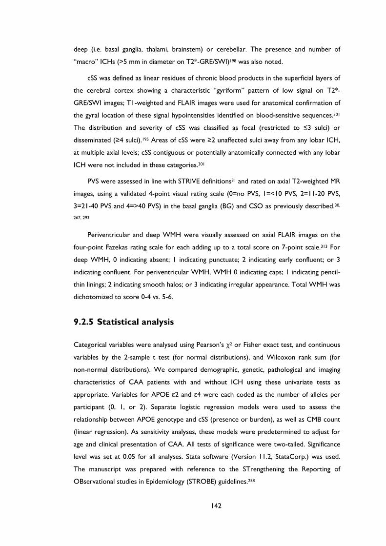
142
deep (i.e. basal ganglia, thalami, brainstem) or cerebellar. The presence and number of
“macro” ICHs (>5 mm in diameter on T2*-GRE/SWI)198 was also noted.
cSS was defined as linear residues of chronic blood products in the superficial layers of
the cerebral cortex showing a characteristic “gyriform” pattern of low signal on T2*-
GRE/SWI images; T1-weighted and FLAIR images were used for anatomical confirmation of
the gyral location of these signal hypointensities identified on blood-sensitive sequences.301
The distribution and severity of cSS was classified as focal (restricted to ≤3 sulci) or
disseminated (≥4 sulci).195 Areas of cSS were ≥2 unaffected sulci away from any lobar ICH,
at multiple axial levels; cSS contiguous or potentially anatomically connected with any lobar
ICH were not included in these categories.301
PVS were assessed in line with STRIVE definitions31 and rated on axial T2-weighted MR
images, using a validated 4-point visual rating scale (0=no PVS, 1=<10 PVS, 2=11-20 PVS,
3=21-40 PVS and 4=>40 PVS) in the basal ganglia (BG) and CSO as previously described.30,
267, 293
Periventricular and deep WMH were visually assessed on axial FLAIR images on the
four-point Fazekas rating scale for each adding up to a total score on 7-point scale.313 For
deep WMH, 0 indicating absent; 1 indicating punctuate; 2 indicating early confluent; or 3
indicating confluent. For periventricular WMH, WMH 0 indicating caps; 1 indicating pencil-
thin linings; 2 indicating smooth halos; or 3 indicating irregular appearance. Total WMH was
dichotomized to score 0-4 vs. 5-6.
9.2.5 Statistical analysis
Categorical variables were analysed using Pearson’s χ2 or Fisher exact test, and continuous
variables by the 2-sample t test (for normal distributions), and Wilcoxon rank sum (for
non-normal distributions). We compared demographic, genetic, pathological and imaging
characteristics of CAA patients with and without ICH using these univariate tests as
appropriate. Variables for APOE ε2 and ε4 were each coded as the number of alleles per
participant (0, 1, or 2). Separate logistic regression models were used to assess the
relationship between APOE genotype and cSS (presence or burden), as well as CMB count
(linear regression). As sensitivity analyses, these models were predetermined to adjust for
age and clinical presentation of CAA. All tests of significance were two-tailed. Significance
level was set at 0.05 for all analyses. Stata software (Version 11.2, StataCorp.) was used.
The manuscript was prepared with reference to the STrengthening the Reporting of
OBservational studies in Epidemiology (STROBE) guidelines.258
143
9.3 Results
Our final cohort consisted of 105 patients with pathological evidence of CAA: 52 from
autopsies, 22 from brain biopsies and 31 with pathological samples from haematoma
evacuations. Fifty-four patients were admitted with symptomatic, spontaneous lobar ICH,
while 51 patients without any symptomatic ICH. Patients without ICH presented either
with cognitive impairment (n=42), transient focal neurological episodes (n=3), or a
combination of other symptoms (n=6; including altered mental status, or seizures, with
findings consistent with inflammatory CAA). The median Clinical Dementia Rating (CDR)
score of patients without ICH and available data (n=27) was 1 (IQR: 0.5-2).
Table 9.1 indicates the severity of CAA, and the presence or absence of neuritic
plaques and neurofibrillary tangles, in the two groups. In general, mild (Vonsattel grade 1)
or moderate to severe (Vonsattel grades 2-4) CAA were equally represented in the
cohorts (p>0.2 for both comparisons). There was no difference in the presence of
vasculopathic changes (vessel-within-vessel appearance and vessel wall necrosis) in the two
groups (29.6% vs. 24%, p= 0.336). Although the prevalence of neuritic plaques was very
similar in the two groups, neuritic plaques in isolation (i.e. with no tangles) were much
more frequent in the ICH patients than in the non-ICH CAA patients (53% vs. 13%,
p<0.0001). More than 90% of pathology samples in both groups had neuritic plaques,
whereas neurofibrillary tangles were more commonly present in the non-ICH patients (87%
vs. 42%, p<0.0001). These associations remained consistent in logistic regression models
controlling for age.
Comparisons of clinical and imaging characteristics between CAA patients with vs. without
ICH are summarised in Table 2. There was a trend for CAA patients with ICH to more
often have APOE ε2 (48.7% vs. 21.4%, p=0.075), whereas non-ICH patients were more
likely to be carriers of APOE ε4 (85.7% vs. 53.9%, p=0.035). The two groups were very
similar in imaging markers of small vessel disease, including WMH burden, high degree of
CSO-PVS and BG-PVS and lobar CMBs counts (
Table 9.2). However, the prevalence of cSS was higher in patients with ICH (51.9% vs.
19.6%, p=0.001), especially disseminated cSS (33.3% vs. 5.9%, p<0.0001). Representative MR
images are showing in Figure 9.1.
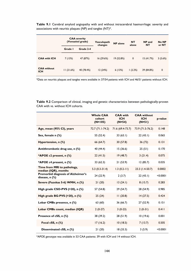
144
Table 9.1 Cerebral amyloid angiopathy with and without intracerebral haemorrhage: severity and
associations with neuritic plaques (NP) and tangles (NT)*.
CAA severity (Vonsattel grade) Vasculopath
changes NP alone
NT alone
NP and NT
No NP or NT
Grade 1 Grade 2-4
CAA with ICH 7 (13%) 47 (87%) 16 (29.6%) 19 (52.8%) 0 15 (41.7%) 3 (5.6%)
CAA without
ICH 11 (21.6%) 40 (78.4%) 12 (24%) 6 (13%) 1 (2.2%) 39 (84.8%) 0
*Data on neuritic plaques and tangles were available in 37/54 patients with ICH and 46/51 patients without ICH.
Table 9.2 Comparison of clinical, imaging and genetic characteristics between pathologically-proven
CAA with vs. without ICH cohorts.
Whole CAA
cohort
(N=105)
CAA with
ICH
(N=54)
CAA without
ICH
(N=51)
p-value
Age, mean (95% CI), years 72.7 (71.1-74.2) 71.6 (69.4-73.7) 73.9 (71.5-76.2) 0.148
Sex, female n (%) 55 (52.4) 33 (63.1) 22 (43.1) 0.065
Hypertension, n (%) 66 (64.7) 30 (57.8) 36 (72) 0.131
Antithrombotic drug use, n (%) 40 (44.4) 15 (36.6) 25 (51) 0.170
*APOE ε2 present, n (%) 22 (41.5) 19 (48.7) 3 (21.4) 0.075
*APOE ε4 present, n (%) 33 (62.3) 21 (53.9) 12 (85.7) 0.035
Time from MRI to pathology,
median (IQR), months 5.3 (0.3-31.4) 1.3 (0.2-11) 23.3 (1.4-50.7) 0.0002
Premorbid diagnosis of Alzheimer’s disease, n (%)
24 (22.9) 2 (3.7) 22 (43.1) <0.0001
Severe (Fazekas 5-6) WMH, n (%) 21 (20) 13 (24.1) 8 (15.7) 0.283
High grade CSO-PVS (>20), n (%) 57 (54.8) 29 (54.7) 28 (54.9) 0.985
High grade BG-PVS (>20), n (%) 25 (24) 11 (20.8) 14 (27.5) 0.424
Lobar CMBs presence, n (%) 63 (60) 36 (66.7) 27 (52.9) 0.151
Lobar CMBs count, median (IQR) 2 (0-27) 3 (0-23) 2 (0-31) 0.411
Presence of cSS, n (%) 38 (39.2) 28 (51.9) 10 (19.6) 0.001
Focal cSS, n (%) 17 (16.2) 10 (18.5) 7 (13.7) 0.505
Disseminated cSS, n (%) 21 (20) 18 (33.3) 3 (5.9) <0.0001
*APOE genotype was available in 53 CAA patients: 39 with ICH and 14 without ICH.
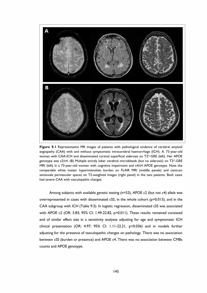
145
Figure 9.1 Representative MR images of patients with pathological evidence of cerebral amyloid
angiopathy (CAA) with and without symptomatic intracerebral haemorrhage (ICH). A. 73-year-old
woman with CAA-ICH and disseminated cortical superficial siderosis on T2*-GRE (left). Her APOE
genotype was ε2/ε4. (B) Multiple strictly lobar cerebral microbleeds (but no siderosis) on T2*-GRE
MRI (left) in a 73-year-old woman with cognitive impairment and ε4/ε4 APOE genotype. Note the
comparable white matter hyperintensities burden on FLAIR MRI (middle panels) and centrum
semiovale perivascular spaces on T2-weighted images (right panel) in the two patients. Both cases
had severe CAA with vasculopathic changes.
Among subjects with available genetic testing (n=53), APOE ε2 (but not ε4) allele was
overrepresented in cases with disseminated cSS, in the whole cohort (p=0.013), and in the
CAA subgroup with ICH (Table 9.3). In logistic regression, disseminated cSS was associated
with APOE ε2 (OR: 5.83; 95% CI: 1.49-22.82, p=0.011). These results remained consisted
and of similar effect size in a sensitivity analyses adjusting for age and symptomatic ICH
clinical presentation (OR: 4.97; 95% CI: 1.11-22.21, p=0.036) and in models further
adjusting for the presence of vasculopathic changes on pathology. There was no association
between cSS (burden or presence) and APOE ε4. There was no association between CMBs
counts and APOE genotype.
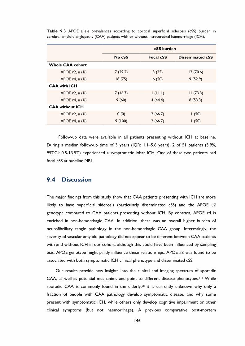
146
Table 9.3 APOE allele prevalences according to cortical superficial siderosis (cSS) burden in
cerebral amyloid angiopathy (CAA) patients with or without intracerebral haemorrhage (ICH).
cSS burden
No cSS Focal cSS Disseminated cSS
Whole CAA cohort
APOE ε2, n (%) 7 (29.2) 3 (25) 12 (70.6)
APOE ε4, n (%) 18 (75) 6 (50) 9 (52.9)
CAA with ICH
APOE ε2, n (%) 7 (46.7) 1 (11.1) 11 (73.3)
APOE ε4, n (%) 9 (60) 4 (44.4) 8 (53.3)
CAA without ICH
APOE ε2, n (%) 0 (0) 2 (66.7) 1 (50)
APOE ε4, n (%) 9 (100) 2 (66.7) 1 (50)
Follow-up data were available in all patients presenting without ICH at baseline.
During a median follow-up time of 3 years (IQR: 1.1–5.6 years), 2 of 51 patients (3.9%,
95%CI: 0.5-13.5%) experienced a symptomatic lobar ICH. One of these two patients had
focal cSS at baseline MRI.
9.4 Discussion
The major findings from this study show that CAA patients presenting with ICH are more
likely to have superficial siderosis (particularly disseminated cSS) and the APOE ε2
genotype compared to CAA patients presenting without ICH. By contrast, APOE ε4 is
enriched in non-hemorrhagic CAA. In addition, there was an overall higher burden of
neurofibrillary tangle pathology in the non-hemorrhagic CAA group. Interestingly, the
severity of vascular amyloid pathology did not appear to be different between CAA patients
with and without ICH in our cohort, although this could have been influenced by sampling
bias. APOE genotype might partly influence these relationships: APOE ε2 was found to be
associated with both symptomatic ICH clinical phenotype and disseminated cSS.
Our results provide new insights into the clinical and imaging spectrum of sporadic
CAA, as well as potential mechanims and point to different disease phenotypes.311 While
sporadic CAA is commonly found in the elderly,58 it is currently unknown why only a
fraction of people with CAA pathology develop symptomatic disease, and why some
present with symptomatic ICH, while others only develop cognitive impairment or other
clinical symptoms (but not haemorrhage). A previous comparative post-mortem
147
histopathological study found that the brain features from CAA patients that are most
consistently related to ICH are severe degree of vascular amyloid deposition and the
presence of fibrinoid necrosis (with or without microaneurysms).72 However, cases of CAA
with symptomatic ICH in our study had a fairly similar vascular amyloid burden and
prevalence of CAA-associated vasculopathic changes compared to cases without ICH. In
addition, the two groups had a very similar profile of putative neuroimaging biomarkers of
CAA severity, including lobar CMBs presence and number, WMH and high degree of CSO-
PVS. It thus seems likely that additional and/or distinct biological pathways from those
involved in amyloid-β accumulation in cortical and leptomeningeal vessels play a role in
determining clinical expression.
The most distinctive neuroimaging feature between the two groups was the much
higher prevalence of disseminated cSS in CAA patients with ICH. The prevalence of cSS in
our histopathology-confirmed CAA-ICH group is in line with the reported prevalence in a
previous imaging study of CAA-ICH.301 In addition, the presence of cSS in the group
without ICH (10%) is more in line with recent imaging studies evaluating cSS in a memory
clinic setting and Alzheimer’s disease,306, 307, 314 and much higher compared to cSS in the
population-based Rotterdam Study (0.7% in 1,062 non-demented subjects ≥60 years).315
Although the pathophysiological mechanisms underlying cSS in CAA are not yet fully
understood, observational data suggest that cSS represents blood residues related to blood
leaking episodes into the subarachnoid space from CAA-affected vessels.190, 195, 301 A
‘secondary’ mechanism due to leakage or expansion of a lobar ICH into the subarachnoid
space cannot be fully excluded; however, similar to other studies, cSS was mostly found
distant from any ICH (and often in the opposite hemisphere), and was also detected even in
cases without any ICH. In addition, cases in which siderosis was clearly connected to a
lobar haematoma were not classified as cSS. Two recent cohort studies have identified cSS
as a particular risk factor for subsequent ICH in CAA.246, 302 In a cohort on 51 patients with
CAA-related cSS and a median follow-up of 35.3 months, new intracranial haemorrhages
were observed in 24 patients (47.1%).246 The majority of new haemorrhages (i.e. 13/18 new
ICH and 4/6 new acute convexity subarachnoid bleeds) were located immediately adjacent
to pre-existing cSS.246 A European multicentre cohort of probable or possible CAA patients
(n=118; median follow-up 24 months), found that cSS was a significant predictor of time
until ICH.302 The ICH risk at 4 years was 25% (95%CI: 7.6-28.3%) for patients without
siderosis, 28.9% (95%CI: 7.7-76.7%) for patients with focal siderosis and 74% (95%CI: 44.1-
95.7) for patients with disseminated cSS (log-rank test: p=0.0031).302 A small autopsy series
of six CAA cases showed that multiple leptomeningeal arteries (and not parenchymal
cortical vessels) can rupture into the subarachnoid space and the brain parenchyma, leading
148
to large lobar haemorrhages.248, 316 All patients had multiple haematomas in the
subarachnoid space (mainly in cerebral sulci), as well as intracerebral haematomas, and each
intracerebral haematoma was connected to the subarachnoid haematomas at the depth of
cerebral sulci or through the lateral margin of the cortex.248 In this neuropathological study
there was evidence of only few ruptured cortical arteries associated with lobar ICH. These
observations might explain the association between disseminated cSS and CAA patients
with ICH in the present study, as well as why patients with disseminated cSS have the
highest risk of future ICH.302
In addition, our study demonstrates that there might be APOE genotype-specific
effects on the imaging and clinical expression of CAA-related disease. While APOE ε4
seems to be more associated with CAA without ICH, APOE ε2 is linked more strongly
with bleeding and cSS. The APOE ε2 allele was previously reported to be associated with
CAA-related lobar ICH, possibly owing to the disease-related vasculopathic abnormalities
seen with this allele,83, 88 and recently with cSS in a cohort of CAA defined by clinico-
radiographic criteria.317 APOE ε4 is also associated with risk of sporadic CAA and ICH, the
severity of which depends on the number of APOE ε4 alleles.77, 318, 319 APOE ε4 might
increase CAA severity by enhancing amyloid-β deposition within small cerebral vessels
whilst APOE ε2 promotes the vasculopathic changes which can lead to vessel rupture.80, 88
Our study raised the interesting possibility APOE ε2 influences pathways causing both cSS
and ICH. Interestingly, after accounting for the presence of vasculopathic changes, the
apparent relation of APOE ε2 to cSS was not reduced, suggesting that vascular amyloid load
and how extensively the vessel wall architecture is disrupted is not the sole driving force
underlying these pathways and hence, these clinical phenotypes. However, any mechanistic
links are clearly complex and need to be treated with caution, as APOE genotype alone
might not be necessary or sufficient to cause or drive these effects. It is important to note
that links may differ according to the presence or absence of Alzheimer’s disease pathology,
particularly for APOE ε2, which has been associated with a reduced risk of late-onset
Alzheimer’s dementia.77 This observation is in line with the differential associations between
neuritic plaques and neurofibrillary tangles data in our CAA patients with and without ICH.
Indeed, there is significant variation in the pathological appearance of CAA and its influence
on Alzheimer’s disease pathology,320, 321 further highlighting the concept of different
phenotypes of CAA which can follow and influence several different pathways. It is
important to acknowledge thought that the overrepresentation of neurofibrillary tangles
among the non-ICH CAA patients might also partly reflect a bias issue, since by definition
these cases presented cognitive impairment, partly driven by neurofibrillary tangle
pathology.
149
Although the natural history of symptomatic CAA patients mainly presenting with
cognitive impairment (i.e. without ICH at baseline) has not been fully investigated, our data
suggest that this CAA phenotype may have a lower risk of developing ICH. It is important
to note that CAA patients without major ICH (microbleeds- or cSS-only CAA) presenting
to stroke services with symptoms other that cognitive impairment (e.g. transient focal
neurological episodes), might still be at significant risk of future bleeding.244, 302 A recent
study on patients presenting with isolated lobar microbleeds showed that they indeed have
a hemorrhagic risk profile suggestive of severe CAA pathology.322 However, the ICH risk
among CMB-only CAA patients was lower compared to CAA patients with past ICH. This
topic requires further investigation in prospective studies, especially given the increased
incidence of ICH in patients with Alzheimer’s disease and the biological association of
Alzheimer’s disease with CAA.323, 324 Whether patients with CAA-related cognitive
impairment/Alzheimer’s disease and cSS have a distinct clinical sub-phenotype (e.g. worse
outcome, high APOE e2 genotype frequency, increased ICH) also requires further study.
The main strengths of this study include the large sample of patients with
histopathologically confirmed CAA and available MRI sequences, the systematic evaluation
of MRI scans for a range of imaging markers of small vessel disease, and the testing of a
prespecified hypothesis. The main limitations, inherent to any clinical-pathology series in
CAA, include: (a) the difference in pathology sampling between autopsied brains and
biopsies/haematomas; (b) the biases regarding which patients get biopsied or autopsied
which might be skewed towards end stage or advanced disease; and (d) potential selection
bias due to the requirement for both pathology and MRI and the unavailability of APOE
genotype data in all patients. The differences in Alzheimer’s pathology between groups
could potentially be explained by a longer duration of disease (taking time from MRI to
pathology as a surrogate for disease duration) in CAA patients without ICH or the different
characteristic between the two groups (including APOE ε4 prevalence). Further molecular
imaging studies assessing amyloid and tau burden in vivo325 may help resolve this question.
Also, the cross-sectional design of the current study did not allow us to assess potential
causality of the reported associations.
In summary, results from this neuropathological-defined cohort of CAA patients with
and without ICH provide multiple lines of evidence for distinct disease phenotypes, and
suggest that cSS and APOE ε2 are related to the haemorrhagic expression of the disease.
Our study emphasizes the widening spectrum of CAA with clinical phenotypes reflecting
different neuroimaging and genetic features and suggests divergent pathophysiological
mechanisms. Although these findings require external validation in larger CAA cohorts,
they may be relevant for future biomarkers studies and disease-modifying CAA trials.300
150
Chapter 10 Cerebral microbleeds,
microangiopathy subtype and recurrent
spontaneous intracerebral haemorrhage
risk: systematic review and meta-analysis
151
ABSTRACT
Objective: To evaluate cerebral microbleeds (CMBs) and recurrent intracerebral
haemorrhage (ICH) and ischaemic stroke risk in spontaneous ICH survivors, stratified by
the MRI-defined presumed underlying microangiopathy – i.e. cerebral amyloid angiopathy
(CAA) vs. non-CAA related ICH.
Methods: Meta-analysis of prospective cohorts with recent spontaneous ICH. We
estimated annualised recurrent symptomatic ICH and ischaemic stroke rates for each study
and pooled odds ratios of recurrent ICH and ischaemic stroke across CMBs groups (i.e. 1,
2-4, 5-10 and >10 CMBs), using a random effects model.
Results: We pooled data from 8 hospital-based studies including 981 patients: 3 CAA-
related ICH cohorts (n=248) and 6 non-CAA related ICH cohorts (n=733). The annual risk
of recurrent ICH was higher in patients with CAA-related ICH compared with non-CAA
related ICH cohorts (9.96%; 95%CI: 5.08-14.84 vs. 1.31%; 95%CI: 0.51-2.11 respectively,
p=0.012). In non-CAA related ICH cohorts, only patients with >10 CMBs had an increased
risk of recurrent ICH (OR: 5.63; 95%CI: 1.79-17.72; p=0.003); CMBs located in both deep
and lobar regions the stronger association with recurrent ICH (OR: 4.01; 95%CI: 1.47-
10.94; p=0.007). In CAA-related ICH cohorts higher numbers of baseline CMBs strongly
predicted an increased risk of recurrence during follow-up: the cumulative OR was 3.32
(95%CI: 1.41-7.81; p=0.006), 4.43 (95%CI: 1.76-11.14; p=0.002) and 3.03 (95%CI: 1.19-7.74;
p=0.02) for 2-4, 5-10 and >10 CMBs respectively. This risk of recurrent ICH was not
significant for patients with a single CMB in any of the cohorts. CMBs were not associated
with ischaemic stroke risk in either CAA or non-CAA related ICH cohorts. The relative
risk for recurrent ICH vs. ischaemic stroke in non-CAA ICH patients with CMBs did not
differ significantly. By contrast, in CAA-related ICH patients with CMBs, the relative risk of
recurrent ICH compared to ischaemic stroke was 31.2 (95% CI: 5.9-164.6; p<0.0005).
Conclusions: In ICH patients, multiple CMBs at baseline are associated with an increased
ICH recurrence risk. The recurrent ICH risk in those with MRI-defined CAA-related ICH is
about 8 times higher than for non-CAA related ICH. The balance of risk for recurrent ICH
vs. ischaemic stroke also differs between CAA and non-CAA related ICH cohorts. Defining
ICH subtypes using MRI is relevant for prognosis, and should be considered in designing
future ICH clinical prevention trials.
152
10.1 Introduction
Spontaneous intracerebral haemorrhage (ICH) presumed to be due to cerebral small vessel
disease31 is one of the most catastrophic forms of stroke.326 It is associated with high
morbidity and mortality,327 but also a substantial risk of recurrence for survivors.220, 253, 254
Understanding the causes and pathophysiology of spontaneous ICH can help stratify the
risk of recurrent ICH or ischaemic cerebrovascular events with relevance for the
development of new treatments. Two main disease processes can cause rupture of small
arteries of the brain leading to spontaneous ICH: hypertensive arteriopathy and sporadic
cerebral amyloid angiopathy (CAA).2 Hypertensive arteriopathy is characterized by
lipohyalinosis and fibrinoid necrosis of small deep perforating arteries, resulting in
haemorrhages particularly in deep brain regions, such as the basal ganglia and thalamus.2
CAA affects the small cortical and leptomeningeal arteries, by the progressive deposition of
amyloid-β in the vessel walls and is an important cause of lobar (but not deep) ICH,
especially in the elderly.28, 328 These two common small vessel pathologies that result in ICH
seem to have intrinsically different risks of recurrent haemorrhage: a history of CAA-
related lobar ICH might carry a significantly higher risk for recurrence compared to deep
ICH due to hypertensive arteriopathy,254, 255, 329 but these risks are currently difficult to
predict.
With the development of more advanced imaging techniques over the past decades,
new neuroimaging markers of small vessel disease have been emerged. Among them,
cerebral microbleeds (CMBs) on blood-sensitive MRI sequences, including T2*-weighted
gradient-recalled echo (T2*-GRE) and susceptibility-weighted imaging (SWI), have become a
relatively common finding in patients with spontaneous ICH and warfarin-related ICH.35, 212
By contrast with other markers of small vessel disease (e.g. leukoaraiosis or lacunes), CMBs
may provide direct evidence of blood leakage from haemorrhagic-prone
microangiopathy.172, 330 The presence of CMBs is also regionally associated with ICH,
making them a possible marker of more bleeding-prone areas.177 In addition, the location of
CMB in the brain appears to reflect the type of the underlying microangiopathy: similar to
macrobleeds, strictly lobar CMBs (cortical-subcortical) are characteristic of CAA-related
small vessel disease (allowing the diagnosis of CAA during life using the Boston criteria202,
203), whilst deep CMBs appear to reflect hypertensive arteriopathy.27, 35 If the risk of ICH
recurrence is related to the underlying arteriopathy type and burden, CMBs may be of
particular value in identifying patients at high risk of recurrence.
153
Some studies indeed show that the burden of CMBs predicts the risk of recurrent
ICH; the relationship with the risk is stronger in patients with CAA-related lobar ICH.180,
227, 331 However, these studies are too small to guide patient management at present; and
more reliable estimates of the risks are urgently needed. We therefore performed a meta-
analysis of all available evidence from published prospective ICH cohorts investigated using
MRI, to investigate the power of CMBs (including CMBs number and location) to predict
recurrent ICH, stratified by the presumed underlying microangiopathy (CAA vs. non-CAA
related ICH). Since CMBs could also be associated with occlusive features of
cerebrovascular disease, as a secondary analysis we also explore their relation with incident
ischaemic stroke in this patient population.
10.2 Methods
10.2.1 Search strategy, selection criteria and data
extraction
PubMed was searched between January 1, 1999 and November 1, 2013 using medical
subject heading (MESH) terms and text words: [“microbleed*” OR “microh(a)emorrhage*”
OR “h(a)emorrhagic lacune” AND [“stroke” OR “intracerebral h(a)emorrhage” OR “brain
h(a)emorrhage”]. Reference lists from all included articles, relevant review articles and the
authors’ own files were also searched for additional eligible reports. Studies were eligible
for inclusion if they: included adult patients with spontaneous symptomatic ICH confirmed
by imaging; had a prospective design, with at least three months of follow-up; assessed the
risk of recurrent symptomatic spontaneous ICH (main outcome) during follow-up; had data
for the presence of CMBs on baseline T2*-GRE MRI; and were published in English. In cases
of multiple publications from the same or overlapping cohorts, only the most recent
comprehensive results from the report with the largest sample size were generally used in
the analysis. We excluded case-control and cross-sectional studies, case reports or case-
series, population-based studies and studies in other patient populations (e.g. CADASIL,
thrombolysis-related ICH etc.).
For each study, we extracted data on the country of the study, time period, clinical
setting, population size, demographic data (including mean age and sex and vascular risk
factors), use of antithrombotic agents, T2*-GRE MRI parameters, number of participants
with at least one CMB at baseline, method and duration of follow-up and number of
participants with the outcome events of interest (recurrent spontaneous symptomatic ICH
154
and symptomatic ischaemic stroke, clearly defined according to defined criteria). For
included cohorts we sought further information from the authors (by email) on total
person-years of follow-up and outcome events (recurrent ICH and symptomatic ischaemic
stroke) stratified by CMBs numbers (1 CMB, 2-4, 5-10 and >10 CMBs) and location (lobar -
in the cortex or subcortical areas of the cerebral hemispheres , deep or mixed CMBs). We
classified each study cohorts as CAA-related ICH or non-CAA related ICH according to
classic Boston criteria,202 based on published information of included subjects in each study
and correspondence with authors.
10.2.2 Statistical analysis
We estimated annualised recurrent symptomatic ICH and ischaemic stroke rates (%/year)
and corresponding 95% confidence intervals (CI) for each study from a poisson regression
model and exact poisson intervals. Pooled rates were calculated using an inverse variance-
based random effects method (DerSimonian-Laird) and stratified by study population
(presumed underlying small vessel disease cause of baseline ICH, i.e. CAA-related ICH vs.
non-CAA ICH). The log(incidence) of recurrent ICH events where compared between
these groups using a significance test with the appropriate degrees of freedom.
We meta-analysed data for recurrent ICH and ischaemic stroke across studies, using a
random effects model with DerSimonian-Laird weights,332 quantifying the strength of any
association using odds ratios (OR) and its associated 95% CI in patients without CMBs vs.
different CMBs groups. To maximise the power of our analyses, for comparisons with zero
events in both groups, we added 0.5 to each group, considered OR=1 and calculated
calculating the standard error (SE), logOR and SE logOR by using the 2-variable input
method. We assessed heterogeneity by I-squared and x-squared statistics and also visually
through inspection of the forest plot and checking for overlapping confidence intervals. We
explored publication bias with funnel plots and the Harbord regression tests for funnel plot
asymmetry. Analyses were stratified by baseline ICH presumed cause (CAA vs. non-CAA
related ICH). As a sensitivity analysis we investigated the influence of one study on the
overall meta-analysis estimate (using the “metaninf” command) and inspect the results
graphically with meta-analysis estimates computed, omitting one study in each turn. We
used meta-regression to explore whether certain confounders could have affected our
results.
All meta-analyses were performed using Stata 11.2 (StataCorp LP, Texas). We
prepared this report with reference to the Preferred Reporting Items for Systematic
155
reviews and Meta-Analyses (PRISMA)333 and the Meta-analysis of Observational Studies in
Epidemiology (MOOSE)334 guidelines.
10.3 Results
Eight unique hospital-based studies with a total of 981 patients (665 with CMBs) met our
predefined inclusion criteria,227, 302, 331, 335-339 and were pooled in meta-analyses. The studies
comprised of three CAA-related ICH cohorts (n=248) 227, 302, 337 and six non-CAA related
ICH cohorts (n=733)331, 335, 336, 338, 339 (i.e. one study included both cohorts227). The basic
characteristics of included cohorts are summarised in Table 10.1. Most studies had similar
inception points (i.e. the start of follow-up), methods of assessment and a high number of
participants lost to follow-up. Most studies used reliable ascertainment methods for
establishing recurrent ICH events at the follow-up.
In these studies, the overall pooled annual risk of recurrent ICH and ischaemic
stroke was 3.20% (95% CI: 1.52-4.88) and 0.87% (95% CI: 0.25-1.48) respectively (Figure
10.1). Patients with CAA-related ICH had a higher annual risk of recurrent ICH compared
with non-CAA related ICH cohorts (9.96%; 95% CI: 5.08-14.84 vs. 1.31%; 95% CI: 0.51-2.11
per year respectively, p=0.012). The ischaemic stroke rates could not be directly compared
in the two patient groups, because in one CAA cohort ischaemic stroke was not an
outcome event,337 and in another study no ischaemic strokes were observed.227 However,
individual non-CAA ICH cohorts seem to have a higher risk of ischaemic stroke at follow-
up when indirectly compared with the CAA cohorts.
All studies used T2*-GRE MRI to detect CMBs at baseline, although imaging
parameters, including echo time and slice thickness varied slightly (Table 10.1). Differences
in demographic, clinical and imaging characteristics between the CMB (+) vs. CMB (-)
groups are noted in Table 10.1. Among patients with CMBs, 79/665 (11.9%; 95% CI: 9.5-
14.6%) experienced recurrent ICH during follow-up, compared to 16/316 (5.1%; 95%CI:
2.9-8.1%) patients without CMBs. The results of the overall analysis showed that those with
any baseline CMBs had a higher risk of recurrent ICH at follow-up (OR: 2.81; 95% CI: 1.61-
4.93); p<0.0005) (Appendix F Figure 1). This risk was not significant for patients with a
single CMB compared to those without CMBs. However, amongst different multiple CMBs
groups (i.e. 2-4, 5-10 and >10 CMBs), there was a graded relationship between CMBs
burden and higher recurrent ICH risk (Appendix F Figure 1). The results were consistent
from study to study (test for heterogeneity p>0.10).
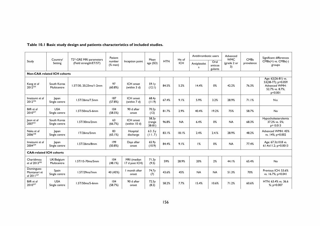
156
Table 10.1 Basic study design and patients characteristics of included studies.
Study Country/ Setting
T2*-GRE MRI parameters (Field strength/ET/ST)
Patient number
(% men)
Inception point Mean
age (SD) HTN
Hx of ICH
Antithrombotic users Advanced
WMC (grade 2 or
3)
CMBs prevalence
Significant differences CMBs(+) vs. CMBs(-)
groups Antiplatelet
s
Oral anticoa
gulants
Non-CAA related ICH cohorts
Kang et al
2012335
South Korea
Multicentre 1.5T/30, 20,23ms/1-2mm
97
(60.8%)
ICH onset
(within 3 d)
59.1y
(12.1) 84.5% 5.2% 14.4% 0% 42.2% 76.3%
Age: 62(26-81) vs.
53(38-77); p=0.059 Advanced WMH: 52.7% vs. 8.7%;
p<0.001
Imaizumi et al 2012336
Japan Single centre
1.5T/26ms/7.5mm 187
(57.8%) ICH onset (within 7 d)
68.4y (11.9)
67.4% 9.1% 5.9% 3.2% 28.9% 71.1% No
Biffi et al
2010227
USA
Single centre 1.5T/50ms/5-6mm
104
(58.5%)
90 d after
onset
70.2y
(12) 81.7% 2.9% 40.4% 19.2% 75% 58.7% No
Jeon et al 2007331
South Korea Single centre
1.5T/30ms/2mm 63
(66.7%) ICH onset
(within 10 d)
58.3y (range
38-81)
96.8% NA 6.4% 0% NA 68.3% Hypocholesterolemia
37.2% vs. 5%;
p= 0.013
Naka et al 2006338
Japan Single centre
1T/26ms/5mm 83
(65.1%) Hospital discharge
63 .5y (11 .7 )
83.1% 18.1% 2.4% 2.4.% 28.9% 48.2% Advanced WMH: 45%
vs. 14%; p=0.002
Imaizumi et al 2004339
Japan Single centre
1.5T/26ms/8mm 199
(50.8%) Days after
onset 65.9y (10.9)
84.4% 9.1% 1% 0% NA 77.4% Age: 67.3±10.8 vs.
61.4±11.2; p=0.0013
CAA-related ICH cohorts
Charidimou et al 2013302
UK/Belgium Multicentre
1.5T/15-70ms/5mm 104
(48.1%) MRI (median
17 d post ICH) 71.3y (9.5)
59% 28.9% 20% 2% 44.1% 65.4% No
Domingues-
Montanari et al 2011337
Spain Single centre
1.5T/29ms/?mm 40 (45%) 1 month after
onset 74.7y (7)
43.6% 45% NA NA 51.3% 70% Previous ICH: 53.6% vs. 16.7%; p=0.041
Biffi et al
2010227
USA
Single centre 1.5T/50ms/5-6mm
104
(58.7%)
90 d after
onset
72.5y
(8.2) 58.2% 7.7% 15.4% 10.6% 71.2% 60.6%
HTN: 63.4% vs. 36.6
%; p=0.007
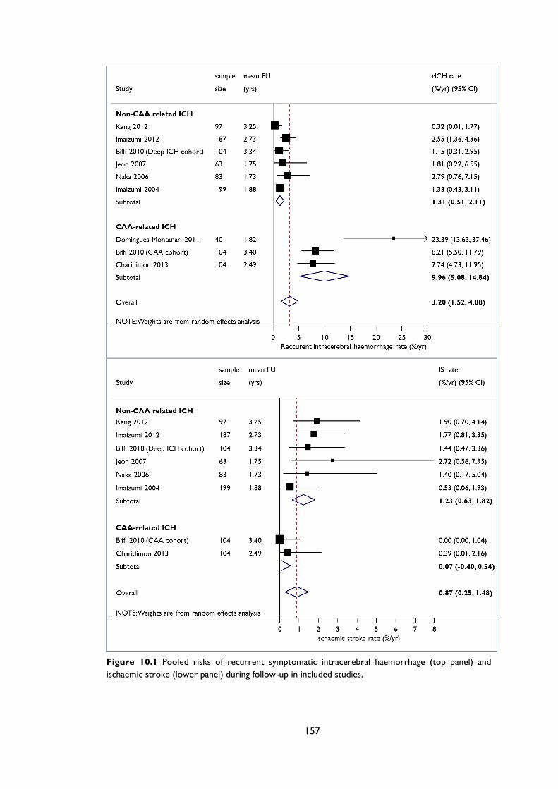
157
Figure 10.1 Pooled risks of recurrent symptomatic intracerebral haemorrhage (top panel) and
ischaemic stroke (lower panel) during follow-up in included studies.
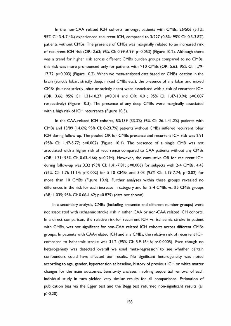
158
In the non-CAA related ICH cohorts, amongst patients with CMBs, 26/506 (5.1%;
95% CI: 3.4-7.4%) experienced recurrent ICH, compared to 3/227 (0.8%; 95% CI: 0.3-3.8%)
patients without CMBs. The presence of CMBs was marginally related to an increased risk
of recurrent ICH risk (OR: 2.63; 95% CI: 0.99-6.99; p=0.053) (Figure 10.2). Although there
was a trend for higher risk across different CMBs burden groups compared to no CMBs,
this risk was more pronounced only for patients with >10 CMBs (OR: 5.63; 95% CI: 1.79-
17.72; p=0.003) (Figure 10.2). When we meta-analysed data based on CMBs location in the
brain (strictly lobar, strictly deep, mixed CMBs etc.), the presence of any lobar and mixed
CMBs (but not strictly lobar or strictly deep) were associated with a risk of recurrent ICH
(OR: 3.66; 95% CI: 1.31-10.27; p=0.014 and OR: 4.01; 95% CI: 1.47-10.94; p=0.007
respectively) (Figure 10.3). The presence of any deep CMBs were marginally associated
with a high risk of ICH recurrence (Figure 10.3).
In the CAA-related ICH cohorts, 53/159 (33.3%; 95% CI: 26.1-41.2%) patients with
CMBs and 13/89 (14.6%; 95% CI: 8-23.7%) patients without CMBs suffered recurrent lobar
ICH during follow-up. The pooled OR for CMBs presence and recurrent ICH risk was 2.91
(95% CI: 1.47-5.77; p=0.002) (Figure 10.4). The presence of a single CMB was not
associated with a higher risk of recurrence compared to CAA patients without any CMBs
(OR: 1.71; 95% CI: 0.63-4.66; p=0.294). However, the cumulative OR for recurrent ICH
during follow-up was 3.32 (95% CI: 1.41-7.81; p=0.006) for subjects with 2-4 CMBs, 4.43
(95% CI: 1.76-11.14; p=0.002) for 5-10 CMBs and 3.03 (95% CI: 1.19-7.74; p=0.02) for
more than 10 CMBs (Figure 10.4). Further analyses within these groups revealed no
differences in the risk for each increase in category and for 2-4 CMBs vs. ≥5 CMBs groups
(RR: 1.035; 95% CI: 0.66-1.62; p=0.879) (data not shown).
In a secondary analysis, CMBs (including presence and different number groups) were
not associated with ischaemic stroke risk in either CAA or non-CAA related ICH cohorts.
In a direct comparison, the relative risk for recurrent ICH vs. ischaemic stroke in patient
with CMBs, was not significant for non-CAA related ICH cohorts across different CMBs
groups. In patients with CAA-related ICH and any CMBs, the relative risk of recurrent ICH
compared to ischaemic stroke was 31.2 (95% CI: 5.9-164.6; p<0.0005). Even though no
heterogeneity was detected overall we used meta-regression to see whether certain
confounders could have affected our results. No significant heterogeneity was noted
according to age, gender, hypertension at baseline, history of previous ICH or white matter
changes for the main outcomes. Sensitivity analyses involving sequential removal of each
individual study in turn yielded very similar results for all comparisons. Estimation of
publication bias via the Egger test and the Begg test returned non-significant results (all
p>0.20).
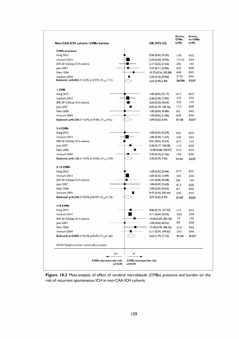
159
Figure 10.2 Meta-analysis of effect of cerebral microbleeds (CMBs) presence and burden on the
risk of recurrent spontaneous ICH in non-CAA ICH cohorts.
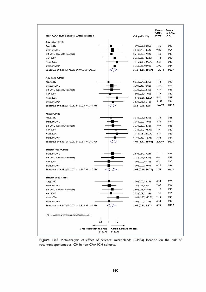
160
Figure 10.3 Meta-analysis of effect of cerebral microbleeds (CMBs) location on the risk of
recurrent spontaneous ICH in non-CAA ICH cohorts.
161
Figure 10.4 Meta-analysis of effect of cerebral microbleeds (CMBs) presence and burden on the
risk of recurrent spontaneous ICH in CAA ICH cohorts.
10.4 Discussion
In this systematic review and meta-analysis of eight cohorts involving 981 survivors with
symptomatic spontaneous ICH, the pooled estimates demonstrated a consistent association
between CMBs presence at baseline and future ICH recurrence. However, the strength of
the association and the magnitude of recurrent ICH risk, especially in relation to CMBs
burden and location, were considerably different depending on the nature and severity of
the underlying microangiopathy – i.e. CAA-related and non-CAA related (presumably
hypertension-related).
Based on data from included cohorts the aggregate rates of recurrent ICH was about
8-fold higher in patients with presumed CAA-related lobar ICH, compared to non-CAA
162
ICH. This is in line with most previous studies149, 340, 341 and a systematic review254 that have
found ICH recurrence is more common following lobar ICH than non-lobar ICH (i.e. deep
ICH) (4.4% vs. 2.1% per year respectively; p=0.002). In the cited review, the risk of
ischaemic stroke (1.1% per year) was lower than the overall risk of recurrent ICH.254 A
more recent meta-analysis also confirmed that ICH survivors seem to be at considerable
risk of serious vascular events, with recurrent ICH being more frequent after lobar ICH.327
In contrast with the previous systematic review, ischaemic stroke appeared to be at least as
common as recurrent ICH over 3 years in four studies with appropriate data.327
However, the included studies in these meta-analyses254, 327 did not have the benefit of
MRI to systematically phenotype the likely underlying arteriopathy. The causal small vessel
disease might have implications for prognosis and treatment, for example, the potential
benefits and hazards of antithrombotic drugs (including antiplatelets and oral
anticoagulant).342, 343 In our meta-analysis, the risk of ischaemic stroke was negligible for
CAA patients; but nearly as high as the risk of recurrent ICH in the non-CAA cohorts. The
prominent rate of recurrent CAA-related ICH and the different rates of subsequent
ischaemic stroke compared to non-CAA related ICH, suggest that vascular mechanisms
(and relative risk of bleeding vs. ischaemia) are fundamentally different between these two
subtypes of small vessel disease.2 It is important to remember that hypertensive small vessel
disease resulting in haemorrhages in deep brain areas, and hypertension in general (which
was more prevalent in non-CAA compared to CAA cohorts; see Table 1), is also a major
risk factor for ischaemic cerebrovascular events. By contrast, CAA is not accounted by
conventional vascular risk factors (such as hypertension) and seems to be a more active
haemorrhage-prone microangiopathy compared to hypertensive arteriopathy: the
progressive accumulation of vascular amyloid-β causes weakening and cracking of cortical
and leptomeningeal vessel walls, resulting in haemorrhage.28 History of previous ICH was
more common in CAA compared to non-CAA cohorts in our meta-analysis. While CAA is
associated with presumed asymptomatic cerebral microinfarcts, symptomatic ischaemic
strokes are rare.197, 328 These characteristics, as well as variation in the use of
antihypertensive therapy might partly account for the different stroke risks in the two
groups.
The differential stroke risks in ICH survivors have implications for prognosis and
secondary prevention decision-making, especially regarding whether to prescribe
antithrombotic treatments in individuals with a history of ICH, an increasingly common
clinical dilemma.342, 344 The key question is whether CMBs are linked to an increased risk of
“macrobleeding”, but available data for this hypothesis are limited.345 The high prevalence of
CMBs in patients with ICH and the relatively low recurrence rates of ICH we found,
163
suggest that there might be only specific sub-group of patients with CMBs that are at
particularly high risk for further haemorrhagic events. Our meta-analysis of ICH cohorts
provides new and stronger evidence for this assumption.
Perhaps unsurprisingly, we did not find a significant association between the presence
of only one CMB and future recurrent ICH risk in the whole ICH population and also in
neither CAA-related, nor non-CAA-related ICH cohorts. However, the pathophysiological
significance of a single CMB is unclear: it might indicate a less severe or less active
microangiopathy, or it might be just the tip of the iceberg of small vessel damage. This issue
is further complicated because the classification of patients into those having single vs.
multiple microbleeds may vary with MRI characteristics used: e.g. higher field strengths,
thinner slices, or susceptibility-weighted imaging seem to increase sensitivity to CMBs
detection.346 All of the included studies have relied on “conventional” T2*-GRE MRI
performed under standard imaging parameters, commonly used in clinical practice. Also, in
one microbleed rating scale validation study, patients with only one potential CMB
accounted for most cases of disagreement and inter-rater variability.238
We have confirmed that CAA-related lobar CMBs are a strong predictor of recurrent
ICH on clinical follow-up, making them good prognostic biomarkers in CAA. Despite this
overall association, the occurrence of CAA-related recurrent lobar ICH was considerably
lower than the overall prevalence of lobar CMBs at baseline. This observation indicates that
particular properties of the CAA-affected vessels as well as additional factors may
determine whether a vessel rupture will result in a CMB or a larger macrobleed.198
However, underscoring a connection between microbleeding and macrobleeding, the
dynamic development of new CMBs on serial T2*-GRE MRI over time also predicts
subsequent recurrent ICH in CAA.180 Recent studies have shown most of these new CAA-
related haemorrhages arise at sites of high baseline amyloid-β deposition, detected in vivo
using PiB PET imaging.176, 285 It is thus possible that in certain circumstances (e.g. with the
use of antithrombotic drugs) CAA-related CMBs, rather than being sealed off by normal
hemostatic mechanisms and surrounding tissue, may enlarge into a symptomatic lobar ICH,
underlying the strong association between CAA and anticoagulant-related ICH.152, 347, 348
Within this context, a decision analysis based on a 69-year-old survivor of a lobar CAA-
related ICH and newly diagnosed non-valvular atrial fibrillation suggested that should not be
anticoagulated with warfarin across the spectrum of thromboembolic and haemorrhagic
risks.349 The strong association of lobar CMBs burden and CAA-related recurrent ICH we
demonstrated, as well as the association with anticoagulation-related ICH152 makes them an
important feature for haemorrhagic risk assessment in older patients at high risk of CAA
pathology. However, it remains unclear whether the presence of strictly lobar CMBs in the
164
absence of symptomatic lobar ICH (i.e. microbleeds-only CAA) which are highly prevalent
in the elderly,174 confers sufficient risk of future ICH. A recent study indicates that this
incident ICH risk is not trivial and warfarin might be a significant predictor of ICH
occurrence in this population (p<0.05) but definitive data are lacking.350
In the non-CAA related ICH cohorts, our results indicate that a certain CMBs load,
specifically the presence of more than ten CMBs, and a mixed distribution in the brain, are
a strong marker of recurrent ICH risk. Since hypertensive small vessel damage primarily
affects deep brain regions, the presence of both deep and lobar CMBs is an indication of
advanced and widespread disease. In all the other CMBs groups the risk of recurrent ICH
might be at least as important as the risk for future ischaemic stroke (and probably all vaso-
occlusive events).327 Consistent with this notion, the relative risks for recurrent ICH
compared to future ischaemic stroke where not different within each CMBs subgroup -
although the patient numbers and the magnitude of the effect sizes were quite low. One
could assume that these relative effects of CMB burden on recurrent ICH and ischaemic
stroke risk might tip the risk-benefit balance in favour of antithrombotic treatment use in
non-CAA ICH patients with less than ten CMBs.
Our meta-analysis has limitations. Some studies had a small sample size, variable
follow-up, and few outcome events, leading to wide confidence intervals around risk
estimates. In these studies, inception points (i.e. the start of follow-up), and methods of
outcome assessment varied. Studies used different imaging parameters, for example echo
time, potentially affecting the detection of CMBs,27 although the prevalence of CMBs was
fairly consistent across studies. Furthermore, studies are subject to selection bias since not
all ICH patients undergo T2*-GRE, thus probably excluding more severe cases of ICH.
However, our findings represent the most up-to-data summary of available data on the
topic and can be generalised in ICH survivors who undergo clinical T2*-GRE MRI. Finally, a
potentially important limitation is possible confounding due to lack of adjustment for other
baseline variables related to future recurrent ICH risk (e.g. antithrombotic drug use,
hypertension treatment, ApoE genotype etc.). Nevertheless, all studies showed a consistent
direction of association between CMBs and recurrent ICH, even when adjusted for these
potential confounders. Furthermore, we did not have pathological confirmation of the small
vessel pathology underlying ICH in each cohort and we acknowledge that the Boston
criteria for CAA classification have imperfect specificity.195, 202 Of note, the type of included
cohorts and the presumed underlying causes of ICH are in line with the higher prevalence
of hypertensive arteriopathy rather than CAA in Asia compared to the United States or
Europe.32 For this reason only one study had concurrent groups of patients with both
CAA-related and non-CAA related ICH.227
165
Spontaneous ICH originates from different cerebral small vessel pathologies which
have intrinsically distinct prognostic significance for future stroke (including recurrent ICH
and ischaemic stroke). Our findings suggest that CMBs on blood-sensitive MRI sequences
are helpful in stratifying the risk of recurrent bleeding in ICH survivors. Furthermore, the
use of MRI is critical for phenotyping the presumed underlying aetiology of spontaneous
ICH, to inform patients and carers, plan clinical services and design clinical trials. The
current data support routine neuroimaging in survivors of spontaneous ICH to define the
microangiopathy subtype and CMBs burden to inform prognosis and relative risk of future
ICH and ischaemic stroke, which could add to risk–benefit analyses for antiplatelet or
anticoagulant use. Whether the risk of recurrent ICH and ischaemic stroke after ICH
change over time and the specific influence of antithrombotic drugs on outcome, are
important questions which remain unanswered. However, in light of the very high risk of
recurrence, in the absence of a compelling indication (e.g. recent coronary stenting, non-
valvular atrial fibrillation) antithrombotics should not be routinely used in survivors of
CAA-related lobar ICH, especially in the presence of lobar CMBs. For non-CAA related
ICH survivors, given that the risk of future ischaemic stroke being almost as high as the risk
of recurrent ICH, the distribution and burden of CMBs may need to be taken into account
in starting antithrombotics. More research is needed to incorporate these data into a more
comprehensive bleeding risk model.

166
Chapter 11 General discussion, future
perspectives and challenges
167
The CAA story is remarkable in that unlike most neurological disorders, the understanding
of the underlying pathology preceded by many years the association with clinical syndromes
and disease. Part of this long delay might be explained by a much lower prevalence of CAA
in the early 20th century,351 because most of the population at the time did not live long
enough to be at risk for developing this age-related disease. As life expectancy increased
significantly and effective control of hypertension has been achieved in the current era,
CAA has become an increasingly prominent cause of lobar intracerebral haemorrhage and
dementia.34 Despite the tremendous contributions from neuropathology (including modern
molecular pathology) towards understating of CAA, there are now compelling reasons for
exploring the disease also from a clinical-radiological perspective. First and foremost, the
advent of advanced MRI has increased our ability to detect and define the consequences of
CAA in routine clinical practise, including more dynamic features of the disease (i.e.
evolution of imaging features over time, incidence of new lesions, clinical recurrence etc.)
which are in a way “frozen” (like a “snapshot”) in neuropathological examination.
Furthermore, the growing knowledge of the underlying pathology, biology, genetics, as well
as of the neuroimaging and neurological correlates of CAA can be translated into better
techniques for diagnosing and ultimately modifying its natural history.
The main objective of this PhD thesis was to gain novel insights into aspects of
potential new CAA biomarkers on applied clinical neuroimaging, which are likely to have
increasing relevance in the field. This was accomplished by (a) investigating the clinical and
radiological spectrum of transient neurological episodes in CAA; (b) exploring the
prevalence, associations and prognostic significance of cSS, a new haemorrhagic marker of
the disease; (c) examining MRI-visible PVS in the centrum semiovale as a new CAA marker
(including pathophysiological features); (d) identifying pathological, neuroimaging and genetic
characteristics in haemorrhagic and non-haemorrhagic CAA with the potential to point
towards different phenotypes of the disease; and (e) summarising the evidence on how
cerebral microbleeds burden, an established imaging marker of cerebral small vessel
disease, relates to recurrent symptomatic ICH according to the presumed MRI-defined
underlying arteriopathy (CAA vs. non-CAA).
In this chapter, I would like first to focus the discussion on the potential clinical aspects
of CAA-related transient focal neurological episodes in the field of cerebrovascular disease
in general. Next, I discuss themes related to the other main thesis findings, namely CAA
imaging biomarkers. These are discussed both as meaningful markers to measure the
disease’s consequences in routine clinical practice, but also in the context of current
knowledge and recently published consensus papers on outcome markers for clinical trials
168
in CAA300 and cerebral small vessels disease imaging standards.31 Finally, suggestions for
future research are given.
11.1 Transient focal neurological episodes in cerebral
amyloid angiopathy: looking beyond TIAs
When vascular neurologists or stroke physicians encounter patients with transient focal
neurological symptoms, they usually consider at least five diagnoses: transient ischaemic
attacks (TIAs), migraine auras, seizures, peripheral nerve compression, or psychogenic
disorders.352 Surveys show that the diagnosis and management of patients with suspected
TIAs, especially by primary care physicians and non-neurologists, remains challenging.353, 354
As recently as 2008, a report suggested that some general practitioners may not refer
patients with transient focal neurological symptoms, while others have a much lower
threshold for referral to minimise any risk.355 It is thus clear that the differential diagnosis of
TIAs is not straightforward,356 despite the simple descriptions given in textbooks. Even for
experienced vascular neurologists, it is sometimes impossible to be sure if a transient
symptom was truly neurological, and even more difficult to be certain if it was of vascular
origin. There is no test to prove a diagnosis of TIA in all cases, although longer attacks are
associated with diffusion-weighted imaging (DWI)-positive lesions,357 which are also
associated with an increased risk of future ischaemic stroke.357 However if the symptoms
are very brief, brain imaging, including DWI can be normal; moreover, taking the history
can be extremely difficult and the inter-observer agreement for the diagnosis of TIA even
amongst experienced neurologists is only modest, with kappa values ranging between 0.65
and 0.78.358-360
In our study (Chapter 2) we have highlighted this common diagnostic dilemma with
reference to the transient focal neurological episodes (TFNE) which are increasingly
recognized in the context of CAA; these attacks can resemble TIAs, migraine auras or
seizures (although they are often not quite typical of any of them), but are probably more
often related to bleeding in the brain (especially cortical superficial siderosis and acute
convexity subarachnoid haemorrhage) rather than ischaemia.361 It is important to note that
TNFEs are not always associated with cSS; there have been reports of transient
neurological symptoms in the presence of acute subcortical cerebral microbleeds alone.362
Since the completion of the study presented here there have been further reports from
small cases series on the topic,363-365 which accord with our data. This is a practical point
with potentially important implications for patient care, because: (a) appropriate blood-
169
sensitive MRI scans can detect small areas of intraparenchymal or subarachnoid bleeding to
establish the diagnosis of CAA and likely origin of the episodes; and (b) giving
antithrombotic drugs in these patients could increase the risk of serious future
intracerebral haemorrhage.
There are similarities between the clinical phenomenology of amyloid spells in our
study and those of some of Miller Fisher’s famous “late-life migraine accompaniments”
(especially the non-visual phenomena):366, 367 “Typical of migrainous accompaniments are the
build-up and migration of visual scintillations, the march of paresthesia, and progression from one
accompaniment to another, characteristics that do not occur in thrombosis and embolism”.366
Some of Miller Fisher’s cases could have been CAA-related, but at that time there was no
access to advanced MRI, and cerebral angiograms were used to "exclude" a vascular cause.
In our multicentre study, blood-sensitive MRI sequences, including T2*-weighted gradient-
recalled echo (T2*-GRE) and susceptibility-weighted imaging (SWI) revealed that TFNE may
be caused by haemorrhagic neuroimaging manifestations of CAA, including superficial
cortical siderosis, convexity subarachnoid haemorrhage or lobar cerebral microbleeds.361
Importantly, and in contrast to Miller Fisher’s migraine accompaniments, CAA-related
TFNE did not have a benign course, but were associated with a high early risk of
symptomatic lobar ICH (about 50% at 2-3 months).361 It is difficult to speculate as to why
Miller Fisher’s patients with migraine accompaniments had such a benign course compared
to “amyloid spells”; perhaps the follow-up available was not very systematic, or some of his
patients had a mild underlying disease process not associated with a high risk of bleeding.
The correct recognition of TFNE in CAA has clinical implications which can be
considered according to two scenarios: first, a patient with suspected or probable CAA
(e.g. a previous lobar intracerebral haemorrhage) who develops a TFNE; and second, a
patient who presents with TFNE but without any previous suspicion of CAA. In both of
these settings, our recent findings clearly suggest a key role for T2*-GRE MRI (or other
blood-sensitive sequences). In the first scenario, where the suspicion of CAA is high
appropriate imaging may identify the possible cause of the TFNE, for example focal
superficial cortical siderosis or acute convexity subarachnoid haemorrhage (depending on
the timing of the MRI),28, 171, 191, 195, 361, 368 but also possibly small cortical DWI lesions.73 The
identification of these potential mechanisms of TNFE should mandate aggressive treatment
of known risk factors for recurrent ICH, with particular attention to blood pressure
control and avoidance of antithrombotics. Anticonvulsant medications may also be helpful if
the underlying mechanism is suspected to be related to seizure activity or cortical
spreading depression.170 In the second scenario, the use of T2*-GRE or SWI may reveal
previously unsuspected CAA by detecting multiple lobar cerebral microbleeds,35 old silent
170
lobar ICHs, cortical superficial siderosis or acute convexity subarachnoid haemorrhage.28,
195, 202 Since in our study TFNE were observed in patients without a history of symptomatic
ICH,361 they may also prove to be a useful diagnostic marker of CAA, potentially allowing
diagnosis earlier in its disease course, before symptomatic ICH occurs. A recent
neuropathological study identified TIA–like episodes as being significant clinical predictors
of severe CAA in persons with pathologically confirmed Alzheimer’s disease.369 As noted by
the authors, the prevalence of a history of TIA-like events in their participants with both
Alzheimer’s disease and advanced CAA on pathology was striking similar to the reported
prevalence of the CAA cohort presented in Chapter 2, at 12.5%.369 We therefore suggest
that the performance of MRI, including not only DWI but also blood-sensitive sequences is
essential in the investigation of older patients with unexplained TFNE, especially if they are
atypical for TIA, recurrent and stereotyped or occur without known risk factors for typical
TIAs.
The magnitude of the problem – i.e. how many patients with TFNE associated with
CAA are missed, is currently unknown. Some indirect estimates can be extrapolated from
studies including patients with relevant clinical and neuroimaging features. A study by
Lovelock et al using data from a population-based cohort (Oxford Vascular Study) scanned
early with CT and a hospital-based stroke clinic cohort scanned with MRI, found that
approximately 5% of consecutive patients presenting with minor stroke have ICH.370 In the
Rotterdam Study population-based cohort, nonspecific transient focal neurological episodes
(not typical of TIAs, including visual symptoms and spreading sensory symptoms) were
found in 1%, 1.8%, 2%, and 2.9%, of patients aged 55-64 years, 65-74 years, 75-84 years, and
85 years or older respectively.371 In the population-based Rotterdam Scan Study cortical
superficial siderosis was found in 0.7% of elderly individuals (mean age 69.6 years), all of
whom had cerebral microbleeds in lobar locations and in close vicinity to the cortical
superficial siderosis.243
Further large prospective studies with systematic enquiry about TFNE and appropriate
brain and detailed standardized vascular imaging are needed, and should ideally be
conducted: (a) in stroke services treating patients with CAA (to get detailed clinical
descriptions of TFNEs before ICH and assess their association with CAA compared with
other types of ICH); and (b) in services assessing suspected TIAs. Although TFNE
diagnosed as TIAs are established as a critical predictor of a high early risk of an ischaemic
stroke,372 some TFNE, in conjunction with appropriate neuroimaging signs, may in the
future provide an early warning sign and window for prevention of symptomatic ICH. With
this rationale in mind, we are further investigating the prevalence, phenomenology and
significance of transient focal neurological symptoms before intracerebral haemorrhage
171
within the context of CROMIS-2 Study II. This is a large UK-based observational case-
control study originally designed to investigate genetic associations with spontaneous and
oral anticoagulation-related intracerebral haemorrhage. Adult (>18y) patients treated at
participating centres with confirmed intracerebral haemorrhage (confirmed on CT or MRI
scans) in the past 1 month, with or without a history of anticoagulant use at the time of the
haemorrhage are eligible for the study. We have specifically designed a questionnaire to
systematically capture any transient focal neurological symptoms before intracerebral
haemorrhage and will further investigate their associations with detailed clinical and imaging
data, as well as 6-month outcome.
11.2 Developing biomarkers for cerebral amyloid
angiopathy (including outcome markers for trials)
As mentioned throughout this thesis, neuroimaging markers have a central role in defining
CAA (and cerebral small vessel disease in general), which falls into at least two basic
categories: to make accurate diagnosis of CAA (or identify individuals “at risk” of
developing the disease) and predict underlying biological and clinical progression (e.g.
bleeding risk stratification). Additionally, neuroimaging or other biomarkers can potentially
measure treatment efficacy and effects (sometimes termed “outcome markers” or
“surrogate markers”). To address all of these requirements in CAA, a combination of
different biomarkers will be needed. Emerging data continue to increase the range of CAA
markers which currently include clinical events (e.g. symptomatic intracerebral
haemorrhage or cognitive impairment), structural neuroimaging brain lesions (both
haemorrhagic and non-haemorrhagic, e.g. cerebral microbleeds, cSS, white matter
hyperintensities, cerebral microinfarcts or CSO-PVS), alterations of vascular physiology, and
direct visualisation of cerebrovascular amyloid using PET-based radioligands.300
There are now grounds for optimism that amyloid-modulating therapies, including
immune-based therapies (amyloid-β immunisation), may soon be possible in CAA. There
are several potential treatment approaches to modify the progression or prevent CAA,
including reducing amyloid-β production, enhancing amyloid-β clearance or protecting small
vessels from its toxic effects. A candidate agent which might delay or inhibit the
progression of CAA is tramiprosate, an ionic compound which binds soluble amyloid-β, and
interferes with the amyloid cascade.373 Tramiprosate has been shown to be a safe treatment
option for patients with suspected CAA in a phase 2 study, supporting future efficacy
trials.374 Emerging data from the use of secretases inhibitors or immunization against
172
amyloid-β in Alzheimer’s disease will be invaluable in guiding further efforts for disease
modification in CAA. A phase 2 randomized controlled trial is currently ongoing at
specialist centres worldwide, in patients with probable CAA, evaluating the safety and
efficacy of Ponezumab, a humanized anti-amyloid monoclonal antibody designed to bind and
remove the vascular amyloid-β (ClinicalTrials.gov Identifier: NCT01821118). Other
potential disease-modifying mechanisms may also be on the horizon: for example in a
transgenic mouse model of CAA, researchers found less severe vascular amyloid
deposition, and improved neurological function, associated with knockout of function of the
CD36 receptor (found on immune and endothelial cells), with preservation of an important
amyloid-β clearing mechanism involving another receptor, LRP-1.375 Thus, blocking the
binding of amyloid-β to CD36 receptor could be a new human therapeutic target in CAA.
Deciding which disease-modifying treatments are the most promising requires: (a)
early-phase trials to show safety, feasibility and efficacy, and (b) late-phase pivotal large scale
clinical trials to establish those treatments suitable for medical use.300 The evaluation and
implementation of such treatments for CAA increases the urgency with which biomarkers
of the disease are needed. By definition, for therapeutic trials, a perfect CAA biomarker
should have some additional desirable features: be representative of an established step in
disease pathogenesis, correlate well with the underlying pathology or disease severity, and
clinical consequences, be efficient for detecting changes in response to treatment, reliably
and reproducibly measurable across many trial sites and cost-effective.300 376 Since there is
currently no ideal CAA biomarker with all these desired features, different surrogates
should be utilised based on the aim, nature or stage of the trial.
For early-phase proof-of-concept studies aimed at identification of promising
candidate treatments, markers with high statistical efficiency for small or short studies
should be utilised. These are unlikely to include clinical events or most of the structural
neuroimaging CAA markers, as they seem to be more prevalent in only selected patients
with advanced disease and might not accumulate at high enough rates to be sensitive
enough and practical to capture disease progression over short time periods.300 For
example, symptomatic intracerebral haemorrhage is somewhat more common in individuals
with multiple cerebral microbleeds as indicated by our findings in Chapter 10 and is likely a
more relevant outcome marker as a recurrent event in the subgroup of patients surviving
one or more lobar intracerebral haemorrhage (9.96%; 95% CI: 5.08-14.84 per year in the
meta-analysis presented here). In addition, symptomatic lobar intracerebral haemorrhage
and most CAA MRI markers probably represent only the “tip of the iceberg” of CAA-
related brain structural damage, as they come very late in the postulated CAA
pathophysiological pathways, from vascular dysfunction to vascular-mediated brain
173
parenchyma injury and clinical dysfunction. Hence, quantitative measures of CAA-related
abnormalities on the vessels themselves or their function might have a clearer potential to
directly detect more subtle (and potentially reversible) effects of the disease in early-phase
studies, although there are big challenges to overcome in running these across multiple
sites.300 Findings from recent studies suggest that a physiological biomarker for direct CAA-
related vascular injury can be altered cerebrovascular reactivity,377 measured by the
functional MRI (fMRI) blood oxygen level-dependent (BOLD) response to visual
stimulation.378-380 These cross-sectional studies have shown that patients with CAA have
reduced and delayed vascular reactivity (lower peak amplitude, longer time to peak and
longer time to return to baseline) compared with age-matched healthy controls,378, 379 and
correlate with established markers of disease severity in CAA.246, 247 Based on these results
highlighting the validity of fMRI as a physiologic biomarker of vascular dysfunction,380 change
in cerebrovascular reactivity as measured by the slope (amplitude over time to peak) from
visual task-evoked BOLD fMRI was recently adopted as a surrogate endpoint measure in
the phase 2 monoclonal antibody study mentioned above (NCT01821118).
Notwithstanding their limitations, compared to physiological biomarkers, clinical
events and haemorrhagic or non-haemorrhagic structural neuroimaging biomarkers of CAA
are probably more clinically meaningful for use in everyday clinical practise, and alone or in
combination are better suited to serve as surrogate markers in late-phase large scale
clinical trials. In addition to disease-modifying trials, imaging markers of CAA can be useful
as surrogate markers in other interventions, e.g. trials of acute blood pressure lowering in
patients with symptomatic ICH. Alternatively, they can be markers affecting prognosis in
specific treatment settings, e.g. intravenous thrombolysis for acute ischaemic stroke in the
presence of microbleeds.381
11.2.1 Cerebral microbleeds, macrobleeds and their inter-
relation
Histopathological correlation studies suggest that radiologically-defined microbleeds
generally correlate with small collections of blood-breakdown products, which have
presumably leaked from fragile small vessels into the brain parenchyma.172, 330 Hence,
cerebral microbleeds differ from other MRI markers of small vessel disease (e.g. lacunes
and white matter hyperintensities), in that they seem to provide more direct evidence of
microvascular leakiness.27 As such, cerebral microbleeds in some populations and disease
settings might have clinical value as markers of increased future risk of symptomatic ICH
174
(“macrobleeding”), potentially raising clinical dilemmas27, 382-384 especially regarding the
safety of antithrombotic drugs, a cornerstone of stroke prevention treatment.
The two sporadic small vessel pathologies which give rise to both microbleeds and
macrobleeds, namely CAA and hypertensive arteriopathy, have distinct natural history and
prognosis. As outlined in Chapter 10, the risk of recurrent bleeding after symptomatic
intracerebral haemorrhage seems to be significantly higher for CAA-related lobar
intracerebral haemorrhage.37, 149 Lobar cerebral microbleeds, suggesting CAA may thus be a
stronger risk factor for future intracerebral haemorrhage than deep cerebral microbleeds.
Furthermore, there is still a controversy about the origin of deep (basal ganglia) cerebral
microbleeds, with a recent pathological study suggesting an association with ischaemia
rather than small haemorrhages.385 It is of course important to bear in mind that CAA and
hypertensive arteriopathy can co-exist and interact, particularly in older populations, and
the interpretation of a “mixed” lobar and deep distribution of cerebral microbleeds
presents a challenge.
In a cross-sectional case-case analysis, cerebral microbleeds were more commonly
found in patients with warfarin-related intracerebral haemorrhage compared to
anticoagulated patients without intracerebral haemorrhage and patients with non-
anticoagulant related spontaneous intracerebral haemorrhage.386 These data suggest that
both deep perforating arteries in the basal ganglia and cerebral white matter (affected by
hypertensive arteriopathy) and CAA-laden leptomeningeal or cortical small vessels
weakened by amyloid-β deposition give rise to ongoing minor self-limiting leaks (i.e.
cerebral microbleeds). It seems plausible that, under certain circumstances (e.g. minor head
trauma, antithrombotic drugs, increased blood pressure, etc.), cerebral microbleeds might
not be effectively “sealed off” and instead expand into a life-threatening intracerebral
haemorrhage.387 Taken together, the associations of cerebral microbleeds with age, specific
small vessel pathologies (particularly CAA) and warfarin-related intracerebral haemorrhage,
and the dynamic evolution of microbleeds over time, suggest an important link between
cerebral microbleeds and future macrobleeding.
Are cerebral microbleeds a prognostic biomarker useful for future ICH risk
assessment? Currently, there is no simple “yes” or “no” answer to this question; rather,
the answer critically depends on the specific patient population under consideration, as well
as cerebral microbleeds location and burden. So, which factors predict the small subgroup
of those with cerebral microbleeds at risk of future macrobleeding? In patients with CAA-
related lobar ICH, longitudinal studies included in the meta-analysis presented here, show
that higher number of lobar microbleeds (especially ≥5 cerebral microbleeds) increases the
175
risk of future recurrent intracerebral haemorrhage,227 making cerebral microbleeds a
powerful risk predictor in this context. It remains unclear whether the presence of cerebral
microbleeds might confer a similar increased risk of recurrence in patients with non-CAA
related intracerebral haemorrhage. The small sample sizes and lower stroke recurrence
rate among non-CAA intracerebral haemorrhage cohorts limit statistical power for
detecting an effect. In ischaemic stroke/TIA patients recent estimates based on all pooled
prospective studies showed that cerebral microbleeds presence was consistently associated
with an increased risk of symptomatic intracerebral haemorrhage (OR: 8.52; 95%CI: 4.23-
17.18).388 This intracerebral haemorrhage risk associated with cerebral microbleeds was
higher among Asian patients compared to Caucasians (OR: 10.4 vs. OR: 3.9 respectively).388
The presence of cerebral microbleeds is also related with an increased risk of future
ischaemic stroke in these populations in whom antithrombotic therapy is commonly used.
Another key question is whether microbleeds indicate an increased risk of ICH in
persons free of clinical stroke. Two prospective population-based studies found that
cerebral microbleeds are associated with both future intracerebral haemorrhage and
ischaemic stroke, even in healthy elderly individuals.389, 390 The number of outcome events
was low in these studies, resulting in wide confidence intervals, and the populations were
Asian, where the predominant microangiopathy is probably hypertension rather than CAA-
related, by contrast with Western populations. Longitudinal data from the Rotterdam scan
study are keenly awaited to shed further light on this topic.
A recent study compared the risk of future symptomatic intracerebral haemorrhage in
patients who presented with strictly lobar microbleeds without a history of intracerebral
haemorrhage (cerebral microbleeds-only CAA) to patients who presented with CAA-
related lobar intracerebral haemorrhage.322 These patients presenting with just lobar
cerebral microbleeds on MRI had a clinical, genetic, and neuroimaging profile suggestive of
severe underlying CAA pathology. In survival analysis the cerebral microbleeds-only group
had a considerable risk of incident intracerebral haemorrhage, although lower compared to
the lobar intracerebral haemorrhage cohort. The use of warfarin in a small subgroup of
cerebral microbleeds-only CAA patients was associated with a significantly increased risk of
future intracerebral haemorrhage after adjusting for other confounders.322
A key question is whether cerebral microbleeds are a useful tool for stratifying the risk
of ICH prior to deciding on antithrombotic or thrombolytic treatments in these patients.
Unfortunately, good quality prospective data in large cohorts are not yet available,
particularly regarding thrombolysis.348 Two meta-analyses391, 392 highlighted a trend of
increased hazard of post-thrombolysis symptomatic intracerebral haemorrhage in ischaemic
176
stroke patients with cerebral microbleeds (RR: 1.90; 95% CI: 0.92 to 3.93; p=0.082).269
CAA may be a particular risk factor for post-thrombolysis intracerebral haemorrhages,
which are often lobar, multiple and occur remote from the infarct in around 20% of
patients.393 Pathological studies also support a role of pre-existing CAA: one study reported
that 70% of patients with thrombolysis-related intracerebral haemorrhage had autopsy-
proven CAA compared to 22% of unselected ICH patients of similar age.394 Finally, an
increased risk of ICH was associated with intravenous thrombolysis in CAA transgenic
mice.392, 395, 396 However, more robust data from large well-designed and adequately
powered prospective studies are clearly needed, especially to investigate the role of lobar
cerebral microbleeds as a marker of CAA in this context.381 An update meta-analysis on
this topic is underway.
Although lobar cerebral microbleeds have great value as a diagnostic biomarker of
CAA and can identify patients with established disease (as part of the Boston criteria), many
key questions remain unanswered. The current data support the notion that the nature and
severity of the underlying arteriopathies, which carry intrinsically different bleeding risks
(i.e. CAA vs. hypertensive arteriopathy), may modify the relationship between cerebral
microbleeds and macrobleeds. More well-designed large scale prospective studies are
needed to draw solid conclusions across different populations (e.g. the CROMIS-2 study in
patients anticoagulated after cardioembolic stroke; www.ucl.ac.uk/cromis-2). Studies should
ideally take into account not only cerebral microbleeds presence, but also their number,
location and progression as well as other markers of small vessel brain injury.397 For now,
the available evidence suggests that in patients with probable CAA the risk of future
intracerebral haemorrhage seems to be substantial, and might aggravated by
anticoagulation.
Several studies have shown that cerebral microbleeds dynamically accumulate over
time, with the burden (presence and number) of baseline cerebral microbleeds predicting
the development of new cerebral microbleeds in both deep and lobar regions.398-400 In the
Rotterdam study, appearance of new lobar cerebral microbleeds was reported in 6.1% of
831 healthy elderly individuals in the general population, rescanned after a mean of 3.4
years from their baseline MRI.399 In the subset with one or more strictly lobar microbleeds
at baseline, possibly reflecting clinically silent CAA, 17.5% developed new lobar
microbleeds.399 In a small study of probable CAA patients with intracerebral haemorrhage
(n=34), 50% demonstrated new incident lobar cerebral microbleeds, after a mean of 15.8
months.180 Although larger systematic studies are need for more accurate estimates, the
apparent substantially higher prevalence and incidence of lobar cerebral microbleeds
compared to symptomatic intracerebral haemorrhage in CAA, makes them an appealing
177
outcome marker to track disease progression and show a relative reduction in incident
events for trials.300 One of the main downsides of cerebral microbleeds as an outcome
marker is their uncertain relevance as a clinically meaningful outcome in patients that will
not go on to develop an intracerebral haemorrhage, or populations with underlying CAA in
whom the risk of bleeding is low (e.g. Alzheimer’s disease). Although strictly lobar cerebral
microbleeds have been independently linked to cognitive impairment in the in the general
elderly population401 as well as in patients with ischaemic stroke,402 their role in
neurological dysfunction is still an area under investigation.
11.2.2 Cortical superficial siderosis
Studies presented in this thesis (Chapter 3 and 4) add to the accumulating evidence that cSS
is a central component of the haemorrhagic manifestations of CAA, expanding its clinical-
imaging spectrum,167, 403 with the potential to identify new mechanisms and subtypes of the
disease. cSS is very common in CAA being found cSS in 60.5% of patients with
histopathologically-proven CAA (n=38; mean age 70 ± 6.4 years) but in none of the
controls with histopathologically-proven non-CAA-related ICH (n=22; mean age 54 ± 18
years).195 The clinical-radiological cohort presented in Chapter 3 further investigated the
strength of this association and found cSS in 40% of probable CAA patients, but less than
5% of patients with a ‘strictly deep’ pattern of ICH;301 cSS was also detected in 15% of
patients with a single lobar ICH or mixed (lobar and deep) haemorrhages but in most of
these cases the presence of lobar CMBs was suggesting some degree of CAA pathology.301
In addition, similar to the previous pathological study,195 cSS was often distant from sites of
lobar haemorrhage and occasionally can occur in the absence of intracerebral
haemorrhage.182, 277 196, 303 These and other observations,203, 404 reinforce the idea that cSS is
a separate bleeding event related to CAA, offering equal evidence to lobar cerebral
microbleeds or intracerebral haemorrhage for its presence. One study included cSS in
modified Boston criteria for CAA diagnosis, showing a potential improvement in
sensitivity,195 but further validation studies are needed to determine whether disseminated
cSS increases the specificity for CAA. In the retrospective imaging study presented here,
disseminated cSS (affecting at least 4 sulci) appeared to be more specific than focal cSS for
CAA.301 Interestingly, cSS was not associated with lobar CMBs burden raising the possibility
that cSS and CMBs may result from different haemorrhagic mechanisms. The population-
based Rotterdam Study in 1,062 non-demented subjects ≥60 years, reported cSS in 7
persons (0.7% prevalence),315 compared to a much higher prevalence of lobar microbleeds
(nearly 14%175).
178
In addition to being a trigger of transient focal neurological symptoms, cSS might have
another clinically meaningful role in CAA, as a marker of vulnerability to future lobar
intracerebral haemorrhage. This has been suggested by case reports and small case
series191, 237, 260, 405 and more recently in a retrospective cohort of patients with CAA-
related cSS.246 The multicentre cohort study of probable or possible CAA presented in
Chapter 3, including those patients without cSS at baseline,302 provides systematic evidence
that cSS is a strong and independed predictor of intracerebral haemorrhage risk in these
patients. It remains unclear whether isolated cSS without other features of CAA confers an
equality high risk of incident intracerebral haemorrhage, e.g. sufficient enough to avoid
antithrombotic treatment. Future studies should explore if cSS is a clinically important
feature for haemorrhagic risk assessment in older patients without symptoms, in the
context of anticoagulants, including recently approved newer direct oral anticoagulants
(dabigatran, rivaroxaban, apixaban, edoxaban).406-409
Why a particular CAA-related bleeding event results in cSS, lobar ICH or CMBs
remains uncertain. A neuropathological series of six autopsy cases of lobar haematomas
caused by CAA showed that multiple leptomeningeal arteries can rupture into the
subarachnoid space and the brain parenchyma:248, 316 all cases had multiple haematomas in
the subarachnoid space (mainly in the cerebral sulci), as well as intracerebral haematomas.
Each intracerebral haematoma was connected to the subarachnoid haematomas at the
depth of cerebral sulci or through the lateral margin of the cortex.248 These observations
might explain why patients with disseminated cSS have the highest risk of ICH;302
disseminated cSS may reflect numerous and widespread leptomeningeal blood vessels
damaged by advanced CAA, providing multiple initiation sites for future ICH.248, 316
Since lobar cerebral microbleeds and CAA are associated with Alzheimer’s
disease,183 some Alzheimer’s disease patients may also be expected to have cSS. cSS, CAA
and Alzheimer’s disease were described on autopsy in two patients with dementia.410 Two
recent imaging studies evaluated cSS in a memory clinic setting.306, 307 Patients with mild
cognitive impairment (MCI) or dementia had prevalence rates of cSS between 2.1% (in
MCI) and 7.1% (any dementia according to ICD-10). 72, 73 Patients fulfilling the criteria for
Alzheimer’s disease showed a prevalence of 4.8%.31, 32 cSS was associated with lower MMSE
score, APOE e4 genotype, higher number of cerebral microbleeds, and white matter
hyperintensities burden. The majority of cSS-affected patients did not have a history of ICH.
Moreover, not all patients with cSS had cerebral microbleeds, so cSS might be the only
diagnostic clue to CAA in these patients. The available data thus support the hypothesis
that cSS is a manifestation of CAA in memory clinic cohorts. Although cSS has been related
to lower MMSE scores, a direct effect of cSS on cognition has currently not been
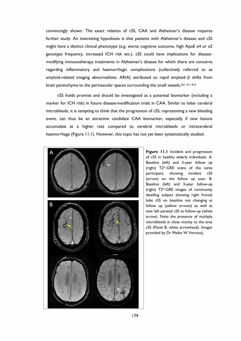
179
convincingly shown. The exact relation of cSS, CAA and Alzheimer’s disease requires
further study. An interesting hypothesis is that patients with Alzheimer’s disease and cSS
might have a distinct clinical phenotype (e.g. worse cognitive outcome, high ApoE e4 or e2
genotype frequency, increased ICH risk etc.). cSS could have implications for disease-
modifying immunotherapy treatments in Alzheimer’s disease for which there are concerns
regarding inflammatory and haemorrhagic complications (collectively referred to as
amyloid-related imaging abnormalities: ARIA) attributed to rapid amyloid–β shifts from
brain parenchyma to the perivascular spaces surrounding the small vessels.261, 411-413
cSS holds promise and should be investigated as a potential biomarker (including a
marker for ICH risk) in future disease-modification trials in CAA. Similar to lobar cerebral
microbleeds, it is tempting to think that the progression of cSS, representing a new bleeding
event, can thus be an attractive candidate CAA biomarker, especially if new lesions
accumulate at a higher rate compared to cerebral microbleeds or intracerebral
haemorrhage (Figure 11.1). However, this topic has not yet been systematically studied.
Figure 11.1 Incident and progression
of cSS in healthy elderly individuals. A.
Baseline (left) and 3-year follow up
(right) T2*-GRE scans of the same
participant, showing incident cSS
(arrow) on the follow up scan. B.
Baseline (left) and 3-year follow-up
(right) T2*-GRE images of community
dwelling subject showing right frontal
lobe cSS on baseline not changing at
follow up (yellow arrows) as well as
new left parietal cSS at follow-up (white
arrow). Note the presence of multiple
microbleeds in close vicinity to the area
cSS (Panel B, white arrowhead). Images
provided by Dr Meike W Vernooij.
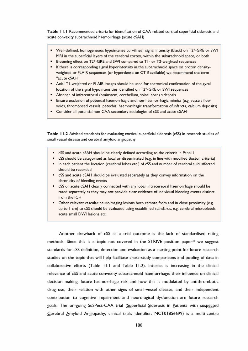
180
Table 11.1 Recommended criteria for identification of CAA-related cortical superficial siderosis and
acute convexity subarachnoid haemorrhage (acute cSAH)
Well-defined, homogeneous hypointense curvilinear signal intensity (black) on T2*-GRE or SWI
MRI in the superficial layers of the cerebral cortex, within the subarachnoid space, or both
Blooming effect on T2*-GRE and SWI compared to T1- or T2-weighted sequences
If there is corresponding signal hyperintensity in the subarachnoid space on proton density-
weighted or FLAIR sequences (or hyperdense on CT if available) we recommend the term
“acute cSAH”
Axial T1-weighted or FLAIR images should be used for anatomical confirmation of the gyral
location of the signal hypointensities identified on T2*-GRE or SWI sequences
Absence of infratentorial (brainstem, cerebellum, spinal cord) siderosis
Ensure exclusion of potential haemorrhagic and non-haemorrhagic mimics (e.g. vessels flow
voids, thrombosed vessels, petechial haemorrhagic transformation of infarcts, calcium deposits)
Consider all potential non-CAA secondary aetiologies of cSS and acute cSAH
Table 11.2 Advised standards for evaluating cortical superficial siderosis (cSS) in research studies of
small vessel disease and cerebral amyloid angiopathy
cSS and acute cSAH should be clearly defined according to the criteria in Panel 1
cSS should be categorised as focal or disseminated (e.g. in line with modified Boston criteria)
In each patient the location (cerebral lobes etc.) of cSS and number of cerebral sulci affected
should be recorded
cSS and acute cSAH should be evaluated separately as they convey information on the
chronicity of bleeding events
cSS or acute cSAH clearly connected with any lobar intracerebral haemorrhage should be
rated separately as they may not provide clear evidence of individual bleeding events distinct
from the ICH
Other relevant vascular neuroimaging lesions both remote from and in close proximity (e.g.
up to 1 cm) to cSS should be evaluated using established standards, e.g. cerebral microbleeds,
acute small DWI lesions etc.
Another drawback of cSS as a trial outcome is the lack of standardised rating
methods. Since this is a topic not covered in the STRIVE position paper31 we suggest
standards for cSS definition, detection and evaluation as a starting point for future research
studies on the topic that will help facilitate cross-study comparisons and pooling of data in
collaborative efforts (Table 11.1 and Table 11.2). Interest is increasing in the clinical
relevance of cSS and acute convexity subarachnoid haemorrhage: their influence on clinical
decision making, future haemorrhage risk and how this is modulated by antithrombotic
drug use, their relation with other signs of small-vessel disease, and their independent
contribution to cognitive impairment and neurological dysfunction are future research
goals. The on-going SuSPect-CAA trial (Superficial Siderosis in Patients with suspected
Cerebral Amyloid Angiopathy; clinical trials identifier: NCT01856699) is a multi-centre
181
study, which aims to prospectively evaluate acute cSAHs and cSS as independent predictors
for future stroke and mortality in patients with possible or probable CAA, as well as to
better define their clinical presentation and outcome. Data from the SuSPect-CAA study
may help define the role of cSS as a neuroimaging biomarker in future CAA treatment
trials.300 Further promising areas of research include correlations of cSS with non-invasive
amyloid-β molecular imaging with PET agents such as Pittsburgh compound B (PiB) to
determine its relationship to amyloid deposition,285, 289 assessment of associations with
ApoE genotype,319 and systematic radiological-pathological correlation studies. These
studies should shed further light on whether cSS identifies distinct phenotypes of CAA or
Alzheimer’s disease.
11.2.3 Non-haemorrhagic CAA markers
MRI-visible perivascular spaces
Perivascular spaces (also termed Virchow-Robin spaces) visible on MRI represent one of
the most recent, and perhaps less well known, candidate non-haemorrhagic biomarkers of
CAA. One of the most important advances in the field was the inclusion of perivascular
spaces as a neuroimaging feature of small vessel disease in the STRIVE consensus paper,31
based on recent data. Here, using a portfolio of hypothesis-driven studies (Chapters 5-8)
direct and robust evidence is provided that the topography of perivascular spaces may
differ according to the underlying arteriopathy, with CAA being preferentially associated
with high numbers of visible perivascular spaces in the centrum semiovale. In summary, the
main new findings presented are that: (a) perivascular spaces are very common in
spontaneous intracerebral haemorrhage; and (b) different mechanisms may account for
perivascular spaces according to their anatomical distribution in intracerebral haemorrhage,
consistent with the known localisation of the underlying pathology. Severe centrum
semiovale perivascular spaces are more prevalent in patients with lobar intracerebral
haemorrhage (probable or possible CAA) and are associated with age, supporting the idea
of being a promising new imaging marker of CAA. By contrast, the severity of basal ganglia
perivascular spaces is associated with markers of hypertensive arteriopathy suggesting
common pathophysiological mechanisms. (c) Severe centrum semiovale perivascular spaces
on MRI are present in 85.7% of patients with pathologically proven CAA but in none of the
control subjects with intracerebral haemorrhage but no pathological evidence of CAA.
Adding centrum semiovale perivascular spaces to current diagnostic criteria might increase
the sensitivity, without reducing specificity hence contribute to improving the in vivo
diagnosis of CAA. (d) MRI-visible perivascular spaces in the centrum semiovale are
182
associated with PET-based cerebrovascular amyloid-β burden, providing important
pathophysiological clues, and further highlighting their possible utility as a surrogate for
vascular amyloid burden.
Looking back in light of these findings, previous studies across different populations
have highlighted strong associations between basal ganglia perivascular spaces severity
(more consistently than centrum semiovale perivascular spaces) and hypertension, white
matter hyperintensities and lacunes, which are all typical neuroimaging features of
hypertensive arteriopathy.30, 270, 275 By contrast, centrum semiovale perivascular spaces were
not associated with conventional vascular risk factors, but only with increasing age,30, 272 so
may relate to a different underlying small vessel arteriopathy. This is supported by a recent
study among memory clinic patients, showing an association between centrum semiovale
perivascular spaces and lobar cerebral microbleeds count and basal ganglia perivascular
spaces and hypertension.274 The new observations presented in this thesis are in line with
these previous studies, but provide more direct evidence pertinent to CAA.
The link between centrum semiovale perivascular spaces on MRI and CAA is
particularly attractive as it may specifically reflect a central and probably early event in the
pathophysiology of the disease: interstitial fluid drainage impairment within the perivascular
spaces, caused by cumulative leptomeningeal and superficial cortical vascular amyloid-β
deposition.274, 276, 299 Data from molecular pathology studies and animal models support the
hypothesis that in CAA, amyloid-β could reach the vessel walls through perivascular spaces,
eventually aggregating within them and causing retrograde perivascular spaces due to
interstitial fluid blockage, setting in motion a vicious cycle which promotes further amyloid
deposition.276, 282 However, the exact mechanisms of perivascular space enlargement and
their appearance on MRI remain poorly understood. The very close anatomical relationship
of this perivascular compartment to the perforating small vessels (as recently demonstrated
in a 7-Tesla study414) suggests that potentially any small vessel pathology could lead to
dysfunction and morphological changes of the perivascular compartment (e.g.
enlargement),263 making them visible on conventional neuroimaging.
From the perspective of a pathologist, evidence from autopsy studies to indicate that
the signal changes seen on imaging are actually expansion of the perivascular spaces is still
very limited.268, 273 More direct correlation studies between MRI-visible perivascular spaces
and underlying pathology-morphology in the setting of CAA are needed if they are to be
assessed as a potential biomarker. In addition, as discussed in the relevant Chapters,
perivascular spaces will need analysis for their spatial and temporal relation to other
markers of CAA, incident appearance over time, and improved protocols for detecting and
183
quantifying them, ideally using volumetric semi-automated methods.415 Whether the
presence of several visible perivascular spaces is clinically significant also remains
controversial, although some studies have associated enlarged perivascular spaces with
worse cognitive function.267
White matter hyperintensities
White matter hyperintensities of presumed vascular origin are another very common non-
haemorrhagic manifestation of CAA on MRI. To date, voxel-based analyses have not
definitely shown different patterns of white matter hyperintensities between patients with
CAA and hypertensive arteriopathy.158, 179, 186 Based on the almost inverse topography of
CAA and hypertensive arteriopathy in the brain, one would expect striking differences in
the regional distribution of white matter hyperintensities (similar to lobar vs. deep cerebral
microbleeds, or perivascular spaces) between these small vessel diseases.186 Surprisingly,
some studies suggest no major differences in overall white matter hyperintensities
topography in CAA compared to hypertensive arteriopathy.158, 179 This relatively preserved
anatomic distribution despite the specific topography across small vessel diseases, with
greatest periventricular lesion burden around the frontal and occipital horns, indicates that
white matter injury might occur through chronic microvascular ischaemia in the most
vulnerable regions.186 Despite the lack of pathological specificity for white matter
hyperintensities,24 amyloid-related alterations in vascular reactivity observed in CAA, are
consistent with this hypothesis.377, 378 A more recent study using a semi-quantitative visual
scale suggested that, compared to normal elderly controls, patients with CAA-related lobar
intracerebral haemorrhage may have a higher prevalence of occipital-predominant (rather
than anterior) white matter hyperintensities.187 Although this finding is interesting and
consistent with the relative predilection of CAA vascular pathology for posterior cortical
regions,109 it requires further investigation. A recent study developed a quantitative method
to measure the antero-posterior distribution of white matter hyperintensities and provided
further lines of evidence that their distribution may indeed represent a promising
neuroimaging marker of CAA, at least in specific populations without ICH.416 In a cohort of
patients without intracerebral haemorrhage (n=59) and with pathological evaluation, more
posterior white matter hyperintensities distribution was an independent predictor of
pathology evidence of CAA, even in cases without lobar microbleeds.416 Also, in a separate
memory clinical cohort (n=259) it was shown that strictly lobar cerebral microbleeds were
associated with more posterior white matter hyperintensities distribution after adjusting
for other confounders.416
184
An easy and clinically meaningful visual assessment tool of the frontal-to-occipital
white matter hyperintensities distribution in patients with different microangiopathies has
not yet been established, and a quantitative method might not be practical or sufficient for
routine clinical use as a diagnostic biomarker. However, patients with advanced CAA have a
larger overall load (i.e. volume) of chronic microvascular white matter hyperintensities
compared to healthy elderly people158 or patients with Alzheimer’s disease alone.284, 417
PET-based cerebrovascular amyloid load is also strongly correlated with ischaemic white
matter hyperintensities in CAA.284 Although data are limited, CAA patients seem to have a
high pace of white matter hyperintensities progression189 Furthermore, damage to white
matter in CAA is associated with cognitive impairment independent of the effects of
haemorrhage in the brain,188, 418 making white matter hyperintensities progression a
reasonable biomarker to measure a clinically meaningful aspect of CAA-related brain
injury.300 In various other studies (including randomised trials) in the field of small vessel
disease, this model has been successfully implemented.2, 419 Whether a reduction in white
matter hyperintensities progression burden is associated with a decrease in the incidence of
cognitive and functional decline in CAA needs to be further tested.
Cerebral microinfarcts
Another non-haemorrhagic manifestation of advanced CAA that likely has an important
cumulative effect on cognitive function, are cerebral microinfarcts.197 These have been
defined histopathologically as tiny areas of infarction (generally up to a maximum of 1-2
mm), only visible on microscopic tissue examination. Because of their small size, they are
typically “invisible” in clinical-radiological studies that rely on conventional structural MRI.
Recently, two approaches have detected microinfarcts in vivo (likely at the large end their
size spectrum). The first approach is the detection of clinically silent small areas of
restricted diffusion on DWI MRI, thought to represent acute microinfarcts, typically
described in CAA-related intracerebral haemorrhage cohorts at a frequency of about
20%.46, 73, 199, 420 The transient appearance of DWI lesions, and their occurrence even in the
chronic post-intracerebral haemorrhage time window,199 suggests they may be part of an
ongoing active process, they must accumulate frequently (around 8-10 large microinfarcts
lesions per person per year – much higher compare to incident lobar microbleeds) and that
the lifetime burden of ischaemia in patients with CAA may therefore be substantial.
Nevertheless, this very transience nature of DWI lesions makes them hard to be used as
meaningful biomarkers. In addition, there is no neuropathological confirmation that these
lesions truly represent cerebral microinfarcts; some may represent ‘healed or healing’
185
microbleeds.421 The second approach for detecting microinfarcts in vivo, utilized ultra-high-
field MRI to reveal small permanent, FLAIR hyperintense and T1 hypointense, structural
lesions which have been correlated with microinfarcts at post-mortem.422 A cross-
validation of microinfarcts found on 7T MRI with 3T MRI will open the way for more
routine detection of these lesions as a biomarker of ongoing damage in CAA. Of course, a
key limitation of both approaches is that with current neuroimaging most burden of
cerebral microinfarcts is left undetectable.423
11.3 The concept of potential cerebral amyloid
angiopathy phenotypes: rational and promise for clinically
meaningful biomarkers
As we have seen, during the past decades, progress has been made in understanding the
pathophysiology and imaging-clinical expression of CAA and cerebral small vessel disease in
general, with trials of disease-modifying drugs being the ultimate goal. What has also
become evident is that CAA in not a uniform, but rather a very heterogeneous entity.
While sporadic CAA is commonly found in the elderly, it is usually mild and clinically silent.
Only a fraction of patients with CAA will go on to develop symptomatic intracerebral
haemorrhage, despite the fact that many more might have evidence of haemorrhagic
manifestations on neuroimaging, such as multiple lobar cerebral microbleeds or cortical
superficial siderosis.28, 37 In other patients, CAA might cause only white matter
hyperintensities and cognitive impairment.416 In a distinct minority of patients, CAA may be
revealed by a striking acute inflammatory encephalopathy and nil else (inflammatory
CAA).162 CAA is also very often present in cases of Alzheimer's syndrome. From these and
other observations a picture arises where CAA might have a spectrum of distinct
phenotypes of neuropathology and neuroimaging manifestations, comorbid conditions and
ultimately clinical expression.167, 403
Currently, at least two pathological subtypes of CAA have been identified: CAA-type
1, characterised by amyloid in cortical capillaries (with or without involvement of other
vessels), and CAA-type 2, where amyloid deposits are restricted to leptomeningeal and
cortical arteries, but not capillaries.38 The APOE ε4 allele is most strongly associated with
CAA-type 1, while APOE ε2 is more associated with CAA-type 2.107 In addition, it is
generally accepted that APOE ε4 promotes vascular amyloid deposition, while APOE ε2
promotes structural vasculopathic changes in amyloid-laden vessels, making them prone to
rupture and intracerebral bleeding. CAA-type 1 (especially capillary, more than arteriolar
186
CAA) appears to be more closely associated with parenchymal amyloid deposition in
Alzheimer’s disease108 and may cause luminal obstruction in the most severe stages,
potentially explaining the low incidence of microbleeds and intracerebral haemorrhage in
patients with Alzheimer’s disease and the association between microinfarcts, ischaemic
white matter hyperintensities and cognitive impairment in CAA.
Thus, APOE genotype probably influences CAA phenotype, but it is likely that this is
not the whole story. Although multiple lobar microbleeds are a marker of increased risk of
intracerebral haemorrhage, an interesting study in a cohort of consecutive CAA patients
(n=46) suggests that cerebral microbleeds arising from CAA-related pathology might be in
some respects distinct from macrohaemorrhages, since they have a bimodal, rather than
continuous, size distribution on MRI.198 In a subset of patients who underwent autopsy,
those with high microbleeds counts (“microbleeders”) had increased wall thickness of
amyloid-positive vessels compared to those with relatively low microbleeds counts
(“macrobleeders”).198 Surprisingly, cortical superficial siderosis, another CAA haemorrhagic
marker associated with high risk of haemorrhage, is not associated with lobar microbleeds
as shown in Chapter 3.301 It is possible that the topography and severity of CAA may affect
the relative likelihood of the small vessel pathology resulting in cerebral microbleeds versus
macrobleeds or cortical superficial siderosis and convexity subarachnoid haemorrhage. For
example, heavy vascular amyloid might actually make cerebral microbleeds more likely than
macrobleeding, and leptomeningeal versus deeper cortical CAA might make siderosis more
likely than cerebral microbleeds. It is also important to bear in mind that ICH may result
from mechanisms not related to the presence of cerebral microbleeds or siderosis.
It is tempting to speculate that multiple different (endo-)phenotypes of CAA exist. This
assumption touches onto the very basic question of what pathophysiological mechanisms
determine whether CAA causes intracerebral haemorrhage or other haemorrhagic
manifestations versus non-haemorrhagic manifestations, or cognitive decline. This question
was partly addressed in the study presented in Chapter 9, and although literature to
support firm conclusions is not yet available, it suggests that a “phenotype approach” in
CAA provides a conceptual model which might allow greater reliance on the clusters of
different surrogate biomarkers in future CAA trials.300 This approach could also strengthen
the links between imaging biomarkers and neurological function. CAA is a dynamic,
chronically progressive disease and future longitudinal studies should determine whether
the described CAA phenotypes represent really distinct entities adhering to their
characteristic imaging feature over time. Maybe one interesting study to perform would be
to gather large numbers of possible and probable CAA cases as part of an international
network and do principal component and cluster analysis, a statistical procedure that
187
converts a set of observations of possibly correlated variables into a set of values of linearly
uncorrelated variables called principal components. This method can potentially allow
determining clusters of clinical-radiological manifestations that could be used to determine
prospectively if these phenotypes are really distinct.
Another key requirement is the identification of the exact role of CAA in the context
of Alzheimer’s disease:320 is amyloid vascular degeneration a silent partner or a key player in
the natural history of neurodegeneration? What are the differences and similarities with
sporadic CAA and what lessons can we learn from immunotherapeutic trials of Alzheimer’s
disease that can be applied to the design of CAA trials? For example, CAA is associated
with cerebral microinfarcts and given the common association between CAA and
Alzheimer pathology, one mechanism contributing to cognitive impairment in Alzheimer’s
disease patients may thus be widespread microinfarcts, at least in a subgroup of subjects
with especially advanced CAA.157, 305 In turn, the pattern of cognitive impairment seen in
advanced sporadic CAA could represent the cumulative effects of the various haemorrhagic
and non-haemorrhagic tissue injuries discussed earlier, in combination with coexistent mild
Alzheimer’s disease pathology. In addition, as shown in Chapter 9, CAA patients with
cognitive impairment and Alzheimer’s disease pathology with an APOE e2 genotype might
be at increased risk of certain haemorrhagic manifestations of small vessel disease, including
intracerebral haemorrhage.83, 424
11.4 Final conclusions and future perspectives
Although the use of the clinical and MRI biomarkers presented here should play a key role,
future studies should aim to provide stronger support and further insights from
pathological-imaging correlations, improved imaging modalities and larger study populations
as part of multicentre collaborations and international networks. Obviously an urgent need
is to improve the sensitivity and specificity of diagnostic criteria for CAA; a large
collaborative effort in underway to explore the value of a range of structural imaging
makers presented in this thesis as part of modified Boston diagnostic criteria. Another key
area of need is the identification of neuroimaging biomarker dynamic progression over time
and whether these also capture clinically relevant changes, which may also contribute in
identifying patients at an earlier stage of the disease, ideally before the serious
complications of symptomatic intracerebral haemorrhage or cognitive impairment has
developed. Early identification will clearly provide the best chance of efficacy for future
disease-modifying treatments; however, a big challenge is that not all people with CAA will
188
go on to develop problems. Advanced imaging methods together with improved
pathophysiological understanding of CAA-related or in general small vessel disease related
degeneration, will validate new promising biomarkers reflecting different steps in CAA
pathogenesis in addition to the known haemorrhagic and non-haemorrhagic structural
damage and their clinical consequences. New promising biomarkers in CAA include
molecular imaging of amyloid in the brain (e.g. using amyloid radioligands, including 11C
Pittsburgh Compound B - PiB),200 quantitative measures of brain structural integrity and
cerebral blood flow, such as diffusion tensor imaging and arterial spin labelling respectively,
fMRI as well as cerebrospinal fluid measures of amyloid and markers of neurodegeneration
or retinal abnormalities. Studies in the near future should systematically investigate how
strongly and specifically each of these biomarkers are related to putative CAA and its
severely as assessed by known structural brain imaging findings (e.g. cerebral microbleeds,
white matter hyperintensities), and to clinical consequences (e.g. cognitive function). The
work described in this thesis has begun to increase the range of potential clinical and MRI
biomarkers for CAA, and should contribute to the ultimate aims of early diagnosis and
disease modification to slow the progression of this common, yet enigmatic and devastating
disease.

189
Appendices
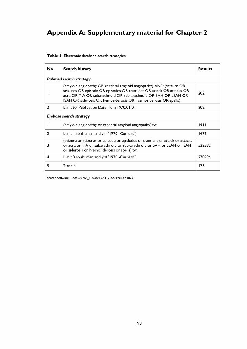
190
Appendix A: Supplementary material for Chapter 2
Table 1. Electronic database search strategies
No Search history Results
Pubmed search strategy
1
(amyloid angiopathy OR cerebral amyloid angiopathy) AND (seizure OR
seizures OR episode OR episodes OR transient OR attack OR attacks OR
aura OR TIA OR subarachnoid OR sub-arachnoid OR SAH OR cSAH OR
fSAH OR siderosis OR hemosiderosis OR haemosiderosis OR spells)
202
2 Limit to: Publication Date from 1970/01/01 202
Embase search strategy
1 (amyloid angiopathy or cerebral amyloid angiopathy).tw. 1911
2 Limit 1 to (human and yr="1970 -Current") 1472
3
(seizure or seizures or episode or epidodes or transient or attack or attacks
or aura or TIA or subarachnoid or sub-arachnoid or SAH or cSAH or fSAH
or siderosis or h?emosiderosis or spells).tw.
522882
4 Limit 3 to (human and yr="1970 -Current") 270996
5 2 and 4 175
Search software used: OvidSP_UI03.04.02.112, SourceID 54875
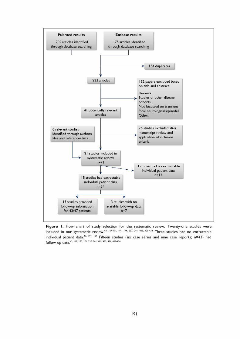
191
Figure 1. Flow chart of study selection for the systematic review. Twenty-one studies were
included in our systematic review.43, 167-171, 191, 194, 237, 241, 405, 425-434 Three studies had no extractable
individual patient data.43, 191, 194 Fifteen studies (six case series and nine case reports; n=43) had
follow-up data.43, 167, 170, 171, 237, 241, 405, 425, 426, 429-434
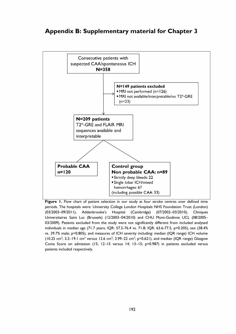
192
Appendix B: Supplementary material for Chapter 3
Figure 1. Flow chart of patient selection in our study at four stroke centres over defined time
periods. The hospitals were: University College London Hospitals NHS Foundation Trust (London)
(03/2003–09/2011), Addenbrooke’s Hospital (Cambridge) (07/2002–03/2010), Cliniques
Universitaires Saint Luc (Brussels) (12/2003–04/2010) and CHU Mont-Godinne UCL (08/2005–
03/2009). Patients excluded from the study were not significantly different from included analysed
individuals in median age (71.7 years; IQR: 57.5-76.4 vs. 71.8; IQR: 63.6-77.5, p=0.205), sex (38.4%
vs. 39.7% male; p=0.805), and measures of ICH severity including: median (IQR range) ICH volume
(10.25 cm3; 3.2–19.1 cm3 versus 12.6 cm3; 3.99–22 cm3; p=0.621), and median (IQR range) Glasgow
Coma Score on admission (15; 12–15 versus 14; 13–15; p=0.987) in patients excluded versus
patients included respectively.
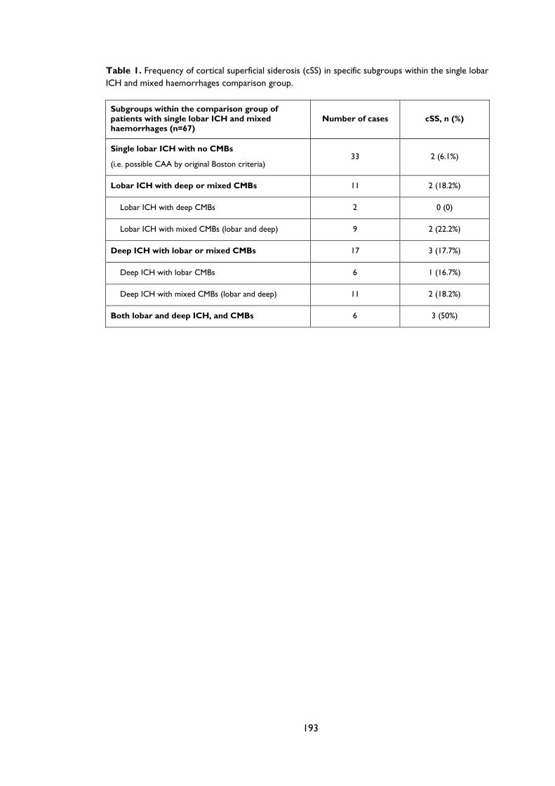
193
Table 1. Frequency of cortical superficial siderosis (cSS) in specific subgroups within the single lobar
ICH and mixed haemorrhages comparison group.
Subgroups within the comparison group of
patients with single lobar ICH and mixed
haemorrhages (n=67)
Number of cases cSS, n (%)
Single lobar ICH with no CMBs
(i.e. possible CAA by original Boston criteria) 33 2 (6.1%)
Lobar ICH with deep or mixed CMBs 11 2 (18.2%)
Lobar ICH with deep CMBs 2 0 (0)
Lobar ICH with mixed CMBs (lobar and deep) 9 2 (22.2%)
Deep ICH with lobar or mixed CMBs 17 3 (17.7%)
Deep ICH with lobar CMBs 6 1 (16.7%)
Deep ICH with mixed CMBs (lobar and deep) 11 2 (18.2%)
Both lobar and deep ICH, and CMBs 6 3 (50%)
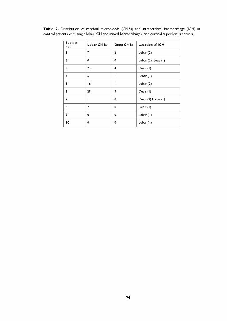
194
Table 2. Distribution of cerebral microbleeds (CMBs) and intracerebral haemorrhage (ICH) in
control patients with single lobar ICH and mixed haemorrhages, and cortical superficial siderosis.
Subject
no. Lobar CMBs Deep CMBs Location of ICH
1 7 2 Lobar (2)
2 0 0 Lobar (2); deep (1)
3 23 4 Deep (1)
4 6 1 Lobar (1)
5 16 1 Lobar (2)
6 28 3 Deep (1)
7 1 0 Deep (2) Lobar (1)
8 2 0 Deep (1)
9 0 0 Lobar (1)
10 0 0 Lobar (1)
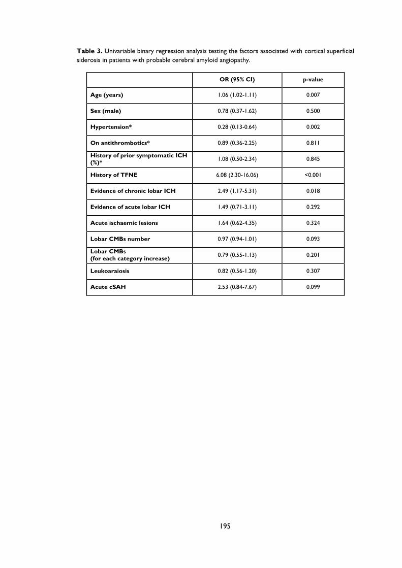
195
Table 3. Univariable binary regression analysis testing the factors associated with cortical superficial
siderosis in patients with probable cerebral amyloid angiopathy.
OR (95% CI) p-value
Age (years) 1.06 (1.02-1.11) 0.007
Sex (male) 0.78 (0.37-1.62) 0.500
Hypertension* 0.28 (0.13-0.64) 0.002
On antithrombotics* 0.89 (0.36-2.25) 0.811
History of prior symptomatic ICH
(%)* 1.08 (0.50-2.34) 0.845
History of TFNE 6.08 (2.30-16.06) <0.001
Evidence of chronic lobar ICH 2.49 (1.17-5.31) 0.018
Evidence of acute lobar ICH 1.49 (0.71-3.11) 0.292
Acute ischaemic lesions 1.64 (0.62-4.35) 0.324
Lobar CMBs number 0.97 (0.94-1.01) 0.093
Lobar CMBs
(for each category increase) 0.79 (0.55-1.13) 0.201
Leukoaraiosis 0.82 (0.56-1.20) 0.307
Acute cSAH 2.53 (0.84-7.67) 0.099
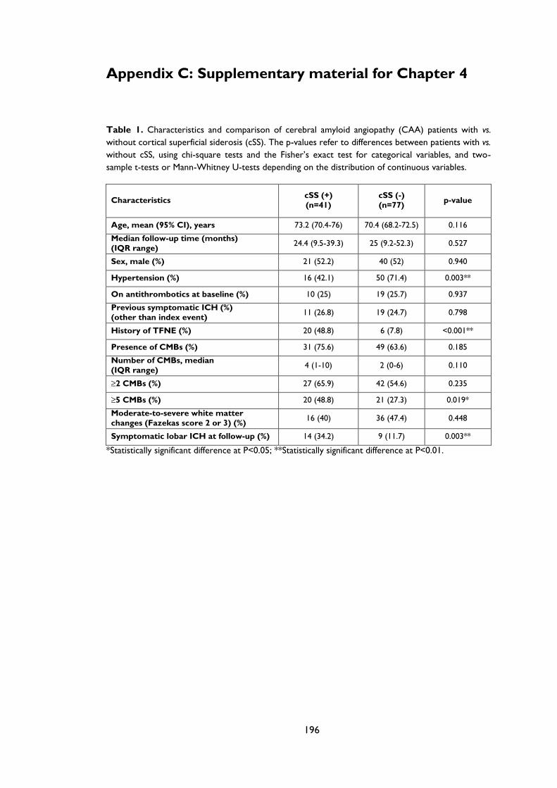
196
Appendix C: Supplementary material for Chapter 4
Table 1. Characteristics and comparison of cerebral amyloid angiopathy (CAA) patients with vs.
without cortical superficial siderosis (cSS). The p-values refer to differences between patients with vs.
without cSS, using chi-square tests and the Fisher’s exact test for categorical variables, and two-
sample t-tests or Mann-Whitney U-tests depending on the distribution of continuous variables.
Characteristics cSS (+)
(n=41)
cSS (-)
(n=77) p-value
Age, mean (95% CI), years 73.2 (70.4-76) 70.4 (68.2-72.5) 0.116
Median follow-up time (months)
(IQR range) 24.4 (9.5-39.3) 25 (9.2-52.3) 0.527
Sex, male (%) 21 (52.2) 40 (52) 0.940
Hypertension (%) 16 (42.1) 50 (71.4) 0.003**
On antithrombotics at baseline (%) 10 (25) 19 (25.7) 0.937
Previous symptomatic ICH (%)
(other than index event) 11 (26.8) 19 (24.7) 0.798
History of TFNE (%) 20 (48.8) 6 (7.8) <0.001**
Presence of CMBs (%) 31 (75.6) 49 (63.6) 0.185
Number of CMBs, median
(IQR range) 4 (1-10) 2 (0-6) 0.110
≥2 CMBs (%) 27 (65.9) 42 (54.6) 0.235
≥5 CMBs (%) 20 (48.8) 21 (27.3) 0.019*
Moderate-to-severe white matter
changes (Fazekas score 2 or 3) (%) 16 (40) 36 (47.4) 0.448
Symptomatic lobar ICH at follow-up (%) 14 (34.2) 9 (11.7) 0.003**
*Statistically significant difference at P<0.05; **Statistically significant difference at P<0.01.

197
Table 2. Different multivariate analysis models of predictors of symptomatic lobar intracerebral
haemorrhage (ICH) during follow-up in patients with cerebral amyloid angiopathy. cSS: cortical
superficial siderosis; CMBs: cerebral microbleeds.
Model 1: Presence of cSS
Model 1A HR (95%CI) p-value
Presence of cSS 2.27 (0.95-5.44) 0.065
CMBs number 1.02 (0.99-1.05) 0.245
Previous symptomatic ICH 2.01 (0.78-5.17) 0.148
Age (for every year increase) 1.07 (1.02-1.13) 0.011
Model 1B HR (95%CI) p-value
Presence of cSS 2.37 (0.98-5.75) 0.057
Presence of ≥2 CMBs 1.09 (0.44-2.67) 0.857
Previous symptomatic ICH 2.04 (0.80-5.23) 0.138
Age (for every year increase) 1.07 (1.01-1.13) 0.016
Model 1C HR (95%CI) p-value
Presence of cSS 2.50 (1.00-6.18) 0.049
Presence of ≥5 CMBs 0.90 (0.37-2.20) 0.815
Previous symptomatic ICH 2.03 (0.79-5.23) 0.141
Age (for every year increase) 1.07 (1.01-1.13) 0.014
198
Model 2: Increasing cSS category (from no, to focal and disseminated cSS)
Model 2A HR (95%CI) p-value
Increasing cSS category
(from no, to focal and disseminated cSS) 1.72 (1.07-2.75) 0.025
CMBs number 1.02 (0.98-1.05) 0.369
Previous symptomatic ICH 1.96 (0.77-5.04) 0.160
Age (for every year increase) 1.08 (1.02-1.14) 0.010
Model 2B HR (95%CI) p-value
Increasing cSS category
(from no, to focal and disseminated cSS) 1.78 (1.11-2.87) 0.018
Presence of ≥2 CMBs 1.03 (0.42-2.53) 0.950
Previous symptomatic ICH 1.97 (0.77-5.05) 0.156
Age (for every year increase) 1.07 (1.01-1.13) 0.015
Model 2C HR (95%CI) p-value
Increasing cSS category
(from no, to focal and disseminated cSS) 1.87 (1.14-3.08) 0.014
Presence of ≥5CMBs 0.79 (0.32-1.97) 0.612
Previous symptomatic ICH 1.96 (0.77-5.04) 0.161
Age (for every year increase) 1.07 (1.02-1.13) 0.012
199
Model 3: Disseminated cSS (vs. all other patients: no and focal cSS)
Model 3A HR (95%CI) p-value
Disseminated cSS
(vs. all other patients: no and focal cSS) 2.78 (1.17-6.58) 0.020
CMBs number 1.01 (0.98-1.05) 0.404
Previous symptomatic ICH 1.98 (0.77-5.06) 0.155
Age (for every year increase) 1.08 (1.02-1.15) 0.007
Model 3B HR (95%CI) p-value
Disseminated cSS
(vs. all other patients: no and focal cSS) 2.97 (1.26-7.01) 0.013
Presence of ≥2 CMBs 1.05 (0.43-2.58) 0.914
Previous symptomatic ICH 1.99 (0.78-5.07) 0.151
Age (for every year increase) 1.08 (1.02-1.14) 0.010
Model 3C HR (95%CI) p-value
Disseminated cSS
(vs. all other patients: no and focal cSS) 3.26 (1.32-8.01) 0.010
Presence of ≥5CMBs 0.79 (0.32-1.98) 0.618
Previous symptomatic ICH 1.99 (0.78-5.10) 0.149
Age (for every year increase) 1.08 (1.02-1.14) 0.008
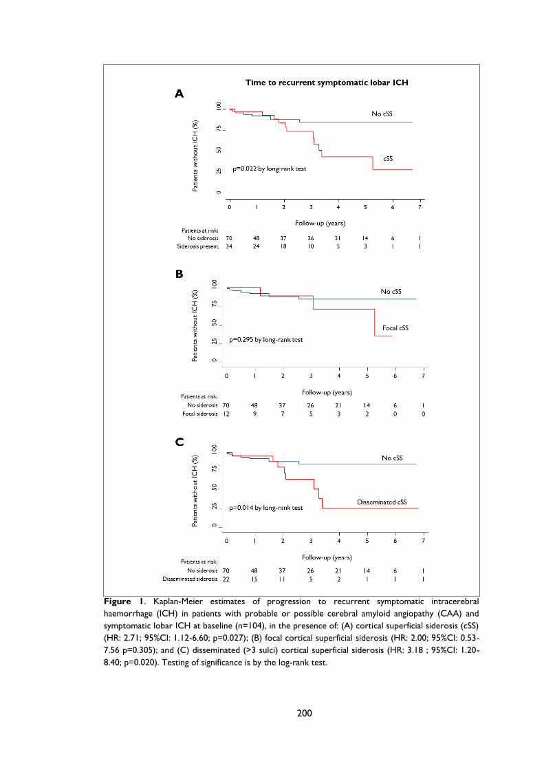
200
Figure 1. Kaplan-Meier estimates of progression to recurrent symptomatic intracerebral
haemorrhage (ICH) in patients with probable or possible cerebral amyloid angiopathy (CAA) and
symptomatic lobar ICH at baseline (n=104), in the presence of: (A) cortical superficial siderosis (cSS)
(HR: 2.71; 95%CI: 1.12-6.60; p=0.027); (B) focal cortical superficial siderosis (HR: 2.00; 95%CI: 0.53-
7.56 p=0.305); and (C) disseminated (>3 sulci) cortical superficial siderosis (HR: 3.18 ; 95%CI: 1.20-
8.40; p=0.020). Testing of significance is by the log-rank test.
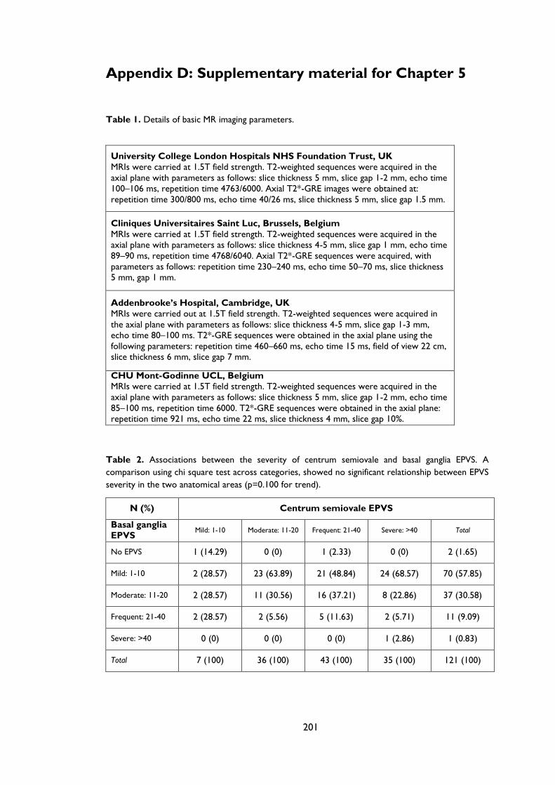
201
Appendix D: Supplementary material for Chapter 5
Table 1. Details of basic MR imaging parameters.
University College London Hospitals NHS Foundation Trust, UK
MRIs were carried at 1.5T field strength. T2-weighted sequences were acquired in the
axial plane with parameters as follows: slice thickness 5 mm, slice gap 1-2 mm, echo time
100–106 ms, repetition time 4763/6000. Axial T2*-GRE images were obtained at:
repetition time 300/800 ms, echo time 40/26 ms, slice thickness 5 mm, slice gap 1.5 mm.
Cliniques Universitaires Saint Luc, Brussels, Belgium
MRIs were carried at 1.5T field strength. T2-weighted sequences were acquired in the
axial plane with parameters as follows: slice thickness 4-5 mm, slice gap 1 mm, echo time
89–90 ms, repetition time 4768/6040. Axial T2*-GRE sequences were acquired, with
parameters as follows: repetition time 230–240 ms, echo time 50–70 ms, slice thickness
5 mm, gap 1 mm.
Addenbrooke’s Hospital, Cambridge, UK
MRIs were carried out at 1.5T field strength. T2-weighted sequences were acquired in
the axial plane with parameters as follows: slice thickness 4-5 mm, slice gap 1-3 mm,
echo time 80–100 ms. T2*-GRE sequences were obtained in the axial plane using the
following parameters: repetition time 460–660 ms, echo time 15 ms, field of view 22 cm,
slice thickness 6 mm, slice gap 7 mm.
CHU Mont-Godinne UCL, Belgium
MRIs were carried at 1.5T field strength. T2-weighted sequences were acquired in the
axial plane with parameters as follows: slice thickness 5 mm, slice gap 1-2 mm, echo time
85–100 ms, repetition time 6000. T2*-GRE sequences were obtained in the axial plane:
repetition time 921 ms, echo time 22 ms, slice thickness 4 mm, slice gap 10%.
Table 2. Associations between the severity of centrum semiovale and basal ganglia EPVS. A
comparison using chi square test across categories, showed no significant relationship between EPVS
severity in the two anatomical areas (p=0.100 for trend).
N (%) Centrum semiovale EPVS
Basal ganglia
EPVS Mild: 1-10 Moderate: 11-20 Frequent: 21-40 Severe: >40 Total
No EPVS 1 (14.29) 0 (0) 1 (2.33) 0 (0) 2 (1.65)
Mild: 1-10 2 (28.57) 23 (63.89) 21 (48.84) 24 (68.57) 70 (57.85)
Moderate: 11-20 2 (28.57) 11 (30.56) 16 (37.21) 8 (22.86) 37 (30.58)
Frequent: 21-40 2 (28.57) 2 (5.56) 5 (11.63) 2 (5.71) 11 (9.09)
Severe: >40 0 (0) 0 (0) 0 (0) 1 (2.86) 1 (0.83)
Total 7 (100) 36 (100) 43 (100) 35 (100) 121 (100)
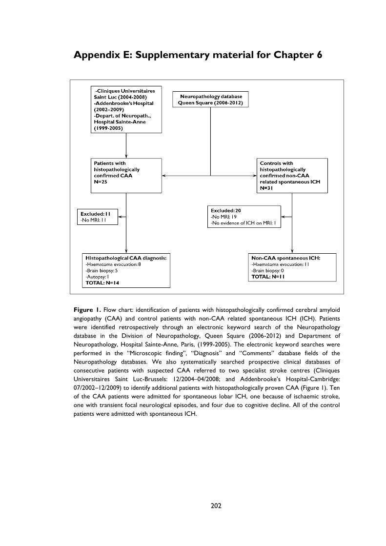
202
Appendix E: Supplementary material for Chapter 6
Figure 1. Flow chart: identification of patients with histopathologically confirmed cerebral amyloid
angiopathy (CAA) and control patients with non-CAA related spontaneous ICH (ICH). Patients
were identified retrospectively through an electronic keyword search of the Neuropathology
database in the Division of Neuropathology, Queen Square (2006-2012) and Department of
Neuropathology, Hospital Sainte-Anne, Paris, (1999-2005). The electronic keyword searches were
performed in the “Microscopic finding”, “Diagnosis” and “Comments” database fields of the
Neuropathology databases. We also systematically searched prospective clinical databases of
consecutive patients with suspected CAA referred to two specialist stroke centres (Cliniques
Universitaires Saint Luc-Brussels: 12/2004–04/2008; and Addenbrooke’s Hospital-Cambridge:
07/2002–12/2009) to identify additional patients with histopathologically proven CAA (Figure 1). Ten
of the CAA patients were admitted for spontaneous lobar ICH, one because of ischaemic stroke,
one with transient focal neurological episodes, and four due to cognitive decline. All of the control
patients were admitted with spontaneous ICH.
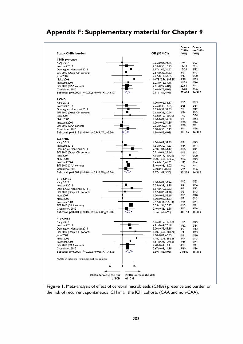
203
Appendix F: Supplementary material for Chapter 9
Figure 1. Meta-analysis of effect of cerebral microbleeds (CMBs) presence and burden on
the risk of recurrent spontaneous ICH in all the ICH cohorts (CAA and non-CAA).
204
References
1. Greenberg SM. Small vessels, big problems. N Engl J Med 2006; 354(14): 1451-3.
2. Pantoni L. Cerebral small vessel disease: from pathogenesis and clinical
characteristics to therapeutic challenges. Lancet Neurol 2010; 9(7): 689-701.
3. Wardlaw JM, Smith C, Dichgans M. Mechanisms of sporadic cerebral small vessel
disease: insights from neuroimaging. Lancet Neurol 2013; 12(5): 483-97.
4. Thompson CS, Hakim AM. Living beyond our physiological means: small vessel
disease of the brain is an expression of a systemic failure in arteriolar function: a unifying
hypothesis. Stroke 2009; 40(5): e322-30.
5. Debette S, Markus HS. The clinical importance of white matter hyperintensities on
brain magnetic resonance imaging: systematic review and meta-analysis. BMJ 2010; 341:
c3666.
6. Altmann-Schneider I, Trompet S, de Craen AJ, et al. Cerebral microbleeds are
predictive of mortality in the elderly. Stroke 2011; 42(3): 638-44.
7. Pantoni L, Sarti C, Alafuzoff I, et al. Postmortem examination of vascular lesions in
cognitive impairment: a survey among neuropathological services. Stroke 2006; 37(4): 1005-
9.
8. Scuteri A, Nilsson PM, Tzourio C, Redon J, Laurent S. Microvascular brain damage
with aging and hypertension: pathophysiological consideration and clinical implications.
Journal of hypertension 2011; 29(8): 1469-77.
9. Fisher CM. Lacunar strokes and infarcts: a review. Neurology 1982; 32(8): 871-6.
10. Gorelick PB, Scuteri A, Black SE, et al. Vascular contributions to cognitive
impairment and dementia: a statement for healthcare professionals from the american heart
association/american stroke association. Stroke 2011; 42(9): 2672-713.
11. The Lancet N. A united approach to vascular disease and neurodegeneration.
Lancet neurology 2012; 11(4): 293.
12. Launer LJ, Hofman A. Frequency and impact of neurologic diseases in the elderly of
Europe: A collaborative study of population-based cohorts. Neurology 2000; 54(11 Suppl 5):
S1-8.
13. Pantoni L, Garcia JH. The significance of cerebral white matter abnormalities 100
years after Binswanger's report. A review. Stroke 1995; 26(7): 1293-301.
14. Lammie GA. Hypertensive cerebral small vessel disease and stroke. Brain Pathol
2002; 12(3): 358-70.
15. Lammie GA, Brannan F, Slattery J, Warlow C. Nonhypertensive cerebral small-
vessel disease. An autopsy study. Stroke 1997; 28(11): 2222-9.
16. Cole FM, Yates P. Intracerebral microaneurysms and small cerebrovascular lesions.
Brain 1967; 90(4): 759-68.
17. Feigin I, Prose P. Hypertensive fibrinoid arteritis of the brain and gross cerebral
hemorrhage: a form of "hyalinosis". Archives of neurology 1959; 1: 98-110.
18. Rosenblum WI. Cerebral hemorrhage produced by ruptured dissecting aneurysm in
miliary aneurysm. Annals of neurology 2003; 54(3): 376-8.
19. Fisher CM. Pathological observations in hypertensive cerebral hemorrhage. J
Neuropathol Exp Neurol 1971; 30(3): 536-50.
205
20. Takebayashi S, Kaneko M. Electron microscopic studies of ruptured arteries in
hypertensive intracerebral hemorrhage. Stroke; a journal of cerebral circulation 1983; 14(1):
28-36.
21. Rosenblum WI. Fibrinoid necrosis of small brain arteries and arterioles and miliary
aneurysms as causes of hypertensive hemorrhage: a critical reappraisal. Acta Neuropathol
2008; 116(4): 361-9.
22. Fisher CM. The arterial lesions underlying lacunes. Acta Neuropathol 1968; 12(1): 1-
15.
23. Fisher CM. Hypertensive cerebral hemorrhage. Demonstration of the source of
bleeding. Journal of neuropathology and experimental neurology 2003; 62(1): 104-7.
24. Gouw AA, Seewann A, van der Flier WM, et al. Heterogeneity of small vessel
disease: a systematic review of MRI and histopathology correlations. J Neurol Neurosurg
Psychiatry 2011; 82(2): 126-35.
25. van Swieten JC, Staal S, Kappelle LJ, Derix MM, van Gijn J. Are white matter lesions
directly associated with cognitive impairment in patients with lacunar infarcts? Journal of
neurology 1996; 243(2): 196-200.
26. Schmidt R, Grazer A, Enzinger C, et al. MRI-detected white matter lesions: do they
really matter? J Neural Transm 2011; 118(5): 673-81.
27. Charidimou A, Werring DJ. Cerebral microbleeds: detection, mechanisms and
clinical challenges Future Neurology 2011; 6(5): 587-611.
28. Charidimou A, Gang Q, Werring DJ. Sporadic cerebral amyloid angiopathy
revisited: recent insights into pathophysiology and clinical spectrum. J Neurol Neurosurg
Psychiatry 2012; 83(2): 124-37.
29. Smith EE, Schneider JA, Wardlaw JM, Greenberg SM. Cerebral microinfarcts: the
invisible lesions. Lancet Neurol 2012; 11(3): 272-82.
30. Doubal FN, MacLullich AM, Ferguson KJ, Dennis MS, Wardlaw JM. Enlarged
perivascular spaces on MRI are a feature of cerebral small vessel disease. Stroke 2010;
41(3): 450-4.
31. Wardlaw JM, Smith EE, Biessels GJ, et al. Neuroimaging standards for research into
small vessel disease and its contribution to ageing and neurodegeneration. Lancet Neurol
2013; 12(8): 822-38.
32. Chen YW, Lee MJ, Smith EE. Cerebral amyloid angiopathy in East and West.
International journal of stroke : official journal of the International Stroke Society 2010; 5(5): 403-
11.
33. Bejot Y, Cordonnier C, Durier J, Aboa-Eboule C, Rouaud O, Giroud M.
Intracerebral haemorrhage profiles are changing: results from the Dijon population-based
study. Brain 2013; 136(Pt 2): 658-64.
34. Lovelock CE, Molyneux AJ, Rothwell PM. Change in incidence and aetiology of
intracerebral haemorrhage in Oxfordshire, UK, between 1981 and 2006: a population-
based study. Lancet neurology 2007; 6(6): 487-93.
35. Greenberg SM, Vernooij MW, Cordonnier C, et al. Cerebral microbleeds: a guide
to detection and interpretation. Lancet Neurol 2009; 8(2): 165-74.
36. Brandner S. Histopathology of cerebral microbleeds. In: Werring DJ, ed. Cerebral
Microbleeds: Pathophysiology to Clinical Practice. Cambridge, England: Cambridge
University Press; 2011: 49-64.
206
37. Vinters HV. Cerebral amyloid angiopathy. A critical review. Stroke 1987; 18(2): 311-
24.
38. Attems J, Jellinger K, Thal DR, Van Nostrand W. Review: Sporadic cerebral amyloid
angiopathy. Neuropathol Appl Neurobiol 2011; 37(1): 75-93.
39. Oppenheim G. Über “drusige Nekrosen” in der Grosshirnrinde. Neurol Centralbl
1909; 28: 410-3.
40. Scholz W. Studien zur pathologie der hirngefabe II: die drusige entartung der
hirnarterien und capillaren
Z gesamte Neurol Psychiatr 1938; 162: 694-715.
41. Neumann MA. Combined amyloid vascular changes and argyrophilic plaques in the
central nervous system. J Neuropathol Exp Neurol 1960; 19: 370-82.
42. Jellinger K. Cerebrovascular amyloidosis with cerebral hemorrhage. J Neurol 1977;
214(3): 195-206.
43. Okazaki H, Reagan TJ, Campbell RJ. Clinicopathologic studies of primary cerebral
amyloid angiopathy. Mayo Clin Proc 1979; 54(1): 22-31.
44. Adeoye O, Broderick JP. Advances in the management of intracerebral
hemorrhage. Nat Rev Neurol 2010; 6(11): 593-601.
45. Qureshi AI, Mendelow AD, Hanley DF. Intracerebral haemorrhage. Lancet 2009;
373(9675): 1632-44.
46. Kimberly WT, Gilson A, Rost NS, et al. Silent ischemic infarcts are associated with
hemorrhage burden in cerebral amyloid angiopathy. Neurology 2009; 72(14): 1230-5.
47. Menon RS, Kidwell CS. Neuroimaging demonstration of evolving small vessel
ischemic injury in cerebral amyloid angiopathy. Stroke 2009; 40(12): e675-7.
48. Johnson KA, Gregas M, Becker JA, et al. Imaging of amyloid burden and distribution
in cerebral amyloid angiopathy. Ann Neurol 2007; 62(3): 229-34.
49. Greenberg SM, Grabowski T, Gurol ME, et al. Detection of isolated
cerebrovascular beta-amyloid with Pittsburgh compound B. Ann Neurol 2008; 64(5): 587-91.
50. Ly JV, Donnan GA, Villemagne VL, et al. 11C-PIB binding is increased in patients
with cerebral amyloid angiopathy-related hemorrhage. Neurology 2010; 74(6): 487-93.
51. About a peculiar disease of the cerebral cortex. By Alois Alzheimer, 1907
(Translated by L. Jarvik and H. Greenson). Alzheimer Dis Assoc Disord 1987; 1(1): 3-8.
52. Divry P. Etude histochimique des plaques séniles. J Belge Neurol Psychiatry 1927; 27:
643–57.
53. Morel F, Wildi E. [Study of various cerebral changes in aged]. Schweizer Archiv fur
Neurologie und Psychiatrie Archives suisses de neurologie et de psychiatrie Archivio svizzero di
neurologia e psichiatria 1955; 76(1-2): 174-223.
54. Morel F. Petite contribution a` l‘e´tude d’une angiopathie apparemment dyshorique
et topistique. Rev Mens Psychiatr Neurol 1950; 120: 352-7.
55. Pantelakis S. [A particular type of senile angiopathy of the central nervous system:
congophilic angiopathy, topography and frequency]. Monatsschrift fur Psychiatrie und
Neurologie 1954; 128(4): 219-56.
56. Jellinger KA. Alzheimer disease and cerebrovascular pathology: an update. J Neural
Transm 2002; 109(5-6): 813-36.
207
57. Jellinger KA, Attems J. Incidence of cerebrovascular lesions in Alzheimer's disease: a
postmortem study. Acta Neuropathol 2003; 105(1): 14-7.
58. Keage HA, Carare RO, Friedland RP, et al. Population studies of sporadic cerebral
amyloid angiopathy and dementia: a systematic review. BMC Neurol 2009; 9: 3.
59. Thal DR, Griffin WS, de Vos RA, Ghebremedhin E. Cerebral amyloid angiopathy
and its relationship to Alzheimer's disease. Acta Neuropathol 2008; 115(6): 599-609.
60. Xuereb JH, Brayne C, Dufouil C, et al. Neuropathological findings in the very old.
Results from the first 101 brains of a population-based longitudinal study of dementing
disorders. Ann N Y Acad Sci 2000; 903: 490-6.
61. MRC-CFAS. Pathological correlates of late-onset dementia in a multicentre,
community-based population in England and Wales. Neuropathology Group of the Medical
Research Council Cognitive Function and Ageing Study (MRC CFAS). Lancet 2001;
357(9251): 169-75.
62. Pfeifer LA, White LR, Ross GW, Petrovitch H, Launer LJ. Cerebral amyloid
angiopathy and cognitive function: the HAAS autopsy study. Neurology 2002; 58(11): 1629-
34.
63. Tanskanen M, Lindsberg PJ, Tienari PJ, et al. Cerebral amyloid angiopathy in a 95+
cohort: complement activation and apolipoprotein E (ApoE) genotype. Neuropathol Appl
Neurobiol 2005; 31(6): 589-99.
64. Kalaria RN, Ballard C. Overlap between pathology of Alzheimer disease and
vascular dementia. Alzheimer Dis Assoc Disord 1999; 13 Suppl 3: S115-23.
65. Ellis RJ, Olichney JM, Thal LJ, et al. Cerebral amyloid angiopathy in the brains of
patients with Alzheimer's disease: the CERAD experience, Part XV. Neurology 1996; 46(6):
1592-6.
66. Mastaglia FL, Byrnes ML, Johnsen RD, Kakulas BA. Prevalence of cerebral vascular
amyloid-beta deposition and stroke in an aging Australian population: a postmortem study. J
Clin Neurosci 2003; 10(2): 186-9.
67. Greenberg SM, Vonsattel JP. Diagnosis of cerebral amyloid angiopathy. Sensitivity
and specificity of cortical biopsy. Stroke 1997; 28(7): 1418-22.
68. Lee SS, Stemmermann GN. Congophilic angiopathy and cerebral hemorrhage. Arch
Pathol Lab Med 1978; 102(6): 317-21.
69. Itoh Y, Yamada M, Hayakawa M, Otomo E, Miyatake T. Cerebral amyloid
angiopathy: a significant cause of cerebellar as well as lobar cerebral hemorrhage in the
elderly. J Neurol Sci 1993; 116(2): 135-41.
70. Campbell DM, Bruins S, Vogel H, Shuer LM, Wijman CA. Intracerebral hemorrhage
caused by cerebral amyloid angiopathy in a 53-year-old man. J Neurol 2008; 255(4): 597-8.
71. Lloyd-Jones DM, Evans JC, Levy D. Hypertension in adults across the age spectrum:
current outcomes and control in the community. JAMA 2005; 294(4): 466-72.
72. Vonsattel JP, Myers RH, Hedley-Whyte ET, Ropper AH, Bird ED, Richardson EP, Jr.
Cerebral amyloid angiopathy without and with cerebral hemorrhages: a comparative
histological study. Ann Neurol 1991; 30(5): 637-49.
73. Gregoire SM, Charidimou A, Gadapa N, et al. Acute ischaemic brain lesions in
intracerebral haemorrhage: multicentre cross-sectional magnetic resonance imaging study.
Brain 2011; 134(Pt 8): 2376-86.
74. Ferreiro JA, Ansbacher LE, Vinters HV. Stroke related to cerebral amyloid
angiopathy: the significance of systemic vascular disease. J Neurol 1989; 236(5): 267-72.
208
75. Broderick J, Brott T, Tomsick T, Leach A. Lobar hemorrhage in the elderly. The
undiminishing importance of hypertension. Stroke 1993; 24(1): 49-51.
76. Arima H, Tzourio C, Anderson C, et al. Effects of perindopril-based lowering of
blood pressure on intracerebral hemorrhage related to amyloid angiopathy: the PROGRESS
trial. Stroke 2010; 41(2): 394-6.
77. Verghese PB, Castellano JM, Holtzman DM. Apolipoprotein E in Alzheimer's disease
and other neurological disorders. Lancet Neurol 2011; 10(3): 241-52.
78. Zannis VI, Breslow JL, Utermann G, et al. Proposed nomenclature of apoE
isoproteins, apoE genotypes, and phenotypes. J Lipid Res 1982; 23(6): 911-4.
79. Mahley RW, Rall SC, Jr. Apolipoprotein E: far more than a lipid transport protein.
Annu Rev Genomics Hum Genet 2000; 1: 507-37.
80. Greenberg SM, Rebeck GW, Vonsattel JP, Gomez-Isla T, Hyman BT.
Apolipoprotein E epsilon 4 and cerebral hemorrhage associated with amyloid angiopathy.
Ann Neurol 1995; 38(2): 254-9.
81. Premkumar DR, Cohen DL, Hedera P, Friedland RP, Kalaria RN. Apolipoprotein E-
epsilon4 alleles in cerebral amyloid angiopathy and cerebrovascular pathology associated
with Alzheimer's disease. Am J Pathol 1996; 148(6): 2083-95.
82. Biffi A, Sonni A, Anderson CD, et al. Variants at APOE influence risk of deep and
lobar intracerebral hemorrhage. Ann Neurol 2010; 68(6): 934-43.
83. Nicoll JA, Burnett C, Love S, et al. High frequency of apolipoprotein E epsilon 2
allele in hemorrhage due to cerebral amyloid angiopathy. Ann Neurol 1997; 41(6): 716-21.
84. Greenberg SM, Briggs ME, Hyman BT, et al. Apolipoprotein E epsilon 4 is
associated with the presence and earlier onset of hemorrhage in cerebral amyloid
angiopathy. Stroke 1996; 27(8): 1333-7.
85. Biffi A, Anderson CD, Jagiella JM, et al. APOE genotype and extent of bleeding and
outcome in lobar intracerebral haemorrhage: a genetic association study. Lancet Neurol
2011; 10(8): 702-9.
86. Montaner J. Genetics of intracerebral haemorrhage: a tsunami effect of APOE
varepsilon2 genotype on brain bleeding size? Lancet Neurol 2011; 10(8): 673-5.
87. O'Donnell HC, Rosand J, Knudsen KA, et al. Apolipoprotein E genotype and the
risk of recurrent lobar intracerebral hemorrhage. The New England journal of medicine 2000;
342(4): 240-5.
88. Greenberg SM, Vonsattel JP, Segal AZ, et al. Association of apolipoprotein E
epsilon2 and vasculopathy in cerebral amyloid angiopathy. Neurology 1998; 50(4): 961-5.
89. McCarron MO, Nicoll JA, Stewart J, et al. The apolipoprotein E epsilon2 allele and
the pathological features in cerebral amyloid angiopathy-related hemorrhage. J Neuropathol
Exp Neurol 1999; 58(7): 711-8.
90. Walker LC, Pahnke J, Madauss M, et al. Apolipoprotein E4 promotes the early
deposition of Abeta42 and then Abeta40 in the elderly. Acta Neuropathol 2000; 100(1): 36-
42.
91. Yamada M, Sodeyama N, Itoh Y, et al. Association of presenilin-1 polymorphism
with cerebral amyloid angiopathy in the elderly. Stroke 1997; 28(11): 2219-21.
92. Yamada M. Cerebral amyloid angiopathy and gene polymorphisms. J Neurol Sci 2004;
226(1-2): 41-4.
209
93. Hamaguchi T, Okino S, Sodeyama N, et al. Association of a polymorphism of the
transforming growth factor-beta1 gene with cerebral amyloid angiopathy. J Neurol Neurosurg
Psychiatry 2005; 76(5): 696-9.
94. Glenner GG, Wong CW. Alzheimer's disease: initial report of the purification and
characterization of a novel cerebrovascular amyloid protein. Biochem Biophys Res Commun
1984; 120(3): 885-90.
95. Roher AE, Lowenson JD, Clarke S, et al. beta-Amyloid-(1-42) is a major component
of cerebrovascular amyloid deposits: implications for the pathology of Alzheimer disease.
Proc Natl Acad Sci U S A 1993; 90(22): 10836-40.
96. Gravina SA, Ho L, Eckman CB, et al. Amyloid beta protein (A beta) in Alzheimer's
disease brain. Biochemical and immunocytochemical analysis with antibodies specific for
forms ending at A beta 40 or A beta 42(43). J Biol Chem 1995; 270(13): 7013-6.
97. Attems J, Lintner F, Jellinger KA. Amyloid beta peptide 1-42 highly correlates with
capillary cerebral amyloid angiopathy and Alzheimer disease pathology. Acta Neuropathol
2004; 107(4): 283-91.
98. Revesz T, Holton JL, Lashley T, et al. Genetics and molecular pathogenesis of
sporadic and hereditary cerebral amyloid angiopathies. Acta Neuropathol 2009; 118(1): 115-
30.
99. Puchtler H, Waldrop FS, Meloan SN. A review of light, polarization and
fluorescence microscopic methods for amyloid. Appl Pathol 1985; 3(1-2): 5-17.
100. Revesz T, Holton JL, Lashley T, et al. Sporadic and familial cerebral amyloid
angiopathies. Brain Pathol 2002; 12(3): 343-57.
101. Olichney JM, Hansen LA, Hofstetter CR, Grundman M, Katzman R, Thal LJ.
Cerebral infarction in Alzheimer's disease is associated with severe amyloid angiopathy and
hypertension. Arch Neurol 1995; 52(7): 702-8.
102. Cadavid D, Mena H, Koeller K, Frommelt RA. Cerebral beta amyloid angiopathy is
a risk factor for cerebral ischemic infarction. A case control study in human brain biopsies. J
Neuropathol Exp Neurol 2000; 59(9): 768-73.
103. Haglund M, Passant U, Sjobeck M, Ghebremedhin E, Englund E. Cerebral amyloid
angiopathy and cortical microinfarcts as putative substrates of vascular dementia. Int J
Geriatr Psychiatry 2006; 21(7): 681-7.
104. Thal DR, Ghebremedhin E, Orantes M, Wiestler OD. Vascular pathology in
Alzheimer disease: correlation of cerebral amyloid angiopathy and
arteriosclerosis/lipohyalinosis with cognitive decline. J Neuropathol Exp Neurol 2003; 62(12):
1287-301.
105. Attems J. Sporadic cerebral amyloid angiopathy: pathology, clinical implications, and
possible pathomechanisms. Acta Neuropathol 2005; 110(4): 345-59.
106. Greenberg SM, Gurol ME, Rosand J, Smith EE. Amyloid angiopathy-related vascular
cognitive impairment. Stroke 2004; 35(11 Suppl 1): 2616-9.
107. Thal DR, Ghebremedhin E, Rub U, Yamaguchi H, Del Tredici K, Braak H. Two
types of sporadic cerebral amyloid angiopathy. J Neuropathol Exp Neurol 2002; 61(3): 282-
93.
108. Thal DR, Papassotiropoulos A, Saido TC, et al. Capillary cerebral amyloid
angiopathy identifies a distinct APOE epsilon4-associated subtype of sporadic Alzheimer's
disease. Acta Neuropathol 2010; 120(2): 169-83.
109. Vinters HV, Gilbert JJ. Cerebral amyloid angiopathy: incidence and complications in
the aging brain. II. The distribution of amyloid vascular changes. Stroke 1983; 14(6): 924-8.
210
110. Attems J, Quass M, Jellinger KA, Lintner F. Topographical distribution of cerebral
amyloid angiopathy and its effect on cognitive decline are influenced by Alzheimer disease
pathology. J Neurol Sci 2007; 257(1-2): 49-55.
111. Biffi A, Greenberg SM. Cerebral amyloid angiopathy: a systematic review. J Clin
Neurol 2011; 7(1): 1-9.
112. Mead S, James-Galton M, Revesz T, et al. Familial British dementia with amyloid
angiopathy: early clinical, neuropsychological and imaging findings. Brain 2000; 123 ( Pt 5):
975-91.
113. Vidal R, Frangione B, Rostagno A, et al. A stop-codon mutation in the BRI gene
associated with familial British dementia. Nature 1999; 399(6738): 776-81.
114. Vidal R, Revesz T, Rostagno A, et al. A decamer duplication in the 3' region of the
BRI gene originates an amyloid peptide that is associated with dementia in a Danish kindred.
Proc Natl Acad Sci U S A 2000; 97(9): 4920-5.
115. Palsdottir A, Abrahamson M, Thorsteinsson L, et al. Mutation in cystatin C gene
causes hereditary brain haemorrhage. Lancet 1988; 2(8611): 603-4.
116. Revesz T, Ghiso J, Lashley T, et al. Cerebral amyloid angiopathies: a pathologic,
biochemical, and genetic view. J Neuropathol Exp Neurol 2003; 62(9): 885-98.
117. Melchor JP, McVoy L, Van Nostrand WE. Charge alterations of E22 enhance the
pathogenic properties of the amyloid beta-protein. J Neurochem 2000; 74(5): 2209-12.
118. Van Nostrand WE, Melchor JP, Cho HS, Greenberg SM, Rebeck GW. Pathogenic
effects of D23N Iowa mutant amyloid beta -protein. J Biol Chem 2001; 276(35): 32860-6.
119. Van Nostrand WE, Melchor JP, Romanov G, Zeigler K, Davis J. Pathogenic effects
of cerebral amyloid angiopathy mutations in the amyloid beta-protein precursor. Ann N Y
Acad Sci 2002; 977: 258-65.
120. Tsubuki S, Takaki Y, Saido TC. Dutch, Flemish, Italian, and Arctic mutations of APP
and resistance of Abeta to physiologically relevant proteolytic degradation. Lancet 2003;
361(9373): 1957-8.
121. Davis J, Xu F, Deane R, et al. Early-onset and robust cerebral microvascular
accumulation of amyloid beta-protein in transgenic mice expressing low levels of a
vasculotropic Dutch/Iowa mutant form of amyloid beta-protein precursor. J Biol Chem 2004;
279(19): 20296-306.
122. Love S, Miners S, Palmer J, Chalmers K, Kehoe P. Insights into the pathogenesis and
pathogenicity of cerebral amyloid angiopathy. Front Biosci 2009; 14: 4778-92.
123. Burgermeister P, Calhoun ME, Winkler DT, Jucker M. Mechanisms of
cerebrovascular amyloid deposition. Lessons from mouse models. Ann N Y Acad Sci 2000;
903: 307-16.
124. Fryer JD, Simmons K, Parsadanian M, et al. Human apolipoprotein E4 alters the
amyloid-beta 40:42 ratio and promotes the formation of cerebral amyloid angiopathy in an
amyloid precursor protein transgenic model. J Neurosci 2005; 25(11): 2803-10.
125. Herzig MC, Van Nostrand WE, Jucker M. Mechanism of cerebral beta-amyloid
angiopathy: murine and cellular models. Brain Pathol 2006; 16(1): 40-54.
126. Bu G. Apolipoprotein E and its receptors in Alzheimer's disease: pathways,
pathogenesis and therapy. Nat Rev Neurosci 2009; 10(5): 333-44.
127. Weller RO, Nicoll JA. Cerebral amyloid angiopathy: both viper and maggot in the
brain. Ann Neurol 2005; 58(3): 348-50.
211
128. Preston SD, Steart PV, Wilkinson A, Nicoll JA, Weller RO. Capillary and arterial
cerebral amyloid angiopathy in Alzheimer's disease: defining the perivascular route for the
elimination of amyloid beta from the human brain. Neuropathol Appl Neurobiol 2003; 29(2):
106-17.
129. Weller RO, Djuanda E, Yow HY, Carare RO. Lymphatic drainage of the brain and
the pathophysiology of neurological disease. Acta Neuropathol 2009; 117(1): 1-14.
130. Schley D, Carare-Nnadi R, Please CP, Perry VH, Weller RO. Mechanisms to explain
the reverse perivascular transport of solutes out of the brain. J Theor Biol 2006; 238(4):
962-74.
131. Weller RO, Yow HY, Preston SD, Mazanti I, Nicoll JA. Cerebrovascular disease is a
major factor in the failure of elimination of Abeta from the aging human brain: implications
for therapy of Alzheimer's disease. Ann N Y Acad Sci 2002; 977: 162-8.
132. Roher AE, Kuo YM, Esh C, et al. Cortical and leptomeningeal cerebrovascular
amyloid and white matter pathology in Alzheimer's disease. Mol Med 2003; 9(3-4): 112-22.
133. Wardlaw JM. Blood-brain barrier and cerebral small vessel disease. J Neurol Sci
2010; 299(1-2): 66-71.
134. Thal DR, Larionov S, Abramowski D, et al. Occurrence and co-localization of
amyloid beta-protein and apolipoprotein E in perivascular drainage channels of wild-type
and APP-transgenic mice. Neurobiol Aging 2007; 28(8): 1221-30.
135. Jack CR, Jr., Knopman DS, Jagust WJ, et al. Hypothetical model of dynamic
biomarkers of the Alzheimer's pathological cascade. Lancet Neurol 2010; 9(1): 119-28.
136. Smith EE, Greenberg SM. Beta-amyloid, blood vessels, and brain function. Stroke
2009; 40(7): 2601-6.
137. Dotti CG, De Strooper B. Alzheimer's dementia by circulation disorders: when
trees hide the forest. Nat Cell Biol 2009; 11(2): 114-6.
138. Iadecola C. Neurovascular regulation in the normal brain and in Alzheimer's
disease. Nat Rev Neurosci 2004; 5(5): 347-60.
139. Rosenberg GA. Inflammation and white matter damage in vascular cognitive
impairment. Stroke 2009; 40(3 Suppl): S20-3.
140. Rosenberg GA, Sullivan N, Esiri MM. White matter damage is associated with
matrix metalloproteinases in vascular dementia. Stroke 2001; 32(5): 1162-8.
141. Rosand J, Muzikansky A, Kumar A, et al. Spatial clustering of hemorrhages in
probable cerebral amyloid angiopathy. Ann Neurol 2005; 58(3): 459-62.
142. Pezzini A, Padovani A. Cerebral amyloid angiopathy-related hemorrhages. Neurol Sci
2008; 29 Suppl 2: S260-3.
143. Pezzini A, Del Zotto E, Volonghi I, Giossi A, Costa P, Padovani A. Cerebral amyloid
angiopathy: a common cause of cerebral hemorrhage. Curr Med Chem 2009; 16(20): 2498-
513.
144. Olichney JM, Hansen LA, Hofstetter CR, Lee JH, Katzman R, Thal LJ. Association
between severe cerebral amyloid angiopathy and cerebrovascular lesions in Alzheimer
disease is not a spurious one attributable to apolipoprotein E4. Arch Neurol 2000; 57(6):
869-74.
145. Ritter MA, Droste DW, Hegedus K, et al. Role of cerebral amyloid angiopathy in
intracerebral hemorrhage in hypertensive patients. Neurology 2005; 64(7): 1233-7.
146. Jellinger KA, Lauda F, Attems J. Sporadic cerebral amyloid angiopathy is not a
frequent cause of spontaneous brain hemorrhage. Eur J Neurol 2007; 14(8): 923-8.
212
147. Attems J, Lauda F, Jellinger KA. Unexpectedly low prevalence of intracerebral
hemorrhages in sporadic cerebral amyloid angiopathy: an autopsy study. J Neurol 2008;
255(1): 70-6.
148. Yamada M, Itoh Y, Otomo E, Hayakawa M, Miyatake T. Subarachnoid haemorrhage
in the elderly: a necropsy study of the association with cerebral amyloid angiopathy. J Neurol
Neurosurg Psychiatry 1993; 56(5): 543-7.
149. Passero S, Burgalassi L, D'Andrea P, Battistini N. Recurrence of bleeding in patients
with primary intracerebral hemorrhage. Stroke 1995; 26(7): 1189-92.
150. Flibotte JJ, Hagan N, O'Donnell J, Greenberg SM, Rosand J. Warfarin, hematoma
expansion, and outcome of intracerebral hemorrhage. Neurology 2004; 63(6): 1059-64.
151. Flaherty ML, Kissela B, Woo D, et al. The increasing incidence of anticoagulant-
associated intracerebral hemorrhage. Neurology 2007; 68(2): 116-21.
152. Rosand J, Hylek EM, O'Donnell HC, Greenberg SM. Warfarin-associated
hemorrhage and cerebral amyloid angiopathy: a genetic and pathologic study. Neurology
2000; 55(7): 947-51.
153. McCarron MO, Nicoll JA. Cerebral amyloid angiopathy and thrombolysis-related
intracerebral haemorrhage. Lancet Neurol 2004; 3(8): 484-92.
154. Sloan MA, Price TR, Petito CK, et al. Clinical features and pathogenesis of
intracerebral hemorrhage after rt-PA and heparin therapy for acute myocardial infarction:
the Thrombolysis in Myocardial Infarction (TIMI) II Pilot and Randomized Clinical Trial
combined experience. Neurology 1995; 45(4): 649-58.
155. Arvanitakis Z, Leurgans SE, Wang Z, Wilson RS, Bennett DA, Schneider JA.
Cerebral amyloid angiopathy pathology and cognitive domains in older persons. Ann Neurol
2011; 69(2): 320-7.
156. Werring DJ, Gregoire SM, Cipolotti L. Cerebral microbleeds and vascular cognitive
impairment. J Neurol Sci 2010; 299(1-2): 131-5.
157. Soontornniyomkij V, Lynch MD, Mermash S, et al. Cerebral microinfarcts
associated with severe cerebral beta-amyloid angiopathy. Brain Pathol 2010; 20(2): 459-67.
158. Holland CM, Smith EE, Csapo I, et al. Spatial distribution of white-matter
hyperintensities in Alzheimer disease, cerebral amyloid angiopathy, and healthy aging. Stroke
2008; 39(4): 1127-33.
159. Iadecola C. The overlap between neurodegenerative and vascular factors in the
pathogenesis of dementia. Acta Neuropathol 2010; 120(3): 287-96.
160. Hachinski V. Shifts in thinking about dementia. JAMA 2008; 300(18): 2172-3.
161. Fotuhi M, Hachinski V, Whitehouse PJ. Changing perspectives regarding late-life
dementia. Nat Rev Neurol 2009; 5(12): 649-58.
162. Chung KK, Anderson NE, Hutchinson D, Synek B, Barber PA. Cerebral amyloid
angiopathy related inflammation: three case reports and a review. J Neurol Neurosurg
Psychiatry 2011; 82(1): 20-6.
163. Wildemann B, Jarius S, Hartmann M, Regula JU, Hametner C. Acute disseminated
encephalomyelitis following vaccination against human papilloma virus. Neurology 2009;
72(24): 2132-3.
164. Kinnecom C, Lev MH, Wendell L, et al. Course of cerebral amyloid angiopathy-
related inflammation. Neurology 2007; 68(17): 1411-6.
213
165. Nicoll JA, Wilkinson D, Holmes C, Steart P, Markham H, Weller RO.
Neuropathology of human Alzheimer disease after immunization with amyloid-beta peptide:
a case report. Nat Med 2003; 9(4): 448-52.
166. Ferrer I, Boada Rovira M, Sanchez Guerra ML, Rey MJ, Costa-Jussa F.
Neuropathology and pathogenesis of encephalitis following amyloid-beta immunization in
Alzheimer's disease. Brain Pathol 2004; 14(1): 11-20.
167. Greenberg SM, Vonsattel JP, Stakes JW, Gruber M, Finklestein SP. The clinical
spectrum of cerebral amyloid angiopathy: presentations without lobar hemorrhage.
Neurology 1993; 43(10): 2073-9.
168. Roch JA, Nighoghossian N, Hermier M, et al. Transient neurologic symptoms
related to cerebral amyloid angiopathy: usefulness of T2*-weighted imaging. Cerebrovasc Dis
2005; 20(5): 412-4.
169. Smith DB, Hitchcock M, Philpott PJ. Cerebral amyloid angiopathy presenting as
transient ischemic attacks. Case report. J Neurosurg 1985; 63(6): 963-4.
170. Finelli PF. Cerebral amyloid angiopathy as cause of convexity SAH in elderly.
Neurologist 2010; 16(1): 37-40.
171. Raposo N, Viguier A, Cuvinciuc V, et al. Cortical subarachnoid haemorrhage in the
elderly: a recurrent event probably related to cerebral amyloid angiopathy. Eur J Neurol
2011; 18(4): 597-603.
172. Fazekas F, Kleinert R, Roob G, et al. Histopathologic analysis of foci of signal loss
on gradient-echo T2*-weighted MR images in patients with spontaneous intracerebral
hemorrhage: evidence of microangiopathy-related microbleeds. AJNR Am J Neuroradiol 1999;
20(4): 637-42.
173. Schrag M, McAuley G, Pomakian J, et al. Correlation of hypointensities in
susceptibility-weighted images to tissue histology in dementia patients with cerebral amyloid
angiopathy: a postmortem MRI study. Acta Neuropathol 2009.
174. Poels MM, Vernooij MW, Ikram MA, et al. Prevalence and risk factors of cerebral
microbleeds: an update of the Rotterdam scan study. Stroke 2010; 41(10 Suppl): S103-6.
175. Vernooij MW, van der Lugt A, Ikram MA, et al. Prevalence and risk factors of
cerebral microbleeds: the Rotterdam Scan Study. Neurology 2008; 70(14): 1208-14.
176. Dierksen GA, Skehan ME, Khan MA, et al. Spatial relation between microbleeds and
amyloid deposits in amyloid angiopathy. Ann Neurol 2010; 68(4): 545-8.
177. Lee SH, Bae HJ, Kwon SJ, et al. Cerebral microbleeds are regionally associated with
intracerebral hemorrhage. Neurology 2004; 62(1): 72-6.
178. Roob G, Lechner A, Schmidt R, Flooh E, Hartung HP, Fazekas F. Frequency and
location of microbleeds in patients with primary intracerebral hemorrhage. Stroke 2000;
31(11): 2665-9.
179. Smith EE, Nandigam KR, Chen YW, et al. MRI markers of small vessel disease in
lobar and deep hemispheric intracerebral hemorrhage. Stroke 2010; 41(9): 1933-8.
180. Greenberg SM, Eng JA, Ning M, Smith EE, Rosand J. Hemorrhage burden predicts
recurrent intracerebral hemorrhage after lobar hemorrhage. Stroke 2004; 35(6): 1415-20.
181. Jeerakathil T, Wolf PA, Beiser A, et al. Cerebral microbleeds: prevalence and
associations with cardiovascular risk factors in the Framingham Study. Stroke 2004; 35(8):
1831-5.
214
182. Sveinbjornsdottir S, Sigurdsson S, Aspelund T, et al. Cerebral microbleeds in the
population based AGES-Reykjavik study: prevalence and location. J Neurol Neurosurg
Psychiatry 2008; 79(9): 1002-6.
183. Cordonnier C, van der Flier WM. Brain microbleeds and Alzheimer's disease:
innocent observation or key player? Brain 2011; 134(Pt 2): 335-44.
184. Ryan NS, Bastos-Leite AJ, Rohrer JD, et al. Cerebral microbleeds in familial
Alzheimer's disease. Brain 2012; 135(Pt 1): e201; author reply e2.
185. Potter GM, Doubal FN, Jackson CA, et al. Counting cavitating lacunes
underestimates the burden of lacunar infarction. Stroke 2010; 41(2): 267-72.
186. Smith EE. Leukoaraiosis and stroke. Stroke 2010; 41(10 Suppl): S139-43.
187. Zhu YC, Chabriat H, Godin O, et al. Distribution of white matter hyperintensity in
cerebral hemorrhage and healthy aging. J Neurol 2012; 259(3): 530-6.
188. Smith EE, Gurol ME, Eng JA, et al. White matter lesions, cognition, and recurrent
hemorrhage in lobar intracerebral hemorrhage. Neurology 2004; 63(9): 1606-12.
189. Chen YW, Gurol ME, Rosand J, et al. Progression of white matter lesions and
hemorrhages in cerebral amyloid angiopathy. Neurology 2006; 67(1): 83-7.
190. Linn J, Herms J, Dichgans M, et al. Subarachnoid hemosiderosis and superficial
cortical hemosiderosis in cerebral amyloid angiopathy. AJNR Am J Neuroradiol 2008; 29(1):
184-6.
191. Kumar S, Goddeau RP, Jr., Selim MH, et al. Atraumatic convexal subarachnoid
hemorrhage: clinical presentation, imaging patterns, and etiologies. Neurology 2010; 74(11):
893-9.
192. Cuvinciuc V, Viguier A, Calviere L, et al. Isolated acute nontraumatic cortical
subarachnoid hemorrhage. AJNR Am J Neuroradiol 2010; 31(8): 1355-62.
193. Kase CS, Nguyen TN. The clinical conundrum of convexal subarachnoid
hemorrhage. Neurology 2010; 74(11): 874-5.
194. Beitzke M, Gattringer T, Enzinger C, Wagner G, Niederkorn K, Fazekas F. Clinical
presentation, etiology, and long-term prognosis in patients with nontraumatic convexal
subarachnoid hemorrhage. Stroke 2011; 42(11): 3055-60.
195. Linn J, Halpin A, Demaerel P, et al. Prevalence of superficial siderosis in patients
with cerebral amyloid angiopathy. Neurology 2010; 74(17): 1346-50.
196. Kumar N, Cohen-Gadol AA, Wright RA, Miller GM, Piepgras DG, Ahlskog JE.
Superficial siderosis. Neurology 2006; 66(8): 1144-52.
197. Smith EE, Schneider JA, Wardlaw JM, Greenberg SM. Cerebral microinfarcts: the
invisible lesions. Lancet Neurol 2012; 11(3): 272-82.
198. Kidwell CS, Greenberg SM. Red meets white: do microbleeds link hemorrhagic and
ischemic cerebrovascular disease? Neurology 2009; 73(20): 1614-5.
199. Auriel E, Gurol ME, Ayres A, et al. Characteristic distributions of intracerebral
hemorrhage-associated diffusion-weighted lesions. Neurology 2012; 79(24): 2335-41.
200. Nordberg A, Rinne JO, Kadir A, Langstrom B. The use of PET in Alzheimer disease.
Nat Rev Neurol 2010; 6(2): 78-87.
201. Bacskai BJ, Frosch MP, Freeman SH, et al. Molecular imaging with Pittsburgh
Compound B confirmed at autopsy: a case report. Arch Neurol 2007; 64(3): 431-4.
202. Knudsen KA, Rosand J, Karluk D, Greenberg SM. Clinical diagnosis of cerebral
amyloid angiopathy: validation of the Boston criteria. Neurology 2001; 56(4): 537-9.
215
203. van Rooden S, van der Grond J, van den Boom R, et al. Descriptive analysis of the
Boston criteria applied to a Dutch-type cerebral amyloid angiopathy population. Stroke
2009; 40(9): 3022-7.
204. Khan MA, Viswanathan A, Greenberg MS. Cerebral microbleeds in relation to
cerebral amyloid angiopathy. In: Werring DJ, ed. Cerebral Microbleeds: Pathophysiology to
Clinical Practice. Cambridge, England: Cambridge University Press; 2011: 109-16.
205. Mittal S, Wu Z, Neelavalli J, Haacke EM. Susceptibility-weighted imaging: technical
aspects and clinical applications, part 2. AJNR Am J Neuroradiol 2009; 30(2): 232-52.
206. Ayaz M, Boikov AS, Haacke EM, Kido DK, Kirsch WM. Imaging cerebral
microbleeds using susceptibility weighted imaging: one step toward detecting vascular
dementia. J Magn Reson Imaging 2010; 31(1): 142-8.
207. Nandigam RN, Viswanathan A, Delgado P, et al. MR imaging detection of cerebral
microbleeds: effect of susceptibility-weighted imaging, section thickness, and field strength.
AJNR Am J Neuroradiol 2009; 30(2): 338-43.
208. Chao CP, Kotsenas AL, Broderick DF. Cerebral amyloid angiopathy: CT and MR
imaging findings. Radiographics 2006; 26(5): 1517-31.
209. Walker DA, Broderick DF, Kotsenas AL, Rubino FA. Routine use of gradient-echo
MRI to screen for cerebral amyloid angiopathy in elderly patients. AJR Am J Roentgenol 2004;
182(6): 1547-50.
210. de Jong D, Kremer BP, Olde Rikkert MG, Verbeek MM. Current state and future
directions of neurochemical biomarkers for Alzheimer's disease. Clin Chem Lab Med 2007;
45(11): 1421-34.
211. Verbeek MM, Kremer BP, Rikkert MO, Van Domburg PH, Skehan ME, Greenberg
SM. Cerebrospinal fluid amyloid beta(40) is decreased in cerebral amyloid angiopathy. Ann
Neurol 2009; 66(2): 245-9.
212. Lee SH, Ryu WS, Roh JK. Cerebral microbleeds are a risk factor for warfarin-
related intracerebral hemorrhage. Neurology 2009; 72(2): 171-6.
213. Anderson CS, Huang Y, Wang JG, et al. Intensive blood pressure reduction in acute
cerebral haemorrhage trial (INTERACT): a randomised pilot trial. Lancet Neurol 2008; 7(5):
391-9.
214. Anderson CS, Heeley E, Huang Y, et al. Rapid blood-pressure lowering in patients
with acute intracerebral hemorrhage. N Engl J Med 2013; 368(25): 2355-65.
215. Greene GM, Godersky JC, Biller J, Hart MN, Adams HP, Jr. Surgical experience
with cerebral amyloid angiopathy. Stroke 1990; 21(11): 1545-9.
216. Leblanc R, Preul M, Robitaille Y, Villemure JG, Pokrupa R. Surgical considerations in
cerebral amyloid angiopathy. Neurosurgery 1991; 29(5): 712-8.
217. Izumihara A, Ishihara T, Iwamoto N, Yamashita K, Ito H. Postoperative outcome of
37 patients with lobar intracerebral hemorrhage related to cerebral amyloid angiopathy.
Stroke 1999; 30(1): 29-33.
218. Mendelow AD, Gregson BA, Rowan EN, Murray GD, Gholkar A, Mitchell PM. Early
surgery versus initial conservative treatment in patients with spontaneous supratentorial
lobar intracerebral haematomas (STICH II): a randomised trial. Lancet 2013; 382(9890):
397-408.
219. Mould WA, Carhuapoma JR, Muschelli J, et al. Minimally invasive surgery plus
recombinant tissue-type plasminogen activator for intracerebral hemorrhage evacuation
decreases perihematomal edema. Stroke 2013; 44(3): 627-34.
216
220. Morgenstern LB, Hemphill JC, 3rd, Anderson C, et al. Guidelines for the
management of spontaneous intracerebral hemorrhage: a guideline for healthcare
professionals from the American Heart Association/American Stroke Association. Stroke
2010; 41(9): 2108-29.
221. Lyden PD, Shuaib A, Lees KR, et al. Safety and tolerability of NXY-059 for acute
intracerebral hemorrhage: the CHANT Trial. Stroke 2007; 38(8): 2262-9.
222. Haley EC, Jr., Thompson JL, Levin B, et al. Gavestinel does not improve outcome
after acute intracerebral hemorrhage: an analysis from the GAIN International and GAIN
Americas studies. Stroke 2005; 36(5): 1006-10.
223. Xi G, Keep RF, Hoff JT. Mechanisms of brain injury after intracerebral
haemorrhage. Lancet Neurol 2006; 5(1): 53-63.
224. Sprigg N, Renton CJ, Dineen RA, Kwong Y, Bath PM. Tranexamic acid for
spontaneous intracerebral hemorrhage: a randomized controlled pilot trial
(ISRCTN50867461). Journal of stroke and cerebrovascular diseases : the official journal of
National Stroke Association 2014; 23(6): 1312-8.
225. Selim M. Deferoxamine mesylate: a new hope for intracerebral hemorrhage: from
bench to clinical trials. Stroke 2009; 40(3 Suppl): S90-1.
226. Kellner CP, Connolly ES, Jr. Neuroprotective strategies for intracerebral
hemorrhage: trials and translation. Stroke 2010; 41(10 Suppl): S99-102.
227. Biffi A, Halpin A, Towfighi A, et al. Aspirin and recurrent intracerebral hemorrhage
in cerebral amyloid angiopathy. Neurology 2010; 75(8): 693-8.
228. Gregoire SM, Jager HR, Yousry TA, Kallis C, Brown MM, Werring DJ. Brain
microbleeds as a potential risk factor for antiplatelet-related intracerebral haemorrhage:
hospital-based, case-control study. J Neurol Neurosurg Psychiatry 2010; 81(6): 679-84.
229. Eckman MH, Wong LK, Soo YO, et al. Patient-specific decision-making for warfarin
therapy in nonvalvular atrial fibrillation: how will screening with genetics and imaging help?
Stroke 2008; 39(12): 3308-15.
230. Bulpitt CJ, Beckett NS, Peters R, et al. Blood pressure control in the Hypertension
in the Very Elderly Trial (HYVET). J Hum Hypertens 2012; 26(3): 157-63.
231. Amarenco P, Bogousslavsky J, Callahan A, 3rd, et al. High-dose atorvastatin after
stroke or transient ischemic attack. N Engl J Med 2006; 355(6): 549-59.
232. Goldstein LB, Amarenco P, Szarek M, et al. Hemorrhagic stroke in the Stroke
Prevention by Aggressive Reduction in Cholesterol Levels study. Neurology 2008; 70(24 Pt
2): 2364-70.
233. Westover MB, Bianchi MT, Eckman MH, Greenberg SM. Statin use following
intracerebral hemorrhage: a decision analysis. Arch Neurol 2011; 68(5): 573-9.
234. Bustamante A, Montaner J. Statin therapy should not be discontinued in patients
with intracerebral hemorrhage. Stroke 2013; 44(7): 2060-1.
235. Goldstein LB. Statin therapy should be discontinued in patients with intracerebral
hemorrhage. Stroke 2013; 44(7): 2058-9.
236. Goldstein LB. Statins after intracerebral hemorrhage: to treat or not to treat. Arch
Neurol 2011; 68(5): 565-6.
237. Katoh M, Yoshino M, Asaoka K, et al. A restricted subarachnoid hemorrhage in the
cortical sulcus in cerebral amyloid angiopathy: could it be a warning sign? Surg Neurol 2007;
68(4): 457-60.
217
238. Gregoire SM, Chaudhary UJ, Brown MM, et al. The Microbleed Anatomical Rating
Scale (MARS): reliability of a tool to map brain microbleeds. Neurology 2009; 73(21): 1759-
66.
239. Inzitari D, Simoni M, Pracucci G, et al. Risk of rapid global functional decline in
elderly patients with severe cerebral age-related white matter changes: the LADIS study.
Archives of internal medicine 2007; 167(1): 81-8.
240. von Elm E, Altman DG, Egger M, Pocock SJ, Gotzsche PC, Vandenbroucke JP.
Strengthening the Reporting of Observational Studies in Epidemiology (STROBE) statement:
guidelines for reporting observational studies. BMJ 2007; 335(7624): 806-8.
241. Karabatsou K, Lecky BR, Rainov NG, Broome JC, White RP. Cerebral amyloid
angiopathy with symptomatic or occult subarachnoid haemorrhage. Eur Neurol 2007; 57(2):
103-5.
242. Viswanathan A, Greenberg SM. Cerebral amyloid angiopathy in the elderly. Annals
of neurology 2011; 70(6): 871-80.
243. Vernooij MW, Ikram MA, Hofman A, Krestin GP, Breteler MM, van der Lugt A.
Superficial siderosis in the general population. Neurology 2009; 73(3): 202-5.
244. Charidimou A, Peeters A, Fox Z, et al. Spectrum of transient focal neurological
episodes in cerebral amyloid angiopathy: multicentre magnetic resonance imaging cohort
study and meta-analysis. Stroke 2012; 43(9): 2324-30.
245. Greenberg SM, Vonsattel JPG, Stakes JW, Gruber M, Finklestein SP. The clinical
spectrum of cerebral amyloid angiopathy: Presentations without lobar hemorrhage.
Neurology 1993; 43 (10): 2073-9.
246. Linn J, Wollenweber FA, Lummel N, et al. Superficial siderosis is a warning sign for
future intracranial hemorrhage. Journal of neurology 2013; 260(1): 176-81.
247. Koeppen AH, Dickson AC, Chu RC, Thach RE. The pathogenesis of superficial
siderosis of the central nervous system. Ann Neurol 1993; 34(5): 646-53.
248. Takeda S, Yamazaki K, Miyakawa T, et al. Subcortical hematoma caused by cerebral
amyloid angiopathy: does the first evidence of hemorrhage occur in the subarachnoid
space? Neuropathology 2003; 23(4): 254-61.
249. Charidimou A, Law R, Werring DJ. Amyloid "spells" trouble. Lancet 2012;
380(9853): 1620.
250. Dreier JP. The role of spreading depression, spreading depolarization and spreading
ischemia in neurological disease. Nat Med 2011; 17(4): 439-47.
251. Dreier JP, Ebert N, Priller J, et al. Products of hemolysis in the subarachnoid space
inducing spreading ischemia in the cortex and focal necrosis in rats: a model for delayed
ischemic neurological deficits after subarachnoid hemorrhage? J Neurosurg 2000; 93(4): 658-
66.
252. Charidimou A, Baron JC, Werring DJ. Transient focal neurological episodes,
cerebral amyloid angiopathy, and intracerebral hemorrhage risk: looking beyond TIAs.
International journal of stroke : official journal of the International Stroke Society 2013; 8(2): 105-
8.
253. Vermeer SE, Algra A, Franke CL, Koudstaal PJ, Rinkel GJ. Long-term prognosis
after recovery from primary intracerebral hemorrhage. Neurology 2002; 59(2): 205-9.
254. Bailey RD, Hart RG, Benavente O, Pearce LA. Recurrent brain hemorrhage is more
frequent than ischemic stroke after intracranial hemorrhage. Neurology 2001; 56(6): 773-7.
218
255. Viswanathan A, Rakich SM, Engel C, et al. Antiplatelet use after intracerebral
hemorrhage. Neurology 2006; 66(2): 206-9.
256. Concato J, Peduzzi P, Holford TR, Feinstein AR. Importance of events per
independent variable in proportional hazards analysis. I. Background, goals, and general
strategy. J Clin Epidemiol 1995; 48(12): 1495-501.
257. Peduzzi P, Concato J, Feinstein AR, Holford TR. Importance of events per
independent variable in proportional hazards regression analysis. II. Accuracy and precision
of regression estimates. J Clin Epidemiol 1995; 48(12): 1503-10.
258. von Elm E, Altman DG, Egger M, Pocock SJ, Gotzsche PC, Vandenbroucke JP. The
Strengthening the Reporting of Observational Studies in Epidemiology (STROBE) statement:
guidelines for reporting observational studies. Lancet 2007; 370(9596): 1453-7.
259. Charidimou A, Jager HR, Fox Z, et al. Prevalence and mechanisms of cortical
superficial siderosis in cerebral amyloid angiopathy. Neurology 2013: In press.
260. Kawahara I, Nakamoto M, Matsuo Y, Tokunaga Y. [A case of cerebral amyloid
angiopathy in which a restricted subarachnoid hemorrhage recurred in the cortical sulcus
following a subcortical hemorrhage]. No shinkei geka Neurological surgery 2010; 38(6): 551-5.
261. Sperling RA, Jack CR, Jr., Black SE, et al. Amyloid-related imaging abnormalities in
amyloid-modifying therapeutic trials: recommendations from the Alzheimer's Association
Research Roundtable Workgroup. Alzheimer's & dementia : the journal of the Alzheimer's
Association 2011; 7(4): 367-85.
262. Ozturk MH, Aydingoz U. Comparison of MR signal intensities of cerebral
perivascular (Virchow-Robin) and subarachnoid spaces. J Comput Assist Tomogr 2002; 26(6):
902-4.
263. Marin-Padilla M, Knopman DS. Developmental aspects of the intracerebral
microvasculature and perivascular spaces: insights into brain response to late-life diseases. J
Neuropathol Exp Neurol 2011; 70(12): 1060-9.
264. Zhu YC, Dufouil C, Mazoyer B, et al. Frequency and location of dilated Virchow-
Robin spaces in elderly people: a population-based 3D MR imaging study. AJNR Am J
Neuroradiol 2011; 32(4): 709-13.
265. Barkhof F. Enlarged Virchow-Robin spaces: do they matter? J Neurol Neurosurg
Psychiatry 2004; 75(11): 1516-7.
266. Wahlund LO, Barkhof F, Fazekas F, et al. A new rating scale for age-related white
matter changes applicable to MRI and CT. Stroke; a journal of cerebral circulation 2001; 32(6):
1318-22.
267. Maclullich AM, Wardlaw JM, Ferguson KJ, Starr JM, Seckl JR, Deary IJ. Enlarged
perivascular spaces are associated with cognitive function in healthy elderly men. J Neurol
Neurosurg Psychiatry 2004; 75(11): 1519-23.
268. Bokura H, Kobayashi S, Yamaguchi S. Distinguishing silent lacunar infarction from
enlarged Virchow-Robin spaces: a magnetic resonance imaging and pathological study.
Journal of neurology 1998; 245(2): 116-22.
269. Patankar TF, Mitra D, Varma A, Snowden J, Neary D, Jackson A. Dilatation of the
Virchow-Robin space is a sensitive indicator of cerebral microvascular disease: study in
elderly patients with dementia. AJNR Am J Neuroradiol 2005; 26(6): 1512-20.
270. Zhu YC, Tzourio C, Soumare A, Mazoyer B, Dufouil C, Chabriat H. Severity of
dilated Virchow-Robin spaces is associated with age, blood pressure, and MRI markers of
small vessel disease: a population-based study. Stroke 2010; 41(11): 2483-90.
219
271. Zhu YC, Dufouil C, Soumare A, Mazoyer B, Chabriat H, Tzourio C. High degree of
dilated Virchow-Robin spaces on MRI is associated with increased risk of dementia. J
Alzheimers Dis 2010; 22(2): 663-72.
272. Charidimou A, Meegahage R, Fox Z, et al. Enlarged perivascular spaces as a marker
of underlying arteriopathy in intracerebral haemorrhage: a multicentre MRI cohort study. J
Neurol Neurosurg Psychiatry 2013; 84(6): 624-9.
273. Cumurciuc R, Guichard JP, Reizine D, Gray F, Bousser MG, Chabriat H. Dilation of
Virchow-Robin spaces in CADASIL. Eur J Neurol 2006; 13(2): 187-90.
274. Martinez-Ramirez S, Pontes-Neto OM, Dumas AP, et al. Topography of dilated
perivascular spaces in subjects from a memory clinic cohort. Neurology 2013; 80(17): 1551-
6.
275. Rouhl RP, van Oostenbrugge RJ, Knottnerus IL, Staals JE, Lodder J. Virchow-Robin
spaces relate to cerebral small vessel disease severity. J Neurol 2008; 255(5): 692-6.
276. Arbel-Ornath M, Hudry E, Eikermann-Haerter K, et al. Interstitial fluid drainage is
impaired in ischemic stroke and Alzheimer's disease mouse models. Acta neuropathologica
2013; 126(3): 353-64.
277. Weller RO, Massey A, Newman TA, Hutchings M, Kuo YM, Roher AE. Cerebral
amyloid angiopathy: amyloid beta accumulates in putative interstitial fluid drainage pathways
in Alzheimer's disease. The American journal of pathology 1998; 153(3): 725-33.
278. Bossuyt PM, Reitsma JB, Bruns DE, et al. The STARD statement for reporting
studies of diagnostic accuracy: explanation and elaboration. Annals of internal medicine 2003;
138(1): W1-12.
279. Weller RO, Subash M, Preston SD, Mazanti I, Carare RO. Perivascular drainage of
amyloid-beta peptides from the brain and its failure in cerebral amyloid angiopathy and
Alzheimer's disease. Brain Pathol 2008; 18(2): 253-66.
280. Deramecourt V, Slade JY, Oakley AE, et al. Staging and natural history of
cerebrovascular pathology in dementia. Neurology 2012; 78(14): 1043-50.
281. Bouvy WH, Biessels GJ, Kuijf HJ, Kappelle LJ, Luijten PR, Zwanenburg JJ.
Visualization of perivascular spaces and perforating arteries with 7 T magnetic resonance
imaging. Investigative radiology 2014; 49(5): 307-13.
282. Carare RO, Hawkes CA, Jeffrey M, Kalaria RN, Weller RO. Review: cerebral
amyloid angiopathy, prion angiopathy, CADASIL and the spectrum of protein elimination
failure angiopathies (PEFA) in neurodegenerative disease with a focus on therapy.
Neuropathology and applied neurobiology 2013; 39(6): 593-611.
283. Klunk WE, Engler H, Nordberg A, et al. Imaging brain amyloid in Alzheimer's
disease with Pittsburgh Compound-B. Ann Neurol 2004; 55(3): 306-19.
284. Gurol ME, Viswanathan A, Gidicsin C, et al. Cerebral amyloid angiopathy burden
associated with leukoaraiosis: A positron emission tomography/magnetic resonance imaging
study. Annals of neurology 2013; 73: 529-36.
285. Gurol ME, Dierksen G, Betensky R, et al. Predicting sites of new hemorrhage with
amyloid imaging in cerebral amyloid angiopathy. Neurology 2012; 79(4): 320-6.
286. Landt J, D'Abrera JC, Holland AJ, et al. Using positron emission tomography and
Carbon 11-labeled Pittsburgh Compound B to image Brain Fibrillar beta-amyloid in adults
with down syndrome: safety, acceptability, and feasibility. Arch Neurol 2011; 68(7): 890-6.
287. Kinahan PE RJ. Analytic 3D Image reconstruction using all detected events. IEEE
Trans Nucl Sci 1989; 36.

220
288. Avants BB, Epstein CL, Grossman M, Gee JC. Symmetric diffeomorphic image
registration with cross-correlation: evaluating automated labeling of elderly and
neurodegenerative brain. Med Image Anal 2008; 12(1): 26-41.
289. Baron JC, Farid K, Dolan E, et al. Diagnostic utility of amyloid PET in cerebral
amyloid angiopathy-related symptomatic intracerebral hemorrhage. Journal of cerebral blood
flow and metabolism : official journal of the International Society of Cerebral Blood Flow and
Metabolism 2014.
290. Rosario BL, Weissfeld LA, Laymon CM, et al. Inter-rater reliability of manual and
automated region-of-interest delineation for PiB PET. Neuroimage 2011; 55(3): 933-41.
291. Nestor PJ, Fryer TD, Smielewski P, Hodges JR. Limbic hypometabolism in
Alzheimer's disease and mild cognitive impairment. Ann Neurol 2003; 54(3): 343-51.
292. Price JC, Klunk WE, Lopresti BJ, et al. Kinetic modeling of amyloid binding in
humans using PET imaging and Pittsburgh Compound-B. Journal of cerebral blood flow and
metabolism : official journal of the International Society of Cerebral Blood Flow and Metabolism
2005; 25(11): 1528-47.
293. Charidimou A, Jaunmuktane Z, Baron JC, et al. White matter perivascular spaces:
An MRI marker in pathology-proven cerebral amyloid angiopathy? Neurology 2014; 82(1):
57-62.
294. Wahlund LO, Barkhof F, Fazekas F, et al. A new rating scale for age-related white
matter changes applicable to MRI and CT. Stroke 2001; 32(6): 1318-22.
295. Yates PA, Sirisriro R, Villemagne VL, Farquharson S, Masters CL, Rowe CC.
Cerebral microhemorrhage and brain beta-amyloid in aging and Alzheimer disease.
Neurology 2011; 77(1): 48-54.
296. Lockhart A, Lamb JR, Osredkar T, et al. PIB is a non-specific imaging marker of
amyloid-beta (Abeta) peptide-related cerebral amyloidosis. Brain : a journal of neurology
2007; 130(Pt 10): 2607-15.
297. Mathis CA, Kuller LH, Klunk WE, et al. In vivo assessment of amyloid-beta
deposition in nondemented very elderly subjects. Annals of neurology 2013; 73(6): 751-61.
298. Aizenstein HJ, Nebes RD, Saxton JA, et al. Frequent amyloid deposition without
significant cognitive impairment among the elderly. Archives of neurology 2008; 65(11): 1509-
17.
299. Hawkes CA, Hartig W, Kacza J, et al. Perivascular drainage of solutes is impaired in
the ageing mouse brain and in the presence of cerebral amyloid angiopathy. Acta
Neuropathol 2011; 121(4): 431-43.
300. Greenberg SM, Salman RA, Biessels GJ, et al. Outcome markers for clinical trials in
cerebral amyloid angiopathy. Lancet Neurol 2014; 13(4): 419-28.
301. Charidimou A, Jager RH, Fox Z, et al. Prevalence and mechanisms of cortical
superficial siderosis in cerebral amyloid angiopathy. Neurology 2013; 81(7): 626-32.
302. Charidimou A, Peeters AP, Jager R, et al. Cortical superficial siderosis and
intracerebral hemorrhage risk in cerebral amyloid angiopathy. Neurology 2013; 81(19):
1666-73.
303. Gutierrez J, Rundek T, Ekind MS, Sacco RL, Wright CB. Perivascular Spaces Are
Associated with Atherosclerosis: An Insight from the Northern Manhattan Study. AJNR Am J
Neuroradiol 2013; 34(9): 1711-6.
304. Pollock H, Hutchings M, Weller RO, Zhang ET. Perivascular spaces in the basal
ganglia of the human brain: their relationship to lacunes. Journal of anatomy 1997; 191 ( Pt
3): 337-46.
221
305. Kovari E, Herrmann FR, Hof PR, Bouras C. The relationship between cerebral
amyloid angiopathy and cortical microinfarcts in brain ageing and Alzheimer's disease.
Neuropathology and applied neurobiology 2013; 39(5): 498-509.
306. Zonneveld HI, Goos JD, Wattjes MP, et al. Prevalence of cortical superficial
siderosis in a memory clinic population. Neurology 2014; 82(8): 698-704.
307. Wollenweber FA, Buerger K, Mueller C, et al. Prevalence of cortical superficial
siderosis in patients with cognitive impairment. Journal of neurology 2014; 261(2): 277-82.
308. Chen W, Song X, Zhang Y. Assessment of the Virchow-Robin Spaces in Alzheimer
disease, mild cognitive impairment, and normal aging, using high-field MR imaging. AJNR Am J
Neuroradiol 2011; 32(8): 1490-5.
309. Adams HH, Cavalieri M, Verhaaren BF, et al. Rating method for dilated Virchow-
Robin spaces on magnetic resonance imaging. Stroke 2013; 44(6): 1732-5.
310. Rannikmae K, Samarasekera N, Martinez-Gonzalez NA, Al-Shahi Salman R, Sudlow
CL. Genetics of cerebral amyloid angiopathy: systematic review and meta-analysis. J Neurol
Neurosurg Psychiatry 2013; 84(8): 901-8.
311. Charidimou A, Jager HR. Developing biomarkers for cerebral amyloid angiopathy
trials: do potential disease phenotypes hold promise? Lancet Neurol 2014; 13(6): 538-40.
312. Brouwers HB, Biffi A, McNamara KA, et al. Apolipoprotein E genotype is associated
with CT angiography spot sign in lobar intracerebral hemorrhage. Stroke 2012; 43(8): 2120-
5.
313. Fazekas F, Chawluk JB, Alavi A, Hurtig HI, Zimmerman RA. MR signal abnormalities
at 1.5 T in Alzheimer's dementia and normal aging. AJR American journal of roentgenology
1987; 149(2): 351-6.
314. Yates PA, Desmond PM, Phal PM, et al. Incidence of cerebral microbleeds in
preclinical Alzheimer disease. Neurology 2014; 82(14): 1266-73.
315. Vernooij MW, Ikram MA, Hofman A, Krestin GP, Breteler MMB, van der Lugt A.
Superficial siderosis in the general population. Neurology 2009; 73(3): 202-5.
316. Takeda S, Hinokuma K, Yamazaki K, et al. The hemorrhage caused by sporadic-type
cerebral amyloid angiopathy occurs primarily in the cerebral sulci. Neuropathology 2012;
32(1): 38-43.
317. Shoamanesh A, Ramirez SM, Oliveira-Filho J, et al. Interrelationship of Cortical
Superficial Siderosis and Microbleeds in Cerebral Amyloid Angiopathy. Neurology In press,
2014.
318. Alonzo NC, Hyman BT, Rebeck GW, Greenberg SM. Progression of cerebral
amyloid angiopathy: accumulation of amyloid-beta40 in affected vessels. J Neuropathol Exp
Neurol 1998; 57(4): 353-9.
319. Rannikmae K, Kalaria RN, Greenberg SM, et al. APOE associations with severe
CAA-associated vasculopathic changes: collaborative meta-analysis. J Neurol Neurosurg
Psychiatry 2014; 85(3): 300-5.
320. Allen N, Robinson AC, Snowden J, Davidson YS, Mann DM. Patterns of cerebral
amyloid angiopathy define histopathological phenotypes in Alzheimer's disease.
Neuropathology and applied neurobiology 2014; 40(2): 136-48.
321. Attems J, Jellinger KA, Lintner F. Alzheimer's disease pathology influences severity
and topographical distribution of cerebral amyloid angiopathy. Acta neuropathologica 2005;
110(3): 222-31.
222
322. van Etten ES, Auriel E, Haley KE, et al. Incidence of symptomatic hemorrhage in
patients with lobar microbleeds. Stroke 2014; 45(8): 2280-5.
323. Tolppanen AM, Lavikainen P, Solomon A, Kivipelto M, Soininen H, Hartikainen S.
Incidence of stroke in people with Alzheimer disease: a national register-based approach.
Neurology 2013; 80(4): 353-8.
324. Chi NF, Chien LN, Ku HL, Hu CJ, Chiou HY. Alzheimer disease and risk of stroke:
a population-based cohort study. Neurology 2013; 80(8): 705-11.
325. Xia CF, Arteaga J, Chen G, et al. [(18)F]T807, a novel tau positron emission
tomography imaging agent for Alzheimer's disease. Alzheimer's & dementia : the journal of the
Alzheimer's Association 2013; 9(6): 666-76.
326. van Asch CJ, Luitse MJ, Rinkel GJ, van der Tweel I, Algra A, Klijn CJ. Incidence, case
fatality, and functional outcome of intracerebral haemorrhage over time, according to age,
sex, and ethnic origin: a systematic review and meta-analysis. Lancet neurology 2010; 9(2):
167-76.
327. Poon MT, Fonville AF, Al-Shahi Salman R. Long-term prognosis after intracerebral
haemorrhage: systematic review and meta-analysis. Journal of neurology, neurosurgery, and
psychiatry 2013.
328. Viswanathan A, Greenberg SM. Cerebral amyloid angiopathy in the elderly. Ann
Neurol 2011; 70(6): 871-80.
329. Weimar C, Benemann J, Terborg C, Walter U, Weber R, Diener HC. Recurrent
stroke after lobar and deep intracerebral hemorrhage: a hospital-based cohort study.
Cerebrovascular diseases (Basel, Switzerland) 2011; 32(3): 283-8.
330. Shoamanesh A, Kwok CS, Benavente O. Cerebral microbleeds: histopathological
correlation of neuroimaging. Cerebrovascular Diseases 2011; 32(6): 528-34.
331. Jeon SB, Kang DW, Cho AH, et al. Initial microbleeds at MR imaging can predict
recurrent intracerebral hemorrhage. J Neurol 2007; 254(4): 508-12.
332. DerSimonian R, Laird N. Meta-analysis in clinical trials. Controlled clinical trials 1986;
7(3): 177-88.
333. Moher D, Liberati A, Tetzlaff J, Altman DG. Preferred reporting items for
systematic reviews and meta-analyses: the PRISMA statement. BMJ 2009; 339: b2535.
334. Stroup DF, Berlin JA, Morton SC, et al. Meta-analysis of observational studies in
epidemiology: a proposal for reporting. Meta-analysis Of Observational Studies in
Epidemiology (MOOSE) group. JAMA 2000; 283(15): 2008-12.
335. Kang DW, Han MK, Kim HJ, et al. New ischemic lesions coexisting with acute
intracerebral hemorrhage. Neurology 2012; 79(9): 848-55.
336. Imaizumi T, Inamura S, Kohama I, Yoshifuji K, Nomura T, Komatsu K.
Antithrombotic Drug Uses and Deep Intracerebral Hemorrhages in Stroke Patients with
Deep Cerebral Microbleeds. Journal of stroke and cerebrovascular diseases : the official journal
of National Stroke Association 2012.
337. Domingues-Montanari S, Hernandez-Guillamon M, Fernandez-Cadenas I, et al. ACE
variants and risk of intracerebral hemorrhage recurrence in amyloid angiopathy.
Neurobiology of aging 2011; 32(3): 551 e13-22.
338. Naka H, Nomura E, Takahashi T, et al. Combinations of the presence or absence of
cerebral microbleeds and advanced white matter hyperintensity as predictors of subsequent
stroke types. AJNR Am J Neuroradiol 2006; 27(4): 830-5.
223
339. Imaizumi T, Horita Y, Hashimoto Y, Niwa J. Dotlike hemosiderin spots on T2*-
weighted magnetic resonance imaging as a predictor of stroke recurrence: a prospective
study. Journal of neurosurgery 2004; 101(6): 915-20.
340. Yen CC, Lo YK, Li JY, Lin YT, Lin CH, Gau YY. Recurrent primary intracerebral
hemorrhage: a hospital based study. Acta neurologica Taiwanica 2007; 16(2): 74-80.
341. Hanger HC, Wilkinson TJ, Fayez-Iskander N, Sainsbury R. The risk of recurrent
stroke after intracerebral haemorrhage. Journal of neurology, neurosurgery, and psychiatry
2007; 78(8): 836-40.
342. Charidimou A, Werring DJ. The dilemma of atrial fibrillation in intracerebral
haemorrhage: how to balance the risks of ischaemia and bleeding. European journal of
neurology : the official journal of the European Federation of Neurological Societies 2013.
343. Wani M, Nga E, Navaratnasingham R. Should a patient with primary intracerebral
haemorrhage receive antiplatelet or anticoagulant therapy? BMJ 2005; 331(7514): 439-42.
344. Horstmann S, Rizos T, Jenetzky E, Gumbinger C, Hacke W, Veltkamp R. Prevalence
of atrial fibrillation in intracerebral hemorrhage. European journal of neurology : the official
journal of the European Federation of Neurological Societies 2013.
345. Vernooij MW. Cerebral microbleeds: do they really predict macrobleeding?
International journal of stroke : official journal of the International Stroke Society 2012; 7(7): 565-
6.
346. Charidimou A, Krishnan A, Werring DJ, Rolf Jager H. Cerebral microbleeds: a guide
to detection and clinical relevance in different disease settings. Neuroradiology 2013; 55(6):
655-74.
347. Haley KE, Greenberg SM, Gurol ME. Cerebral microbleeds and macrobleeds:
should they influence our recommendations for antithrombotic therapies? Current cardiology
reports 2013; 15(12): 425.
348. Charidimou A, Shakeshaft C, Werring DJ. Cerebral microbleeds on magnetic
resonance imaging and anticoagulant-associated intracerebral hemorrhage risk. Frontiers in
neurology 2012; 3: 133.
349. Eckman MH, Rosand J, Knudsen KA, Singer DE, Greenberg SM. Can patients be
anticoagulated after intracerebral hemorrhage? A decision analysis. Stroke; a journal of
cerebral circulation 2003; 34(7): 1710-6.
350. van Etten E, Auriel E, Haley KE, et al. Warfarin increases risk of future intracerebral
hemorrhage in patients presenting with isolated lobar microbleeds on MRI. Stroke; a journal
of cerebral circulation 2013; 44(Abstract ): TP301.
351. Smith EE, Eichler F. Cerebral amyloid angiopathy and lobar intracerebral
hemorrhage. Arch Neurol 2006; 63(1): 148-51.
352. Sorensen AG, Ay H. Transient ischemic attack: definition, diagnosis, and risk
stratification. Neuroimaging clinics of North America 2011; 21(2): 303-13, x.
353. Rothwell PM, Buchan A, Johnston SC. Recent advances in management of transient
ischaemic attacks and minor ischaemic strokes. Lancet Neurol 2006; 5(4): 323-31.
354. Kelly J, Hunt BJ, Lewis RR, Rudd A. Transient ischaemic attacks: under-reported,
over-diagnosed, under-treated. Age and ageing 2001; 30(5): 379-81.
355. Jagadesham VP, Aparajita R, Gough MJ. Can the UK guidelines for stroke be
effective? Attitudes to the symptoms of a transient ischaemic attack among the general
public and doctors. Clin Med 2008; 8(4): 366-70.
224
356. Nadarajan V, Perry RJ, Johnson J, Werring DJ. Transient ischaemic attacks: mimics
and chameleons. Practical neurology 2014; 14(1): 23-31.
357. Easton JD, Saver JL, Albers GW, et al. Definition and evaluation of transient
ischemic attack: a scientific statement for healthcare professionals from the American Heart
Association/American Stroke Association Stroke Council; Council on Cardiovascular
Surgery and Anesthesia; Council on Cardiovascular Radiology and Intervention; Council on
Cardiovascular Nursing; and the Interdisciplinary Council on Peripheral Vascular Disease.
The American Academy of Neurology affirms the value of this statement as an educational
tool for neurologists. Stroke 2009; 40(6): 2276-93.
358. Kraaijeveld CL, van Gijn J, Schouten HJ, Staal A. Interobserver agreement for the
diagnosis of transient ischemic attacks. Stroke 1984; 15(4): 723-5.
359. Koudstaal PJ, van Gijn J, Staal A, Duivenvoorden HJ, Gerritsma JG, Kraaijeveld CL.
Diagnosis of transient ischemic attacks: improvement of interobserver agreement by a
check-list in ordinary language. Stroke 1986; 17(4): 723-8.
360. Ferro JM, Falcao I, Rodrigues G, et al. Diagnosis of transient ischemic attack by the
nonneurologist. A validation study. Stroke 1996; 27(12): 2225-9.
361. Charidimou A, Peeters A, Fox Z, et al. Spectrum of Transient Focal Neurological
Episodes in Cerebral Amyloid Angiopathy: Multicentre Magnetic Resonance Imaging Cohort
Study and Meta-Analysis. Stroke 2012.
362. Teo JTH, Ramadan H, Gregoire SM, et al. Can cerebral microbleeds cause an acute
stroke syndrome? Neurology Clinical Practice 2011; 1(1): 75-7.
363. Paterson RW, Uchino K, Emsley HC, Pullicino P. Recurrent stereotyped episodes
in cerebral amyloid angiopathy: response to migraine prophylaxis in two patients.
Cerebrovascular diseases extra 2013; 3(1): 81-4.
364. Cuinat L, Nasr N, Kamsu JM, Tanchoux F, Bonneville F, Larrue V. Meningeal disease
masquerading as transient ischemic attack. Journal of stroke and cerebrovascular diseases : the
official journal of National Stroke Association 2014; 23(6): 1738-43.
365. Apoil M, Cogez J, Dubuc L, et al. Focal Cortical Subarachnoid Hemorrhage
Revealed by Recurrent Paresthesias: A Clinico-Radiological Syndrome Strongly Associated
with Cerebral Amyloid Angiopathy. Cerebrovasc Dis 2013; 36(2): 139-44.
366. Fisher CM. Late-life migraine accompaniments as a cause of unexplained transient
ischemic attacks. The Canadian journal of neurological sciences Le journal canadien des sciences
neurologiques 1980; 7(1): 9-17.
367. Fisher CM. Late-life migraine accompaniments--further experience. Stroke 1986;
17(5): 1033-42.
368. Kumar N. Neuroimaging in superficial siderosis: an in-depth look. AJNR Am J
Neuroradiol 2010; 31(1): 5-14.
369. Ringman JM, Sachs MC, Zhou Y, Monsell SE, Saver JL, Vinters HV. Clinical
Predictors of Severe Cerebral Amyloid Angiopathy and Influence of APOE Genotype in
Persons With Pathologically Verified Alzheimer Disease. JAMA neurology 2014; 71(7): 878-
83.
370. Lovelock CE, Redgrave JN, Briley D, Rothwell PM. Reliable estimation of the
proportion of minor stroke due to intracerebral haemorrhage. International journal of stroke :
official journal of the International Stroke Society 2009; 4(1): 6-10.
371. Bots ML, van der Wilk EC, Koudstaal PJ, Hofman A, Grobbee DE. Transient
neurological attacks in the general population. Prevalence, risk factors, and clinical
relevance. Stroke 1997; 28(4): 768-73.
225
372. Giles MF, Rothwell PM. Risk of stroke early after transient ischaemic attack: a
systematic review and meta-analysis. Lancet neurology 2007; 6(12): 1063-72.
373. Gervais F, Paquette J, Morissette C, et al. Targeting soluble Abeta peptide with
Tramiprosate for the treatment of brain amyloidosis. Neurobiol Aging 2007; 28(4): 537-47.
374. Greenberg SM, Rosand J, Schneider AT, et al. A phase 2 study of tramiprosate for
cerebral amyloid angiopathy. Alzheimer Dis Assoc Disord 2006; 20(4): 269-74.
375. Park L, Zhou J, Zhou P, et al. Innate immunity receptor CD36 promotes cerebral
amyloid angiopathy. Proc Natl Acad Sci U S A 2013; 110(8): 3089-94.
376. Mayeux R. Biomarkers: potential uses and limitations. NeuroRx : the journal of the
American Society for Experimental NeuroTherapeutics 2004; 1(2): 182-8.
377. Smith EE, Vijayappa M, Lima F, et al. Impaired visual evoked flow velocity response
in cerebral amyloid angiopathy. Neurology 2008; 71(18): 1424-30.
378. Dumas A, Dierksen GA, Gurol ME, et al. Functional magnetic resonance imaging
detection of vascular reactivity in cerebral amyloid angiopathy. Ann Neurol 2012; 72(1): 76-
81.
379. Peca S, McCreary CR, Donaldson E, et al. Neurovascular decoupling is associated
with severity of cerebral amyloid angiopathy. Neurology 2013; 81(19): 1659-65.
380. Gurol ME, Greenberg SM. A physiologic biomarker for cerebral amyloid
angiopathy. Neurology 2013; 81(19): 1650-1.
381. Charidimou A, Fox Z, Werring DJ. Do cerebral microbleeds increase the risk of
intracerebral hemorrhage after thrombolysis for acute ischemic stroke? Int J Stroke 2013;
8(3): E1-2.
382. Werring D. Cerebral Microbleeds: Pathophysiology to Clinical Practice: Cambridge
University Press; 2011.
383. Kakar P, Charidimou A, Werring DJ. Cerebral microbleeds: a new dilemma in
stroke medicine. JRSM Cardiovasc Dis 2012; 1(8): 2048004012474754.
384. Wang Z, Soo YO, Mok VC. Cerebral Microbleeds: Is Antithrombotic Therapy Safe
to Administer? Stroke; a journal of cerebral circulation 2014.
385. Janaway BM, Simpson JE, Hoggard N, et al. Brain haemosiderin in older people:
pathological evidence for an ischaemic origin of magnetic resonance imaging (MRI)
microbleeds. Neuropathol Appl Neurobiol 2014; 40(3): 258-69.
386. Lovelock CE, Cordonnier C, Naka H, et al. Antithrombotic drug use, cerebral
microbleeds, and intracerebral hemorrhage: a systematic review of published and
unpublished studies. Stroke 2010; 41(6): 1222-8.
387. Hart RG, Boop BS, Anderson DC. Oral anticoagulants and intracranial hemorrhage.
Facts and hypotheses. Stroke; a journal of cerebral circulation 1995; 26(8): 1471-7.
388. Charidimou A, Kakar P, Fox Z, Werring DJ. Cerebral microbleeds and recurrent
stroke risk: systematic review and meta-analysis of prospective ischemic stroke and
transient ischemic attack cohorts. Stroke; a journal of cerebral circulation 2013; 44(4): 995-
1001.
389. Bokura H, Saika R, Yamaguchi T, et al. Microbleeds are associated with subsequent
hemorrhagic and ischemic stroke in healthy elderly individuals. Stroke 2011; 42(7): 1867-71.
390. Nishikawa T, Ueba T, Kajiwara M, Fujisawa I, Miyamatsu N, Yamashita K. Cerebral
microbleeds predict first-ever symptomatic cerebrovascular events. Clin Neurol Neurosurg
2009; 111(10): 825-8.
226
391. Shoamanesh A, Kwok CS, Lim PA, Benavente OR. Postthrombolysis intracranial
hemorrhage risk of cerebral microbleeds in acute stroke patients: a systematic review and
meta-analysis. International journal of stroke : official journal of the International Stroke Society
2013; 8(5): 348-56.
392. Charidimou A, Kakar P, Fox Z, Werring DJ. Cerebral microbleeds and the risk of
intracerebral haemorrhage after thrombolysis for acute ischaemic stroke: systematic review
and meta-analysis. J Neurol Neurosurg Psychiatry 2013; 84(3): 277-80.
393. Ahmed N, Wahlgren N, Grond M, et al. Implementation and outcome of
thrombolysis with alteplase 3-4.5 h after an acute stroke: an updated analysis from SITS-
ISTR. Lancet Neurol 2010; 9(9): 866-74.
394. McCarron MO, Nicoll JA. Cerebral amyloid angiopathy and thrombolysis-related
intracerebral haemorrhage. Lancet Neurol 2004; 3(8): 484-92.
395. Winkler DT, Biedermann L, Tolnay M, et al. Thrombolysis induces cerebral
hemorrhage in a mouse model of cerebral amyloid angiopathy. Ann Neurol 2002; 51(6): 790-
3.
396. Reuter B, Grudzenski S, Chatzikonstantinou E, et al. Thrombolysis in Experimental
Cerebral Amyloid Angiopathy and the Risk of Secondary Intracerebral Hemorrhage. Stroke
2014.
397. Wardlaw JM, Smith EE, Biessels GJ, et al. Neuroimaging standards for research into
small vessel disease and its contribution to ageing and neurodegeneration. Lancet Neurol
2013; 12(8): 822-38.
398. Gregoire SM, Brown MM, Kallis C, Jager HR, Yousry TA, Werring DJ. MRI
detection of new microbleeds in patients with ischemic stroke: five-year cohort follow-up
study. Stroke 2010; 41(1): 184-6.
399. Poels MM, Ikram MA, van der Lugt A, et al. Incidence of Cerebral Microbleeds in
the General Population: The Rotterdam Scan Study. Stroke 2011; 42(3): 656-61.
400. Goos JD, Henneman WJ, Sluimer JD, et al. Incidence of cerebral microbleeds: a
longitudinal study in a memory clinic population. Neurology 2010; 74(24): 1954-60.
401. Poels MM, Ikram MA, van der Lugt A, et al. Cerebral microbleeds are associated
with worse cognitive function: the Rotterdam Scan Study. Neurology 2012; 78(5): 326-33.
402. Gregoire SM, Scheffler G, Jager HR, et al. Strictly lobar microbleeds are associated
with executive impairment in patients with ischemic stroke or transient ischemic attack.
Stroke 2013; 44(5): 1267-72.
403. Maia LF, Mackenzie IR, Feldman HH. Clinical phenotypes of Cerebral Amyloid
Angiopathy. J Neurol Sci 2007; 257(1-2): 23-30.
404. De Reuck J, Deramecourt V, Cordonnier C, et al. Superficial siderosis of the
central nervous system: a post-mortem 7.0-tesla magnetic resonance imaging study with
neuropathological correlates. Cerebrovasc Dis 2013; 36(5-6): 412-7.
405. Profice P, Pilato F, Della GM, et al. Recurrent subarachnoid bleeding and superficial
siderosis in a patient with histopathologically proven cerebral amyloid angiopathy. Case Rep
Neurol 2011; 3(2): 124-8.
406. Connolly SJ, Ezekowitz MD, Yusuf S, et al. Dabigatran versus warfarin in patients
with atrial fibrillation. N Engl J Med 2009; 361(12): 1139-51.
407. Patel MR, Mahaffey KW, Garg J, et al. Rivaroxaban versus warfarin in nonvalvular
atrial fibrillation. The New England journal of medicine 2011; 365(10): 883-91.
227
408. Granger CB, Alexander JH, McMurray JJ, et al. Apixaban versus warfarin in patients
with atrial fibrillation. The New England journal of medicine 2011; 365(11): 981-92.
409. Giugliano RP, Ruff CT, Braunwald E, et al. Edoxaban versus warfarin in patients with
atrial fibrillation. N Engl J Med 2013; 369(22): 2093-104.
410. Feldman HH, Maia LF, Mackenzie IR, Forster BB, Martzke J, Woolfenden A.
Superficial siderosis: a potential diagnostic marker of cerebral amyloid angiopathy in
Alzheimer disease. Stroke 2008; 39(10): 2894-7.
411. Sperling R, Salloway S, Brooks DJ, et al. Amyloid-related imaging abnormalities in
patients with Alzheimer's disease treated with bapineuzumab: a retrospective analysis.
Lancet Neurol 2012; 11(3): 241-9.
412. Salloway S, Sperling R, Gilman S, et al. A phase 2 multiple ascending dose trial of
bapineuzumab in mild to moderate Alzheimer disease. Neurology 2009; 73(24): 2061-70.
413. Ostrowitzki S, Deptula D, Thurfjell L, et al. Mechanism of amyloid removal in
patients with Alzheimer disease treated with gantenerumab. Arch Neurol 2012; 69(2): 198-
207.
414. Bouvy WH, Biessels GJ, Kuijf HJ, Kappelle LJ, Luijten PR, Zwanenburg JJ.
Visualization of Perivascular Spaces and Perforating Arteries With 7-T Magnetic Resonance
Imaging. Investigative radiology 2014.
415. Hernandez MD, Piper RJ, Wang X, Deary IJ, Wardlaw JM. Towards the automatic
computational assessment of enlarged perivascular spaces on brain magnetic resonance
images: A systematic review. Journal of magnetic resonance imaging : JMRI 2013.
416. Thanprasertsuk S, Martinez-Ramirez S, Pontes-Neto OM, et al. Posterior white
matter disease distribution as a predictor of amyloid angiopathy. Neurology 2014.
417. Gurol ME, Irizarry MC, Smith EE, et al. Plasma beta-amyloid and white matter
lesions in AD, MCI, and cerebral amyloid angiopathy. Neurology 2006; 66(1): 23-9.
418. Viswanathan A, Patel P, Rahman R, et al. Tissue microstructural changes are
independently associated with cognitive impairment in cerebral amyloid angiopathy. Stroke
2008; 39(7): 1988-92.
419. Schmidt R, Scheltens P, Erkinjuntti T, et al. White matter lesion progression: a
surrogate endpoint for trials in cerebral small-vessel disease. Neurology 2004; 63(1): 139-44.
420. Menon RS, Burgess RE, Wing JJ, et al. Predictors of highly prevalent brain ischemia
in intracerebral hemorrhage. Ann Neurol 2012; 71(2): 199-205.
421. Shoamanesh A, Catanese L, Sakai O, Pikula A, Kase CS. Diffusion-weighted imaging
hyperintensities in intracerebral hemorrhage: microinfarcts or microbleeds? Ann Neurol
2013; 73(6): 795-6.
422. van Veluw SJ, Zwanenburg JJ, Engelen-Lee J, et al. In vivo detection of cerebral
cortical microinfarcts with high-resolution 7T MRI. Journal of cerebral blood flow and
metabolism : official journal of the International Society of Cerebral Blood Flow and Metabolism
2013; 33(3): 322-9.
423. Westover MB, Bianchi MT, Yang C, Schneider JA, Greenberg SM. Estimating
cerebral microinfarct burden from autopsy samples. Neurology 2013; 80(15): 1365-9.
424. McCarron MO, Nicoll JA. Apolipoprotein E genotype and cerebral amyloid
angiopathy-related hemorrhage. Ann N Y Acad Sci 2000; 903: 176-9.
425. Chamouard JM, Duyckaerts C, Rancurel G, Poisson M, Buge A. [Transient ischemic
attack in amyloid angiopathy]. Rev Neurol (Paris) 1988; 144(10): 598-602.
228
426. Case records of the Massachusetts General Hospital. Weekly clinicopathological
exercises. Case 22-1996. Cerebral hemorrhage in a 69-year-old woman receiving warfarin.
N Engl J Med 1996; 335(3): 189-96.
427. Greenberg SM, Finklestein SP, Schaefer PW. Petechial hemorrhages accompanying
lobar hemorrhage: detection by gradient-echo MRI. Neurology 1996; 46(6): 1751-4.
428. Peysson S, Nighoghossian N, Derex L, Jouvet A, Hermier M, Trouillas P. [Cerebral
amyloid angiopathy revealed by transient ischemic events: contribution of MRI to diagnosis
and pathophysiology study]. Rev Neurol (Paris) 2003; 159(2): 203-5.
429. Kleinig TJ, Kiley M, Thompson PD. Acute convexity subarachnoid haemorrhage: a
cause of aura-like symptoms in the elderly. Cephalalgia 2008; 28(6): 658-63.
430. Izenberg A, Aviv RI, Demaerschalk BM, et al. Crescendo transient Aura attacks: a
transient ischemic attack mimic caused by focal subarachnoid hemorrhage. Stroke 2009;
40(12): 3725-9.
431. Brunot S, Osseby GV, Rouaud O, et al. Transient ischaemic attack mimics revealing
focal subarachnoid haemorrhage. Cerebrovasc Dis 2010; 30(6): 597-601.
432. Dhollander I, Nelissen N, Van Laere K, et al. In vivo amyloid imaging in cortical
superficial siderosis. J Neurol Neurosurg Psychiatry 2011; 82(4): 469-71.
433. Gasca-Salas C, Garcia de Eulate R, Pastor P. [The use of SWI-MRI to differentiate
between seizures and transient ischemic attacks in a patient with cerebral amyloid
angiopathy]. An Sist Sanit Navar 2011; 34(2): 317-21.
434. Emsley HC, Kowalewska-Zietek J, Gulati RS, Wuppalapati S. When stopping the
antiplatelet drugs stopped the 'TIAs'. Practical Neurology 2012; 12(1): 36-9.