Embed Size (px)
Citation preview
Biochimica et Biophysica Acta 1863 (2016) 1728–1748
Contents lists available at ScienceDirect
Biochimica et Biophysica Acta
j ourna l homepage: www.e lsev ie r .com/ locate /bbamcr
Cardiomyocytes from human pluripotent stem cells: From laboratorycuriosity to industrial biomedical platform☆
Chris Denning a,⁎, Viola Borgdorff a, James Crutchley a, Karl S.A. Firth a, Vinoj George a, Spandan Kalra a,Alexander Kondrashov a, Minh Duc Hoang a, Diogo Mosqueira a, Asha Patel a, Ljupcho Prodanov a,Divya Rajamohan a, William C. Skarnes b, James G.W. Smith a, Lorraine E. Young a
a Department of Stem Cell Biology, Centre for Biomolecular Sciences, University of Nottingham, NG7 2RD, United Kingdomb Wellcome Trust Sanger Institute, Wellcome Trust Genome Campus, Hinxton, Cambridge, United Kingdom
☆ This article is part of a Special Issue entitled: CardiomDevelopmental and Environmental Cues in the HeartHughes Abriel.⁎ Corresponding author.
E-mail address: [email protected] (C. D
http://dx.doi.org/10.1016/j.bbamcr.2015.10.0140167-4889/© 2015 The Authors. Published by Elsevier B.V
a b s t r a c t
a r t i c l e i n f oArticle history:Received 2 September 2015Received in revised form 12 October 2015Accepted 20 October 2015Available online 31 October 2015
Keywords:Human embryonic stem cellsHuman induced pluripotent stem cellsCas9/CRISPR genome editingCardiomyocytesDrug screeningDisease modellingMaturation factorsMuscular thin filmsEngineered heart tissueAutomated scalabilityHigh content platformsCalcium imagingElectrophysiologyMitochondriaContractility
Cardiomyocytes from human pluripotent stem cells (hPSCs-CMs) could revolutionise biomedicine. Globalburden of heart failure will soon reach USD $90bn, while unexpected cardiotoxicity underlies 28% of drugwithdrawals. Advances in hPSC isolation, Cas9/CRISPR genome engineering and hPSC-CM differentiation haveimproved patient care, progressed drugs to clinic and opened a new era in safety pharmacology. Nevertheless,predictive cardiotoxicity using hPSC-CMs contrasts from failure to almost total success. Since this likely relatesto cell immaturity, efforts are underway to use biochemical and biophysical cues to improve many of the ~30structural and functional properties of hPSC-CMs towards those seen in adult CMs. Other developments neededfor widespread hPSC-CM utility include subtype specification, cost reduction of large scale differentiation andelimination of the phenotyping bottleneck. This review will consider these factors in the evolution of hPSC-CMtechnologies, as well as their integration into high content industrial platforms that assess structure, mitochon-drial function, electrophysiology, calcium transients and contractility. This article is part of a Special Issueentitled: Cardiomyocyte Biology: Integration of Developmental and Environmental Cues in the Heart edited byMarcus Schaub and Hughes Abriel.
© 2015 The Authors. Published by Elsevier B.V. This is an open access article under the CC BY-NC-ND license(http://creativecommons.org/licenses/by-nc-nd/4.0/).
1. Introduction
Human embryonic stem cells (hESCs) were first isolated from blas-tocyst stage embryos in 1998 [1], with later demonstration of humaninduced pluripotent stem cell (hiPSC) reprogramming from somaticcells by just four genetic factors [2]. The ability to culture these humanpluripotent stem cell (hPSC) populations long-term and yet inducetheir differentiation into a wide variety of cell types potentially offersa new era in biomedicine, particularly for understanding human devel-opment, drug screening, disease modelling and cell therapy to replacelost or damaged tissues.
The socioeconomic drivers for development of new technologiesto address these biomedical areas are strong. The UK Department
yocyte Biology: Integration ofedited by Marcus Schaub and
enning).
. This is an open access article under
for Innovation, Research & Skills [3] concluded that 80% of healthcarecosts go towards treating the late stages of illnesses, which in thefuture could be cured early or better managed by regenerativemedicine and cell replacement approaches. The global burden ofheart failure is currently USD $45bn, with a forecast of USD $90bnby 2030. These sobering statistics have prompted more than a decadeof autologous stem cell trials to treat heart failure, predominantlyusing cell populations derived the bone marrow. However, the effica-cy of such treatments has been called into doubt with the realisationthat only trials containing flaws (e.g. design or reporting errors)showed positive outcomes, while error-free trials showed no benefit[4].
The pharmaceutical industry faces equivalent challenges. Averagedrug development duration is 10–15 years with costs as high as USD$11bn [5]. Between 1980 and 2009, ~1 in 7 licenced drugs deemed effica-cious in Phase III trials had to be withdrawn from the market. The mainreasons included unanticipated side-effects, such as cardiotoxicity, hepa-totoxicity and gastro-intestinal issues [6]. Unexpected cardiotoxicity was
the CC BY-NC-ND license (http://creativecommons.org/licenses/by-nc-nd/4.0/).
1729C. Denning et al. / Biochimica et Biophysica Acta 1863 (2016) 1728–1748
implicated in 28% of drug withdrawals in the USA [7]. For the heart, sideeffects can damage the structural integrity and survival of cardiomyocytes(CMs), as is the case with the anti-inflammatory drug, Vioxx [8], andmany anti-cancer drugs, such as doxorubicin [9]. Beat regularity and du-ration (QT prolongation or shortening) can also be affected, potentiallyleading to polymorphic ventricular tachyarrhythmia, seizures and suddendeath. In 2010 thiswas the reason for theUS Food andDrug Administra-tion (FDA) requesting withdrawal of propoxyphene, an opioid pain re-lievermarketed by Xanodyne Pharmaceuticals [10], and of sibutramine,a weight loss agent marketed by Abbott Laboratories [11]. In the worstcases, adverse side effects lead to fatalities, as was the case with theserotonin agonist, cisparide, which caused 125 deaths before its useceased [12].
1.1. Current safety assessment platforms are suboptimal
Underlying poor predictivity of cardiotoxicity are suboptimal safetyassessment platforms. While human primary CMs would be thein vitro model of choice, their large-scale use is hindered by limitedavailability, poor consistency, almost non-existent proliferation andde-differentiation in culture [13]. Consequently, various other modelsare used. Provisional safety screens often involve aneuploid tumour celllines (e.g. CHO or HEK cells) genetically engineered to over-express anion channel of choice. While high throughput, these cells do not replicatethe complexity of the working CM and can lead to false negatives or pos-itive. Thus, themulti-channel blocking drug, verapamil, is considered safeand “QT-neutral” on account of dual blocking of potassium IKr and calciumIca,L channels but can be flagged as potentially harmful in the single ionchannel assays [14]. Ex vivo systems, such as ventricular wedge prepara-tions [15] and Purkinje fibres [16], have been extensively used in physio-logical and pharmacological studies, but low-throughput and inter-species differences are limitations.
Species differences are particularly highlighted in the mouse [13].While this species benefits from genetic tractability via gene targeting,the beat rate of the mouse heart is ~10 times faster than human(500 bpm vs 60 bpm) and has an electrocardiogram duration 5–10times shorter (450 ms vs 50-100ms). Increases in heart rate are associ-ated with increased force of contraction in humans but decreased forceinmice [17].Whereas repolarisation of themouse CMs is driven primar-ily by Ito, IK,slow1, IK,slow2, ISS ion channels, this role is achieved by the po-tassium channels, IKr and IKr in human cells [18]. There are speciesdifferences in the role of the regulatory molecule, phospholamban,while expression of structural genes also varies. In humans, expressionof alpha and beta myosin heavy chains (α−/β-MHC) locates to theatria and ventricles, respectively, but in the mouse αMHC is expressedin both locations. There are also differences in developmental progres-sion and location of themyosin light chains,MLC2a andMLC2v. The sur-face marker, SIRPA, is expressed on human but not mouse CMs. Suchdifferences mean that mice are at least 10× more tolerant to 37% ofdrugs than humans. Issues extend to rats and dogs, which tolerate 4.5-to 100-fold the concentration of various chemotherapeutic agentsthan humans (e.g. ThioTEPA, Myleran, Actinomycin-D, Mitomycin C,Mithramycin, Fludarabine) [19].
Reducing drug attrition by 5% in Phase 1 clinical development couldreduce drug development costs by 5.5–7.1% [20] equating to savings ofabout USD $100m. Thus, there has been considerable effort invested infinding additional tools for safety assessment, which include hPSC-CMs.
1.2. Evolution of hPSC-CM differentiation
With the issues above, it was a certain degree of excitement that, in2000, Joseph Itskovitz-Eldor's team demonstrated contracting struc-tures containing CMs could be produced by spontaneous differentiationof hESCs via three-dimensional embryoid bodies [21]. Subsequent re-search has shown that CMs derived from both hESC and hiPSC displaymany of the structural and functional features associated with heart
cells (for review [13]). This promoted development and evaluationof three general strategies to improve differentiation efficiency: 3-dimensional aggregates known as embryoid bodies; co-cultures withan inducer END-2 cell line; 2-dimensional monolayers (reviewed in[22]). Initially, these approaches produced purities of b50% hPSC-CMsand additional enrichmentwas needed to go beyond 90% purity. Genet-ic selection strategies were developed first. These employed random in-tegration into the hESC genome of expression cassettes that coupledcardiac specific promoters (e.g. MYH6 encoding αMHC) with puromy-cin antibiotic resistance [23]. Gene targeting allowed refinement by pre-cise positioning of the NeoR gene downstream of MHY6 [24]; thisapproach is still used for commercial production by Cellular DynamicsInternational (CDI) since it enables selection of CMs at mass scale.
Purity of hPSC-CMs has also been improved by FACS sorting for cellsexpressing markers associated with cardiac progenitors (CD15; SSEA1[25]) or CMs (e.g. VCAM1, SIRPA [26]) and for cells incubated with mo-lecular beacons targeting mRNAs that encode cardiac troponin andMHCs [27]. Sorting has been achieved following staining with themito-chondrial dye, TMRM, on the basis that highly metabolic CMs will havethe highest numbers of mitochondria and hence have high level of fluo-rescence [28]. However, these approaches are neither economically norpractically viable for large scale production of hPSC-CMs due to slowsort speeds (maximum 70,000 cells/s) and poor survival after couplingdissociation with FACS. This approach would require over 2 weeks toharvest 10 billion hPSC-CMs, which is the number estimated from pri-mate studies to be required for transplantation to restore function intothe infarcted human heart of a single patient [29].
In an effort to develop mass enrichment strategies that do not re-quire bespoke genetic modification approaches for each hPSC line,Tohyama et al. [30] exploited the metabolic differences between CMsand the contaminating (predominantly) fibroblast populations. Whilefibroblasts rely on glucose-based metabolism, hPSC-CMs can utiliseboth lactate and glucose. Purities of 99% were achieved by incubatinghPSC-CMs in medium that contained 4 mM lactate but lacked glucoseand this approach is becoming popular. Microfluidic approaches hasalso been used to separate cells on the basis of size [31] or electrophys-iological signals [32] but the considerable heterogeneity in differentiat-ed hPSC-CM cultures means these physical approaches will requirefurther validation.
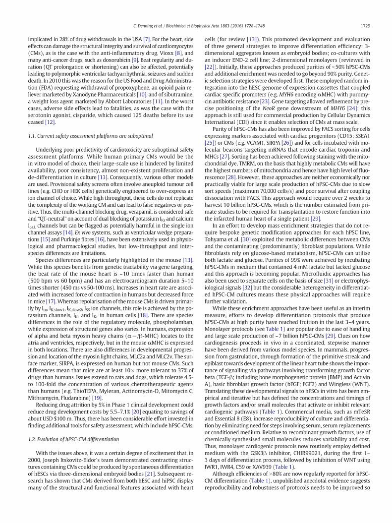
While these enrichment approaches have been useful as an interimmeasure, efforts to develop differentiation protocols that producehPSC-CMs at high purity have reached fruition in the last 3–4 years.Monolayer protocols (see Table 1) are popular due to ease of handlingand large scale production of ~7 billion hPSC-CMs [29]. Clues on howcardiogenesis proceeds in vivo in a coordinated, stepwise mannerhave been derived from various model species. In mammals, progres-sion from gastrulation, through formation of the primitive streak andepiblast towards development of the linear heart tube shows the impor-tance of signalling via pathways involving transforming growth factorbeta (TGF-β; including bone morphogenetic protein [BMP] and ActivinA), basic fibroblast growth factor (bFGF; FGF2) and Wingless (WNT).Translating these developmental signals to hPSCs in vitro has been em-pirical and iterative but has defined the concentrations and timings ofgrowth factors and/or small molecules that activate or inhibit relevantcardiogenic pathways (Table 1). Commercial media, such as mTeSRand Essential 8 (E8), increase reproducibility of culture and differentia-tion by eliminating need for steps involving serum, serum replacementsor conditioned medium. Relative to recombinant growth factors, use ofchemically synthesised small molecules reduces variability and cost.Thus, monolayer cardiogenic protocols now routinely employ definedmedium with the GSK3β inhibitor, CHIR99021, during the first 1–3 days of differentiation process, followed by inhibition of WNT usingIWR1, IWR4, C59 or XAV939 (Table 1).
Although efficiencies of N80% are now regularly reported for hPSC-CM differentiation (Table 1), unpublished anecdotal evidence suggestsreproducibility and robustness of protocols needs to be improved so
Table 1Methods for monolayer differentiation of hPSC-CMs. Abbreviations: MEF-CM, mouse embryonic fibroblast-conditioned medium; E8, Essential 8 medium; Reprog, Reprogramming method; N/A, not applicable; Retro, retrovirus; Lenti, lentivirus;Epi, Epsiomal; AA, activin A; V, ventricular; A, atrial; N, nodal (or pacemaker).
Timeline
hPSC line(s) Medium Substrate Reprog Differentiation procedure Efficiency Subtypes Ref
2007 hESC: H7 MEF–CM Matrigel N/A 30% V,A 198
2011
hESC: H7 MEF–CM Matrigel N/A 70% V,A,N 199
hESC: KhES1; hiPSC lines:201B6, 201B7, 253G1,
253G4MEF–CM Matrigel Retro; Lenti 60% V,A 200
Engineered hESC: H3, M1 MEF–CM Matrigel None 40% V,A 78
2012hESC: H9, H13, H14
hiPSC:IMR90C4mTESR1 Matrigel Epi; Lenti 85% V,A 201
2013hESC: H1, H9
hiPSC: hAFDC–iPS–36mTESR1 Matrigel Retro 80% V,A 202
2014hESC: H7, H9
hiPSC: 58FSDNC3,64FSDNC1
E8Matrigel, Laminin,
VitronectinSendai 90% V,A 174
2015 hPSCs: Line N/S.E8, mTESR,MEF–CM
Matrigel N/S 80% V,A 203
AA BMP4
RPMI/B27
BMP4;FGF2 AA Noggin DKK1+Rai/RA DKK1
RPMI/B27
AA BMP4 + FGF2 DKK1
RPMI/B27 (–insulin) RPMI/B27
CHIR IWP2; IWP4
RPMI/B27 (–insulin) RPMI/B27
AA; BMP4; FGF2;VEGFA; SCF
LI–APEL LI–PEL
CHIR; BMP4;Ascorbate
BMP4; IWR1
CIM RPMI/B27 (–insulin) RPMI/B27
CHIR WNT–C59
CDM3
BMP4; AA; CHIR XAV939
Cardiac differentiation medium
D0 D1 D2 D3 D4 D5 D6 D7 D8 D9 D10 D11 D12
1730C.D
enningetal./Biochim
icaetBiophysica
Acta
1863(2016)
1728–1748
1731C. Denning et al. / Biochimica et Biophysica Acta 1863 (2016) 1728–1748
there is greater consistency between lines and laboratories. Also, proto-cols yield a mixed population of CM subtypes, including ventricular-,atrial- and pacemaker-like cells. Cultures containing a single subtypeare preferred; for example, ventricular cells are needed to evaluatedrugs that have Torsades de Pointe liabilities or for transplantationafter myocardial infarction. In this regard, there have been recent excit-ing developments. Birket and colleagues [33] combined a complex butelegant double transgenic approach, wherein an NKX2.5-GFP targetedhESC line was further transfected with an inducible MYC expressionconstruct. In the presence of insulin-like growth factor-1 (IGF-1) and ahedgehog pathway agonist, cardiovascular progenitor cells could be iso-lated and proliferated for over 40 population doublings. Moreover,modulating exogenous BMP, FGF, WNT and RA signalling led to multi-lineage differentiation, as well as directed specification to pacemakerand ventricular cells. This report was remarkable because it not onlyshowed long-term proliferation of hPSC-derived cardiac progenitors(in 11 other reports using mouse and human PSCs, maximum expansionwas 4-fold [34]), but it was the first robust demonstration of subtypespecification. In an alternative approach, modulation of retinoic acid sig-nalling during hESC differentiation was used to generate atrial- andventricular-like CMs. These CM subtypes were used to show that themulti-ion channel blocker, vernakalant, and Kv1.5 blocker, XEN-D0101,caused a reduction in early repolarization only in the atrial cells [35], pro-viding a novel preclinical test platform for these drug classes.
1.3. Genotypes are now readily captured using hiPSC reprogramming
Improvements in CM differentiation have been paralleled byadvances in hiPSC production methods. The original landmark papersby Shinya Yamanaka's teamand the reports thereafter described low ef-ficiency (b0.1%) production of hiPSCs via integrating retroviruses [2]in undefined medium on mitotically-inactivated mouse embryonicfibroblasts. Technologies have evolved so that academic and commer-cial labs now produce hiPSC lines at efficiencies of ~4.4% using non-integrating approaches in defined medium on recombinant matrices[36] Indeed, large scale banking schemes includingHuman Induced Plu-ripotent Stem Cells Initiative (HIPSCI), StemBANCC/IMI, California Insti-tute for RegenerativeMedicine and NewYork StemCell Foundationwillcreate hiPSC lines from 7000 normal or diseased skin biopsy donorsusing Sendai-virus, episomes or mRNA with a combination of SOX2,c-MYC, OCT4, KLF4 and/or LIN28 [37]. Nevertheless, each integration-free method has pros and cons and there is not yet consensus onwhich reprogramming method is best. Episomal plasmids have lowerreprogramming efficiencies and the potential for residual plasmid inte-gration; Sendai-virus require higher biosafety containment levels andare relatively costly; mRNA reprogramming is labour intensive, requir-ing repeated (daily) transduction and costly Pluriton medium. Inaddition, there are licencing cost implications and restrictions to be con-sidered for commercial use for Sendai-virus and mRNA approaches.
1.4. hPSC-CMs are becoming valuable for in vitro and in vivo biomedicalapplication
The relative ease of efficient reprogramming and directed cardio-genesis has accelerated progress towards biomedical application. This ishelped by hiPSCs largely eliminating ethical or legal restrictions thatprohibited use of hESCs in many companies and countries. For predictivecardiotoxicity, many reports show hPSC-CMs are effective in safetyscreening. In the 13 years from 2000 to 2013, pharmacological responsesof hPSC-CMs to only 60 different compounds had been demonstrated[13]. Thesenumbers arenowbeing exceededby single studies; one reportassessed impact of 131 compounds of hPSC-CM function [38].
Accuracy of the assays is also improving and gaining interest fromthe pharmaceutical industry. Using hPSC-CMs, AstraZeneca showed70% specificity and 87% sensitivity for a 51 compound screen [39],while a study commissioned by J&J recorded an accuracy of 90% following
blind testing of electrical toxicity in 20 compounds [40]. Work fromGlaxoSmithKline cross-compared pharmacological responses of hPSC-CMs and animal models, concluding that the human cells offered a reli-able and cost-effective surrogate to preclinical in vitro testing [41]. Directcomparison between CMs isolated from hPSCs or dog and rabbit heartsshowed the human cells more accurately predicted moxifloxacin-induced cardiotoxicity [42]. Studies have extended to screening antiviralsas a treatment for B3-strain of coxsackievirus, a major causative agent forviralmyocarditis [43]. Notably, hPSC-CMswere used to show that toxicitywas reducedwhen the anti-cancer drug, doxorubicin, was delivered via aHER2-targeted liposomal pathway; this assisted the decision to advanceto Phase I testing [44]. Such studies have led the CIPA initiative (Compre-hensive In Vitro Proarrhythmia Assay) to propose integration of hPSC-CMs into the ICH (International Conference on Harmonisation) S7a/band E14 guidelines by the end of 2015. These guidelines have been themainstay over the last decade of preclinical assessment of cardiac electro-physiology for new drugs [45].
Patient-specific hiPSC-CMs are being used increasingly to evaluatealtered phenotype and drug rescue of various channelopathies affectingthe heart, including long QT syndrome (LQTS)-1 [46–48], −2 [49–53],−3 [54], −8 [55], LQTS3/Brugada overlap [56] and catecholaminergicpolymorphic ventricular tachycardia (CPVT) [57–59]. Disorders that af-fect structure, contractility and survival have also been modelled, suchas Duchenne muscular dystrophy (DMD) [60], dilated cardiomyopathy[61,62], hypertrophic cardiomyopathy (HCM) [63,64], Leopard Syndrome[65], Barth Syndrome [66,67] and arrhythmogenic right ventricularcardiomyopathy (ARVC) [68–71]. These have been used to understanddisease mechanisms and evaluate novel therapeutics. Thus, dantroleneabolished isoprenaline-induced arrhythmias in CPVT1 hiPSC-CMs[58], while trichostatin A was shown to prevent hypertrophy inHCM hiPSC-CMs [64]. Tests for efficacy of genetic intervention in-clude oligonucleotide-mediated exon skipping and allele-specificRNAi to correct DMD [72] and LQTS2 [53] hiPSC-CMs, respectively.Most notably, the inability to manage effectively treatment of an in-dividual with complex LQTS was addressed by deriving hiPSC-CMsand performing multi-parameter in vitro drug testing until a suit-able combinatorial regime was identified. This treatment was usedin the clinic to improve the patient's care [73] showing feasibilityof personalised medicine.
Nevertheless, while the examples above show the potential offeredby hiPSC-CMs, there are several reports of deficiencies relative to theirhESC derived counterparts. Thus, Foldes and colleagues [74] describedrobust hypertrophic responses to phenylephrine in hESC-CMs but nothiPSC-CMs. This was irrespective of the reprogramming or differentia-tion method used. Indeed, a hESC line was differentiated to fibroblasts,which were reprogrammed to hiPSC. When this hiPSC line and theparental hESC line were differentiated to CMs, only the cells derivedfrom hESCs showed hypertrophy, despite the cells sharing the same ge-notype. Similar issues have been reported for improper reprogrammingand disease modelling in hiPSC from patients with Fragile-X relative tohESCs derived from pre-implantation genetic diagnosis embryos [75].
Beyond their in vitro use, hPSC-CMs are also being evaluated fortreatment of damaged of diseased heart. Pilot studies using hPSC-CM engraftment into mouse, rat, guinea-pig and pig models of myo-cardial infarction were escalated to pigtail macaque non-human pri-mates in mid-2014 [29]. In the primate studies, 1 billion cryopreservedhPSC-CMs were transplanted in a complex pro-survival cocktail to theinfarct site of each of 7 animals. Transplanted cells led to extensiveremuscularisation, accompanied by host vasculature perfusion, electro-mechanical junction formation between graft & host, and synchronouscalcium transients. Nonetheless, there were two cautionary notes. Whilethe hPSC-CMs constituted a graft size of up to 5.3% of the left ventricularmass, survival of transplanted cells was less than 10% (b108 of 109 cells)despite the powerful pro-survival cocktail. Secondly, although the ma-caques remained free of distress, continuous electrocardiogram record-ings showed that all animals receiving hESC-CMs developed ventricular
1732 C. Denning et al. / Biochimica et Biophysica Acta 1863 (2016) 1728–1748
arrhythmias. There are similarities but also differences between these pri-mate studies and results from transplantation into swine. In a study inpigs [76], the tri-lineage differentiation potential of hiPSCs was exploitedto produce CMs, endothelial cells and smooth muscle cells. A total of 6million cells (2million of each lineage)were complexedwith a 3D epicar-dial fibrin patch loaded with microspheres to allow prolonged release ofthe pro-survival factor, insulin-like growth factor 1 (IGF-1). The complexwas then transplanted into a porcinemodel ofmyocardial infarction. Sim-ilar to the primate study, over a 4 week period cell survival was around9%, although without the fibrin patch was reduced to 3–4%. Surprisingly,given the low cell numbers transplanted (160-fold less than the primatestudy), there were improvements in myocardial wall stress, metabolismand contractile performance. However, distinct to macaques, develop-ment of ventricular arrhythmias was not reported in the pigs; whetherthis important difference is down to the animal model, cell types, num-bers or preparation method, inclusion of different survival factors ortransplant route are now all questions that need to be addressed. More-over, these reports have not included methods to improve vasculatureto the grafted cells and this will be a consideration for the future.
The preclinical studies have led to the first clinical trial for the heartusing hPSC derivatives. Menasché and co-workers [77] sought to directdifferentiation from hESCs and then use immunomagnetic sorting toisolate ISL-1+/SSEA-1+ cardiac progenitor cells. These were embed-ded into a fibrin scaffold, whichwas surgically delivered onto the infarctarea in a 68-year-old patient suffering from severe heart failure. At the3 month follow-up stage, the patient showed no complications, suchas arrhythmias, tumour formation or immunosuppression-related ad-verse events, but was symptomatically improved, wherein echocardio-graphically showed the damaged region of the heart regainedcontractility. The progress of this patient, and those who follow, willbe keenly awaited. Nevertheless, nearly a year on it is not clear whetherany further patients have been recruited to this trial, even though theexpected start and end dates are 2013 and 2017 to treat a total of 6 pa-tients. This perhaps highlights the challenges of coordinating complexprocesses of large scale cardiac progenitor cell production, surgicalprocedures and immunosuppression regimenswith highly selective pa-tient inclusion criteria. Thus, for inclusion, patients must display severeleft ventricular systolic dysfunction with left ventricular ejection frac-tion (LVEF) ≤ 35% as assessed by echocardiography or scintigraphyand have an echocardiography history of myocardial infarction with aresidual akinesia involving more than 2 of 16 contiguous segments.They will show New York Heart Association (NYHA) Class III or IV,despite optimal standard of care including diuretics and angiotensin re-ceptor blockers and, if possible, beta blockers and aldosterone blockers,as well as previous implantation of an automatic internal defibrillatorassociated to ventricular resynchronization [78]. If the current Phase Itrial continues to provide optimism as a new treatment route for pa-tients, the issues of bioprocess, surgery and patient selection willneed to be reviewed carefully. Indeed, it will only be through largertrials that true improvements in the patient's heart function can beattributed to cell transplantation rather than natural recovery or im-pact of past treatment (e.g. bypass surgery, drug treatment, ventric-ular resynchronization).
1.5. Genome editing marks a new era for in vitro genotype modelling
Until hiPSC approaches provided a route to capturing awide range ofpatient-relevant genotypes, reliance had been on establishing hESClines from pre-implantation genetic diagnosis (PGD) embryos [79] orby gene targeting [80]. However, PGD is available for only a limitednumber of genetic conditions, few scientists have access to these fa-cilities and the use of embryos (even those that harbour detrimentalgenetic lesions) is ethically sensitive in many countries. Similarly,gene targeting by homologous recombination was initially achievedin a few laboratories to create knockouts (e.g. HPRT1 to model of themetabolic disorder, Lesch Nyhan syndrome [80]) or reporter constructs
downstream of developmentally important genes, such as NKX2.5 [81].In rare cases, creation of isogenic pairswas used to study role ofmutationsin genes such as KCNH2, which underlies the sudden cardiac death condi-tion of LQTS2 [82]. Further progresswas stymied because of low recombi-nation frequency (1 in 106–109 cells) in most mammalian cells, whichmade the generation of isogenic models almost unachievable becausethis often requires biallelic targeting. However, progress in genomeediting tools now allows rapid engineering of the genotypes available inhPSCs. If there is not the need for patient history to draw in vitro-in vivocorrelations, the speed, flexibility, ease and low cost of gene targetingwill be used in preference to hiPSC reprogramming to capture specific ge-notypes; indeed, in our own laboratory, this is the situation for somediseases.
For gene targeting, it has been known for 25 years that introductionof specific double strand breaks at the target locus can improve efficien-cy. In human cells, complexing the Fok1 endonuclease to a pair of zincfinger nucleases (ZFNs) [83] allowed double strand breaks at a modelGFP locus in 293 T cells [84] and endogenous PIG-A locus in hPSCs [84]resulting in targeting efficiency improvements of 200- to 2000-fold.However, the complex design and construction for each ZFN attracteda high commercial cost of USD $25,000. The advent of transcriptionactivator-like effector nucleases (TALENs) used the same principle asZFNs, relying on a dimeric protein-based DNA binding domain coupledto endonuclease. Construction kits, such as GoldenGate [85] and FLASHassembly [86], meant individual labs could produce their own TALENvectors and hence reduce costs by 20-fold relative to ZFNs. Moreover,TALENs showed greater specificity in hPSCs, with less off-target activityand toxicity in comparison to ZFNs [87].
The real breakthrough came with the development of the Cas9/CRISPR (Clustered Regularly Interspaced Short Palindromic Repeat) sys-tem (Fig. 1) [88] which is derived from various strains of bacteria and isoften described as their immune system. The adapted version of thissystem relies on 100 base site-specific guide RNA (gRNA) to direct theCas9 endonuclease to the target site, which eliminates the need fortime-consuming production of DNA binding-endonuclease fusion pro-teins [89]. Such approaches have been used to perform correctionmethods using exon skipping, frameshifting and exon knock-in intohiPSC lines carrying mutations in DMD, which underlie severe muscledegenerative disease [90].
There have been several further refinements in the Cas9/CRISPR sys-tem (Fig. 1). Use of dual guide RNA/Cas9-Nickase (D10A) reduces off-target activity to an almost undetectable level [91]. Moreover, pre-synthesised in vitro transcribed gRNAs can be complexedwith recombi-nant Cas9 protein and transfected by nucleofection, providing a rapidroute to gene knockout or small changes in sequence, including substi-tutions [92]. Thus, in the absence of a homology domain, Cas9-inducedDNA cleavage leads to non-homologous end joining and causes inser-tions or deletions, known as ‘indels’, that can cause gene knockouts[93]. To induce base substitutions, small changes or deletions of up to100 kbases, the conventional ~5–15 kb targeting vector can be replacedwith a ~ 100–120nucleotide single-strandedDNAoligo [94].With this ap-proach, Kimet al. [92] showedup to 79% targeting efficiency inCCR5 locusin human leukaemia K562 cell line, BJ fibroblasts and H9 hESCs.
The recombinant Cas9 approach has other advantages. Since it is ac-tive immediately upon entry to the nucleus but continues to function foronly 24 h, off-target effects and toxicities are low, while targetingfrequency is high and eliminates the need for drug selection. Ourown work corroborates these findings (Fig. 1). We used Amaxa 4Dnucleofection to deliver a combination of gRNA, recombinant Cas9protein and 110-nucleotide single-stranded DNA oligo templateinto ReBl-PAT hiPSCs, which resulted in a targeting frequency atthe β2-adrenoceptor locus of 33%, of which 25% of clones werebiallelic. Such efficiencies can be expected to rise further with thefinding that small molecules can enhance targeting by up to 9-foldin hPSCs [95], which will ultimately lead to panels of isogenic pairsin hPSCs to study disease mechanisms and therapies.
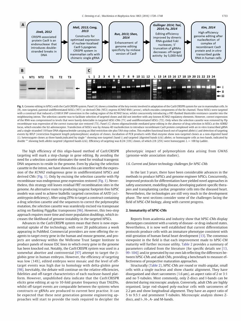
Fig. 1.Genome editing in hPSCswith the Cas9/CRISPR system. Panel (A) shows a timeline of the key events involved in adaptation of the Cas9/CRISPR system for use inmammalian cells. In(B), non-targeted, parental undifferentiated hESCs (NT1) or derived CMs (NT2) express KCNH2 RNA (arrow), which encodes components of the Ikr channel. These hESCs were targetedwith a construct that induced a G1681A SNP conversion in the coding region of the KCNH2 locus, whilst concurrently introducing a FRT-flanked blasticidin resistance cassette into theneighbouring intron. The selection cassette was to facilitate selection of targeted clones and did not interfere with any known KCNH2 regulatory elements. However, correct expressionof the RNA was compromised to levels that were barely detectable in targeted hESC-CMs (T1) and undifferentiated hESCs (T2). Only when the selection cassette was removed by Flprecombinase was expression of the correct transcript size restored (T3). Panel (C) shows oligonucleotide-mediated gene editing in the absence of drug selection in hESCs at the ADRB2locus, which encodes the b2-adrenoceptor. Transfection of hESCs was by Amaxa 4D nucleofection to introduce recombinant Cas9 protein complexed with an in vitro transcribed gRNAand a single-stranded 110 base DNA oligonucleotide carrying an XbaI restriction site plus TAA stop codon. This enables functional knock out of targeted allele(s) and detection of targetingevents by RFLP (restriction fragment length polymorphism) analysis of clones. Incubation of PCR products with XbaI enzyme show non-targeted clones as a non-digested band(i); heterozygote clones as three bands,indicated by single * showing non-targeted (band i) and targeted (digested bands ii/iii) alleles; or homozygote cells as two bands, indicated bydouble ** showing both alleles targeted (digested bands ii/iii). Efficiency of targeting was 8/24 (33%) clones, of which 2/8 (25%) were homozygote. L = 100 bp Ladder.
1733C. Denning et al. / Biochimica et Biophysica Acta 1863 (2016) 1728–1748
The high efficiency of this oligo-based method of Cas9/CRISPRtargeting will mark a step-change in gene editing. By avoiding theneed for a selection cassette eliminates the need for residual transgenicDNA sequences to reside in the genome. Even by placing the selectioncassette in the intron, we have shown this can interferewith the expres-sion of the KCNH2 endogenous gene in undifferentiated hPSCs andderived CMs (Fig. 1). Only by excising the selection cassette with Flprecombinase was endogenous gene expression restored (Fig. 1). Never-theless, this strategy still leaves residual FRT recombination sites in thegenome. An alternative route to producing isogenic footprint-free hiPSCmodels was used to achieve biallellic targeted correction of AATD (α1-antitrypsin deficiency). After using ZFNs to target a construct containinga drug selection cassette and the sequences to correct the polymorphicmutation, the selection cassette was seamlessly excised via transposaseacting on flanking PiggyBac transposons [96]. However, this two-stepapproach requiresmore time andmore population doublings, which in-creases the likelihood of genome instability in the targeted hPSCs.
Advances in the Cas9/CRISPR system mean that there is now expo-nential uptake of the technology, with over 20 publications a weekappearing in PubMed. Commercial providers are now offering the re-agents to target every gene in the human andmouse genome, and pro-jects are underway within the Wellcome Trust Sanger Institute toproduce panels of mouse ESC lines in which every gene in the genomehas been knocked out. Notably, the Cas9/CRISPR system was used in asomewhat abortive and controversial [97] attempt to target the β-globin gene in human embryos. However, the efficiency of targetingwas low (14%), edited embryos were mosaic and the level of off-target events was high due to homology with delta-globin gene[98]. Inevitably, the debate will continue on the relative efficiencies,fidelities and off-target characteristics of each nuclease-based plat-form. However, unpublished data indicate that the Cas9/CRISPRelicits gene editing at up to 10-fold greater frequency than TALENs,whilst off-target events are comparable between the systems whenconstructs or gRNAs are produced to current best practise. It canbe expected that these next generation genome engineering ap-proaches will start to provide the tools required to decipher the
phenotypic impact of polymorphism data arising from GWAS(genome-wide association studies).
1.6. Current and future technology challenges for hPSC-CMs
In the last 3 years, there have been considerable advances in themethods to produce hiPSCs and genome engineer hPSCs. Concurrently,improved protocols for differentiation have yielded novel approaches insafety assessment,modelling disease, developing patient-specific thera-pies and transplanting cardiac progenitor cells into the diseased heart.Nevertheless, the technologies are still very much in the developmentphase. The next sections consider some of the challenges facing thefield of hPSC-CM biology, along with current progress.
2. Immaturity of hPSC-CMs
Reports from academia and industry show that hPSC-CMs displayphenotypes consistentwith a variety of disease- or drug-induced states.Nevertheless, it is now well established that current differentiationprotocols produce cells with an immature phenotype consistent withmid-gestation of human foetal heart development [13]. The consensusviewpoint in the field is that each improvement made to hPSC-CMmaturity will further increase utility. Table 2 provides a summary ofparameters collated from the literature (for specific details see [13,99–104]) and/or generated by our own lab reflecting the differences be-tween hPSC-CMs and adult CMs, providing a benchmark to measure ef-fectiveness of prospective maturation approaches.
Structurally (Table 2), hPSC-CMs are round or multi-angular, smallcells with a single nucleus and show chaotic alignment. They havedisorganised and short sarcomeres (1.6 μm), an aspect ratio of 2 or 3:1and no T-tubules. Most commonly, only Z-discs and I-bands can bedetected during microscopic analysis. Conversely, adult CMs are highlyorganised, large rod-shaped poly-nuclear cells with sarcomeres of2.2 μm and show longitudinal alignment. They have an aspect ratio of5 to 9.5:1 and prominent T-tubules. Microscopic analysis shows Z-discs, and I-, H-, A- and M-bands.
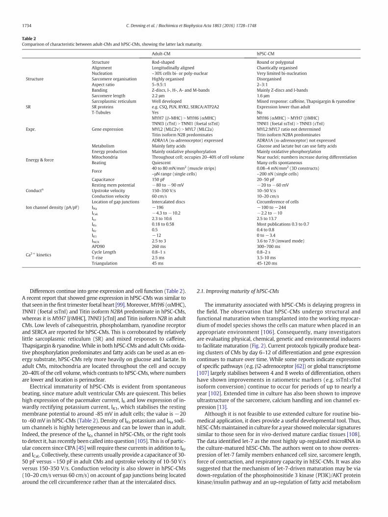
Table 2Comparison of characteristic between adult-CMs and hPSC-CMs, showing the latter lack maturity.
Adult-CM hPSC-CM
Structure
Structure Rod-shaped Round or polygonalAlignment Longitudinally aligned Chaotically organisedNucleation ~30% cells bi- or poly-nuclear Very limited bi-nucleationSarcomere organisation Highly organised DisorganisedAspect ratio 5–9.5:1 2–3:1Banding Z-discs, I-, H-, A- and M-bands Mainly Z-discs and I-bandsSarcomere length 2.2 μm 1.6 μm
SRSarcoplasmic reticulum Well developed Mixed response: caffeine, Thapsigargin & ryanodineSR proteins e.g. CSQ, PLN, RYR2, SERCA/ATP2A2 Expression lower than adultT-Tubules Yes No
Expr. Gene expression
MYH7 (β-MHC) N MYH6 (αMHC)TNNI3 (cTnI) N TNNI1 (foetal ssTnI)MYL2 (MLC2v) N MYL7 (MLC2a)Titin isoform N2B predominatesADRA1A (α-adrenoceptor) expressed
MYH6 (αMHC) N MYH7 (βMHC)TNNI1 (foetal ssTnI) N TNNI3 (cTnI)MYL2:MYL7 ratio not determinedTitin isoform N2BA predominatesADRA1A (α-adrenoceptor) not expressed
Energy & force
Metabolism Mainly fatty acids Glucose and lactate but can use fatty acidsEnergy production Mainly oxidative phosphorylation Mainly oxidative phosphorylationMitochondria Throughout cell; occupies 20–40% of cell volume Near nuclei; numbers increase during differentiationBeating Quiescent Many cells spontaneous
Force40 to 80 mN/mm2 (muscle strips)~μN range (single cells)
0.08–4 mN/mm2 (3D constructs)~200 nN (single cells)
Conductn
Capacitance 150 pF 20–50 pFResting mem potential −80 to −90 mV −20 to −60 mVUpstroke velocity 150–350 V/s 10–50 V/sConduction velocity 60 cm/s 10–20 cm/sLocation of gap junctions Intercalated discs Circumference of cells
Ion channel density (pA/pF) INa −196 −100 to −244ICaL −4.3 to −10.2 −2.2 to −10Ito 2.3 to 10.6 2.5 to 13.7IKs 0.18 to 0.58 Most publications 0.3 to 0.7IKr 0.5 0.4 to 0.8IK1 −12 0 to −3.4INCX 2.5 to 3 3.6 to 7.9 (inward mode)
Ca2+ kinetics
APD90 260 ms 300–700 msCycle Length 0.8–1 s 0.8–2 sT-rise 2.5 ms 3.5-10 msTriangulation 45 ms 45-120 ms
1734 C. Denning et al. / Biochimica et Biophysica Acta 1863 (2016) 1728–1748
Differences continue into gene expression and cell function (Table 2).A recent report that showed gene expression in hPSC-CMs was similar tothat seen in thefirst trimester foetal heart [99].Moreover,MYH6 (αMHC),TNNI1 (foetal ssTnI) and Titin isoform N2BA predominate in hPSC-CMs,whereas it is MYH7 [βMHC], TNNI3 [cTnI] and Titin isoform N2B in adultCMs. Low levels of calsequestrin, phospholambam, ryanodine receptorand SERCA are reported for hPSC-CMs. This is corroborated by relativelylittle sarcoplasmic reticulum (SR) and mixed responses to caffeine,Thapsigargin & ryanodine.While in both hPSC-CMs and adult CMs oxida-tive phosphorylation predominates and fatty acids can be used as an en-ergy substrate, hPSC-CMs rely more heavily on glucose and lactate. Inadult CMs, mitochondria are located throughout the cell and occupy20–40% of the cell volume, which contrasts to hPSC-CMs, where numbersare lower and location is perinuclear.
Electrical immaturity of hPSC-CMs is evident from spontaneousbeating, since mature adult ventricular CMs are quiescent. This belieshigh expression of the pacemaker current, If, and low expression of in-wardly rectifying potassium current, IK1, which stabilises the restingmembrane potential to around -85 mV in adult cells; the value is −20to -60 mV in hPSC-CMs (Table 2). Density of IKs potassium and INa sodi-um channels is highly heterogeneous and can be lower than in adult.Indeed, the presence of the IKs channel in hPSC-CMs, or the right toolsto detect it, has recently been called into question [105]. This is of partic-ular concern since CIPA [45]will require these currents in addition to IKrand ICaL. Collectively, these currents usually provide a capacitance of 30-50 pF versus ~150 pF in adult CMs and upstroke velocity of 10-50 V/sversus 150-350 V/s. Conduction velocity is also slower in hPSC-CMs(10–20 cm/s versus 60 cm/s) on account of gap junctions being locatedaround the cell circumference rather than at the intercalated discs.
2.1. Improving maturity of hPSC-CMs
The immaturity associated with hPSC-CMs is delaying progress inthe field. The observation that hPSC-CMs undergo structural andfunctional maturation when transplanted into the working myocar-dium of model species shows the cells can mature when placed in anappropriate environment [106]. Consequently, many investigatorsare evaluating physical, chemical, genetic and environmental inducersto facilitate maturation (Fig. 2). Current protocols typically produce beat-ing clusters of CMs by day 6–12 of differentiation and gene expressioncontinues to mature over time. While some reports indicate expressionof specific pathways (e.g. β2-adrenoceptor [62]) or global transcriptome[107] largely stabilises between 4 and 8 weeks of differentiation, othershave shown improvements in ratiometric markers (e.g. ssTnI:cTnIisoform conversion) continue to occur for periods of up to nearly ayear [102]. Extended time in culture has also been shown to improveultrastructure of the sarcomere, calcium handling and ion channel ex-pression [13].
Although it is not feasible to use extended culture for routine bio-medical application, it does provide a useful developmental tool. Thus,hESC-CMsmaintained in culture for a year showedmolecular signaturessimilar to those seen for in vivo-derived mature cardiac tissues [108].The data identified let-7 as the most highly up-regulated microRNA inthe culture-matured hESC-CMs. The authors went on to show overex-pression of let-7 family members enhanced cell size, sarcomere length,force of contraction, and respiratory capacity in hESC-CMs. It was alsosuggested that the mechanism of let-7-driven maturation may be viadown-regulation of the phosphoinositide 3 kinase (PI3K)/AKT proteinkinase/insulin pathway and an up-regulation of fatty acid metabolism

Fig. 2. Schematic of in vitromaturation strategies for hPSC-CMs. (A)Methods include biophysical stimuli such asmechanical cues, electrical stimulation, optimising substrate stiffness andtopography. Biochemical cues can bepresented as soluble factors or substrate ligandswithin biological or syntheticmatrices. Geneticmanipulation such as forced expression ofmissing ionchannels has also been adopted as a maturation strategy. (B) The aim of these strategies is to drive the polygonal morphology and disorganised myofibril banding of immature CMstowards a more mature state indicated by rod shaped morphology and parallel myofibrils (see also Table 2).
1735C. Denning et al. / Biochimica et Biophysica Acta 1863 (2016) 1728–1748
[108]. Similar approaches to over-express components of the CMmachinery, such as miR-1, calsequestrin or Kir2.1 have also facilitatedmaturation of hPSC-CMs [13].
2.2. Maturation: medium additives
Adding supplements to the culture medium is potentially a straight-forward way to modulate hPSC-CM maturation. Table 3 shows factorsthat have undergone some level of testing in mouse and human cellsto evaluate their ability to induce CM maturity. Triiodothyronine (T3)is essential for normal cardiac development and during the perinatal
Table 3Factors with potential for facilitating maturation hPSC-CMs.
Factor Known function Model s
Insulin Regulates glucose uptake and postnatal cardiac growthARVD hIBMX Induction of adipogenesis
Dexamethasone Induction of adipogenesis
CorticosteroneStructural and functional maturation of the foetal heartin vivo
Mousecardiom
PPARα Regulator of fatty acid metabolism in adult CMs ARVD h
PGC-1α (PPARγcoactivator 1α)
Promotes cardiac mitochondrial biogenesis hESC-C
Klf15Glucocorticoid receptor target that interacts with PPARαto regulate cardiac lipid metabolism
Cardiacfrom m
13-HODE Component of oxidised low-density lipoprotein via PPARγARVD hRosiglitazone PPARγ activator that increases adiponectin in CMs
Indomethacin Mediates agonists for PPARγ to regulate adipogenesisInsulin-like growthfactor
IGF1 receptor induces heart growth via the PI3K pathway mESC
T3 Thyroid hormone essential for optimal heart development hiPSC-C
EPA Fish oil that affects developmental bioenergetics mESC
Abbreviations: IBMX, 3-isobutyl-1-methylxanthine; ARVD, arrhythmogenic right ventricular dydecadienoic acid; T3, Tri-iodo-L-thyronine; EPA, eicosapentaenoic acid.
period, it regulates isoform switching of severalmyocardial proteins, in-cludingMHCand titin. Incubation of hPSC-CMswith T3 for 1–2weeks ledto changes consistentwithmaturation, including 11-fold up-regulation ofαMHC, lower proliferation rates (but not increased bi-nucleation), 1.5-fold increase in twitch force (to ~12nN/cell), higher calcium-derivedmaximal upstroke and decay velocities enhanced oxygen consumptionrates [109].
An alternative approach was taken byWen and colleagues to modelthe late onset disorder, arrhythmogenic right ventricular dysplasia(ARVD) [110]. A combination of insulin, dexamethasone (a glucocorti-coid) and IBMX (3-isobutyl-1-methil-xanthine; a phosphodiesterase
ystem Effect Ref
iPSC-CMInduction of adult-like metabolism in model of adult onsetdisease
[70]
foetalyocytes
Improve contractility, Z-disc assembly, mature myofibrilsand mitochondrial capacity
[207]
iPSC-CMCo-activation of PPARα and PPARγ promoted lipogenesis,apoptosis & channel deregulation
[110]
MControlling PGC-1α and reactive oxygen species implied inrecapitulating mature phenotypes
[208]
progenitorsouse hearts
Cells with plakoglobin mutation showed increased Klf15,CEBPα, Wnt5b
[209]
iPSC-CMInduced lipogenesis and apoptosis in model of adult onsetdisease
[70]
Insulin or IGF1/2 during early differentiation increasedmesodermal cell proliferation
[210]
M T3 drives maturation [109]Increases in gene expression associated with cardiacdevelopment
[211]
splasia; PPARα, peroxisome proliferator-activated receptorα; 13-HODE, 13-hydroxyocta-
1736 C. Denning et al. / Biochimica et Biophysica Acta 1863 (2016) 1728–1748
inhibitor) was used to drive metabolic maturation by increasing fattyacid synthesis and triggering activation of PPARα, which led to en-hanced mitochondrial oxidative phosphorylation. Further additionof the PPARγ activators, rosiglitazone and indomethacin, to the mediumcaused abnormal PPARγ activation in ARVD hiPSC-CMs. This unveiledthe pathological phenotypes associated with this condition, which in-clude exaggerated lipogenesis, apoptosis, Na+ channel down-regulationand defective intracellular Ca2+handling. However, it is unlikely thatme-dium additives alone will induce complete maturation of CMs, hence theeffect of modulating other components of the cell environment are beinginvestigated.
2.3. Maturation: biophysical cues
Unlike the hESC-CMs, primary CMs in the atria and ventricles ofadult human heart do not exhibit spontaneous beating. Instead, theyare innervated by the autonomic nervous system via nodal CMs,which determines pace and contractility. To mimic this excitation–con-traction coupling, Radisic and colleagues applied extrinsic electricalfield stimulation to neonatal rat ventricular myocytes (NRVM) [111].Compared to non-stimulated cells, field stimulation induced elongatedmorphology concurrent with increased sarcomere volume and num-bers of mitochondria, intercalated discs, gap junctions and contractility.Thiswork has been translated to hPSC-CMs [112],wherein three dimen-sional (3D) configurations have been subjected to electrical field stimu-lation of increasing frequency (see 3D engineering section below).
Other investigators have assessed the impact of mechanical cues tomimic the intra- and extra-cellular stresses that CMs experience. Thiscan be achieved by altering cyclic stretch,mechanical load and substratestiffness. Thus, Mihica and co-workers [113] reported hESC-CMs seededonto gelatin-based scaffolds and stressed with cyclical stretchingshowed several hallmarks of maturation, namely, cell elongation, in-creased expression of gap junction proteins and ion channels,while imag-ing confirmed shorter calcium cycle durations. Implantation of the hPSC-CM constructs under the epicardium of ischemic rat hearts demonstratedenhanced survival and engraftment in the stretched constructs.
Modulating substrate stiffness provides an alternative route tovarying the level of load experienced by the CMs. The rationale is thatsubstrate stiffness of the myocardium changes dynamically during de-velopment. In the mouse, the elastic modulus increases from 12kPa inthe embryonic heart to 39kPa in the neonate [114]. In the humanheart, the modulus is 10kPa at the start of diastole but increases to500kPa at the end [115]. While these dynamic ranges are known toexist, there is considerable variation in the literature as towhat the ‘cor-rect’ range of elastic moduli to translate from in vivo to in vitro. Whencultured on substrates that mimic the elasticity of the developing myo-cardium (i.e. 1-11kPa, values for rat and quail), CMs from chicken em-bryos produced contractile force and developed actomyosin striations[116]. In contrast, CMs cultured on harder substrates (34kPa) designedto mimic post-infarct fibrotic scar tissue cells overstrained themselves,lack striated myofibrils and stop beating [116]. For neonatal rat ventric-ular CMs cultured on hydrogels of 90kPa elastic modulus, there was ahigh level of sarcomeric content and microtubule polymerisation rela-tive to cells cultured on 13kPa hydrogels [117]. The same study alsoshowed that peak systolic force was generated from CMs seeded tomicro-patterned shapes of ~7:1 aspect ratio/13kPa substrates but ~2:1aspect ratio/90kPa substrates [117]. In another report, neonatal rat ven-tricular CMs cultured on collagen-coated polyacrylamide gels with anelastic modulus 10kPa showed enhanced maturation, as evidenced byincreases in sarcomere alignment, mechanical force, improved calciumtransients and sarcoplasmic calcium stores relative to cells on substrateswith higher elastic moduli [118]. Most recently, single hPSC-CMs werecultured on 10kPa polyacrylamide substrates patterned with Matrigelin 2,000μm2 rectangles of aspect ratio between 5:1 and 7:1. The keyfindings were that translation of sarcomere shortening to mechanicaloutput was highest in 7:1, while increased substrate stiffness or applied
overstretch perturbedmyofibril structure andmechanical output in 7:1hPSC-CMs [119]. It is possible these discrepancies reflect the differentapproaches used to measure elastic modulus or differences in the celltypes, as well as their isolation and culture methods. Irrespective ofthe reasons, the diversity of data makes it difficult to pinpoint conclu-sions and a careful analysis of the impact of elastic modulus on hPSC-CM function is needed.
2.4. Maturation: chemical cues from the substrate
The substrate chemistry and structure can have a significant influ-ence on thematurity of hPSC-CMs. It is known that different extracellu-lar matrices can influence structure and cell behaviour, withphenylephrine-induced maturation absent when neonatal rat ventricu-lar CMs were cultured on gelatin but present on fibronectin or laminin[120]. This has prompted investigations into the impact of syntheticpolymers on hPSC-CMs. A library of combinatorial polymers was usedto identify a mixture of 4% polyethylene glycol:96% carboxylated PCLas enabling the greatest level of contractility and mitochondrial func-tion. This was concurrent with increases in expression of MLC2v andintegrin α7, as well as a modest level of isoform switch from foetalssTnI to the postnatal cTnI [121]. Patel and co-workers [122] screenedalmost 700 polymers for their utility as growth substrates for hPSC-CMs. These were refined down to identify chemically-defined methac-rylate co-polymers (isobornyl and tert-butylamino-ethyl) on whichhPSC-CMs exhibited a 6-fold faster upstroke velocity and significantlylonger sarcomeres relative to gelatin controls. This copolymer also en-hanced detection of the anti-cancer drug, doxorubicin, by up to 10-foldwhen myofibril disruption was used as the parameter for cardiotoxicity.
Combining substrates and enhanced maturation medium has alsobeen investigated. Single hPSC- or second trimester human foetal-CMswere seeded to gelatin patterned lines on an acrylamide substrate load-ed with fluorescent beads, which allowed measurement of contractionforce [123].While hPSC-CMs showed distinctly lower contraction stressthan the foetal counterparts (~0.25mN vs ~0.4mN/mm2), incubationwith a proprietary commercial medium containing T3 promotedcontraction force to beyond that seen in the foetal cells (~0.5mN vs~0.4mN/mm2). Concurrently, there was evidence of improved electro-physiology (upstroke velocities, action potential amplitudes, restingmembrane potentials), sarcomeric organisation and cardiac-specificgene expression.
Nevertheless, while these improvements are encouraging, the datashowed that the hPSC-CMs mirror the late-stage foetus rather thanthe adult myocardium.
2.5. Development of muscular thin films (MTFs)
Notwithstanding the variables above, relative to unpatternedsubstrates, hPSC-CMs seeded onto fibronectin-coated micro-groovedpolydimethylsiloxane (PDMS) scaffolds (~1.8 MPa) showed cellularalignment, sarcomeric organisation, enhanced calcium properties andheightened responses to caffeine, suggesting improved cycling [124].Micro-patterned PDMS can also be incorporated into muscular thinfilms (MTFs), which have tunable stiffness and flexibility to mimichealthy as well as diseased myocardium conditions [125]. Shorteningof CMs during synchronous contraction causes the MTF to flex andadopt a pseudo 3D conformation, thereby enabling the force of contrac-tion to be calculated [126]. The MTF platform has been used to evaluatefunction of various cell types. Feinberg and co-workers [127] investigat-ed the impact of architectures comprising isotropic (ISO) monolayers,anisotropic (ANISO) monolayers and 20 μm wide 20 μm spaced lines(LINES) on neonatal rat ventricular CMs. Relative to the ISO configura-tion, ANISO and LINES showed uniaxial alignment, enhanced calciumhandling and conduction velocity, and a 10-fold increase in peak systolicstress [127]. Treatment of human umbilical arterial vascular smoothmuscle cells as an anisotropic monolayer on MTFs showed application
1737C. Denning et al. / Biochimica et Biophysica Acta 1863 (2016) 1728–1748
of 50 nMendothelin-1 increased basal contractile stress from ~17kPa to~22kPa [128]. The same study went on to show MTFs seeded withneonatal rat ventricular CMs generated a peak systole stress of ~9kPa,similar to contractility measurements performed on papillary musclefrom adult rats [128].
The MTF platform has been extended to modelling of themitochon-drial myopathy, Barth syndrome, which is caused by mutations in theX-linked gene, Tafazzin (TAZ). Patient-derived hiPSCs with mutationsin TAZ were differentiated to CMs and cultured as self-organising lami-nar, anisotropic MTF myocardium constructs for “heart-on-a-chip”analyses [129]. Control CMs showed better sarcomeric alignment thanthe disease samples. During electrical field stimulation from 1 to 5 Hzin galactose-containing medium, control hiPSC-CM MTFs produced atwitch stress of 250 Pa relative to the significantly weaker values of100 Pa in the diseased tissues. This recapitulated the Barth Syndromemyopathic phenotype and provides a basis for further investigation ofthe mechanisms that underlie the condition in the engineered tissue.The development of MTFs that have electrodes incorporated for electri-cal field stimulation and micro-fluidic channels for drug loading willallow the platform to be used for cost-effective and scalable for pharma-cological testing [126].
Nevertheless, 2D systems and MTFs lack the structure of the adultheart. In part, this may be due to improper cell attachment to the sub-strate. Bidirectional translation of mechanical forces between the con-tractile apparatus and ECM is governed by integrins [130]. While thereare similarities in integrin expression between adult-CM and hPSC-CMs, the adult cells typically attach to surfaces with their distal cellularregions (costameres integrin rich area) [131], whereas hPSC-CMs formintegrin attachments along their basal surface. In addition, sarcomeresin adult-CMs are aligned in perpendicular direction from the cell axis,which results in higher probability of actin-myosin cross-bridge forma-tion and hence greater contractile force. Collectively, these differenceslead to different mechanotransduction mechanism and contractility pat-terns between adult- and hPSC-CMs. It may be that nano-patterning thesubstrate could promote better adhesion and alignment in hPSC-CMsand will need to be tested.
2.6. Engineering heart tissues in three dimensions
In parallel to the development of the various 2D and MTF systemsavailable, 3D approaches to incorporate hPSC-CMs have been investi-gated. The 3D systems that have been validated to a higher level includeengineered heart tissues (EHTs [132]), cardiac microtissues (CMTs ormicrotissue gauges; μTUGs [133]) and cardiac biowires [112]. EHTsand CMTs rely on casting cell-hydrogel mixtures in moulds featuringelastic anchors that guide cardiac tissue organisation in an aligned con-formation. This enables the CMs to perform contractile work against theanchors, thus developing an auxotonic tension that resembles physio-logical conditions. EHT fabrication involves encapsulating CMs in fibringels between two silicon posts, whereas CMTs are based on CM-seeded fibrin/collagen gels tethered to PDMS cantilevers. Functionalevaluation of CM contractility can be monitored indirectly in the EHTs byvideo-optically analysing the deflection of the silicon posts with knownmechanical properties. In CMTs, direct force reporting is via a micro-electromechanical sensor coupled to the cantilevers. Importantly, CMsin these systems can be loaded with calcium- or voltage-sensitive dyes,enabling functional analysis of calcium transients and electrophysiology[134]. Cardiac biowires consist of a cell-laden collagen gel around a surgi-cal suture placed in a PDMS mould.
These 3D systems each have pros and cons. EHTs and biowires areproduced in centimetre-scale that require 0.5–1 million CMs per unit.For EHTs, format is 24-well requiring 12–24 million hPSC-CMs, whichis costly at current commercial rates of USD$1000/million cells. In con-trast, a million cells are sufficient to produce 100–200 microscale CMTsand so 96-well formats are feasible. Measurements in EHTs are possibleover several weeks, potentially enabling acute and chronic drug effects
to bemonitored,whereas long-termanalysis in CMTs ismore difficult asthey are harder to handle. Dynamic load of the silicon posts in EHT andcantilevers in CMTs can be varied tomimic heart failure. Biowires do notpermit measurement of contractility, which is a major disadvantage.
The 3D configuration has been shown to enhance CM maturation.CMs in EHTs align along the force lines between the silicon posts, with3.4-fold improved longitudinal orientation relative to embryoid body-CMs. Sarcomeres in the EHTs become evenly distributed both aroundthe nuclei and in the periphery of the cells, although connexin-43 isstill expressed along the sarcolemma (unlike adult CMs that expressthis gap junctional protein in the intercalated discs). Elevated expres-sion of adult isoforms of sarcomeric genes (e.g. MYH7 encoding β-MHC) also occurs. EHTs display key responses to physiological andpharmacological stimulation, such as increased contractile forces athigher extracellular Ca2+ concentrations and upon treatment with β-adrenergic agonists [135]. CMTs developed with hESC-CMs promotecell alignment and expression of mature CMmarkers such as BNP.
These improvements in maturation appear to be further enhancedby incorporating electrical stimulation, even if not always as anticipated.Beat rate is ~3 Hz in human foetal hearts beat but ~1 Hz in adults. Thissuggests that reduction in rate might correlate with maturation but theopposite was observed in hPSC-CMs. Thus, increasing stimulation fre-quency from 1 to 6 Hz in hPSC-CM biowires caused maturation, as evi-denced by improvements in structure and function [112]. It may be thatforcing mechanical stress by pacing is more important than the electri-cal stimulus per se. Nevertheless, stimulated biowires had myofibrilswith a higher degree of ultrastructural organisation (aligned Z-discsdisplaying up to two I-bands per disc; organised sarcomeres showingup to 0.4-H zones per sarcomere) and enhanced expression of cardiaccontractile proteins, such as sarcomeric α-actinin, actin and cTnT. Con-duction velocity increased (~15 vs ~10 cm/s) and therewas greater sar-coplasmic reticulum maturity, with caffeine treatment resulting inhigher cytosolic Ca2+ transients. Electrical stimulation of biowires in-duced higher IKr (~0.81 vs 0.52pA/pF) and IK1 (~1.53 vs 0.94pA/pF) cur-rents [112]. Similar data have been created for hPSC-CM derived EHTs,wherein electrical stimulation improved Ca2+ transients, contractionforce and response to isoprenaline [134].
Nevertheless, 3D platforms have not produced fully mature CMsand are absent for properties including (i) formation of T-tubules,(ii) expression of the full array of sarcomeric proteins (including α-sarcomeric protein and myosin-binding protein C), (iii) physiologicalpotassium ion channel densities, and (iv) contraction forces. For exam-ple, infarcted heart muscle has a twitch force of 40–80 mN/mm2, whichis ~30- or 600-fold greater that EHTs comprising rat-CMs (2-4mN/mm2) or hPSC-CMs (0.08–0.12mN/mm2) [136]. Whether these param-eters can be improved by combining electrical pacing, medium supple-mentation with adrenergic agonists, thyroid hormones and growthfactors and/or co-culture with supporting cell types (e.g. cardiac fibro-blasts [137]) requires further investigation.
As well as use in vitro, the utility of EHTs in correcting myocardialfunction deficit in animal models of heart failure has been tested. Moststudies have used allogeneic transplantation of rat EHTs or xeno-grafting of human EHTs into myocardial infarcted immunosuppressedrats. By 4–12 weeks post transplantation, ~25–30% of grafted cells sur-vive intervention [138,139] and show electrical integration without ar-rhythmias [140]. Of note, EHT-grafted hearts showed maximumconduction velocities similar to non-infarcted rat myocardium (VT =0.19 m/s vs VT = 0.16 m/s, respectively) and host-derived angiogenesiswith a ~ 2.8-fold increase in vascular density in the EHT-borderzone re-gion [139]. Grafted hearts showed slowing of disease progression, evi-denced by improved fractional shortening and lower maximum leftventricular volume [140]. Nonetheless, recovery of the infarcted heartafter EHT transplantationwas not to ‘healthy’ levels and additional chal-lenges to overcome include: i) reducing immunogenicity of the graft bydeveloping defined, xeno/serum-free culture and EHT fabricationconditions; ii) scaling graft size to include the cell quantities (≥1010
1738 C. Denning et al. / Biochimica et Biophysica Acta 1863 (2016) 1728–1748
cells) needed for the human heart; iii) increasing graft complexity to in-clude not only cardiomyocytes, but also smoothmuscle cells and cardiacfibroblasts. Further inclusion of endothelial cells should help to over-come the important issues of vascularization, which could reduce thelevels of cell death seen in the transplanted EHTs [138]. Integration ofpre-formed vascular structures [141] or of additional extracellular ma-trix (e.g. collagen +Matrigel) has already shown progress to improvedvasculargenesis of EHT-derived grafts in animal models of heart disease[142] and will be an area to explore further in the future.
In summary, different physical, chemical, genetic and environmentalfactors have been shown to mature hPSC-CMs in 2D and 3D configura-tions. The varying successmay be due to biology or to differences in tech-nique. Recent work by Du and colleagues showed that non-invasiveoptical mapping of action potential morphology in hiPSC-CMs seeded asconfluent monolayers or as sparse cultures did not predict cardiac cham-ber specificity but, instead, was dependent on cell density [143]. Thus, es-tablishing experimental standards will ensure greater comparabilitybetween reports.
3. Towards industrial scalability of hPSC-CM platforms
Most of the studies described above have been carried out by indi-vidual academic or industrial labs using less than 5 hPSC lines in con-junction with low throughput technology to test low numbers ofparameters and so require only a fewmillion hPSC-CMs. For these tech-nologies to be used as widespread commercial tools, there will be aneed to upscale culture and differentiation. Analysis of a hundred com-pounds over 6 x ½ log doses for 10 replicates using existing 96-well cal-cium imaging systems [144] or 24-well multiplexed EHTs [135] wouldrequire approximately 500 million or 3 billion hPSC-CMs, respectively.For in vivo use, recent studies have shown that transplanting 1 billionhPSC-CMs into the infarcted hearts of pigtailed macaques led to sub-stantial remuscularisation [29] but to achieve the same in the largerhuman heart would require at least 10 billion CMs. Current commercialrates for 1million hPSC-CMs costs are USD 1000, meaning at the in vitroand in vivo scales above permitted budgets of most companies orhealthcare providers would be exceeded.
3.1. Scale-up of hPSC culture and CM differentiation
The three core requirements for adherent culture of hPSCs aremedi-um, matrix and passaging method. The labour intensive nature of me-chanical dissection of individual colonies was never compatible withcommercial upscaling and soon gave way to bulk passaging methods[145]. Initially, clump passaging methods used collagenase, dispase orcell scraping, but have largely been replaced by small clump passagingwith EDTA and by Accutase and TrypLE enzymes that produce singlecells or small clusters (2–5 cells). The latter enzymatic approaches arecompatible with single cell cloning needed for genome editing technol-ogies and automated cell counting for integration of hPSCs into roboticculture platforms.
Similar progress has been achieved with culture medium. Poorlydefined serum, serum replacements and conditioned medium weresuperseded by more refined medium such as StemPro, Nutristem,mTeSR and TeSR2 celiz [146], some of which were available as“xeno-free” formulation and potentially compatible with clinical-grade Good Manufacturing Practice (GMP). However, these mediaoften showed considerable batch to batch variability. Developmentof Essential 8 (E8) has addressed this issue and is becoming the de-fined medium of choice for many laboratories. It can also be coupledwith Sendai-virus and Essential 6 medium to enable efficientintegration-free reprogramming of somatic cells into hiPSCs and iscompatible with high efficiency monolayer differentiation of hPSCsto CMs (see Table 1). Nevertheless, the high cost of commercially-produced E8 (~USD $450 per litre) means many research labs are
using the published formulation to make their own medium foraround USD $70 per litre.
Perhaps the most challenging part of the culture system to define isthe matrix. Very few labs now rely on the early methods of usingmitotically-inactivated mouse or human feeder cells to support hPSCcultures. However, use of cell derived (e.g. Matrigel, Geltrex) or recom-binant (e.g. laminin, collagen, fibronectin, vitronectin, E-cadherin) arefrequently used but are expensive, variable and/or labile [145]. De-velopments in the field have included formulation of humanisedversions of the proteins (e.g. CellStart), peptide-polymer conjugates(e.g. Synthemax) and plasma-treated polystyrene. Recently, Celizand co-workers [145] employed a high throughput materials dis-covery approach by microarray screening of 909 unique polymersto identify the first synthetic polymeric substrate that achievedboth hPSC expansion in commercially-available StemPro and mTeSRmedia and subsequent multi-lineage differentiation, including to CMs.Nevertheless, the compatibility of the polymer substrate with E8 wasnot shown and the largest culture format was 6-well so further develop-ment is needed.
In parallel with the development of improved culture systems, therehave been various attempts to evaluate the compatibility of culture pro-tocols on automated liquid handling platforms for robotic scale-up ofhPSCs and their differentiated derivatives. Automated systems havebeen shown to improve the consistency, quality and failure rates oftenreported frommanual handling of cell cultures [147]. The CompacT Se-lecT systemwas used to demonstrate feasibility of producing ~2.5 x 10⁹
undifferentiated hESCs, although this early report used mouse embry-onic fibroblast conditioned medium coupled with Matrigel [148]. TheBiomek FXP liquid handler workstation has been used to automate thecardiomyogenic differentiation of mouse ESC in a 384 well plate format[149]. However, in a tour de force of automation Paull and co-workers[150] development a modular system that enabled 1008 mRNAreprogramming events from human adult and control fibroblasts to beprocessed in batches of 48 samples per run. This resulted in 221 suc-cesses, as judged by presence of nascent TRA-1-60+ hiPSC colonies.The automated systemwas used to induce spontaneous or directed dif-ferentiation of several of the lines into lineages includingmidbrain-typedopaminergic neurons, hepatocytes, metanephric mesenchyme and ol-igodendrocytes. CMs were also produced at efficiencies of ~40–65%,similar to cultures handled manually. In our own lab, we have dem-onstrated the feasibility of automating large scale production ofhPSC-CMs in 90cm2 Roboflask™ format on a Tecan Evoware LiquidHandling Platform (Fig. 3). This custom built system has a capacityof ~100 x 90cm2 Roboflasks™, giving a potential maximum batchproduction yield of ̴3 x 10⁹ hPSC-CMs in a fully defined and repro-ducible manner.
An alternative route to scaling hPSCs and differentiated lineages isthe use of suspension bioreactors. Undifferentiated hPSCs have beenupscaled across 10 passages as multi-cellular aggregates in stirredtank bioreactors in suspension [151], including in mTeSR or E8medium[152], whilst retaining normal karyotype, expression of pluripotency-associated markers and multi-lineage differentiation potential. Whileit has been long-established that mouse PSCs can be differentiated instirred bioreactors to yield N3 × 109 CMs in stirred bioreactors [153],only more recently have suspension cultures been successfully usedfor cardiomyogenesis in hPSCs. Thus, in 100 ml bioreactors, batch andcyclic perfusion controlled feeding strategies with induction of usingthe GSK3 inhibitor, CHIR99021, produced 40 million hPSC-CMs [154].Based on electrophysiology, 85% cells were ventricular subtype, al-though nomolecular characterisation was performed and the use of ac-tion potential morphology to assign subtype has been questionedrecently du [143]. Elegant work has also showed pipeline conversionof mouse fibroblasts into iPSCs and then into iPSC-CMs in a single sus-pension bioreactor [155]. The challenge now is to translate the high ef-ficiency ‘inducible secondary’ iPSC reprogramming into a technologythat is compatible with human cells.
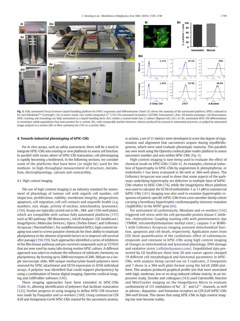
Fig. 3. Fully automated Tecan Evoware Liquid Handling platform for hPSCs expansion and differentiation. Panel (A) shows the anatomy of the automated platform. hPSCs cultured in92 cm2 Roboflasks™ (Corning®) (Ai) in tower stacks (Aii) within integrated 37 °C/5% CO2 automated incubators (LiCONiC Instruments) (Aiii). All media exchanges, cell dissociations,hPSC counting and reseedings are fully automated on a liquid handling deck (Aiv) within a custom build class 2 cabinet (Bigneat Ltd) (Av). In (B), automated hPSC-CM differentiationin monolayer yields populations that stain positive for α-actinin (Bi), with comparable purities between cultures produced by manual or automated processes, as judged by automatedimage analysis to a-actinin (Bii) or flow cytometry for cTnT or a-actinin (Biii).
1739C. Denning et al. / Biochimica et Biophysica Acta 1863 (2016) 1728–1748
4. Towards industrial phenotyping of hPSC-CMs
For in vitro assays, such as safety assessment, there will be a need tointegrate hPSC-CMs into existing or new platforms to assess cell function.In parallel with issues above of hPSC-CM maturation, cell phentopyingis rapidly becoming a bottleneck. In the following sections, we considersome of the platforms that have been (or might be) used for themedium- to high-throughput measurement of structure, metabo-lism, electrophysiology, calcium and contractility.
4.1. High content imaging
The use of high content imaging is an industry standard for assess-ment of physiology of tumour cell with regards cell number, cellshape/size, proliferation, viability, membrane integrity, phagocytosis,apoptosis, cell migration, cell-cell contacts and organelle health (e.g.numbers, size, shape, activity of nucleus, mitochondria, lysosomes)[156]. Assays are typically carried out in 96-, 384- and 1536-well plates,which are compatible with various fully automated platforms [157]such as BD pathway (BD Biosciences), InCell Analyser (GE-healthcare),ImageXpress (Molecular Devices), Opera (Perkin Elmer) and CellomicsArrayscan (ThermoFisher). For undifferentiated hPSCs, high content im-agingwas used to screen putative chemicals for their ability tomaintainpluripotency in the absence of growth factors or to improve cell survivalafter passage [158,159]. Such approaches identified a series of inhibitorsof the Rho kinase pathway and pro-survival compounds such as Y27632that are now used bymany labs during routine hPSC culture. A differentapproach was taken to evaluate the influence of substrate chemistry onpluripotency. By formingup to 2000microspots of 200–300 μmona sin-gle microscope slide, 909 unique methacrylate-based polymers wereassessed for hPSC attachment and OCT4 expression in 4356 individualassays. A polymer was identified that could support pluripotency byusing a combination of Imstar digital imaging, Operetta confocal imag-ing and CellProfiler software [145].
These imaging approaches have been extended to hPSC-CMs(Table 4), allowing identification of polymers that facilitate maturation[122]. Further progress in using imaging to define hPSC-CM structurewas made by Pasqualini and co-workers [160]. Using commercial CDIiCell and Axiogenesis CorAt hPSC-CMs stained for the sarcomeric protein,
α-actinin, a set of 11metrics were developed to score the degree of orga-nisation and alignment that sarcomeres acquire during myofibrillo-genesis, which were used evaluate phenotypic maturity. This parallelsour ownwork using the Operetta confocal plate reader platform to assesssarcomere number and area within hPSC-CMs (Fig. 4).
High content imaging is now being used to evaluate the effect ofchemical insult on hPSC-CMs (Table 4). As examples, chemical induc-tion of hypertrophy in hPSC-CMs by angiotensin II, phenylephrine, orendothelin-1 has been evaluated in 96-well or 384-well plates. TheCellomics Arrayscan was used to show that some aspects of the path-ways underlying hypertrophy are defective in multiple lines of hiPSC-CMs relative to hESC-CMs [74], while the ImageXpress Micro platformwas used to calculate the EC50 of enthothelin-1 as 11 pM in commercialhiPSC-CMs [161]. Imaging was also used to examine hypertrophic re-sponses of patient-specific hiPSC-CMs froma ten-member family cohortcarrying a hereditary hypertrophic cardiomyopathy missense mutation(Arg663His) in the MYH7 gene [63].
For assessment of cardiotoxicity, Mioulane and co-workers [162]triggered cell stress with the cell-permeable protein kinase C inhib-itor, chelerythrine. Coupling staining cells with potentiometric dye(TMRM; tetramethylrhodamine methyl ester), caspase-3 or BOBO-1 with Cellomics Arrayscan imaging assessed mitochondrial func-tion, apoptosis and cell death, respectively. Application notes fromCDI show quantification of the cardiotoxic effect of valinomycin,etoposide and rotenone in hPSC-CMs using high content imagingof changes in mitochondrial and lysosomal physiology, DNA damageand oxidative stress (cellulardynamics.com). Unpublished data pre-sented by GE-healthcare show how 26 anti-cancer agents changed19 different cell morphological and functional parameters in hPSC-CMs, with analysis being carried out on 3 replicates, 2 timepointsand 7 doses in a 384-well plate format using the InCell 2000 plat-form. This analysis produced graphical profile sets that were associatedwith high, moderate, low or no drug-induced cellular toxicity. In an im-pressive study, Sirenko and colleagues [163] used CalceinAM, Hoechstand MitoTracker imaging on the ImageXpress Micro to evaluatecardiotoxicity of 131 modulators of Na+, K+ and Ca2+ channels, as wellas adreno-, dopamine- and histamine-receptors on CDI iCell hiPSC-CMs384-well format. This shows that using hPSC-CMs in high content imag-ing has now become reality.
1740 C. Denning et al. / Biochimica et Biophysica Acta 1863 (2016) 1728–1748
4.2. Mitochondrial function
Mitochondria can be influenced by developmental stage, diseasestate and toxic insult. Hence, analysis of function in hPSC-CMs hasbeen evaluated to gauge maturity, model disease and quantify toxicity(Table 4). As well as the imaging systems discussed above, function ofmitochondria can be determined bymeasuring glycolysis and oxidativephosphorylation (OXPHOS), for which the preferred substrates areglucose in immature CMs and fatty acids in mature CMs, respectively.Initial platforms for evaluation were low throughput. This promptedthe development ofMitoXpress™, a long-decay phosphorescent oxygenprobe [164], which allows estimation of IC50 values of 25 drugs/dayusing a 96-well format [165]. As an alternative platform, the SeahorseExtracellular Flux Analysers uses 24- or 96-well custom plates to deter-mine in vitro oxygen consumption rate (OCR) and extracellular acidifica-tion rate (ECAR) to assess OXPHOS, glycolysis and fatty acid oxidation.These approaches have been used to confirm a preference of hPSC-CMsfor glycolysis when cultured in medium containing glucose, consistentwith developmentally immature state. However, hPSC-CMs can undergosome level of maturation by culturing in galactose/fatty acid-containingmedium to force transition towards OXPHOS or treating with T3 [109]or overexpressing let7 miRNA family [108].
The Seahorse platform is now becoming a common tool in evaluatingdisease state in hPSC-CMs (Table 4). Changes in OCR, ECAR, glycolysis,fatty acid oxidation, maximum respiration capacity and ATP generationhave been studied in hPSC-CMmodels of Barth syndrome [67], Duchennemuscular dystrophy [166], Pompe's disease [167], ARVD [70] and type Idiabetes [168]. Usually, this involves recording basal levels of activ-ity from the hPSC-CMs and then subjecting them to a sequentialtreatment of electron transport chain inhibitors (e.g. oligomycin, ro-tenone, antimycin-A) or uncouplers (FCCP). The same approachesare now being adopted for compound screening. Enzo Life Sciencesused Axiogenesis Cor.4 U hiPSC-CMs to measure oxygen consump-tion and glycolytic flux in real-time via oxygen and pH sensorprobes in 96- or 384-well to inform on drug induced toxicity andmitochondrial function.
4.3. Electrophysiology and calcium
The use of sharp glass electrodes to access the cell interior viaconventional patch clamp electrophysiology is considered the gold-standard for studying ion channel activity. No other technique offersmore insight into the functionality, kinetics, gating properties and phar-macology of ion channels. However, the complexity of this low through-put, labour-intensive approach allows only 10–15 data points a day,which is insufficient to meet the demands of both academic and indus-trial labs. This has prompted the development of various platforms fordirect or indirect measurement of electrophysiology and/or calciumflux, which are now being applied to hPSC-CMs (Table 4).
4.3.1. Planar patch clamp electrophysiologyTo overcome the low throughput of conventional sharp electrodes,
several ‘planar’ patch clamp platforms have been developed that canprocess 16 to 384 multiple recordings in parallel. While these systemsdiffer in their technical specifications and level of automation, they in-crease data throughput 10- to 100-fold depending on the ion channelunder investigation and the platform used [169]. By reducing the com-plexity of the process, they alsomake patch clamping accessible tomoreusers, regardless of previous experience in electrophysiology [170]. Theuse of platforms including IonWorks Quattro [171], PatchXpress [172],Patchliner [173], SynchroPatch [174] and Opatch [175] has been main-stream for the analysis of recombinant cell lines overexpressing singleion channels, and they are nowbeing trialled for usewithmore complexhPSC-CMs [24,175–177] with varying degrees of success (Table 4).
The PatchXpress has been used with high purity hiPSC-CMs to si-multaneously record from 16 channels [24]. The authors measured
densities of INa, Ica and IKr currents in the presence or absence of theirrespective pharmacological inhibitors, tetrodotoxin, nifedipine andE4031. However, the study used an engineered hiPSC line with a neo-mycin phosphotransferse drug resistance cassette targeted down-stream of the MYH6 (αMHC) locus to enrich the CMs, a strategy thatis not easily adopted acrossmultiple hPSC lines. Seal rateswere relative-ly low at ~50% across 58 wells, as was the quality of the seal (~200megaOhms [MΩ]). Genetically purified hiPSC-CMs have also beenused on the CytoPatch2 platform [178]. Analysis in current clampmode enabled retrieval of ventricular, atrial/nodal and S-type action po-tentialmorphologies. By achieving seal resistances over 1GΩ, the actionpotential durations of ventricular-like cells were shown to be decreasedby nifedipine and tetrodotoxin, but increased by cisapride [178]. In ourown work (submitted CD, DR), we have used high efficiency differenti-ation protocols to produce CM purities of N80% across 6 healthy or dis-eased hPSC lines. Processingwith the Patchliner platform enabled catchrates of ~81%, seal rates of ~80% and seal qualities of up to N2GΩ. Anal-ysis in voltage clampmode, allowed evaluation of INa, Ica,L and IK currentdensities and proof of principle for pharmacological testing with tetro-dotoxin, nifedipine and E-4031. These studies provide an initial demon-stration that planar patch systems can be used to investigate specificionic currents in single hPSC-CMs.
4.3.2. Multi-electrode arrays (MEAs) and nanopillarsThe invasive nature of planar patch permits only short-term
(minutes to hours) recordings from single cells. However, someapplica-tions, including acute and chronic toxicity, may benefit from long-termrecordings (days/weeks) from multi-cell clusters. Multi-electrode ar-rays (MEAs) comprise glass slides photo-etched with arrays of micro-electrodes that can record field potentials from cell types for periodsfrom hours to days. Most analysis with MEAs has been done using lowthroughput single-well formats using multi-cellular clusters of hPSC-CMs in 2Dmonolayers or 3D structures, including evaluation of drug re-sponses in hiPSC-CMs carrying KCNH2 (IKr) mutations that underlieLQTS2 [51]. Nevertheless, there have been some reports of multiplexedMEAs being used for evaluation of cardiotoxicity in hPSC-CMs (Table 4).By seeding hESC-CMs to 4-well MEAs, field potential duration (FPD)was calculated after challenge with escalating doses of 12 cardiac ornon-cardiac drugs. This showed that the dose known to cause QT pro-longation in patients corresponded with increases in FPD in hESC-CMs[179]. In a follow-up paper, this approach was extended to evaluatethe effect of the IKs inhibitors, HMR1556 and JNJ303, on hPSC-CMs.However, prolongation could only be detected when the high densityIKr current was reduced by pharmacology (dofetilide or sotalol) or ge-netics (hiPSC-CMs with a mutation in KCNH2) [40], supporting the no-tion that IKs is a weak current in hPSC-CMs [105]. Positive in vitro-invivo correlations have also been made with the hPSC-CM model beingused in conjunction with a 48-well MEA system. Recordings weremade 0.5, 1, 2 and 4 h post exposure to compounds with a combinationof IKr, QT and clinical proarrhythmia properties, including verapamil,ranolazine, flecainide, amiodarone, ouabain, cisapride and terfenadine[180].
However, a contradictory report used theMaestroMEA Platform (1-to 96-wells) to perform blinded evaluation of field potential duration,Na+ slope/amplitude, beat rate and early after-depolarizations to 15IKr, INa or IKs blockers in hPSC-CMs (Table 4). The authors concludedthat these parameters were unable to distinguish torsadogenic from be-nign compounds, with the suggestion that hPSC-CMs on MEAs wouldhave a high false-positive rate in regard to proarrhythmic risk [181]. Itis not clear why these discrepancies have arisen but will be importantto identify to ensure consistency of analysis in the MEA platform. It ispossible differences in cell seeding density or process reduces contactwith the electrode, which could reduce signal strength, quality and ac-curacy of cellular action potentials recordings, as could the proportionof quiescent CMs in the cell preparations [182]. Additional factors thatwill need to be borne in mind as new generations of 96-well MEAs
1741C. Denning et al. / Biochimica et Biophysica Acta 1863 (2016) 1728–1748
capable of electrical pacing are developedwill include the need for lighttransmission, reducing plate cost and improved level of user-friendlysoftware interface that automates data analysis.
Recent technological improvements have allowed themerger of theadvantages of substrate-integrated extracellular MEAs with intracellularelectrodes. These switch between extra- and intra-cellular recordingsusing vertical nanopillar electrodes capable of nano-scale electroporationand re-sealing events.While such systems are capable of detecting actionpotential waveforms of hPSC-CMs [183], the transient nature of the intra-cellular recordings currently limits their use for drug screening purposes.
4.3.3. Optical imagingThe challenges of direct recording of electrophysiology or use of
impedance-based approaches have stimulated the development of opti-cal mapping to measure action potentials and calcium wave propaga-tion using voltage-sensitive dyes (VSDs) [184] like the ANEPPS dyes orgenetically-encoded voltage indicators (GEVIs) [185]. These techniquesallow action potential detection with high spatial and temporal resolu-tion, and are amenable to scale-up. Fluorescence data can be easily andrapidly acquired simultaneously from several cells in a single field ofview, permitting the analysis of over 440 single hESC-CMs in a day(Table 4) [186]. Furthermore, the potential to carry out sequential mea-surements would allow each cell to be used as its own control helpingovercome the inherent heterogeneity of preparations of hPSC-CMs(Herron et al., 2012).
Optical imaging has been used tomeasure calcium transients or beatrates in hPSC-CMs, with high throughput platforms including FLIPRtetra, ImageXpress Micro, FDSS/μCELL and CellOPTIQ in 96- to 384-well formats (Table 4). In particular, the 96-well cellOPTIQ showednifedipine caused a dose dependent decrease in APD75 illustrating its ef-fect on Ca2+ channels [144], while the FDSS/μCELLwas used to monitorchanges in astemizole-induced calcium transients in Cor.4 U CMs in a384-well format [187]. Using the FLIPR tetra in conjunction with aCalcium 5 kit enabled the beat rate, peak shape and regularity to bemonitored in response to 131 compounds comprising cardiac glyco-sides, anti-arrhythmics, α−/β-adrenoceptors, Ca2+ blockers and anti-histamines [187].
However, fluorescent dyes are inherently phototoxic, which cancause temporal degradation of sample and signal quality over time,thereby limiting recording times [185]. This can be overcome by intro-ducing genetically encoded voltage indicators (GEVIs), such as ArcLightGEVI, which has recently been used to image hiPSC-CM action poten-tials during drug screening [186]. In this study, ArcLight allowed the ac-curate prediction of arrhythmic effects, which was comparable to patchclamp measurements [186]. Unlike VSDs, GEVIs also allow specific cellpopulations to be targeted and provide homogenous signal intensitiesindependent of cell uptake [186]. Nevertheless, VSDs or GEVIs providesurrogates of membrane potentials and tend to overestimate action po-tential duration [185,188].
4.3.4. Contraction and force generationExcitation–contraction coupling leads to force generation, which is
the ultimate goal of theworkingmyocardium. As such, variousmethodshave been developed to measure contractility of CMs, including atomicforce microscopy [103], flexible cantilevers [189] strain gauges [190],magnetic beads [191], optical edge detection [192], polyacrylamidegels [193], muscular thin films [194], carbon fibre deflection [195],microposts [196] and image based label-free method [197]. Eachmethodprepares, measures and reports force in a different way (e.g. clamping,bonding, poking), whichmakes cross-comparison of the values producedrather challenging. Nevertheless, force generation has been measuredfromhPSC-CMs in presence or absence of pharmacology (Table 4). Twitchforces for hPSCs-CM range from 2-260nN for single hPSC-CMs [123,196,198] and 0.08–4.4 mN/mm2 for 3D hPSC-CM derived EHTs [199]. This isrelatively low compared with the microN range of single adult CM [124]and 40-80mN/mm2 range of muscle human strips [200]. This reinforces
the need to evaluate the effect on hPSC-CM contractility of the differentmaturation strategies discussed above, including incorporation of non-cardiomyocyte populations found in the heart (i.e. endothelial cells, fibro-blasts, smooth muscle cells).
Translating these approaches into medium- to high-throughput ischallenging and only a few examples exist. The use of impedance to re-port on contractility is becoming popular even though this is a surrogatemeasure and its accuracy needs to be established more thoroughly. Thepremise is that as CMs contract and relax, nano-scale changes in mor-phology andmembrane structure alter the level of impedance betweencell and underlying substrate. These characteristics are exploited byelectric cell-substrate impedance sensing platforms, which incorporategold-film electrodes into the bottom of tissue-culture plates [201,202].Rapid (12.9 ms update rate/plate) data acquisition frequencies [203]allow the resolution of subtle changes in CMbeat profiling including ar-rhythmic events and the continuous monitoring of cell index is usefulfor cardiotoxicity profiling [204]. Of the impedance platforms, thexCELLigence has been most widely used on hPSC-CMs. This showedtesting of a panel of 49 cardioactive compounds on hiPSC-CM hadsensitivity, specificity and accuracy of 90%, 74%, and 82%, respective-ly, which compared favourably when compared to in vitro data fordog (83%, 84% and 82%) and rat (77%, 74%, and 74%) CMs [205].The CardioExcyte96 [206] is a newer platform that measures bothimpedance and extracellular field potentials, allowing study ofhPSC-CM beat rates, amplitudes and electrophysiology (Table 4).Nonetheless, our own experiences are that improvements are need-ed in the number of wells that lead to productive recordings (oftenas low as 15%), as well as to signal recording and data analysis,which are predominately handled as manually processes and limitthroughput.
It is probable that direct measurement of contractility in 3D EHTswill provide a more accurate readout than the indirect method ofimpedance. Multiplexing hPSC-CM EHTs in a 24-well format hasshown differences between the maximal twitch forces (0.22, 0.05, and0.08mN) and sensitivity to external Ca2+ (EC50: 0.15, 0.39, and1.05 mM Ca2+) of rat, mouse, and human EHTs. Evaluating the impactof drug panels on contractility in hPSC-CMderived EHTs is now also un-derway, which will provide an important resource to the pharmaceuti-cal industry [135].
5. Conclusions & future challenges
Over the last 3–5 years, hPSC-CM technologies have transformed.Challenges of hPSC culture have largely been overcome by the develop-ment of defined medium, while differentiation strategies to produceCMs at scale are being refined rapidly. New tools for producingintegration-free hiPSC and engineered Cas9/CRISPR cells mean thatthe range of genotypes available is broader than ever. Isogenic pairs ofhealthy and diseased hPSCs provide sets of cell lines in which only themutation differs in an otherwise identical genetic background. This willoffer unrivalled opportunities to examine impact of polymorphismsidentified from genome-wide association studies (GWAS). These ad-vances have enticed interest from the pharmaceutical industry with thepromise of replacing suboptimal drug safety assessment platforms withphysiologically-relevant human cardiomyocytes. Indeed, there havebeen spectacular successes with hPSC-CMs being used to refine patienttreatment, assist in progressing drugs to Phase I clinical trial andevaluate tens if not hundreds of drugs in medium content assays.Such observations have prompted the CIPA initiative, which willwrite a new chapter of safety assessment guidelines using hPSC-CMs. The progression of hPSC-derived cardiac cells to the clinic isalso an important milestone.
Nevertheless, the excitement needs to be temperedwith the realisa-tion that, relative to many in vitro models, hPSC-CMs are expensive toproduce and beyond the tolerated ‘price point per well’ of most Pharmascreening budgets. Consistency of hPSC-CM production needs to be
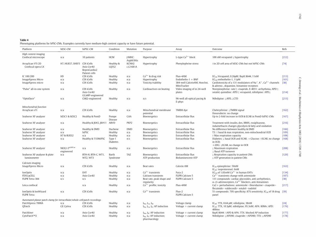
Table 4Phenotyping platforms for hPSC-CMs. Examples currently have medium-high content capacity or have future potential.
Platform hESC-CM hiPSC-CM Condition Mutation Purpose Assay Outcome Refs
High content imagingConfocal microscope n/a 10 patients HCM βMHC:
Arg663HisHypertrophy L-type Ca2+ block 100 nM verapamil ↓ hypertrophy [212]
ArrayScan VTi 2DConfocal opera LX
H7, HUES7, SHEF3 CDI iCellsAxio Cor4UReproCardio2Patient cells
Healthy &LQTS2
KCNH2c.G1681A
Hypertrophy Phenylephrine stress ↑ in 2D cell area of hESC-CMs but not hiPSC-CMs [74]
IC 100/200 H9 CDI iCells Healthy n/a Ca2+ & drug risk Fluo-4AM EC50 Verapamil, 0.26μM; BayK 8644, 11nM [213]ImageXpress Micro n/a CDI iCells Healthy n/a Hypertrophy Endothelin-1 + BNP EC50 enthothelin-1, 11pM [161]ImageXpress micro n/a CDI iCells Healthy n/a Toxicity/viability 384-well CalceinAM, Hoechst,
MitoTrackerCardiotoxicity of a 131 modulators of Na+, K+, Ca2+ channels& adreno-, dopamine, histamine receptors
[38]
“Pulse” all-in-one system n/a CDI iCellsAxio Cor4UGCaMP-engineered
Healthy n/a Cardioactives on beating Video imaging of in 24-wellplates
Norepinephrine: rate↑; cisapride, E-4031: arrhythmia, APD↑;sotalol, quinidine: APD↑; verapamil, nifedipine: APD↓ [214]
“OptoDyce” n/a ChR2-engineered Healthy n/a n/s 96-well all-optical pacing &E-phys
Nifedipine: ↓APD, ↓CTD [215]
Mitochondrial functionArrayScan vTi n/a CDI iCells Healthy n/a Mitochondrial membrane
potentialTMRM dye Chelerythrine: ↓TMRM signal
Doxorubicin: no change[162]
Seahorse XF analyser hESC1 & hESC2 Healthy & PomD PompeDisease
GAA Bioenergetics Extracellular flux Up to 2-fold increase in OCR & ECAR in PomD hiPSC-CMs [167]
Seahorse XF analyser n/a Healthy & JK#2, JK#11 ARVD PKP2 Bioenergetics Extracellular flux Treatment with insulin, dex, IBMX, rosiglitazone,indamethacin changes glycolysis & fatty acid oxidation
[216]
Seahorse XF analyser n/a Healthy & DMD Duchene DMD Bioenergetics Extracellular flux No difference between healthy & DMD [166]Seahorse XF analyser n/a hiPSC Healthy n/a Bioenergetics Extracellular flux T3: ↑ basal & max respiration, non-mitochondrial OCR [109]Seahorse XF analyser H7 & RUES2 n/a Healthy n/a Bioenergetics Extracellular flux ↑ Max respiration capacity [108]Seahorse XF analyser n/a Healthy & T1DMiPSC Type 1
Diabetesn/s Bioenergetics Extracellular flux Diabetes: ↓ basal OCR and ECAR; +Glucose: ↑ECAR, no change
in OCR;+2DG: ↓ECAR, no change in OCR
[168]
Seahorse XF analyser NKX2-5eGFP/w
engineeredn/a Healthy n/a Bioenergetics Extracellular flux ↓ Maximum respiration
↓ Basal ATP turnover[208]
Seahorse XF analyser & plateluminometer
n/a BTH-H, BTH-C, WT1,WT2, WT3
BarthSyndrome
TAZ Bioenergetics Extracellular flux ↓ Respiration capacity in patient CMs [129]ATP production Bioluminescent ATP ↓ ATP generation in patient CMs
Calcium imagingImageXpress Micro n/a CDI iCells Healthy n/a Beat rates Calcein AM IC50 epinephrine: 50nM
IC50: isoproterenol: 6nM[163]
IonOptix n/a EHT Healthy n/a Ca2+ transients Fura-2 EC50 of 1.05mM Ca2+ in human EHTs [134]FDSS/μCELL n/a Axio Cor4U Healthy n/a Calcium transients FLIPR Calcium 5 Ca2+ transients change with astemizole [187]FLIPR Tetra 384 n/s n/s Healthy n/a Beat rate, peak shape and
regularityFLIPR Calcium 5 131 compounds: cardiac glycosides, anti-arrhythmics,
α-/β-adrenoceptors, Ca2+ blockers, anti-histamines[38]
Leica confocal n/a Healthy n/a Ca2+ profile; toxicity Fluo-4AM Ca2+ perturbations: astemizoleN thioridazineN cisaprideNflecainideN valdecoxibN sotalolN nadolol
[217]
IonOptix & IonWizard n/a CDI iCells Healthy n/a Ca2+ transients Fluo-2 51 compounds: 70% specificity; 87% sensitivity; IC50 of 18 drugpanel
[39]FLIPR Tetra FLIPR Calcium 5
Automated planar patch clamp for intracellular/whole-cell/patch recordingsPatchXpress 7000A n/a CDI iCells Healthy n/a INa, ICa, IKr Voltage clamp IC50: TTX, 0.64 μM; nifedipine, 38nM [24]QPatch GE Cytiva CDI iCells Healthy n/a INa, ICa, IK; AP induction Voltage + current clamp IC50: TTX, 10.3μM; nifedipine, 95.3nM; APA: 80Mv; APD:
400ms[176]
Patchliner n/a Axio Cor4U Healthy n/a INa, IK; AP induction Voltage + current clamp BayK 8644: ↑APD & APA; TTX: blocked AP induction [177]CytoPatch™2 n/a Axio Cor4U Healthy n/a INa, IK; AP induction;
pharmacologyVoltage + current clamp Nifedipine: ↓APD90; cisapride: ↑APD90; TTX: ↓APD90 [178]
1742C.D
enningetal./Biochim
icaetBiophysica
Acta
1863(2016)
1728–1748
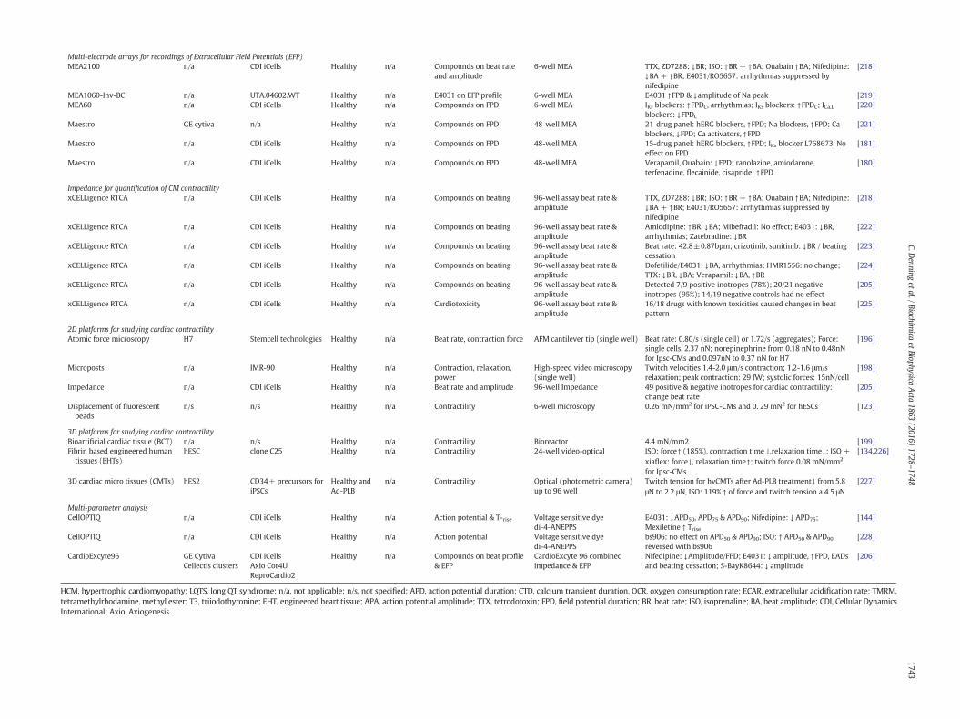
Multi-electrode arrays for recordings of Extracellular Field Potentials (EFP)MEA2100 n/a CDI iCells Healthy n/a Compounds on beat rate
and amplitude6-well MEA TTX, ZD72 8: ↓BR; ISO: ↑BR + ↑BA; Ouabain ↑BA; Nifedipine:
↓BA + ↑B E4031/RO5657: arrhythmias suppressed bynifedipine
[218]
MEA1060-Inv-BC n/a UTA.04602.WT Healthy n/a E4031 on EFP profile 6-well MEA E4031 ↑F & ↓amplitude of Na peak [219]MEA60 n/a CDI iCells Healthy n/a Compounds on FPD 6-well MEA IKr blocke ↑FPDC, arrhythmias; IKs blockers: ↑FPDC; ICa,L
blockers: PDC
[220]
Maestro GE cytiva n/a Healthy n/a Compounds on FPD 48-well MEA 21-drug p el: hERG blockers, ↑FPD; Na blockers, ↑FPD; Cablockers, PD; Ca activators, ↑FPD
[221]
Maestro n/a CDI iCells Healthy n/a Compounds on FPD 48-well MEA 15-drug p el: hERG blockers, ↑FPD; IKs blocker L768673, Noeffect on D
[181]
Maestro n/a CDI iCells Healthy n/a Compounds on FPD 48-well MEA Verapami uabain: ↓FPD; ranolazine, amiodarone,terfenadin , flecainide, cisapride: ↑FPD
[180]
Impedance for quantification of CM contractilityxCELLigence RTCA n/a CDI iCells Healthy n/a Compounds on beating 96-well assay beat rate &
amplitudeTTX, ZD72 8: ↓BR; ISO: ↑BR + ↑BA; Ouabain ↑BA; Nifedipine:↓BA + ↑B E4031/RO5657: arrhythmias suppressed bynifedipine
[218]
xCELLigence RTCA n/a CDI iCells Healthy n/a Compounds on beating 96-well assay beat rate &litude
Amlodipi : ↑BR, ↓BA; Mibefradil: No effect; E4031: ↓BR,arrhythm ; Zatebradine: ↓BR
[222]
xCELLigence RTCA n/a CDI iCells Healthy n/a Compounds on beating 96-well assay beat rate &litude
Beat rate: 2.8±0.87bpm; crizotinib, sunitinib: ↓BR / beatingcessation
[223]
xCELLigence RTCA n/a CDI iCells Healthy n/a Compounds on beating 96-well assay beat rate &litude
Dofetilide 4031: ↓BA, arrhythmias; HMR1556: no change;TTX: ↓BR, BA; Verapamil: ↓BA, ↑BR
[224]
xCELLigence RTCA n/a CDI iCells Healthy n/a Compounds on beating 96-well assay beat rate &litude
Detected 9 positive inotropes (78%); 20/21 negativeinotropes 5%); 14/19 negative controls had no effect
[205]
xCELLigence RTCA n/a CDI iCells Healthy n/a Cardiotoxicity 96-well assay beat rate &litude
16/18 dru with known toxicities caused changes in beatpattern
[225]
2D platforms for studying cardiac contractilityAtomic force microscopy H7 Stemcell technologies Healthy n/a Beat rate, contraction force AFM cantilever tip (single well) Beat rate: .80/s (single cell) or 1.72/s (aggregates); Force:
single cel 2.37 nN; norepinephrine from 0.18 nN to 0.48nNfor Ipsc-C s and 0.097nN to 0.37 nN for H7
[196]
Microposts n/a IMR-90 Healthy n/a Contraction, relaxation,power
High-speed video microscopy(single well)
Twitch ve cities 1.4-2.0 μm/s contraction; 1.2-1.6 μm/srelaxation peak contraction: 29 fW; systolic forces: 15nN/cell
[198]
Impedance n/a CDI iCells Healthy n/a Beat rate and amplitude 96-well Impedance 49 positiv & negative inotropes for cardiac contractility:change be rate
[205]
Displacement of fluorescentbeads
n/s n/s Healthy n/a Contractility 6-well microscopy 0.26 mN/ 2 for iPSC-CMs and 0. 29 mN2 for hESCs [123]
3D platforms for studying cardiac contractilityBioartificial cardiac tissue (BCT) n/a n/s Healthy n/a Contractility Bioreactor 4.4 mN/m 2 [199]Fibrin based engineered human
tissues (EHTs)hESC clone C25 Healthy n/a Contractility 24-well video-optical ISO: force (185%), contraction time ↓,relaxation time↓; ISO +
xiaflex: fo e↓, relaxation time↑; twitch force 0.08 mN/mm2
for Ipsc-C s
[134,226]
3D cardiac micro tissues (CMTs) hES2 CD34+ precursors foriPSCs
Healthy andAd-PLB
n/a Contractility Optical (photometric camera)up to 96 well
Twitch te ion for hvCMTs after Ad-PLB treatment↓ from 5.8μN to 2.2 , ISO: 119% ↑ of force and twitch tension a 4.5 μN
[227]
Multi-parameter analysisCellOPTIQ n/a CDI iCells Healthy n/a Action potential & T-rise Voltage sensitive dye
di-4-ANEPPSE4031: ↓A D50, APD75 & APD90; Nifedipine: ↓ APD75;Mexiletin Trise
[144]
CellOPTIQ n/a CDI iCells Healthy n/a Action potential Voltage sensitive dyedi-4-ANEPPS
bs906: no ffect on APD50 & APD90; ISO: ↑ APD50 & APD90
reversed th bs906[228]
CardioExcyte96 GE CytivaCellectis clusters
CDI iCellsAxio Cor4UReproCardio2
Healthy n/a Compounds on beat profile& EFP
CardioExcyte 96 combinedimpedance & EFP
Nifedipin ↓Amplitude/FPD; E4031: ↓ amplitude, ↑FPD, EADsand beati cessation; S-BayK8644: ↓ amplitude
[206]
HCM, hypertrophic cardiomyopathy; LQTS, long QT syndrome; n/a, not applicable; n/s, not specified; APD, action potential duration; CTD, calcium transient duration, OCR, oxygen nsumption rate; ECAR, extracellular acidification rate; TMRM,tetramethylrhodamine, methyl ester; T3, triiodothyronine; EHT, engineered heart tissue; APA, action potential amplitude; TTX, tetrodotoxin; FPD, field potential duration; BR, beat ra ; ISO, isoprenaline; BA, beat amplitude; CDI, Cellular DynamicsInternational; Axio, Axiogenesis.
1743C.D
enningetal./Biochim
icaetBiophysica
Acta
1863(2016)
1728–1748
8R;
PDrs:↓Fan↓FanFPl, Oe
8R;
neias4
/E↓7/(9gs
0ls,Mlo;eatmm
m↑rcMnsμN
Pe ↑e
wie:ng
cote
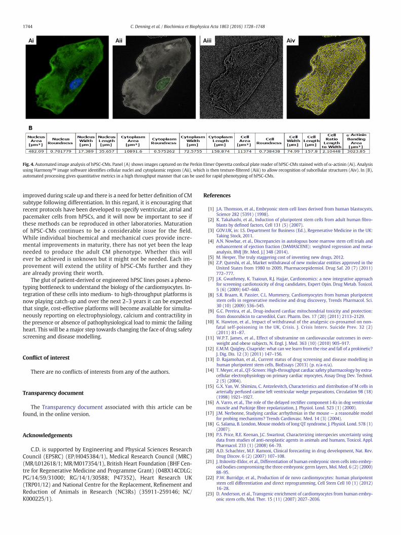
Fig. 4. Automated image analysis of hPSC-CMs. Panel (A) shows images captured on the Perkin Elmer Operetta confocal plate reader of hPSC-CMs stained with ofα-actinin (Ai). Analysisusing Harmony™ image software identifies cellular nuclei and cytoplasmic regions (Aii), which is then texture-filtered (Aiii) to allow recognition of subcellular structures (Aiv). In (B),automated processing gives quantitative metrics in a high throughput manner that can be used for rapid phenotyping of hPSC-CMs.
1744 C. Denning et al. / Biochimica et Biophysica Acta 1863 (2016) 1728–1748
improved during scale up and there is a need for better definition of CMsubtype following differentiation. In this regard, it is encouraging thatrecent protocols have been developed to specify ventricular, atrial andpacemaker cells from hPSCs, and it will now be important to see ifthese methods can be reproduced in other laboratories. Maturationof hPSC-CMs continues to be a considerable issue for the field.While individual biochemical and mechanical cues provide incre-mental improvements in maturity, there has not yet been the leapneeded to produce the adult CM phenotype. Whether this willever be achieved is unknown but it might not be needed. Each im-provement will extend the utility of hPSC-CMs further and theyare already proving their worth.
The glut of patient-derived or engineered hPSC lines poses a pheno-typing bottleneck to understand the biology of the cardiomyocytes. In-tegration of these cells into medium- to high-throughput platforms isnow playing catch-up and over the next 2–3 years it can be expectedthat single, cost-effective platforms will become available for simulta-neously reporting on electrophysiology, calcium and contractility inthe presence or absence of pathophysiological load to mimic the failingheart. This will be amajor step towards changing the face of drug safetyscreening and disease modelling.
Conflict of interest
There are no conflicts of interests from any of the authors.
Transparency document
The Transparency document associated with this article can befound, in the online version.
Acknowledgements
C.D. is supported by Engineering and Physical Sciences ResearchCouncil (EPSRC) (EP/H045384/1), Medical Research Council (MRC)(MR/L012618/1; MR/M017354/1), British Heart Foundation (BHF Cen-tre for Regenerative Medicine and Programme Grant) (04BX14CDLG;PG/14/59/31000; RG/14/1/30588; P47352), Heart Research UK(TRP01/12) and National Centre for the Replacement, Refinement andReduction of Animals in Research (NC3Rs) (35911-259146; NC/K000225/1).
References
[1] J.A. Thomson, et al., Embryonic stem cell lines derived from human blastocysts,Science 282 (5391) (1998).
[2] K. Takahashi, et al., Induction of pluripotent stem cells from adult human fibro-blasts by defined factors, Cell 131 (5) (2007).
[3] GOV.UK, in: I.S. Department for Business (Ed.), Regenerative Medicine in the UK:Taking Stock, 2011.
[4] A.N. Nowbar, et al., Discrepancies in autologous bone marrow stem cell trials andenhancement of ejection fraction (DAMASCENE): weighted regression and meta-analysis, BMJ [Br. Med. J.] 348 (2014).
[5] M. Herper, The truly staggering cost of inventing new drugs, 2012.[6] Z.P. Qureshi, et al., Market withdrawal of new molecular entities approved in the
United States from 1980 to 2009, Pharmacoepidemiol. Drug Saf. 20 (7) (2011)772–777.
[7] J.K. Gwathmey, K. Tsaioun, R.J. Hajjar, Cardionomics: a new integrative approachfor screening cardiotoxicity of drug candidates, Expert Opin. Drug Metab. Toxicol.5 (6) (2009) 647–660.
[8] S.R. Braam, R. Passier, C.L. Mummery, Cardiomyocytes from human pluripotentstem cells in regenerative medicine and drug discovery, Trends Pharmacol. Sci.30 (10) (2009) 536–545.
[9] G.C. Pereira, et al., Drug-induced cardiac mitochondrial toxicity and protection:from doxorubicin to carvedilol, Curr. Pharm. Des. 17 (20) (2011) 2113–2129.
[10] K. Hawton, et al., Impact of withdrawal of the analgesic co-proxamol on non-fatal self-poisoning in the UK, Crisis. J. Crisis Interv. Suicide Prev. 32 (2)(2011) 81–87.
[11] W.P.T. James, et al., Effect of sibutramine on cardiovascular outcomes in over-weight and obese subjects, N. Engl. J. Med. 363 (10) (2010) 905–917.
[12] E.M.M. Quigley, Cisapride: what can we learn from the rise and fall of a prokinetic?J. Dig. Dis. 12 (3) (2011) 147–156.
[13] D. Rajamohan, et al., Current status of drug screening and disease modelling inhuman pluripotent stem cells, BioEssays (2013) (p. n/a-n/a).
[14] T. Meyer, et al., QT-Screen: High-throughput cardiac safety pharmacology by extra-cellular electrophysiology on primary cardiac myocytes, Assay Drug Dev. Technol.2 (5) (2004).
[15] G.X. Yan, W. Shimizu, C. Antzelevitch, Characteristics and distribution of M cells inarterially perfused canine left ventricular wedge preparations, Circulation 98 (18)(1998) 1921–1927.
[16] A. Varro, et al., The role of the delayed rectifier component I-Ks in dog ventricularmuscle and Purkinje fibre repolarization, J. Physiol. Lond. 523 (1) (2000).
[17] J.M. Nerbonne, Studying cardiac arrhythmias in the mouse — a reasonable modelfor probing mechanisms? Trends Cardiovasc. Med. 14 (3) (2004).
[18] G. Salama, B. London, Mouse models of long QT syndrome, J. Physiol. Lond. 578 (1)(2007).
[19] P.S. Price, R.E. Keenan, J.C. Swartout, Characterizing interspecies uncertainty usingdata from studies of anti-neoplastic agents in animals and humans, Toxicol. Appl.Pharmacol. 233 (1) (2008) 64–70.
[20] A.D. Schachter, M.F. Ramoni, Clinical forecasting in drug development, Nat. Rev.Drug Discov. 6 (2) (2007) 107–108.
[21] J. Itskovitz-Eldor, et al., Differentiation of human embryonic stem cells into embry-oid bodies compromising the three embryonic germ layers, Mol. Med. 6 (2) (2000)88–95.
[22] P.W. Burridge, et al., Production of de novo cardiomyocytes: human pluripotentstem cell differentiation and direct reprogramming, Cell Stem Cell 10 (1) (2012)16–28.
[23] D. Anderson, et al., Transgenic enrichment of cardiomyocytes from human embry-onic stem cells, Mol. Ther. 15 (11) (2007) 2027–2036.
1745C. Denning et al. / Biochimica et Biophysica Acta 1863 (2016) 1728–1748
[24] J. Ma, et al., High purity human-induced pluripotent stem cell-derivedcardiomyocytes: electrophysiological properties of action potentials and ionic cur-rents, Am. J. Physiol. Heart Circ. Physiol. 301 (5) (2011) H2006–H2017.
[25] D. Calderon, et al., Immune response to human embryonic stem cell-derived cardi-ac progenitors and adipose-derived stromal cells, J. Cell. Mol. Med. 16 (7) (2012)1544–1552.
[26] R.J. Skelton, et al., SIRPA, VCAM1 and CD34 identify discrete lineages during earlyhuman cardiovascular development, Stem Cell Res. 13 (1) (2014) 172–179.
[27] B.M. Wile, et al., Molecular beacon-enabled purification of living cells by targetingcell type-specific mRNAs, Nat. Protoc. 9 (10) (2014) 2411–2424.
[28] F. Hattori, et al., Nongenetic method for purifying stem cell-derived cardiomyocytes,Nat. Methods 7 (1) (2010) 61–66.
[29] J.J. Chong, et al., Human embryonic-stem-cell-derived cardiomyocytes regeneratenon-human primate hearts, Nature 510 (7504) (2014) 273–277.
[30] S. Tohyama, et al., Distinctmetabolic flow enables large-scale purification ofmouseand human pluripotent stem cell-derived cardiomyocytes, Cell Stem Cell 12 (1)(2013) 127–137.
[31] W. Zhang, et al., Microfluidics separation reveals the stem-cell-like deformability oftumor-initiating cells, Proc. Natl. Acad. Sci. U. S. A. 109 (46) (2012) 18707–18712.
[32] F.B. Myers, et al., Label-free electrophysiological cytometry for stem cell-derivedcardiomyocyte clusters, Lab Chip 13 (2) (2013) 220–228.
[33] M.J. Birket, et al., Expansion and patterning of cardiovascular progenitors derivedfrom human pluripotent stem cells, Nat. Biotechnol. (2015).
[34] M.J. Birket, C.L. Mummery, Pluripotent stem cell derived cardiovascularprogenitors–a developmental perspective, Dev. Biol. 400 (2) (2015) 169–179.
[35] H.D. Devalla, et al., Atrial-like cardiomyocytes from human pluripotent stem cellsare a robust preclinical model for assessing atrial-selective pharmacology, EMBOMol. Med. 7 (4) (2015) 394–410.
[36] L. Warren, et al., Highly efficient reprogramming to pluripotency and directeddifferentiation of human cells with synthetic modified mRNA, Cell Stem Cell 7(5) (2010).
[37] F.A. Soares, et al., International coordination of large-scale human induced pluripotentstem cell initiatives:wellcome trust and ISSCRworkshopswhite paper, Stem Cell Rep.3 (6) (2014) 931–939.
[38] O. Sirenko, et al., Assessment of beating parameters in human induced pluripotentstem cells enables quantitative in vitro screening for cardiotoxicity, Toxicol. Appl.Pharmacol. 273 (3) (2013) 500–507.
[39] A. Pointon, et al., Assessment of cardiomyocyte contraction in human-induced plurip-otent stem cell-derived cardiomyocytes, Toxicol. Sci. 144 (2) (2015) 227–237.
[40] S.R. Braam, et al., Repolarization reserve determines drug responses in human plurip-otent stem cell derived cardiomyocytes, Stem Cell Res. 10 (1) (2013) 48–56.
[41] K. Harris, et al., Comparison of electrophysiological data from human-inducedpluripotent stem cell–derived cardiomyocytes to functional preclinical safetyassays, Toxicol. Sci. 134 (2) (2013) 412–426.
[42] L. Nalos, et al., Comparison of the I(Kr) blockers moxifloxacin, dofetilide andE-4031 in five screening models of pro-arrhythmia reveals lack of specificity ofisolated cardiomyocytes, Br. J. Pharmacol. 165 (2) (2012) 467–478.
[43] A. Sharma, et al., Human induced pluripotent stem cell-derived cardiomyocytes asan in vitro model for coxsackievirus B3-induced myocarditis and antiviral drugscreening platform, Circ. Res. 115 (6) (2014) 556 − +.
[44] J.G. Reynolds, et al., HER2-targeted liposomal doxorubicin displays enhanced anti-tumorigenic effects without associated cardiotoxicity, Toxicol. Appl. Pharmacol.262 (1) (2012) 1–10.
[45] P.T. Sager, et al., Rechanneling the cardiac proarrhythmia safety paradigm: Ameet-ing report from the cardiac safety research consortium, Am. Heart J. 167 (3) (2014)292–300.
[46] A. Moretti, et al., Patient-specific induced pluripotent stem-cell models for long-QTsyndrome, N. Engl. J. Med. 363 (15) (2010) 1397–1409.
[47] T. Egashira, et al., Disease characterization using LQTS-specific induced pluripotentstem cells, 95 (2012) 419–429.
[48] M. Zhang, et al., Recessive cardiac phenotypes in induced pluripotent stem cellmodels of Jervell and Lange-Nielsen syndrome: disease mechanisms and pharma-cological rescue, Proc. Natl. Acad. Sci. 111 (50) (2014) E5383–E5392.
[49] I. Itzhaki, et al., Modelling the long QT syndrome with induced pluripotent stemcells, Nature 471 (7337) (2011) 225–229.
[50] A.L. Lahti, et al., Human disease model for long QT syndrome type 2 using iPS cellsdemonstrates arrhythmogenic characteristics in cell culture, Dis. Model. Mech.(2011).
[51] E. Matsa, et al., Drug evaluation in cardiomyocytes derived from human inducedpluripotent stem cells carrying a long QT syndrome type 2 mutation, Eur. Heart J.32 (8) (2011) 952–962.
[52] M. Bellin, et al., Isogenic human pluripotent stem cell pairs reveal the role of aKCNH2 mutation in long-QT syndrome, vol. 322013 3161–3175.
[53] E. Matsa, et al., Allele-specific RNA interference rescues the long-QT syndromephenotype in human-induced pluripotency stem cell cardiomyocytes, Eur. HeartJ. 35 (16) (2014) 1078–1087.
[54] D.Ma, et al., Modeling type 3 long QT syndromewith cardiomyocytes derived frompatient-specific induced pluripotent stem cells, Int. J. Cardiol. 168 (6) (2013)5277–5286.
[55] M. Yazawa, et al., Using induced pluripotent stem cells to investigate cardiac phe-notypes in Timothy syndrome, Nature 471 (7337) (2011).
[56] R.P. Davis, et al., Cardiomyocytes derived from pluripotent stem cells recapitulateelectrophysiological characteristics of an overlap syndrome of cardiac sodiumchannel disease, Circulation 125 (25) (2012).
[57] A. Fatima, et al., In vitro modeling of ryanodine receptor 2 dysfunction usinghuman induced pluripotent stem cells, Cell. Physiol. Biochem. 28 (4) (2011).
[58] C.B. Jung, et al., Dantrolene rescues arrhythmogenic RYR2 defect in a patient-specific stem cell model of catecholaminergic polymorphic ventricular tachycardia,EMBO Mol. Med. 4 (3) (2012).
[59] I. Itzhaki, et al., Modeling of catecholaminergic polymorphic ventricular tachycar-dia with patient-specific human-induced pluripotent stem cells, J. Am. Coll. Cardiol.60 (11) (2012) 990–1000.
[60] B. Lin, et al., Modeling and study of the mechanism of dilated cardiomyopathyusing induced pluripotent stem cells derived from individuals with Duchennemuscular dystrophy, Dis. Model. Mech. 8 (5) (2015) 457–466.
[61] N. Sun, et al., Patient-specific induced pluripotent stem cells as a model for familialdilated cardiomyopathy, vol. 4.2012 (130ra47-130ra47).
[62] H. Wu, et al., Epigenetic regulation of phosphodiesterases 2A and 3A underliescompromised β-adrenergic signaling in an iPSC model of dilated cardiomyopathy,Cell Stem Cell 17 (1) (2015) 89–100.
[63] F. Lan, et al., Abnormal calcium handling properties underlie familial hypertrophiccardiomyopathy pathology in patient-specific induced pluripotent stem cells, CellStem Cell 12 (1) (2013) 101–113.
[64] L. Han, et al., Study familial hypertrophic cardiomyopathy using patient-specific in-duced pluripotent stem cells, vol. 104.2014 258–269.
[65] X. Carvajal-Vergara, et al., Patient-specific induced pluripotent stem-cell-derivedmodels of LEOPARD syndrome, Nature 465 (7299) (2010) (p. 808-U12).
[66] J. Dudek, et al., Cardiolipin deficiency affects respiratory chain function and organi-zation in an induced pluripotent stem cell model of Barth syndrome, Stem Cell Res.11 (2) (2013) 806–819.
[67] G. Wang, et al., Modeling the mitochondrial cardiomyopathy of Barth syndromewith induced pluripotent stem cell and heart-on-chip technologies, Nat. Med. 20(6) (2014) 616–623.
[68] D. Ma, et al., Generation of patient-specific induced pluripotent stem cell-derivedcardiomyocytes as a cellular model of arrhythmogenic right ventricular cardiomy-opathy, vol. 342013 1122–1133.
[69] O. Caspi, et al., Modeling of arrhythmogenic right ventricular cardiomyopathywithhuman induced pluripotent stem cells, Circ. Cardiovasc. Genet. 6 (6) (2013)557–568.
[70] C. Kim, et al., Studying arrhythmogenic right ventricular dysplasia with patient-specific iPSCs, Nature 494 (7435) (2013) 105–110.
[71] J.-Y. Wen, et al., Maturation-based model of arrhythmogenic right ventricular dys-plasia using patient-specific induced pluripotent stem cells, Circ. J. 79 (7) (2015)1402–1408.
[72] E. Dick, et al., Exon skipping and gene transfer restore dystrophin expression inhuman induced pluripotent stem cells-cardiomyocytes harboring DMDmutations,Stem Cells Dev. 22 (20) (2013) 2714–2724.
[73] C. Terrenoire, et al., Induced pluripotent stem cells used to reveal drug actions in along QT syndrome family with complex genetics, J. Gen. Physiol. 141 (1)(2013) 61–72.
[74] G. Foldes, et al., Aberrant alpha-adrenergic hypertrophic response incardiomyocytes from human induced pluripotent cells, Stem Cell Rep. 3 (5)(2014) 905–914.
[75] S.D. Sheridan, et al., Epigenetic Characterization of the FMR1 gene and aberrantneurodevelopment in human induced pluripotent stem cell models of fragile Xsyndrome, PLoS One 6 (10) (2011).
[76] L. Ye, et al., Cardiac repair in a porcine model of acute myocardial infarction withhuman induced pluripotent stem cell-derived cardiovascular cells, Cell Stem Cell15 (6) (2014) 750–761.
[77] P. Menasche, et al., Human embryonic stem cell-derived cardiac progenitors for se-vere heart failure treatment: first clinical case report, Eur. Heart J. 36 (30) (2015)2011–2017.
[78] P. Menasché, Transplantation of human embryonic stem cell-derived CD15+ Isl-1 + progenitors in severe heart failure, 2013 (ClinicalTrials.gov Internet).
[79] I. Mateizel, et al., Derivation of human embryonic stem cell lines from embryos ob-tained after IVF and after PGD for monogenic disorders, Hum. Reprod. 21 (2)(2006) 503–511.
[80] A. Urbach, M. Schuldiner, N. Benvenisty, Modeling for Lesch–Nyhan disease bygene targeting in human embryonic stem cells, Stem Cells 22 (4) (2004)635–641.
[81] D.A. Elliott, et al., NKX2-5(eGFP/w) hESCs for isolation of human cardiac progeni-tors and cardiomyocytes, Nat. Methods 8 (12) (2011) 1037–1040.
[82] M. Bellin, et al., Isogenic human pluripotent stem cell pairs reveal the role of aKCNH2 mutation in long-QT syndrome, EMBO J. 32 (24) (2013) 3161–3175.
[83] Y.G. Kim, J. Cha, S. Chandrasegaran, Hybrid restriction enzymes: zinc finger fu-sions to Fok I cleavage domain, Proc. Natl. Acad. Sci. U. S. A. 93 (3) (1996)1156–1160.
[84] M.H. Porteus, D. Baltimore, Chimeric nucleases stimulate gene targeting in humancells, Science 300 (5620) (2003) 763.
[85] T. Cermak, et al., Efficient design and assembly of custom TALEN and other TALeffector-based constructs for DNA targeting, Nucleic Acids Res. 39 (12) (2011) e82.
[86] D. Reyon, et al., FLASH assembly of TALENs for high-throughput genome editing,Nat. Biotechnol. 30 (5) (2012) 460–465.
[87] C.Mussolino, et al., A novel TALE nuclease scaffold enables high genome editing activ-ity in combination with low toxicity, Nucleic Acids Res. 39 (21) (2011) 9283–9293.
[88] P.D. Hsu, E.S. Lander, F. Zhang, Development and applications of CRISPR-Cas9 forgenome engineering, Cell 157 (6) (2014) 1262–1278.
[89] P. Mali, et al., RNA-guided human genome engineering via Cas9, Science 339(6121) (2013) 823–826.
[90] H.L. Li, et al., Precise correction of the dystrophin gene in duchenne muscular dys-trophy patient induced pluripotent stem cells by TALEN and CRISPR-Cas9, StemCell Rep. 4 (1) (2015) 143–154.
1746 C. Denning et al. / Biochimica et Biophysica Acta 1863 (2016) 1728–1748
[91] F.A. Ran, et al., Double nicking by RNA-guided CRISPR Cas9 for enhanced genomeediting specificity, Cell 154 (6) (2013) 1380–1389.
[92] S. Kim, et al., Highly efficient RNA-guided genome editing in human cells via deliv-ery of purified Cas9 ribonucleoproteins, Genome Res. 24 (6) (2014) 1012–1019.
[93] L. Cong, et al., Multiplex genome engineering using CRISPR/Cas systems, Science339 (6121) (2013) 819–823.
[94] F. Chen, et al., High-frequency genome editing using ssDNA oligonucleotides withzinc-finger nucleases, Nat. Methods 8 (9) (2011) 753–755.
[95] C. Yu, et al., Small molecules enhance CRISPR genome editing in pluripotent stemcells, Cell Stem Cell 16 (2) (2015) 142–147.
[96] K. Yusa, et al., Targeted gene correction of alpha1-antitrypsin deficiency in inducedpluripotent stem cells, Nature 478 (7369) (2011) 391–394.
[97] E. Lanphier, et al., Don't edit the human germ line, Nature 519 (7544) (2015)410–411.
[98] P. Liang, et al., CRISPR/Cas9-mediated gene editing in human tripronuclear zygotes,Protein Cell 6 (5) (2015) 363–372.
[99] C.W. van den Berg, et al., Transcriptome of human foetal heart compared withcardiomyocytes from pluripotent stem cells, Development (2015).
[100] X. Yang, L. Pabon, C.E. Murry, Engineering adolescence: maturation of human plu-ripotent stem cell-derived cardiomyocytes, Circ. Res. 114 (3) (2014) 511–523.
[101] C.C. Veerman, et al., Immaturity of human stem-cell-derived cardiomyocytes inculture: fatal flaw or soluble problem? Stem Cells Dev. 24 (9) (2015) 1035–1052.
[102] F.B. Bedada, et al., Acquisition of a quantitative, stoichiometrically conservedratiometric marker of maturation status in stem cell-derived cardiac myocytes,Stem Cell Rep. 3 (4) (2014) 594–605.
[103] A. Blazeski, et al., Cardiomyocytes derived from human induced pluripotent stemcells asmodels for normal and diseased cardiac electrophysiology and contractility,Prog. Biophys. Mol. Biol. 110 (2–3) (2012) 166–177.
[104] M.K. Jonsson, et al., Application of human stem cell-derived cardiomyocytes insafety pharmacology requires caution beyond hERG, J. Mol. Cell. Cardiol. 52 (5)(2012) 998–1008.
[105] T. Christ, A. Horvath, T. Eschenhagen, LQT1-phenotypes in hiPSC: are we measur-ing the right thing? Proc. Natl. Acad. Sci. U. S. A. 112 (16) (2015) (p. (E1968-E1968)).
[106] L.W. van Laake, et al., Human embryonic stem cell-derived cardiomyocytes surviveand mature in the mouse heart and transiently improve function after myocardialinfarction, Stem Cell Res. 1 (1) (2007) 9–24.
[107] I. Piccini, et al., Human pluripotent stem cell-derived cardiomyocytes: genome-wide expression profiling of long-term in vitro maturation in comparison tohuman heart tissue, Genomic Data 4 (2015) 69–72.
[108] K.T. Kuppusamy, et al., Let-7 family of microRNA is required for maturation andadult-like metabolism in stem cell-derived cardiomyocytes, Proc. Natl. Acad. Sci.U. S. A. 112 (21) (2015) E2785–E2794.
[109] X. Yang, et al., Tri-iodo-l-thyronine promotes the maturation of humancardiomyocytes-derived from induced pluripotent stem cells, J. Mol. Cell. Cardiol.72 (2014) 296–304.
[110] J.Y. Wen, et al., Maturation-based model of arrhythmogenic right ventricular dys-plasia using patient-specific induced pluripotent stem cells, Circ. J. 79 (7) (2015)1402–1408.
[111] M. Radisic, et al., Functional assembly of engineered myocardium by electricalstimulation of cardiac myocytes cultured on scaffolds, Proc. Natl. Acad. Sci. U. S.A. 101 (52) (2004) 18129–18134.
[112] S.S. Nunes, et al., Biowire: a platform for maturation of human pluripotent stemcell-derived cardiomyocytes, Nat. Methods 10 (8) (2013) 781–787.
[113] A. Mihic, et al., The effect of cyclic stretch onmaturation and 3D tissue formation ofhuman embryonic stem cell-derived cardiomyocytes, Biomaterials 35 (9) (2014)2798–2808.
[114] J.G. Jacot, J.C. Martin, D.L. Hunt, Mechanobiology of cardiomyocyte development, J.Biomech. 43 (1) (2010) 93–98.
[115] L.D. Huyer, et al., Biomaterial based cardiac tissue engineering and its applications,Biomed. Mater. 10 (3) (2015) 034004.
[116] A.J. Engler, C. C.-K., C.P. J., M. R., H. T., D.W. S., J.W. S., Embryonic cardiomyocytesbeat best on a matrix with heart-like elasticity: scar-like rigidity inhibits beating,Cell Sci. 121 (22) (2008) 3794–3802.
[117] M.L. McCain, et al., Matrix elasticity regulates the optimal cardiac myocyteshape for contractility, Am. J. Physiol. Heart Circ. Physiol. 306 (11) (2014)H1525–H1539.
[118] J.G. Jacot, A.D. McCulloch, J.H. Omens, Substrate stiffness affects the functionalmaturation of neonatal rat ventricular myocytes, Biophys. J. 95 (7) (2008)3479–3487.
[119] A.J.S. Ribeiro, et al., Contractility of single cardiomyocytes differentiated from plu-ripotent stem cells drepends on physiological shape and substrate stiffness, Proc.Natl. Acad. Sci. (2015).
[120] J.M. Taylor, J.D. Rovin, J.T. Parsons, A role for focal adhesion kinase in phenylephrine-induced hypertrophy of rat ventricular cardiomyocytes, J. Biolumin. Chemilumin.275 (25) (2000) 19250–19257.
[121] Y.W. Chun, et al., Combinatorial polymer matrices enhance in vitro maturation ofhuman induced pluripotent stem cell-derived cardiomyocytes, Biomaterials 67(2015) 52–64.
[122] A.K. Patel, et al., A defined synthetic substrate for serum-free culture of humanstem cell derived cardiomyocytes with improved functional maturity identi-fied using combinatorial materials microarrays, Biomaterials 61 (2015)257–265.
[123] M.C. Ribeiro, et al., Functional maturation of human pluripotent stem cell derivedcardiomyocytes in vitro—correlation between contraction force and electrophysi-ology, Biomaterials 51 (2015) 138–150.
[124] C. Rao, et al., The effect of microgrooved culture substrates on calcium cycling ofcardiac myocytes derived from human induced pluripotent stem cells, Biomate-rials 34 (10) (2013) 2399–2411.
[125] R.N. Palchesko, et al., Development of polydimethylsiloxane substrates with tun-able elastic modulus to study cell mechanobiology in muscle and nerve, PLoSOne 7 (12) (2012) e51499.
[126] A. Grosberg, A.P. N., J.A. G., M.D. B., M.L. M., K.K. P., Muscle on a chip: in vitro con-tractility assays for smooth and striated muscle, J. Pharmacol. Toxicol. Methods 65(3) (2012) 126–135.
[127] A.W. Feinberg, et al., Controlling the contractile strength of engineeredcardiac muscle by hierarchal tissue architecture, Biomaterials 33 (23)(2012) 5732–5741.
[128] P.W. Alford, et al., Biohybrid thin films for measuring contractility in engineeredcardiovascular muscle, Biomaterials 31 (13) (2010) 3613–3621.
[129] G. Wang, et al., Modeling the mitochondrial cardiomyopathy of Barth syndromewith induced pluripotent stem cell and heart-on-chip technologies, Nat. Med. 20(6) (2014) 616–623.
[130] J.Y. Kresh, A. Chopra, Intercellular and extracellular mechanotransduction in cardi-ac myocytes, Pflugers Arch. 462 (1) (2011) 75–87.
[131] T.K.R. Borg, E. K.; Lundgren, K. Borg, B. Obrink, Recognition of extracellularmatrix components by neonatal and adult cardiac myocytes, Dev. Biol. 104(1984) 86–96.
[132] S. Schaaf, et al., Human engineered heart tissue as a versatile tool in basic researchand preclinical toxicology, PLoS One 6 (10) (2011) e26397.
[133] T. Boudou, et al., A microfabricated platform to measure and manipulate the me-chanics of engineered cardiac microtissues, Tissue Eng. A 18 (9–10) (2012)910–919.
[134] A. Stoehr, et al., Automated analysis of contractile force and Ca2+ transients inengineered heart tissue, Am. J. Physiol. Heart Circ. Physiol. 306 (9) (2014)H1353–H1363.
[135] A. Eder, et al., Human engineered heart tissue as a model system for drug testing,Adv. Drug Deliv. Rev. (2015).
[136] H. Cao, et al., Electrical and mechanical strategies to enable cardiac repair and re-generation, Biomed. Eng. IEEE Rev. 8 (2015) 114–124.
[137] H. Saini, et al., 3D cardiac microtissues encapsulated with the co-cultureof cardiomyocytes and cardiac fibroblasts, Adv. Healthcare Mater. (2015)(p. n/a-n/a.).
[138] J. Riegler, et al., Human engineered heart muscles engraft and survive long term ina rodent myocardial infarction model, Circ. Res. 117 (8) (2015) 720–730.
[139] L. Zhang, et al., Derivation and high engraftment of patient-specific cardiomyocytesheet using induced pluripotent stem cells generated from adult cardiac fibroblast,Circ. Heart Fail. 8 (1) (2015) 156–166.
[140] W.-H. Zimmermann, et al., Engineered heart tissue grafts improve systolic anddiastolic function in infarcted rat hearts, Nat. Med. 12 (4) (2006) 452–458.
[141] I. Vollert, et al., In vitro perfusion of engineered heart tissue through endothelializedchannels, Tissue Eng. A 20 (3–4) (2013) 854–863.
[142] S. Leontyev, et al., Transplantation of engineered heart tissue as a biological cardiacassist device for treatment of dilated cardiomyopathy, Eur. J. Heart Fail. 15 (1)(2013) 23–35.
[143] D.T. Du, et al., Action potential morphology of human induced pluripotent stemcell-derived cardiomyocytes does not predict cardiac chamber specificity and isdependent on cell density, Biophys. J. 108 (1) (2015) 1–4.
[144] G. Smith, M.A. Craig, B. Anson, M. Hortigon-Vinagre, F. Burton, R. Wallis, I. Ghouri,Optical measurements of electrical activity fromhiPSC-derived cardiomyocytes is arobust and high-throughput method for measuring NCE-effects on the cardiac ac-tion potential, SOT Annual Meeting, 2013.
[145] A.D. Celiz, et al., Discovery of a novel polymer for human pluripotent stem cell ex-pansion and multilineage differentiation, Adv. Mater. (2015).
[146] !!! INVALID CITATION !!![147] W. Hussain, et al., Reproducible culture and differentiation of mouse embryonic
stem cells using an automated microwell platform, Biochem. Eng. J. 77 (100)(2013) 246–257.
[148] R.J. Thomas, et al., Automated, scalable culture of human embryonic stem cells infeeder-free conditions, Biotechnol. Bioeng. 102 (6) (2009) 1636–1644.
[149] M.P. Kowalski, et al., Controlling embryonic stem cell growth and differentiation byautomation: enhanced and more reliable differentiation for drug discovery, J.Biomol. Screen. 17 (9) (2012) 1171–1179.
[150] D. Paull, et al., Automated, high-throughput derivation, characterization and differ-entiation of induced pluripotent stem cells, Nat. Methods (2015).
[151] D. Steiner, et al., Derivation, propagation and controlled differentiation ofhuman embryonic stem cells in suspension, Nat. Biotechnol. 28 (4) (2010)361–364.
[152] A. Elanzew, et al., A reproducible and versatile system for the dynamic expansionof human pluripotent stem cells in suspension, Biotechnol. J. (2015).
[153] M. Schroeder, et al., Differentiation and lineage selection of mouse embryonicstem cells in a stirred bench scale bioreactor with automated process control,Biotechnol. Bioeng. 92 (7) (2005) 920–933.
[154] H. Kempf, et al., Controlling expansion and cardiomyogenic differentiation ofhuman pluripotent stem cells in scalable suspension culture, Stem Cell Rep. 3(6) (2014) 1132–1146.
[155] D.A. Fluri, et al., Derivation, expansion and differentiation of induced pluripotent stemcells in continuous suspension cultures, Nat. Methods 9 (5) (2012) 509–516.
[156] J.M. Zock, Applications of high content screening in life science research, Comb.Chem. High Throughput Screen. 12 (9) (2009) 870–876.
[157] F. Zanella, J.B. Lorens, W. Link, High content screening: seeing is believing, TrendsBiotechnol. 28 (5) (2010) 237–245.
1747C. Denning et al. / Biochimica et Biophysica Acta 1863 (2016) 1728–1748
[158] S.C. Desbordes, et al., High-throughput screening assay for the identification ofcompounds regulating self-renewal and differentiation in human embryonicstem cells, Cell Stem Cell 2 (6) (2008) 602–612.
[159] I. Barbaric, P.J. Gokhale, P.W. Andrews, High-content screening of small com-pounds on human embryonic stem cells, Biochem. Soc. Trans. 38 (4) (2010)1046–1050.
[160] F.S. Pasqualini, et al., Structural phenotyping of stem cell-derived cardiomyocytes,Stem Cell Rep. 4 (3) (2015) 340–347.
[161] C. Carlson, et al., Phenotypic screening with human iPS cell-derived cardiomyocytes:HTS-compatible assays for interrogating cardiac hypertrophy, J. Biomol. Screen. 18(10) (2013) 1203–1211.
[162] M. Mioulane, et al., Development of high content imaging methods for cell deathdetection in human pluripotent stem cell-derived cardiomyocytes, J. Cardiovasc.Transl. Res. 5 (5) (2012) 593–604.
[163] O. Sirenko, et al., Multiparameter in vitro assessment of compound effects on car-diomyocyte physiology using iPSC cells, J. Biomol. Screen. 18 (1) (2013) 39–53.
[164] Y. Will, et al., Analysis of mitochondrial function using phosphorescent oxygen-sensitive probes, Nat. Protoc. 1 (6) (2006) 2563–2572.
[165] J. Hynes, et al., Investigation of drug-induced mitochondrial toxicity usingfluorescence-based oxygen-sensitive probes, Toxicol. Sci. 92 (1) (2006) 186–200.
[166] X. Guan, et al., Dystrophin-deficient cardiomyocytes derived from humanurine: new biologic reagents for drug discovery, Stem Cell Res. 12 (2)(2014) 467–480.
[167] H.P. Huang, et al., Human Pompe disease-induced pluripotent stem cells for path-ogenesis modeling, drug testing and disease marker identification, Hum. Mol.Genet. 20 (24) (2011) 4851–4864.
[168] C. Kikuchi, et al., Comparison of cardiomycyte differentiation potential betweentype 1 diabetic donor- and non-diabetic donor-derived induced pluripotent stemcells, Cell Transplant. (2015).
[169] A. Brueggemann, et al., Planar patch clamp: advances in electrophysiology,Methods Mol. Biol. 491 (2008) 165–176.
[170] R. Shroder, et al., in: Sophion (Ed.), Exploring Stem Cell-Derived Cardiomyocyteswith Automated Patch Clamp Techniques, 2012.
[171] T.J. Dale, et al., Population patch clamp electrophysiology: a breakthrough technol-ogy for ion channel screening, Mol. BioSyst. 3 (10) (2007) 714–722.
[172] B.T. Donovan, et al., Utility of frozen cell lines in medium throughput electrophys-iology screening of hERG and NaV1.5 blockade, J. Pharmacol. Toxicol. Methods 64(3) (2011) 269–276.
[173] S. Stoelzle, et al., State-of-the-art automated patch clamp devices: heat activation,action potentials, and high throughput in ion channel screening, Front. Pharmacol.2 (2011).
[174] L. Polonchuk, in: M. Martina, S. Taverna (Eds.), Industrializing Electrophysiology:HT Automated Patch Clamp on SyncroPatch® 96 Using Instant Frozen Cells, inPatch-Clamp Methods and Protocols, Springer, New York 2014, pp. 81–92.
[175] Friis, S., et al., Current clamp of stem cell derived cardiomyocytes on Qpatch.Biophys. J. 106(2): p. 764a.
[176] R.L. Schroder, et al., Exploring stem cell-derived cardiomyocytes with automatedpatch clamp techniques, Biophys. J. 102 (3) (2012) (p. 544A-544A).
[177] N. Becker, et al., Minimized cell usage for stem cell-derived and primary cells on anautomated patch clamp system, J. Pharmacol. Toxicol. Methods 68 (1) (2013) 82–87.
[178] O. Scheel, et al., Action potential characterization of human induced pluripotentstem cell–derived cardiomyocytes using automated patch-clamp technology,Assay Drug Dev. Technol. 12 (8) (2014) 457–469.
[179] S.R. Braam, et al., Prediction of drug-induced cardiotoxicity using human embryon-ic stem cell-derived cardiomyocytes, Stem Cell Res. 4 (2) (2010) 107–116.
[180] K.H. Gilchrist, et al., High-throughput cardiac safety evaluation and multi-parameter arrhythmia profiling of cardiomyocytes using microelectrode arrays,Toxicol. Appl. Pharmacol. (2015).
[181] Y. Qu, H.M. Vargas, Proarrhythmia risk assessment in human induced pluripotentstem cell-derived cardiomyocytes using the maestro MEA platform, Toxicol. Sci.(2015).
[182] M.E. Spira, A. Hai, Multi-electrode array technologies for neuroscience and cardiol-ogy, Nat. Nanotechnol. 8 (2) (2013) 83–94.
[183] P.W. Burridge, et al., Chemically defined generation of human cardiomyocytes, Nat.Methods 11 (8) (2014) 855–860.
[184] B.M. Salzberg, H.V. Davila, L.B. Cohen, Optical recording of impulses in individualneurones of an invertebrate central nervous system, Nature 246 (5434) (1973)508–509.
[185] M.-L. Chang Liao, et al., Sensing cardiac electrical activity with a cardiac myocytetargeted optogenetic voltage indicator, Circ. Res. (2015).
[186] J.S. Leyton-Mange, et al., Rapid cellular phenotyping of human pluripotent stemcell-derived cardiomyocytes using a genetically encoded fluorescent voltage sen-sor, Stem Cell Rep. 2 (2) (2014) 163–170.
[187] Kettenhofen, R., A Gossmann, M Bertram, B Luerman, G Rascher-Eggstein, G, Thenewcomer is getting up and running: human iPS cell-derived cardiomyocyte im-plementation inmultiple cardiac safety assessment assays, in Safety PharmacologySociety Annual Meeting. 2014.
[188] A. Lopez-Izquierdo, et al., A near-infrared fluorescent voltage-sensitive dye allowsfor moderate-throughput electrophysiological analyses of human induced pluripo-tent stem cell-derived cardiomyocytes, Am. J. Physiol. Heart Circ. Physiol. 307 (9)(2014) H1370–H1377.
[189] Y. Tanaka, et al., Demonstration of a PDMS-based bio-microactuator using culturedcardiomyocytes to drive polymer micropillars, Lab Chip 6 (2) (2006) 230–235.
[190] C. Vannier, H. Chevassus, G. Vassort, Ca-dependence of isometric force kinetics insingle skinned ventricular cardiomyocytes from rats, Cardiovasc. Res. 32 (3)(1996) 580–586.
[191] S. Yin, et al., Measuring single cardiac myocyte contractile force via moving a mag-netic bead, Biophys. J. 88 (2) (2005) 1489–1495.
[192] I.F. Edes, et al., Rate of tension redevelopment is not modulated by sarcomerelength in permeabilized human, murine, and porcine cardiomyocytes, Am. J. Phys-iol. Regul. Integr. Comp. Physiol. 293 (1) (2007) R20–R29.
[193] L.B. Hazeltine, et al., Effects of substrate mechanics on contractility ofcardiomyocytes generated from human pluripotent stem cells, Int. J. Cell Biol.(2012) 508294 (2012).
[194] J. Shim, et al., Modeling of cardiac muscle thin films: pre-stretch, passive and activebehavior, J. Biomech. 45 (5) (2012) 832–841.
[195] G. Iribe, M. Helmes, P. Kohl, Force-length relations in isolated intactcardiomyocytes subjected to dynamic changes in mechanical load, Am. J. Physiol.Heart Circ. Physiol. 292 (3) (2007) H1487–H1497.
[196] J. Liu, et al., Atomic force mechanobiology of pluripotent stem cell-derivedcardiomyocytes, PLoS One 7 (5) (2012) e37559.
[197] T. Hayakawa, et al., Image-based evaluation of contraction-relaxation kinetics ofhuman-induced pluripotent stem cell-derived cardiomyocytes: correlation andcomplementarity with extracellular electrophysiology, J. Mol. Cell. Cardiol. 77(2014) 178–191.
[198] M.L. Rodriguez, et al., Measuring the contractile forces of human induced pluripo-tent stem cell-derived cardiomyocytes with arrays of microposts, J. Biomech. Eng.136 (5) (2014) 051005.
[199] G. Kensah, et al., Murine and human pluripotent stem cell-derived cardiac bodiesform contractile myocardial tissue in vitro, Eur. Heart J. 34 (15) (2013) 1134–1146.
[200] G. Hasenfuss, et al., Energetics of isometric force development in control andvolume-overload human myocardium. Comparison with animal species, Circ.Res. 68 (3) (1991) 836–846.
[201] I. Giaever, C.R. Keese, Monitoring fibroblast behavior in tissue culture with an ap-plied electric field, Proc. Natl. Acad. Sci. U. S. A. 81 (12) (1984) 3761–3764.
[202] I. Giaever, C.R. Keese, A morphological biosensor for mammalian cells, Nature 366(6455) (1993) 591–592.
[203] S.D. Lamore, et al., Cellular impedance assays for predictive preclinical drug screen-ing of kinase inhibitor cardiovascular toxicity, Toxicol. Sci. 135 (2) (2013) 402–413.
[204] M. Peters, et al., Human stem cell-derived cardiomyocytes in cellular impedanceassays: bringing cardiotoxicity screening to the front line, Cardiovasc. Toxicol. 15(2) (2015) 127–139.
[205] C.W. Scott, et al., An impedance-based cellular assay using human iPSC-derivedcardiomyocytes to quantify modulators of cardiac contractility, Toxicol. Sci. 142(2) (2014) 331–338.
[206] L. Doerr, et al., New easy-to-use hybrid system for extracellular potential and im-pedance recordings, J. Lab. Autom. 20 (2) (2015) 175–188.
[207] E.A. Rog-Zielinska, et al., Glucocorticoids promote structural and functional matu-ration of foetal cardiomyocytes: a role for PGC-1alpha, Cell Death Differ. 22 (7)(2015) 1106–1116.
[208] M.J. Birket, et al., PGC-1α and reactive oxygen species regulate human embry-onic stem cell-derived cardiomyocyte function, Stem Cell Rep. 1 (6) (2013)560–574.
[209] R. Lombardi, et al., Nuclear plakoglobin is essential for differentiation of cardiacprogenitor cells to adipocytes in arrhythmogenic right ventricular cardiomyopa-thy, Circ. Res. 109 (12) (2011) 1342–1353.
[210] M.C. Engels, et al., Insulin-like growth factor promotes cardiac lineage inductionin vitro by selective expansion of early mesoderm, Stem Cells 32 (6) (2014)1493–1502.
[211] P. Shabani, et al., Exogenous treatment with eicosapentaenoic acid supports matu-ration of cardiomyocytes derived from embryonic stem cells, Biochem. Biophys.Res. Commun. 461 (2) (2015) 281–286.
[212] F. Lan, et al., Abnormal calcium handling properties underlie familial hypertrophiccardiomyopathy pathology in patient-specific induced pluripotent stem cells, CellStem Cell 12 (1) (2013) 101–113.
[213] F. Cerignoli, et al., High throughput measurement of Ca(2)(+) dynamics for drugrisk assessment in human stem cell-derived cardiomyocytes by kinetic image cy-tometry, J. Pharmacol. Toxicol. Methods 66 (3) (2012) 246–256.
[214] M. Maddah, et al., A non-invasive platform for functional characterization of stem-cell-derived cardiomyocytes with applications in cardiotoxicity testing, Stem CellRep. 4 (4) (2015) 621–631.
[215] A. Klimas, et al., OptoDyCE: automated system for high-throughput all-optical dy-namic cardiac electrophysiology, 2015.
[216] C. Kim, et al., Studying arrhythmogenic right ventricular dysplasia with patient-specific iPSCs, Nature 494 (7435) (2013) 105–110.
[217] K.J. Lewis, et al., A new system for profiling drug-induced calcium signal perturba-tion in human embryonic stem cell-derived cardiomyocytes, J. Biomol. Screen. 20(3) (2015) 330–340.
[218] L. Guo, et al., Estimating the risk of drug-induced proarrhythmia using human in-duced pluripotent stem cell-derived cardiomyocytes, Toxicol. Sci. 123 (1) (2011).
[219] P. Pradhapan, et al., CardiomyocyteMEA data analysis (CardioMDA)— a novel fieldpotential data analysis software for pluripotent stem cell derived cardiomyocytes,PLoS One 8 (9) (2013) e73637.
[220] Y. Nozaki, et al., Availability of human induced pluripotent stem cell-derivedcardiomyocytes in assessment of drug potential for QT prolongation, Toxicol.Appl. Pharmacol. 278 (1) (2014) 72–77.
[221] M. Clements, N. Thomas, High-throughput multi-parameter profiling of electro-physiological drug effects in human embryonic stem cell derived cardiomyocytesusing multi-electrode arrays, Toxicol. Sci. 140 (2) (2014) 445–461.
[222] M.K.B. Jonsson, Q.-D. Wang, B. Becker, Impedance-based detection of beatingrhythm and proarrhythmic effects of compounds on stem cell-derivedcardiomyocytes, Assay Drug Dev. Technol. 9 (6) (2011) 589–599.

1748 C. Denning et al. / Biochimica et Biophysica Acta 1863 (2016) 1728–1748
[223] K.R. Doherty, et al., Multi-parameter in vitro toxicity testing of crizotinib, sunitinib,erlotinib, and nilotinib in human cardiomyocytes, Toxicol. Appl. Pharmacol. 272(1) (2013) 245–255.
[224] H.M. Himmel, Drug-induced functional cardiotoxicity screening in stem cell-derived human and mouse cardiomyocytes: effects of reference compounds, J.Pharmacol. Toxicol. Methods 68 (1) (2013) 97–111.
[225] K.R. Doherty, et al., Structural and functional screening in human induced-pluripotent stem cell-derived cardiomyocytes accurately identifies cardiotoxicityof multiple drug types, Toxicol. Appl. Pharmacol. 285 (1) (2015) 51–60.
[226] M.N. Hirt, et al., Functional improvement and maturation of rat and humanengineered heart tissue by chronic electrical stimulation, J. Mol. Cell. Cardiol. 74(2014) 151–161.
[227] G. Chen, et al., Phospholamban as a crucial determinant of the inotropic responseof human pluripotent stem cell-derived ventricular cardiomyocytes andengineered 3-dimensional tissue constructs, Circ. Arrhythm. Electrophysiol. 8 (1)(2015) 193–202.
[228] T.P. Martin,M.P. Hortigon-Vinagre, J.E. Findlay, C. Elliott, S. Currie, G.S. Baillie, Targeteddisruption of the heat shock protein 20-phosphodiesterase 4D (PDE4D) interactionprotects against pathological cardiac remodelling in a mouse model of hypertrophy,FEBS Open Bio 4 (2014) 923–927, http://dx.doi.org/10.1016/j.fob.2014.10.011eCollection 2014.
![Biochimica et Biophysica Acta - immed.org considerations/09.07.2017 updates/Membrane... · G.L. Nicolson, M.E. Ash / Biochimica et Biophysica Acta 1859 (2017) 1704–1724 1705 [8]](https://img.pdfslide.net/doc/110x75/5c684f1e09d3f2f5638b5509/biochimica-et-biophysica-acta-immed-considerations09072017-updatesmembrane.jpg)