Embed Size (px)
Citation preview
Biochimica et Biophysica Acta xxx (2011) xxx–xxx
BBAMCR-16480; No. of pages: 11; 4C: 2, 3, 4, 6, 8
Contents lists available at ScienceDirect
Biochimica et Biophysica Acta
j ourna l homepage: www.e lsev ie r.com/ locate /bbamcr
The elusive middle domain of Hsp104 and ClpB: Location and function☆
Morgan E. DeSantis a,b, James Shorter a,b,⁎a Department of Biochemistry and Biophysics, Perelman School of Medicine at The University of Pennsylvania, 805b Stellar-Chance Laboratories, 422 Curie Boulevard, Philadelphia, PA19104, USAb Biochemistry and Molecular Biophysics Graduate Group, Perelman School of Medicine at The University of Pennsylvania, Philadelphia, PA 19104, USA
☆ This article is part of a Special Issue entitled: AAA A⁎ Corresponding author.
E-mail address: [email protected] (J. Sh
0167-4889/$ – see front matter © 2011 Elsevier B.V. Aldoi:10.1016/j.bbamcr.2011.07.014
Please cite this article as: M.E. DeSantis, J. SActa (2011), doi:10.1016/j.bbamcr.2011.07
a b s t r a c t
a r t i c l e i n f oArticle history:Received 2 June 2011Received in revised form 13 July 2011Accepted 18 July 2011Available online xxxx
Keywords:Hsp104ClpBHexamerAggregatePrion
Hsp104 in yeast and ClpB in bacteria are homologous, hexameric AAA+ proteins and Hsp100 chaperones,which function in the stress response as ring-translocases that drive protein disaggregation and reactivation.Both Hsp104 and ClpB contain a distinctive coiled-coil middle domain (MD) inserted in the first AAA+domain, which distinguishes them from other AAA+ proteins and Hsp100 family members. Here, we focus onrecent developments concerning the location and function of the MD in these hexameric molecular machines,which remains an outstanding question. While the atomic structure of the hexameric assembly of Hsp104 andClpB remains uncertain, recent advances have illuminated that the MD is critical for the intrinsic disaggregaseactivity of the hexamer and mediates key functional interactions with the Hsp70 chaperone system (Hsp70and Hsp40) that empower protein disaggregation. This article is part of a Special Issue entitled: AAA ATPases:structure and function.
TPases: structure and function.
orter).
l rights reserved.
horter, The elusive middle domain of Hsp104.014
© 2011 Elsevier B.V. All rights reserved.
1. Introduction
Hsp104 and ClpB are homologous protein disaggregases, whichare classified in the Hsp100 family of proteins [1–9]. This family, inturn, is a member of the AAA+ (ATPases Associated with variouscellular Activities) super-family [10–12]. Hsp104, which is found inSaccharomyces cerevisiae, has two main functions. First, in collabo-rationwith Hsp70 andHsp40, Hsp104 confers thermo- and chemical-tolerance to yeast by resolubilizing stress-induced protein aggre-gates and restoring proteins to native structure and function (Fig. 1a)[13–18]. These aggregates are typically disordered or amorphous instructure [19,20]. Additionally, Hsp104 can directly remodel amyloidand this activity governs prion inheritance in yeast (Fig. 1b) [21–33].Prions, which are proteins that adopt an infectious amyloid fold, arestructurally distinct from disordered aggregates in that they formordered assemblies with a characteristic ‘cross-β’ structure [20,34–39]. Unlike their mammalian counterparts, yeast prions can conferselective advantages, which are only made possible by the Hsp104-catalyzed remodeling activities that facilitate stable prion inheri-tance through successive generations [22,27,40–42]. Curiously,Hsp104 is absent from metazoan lineages [43]. Thus, it has beensuggested that the ability of Hsp104 to remodel amyloid conformersas well as toxic preamyloid oligomers might even be harnessed,engineered and potentiated for therapeutics against numerous
neurodegenerative amyloidoses [6,39,43–45]. Despite sharing over50% identity with Hsp104, the bacterial protein ClpB does not possessthe same dual functionality as Hsp104. Like Hsp104, ClpB is able todisaggregate amorphous substrates in response to environmentalstresses that induce widespread protein aggregation [46–50].However, unlike Hsp104, ClpB appears to be ineffective at remodel-ing amyloid conformers [21,51,52].
Significant efforts have been made toward gaining a structuralunderstanding of Hsp104 and ClpB. Like many AAA+ proteins,Hsp104 and ClpB are functional as ring-shaped hexamers [1,53,54],which are thought to drive protein disaggregation by directlytranslocating substrates through their central channel (Fig. 1a, b)[55–60]. However, there is still no general consensus about the grossdomain organization within these hexameric molecular machines[53,61–66]. Thus, how the hexamer couples conformational change togenerate the mechanical force necessary to drive protein disaggrega-tion continues to remain uncertain. A point of particular contention isthe location and orientation of the unique coiled-coil insertion,termed the Middle Domain (MD), within the hexameric assembly.Despite a lack of detailed structural information, a number of studieshave recently revealed key mechanistic insights concerning how ClpBand Hsp104 functionally interact with their respective Hsp70chaperone system (Hsp70 and Hsp40), which is also activated duringthe stress response [18,67–70]. Here, the elusive MD plays a criticalrole that mediates the functional interaction between Hsp104/ClpBand the Hsp70 chaperone system [67–69]. In this review, we will firstoutline the debate concerning the quaternary structural organizationof Hsp104 and ClpB and then explore the implications that the MDmediates functional interactions with Hsp70.
and ClpB: Location and function, Biochim. Biophys.
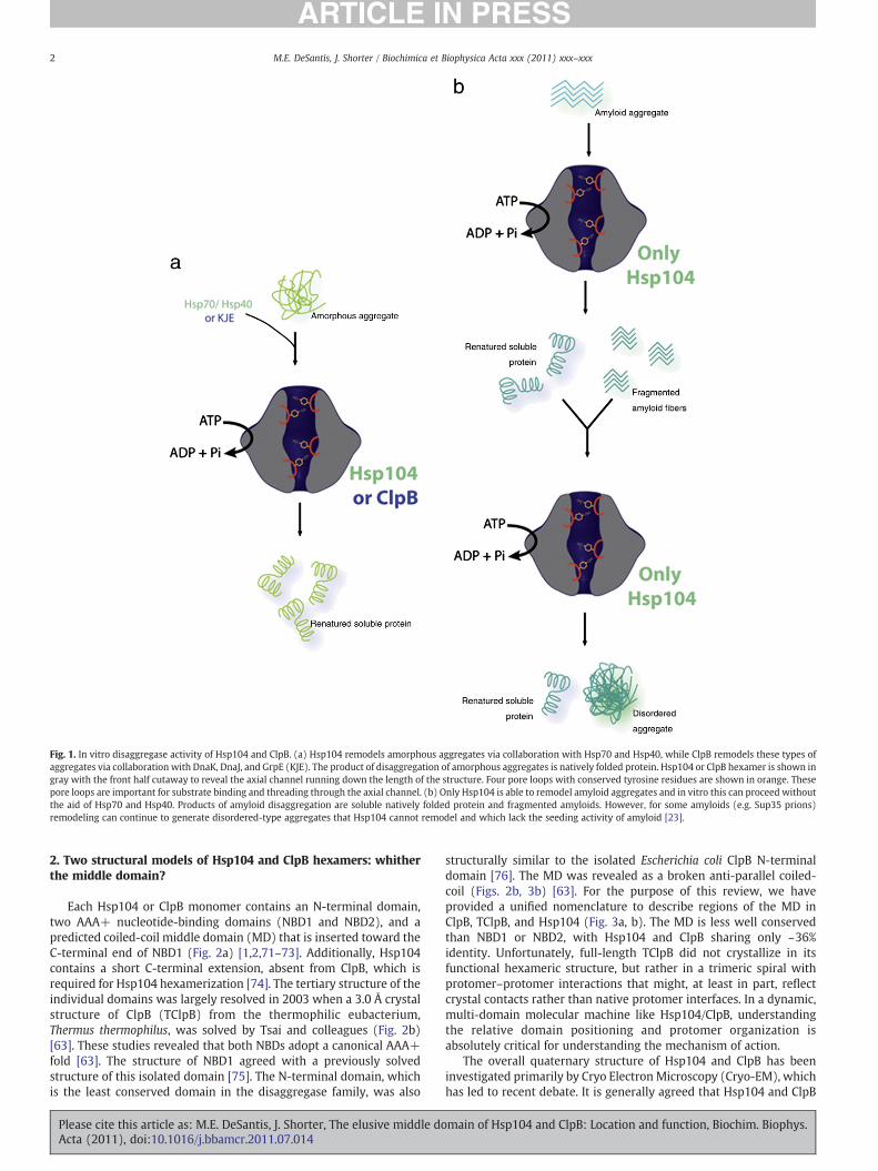
Fig. 1. In vitro disaggregase activity of Hsp104 and ClpB. (a) Hsp104 remodels amorphous aggregates via collaboration with Hsp70 and Hsp40, while ClpB remodels these types ofaggregates via collaboration with DnaK, DnaJ, and GrpE (KJE). The product of disaggregation of amorphous aggregates is natively folded protein. Hsp104 or ClpB hexamer is shown ingray with the front half cutaway to reveal the axial channel running down the length of the structure. Four pore loops with conserved tyrosine residues are shown in orange. Thesepore loops are important for substrate binding and threading through the axial channel. (b) Only Hsp104 is able to remodel amyloid aggregates and in vitro this can proceed withoutthe aid of Hsp70 and Hsp40. Products of amyloid disaggregation are soluble natively folded protein and fragmented amyloids. However, for some amyloids (e.g. Sup35 prions)remodeling can continue to generate disordered-type aggregates that Hsp104 cannot remodel and which lack the seeding activity of amyloid [23].
2 M.E. DeSantis, J. Shorter / Biochimica et Biophysica Acta xxx (2011) xxx–xxx
2. Two structural models of Hsp104 and ClpB hexamers: whitherthe middle domain?
Each Hsp104 or ClpB monomer contains an N-terminal domain,two AAA+ nucleotide-binding domains (NBD1 and NBD2), and apredicted coiled-coil middle domain (MD) that is inserted toward theC-terminal end of NBD1 (Fig. 2a) [1,2,71–73]. Additionally, Hsp104contains a short C-terminal extension, absent from ClpB, which isrequired for Hsp104 hexamerization [74]. The tertiary structure of theindividual domains was largely resolved in 2003 when a 3.0 Å crystalstructure of ClpB (TClpB) from the thermophilic eubacterium,Thermus thermophilus, was solved by Tsai and colleagues (Fig. 2b)[63]. These studies revealed that both NBDs adopt a canonical AAA+fold [63]. The structure of NBD1 agreed with a previously solvedstructure of this isolated domain [75]. The N-terminal domain, whichis the least conserved domain in the disaggregase family, was also
Please cite this article as: M.E. DeSantis, J. Shorter, The elusive middle doActa (2011), doi:10.1016/j.bbamcr.2011.07.014
structurally similar to the isolated Escherichia coli ClpB N-terminaldomain [76]. The MD was revealed as a broken anti-parallel coiled-coil (Figs. 2b, 3b) [63]. For the purpose of this review, we haveprovided a unified nomenclature to describe regions of the MD inClpB, TClpB, and Hsp104 (Fig. 3a, b). The MD is less well conservedthan NBD1 or NBD2, with Hsp104 and ClpB sharing only ~36%identity. Unfortunately, full-length TClpB did not crystallize in itsfunctional hexameric structure, but rather in a trimeric spiral withprotomer–protomer interactions that might, at least in part, reflectcrystal contacts rather than native protomer interfaces. In a dynamic,multi-domain molecular machine like Hsp104/ClpB, understandingthe relative domain positioning and protomer organization isabsolutely critical for understanding the mechanism of action.
The overall quaternary structure of Hsp104 and ClpB has beeninvestigated primarily by Cryo ElectronMicroscopy (Cryo-EM), whichhas led to recent debate. It is generally agreed that Hsp104 and ClpB
main of Hsp104 and ClpB: Location and function, Biochim. Biophys.

3M.E. DeSantis, J. Shorter / Biochimica et Biophysica Acta xxx (2011) xxx–xxx
are native hexamers and that oligomerization is promoted byincreased protein concentration [53,77–79], low salt [77,80], and thepresence of ADP or ATP [53,54,79–81]. Curiously, however, while
Fig. 2. Predicted structures and hexameric models of Hsp104 and ClpB. (a) Domain organiNucleotide binding domain 1 (NBD1) shown in cyan, Middle domain (MD) in yellow, Nucextension (C) shown in green. Sequence numbering for ClpB is shown on top and for Hsp104part (a). A 180° rotation about the vertical axis is shown on the right. (c) The Tsai model for thCryo-EM envelopes generated with TClpB is shown on left. (d) The Saibil model, which usestructure of hexameric, full-length ClpC is shown on the right. The adaptor protein MecA wasthe N-terminus is shown on the bottom. One subunit is colored as described in part (a). Th
Please cite this article as: M.E. DeSantis, J. Shorter, The elusive middle doActa (2011), doi:10.1016/j.bbamcr.2011.07.014
nucleotide binding to NBD1 is critical for ClpB hexamerization [80],this situation is reversed in Hsp104where nucleotide binding to NBD2is key [53,78,79]. The underlying reason for this switch between NBDs
zation of one monomer of Hsp104 and ClpB. N-terminal domain (N) shown in purple,leotide binding domain 2 (NBD2) in dark blue. Only Hsp104 has the short C-terminalis shown on the bottom. (b) TClpB crystal structure. Domain coloring corresponds withe hexameric quaternary structure of TClpB. The Tsai model, whichwas initially based ond Hsp104 to generate Cryo-EM density, is shown in the middle. (e) The 6.93 Å crystalomitted for clarity. A side view is shown on top and a view down the axial channel frome other five subunits are in gray.
main of Hsp104 and ClpB: Location and function, Biochim. Biophys.

4 M.E. DeSantis, J. Shorter / Biochimica et Biophysica Acta xxx (2011) xxx–xxx
is unknown and continues to remain puzzling and unaddressed. NBD1contributes the majority of basal ATPase activity in Hsp104 [53,78,79],whereas both NBDs contribute to basal ATPase activity in ClpB [80].The ATPase activities of both NBDs are required for the full repertoireof protein-remodeling activities and are modulated by allostericcommunicat ion within and between NBD1 and NBD2[73,77,79,80,82–84]. Gross domain position and the protomer–protomer interfacial packing of Hsp104 and ClpB hexamers stillremain uncertain. Of particular interest is the position of the coiled-coil MD, which is necessary for disaggregase activity and is unique tothe Hsp100 chaperones that function primarily in disaggregation[63,80,85]. In the TClpB crystal structure, this domain was juttingobliquely from the axis of the other domains [63] (Fig. 2b). Thus, in theoriginal Cryo-EM reconstructions of TClpB in the presence of AMP-PNP (a non-hydrolyzable ATP analog) the MD was assigned toprotrusions that appeared to emanate from one tier of the hexamer[63] (Fig. 2c, Tsai model).
In subsequent studies, to determine any conformational reorgani-zations that take place through the ATPase cycle, Cryo-EM envelopesof TClpB in the ADP and apo state were reconstructed as well as theenvelope of the Double Walker B TClpB mutant (E271A:E668A) in thepresence of ATP [61]. This mutant binds but does not hydrolyze ATP atboth NBDs and has increased affinity for substrate [61,86]. In all states,a two-tiered hexamer with an axial channel running through the
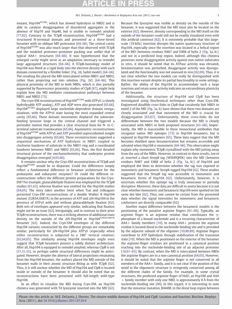
Fig. 3. Middle domain nomenclature. (a) An alignment of the MD from E. coli ClpB, T. thermpurple, helix 3 is colored in light blue, and helix four is colored in yellow. Motif 1 (also caArrowheads denote key residues discussed in the text. (b) Close up of the MD in the TClpB crfor clarity. Arrows point to side chains (shown as sticks) of key residues discussed in the te
Please cite this article as: M.E. DeSantis, J. Shorter, The elusive middle doActa (2011), doi:10.1016/j.bbamcr.2011.07.014
center was clearly visible [61]. However, the N-terminal domain wasnot visible as electron density [61]. In the AMP-PNP-bound state, theTClpB envelope shows clear, well defined protrusions on the outsideof the hexamer, which, when the individual domains of the TClpBmonomeric crystal structure were fitted as rigid bodies into thedensity, overlapped partially with predicted MD density [61,63]. Itwas suggested that the exterior position of the MD might enable it toact as a ‘crowbar’ to pry apart large aggregates [63]. Glover andLindquist had originally suggested that Hsp104 might possess a‘crowbar activity’, but did not ascribe this activity to any particulardomain [16]. All the other nucleotide states of TClpB (ATP, ADP, andapo) do not have such large protrusions of density that could correlatewith a MD projection. It was suggested that this might be due to theinherent mobility of the coiled-coil MD [61]. Indeed, the maindifference between the different nucleotide states was the length ofthese radially extending protrusions [61]. By contrast, the positions ofthe AAA+ domains remained almost identical in the variousnucleotide states [61]. Consequently, these reconstructions do notreadily clarify the mechanochemical coupling events that drivesubstrate translocation through the central channel.
This structural model of the hexamer was challenged by Cryo-EMreconstructions of Hsp104 by Saibil and colleagues [64–66]. In thesestudies, Cryo-EM reconstructions were generated of Hsp104 lackingits N-terminal domain, ΔN-Hsp104, and an NBD2 sensor-1 Hsp104
ophilus ClpB, and S. cerevisiae Hsp104. Helix 1 is colored in green, helix 2 is colored inlled wing 2) is boxed in black while motif 2 (also called wing 1) is boxed in orange.ystal structure. Each helix and motif is colored as indicated in part (a). NBD2 is omittedxt.
main of Hsp104 and ClpB: Location and function, Biochim. Biophys.
5M.E. DeSantis, J. Shorter / Biochimica et Biophysica Acta xxx (2011) xxx–xxx
mutant, Hsp104N728A, which has slowed hydrolysis in NBD2 and isable to catalyze disaggregation of disordered aggregates in theabsence of Hsp70 and Hsp40, but is unable to remodel amyloid[77,82]. Contrary to the TClpB reconstruction, Hsp104N728A had astructured N-terminal domain and presented as a three-tieredhexamer with no oblique MD protrusion [64–66]. The central cavityof Hsp104N728A was also much larger than that observed with TClpBand the modeled protomer–protomer packing was unlike that oftypical AAA+ structures [64–66]. It was hypothesized that theenlarged cavity might serve as an adaptation necessary to remodellarge aggregated structures [64–66]. A TClpB-homology model ofHsp104 was fitted as a rigid body into the electron density with eachdomain connected by a flexible linker (Fig. 2d, Saibil model) [64–66].The resulting fits placed the MD intercalated within NBD1 and NBD2,rather than projecting out into solution (Fig. 2d) [64–66]. Thisphysical proximity of the MD to both NBDs, which is also partiallysupported by fluorescence proximity studies of ClpB [87], might helpexplain how the MD mediates communication pathways betweenNBD1 and NBD2 [73].
The cryo-EM reconstructions of Hsp104N728A with ATPγS (a slowlyhydrolysable ATP analog), ATP and ADP were also generated [65,66].Hsp104N728A displayed large nucleotide-dependent domain reorga-nizations, with the ATPγS state having the most expanded centralcavity [65,66]. These domain movements displaced the substrate-binding tyrosine loops in the central channel and triggered aperistaltic motion that provides a clear structural basis for N- to C-terminal substrate translocation [65,66]. Asymmetric reconstructionsof Hsp104N728A with ATPγS and ATP provided unprecedented insightinto disaggregase activity [65,66]. These reconstructions suggested asequential mechanism of ATP hydrolysis in NBD1 coupled toclockwise handover of substrate in the NBD1 ring and a coordinatedhandover between NBD1 and NBD2 [65,66]. Thus, the first detailedstructural picture of the mechanochemistry that underpins proteindisaggregation emerged [4,65,66].
It remains unclear why the Cryo-EM reconstructions of TClpB andHsp104N728A would be so disparate. Could the differences simplyreflect a fundamental difference in hexamer architecture for theprokaryotic and eukaryotic enzymes? Or could the different re-constructions reflect the different protein preparations for the Cryo-EM? For example, glutaraldehyde fixationwas employed for the TClpBstudies [61,63], whereas fixative was omitted for the Hsp104 studies[64,65]. The story takes another twist when Tsai and colleaguespresented Cryo-EM reconstructions of a double Walker B Hsp104mutant (E285A:E687A) in the presence of ATP and ΔN-Hsp104 in thepresence of ATPγS with and without glutaraldehyde fixation [62].Both sets of envelopes appeared very similar, indicating that fixationmight not be an issue [62]. Intriguingly, and in contrast to their priorTClpB reconstructions, there was a striking absence of additional massdensity on the outside of the ΔN-Hsp104 or Hsp104E285A:E687A
hexamer [62]. Indeed, the Cryo-EM envelopes of the differentHsp104 variants constructed by the different groups are remarkablysimilar, particularly for ΔN-Hsp104 plus ATPγS (especially wheneither reconstruction is subjected to a 180° vertical rotation)[62,64,65]. This similarity among Hsp104 envelopes might evensuggest that TClpB hexamers possess a subtly distinct architecture.After all, Hsp104 is equipped to remodel amyloid, whereas ClpB is not[21,51,52], so perhaps subtle structural differences might be antici-pated. However, despite the absence of lateral projections emanatingfrom the Hsp104 hexamer, the authors placed the MD outside of thehexamer walls in their atomic structure fitting as with TClpB [62].Thus, it is still unclear whether the MD of Hsp104 and ClpB is locatedinside or outside of the hexamer. It should also be noted that noreconstructions have been presented with full-length wild-typeHsp104.
In an effort to visualize the MD during Cryo-EM, an Hsp104chimera was generated with T4 lysozyme inserted into the MD [62].
Please cite this article as: M.E. DeSantis, J. Shorter, The elusive middle doActa (2011), doi:10.1016/j.bbamcr.2011.07.014
Because the lysozyme was visible as density on the outside of thehexamer, it was suggested that the MD must also be located on theexterior [62]. However, density corresponding to the MD itself on theoutside of the hexamer could still not be readily visualized even withthis artificial construct [62]. It is extremely probable that this large-scale (19 kDa) insertion disrupts the native quaternary structure ofHsp104, especially since the insertion was located in a helical regionof the MD (between residues N467 and E468 of helix 2 (Fig. 3a, b))and not in a predicted loop region. Indeed, although this chimerapossesses some disaggregation activity against non-native substratesin vitro, it should be noted that its ATPase activity was elevated,hexamerization was perturbed, disaggregase activity was dysregu-lated and the functionality was not assessed in vivo [62,69]. Thus, it isnot clear whether the two models can really be distinguished withthis insertion variant despite its partial functionality in some settings.Rather, the ability of the Hsp104 to accommodate such a largeinsertion and retain some activity indicates an extraordinary plasticityof the hexamer.
Additionally, the structure of Hsp104 and ClpB has beeninvestigated using biochemical techniques other than Cryo-EM.Engineered disulfide cross-links in ClpB that covalently link NBD1 tomotif 2 of the MD (Fig. 3a, b) have shown that these two domains areclosely associated and that movement of the MD is crucial fordisaggregation [63,67]. Unfortunately, these cross-links do notdifferentiate between the two models because the MD is closelyassociated with NBD1 in both proposed structures [61–66]. Impor-tantly, the MD is inaccessible to three monoclonal antibodies thatrecognize native MD epitopes [73] in Hsp104 hexamers, but isexposed in Hsp104 monomers [65]. These data suggest that the MDbecomes shielded upon hexamerization but remains accessible andsolvated when Hsp104 is monomeric [64–66]. This observation mightexplain why monomeric TClpB crystallized with the MD jutting awayfrom the axis of the NBDs. However, in contrast to these results, Lee etal. inserted a short StrepII tag (WSHPQFEK) into the MD (betweenresidues N467 and E468 of helix 2 (Fig. 3a, b)) of Hsp104 andemployed dot blots to determine whether the tag was exposed inmonomeric and hexameric StrepII-tagged Hsp104 [62]. These studiessuggested that the StrepII tag was accessible in monomeric andhexameric forms of Hsp104 [62]. Unfortunately, however, it isuncertain whether this epitope tag is truly innocuous or partiallydisruptive. Moreover, these data are difficult to assess because it is notclear whethermonomeric and hexameric Hsp104were spotted on thesame dot blot [62]. Thus, one cannot be certain from the presenteddata whether the signal intensities for monomeric and hexamericconformers are directly comparable [62].
Another major difference between the hexameric models is thepositioning of the putative arginine fingers [61–66]. Typically, anarginine finger is an arginine residue that coordinates the γ-phosphate of a bound nucleotide and is a recurring characteristic ofAAA+ family members [10]. In many AAA+ proteins the arginineresidue is located distal to the nucleotide-binding site and is providedby the adjacent subunit of the oligomer [10,88,89]. Arginine fingerscontribute to ATP hydrolysis through stabilization of the transitionstate [10]. When the MD is positioned on the exterior of the hexamerthe arginine-finger residues are positioned in a canonical positionreaching into the nucleotide-binding site of an adjacent protomer[10,61–63]. By contrast, when the MD is intercalated between NBDsthe arginine fingers are in a non-canonical position [64,65]. However,it should be noted that the arginine finger is not conserved in allmembers of the AAA+ family, and it is not clear if the position of thismotif in the oligomeric structure is stringently conserved among allthe different clades of the family. For example, in some crystalstructures, the predicted arginine finger of HslU, an Hsp100 and AAA+ family member with only one NBD, is approximately 8 Å from thenucleotide-binding site [90]. In this regard, it is interesting to notethat the missense mutation, R444M, in the distal loop region between
main of Hsp104 and ClpB: Location and function, Biochim. Biophys.

6 M.E. DeSantis, J. Shorter / Biochimica et Biophysica Acta xxx (2011) xxx–xxx
helices 1 and 2 in theMD (Fig. 3a,b) reduces ATPase activity of Hsp104and impairs thermotolerance in a dominant-negative manner anddisrupts amyloid remodeling functionality [64]. This deficiency maysuggest a close contact between the distal loop of the MD and theNBD2 [64]. Thus, it is possible that other conserved arginines mightfulfill the role of the arginine finger in Hsp104.
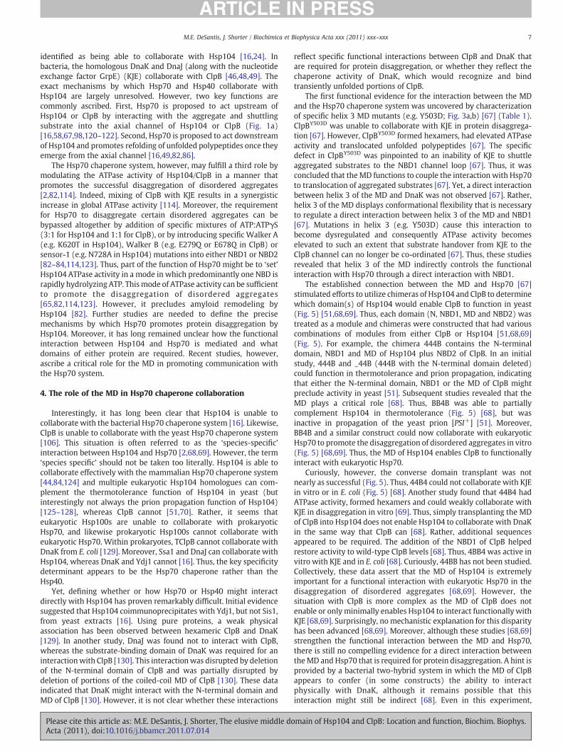
Finally, a crystal structure of another Hsp100 family member fromBacillus subtilis, ClpC, was recently solved and has weighed in on thedebate [91] (Fig. 2e). Like ClpB and Hsp104, ClpC is an AAA+disaggregase involved in modulation of stress response [92] andprotein quality control [93]. Typically, ClpC passes disaggregatedsubstrates to the chambered protease, ClpP, for degradation [91–94].While ClpC is natively a hexamer, it requires an adaptor protein,MecA, to oligomerize and form an active enzyme [95,96]. Byemploying a variant with four loop deletions and two ATPase-obliterating mutations, a 6.93 Å crystal structure of hexameric ClpC incomplex with the adaptor protein MecA was obtained [91].Interestingly, the packing within the NBD1 ring corresponded wellwith that proposed for TClpB [61,63,91] (Fig. 4). Additionally, the MDof ClpC, which is approximately half the length of the MD of ClpB [96],was also jutting out and away from the NBDs and was distinctlylocated on the outside of the hexamer [91]. These data provideindependent support for the TClpB hexameric model. However, thecoiled-coil domain of ClpC is considerably shorter than that of Hsp104or ClpB and must interact with MecA so that ClpC can hexamerize[91]. Thus, the MD of ClpC plays a very different role to the MD of ClpBor Hsp104 where it is dispensable for hexamerization [69,80].
In closing this section, we suggest that further biochemicalcharacterization and alternative techniques are urgently needed todiscern which hexameric model is correct or whether a revised modelis required for Hsp104 and ClpB. It is probable that both models arepartially correct because Hsp104 and ClpB are large, oligomericmachines that possess significant domain plasticity [62,63,69].Potentially, the MD might be able to occupy both structuralconformations depending on the stage of the disaggregase cycle.Two factors are likely to have greatly increased the difficulty inobtaining high-resolution structural information on the Hsp104 orClpB hexamer. First, the hexamer is highly dynamic and monomersare exchanged on the minute timescale [97,98]. Second, individualsubunits or domains likely adopt different conformations within thehexamer [65,66]. Future studies employing a full gamut of comple-mentary techniques will be essential to refine our understanding ofthe hexameric structure of Hsp104 and ClpB.
3. The function of the MD
Despite the ambiguity in MD location, several studies have probedMD function. For instance, in ClpB, deletion of the MD causes a loss of
Fig. 4.Overlay of ClpB and ClpCmonomers. ClpB and ClpCmonomers were aligned by their Ain Fig. 2 with the N domain in purple, NBD1 in cyan, MD in yellow, and NBD2 in dark blue. AllN-terminal domain (marked with an arrow) and the different angle of the ClpC MD (mark
Please cite this article as: M.E. DeSantis, J. Shorter, The elusive middle doActa (2011), doi:10.1016/j.bbamcr.2011.07.014
thermotolerance function [80]. Furthermore, partial truncations of theClpBMD cause decreased ATPase activity, hexamerization defects, andimpaired disaggregation [99]. Even point mutations in the MD havebeen implicated in altered ATPase activity, loss of thermotoleranceactivity, and/or destabilization of the hexamer [64,67,85,100]. Thedynamic mobility of the MD also appears critical for Hsp104/ClpBfunction, as crosslinks that hinder MD flexibility reduce or ablatedisaggregation activity of ClpB [63,67]. Additionally, the MD appearsto be involved in facilitating NBD1 and NBD2 communication [73]:when motif 2 is covalently attached to NBD1, ATPase activity in NBD2increases by ~30-fold [87]. Clearly, the structural integrity and relativemobility of the MD must be maintained for full disaggregasefunctionality.
At first glance, the MD appears to differentiate Hsp100 proteinsthat possess disaggregase activity from other Hsp100s. The disag-gregases: ClpB and ClpC [98,101,102] in bacteria, Hsp104 and Hsp78[103–106] in yeast, as well as Hsp101 [107–110] in plants, all containa coiled-coil insertion toward the C-terminal end of NBD1. ClpE isanother bacterial homologue that contains a coiled-coil insertion[111,112]. Genetic studies have implicated ClpE as part of the cellulardisaggregation machinery, but this has yet to be demonstrated withpure protein biochemistry [113]. While there are several examples ofHsp100 proteins that do not contain a MD and also do not possessdisaggregase activity [114], the correlation between MD and disag-gregase function is not quite so straightforward. For instance, ClpV,another bacterial Hsp100 protein, also contains a predicted MDinsertion between its two AAA+ domains but does not possess anydisaggregation activity against substrates in vivo [115]. Additionally,ClpA, another Hsp100 and AAA+ protein with two NBDs permonomer found in E. coli (but not in yeast), displays disaggregaseactivity against disordered aggregates but does not harbor a coiled-coil MD [82,116]. Indeed, in collaboration with its adaptor protein,ClpS, ClpA disaggregates substrates and delivers them to ClpP fordegradation [116]. ClpA disaggregates substrates without any need forthe Hsp70 chaperone system [114,116], indicating that Hsp70 is notabsolutely required for disaggregation per se. Importantly, however,the Hsp70 chaperone system inhibits protein disaggregation by ClpA[116]. In this way, DnaK prevents degradation of aggregated sub-strates by ClpAP in E. coli and permits their disaggregation andreactivation by ClpB [116]. Since the MD is a major distinguishingfeature of ClpB compared to ClpA, these studies provided the first hintthat the MD might contribute to collaboration with Hsp70.
Hsp104 and ClpB require collaboration with Hsp70 and Hsp40chaperones for successful disaggregation of amorphous aggregates,both in vitro and in vivo [13,16,47,49,117,118]. Hsp70 and Hsp40 canalso ameliorate the amyloid-remodeling activity of Hsp104[24,28,44,51,119]. In yeast, members of the Hsp70 chaperone family(e.g. Ssa1, Ssb1) and Hsp40 family (e.g. Ydj1 and Sis1) have been
AA+ domains using Pymol. The resulting RMSDwas 3.4. Domains for ClpB are colored asdomains in ClpC are colored in orange. Note the drastically different position of the ClpBed with an asterisk).
main of Hsp104 and ClpB: Location and function, Biochim. Biophys.
7M.E. DeSantis, J. Shorter / Biochimica et Biophysica Acta xxx (2011) xxx–xxx
identified as being able to collaborate with Hsp104 [16,24]. Inbacteria, the homologous DnaK and DnaJ (along with the nucleotideexchange factor GrpE) (KJE) collaborate with ClpB [46,48,49]. Theexact mechanisms by which Hsp70 and Hsp40 collaborate withHsp104 are largely unresolved. However, two key functions arecommonly ascribed. First, Hsp70 is proposed to act upstream ofHsp104 or ClpB by interacting with the aggregate and shuttlingsubstrate into the axial channel of Hsp104 or ClpB (Fig. 1a)[16,58,67,98,120–122]. Second, Hsp70 is proposed to act downstreamof Hsp104 and promotes refolding of unfolded polypeptides once theyemerge from the axial channel [16,49,82,86].
The Hsp70 chaperone system, however, may fulfill a third role bymodulating the ATPase activity of Hsp104/ClpB in a manner thatpromotes the successful disaggregation of disordered aggregates[2,82,114]. Indeed, mixing of ClpB with KJE results in a synergisticincrease in global ATPase activity [114]. Moreover, the requirementfor Hsp70 to disaggregate certain disordered aggregates can bebypassed altogether by addition of specific mixtures of ATP:ATPγS(3:1 for Hsp104 and 1:1 for ClpB), or by introducing specific Walker A(e.g. K620T in Hsp104), Walker B (e.g. E279Q or E678Q in ClpB) orsensor-1 (e.g. N728A in Hsp104) mutations into either NBD1 or NBD2[82–84,114,123]. Thus, part of the function of Hsp70 might be to ‘set’Hsp104 ATPase activity in a mode in which predominantly one NBD israpidly hydrolyzing ATP. Thismode of ATPase activity can be sufficientto promote the disaggregation of disordered aggregates[65,82,114,123]. However, it precludes amyloid remodeling byHsp104 [82]. Further studies are needed to define the precisemechanisms by which Hsp70 promotes protein disaggregation byHsp104. Moreover, it has long remained unclear how the functionalinteraction between Hsp104 and Hsp70 is mediated and whatdomains of either protein are required. Recent studies, however,ascribe a critical role for the MD in promoting communication withthe Hsp70 system.
4. The role of the MD in Hsp70 chaperone collaboration
Interestingly, it has long been clear that Hsp104 is unable tocollaborate with the bacterial Hsp70 chaperone system [16]. Likewise,ClpB is unable to collaborate with the yeast Hsp70 chaperone system[106]. This situation is often referred to as the ‘species-specific’interaction between Hsp104 and Hsp70 [2,68,69]. However, the term‘species specific’ should not be taken too literally. Hsp104 is able tocollaborate effectively with the mammalian Hsp70 chaperone system[44,84,124] and multiple eukaryotic Hsp104 homologues can com-plement the thermotolerance function of Hsp104 in yeast (butinterestingly not always the prion propagation function of Hsp104)[125–128], whereas ClpB cannot [51,70]. Rather, it seems thateukaryotic Hsp100s are unable to collaborate with prokaryoticHsp70, and likewise prokaryotic Hsp100s cannot collaborate witheukaryotic Hsp70. Within prokaryotes, TClpB cannot collaborate withDnaK from E. coli [129]. Moreover, Ssa1 and DnaJ can collaborate withHsp104, whereas DnaK and Ydj1 cannot [16]. Thus, the key specificitydeterminant appears to be the Hsp70 chaperone rather than theHsp40.
Yet, defining whether or how Hsp70 or Hsp40 might interactdirectly with Hsp104 has proven remarkably difficult. Initial evidencesuggested that Hsp104 coimmunoprecipitates with Ydj1, but not Sis1,from yeast extracts [16]. Using pure proteins, a weak physicalassociation has been observed between hexameric ClpB and DnaK[129]. In another study, DnaJ was found not to interact with ClpB,whereas the substrate-binding domain of DnaK was required for aninteractionwith ClpB [130]. This interactionwas disrupted by deletionof the N-terminal domain of ClpB and was partially disrupted bydeletion of portions of the coiled-coil MD of ClpB [130]. These dataindicated that DnaK might interact with the N-terminal domain andMD of ClpB [130]. However, it is not clear whether these interactions
Please cite this article as: M.E. DeSantis, J. Shorter, The elusive middle doActa (2011), doi:10.1016/j.bbamcr.2011.07.014
reflect specific functional interactions between ClpB and DnaK thatare required for protein disaggregation, or whether they reflect thechaperone activity of DnaK, which would recognize and bindtransiently unfolded portions of ClpB.
The first functional evidence for the interaction between the MDand the Hsp70 chaperone system was uncovered by characterizationof specific helix 3 MD mutants (e.g. Y503D; Fig. 3a,b) [67] (Table 1).ClpBY503D was unable to collaborate with KJE in protein disaggrega-tion [67]. However, ClpBY503D formed hexamers, had elevated ATPaseactivity and translocated unfolded polypeptides [67]. The specificdefect in ClpBY503D was pinpointed to an inability of KJE to shuttleaggregated substrates to the NBD1 channel loop [67]. Thus, it wasconcluded that theMD functions to couple the interaction with Hsp70to translocation of aggregated substrates [67]. Yet, a direct interactionbetween helix 3 of the MD and DnaK was not observed [67]. Rather,helix 3 of the MD displays conformational flexibility that is necessaryto regulate a direct interaction between helix 3 of the MD and NBD1[67]. Mutations in helix 3 (e.g. Y503D) cause this interaction tobecome dysregulated and consequently ATPase activity becomeselevated to such an extent that substrate handover from KJE to theClpB channel can no longer be co-ordinated [67]. Thus, these studiesrevealed that helix 3 of the MD indirectly controls the functionalinteraction with Hsp70 through a direct interaction with NBD1.
The established connection between the MD and Hsp70 [67]stimulated efforts to utilize chimeras of Hsp104 and ClpB to determinewhich domain(s) of Hsp104 would enable ClpB to function in yeast(Fig. 5) [51,68,69]. Thus, each domain (N, NBD1, MD and NBD2) wastreated as a module and chimeras were constructed that had variouscombinations of modules from either ClpB or Hsp104 [51,68,69](Fig. 5). For example, the chimera 444B contains the N-terminaldomain, NBD1 and MD of Hsp104 plus NBD2 of ClpB. In an initialstudy, 444B and _44B (444B with the N-terminal domain deleted)could function in thermotolerance and prion propagation, indicatingthat either the N-terminal domain, NBD1 or the MD of ClpB mightpreclude activity in yeast [51]. Subsequent studies revealed that theMD plays a critical role [68]. Thus, BB4B was able to partiallycomplement Hsp104 in thermotolerance (Fig. 5) [68], but wasinactive in propagation of the yeast prion [PSI+] [51]. Moreover,BB4B and a similar construct could now collaborate with eukaryoticHsp70 to promote the disaggregation of disordered aggregates in vitro(Fig. 5) [68,69]. Thus, the MD of Hsp104 enables ClpB to functionallyinteract with eukaryotic Hsp70.
Curiously, however, the converse domain transplant was notnearly as successful (Fig. 5). Thus, 44B4 could not collaborate with KJEin vitro or in E. coli (Fig. 5) [68]. Another study found that 44B4 hadATPase activity, formed hexamers and could weakly collaborate withKJE in disaggregation in vitro [69]. Thus, simply transplanting the MDof ClpB into Hsp104 does not enable Hsp104 to collaborate with DnaKin the same way that ClpB can [68]. Rather, additional sequencesappeared to be required. The addition of the NBD1 of ClpB helpedrestore activity to wild-type ClpB levels [68]. Thus, 4BB4 was active invitro with KJE and in E. coli [68]. Curiously, 44BB has not been studied.Collectively, these data assert that the MD of Hsp104 is extremelyimportant for a functional interaction with eukaryotic Hsp70 in thedisaggregation of disordered aggregates [68,69]. However, thesituation with ClpB is more complex as the MD of ClpB does notenable or only minimally enables Hsp104 to interact functionally withKJE [68,69]. Surprisingly, no mechanistic explanation for this disparityhas been advanced [68,69]. Moreover, although these studies [68,69]strengthen the functional interaction between the MD and Hsp70,there is still no compelling evidence for a direct interaction betweentheMD and Hsp70 that is required for protein disaggregation. A hint isprovided by a bacterial two-hybrid system in which the MD of ClpBappears to confer (in some constructs) the ability to interactphysically with DnaK, although it remains possible that thisinteraction might still be indirect [68]. Even in this experiment,
main of Hsp104 and ClpB: Location and function, Biochim. Biophys.
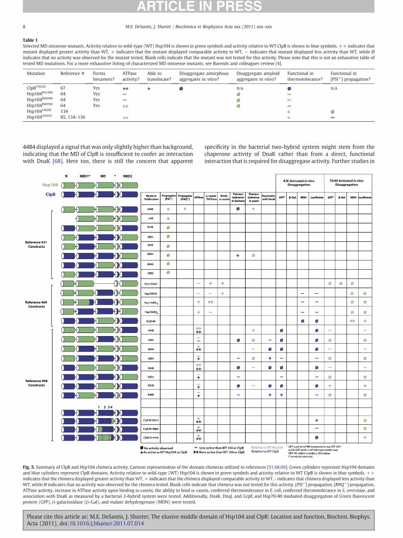
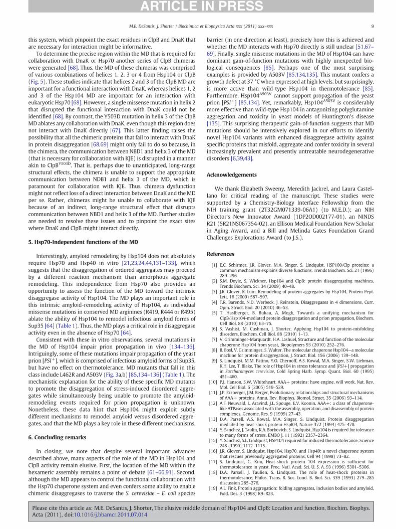
Table 1Selected MDmissense mutants. Activity relative to wild-type (WT) Hsp104 is shown in green symbols and activity relative to WT ClpB is shown in blue symbols. ++ indicates thatmutant displayed greater activity than WT, + indicates that the mutant displayed comparable activity to WT, − indicates that mutant displayed less activity than WT, while Øindicates that no activity was observed for the mutant tested. Blank cells indicate that the mutant was not tested for this activity. Please note that this is not an exhaustive table oftested MD mutations. For a more exhaustive listing of characterized MD missense mutants, see Barends and colleagues review [4].
Mutation Reference # Formshexamers?
ATPaseactivity?
Able totranslocate?
Disaggregate amorphousaggregates in vitro?
Disaggregate amyloidaggregates in vitro?
Functional inthermotolerance?
Functional in[PSI+] propagation?
ClpBY503D 67 Yes n/a n/aHsp104R419M 64 YesHsp104R444M 64 YesHsp104R495M 64 YesHsp104L462R 134Hsp104A503V 85, 134–136
8 M.E. DeSantis, J. Shorter / Biochimica et Biophysica Acta xxx (2011) xxx–xxx
44B4 displayed a signal that was only slightly higher than background,indicating that the MD of ClpB is insufficient to confer an interactionwith DnaK [68]. Here too, there is still the concern that apparent
Fig. 5. Summary of ClpB and Hsp104 chimera activity. Cartoon representation of the domainand blue cylinders represent ClpB domains. Activity relative to wild-type (WT) Hsp104 is shindicates that the chimera displayed greater activity thanWT, + indicates that the chimera dWT, while Ø indicates that no activity was observed for the chimera tested. Blank cells indicaATPase activity, increase in ATPase activity upon binding α-casein, the ability to bind α-caseassociation with DnaK as measured by a bacterial 2-hybrid system were tested. Additionalprotein (GFP), β-galactosidase (β-Gal), and malate dehydrogenase (MDH) were tested.
Please cite this article as: M.E. DeSantis, J. Shorter, The elusive middle doActa (2011), doi:10.1016/j.bbamcr.2011.07.014
specificity in the bacterial two-hybrid system might stem from thechaperone activity of DnaK rather than from a direct, functionalinteraction that is required for disaggregase activity. Further studies in
chimeras utilized in references [51,68,69]. Green cylinders represent Hsp104 domainsown in green symbols and activity relative to WT ClpB is shown in blue symbols. ++isplayed comparable activity toWT, - indicates that chimera displayed less activity thante that chimera was not tested for this activity. [PSI+] propagation, [RNQ+] propagation,in, conferred thermotolerance in E. coli, conferred thermotolerance in S. cerevisiae, andly, DnaK, DnaJ, and GrpE and Hsp70/40 mediated disaggregation of Green fluorescent
main of Hsp104 and ClpB: Location and function, Biochim. Biophys.
9M.E. DeSantis, J. Shorter / Biochimica et Biophysica Acta xxx (2011) xxx–xxx
this system, which pinpoint the exact residues in ClpB and DnaK thatare necessary for interaction might be informative.
To determine the precise region within the MD that is required forcollaboration with DnaK or Hsp70 another series of ClpB chimeraswere generated [68]. Thus, the MD of these chimeras was comprisedof various combinations of helices 1, 2, 3 or 4 from Hsp104 or ClpB(Fig. 5). These studies indicate that helices 2 and 3 of the ClpB MD areimportant for a functional interaction with DnaK, whereas helices 1, 2and 3 of the Hsp104 MD are important for an interaction witheukaryotic Hsp70 [68]. However, a singlemissensemutation in helix 2that disrupted the functional interaction with DnaK could not beidentified [68]. By contrast, the Y503D mutation in helix 3 of the ClpBMD ablates any collaborationwith DnaK, even though this region doesnot interact with DnaK directly [67]. This latter finding raises thepossibility that all the chimeric proteins that fail to interact with DnaKin protein disaggregation [68,69] might only fail to do so because, inthe chimera, the communication between NBD1 and helix 3 of theMD(that is necessary for collaboration with KJE) is disrupted in a mannerakin to ClpBY503D. That is, perhaps due to unanticipated, long-rangestructural effects, the chimera is unable to support the appropriatecommunication between NDB1 and helix 3 of the MD, which isparamount for collaboration with KJE. Thus, chimera dysfunctionmight not reflect loss of a direct interaction between DnaK and theMDper se. Rather, chimeras might be unable to collaborate with KJEbecause of an indirect, long-range structural effect that disruptscommunication between NBD1 and helix 3 of the MD. Further studiesare needed to resolve these issues and to pinpoint the exact siteswhere DnaK and ClpB might interact directly.
5. Hsp70-Independent functions of the MD
Interestingly, amyloid remodeling by Hsp104 does not absolutelyrequire Hsp70 and Hsp40 in vitro [21,23,24,44,131–133], whichsuggests that the disaggregation of ordered aggregates may proceedby a different reaction mechanism than amorphous aggregateremodeling. This independence from Hsp70 also provides anopportunity to assess the function of the MD toward the intrinsicdisaggregase activity of Hsp104. The MD plays an important role inthis intrinsic amyloid-remodeling activity of Hsp104, as individualmissense mutations in conserved MD arginines (R419, R444 or R495)ablate the ability of Hsp104 to remodel infectious amyloid forms ofSup35 [64] (Table 1). Thus, the MD plays a critical role in disaggregaseactivity even in the absence of Hsp70 [64].
Consistent with these in vitro observations, several mutations inthe MD of Hsp104 impair prion propagation in vivo [134–136].Intriguingly, some of these mutations impair propagation of the yeastprion [PSI+], which is comprised of infectious amyloid forms of Sup35,but have no effect on thermotolerance. MD mutants that fall in thisclass include L462R and A503V (Fig. 3a,b) [85,134–136] (Table 1). Themechanistic explanation for the ability of these specific MD mutantsto promote the disaggregation of stress-induced disordered aggre-gates while simultaneously being unable to promote the amyloid-remodeling events required for prion propagation is unknown.Nonetheless, these data hint that Hsp104 might exploit subtlydifferent mechanisms to remodel amyloid versus disordered aggre-gates, and that the MD plays a key role in these different mechanisms.
6. Concluding remarks
In closing, we note that despite several important advancesdescribed above, many aspects of the role of the MD in Hsp104 andClpB activity remain elusive. First, the location of the MD within thehexameric assembly remains a point of debate [61–66,91]. Second,although the MD appears to control the functional collaboration withthe Hsp70 chaperone system and even confers some ability to enablechimeric disaggregases to traverse the S. cerevisiae – E. coli species
Please cite this article as: M.E. DeSantis, J. Shorter, The elusive middle doActa (2011), doi:10.1016/j.bbamcr.2011.07.014
barrier (in one direction at least), precisely how this is achieved andwhether the MD interacts with Hsp70 directly is still unclear [51,67–69]. Finally, single missense mutations in the MD of Hsp104 can havedominant gain-of-function mutations with highly unexpected bio-logical consequences [85]. Perhaps one of the most surprisingexamples is provided by A503V [85,134,135]. This mutant confers agrowth defect at 37 °Cwhen expressed at high levels, but surprisingly,is more active than wild-type Hsp104 in thermotolerance [85].Furthermore, Hsp104A503V cannot support propagation of the yeastprion [PSI+] [85,134]. Yet, remarkably, Hsp104A503V is considerablymore effective than wild-type Hsp104 in antagonizing polyglutamineaggregation and toxicity in yeast models of Huntington's disease[135]. This surprising therapeutic gain-of-function suggests that MDmutations should be intensively explored in our efforts to identifynovel Hsp104 variants with enhanced disaggregase activity againstspecific proteins that misfold, aggregate and confer toxicity in severalincreasingly prevalent and presently untreatable neurodegenerativedisorders [6,39,43].
Acknowledgements
We thank Elizabeth Sweeny, Meredith Jackrel, and Laura Castel-lano for critical reading of the manuscript. These studies weresupported by a Chemistry-Biology Interface Fellowship from theNIH training grant (2T32GM071339-06A1) (to M.E.D.); an NIHDirector's New Innovator Award (1DP2OD002177-01), an NINDSR21 (5R21NS067354-02), an Ellison Medical Foundation New Scholarin Aging Award, and a Bill and Melinda Gates Foundation GrandChallenges Explorations Award (to J.S.).
References
[1] E.C. Schirmer, J.R. Glover, M.A. Singer, S. Lindquist, HSP100/Clp proteins: acommon mechanism explains diverse functions, Trends Biochem. Sci. 21 (1996)289–296.
[2] S.M. Doyle, S. Wickner, Hsp104 and ClpB: protein disaggregating machines,Trends Biochem. Sci. 34 (2009) 40–48.
[3] J.R. Glover, R. Lum, Remodeling of protein aggregates by Hsp104, Protein Pept.Lett. 16 (2009) 587–597.
[4] T.R. Barends, N.D. Werbeck, J. Reinstein, Disaggregases in 4 dimensions, Curr.Opin. Struct. Biol. 20 (2010) 46–53.
[5] T. Haslberger, B. Bukau, A. Mogk, Towards a unifying mechanism forClpB/Hsp104-mediated protein disaggregation and prion propagation, Biochem.Cell Biol. 88 (2010) 63–75.
[6] S. Vashist, M. Cushman, J. Shorter, Applying Hsp104 to protein-misfoldingdisorders, Biochem. Cell Biol. 88 (2010) 1–13.
[7] V. Grimminger-Marquardt, H.A. Lashuel, Structure and function of the molecularchaperone Hsp104 from yeast, Biopolymers 93 (2010) 252–276.
[8] B. Bosl, V. Grimminger, S. Walter, Themolecular chaperone Hsp104—a molecularmachine for protein disaggregation, J. Struct. Biol. 156 (2006) 139–148.
[9] S. Lindquist, M.M. Patino, Y.O. Chernoff, A.S. Kowal, M.A. Singer, S.W. Liebman,K.H. Lee, T. Blake, The role of Hsp104 in stress tolerance and [PSI+] propagationin Saccharomyces cerevisiae, Cold Spring Harb. Symp. Quant. Biol. 60 (1995)451–460.
[10] P.I. Hanson, S.W. Whiteheart, AAA+ proteins: have engine, will work, Nat. Rev.Mol. Cell Biol. 6 (2005) 519–529.
[11] J.P. Erzberger, J.M. Berger, Evolutionary relationships and structural mechanismsof AAA+ proteins, Annu. Rev. Biophys. Biomol. Struct. 35 (2006) 93–114.
[12] A.F. Neuwald, L. Aravind, J.L. Spouge, E.V. Koonin, AAA+: a class of chaperone-like ATPases associated with the assembly, operation, and disassembly of proteincomplexes, Genome. Res. 9 (1999) 27–43.
[13] D.A. Parsell, A.S. Kowal, M.A. Singer, S. Lindquist, Protein disaggregationmediated by heat-shock protein Hspl04, Nature 372 (1994) 475–478.
[14] Y. Sanchez, J. Taulin, K.A. Borkovich, S. Lindquist, Hsp104 is required for toleranceto many forms of stress, EMBO J. 11 (1992) 2357–2364.
[15] Y. Sanchez, S.L. Lindquist, HSP104 required for induced thermotolerance, Science248 (1990) 1112–1115.
[16] J.R. Glover, S. Lindquist, Hsp104, Hsp70, and Hsp40: a novel chaperone systemthat rescues previously aggregated proteins, Cell 94 (1998) 73–82.
[17] S. Lindquist, G. Kim, Heat-shock protein 104 expression is sufficient forthermotolerance in yeast, Proc. Natl. Acad. Sci. U. S. A. 93 (1996) 5301–5306.
[18] D.A. Parsell, J. Taulien, S. Lindquist, The role of heat-shock proteins inthermotolerance, Philos. Trans. R. Soc. Lond. B. Biol. Sci. 339 (1993) 279–285discussion 285–276.
[19] A.L. Fink, Protein aggregation: folding aggregates, inclusion bodies and amyloid,Fold. Des. 3 (1998) R9–R23.
main of Hsp104 and ClpB: Location and function, Biochim. Biophys.

10 M.E. DeSantis, J. Shorter / Biochimica et Biophysica Acta xxx (2011) xxx–xxx
[20] L. Wang, D. Schubert, M.R. Sawaya, D. Eisenberg, R. Riek, Multidimensionalstructure–activity relationship of a protein in its aggregated states, Angew.Chem. Int. Ed. 49 (2010) 3904–3908.
[21] J. Shorter, S. Lindquist, Hsp104 catalyzes formation and elimination of self-replicating Sup35 prion conformers, Science 304 (2004) 1793–1797.
[22] J. Shorter, S. Lindquist, Prions as adaptive conduits of memory and inheritance,Nat. Rev. Genet. 6 (2005) 435–450.
[23] J. Shorter, S. Lindquist, Destruction or potentiation of different prions catalyzedby similar Hsp104, Remodeling Act. 23 (2006) 425–438.
[24] J. Shorter, S. Lindquist, Hsp104, Hsp70 and Hsp40 interplay regulates formation,growth and elimination of Sup35 prions, EMBO J. 27 (2008) 2712–2724.
[25] Y.O. Chernoff, S.L. Lindquist, B. Ono, S.G. Inge-Vechtomov, S.W. Liebman, Role ofthe chaperone protein Hsp104 in propagation of the yeast prion-like factor[PSI+], Science 268 (1995) 880–884.
[26] H. Moriyama, H.K. Edskes, R.B. Wickner, [URE3] Prion propagation in Saccharo-myces cerevisiae: requirement for chaperone Hsp104 and curing by over-expressed chaperone Ydj1p, Mol. Cell. Biol. 20 (2000) 8916–8922.
[27] S. Alberti, R. Halfmann, O. King, A. Kapila, S. Lindquist, A systematic surveyidentifies prions and illuminates sequence features of prionogenic proteins, Cell137 (2009) 146–158.
[28] E.A. Sweeny, J. Shorter, Prion proteostasis: Hsp104 meets its supporting cast,Prion 2 (2008) 135–140.
[29] N. Sondheimer, S. Lindquist, Rnq1: an epigenetic modifier of protein function inyeast, Mol. Cell. 5 (2000) 163–172.
[30] Z. Du, K.W. Park, H. Yu, Q. Fan, L. Li, Newly identified prion linked to thechromatin-remodeling factor Swi1 in Saccharomyces cerevisiae, Nat. Genet. 40(2008) 460–465.
[31] B.K. Patel, J. Gavin-Smyth, S.W. Liebman, The yeast global transcriptional co-repressor protein Cyc8 can propagate as a prion, Nat. cell biol. 11 (2009)344–349.
[32] S. DiSalvo, A. Derdowski, J.A. Pezza, T.R. Serio, Dominant prion mutants inducecuring through pathways that promote chaperone-mediated disaggregation,Nat. Struct. Mol. Biol. 18 (2011) 486–492.
[33] P. Satpute-Krishnan, S.X. Langseth, T.R. Serio, Hsp104-dependent remodeling ofprion complexes mediates protein-only inheritance, PLoS Biol. 5 (2007) e24.
[34] M. Sunde, L.C. Serpell, M. Bartlam, P.E. Fraser, M.B. Pepys, C.C.F. Blake, Commoncore structure of amyloid fibrils by synchrotron X-ray diffraction, J. Mol. Biol. 273(1997) 729–739.
[35] R. Nelson, M.R. Sawaya, M. Balbirnie, A.O. Madsen, C. Riekel, R. Grothe, D.Eisenberg, Structure of the cross-beta spine of amyloid-like fibrils, Nature 435(2005) 773–778.
[36] P.M. Tessier, S. Lindquist, Unraveling infectious structures, strain variants andspecies barriers for the yeast prion [PSI+], Nat. Struct. Mol. Biol. 16 (2009)598–605.
[37] A. Aguzzi, T. O'Connor, Protein aggregation diseases: pathogenicity andtherapeutic perspectives, Nat. Rev. Drug Discov. 9 (2010) 237–248.
[38] A. Aguzzi, L. Rajendran, The transcellular spread of cytosolic amyloids, prions,and prionoids, Neuron 64 (2009) 783–790.
[39] M. Cushman, B.S. Johnson, O.D. King, A.D. Gitler, J. Shorter, Prion-like disorders:blurring the divide between transmissibility and infectivity, J. Cell Sci. 123(2010) 1191–1201.
[40] R. Halfmann, S. Lindquist, Epigenetics in the extreme: prions and the inheritanceof environmentally acquired traits, Science 330 (2010) 629–632.
[41] J. Shorter, Emergence and natural selection of drug-resistant prions, Mol. Biosyst.6 (2010) 1115–1130.
[42] H.L. True, S.L. Lindquist, A yeast prion provides amechanism for genetic variationand phenotypic diversity, Nature 407 (2000) 477–483.
[43] J. Shorter, Hsp104: a weapon to combat diverse neurodegenerative disorders,Neurosignals 16 (2008) 63–74.
[44] C. Lo Bianco, J. Shorter, E. Ragulier, H. Lashuel, T. Iwatsubo, S. Lindquist, P. Aebischer,Hsp104 antagonizes alpha-synuclein aggregation and reduces dopaminergic degen-eration in a rat model of Parkinson disease, J. Clin. Investig. 118 (2008) 3087–3097.
[45] C. Vacher, L. Garcia-Oroz, D.C. Rubinsztein, Overexpression of yeast hsp104reduces polyglutamine aggregation and prolongs survival of a transgenic mousemodel of Huntington's disease, Hum. Mol. Genet. 14 (2005) 3425–3433.
[46] K. Motohashi, Y. Watanabe, M. Yohda, M. Yoshida, Heat-inactivated proteins arerescued by the DnaK.J-GrpE set and ClpB chaperones, Proc. Natl. Acad. Sci. U. S. A.96 (1999) 7184–7189.
[47] A. Mogk, T. Tomoyasu, P. Goloubinoff, S. Rudiger, D. Roder, H. Langen, B. Bukau,Identification of thermolabile E. coli proteins: prevention and reversion ofaggregation by DnaK and ClpB, EMBO J. 18 (1999) 6934–6949.
[48] M. Zolkiewski, ClpB cooperates with DnaK, DnaJ, and GrpE in suppressingprotein aggregation. A novel multi-chaperone system from Escherichia coli, J.Biol. Chem. 274 (1999) 28083–28086.
[49] P. Goloubinoff, A. Mogk, A.P.B. Zvi, T. Tomoyasu, B. Bukau, Sequential mechanismof solubilization and refolding of stable protein aggregates by a bichaperonenetwork, Proc. Natl. Acad. Sci. U. S. A. 96 (1999) 13732–13737.
[50] C.L. Squires, S. Pedersen, B.M. Ross, C. Squires, ClpB is the Escherichia coli heatshock protein F84.1, J. Bacteriol. 173 (1991) 4254–4262.
[51] K.A. Tipton, K.J. Verges, J.S. Weissman, In vivo monitoring of the prion replicationcycle reveals a critical role for sis1 in delivering substrates to Hsp104, Cell 32(2008) 584–591.
[52] M.P. Hinault, A.F. Cuendet, R.U. Mattoo, M. Mensi, G. Dietler, H.A. Lashuel, P.Goloubinoff, Stable alpha-synuclein oligomers strongly inhibit chaperoneactivity of the Hsp70 system by weak interactions with J-domain co-chaperones,J. Biol. Chem. 285 (2010) 38173–38182.
Please cite this article as: M.E. DeSantis, J. Shorter, The elusive middle doActa (2011), doi:10.1016/j.bbamcr.2011.07.014
[53] D.A. Parsell, A.S. Kowal, S. Lindquist, Saccharomyces cerevisiae Hsp104 protein.Purification and characterization of ATP-induced structural changes, J. Biol.Chem. 269 (1994) 4480–4487.
[54] M. Zolkiewski, M. Kessel, A. Ginsburg, M.R. Maurizi, Nucleotide-dependentoligomerization of C1pB from Escherichia coli, Protein Sci. 8 (1999) 1899–1903.
[55] R. Lum, M. Niggemann, J.R. Glover, Peptide and protein binding in the axialchannel of Hsp104: insights into the mechanism of protein unfolding, J. Biol.Chem. 283 (2008) 30139–30150.
[56] R. Lum, J.M. Tkach, E. Vierling, J.R. Glover, Evidence for an unfolding/threadingmechanism for protein disaggregation by Saccharomyces cerevisiae Hsp104, J.Biol. Chem. 279 (2004) 29139–29146.
[57] C. Schlieker, J. Weibezahn, H. Patzelt, P. Tessarz, C. Strub, K. Zeth, A. Erbse, J.Schneider-Mergener, J.W. Chin, P.G. Schultz, B. Bukau, A. Mogk, Substraterecognition by the AAA+ chaperone ClpB, Nat. Struct. Mol. Biol. 11 (2004)607–615.
[58] J. Weibezahn, P. Tessarz, C. Schlieker, R. Zahn, Z. Maglica, S. Lee, H. Zentgraf, E.U.Weber-Ban, D.A. Dougan, F.T.F. Tsai, A. Mogk, B. Bukau, Thermotolerancerequires refolding of aggregated proteins by substrate translocation through thecentral pore of ClpB, Cell 119 (2004) 653–665.
[59] J. Shorter, S. Lindquist, Navigating the ClpB channel to solution, Nat. Struct. Mol.Biol. 12 (2005) 4–6.
[60] A.L. Horwich, Chaperoned protein disaggregation—the ClpB ring uses its centralchannel, Cell 119 (2004) 579–581.
[61] S. Lee, J.-M. Choi, F.T.F. Tsai, Visualizing the ATPase cycle in a proteindisaggregating machine: structural basis for substrate binding by ClpB, Mol.Cell 25 (2007) 261–271.
[62] S. Lee, B. Sielaff, J. Lee, F.T.F. Tsai, CryoEM structure of Hsp104 and its mechanisticimplication for protein disaggregation, Proc. Natl. Acad. Sci. 107 (2010)8135–8140.
[63] S. Lee, M.E. Sowa, Y.-h. Watanabe, P.B. Sigler, W. Chiu, M. Yoshida, F.T.F. Tsai, Thestructure of ClpB: a molecular chaperone that rescues proteins from anaggregated state, Cell 115 (2003) 229–240.
[64] P. Wendler, J. Shorter, C. Plisson, A.G. Cashikar, S. Lindquist, H.R. Saibil, AtypicalAAA+ subunit packing creates an expanded cavity for disaggregation by theprotein-remodeling factor Hsp104, Cell 131 (2007) 1366–1377.
[65] P. Wendler, J. Shorter, D. Snead, C. Plisson, D.K. Clare, S. Lindquist, H.R. Saibil,Motor mechanism for protein threading through Hsp104, Mol. Cell 34 (2009)81–92.
[66] P. Wendler, H.R. Saibil, Cryo electron microscopy structures of Hsp100 proteins:crowbars in or out? Biochem. Cell Biol. 88 (2010) 89–96.
[67] T. Haslberger, J. Weibezahn, R. Zahn, S. Lee, F.T.F. Tsai, B. Bukau, A. Mogk, Mdomains couple the ClpB threading motor with the DnaK chaperone activity,Mol. Cell 25 (2007) 247–260.
[68] M. Miot, M. Reidy, S.M. Doyle, J.R. Hoskins, D.M. Johnston, O. Genest, M.-C. Vitery,D.C. Masison, S. Wickner, Species-specific collaboration of heat shock proteins(Hsp) 70 and 100 in thermotolerance and protein disaggregation, Proc. Natl.Acad. Sci. 108 (2011) 6915–6920.
[69] B. Sielaff, F.T.F. Tsai, The M-domain controls Hsp104 protein remodeling activityin an Hsp70/Hsp40-dependent manner, J. Mol. Biol. 402 (2010) 30–37.
[70] D.A. Parsell, S. Lindquist, The function of heat-shock proteins in stress tolerance:degradation and reactivation of damaged proteins, Annu. Rev. Genet. 27 (1993)437–496.
[71] D.A. Parsell, Y. Sanchez, J.D. Stitzel, S. Lindquist, Hspl04 is a highly conservedprotein with two essential nucleotide-binding sites, Nature 353 (1991) 270–273.
[72] M.E. Barnett, A. Zolkiewska, M. Zolkiewski, Structure and activity of ClpB fromEscherichia coli, J. Biol. Chem. 275 (2000) 37565–37571.
[73] A.G. Cashikar, E.C. Schirmer, D.A. Hattendorf, J.R. Glover, M.S. Ramakrishnan,D.M. Ware, S.L. Lindquist, Defining a pathway of communication from the C-terminal peptide binding domain to the N-terminal ATPase domain in a AAAprotein, Mol. Cell 9 (2002) 751–760.
[74] R.G. Mackay, C.W. Helsen, J.M. Tkach, J.R. Glover, The C-terminal extension ofsaccharomyces cerevisiae Hsp104 plays a role in oligomer assembly, Biochem-istry 47 (2008) 1918–1927.
[75] J. Li, B. Sha, Crystal structure of E. coli Hsp100 ClpB nucleotide-binding domain 1(NBD1) and mechanistic studies on ClpB ATPase activity, J. Mol. Biol. 318 (2002)1127–1137.
[76] J. Li, B. Sha, Crystal structure of the E. coli Hsp100 ClpB N-terminal domain,Structure (Camb) 11 (2003) 323–328.
[77] D.A. Hattendorf, S.L. Lindquist, Cooperative kinetics of both Hsp104 ATPasedomains and interdomain communication revealed by AAA sensor-1 mutants.EMBO J. 21 (2002) 12–21.
[78] E.C. Schirmer, C. Queitsch, A.S. Kowal, D.A. Parsell, S. Lindquist, The ATPaseactivity of Hsp104, effects of environmental conditions and mutations, J. Biol.Chem. 273 (1998) 15546–15552.
[79] E.C. Schirmer, D.M. Ware, C. Queitsch, A.S. Kowal, S.L. Lindquist, Subunitinteractions influence the biochemical and biological properties of Hsp104, Proc.Natl. Acad. Sci. U. S. A. 98 (2001) 914–919.
[80] A. Mogk, C. Schlieker, C. Strub, W. Rist, J. Weibezahn, B. Bukau, Roles of individualdomains and conserved motifs of the AAA+ protein ClpB in oligomerization,ATP-hydrolysis and chaperone activity, J. Biol. Chem. (2003) M209686200.
[81] J.M. Tkach, J.R. Glover, Amino acid substitutions in the C-terminal AAA+moduleof Hsp104 prevent substrate recognition by disrupting oligomerization andcause high temperature inactivation, J. Biol. Chem. 279 (2004) 35692–35701.
[82] S.M. Doyle, J. Shorter, M. Zolkiewski, J.R. Hoskins, S. Lindquist, S. Wickner,Asymmetric deceleration of ClpB or Hsp104 ATPase activity unleashes protein-remodeling activity, Nat. Struct. Mol. Biol. 14 (2007) 114–122.
main of Hsp104 and ClpB: Location and function, Biochim. Biophys.
11M.E. DeSantis, J. Shorter / Biochimica et Biophysica Acta xxx (2011) xxx–xxx
[83] T.M. Franzmann, A. Czekalla, S.G. Walter, Regulatory circuits of the AAA+disaggregase Hsp104, J. Biol. Chem. 286 (2011) 17992–18001.
[84] A. Schaupp, M. Marcinowski, V. Grimminger, B. Bosl, S. Walter, Processing ofproteins by the molecular chaperone Hsp104, J. Mol. Biol. 370 (2007) 674–686.
[85] E.C. Schirmer, O.R. Homann, A.S. Kowal, S. Lindquist, Dominant gain-of-functionmutations in Hsp104p reveal crucial roles for themiddle region, Mol. Biol. Cell 15(2004) 2061–2072.
[86] J. Weibezahn, C. Schlieker, B. Bukau, A. Mogk, Characterization of a trap mutantof the AAA+ chaperone ClpB, J. Biol. Chem. 278 (2003) 32608–32617.
[87] Y.-h. Watanabe, M. Takano, M. Yoshida, ATP binding to nucleotide bindingdomain (NBD)1 of the ClpB chaperone induces motion of the long coiled-coil,stabilizes the hexamer, and activates NBD2, J. Biol. Chem. 280 (2005)24562–24567.
[88] T. Ogura, S.W. Whiteheart, A.J. Wilkinson, Conserved arginine residuesimplicated in ATP hydrolysis, nucleotide-sensing, and inter-subunit interactionsin AAA and AAA+ ATPases, J. Struct. Biol. 146 (2004) 106–112.
[89] A.N. Lupas, J. Martin, AAA proteins, Curr. Opin. Struct. Biol. 12 (2002) 746–753.[90] M. Bochtler, C. Hartmann, H.K. Song, G.P. Bourenkov, H.D. Bartunik, R. Huber, The
structures of HsIU and the ATP-dependent protease HsIU–HsIV, Nature 403(2000) 800–805.
[91] F. Wang, Z. Mei, Y. Qi, C. Yan, Q. Hu, J. Wang, Y. Shi, Structure and mechanism ofthe hexameric MecA-ClpC molecular machine, Nature 471 (2011) 331–335.
[92] J. Kirstein, D. Zuhlke, U. Gerth, K. Turgay, M. Hecker, A tyrosine kinase and itsactivator control the activity of the CtsR heat shock repressor in B. subtilis, EMBOJ. 24 (2005) 3435–3445.
[93] E. Kruger, E. Witt, S. Ohlmeier, R. Hanschke, M. Hecker, The clp proteases ofBacillus subtilis are directly involved in degradation of misfolded proteins, J.Bacteriol. 182 (2000) 3259–3265.
[94] J. Kirstein, N.Moliere, D.A. Dougan, K. Turgay, Adapting themachine: adaptor proteinsfor Hsp100/Clp and AAA+ proteases, Nat. Rev. Microbiol. 7 (2009) 589–599.
[95] T. Schlothauer, A. Mogk, D.A. Dougan, B. Bukau, K. Turgay, MecA, an adaptor proteinnecessary for ClpC chaperone activity, Proc. Natl. Acad. Sci. 100 (2003) 2306–2311.
[96] J. Kirstein, T. Schlothauer, D.A. Dougan, H. Lilie, G. Tischendorf, A. Mogk, B. Bukau,K. Turgay, Adaptor protein controlled oligomerization activates the AAA+protein ClpC, EMBO J. 25 (2006) 1481–1491.
[97] N.D. Werbeck, S. Schlee, J. Reinstein, Coupling and dynamics of subunits in thehexameric AAA+ chaperone ClpB, J. Mol. Biol. 378 (2008) 178–190.
[98] T. Haslberger, A. Zdanowicz, I. Brand, J. Kirstein, K. Turgay, A. Mogk, B. Bukau,Protein disaggregation by the AAA+ chaperone ClpB involves partial threadingof looped polypeptide segments, Nat. Struct. Mol. Biol. 15 (2008) 641–650.
[99] S. Kedzierska, V. Akoev, M.E. Barnett, M. Zolkiewski, Structure and function of themiddledomainof ClpB from Escherichia coli, Biochemistry 42 (2003) 14242–14248.
[100] Y.h. Watanabe, Y. Nakazaki, R. Suno, M. Yoshida, Stability of the two wings of thecoiled-coil domain of ClpB chaperone is critical for its disaggregation activity,Biochem. J. 421 (2009) 71–77.
[101] E. Kruger, U. Volker, M. Hecker, Stress induction of clpC in Bacillus subtilis and itsinvolvement in stress tolerance, J. Bacteriol. 176 (1994) 3360–3367.
[102] K. Turgay, L.W. Hamoen, G. Venema, D. Dubnau, Biochemical characterization ofa molecular switch involving the heat shock protein ClpC, which controls theactivity of ComK, the competence transcription factor of Bacillus subtilis, GenesDev. 11 (1997) 119–128.
[103] C. Leidhold, B.v. Janowsky, D. Becker, T. Bender, W. Voos, Structure and functionof Hsp78, the mitochondrial ClpB homolog, J. Struct. Biol. 156 (2006) 149–164.
[104] A. Lewandowska, M. Gierszewska, J. Marszalek, K. Liberek, Hsp78 chaperonefunctions in restoration of mitochondrial network following heat stress,Biochim. et Biophy. Acta (BBA) - Mol. Cell Res. 1763 (2006) 141–151.
[105] K. Röttgers, N. Zufall, B. Guiard, W. Voos, The ClpB homolog Hsp78 is required forthe efficient degradation of proteins in the mitochondrial matrix, J. Biol. Chem.277 (2002) 45829–45837.
[106] J. Krzewska, T. Langer, K. Liberek, Mitochondrial Hsp78, a member of theClp/Hsp100 family in Saccharomyces cerevisiae, cooperates with Hsp70 in proteinrefolding, FEBS Lett. 489 (2001) 92–96.
[107] J. Nieto-Sotelo, K.B. Kannan, L.M. Martínez, C. Segal, Characterization of a maizeheat-shock protein 101 gene, HSP101, encoding a ClpB/Hsp100 proteinhomologue, Gene 230 (1999) 187–195.
[108] C. Queitsch, S.-W. Hong, E. Vierling, S. Lindquist, Heat shock protein 101 plays acrucial role in thermotolerance in Arabidopsis, Plant Cell 12 (2000) 479–492.
[109] Y. Lee, R.T. Nagao, J.L. Key, A soybean 101-kD heat shock protein complements ayeast HSP104 deletion mutant in acquiring thermotolerance, Plant Cell Online 6(1994) 1889–1897.
[110] D.R. Gallie, D. Fortner, J. Peng, D. Puthoff, ATP-dependent hexameric assemblyof theheat shock protein Hsp101 involves multiple interaction domains and a functionalC-proximal nucleotide-binding domain, J. Biol. Chem. 277 (2002) 39617–39626.
Please cite this article as: M.E. DeSantis, J. Shorter, The elusive middle doActa (2011), doi:10.1016/j.bbamcr.2011.07.014
[111] I. Derré, G. Rapoport, K. Devine, M. Rose, T. Msadek, ClpE, a novel type of HSP100ATPase, is part of the CtsR heat shock regulon of Bacillus subtilis, Mol. Microbiol.32 (1999) 581–593.
[112] H. Ingmer, F.K. Vogensen, K. Hammer, M. Kilstrup, Disruption and analysis of theclpB, clpC, and clpE genes in Lactococcus lactis: ClpE, a new Clp family in gram-positive bacteria, J. Bacteriol. 181 (1999) 2075–2083.
[113] M. Miethke, M. Hecker, U. Gerth, Involvement of Bacillus subtilis ClpE in CtsRdegradation and protein quality control, J. Bacteriol. 188 (2006) 4610–4619.
[114] S.M. Doyle, J.R. Hoskins, S. Wickner, Collaboration between the ClpB AAA+remodeling protein and the DnaK chaperone system, Proc. Natl. Acad. Sci. 104(2007) 11138–11144.
[115] C. Schlieker, H. Zentgraf, P. Dersch, A. Mogk, ClpV, a unique Hsp100/Clp memberof pathogenic proteobacteria, Biol. Chem. 386 (2005) 1115–1127.
[116] D.A. Dougan, B.G. Reid, A.L. Horwich, B. Bukau, ClpS, a substrate modulator of theClpAP machine, Mol. Cell 9 (2002) 673–683.
[117] Y. Sanchez, D.A. Parsell, J. Taulien, J.L. Vogel, E.A. Craig, S. Lindquist, Geneticevidence for a functional relationship between Hsp104 and Hsp70, J. Bacteriol.175 (1993) 6484–6491.
[118] J.L. Vogel, D.A. Parsell, S. Lindquist, Heat-shock proteins Hsp104 and Hsp70reactivate mRNA splicing after heat inactivation, Curr. Biol. 5 (1995) 306–317.
[119] T. Higurashi, J.K. Hines, C. Sahi, R. Aron, E.A. Craig, Specificity of the J-protein Sis1in the propagation of 3 yeast prions, Proc. Natl. Acad. Sci. U. S. A. 105 (2008)16596–16601.
[120] P. Tessarz, A. Mogk, B. Bukau, Substrate threading through the central pore of theHsp104 chaperone as a common mechanism for protein disaggregation andprion propagation, Mol. Microbiol. 68 (2008) 87–97.
[121] S. Zietkiewicz, J. Krzewska, K. Liberek, Successive and synergistic action of theHsp70 and Hsp100 chaperones in protein disaggregation, J. Biol. Chem. 279(2004) 44376–44383.
[122] S. Ziętkiewicz, A. Lewandowska, P. Stocki, K. Liberek, Hsp70 chaperone machineremodels protein aggregates at the initial step of Hsp70–Hsp100-dependentdisaggregation, J. Biol. Chem. 281 (2006) 7022–7029.
[123] J.R. Hoskins, S.M. Doyle, S. Wickner, Coupling ATP utilization to proteinremodeling by ClpB, a hexameric AAA+ protein, Proc. Natl. Acad. Sci. U. S. A.106 (2009) 22233–22238.
[124] D.D. Mosser, S. Ho, J.R. Glover, Saccharomyces cerevisiae Hsp104 enhances thechaperone capacity of human cells and inhibits heat stress-induced proapoptoticsignaling, Biochemistry 43 (2004) 8107–8115.
[125] J.F. Zenthon, F. Ness, B. Cox, M.F. Tuite, The [PSI+] prion of Saccharomycescerevisiae can be propagated by an Hsp104 orthologue from Candida albicans,Eukaryot Cell 5 (2006) 217–225.
[126] E.C. Schirmer, S. Lindquist, E. Vierling, An Arabidopsis heat shock proteincomplements a thermotolerance defect in yeast, Plant Cell 6 (1994) 1899–1909.
[127] P. Senechal, G. Arseneault, A. Leroux, S. Lindquist, L.A. Rokeach, The Schizosac-charomyces pombe Hsp104 disaggregase is unable to propagate the [PSI+] prion,PLoS One 4 (2009) e6939.
[128] Y.R. Lee, R.T. Nagao, J.L. Key, A soybean 101-kD heat shock protein complementsa yeast HSP104 deletion mutant in acquiring thermotolerance, Plant Cell 6(1994) 1889–1897.
[129] S. Schlee, P. Beinker, A. Akhrymuk, J. Reinstein, A chaperone network for theresolubilization of protein aggregates: direct interaction of ClpB and DnaK, J. Mol.Biol. 336 (2004) 275–285.
[130] S. Kedzierska, L.S. Chesnokova, S.N. Witt, M. Zolkiewski, Interactions within theClpB/DnaK bi-chaperone system from Escherichia coli, Arch. Biochem. Biophys.444 (2005) 61–65.
[131] Y.H. Liu, Y.L. Han, J. Song, Y. Wang, Y.Y. Jing, Q. Shi, C. Tian, Z.Y. Wang, C.P. Li, J.Han, X.P. Dong, Heat shock protein 104 inhibited the fibrillization of prionpeptide 106–126 and disassembled prion peptide 106–126 fibrils in vitro, Int. J.Biochem. Cell Biol. 43 (2011) 768–774.
[132] S. Narayanan, S. Walter, B. Reif, Yeast prion-protein, sup35, fibril formationproceeds by addition and substraction of oligomers, Chembiochem 7 (2006)757–765.
[133] J. Savistchenko, J. Krzewska, N. Fay, R. Melki, Molecular chaperones and theassembly of the prion Ure2p in vitro, J. Biol. Chem. 283 (2008) 15732–15739.
[134] A. Takahashi, H. Hara, H. Kurahashi, Y. Nakamura, A systematic evaluation of thefunction of the protein-remodeling factor Hsp104 in [PSI+] prion propagation inS. cerevisiae by comprehensive chromosomal mutations, Prion 1 (2007) 69–77.
[135] K.C. Gokhale, G.P. Newnam, M.Y. Sherman, Y.O. Chernoff, Modulation of prion-dependent polyglutamine aggregation and toxicity by chaperone proteins in theyeast model, J. Biol. Chem. 280 (2005) 22809–22818.
[136] H. Kurahashi, Y. Nakamura, Channel mutations in Hsp104 hexamer distinctivelyaffect thermotolerance and prion-specific propagation, Mol. Microbiol. 63(2007) 1669–1683.
main of Hsp104 and ClpB: Location and function, Biochim. Biophys.