Embed Size (px)
Citation preview
69
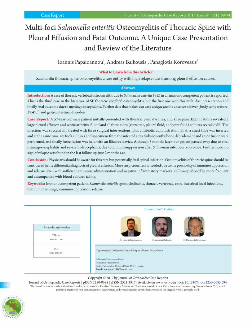
Multi-foci Salmonella enteritis Osteomyelitis of Thoracic Spine with Pleural Effusion and Fatal Outcome. A Unique Case Presentation
and Review of the Literature
Ioannis Papaioannou1, Andreas Baikousis1, Panagiotis Korovessis1
Abstract
Introduction: A case of thoracic vertebral osteomyelitis due to Salmonella enteritis (SE) in an immunocompetent patient is reported. This is the third case in the literature of SE thoracic vertebral osteomyelitis, but the first one with this multi-foci presentation and finally fatal outcome due to meningoencephalitis. Further data that makes our case unique are the absence of fever (body temperature: 37.4°C) and gastrointestinal disorders.
Case Report: A 57-year-old male patient initially presented with thoracic pain, dyspnea, and knee pain. Examinations revealed a large pleural effusion and septic arthritis. Blood and all these sides (vertebrae, pleural fluid, and joint fluid) cultures revealed SE. The infection was successfully treated with three surgical interventions, plus antibiotic administration. First, a chest tube was inserted and at the same time, we took cultures and specimens from the infected sites. Subsequently, bone debridement and spine fusion were performed, and finally, knee fusion was held with an Illizarov device. Although 8 months later, our patient passed away due to viral meningoencephalitis and severe hydrocephalus, due to immunosuppression after Salmonella infection recurrence. Furthermore, no sign of relapse was found in the last follow-up, just 2 months ago.
Conclusion: Physicians should be aware for this rare but potentially fatal spinal infection. Osteomyelitis of thoracic spine should be considered in the differential diagnosis of pleural effusion. More suspiciousness is needed due to the possibility of immunosuppression and relapse, even with sufficient antibiotic administration and negative inflammatory markers. Follow-up should be more frequent and accompanied with blood cultures taking.
Keywords: Immunocompetent patient, Salmonella enteritis spondylodiscitis, thoracic vertebrae, extra-intestinal focal infections, titanium mesh cage, immunosuppression, relapse.
Case Report Journal of Orthopaedic Case Reports 2017 Jan-Feb: 7(1):69-74
What to Learn from this Article?
Salmonella thoracic spine osteomyelitis a rare entity with high relapse rate is among pleural effusion causes.
Author’s Photo Gallery
Access this article online
Website:
www.jocr.co.in
DOI:
2250-0685.6941Departement of Orthopaedic, General Hospital of Patras, Patras, Greece.
Address of CorrespondenceDr. Ioannis Papaioannou, Pavlou Pavlopoulou 15, Aroi, Patras, 26331, Greece. E-mail: [email protected]
Dr. Ioannis Papaioannou Dr. Andreas Baikousis Dr. Panagiotis Korovessis
Copyright © 2017 by Journal of Orthpaedic Case ReportsJournal of Orthopaedic Case Reports | pISSN 2250-0685 | eISSN 2321-3817 | Available on www.jocr.co.in | doi: 10.13107/jocr.2250-0685.694
This is an Open Access article distributed under the terms of the Creative Commons Attribution Non-Commercial License (http://creativecommons.org/licenses/by-nc/3.0) which permits unrestricted non-commercial use, distribution, and reproduction in any medium, provided the original work is properly cited.
www.jocr.co.in
70
Journal of Orthopaedic Case Reports | Volume 7 | Issue 1 | Jan - Feb 2017 | Page 69-74
Introduction
Salmonella vertebral osteomyelitis (SVO) is not a reportable disease, and it is very likely that this entity is underdiagnosed. This uncommon entity can be a relatively benign condition that responds well to appropriate treatment or can result in a fatal outcome as in our case. The purpose of this paper is to appose a unique case of simultaneous presence of thoracic spondylodiscitis, thorax empyema, and knee arthritis caused by Salmonella enteritis (SE) in an immunocompetent patient. To the best of our knowledge [1, 2], this is the third case of SVO by enteritis species in thoracic spine but the first with multi-foci presentation and finally fatal late onset outcome due to severe viral meningoencephalitis.
Case Report
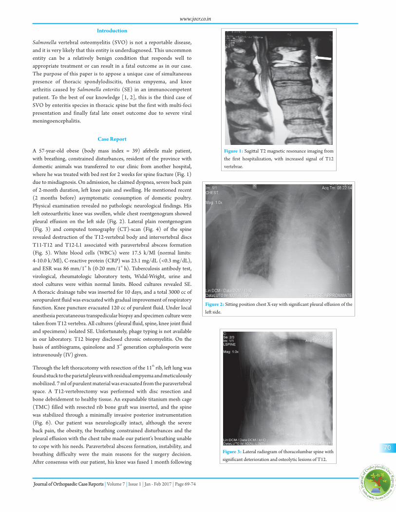
A 57-year-old obese (body mass index = 39) afebrile male patient, with breathing, constrained disturbances, resident of the province with domestic animals was transferred to our clinic from another hospital, where he was treated with bed rest for 2 weeks for spine fracture (Fig. 1) due to misdiagnosis. On admission, he claimed dyspnea, severe back pain of 2-month duration, left knee pain and swelling. He mentioned recent (2 months before) asymptomatic consumption of domestic poultry. Physical examination revealed no pathologic neurological findings. His left osteoarthritic knee was swollen, while chest roentgenogram showed pleural effusion on the left side (Fig. 2). Lateral plain roentgenogram (Fig. 3) and computed tomography (CT)-scan (Fig. 4) of the spine revealed destruction of the T12-vertebral body and intervertebral discs T11-T12 and T12-L1 associated with paravertebral abscess formation (Fig. 5). White blood cells (WBC’s) were 17.5 k/Μl (normal limits: 4-10.0 k/Μl), C-reactive protein (CRP) was 23.1 mg/dL (<0.3 mg/dL), and ESR was 86 mm/1st h (0-20 mm/1st h). Tuberculosis antibody test, virological, rheumatologic laboratory tests, Widal-Wright, urine and stool cultures were within normal limits. Blood cultures revealed SE. A thoracic drainage tube was inserted for 10 days, and a total 3000 cc of seropurulent fluid was evacuated with gradual improvement of respiratory function. Knee puncture evacuated 120 cc of purulent fluid. Under local anesthesia percutaneous transpedicular biopsy and specimen culture were taken from T12 vertebra. All cultures (pleural fluid, spine, knee joint fluid and specimens) isolated SE. Unfortunately, phage typing is not available in our laboratory. T12 biopsy disclosed chronic osteomyelitis. On the basis of antibiograms, quinolone and 3rd generation cephalosporin were intravenously (IV) given.
Through the left thoracotomy with resection of the 11th rib, left lung was found stuck to the parietal pleura with residual empyema and meticulously mobilized. 7 ml of purulent material was evacuated from the paravertebral space. A T12-vertebrectomy was performed with disc resection and bone debridement to healthy tissue. An expandable titanium mesh cage (TMC) filled with resected rib bone graft was inserted, and the spine was stabilized through a minimally invasive posterior instrumentation (Fig. 6). Our patient was neurologically intact, although the severe back pain, the obesity, the breathing constrained disturbances and the pleural effusion with the chest tube made our patient’s breathing unable to cope with his needs. Paravertebral abscess formation, instability, and breathing difficulty were the main reasons for the surgery decision. After consensus with our patient, his knee was fused 1 month following
Figure 1: Sagittal T2 magnetic resonance imaging from the first hospitalization, with increased signal of T12 vertebrae.
Figure 2: Sitting position chest X-ray with significant pleural effusion of the left side.
Figure 3: Lateral radiogram of thoracolumbar spine with significant deterioration and osteolytic lesions of T12.
www.jocr.co.in
71
Journal of Orthopaedic Case Reports | Volume 7 | Issue 1 | Jan - Feb 2017 | Page 69-74
admission with an Illizarov device. The post-operative course was complicated with superficial wound infection at the thoracotomy skin incision, knee arthrodesis incision and the insertion of the left L2 screw close to the thoracotomy wound with Acinetobacter. All sites required local debridement and secondary closure. The left knee fused completely 3 months following surgery. On the basis of antibiotic susceptibility pattern, quinolone and ceftriaxone were IV given. The patient received 3 months IV antibiotic treatment and was discharged with instructions for a monthly check of inflammatory markers. Antibiograms is the safe way for the antibiotics selection, although ciprofloxacin, 3rd generation cephalosporin, ampicillin, and chloramphenicol are the most common antibiotics in such infections. The IV administration was combined with our patient hospitalization due to his impaired general condition and the post-operative complications. The duration of the antibiotic treatment was determined on the basis of the evidence-based guidelines, to minimize relapse possibility. At the 6-month follow-up, CRP and ESR remained within normal limits. Radiograms and CT scan showed a good result of fusion in both sites (Fig. 7 and 8). The patient was free of pain, and he had started to return to his daily routine. There was not any sign of recurrence. Although 2 months later, our patient became anxious and his family found him with an altered state of consciousness and cognition. On admission to the hospital, our patient lost his consciousness and had to be intubated and transferred to intensive care unit. Brain CT scan revealed edema and hydrocephalus that required drainage tube installation (Fig. 9). The lumbar puncture of cerebrospinal fluid was indicative of viral infection. EEG showed slow waves and based on the previous medical history all possible cultures were taken. Blood cultures revealed again SE. Our patient did not recover from this meningoencephalitis, and his relatives decided to disconnect him from the ventilator on the day 11th being clinically dead. We believe that SE infection recurrence caused immunosuppression to this impaired patient making him unable to manage this central nervous system viral infection.
Discussion
Salmonella causes a broad spectrum of human illnesses from gastroenteritis, typhoid fever, and bacteremia to the asymptomatic carrier state [3]. SE primarily affects gastrointestinal, and rarely other systems such as musculoskeletal, brain and thyroid gland [4]. Vertebral osteomyelitis has been an uncommon complication of Salmonella infection by either hematogenous or contiguous spread. Osteomyelitis accounts approximately 0.5% of all cases [5]. Spondylodiscitis caused by SE is extremely rare with only nine cases involving lumbar [4], cervical [2] and thoracic [3] spine, including our case. Features of cervical and lumbar spine presented briefly in Table 1 [6]. In Table 2, we quote the features of thoracic spine SE reported to date.
Salmonella osteomyelitis is usually seen when the patient is immunologically compromised. In addition to sickle cell disease, conditions that may compromise the immune system include chronic alcoholism, chronic lymphocytic leukemia, systemic lupus erythematosus, neoplastic disease, long-term steroid intake [7], and possibly HIV. None of these predisposing factors found to our patient. Salmonella species can easily enter the systemic circulation, causing bacteremia, with positive blood cultures. Symptomatic extra-intestinal focal infections as joint involvement and chest empyema may occur after a prolonged asymptomatic period as in this male patient [8].
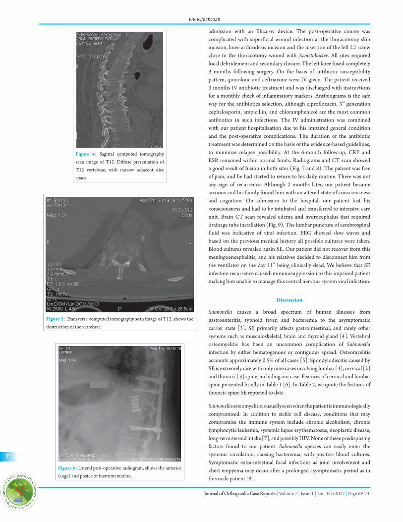
Figure 4: Sagittal computed tomography scan image of T12. Diffuse presentation of T12 vertebrae, with narrow adjacent disc space.
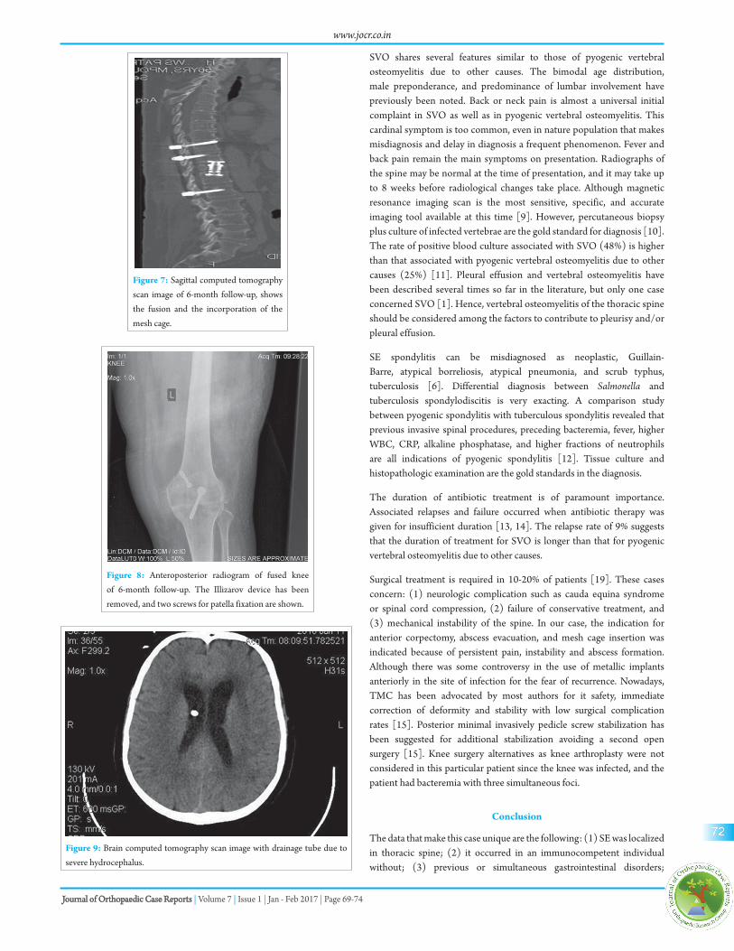
Figure 5: Transverse computed tomography scan image of T12, shows the destruction of the vertebrae.
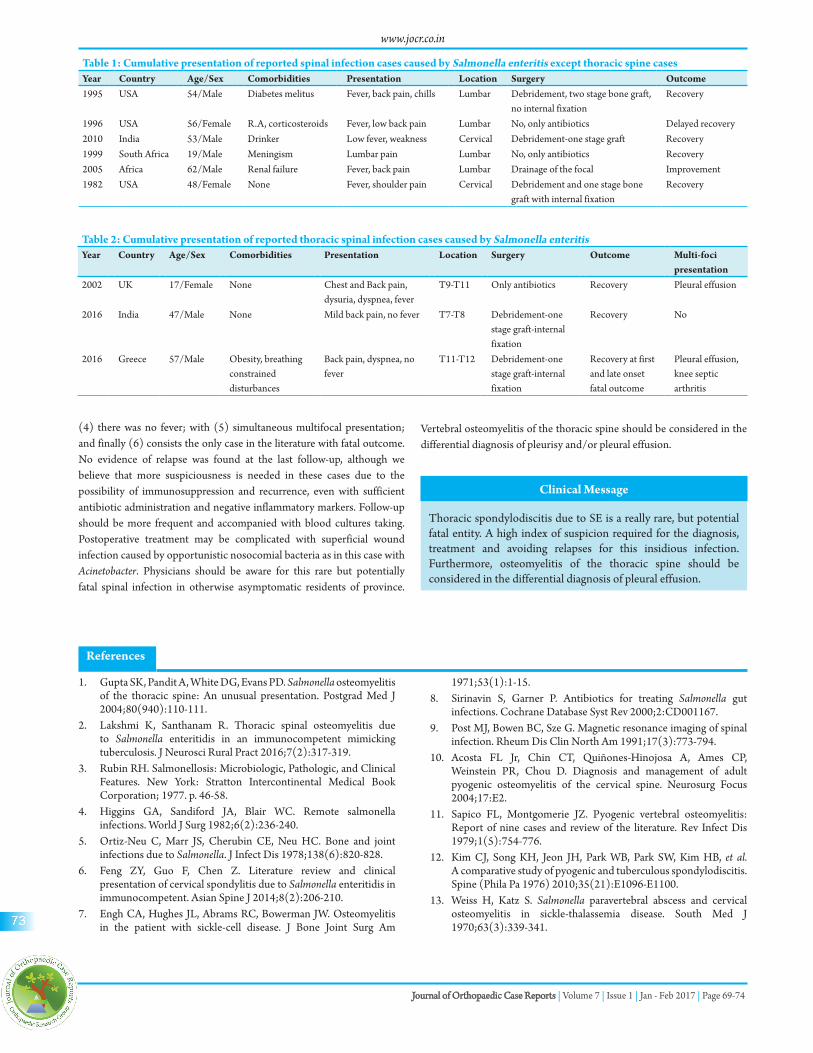
Figure 6: Lateral post-operative radiogram, shows the anterior (cage) and posterior instrumentation.
www.jocr.co.in
72
Journal of Orthopaedic Case Reports | Volume 7 | Issue 1 | Jan - Feb 2017 | Page 69-74
SVO shares several features similar to those of pyogenic vertebral osteomyelitis due to other causes. The bimodal age distribution, male preponderance, and predominance of lumbar involvement have previously been noted. Back or neck pain is almost a universal initial complaint in SVO as well as in pyogenic vertebral osteomyelitis. This cardinal symptom is too common, even in nature population that makes misdiagnosis and delay in diagnosis a frequent phenomenon. Fever and back pain remain the main symptoms on presentation. Radiographs of the spine may be normal at the time of presentation, and it may take up to 8 weeks before radiological changes take place. Although magnetic resonance imaging scan is the most sensitive, specific, and accurate imaging tool available at this time [9]. However, percutaneous biopsy plus culture of infected vertebrae are the gold standard for diagnosis [10]. The rate of positive blood culture associated with SVO (48%) is higher than that associated with pyogenic vertebral osteomyelitis due to other causes (25%) [11]. Pleural effusion and vertebral osteomyelitis have been described several times so far in the literature, but only one case concerned SVO [1]. Hence, vertebral osteomyelitis of the thoracic spine should be considered among the factors to contribute to pleurisy and/or pleural effusion.
SE spondylitis can be misdiagnosed as neoplastic, Guillain-Barre, atypical borreliosis, atypical pneumonia, and scrub typhus, tuberculosis [6]. Differential diagnosis between Salmonella and tuberculosis spondylodiscitis is very exacting. A comparison study between pyogenic spondylitis with tuberculous spondylitis revealed that previous invasive spinal procedures, preceding bacteremia, fever, higher WBC, CRP, alkaline phosphatase, and higher fractions of neutrophils are all indications of pyogenic spondylitis [12]. Tissue culture and histopathologic examination are the gold standards in the diagnosis.
The duration of antibiotic treatment is of paramount importance. Associated relapses and failure occurred when antibiotic therapy was given for insufficient duration [13, 14]. The relapse rate of 9% suggests that the duration of treatment for SVO is longer than that for pyogenic vertebral osteomyelitis due to other causes.
Surgical treatment is required in 10-20% of patients [19]. These cases concern: (1) neurologic complication such as cauda equina syndrome or spinal cord compression, (2) failure of conservative treatment, and (3) mechanical instability of the spine. In our case, the indication for anterior corpectomy, abscess evacuation, and mesh cage insertion was indicated because of persistent pain, instability and abscess formation. Although there was some controversy in the use of metallic implants anteriorly in the site of infection for the fear of recurrence. Nowadays, TMC has been advocated by most authors for it safety, immediate correction of deformity and stability with low surgical complication rates [15]. Posterior minimal invasively pedicle screw stabilization has been suggested for additional stabilization avoiding a second open surgery [15]. Knee surgery alternatives as knee arthroplasty were not considered in this particular patient since the knee was infected, and the patient had bacteremia with three simultaneous foci.
Conclusion
The data that make this case unique are the following: (1) SE was localized in thoracic spine; (2) it occurred in an immunocompetent individual without; (3) previous or simultaneous gastrointestinal disorders;
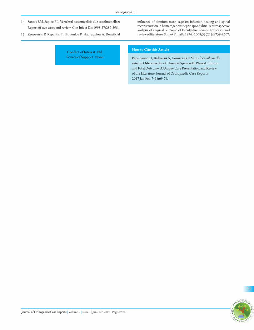
Figure 7: Sagittal computed tomography scan image of 6-month follow-up, shows the fusion and the incorporation of the mesh cage.
Figure 8: Anteroposterior radiogram of fused knee of 6-month follow-up. The Illizarov device has been removed, and two screws for patella fixation are shown.
Figure 9: Brain computed tomography scan image with drainage tube due to severe hydrocephalus.
www.jocr.co.in
73
Journal of Orthopaedic Case Reports | Volume 7 | Issue 1 | Jan - Feb 2017 | Page 69-74
(4) there was no fever; with (5) simultaneous multifocal presentation; and finally (6) consists the only case in the literature with fatal outcome. No evidence of relapse was found at the last follow-up, although we believe that more suspiciousness is needed in these cases due to the possibility of immunosuppression and recurrence, even with sufficient antibiotic administration and negative inflammatory markers. Follow-up should be more frequent and accompanied with blood cultures taking. Postoperative treatment may be complicated with superficial wound infection caused by opportunistic nosocomial bacteria as in this case with Acinetobacter. Physicians should be aware for this rare but potentially fatal spinal infection in otherwise asymptomatic residents of province.
Table 1: Cumulative presentation of reported spinal infection cases caused by Salmonella enteritis except thoracic spine casesYear Country Age/Sex Comorbidities Presentation Location Surgery Outcome1995 USA 54/Male Diabetes melitus Fever, back pain, chills Lumbar Debridement, two stage bone graft,
no internal fixationRecovery
1996 USA 56/Female R.A, corticosteroids Fever, low back pain Lumbar No, only antibiotics Delayed recovery2010 India 53/Male Drinker Low fever, weakness Cervical Debridement-one stage graft Recovery1999 South Africa 19/Male Meningism Lumbar pain Lumbar No, only antibiotics Recovery2005 Africa 62/Male Renal failure Fever, back pain Lumbar Drainage of the focal Improvement1982 USA 48/Female None Fever, shoulder pain Cervical Debridement and one stage bone
graft with internal fixationRecovery
Table 2: Cumulative presentation of reported thoracic spinal infection cases caused by Salmonella enteritisYear Country Age/Sex Comorbidities Presentation Location Surgery Outcome Multi-foci
presentation2002 UK 17/Female None Chest and Back pain,
dysuria, dyspnea, feverT9-T11 Only antibiotics Recovery Pleural effusion
2016 India 47/Male None Mild back pain, no fever T7-T8 Debridement-one stage graft-internal fixation
Recovery No
2016 Greece 57/Male Obesity, breathing constrained disturbances
Back pain, dyspnea, no fever
T11-T12 Debridement-one stage graft-internal fixation
Recovery at first and late onset fatal outcome
Pleural effusion, knee septic arthritis
Clinical Message
Thoracic spondylodiscitis due to SE is a really rare, but potential fatal entity. A high index of suspicion required for the diagnosis, treatment and avoiding relapses for this insidious infection. Furthermore, osteomyelitis of the thoracic spine should be considered in the differential diagnosis of pleural effusion.
Vertebral osteomyelitis of the thoracic spine should be considered in the differential diagnosis of pleurisy and/or pleural effusion.
References
1. Gupta SK, Pandit A, White DG, Evans PD. Salmonella osteomyelitis of the thoracic spine: An unusual presentation. Postgrad Med J 2004;80(940):110-111.
2. Lakshmi K, Santhanam R. Thoracic spinal osteomyelitis due to Salmonella enteritidis in an immunocompetent mimicking tuberculosis. J Neurosci Rural Pract 2016;7(2):317-319.
3. Rubin RH. Salmonellosis: Microbiologic, Pathologic, and Clinical Features. New York: Stratton Intercontinental Medical Book Corporation; 1977. p. 46-58.
4. Higgins GA, Sandiford JA, Blair WC. Remote salmonella infections. World J Surg 1982;6(2):236-240.
5. Ortiz-Neu C, Marr JS, Cherubin CE, Neu HC. Bone and joint infections due to Salmonella. J Infect Dis 1978;138(6):820-828.
6. Feng ZY, Guo F, Chen Z. Literature review and clinical presentation of cervical spondylitis due to Salmonella enteritidis in immunocompetent. Asian Spine J 2014;8(2):206-210.
7. Engh CA, Hughes JL, Abrams RC, Bowerman JW. Osteomyelitis in the patient with sickle-cell disease. J Bone Joint Surg Am
1971;53(1):1-15.8. Sirinavin S, Garner P. Antibiotics for treating Salmonella gut
infections. Cochrane Database Syst Rev 2000;2:CD001167.9. Post MJ, Bowen BC, Sze G. Magnetic resonance imaging of spinal
infection. Rheum Dis Clin North Am 1991;17(3):773-794.10. Acosta FL Jr, Chin CT, Quiñones-Hinojosa A, Ames CP,
Weinstein PR, Chou D. Diagnosis and management of adult pyogenic osteomyelitis of the cervical spine. Neurosurg Focus 2004;17:E2.
11. Sapico FL, Montgomerie JZ. Pyogenic vertebral osteomyelitis: Report of nine cases and review of the literature. Rev Infect Dis 1979;1(5):754-776.
12. Kim CJ, Song KH, Jeon JH, Park WB, Park SW, Kim HB, et al. A comparative study of pyogenic and tuberculous spondylodiscitis. Spine (Phila Pa 1976) 2010;35(21):E1096-E1100.
13. Weiss H, Katz S. Salmonella paravertebral abscess and cervical osteomyelitis in sickle-thalassemia disease. South Med J 1970;63(3):339-341.
www.jocr.co.in
74
Journal of Orthopaedic Case Reports | Volume 7 | Issue 1 | Jan - Feb 2017 | Page 69-74
14. Santos EM, Sapico FL. Vertebral osteomyelitis due to salmonellae:
Report of two cases and review. Clin Infect Dis 1998;27:287-295.
15. Korovessis P, Repantis T, Iliopoulos P, Hadjipavlou A. Beneficial
influence of titanium mesh cage on infection healing and spinal reconstruction in hematogenous septic spondylitis: A retrospective analysis of surgical outcome of twenty-five consecutive cases and review of literature. Spine (Phila Pa 1976) 2008;33(21):E759-E767.
Conflict of Interest: Nil. Source of Support: None
How to Cite this Article
Papaioannou I, Baikousis A, Korovessis P. Multi-foci Salmonella enteritis Osteomyelitis of Thoracic Spine with Pleural Effusion and Fatal Outcome. A Unique Case Presentation and Review of the Literature. Journal of Orthopaedic Case Reports 2017 Jan-Feb;7(1):69-74.