Embed Size (px)
Citation preview
Int J Clin Exp Pathol 2014;7(11):8211-8216www.ijcep.com /ISSN:1936-2625/IJCEP0002517
Case ReportSacrococcygeal teratoma with nephroblastic elements: a case report and review of literature
Yangyang Ma1, Jicui Zheng2, Haitao Zhu2, Chun Shen2, Shan Zheng2, Xianmin Xiao2, Lian Chen1
1Department of Pathology, 2Department of Surgery, Fudan University Children’s Hospital, Shanghai, China
Received September 15, 2014; Accepted November 1, 2014; Epub October 15, 2014; Published November 1, 2014
Abstract: Sacrococcygeal teratoma with nephroblastic elements is very rare. Only 8 cases have been reported up to date. 3 cases were misdiagnosed as extrarenal nephroblastomas and were administered excessive treatments such as chemotherapy and radiation therapy. It has a great significance to distinguish sacrococcygeal teratoma with nephroblastic elements from extrarenal nephroblastoma for their different biological behaviors, therapies and prognoses. Here we report one case of sacrococcygeal teratoma with nephroblastic elements in a newborn with review of the literature. This is the first case of sacrococcygeal teratoma with nephroblastic elements in Chinese to our knowledge.
Keywords: Sacrococcygeal teratoma, nephroblastic elements, extrarenal nephroblastoma
Introduction
Sacrococcygeal teratoma is one of the most common neoplasms in newborns with an inci-dence of 1/21,000 births [1]. Although most sacrococcygeal teratomas are mature, a variety of immature components may be present. Occurrence of nephroblastic elements is very rare in sacrococcygeal teratomas. Only 8 cases of sacrococcygeal teratomas with nephroblas-tic elements (STWNE) have been reported up to now. Here we describe another case of sacro-coccygeal teratoma with nephroblastic ele-ments and review the literature.
Case report
A 3700 g boy was delivered by caesarean sec-tion at full-term gestation. He was admitted to our hospital at birth for a sacrococcygeal mass. The mass was first detected in the fetus at 24 weeks’ gestation by ultrasound. An exgenous mass measuring 10 × 8 × 5 cm in the sacrococ-cygeal region was found on physical examina-tion which was confirmed by CT scan and MRI scan afterwards (Figure 1). Both of the kidneys were normal. The serum α-fetoprotein (AFP) concentration was more than 121000 ng/ml at birth. Given that the mass might be a teratoma,
a tumorectomy was performed on the 8th day after birth. The mass was entirely removed. No additional treatments such as chemotherapy and radiation therapy were given postoperative-ly. The child is still alive without evidence of tumor recurrence and metastasis 8 months after surgery. Serum AFP levels were monitored in follow-up and fell to 45.1 ng/ml at 8 months after sugery. Written informed consent was got from the child’s parents.
Pathological findings
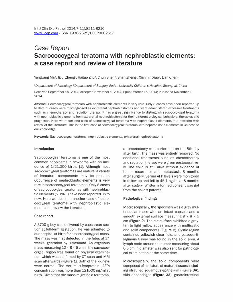
Macroscopically, the specimen was a gray mul-tinodular mass with an intact capsule and a smooth external surface measuring 9 × 8 × 5 cm (Figure 2). The cut surface exhibited a gray-tan to light yellow appearance with multicystic and solid components (Figure 2). Cystic region contained yellowish clear fluid, and osteocarti-laginous tissue was found in the solid area. A lymph node around the tumor measuring about 0.5 cm in diameter was also sent for pathologi-cal examination at the same time.
Microscopically, the solid components were composed of a mixture of mature tissues includ-ing stratified squamous epithelium (Figure 3A), skin appendages (Figure 3A), gastrointestinal
Sacrococcygeal teratoma with nephroblastic elements
8212 Int J Clin Exp Pathol 2014;7(11):8211-8216
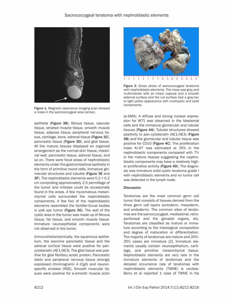
epithelia (Figure 3B), fibrous tissue, vascular tissue, striated muscle tissue, smooth muscle tissue, adipose tissue, peripheral nervous tis-sue, cartilage, bone, adrenal tissue (Figure 3C), pancreatic tissue (Figure 3D), and glial tissue. All the mature tissues displayed an organoid arrangement as the normal skin tissue, intesti-nal wall, pancreatic tissue, adrenal tissue, and so on. There were focal areas of nephroblastic elements under the gastrointestinal epithelia in the form of primitive round cells, immature glo-merular structures and tubules (Figure 3E and 3F). The nephroblastic elements were 0.2 × 0.2 cm comprising approximately 2.5 permillage of the tumor and mitoses could be occasionally found in the areas. A few myxomatous mesen-chymal cells surrounded the nephroblastic components. A few foci of the nephroblastic elements resembled the Schiller-Duval bodies in yolk sac tumor (Figure 3G). The wall of the cystic area in the tumor was made up of fibrous tissue, fat tissue, and smooth muscle tissue. Immature neuroepithelial components were not observed in the tumor.
Immunohistochemically, the squamous epithe-lium, the exocrine pancreatic tissue and the adrenal cortical tissue were positive for pan-cytokeratin (AE1/AE3). The glial tissue was pos-itive for glial fibrillary acidic protein. Pancreatic islets and peripheral nervous tissue strongly expressed chromogranin A (CgA) and neuron-specific enolase (NSE). Smooth muscular tis-sues were positive for α-smooth muscle actin
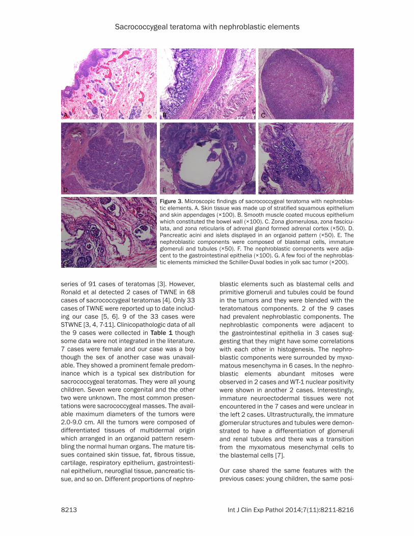
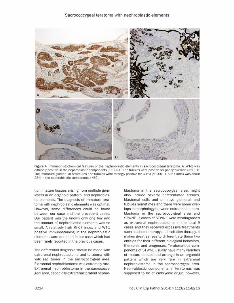
(α-SMA). A diffuse and strong nuclear expres-sion for WT1 was observed in the blastemal cells and the immature glomerular and tubular tissues (Figure 4A). Tubular structures showed positivity to pan-cytokeratin (AE1/AE3) (Figure 4B) and the glomerular and tubular tissue was positive for CD10 (Figure 4C). The proliferation index Ki-67 was estimated at 25% in the nephroblastic components compared with 7% in the mature tissues suggesting the nephro-blastic components may have a relatively high-er proliferative activity (Figure 4D). The diagno-sis was immature solid-cystic teratoma grade I with nephroblastic elements and no tumor cell was detected in the lymph node.
Discussion
Teratomas are the most common germ cell tumor that consists of tissues derived from the three germ cell layers (ectoderm, mesoderm, and endoderm). The common sites of terato-mas are the sacroccocygeal, mediastinal, retro-peritoneal and the gonadal organs, etc. Teratomas are classified as mature or imma-ture according to the histological composition and degree of maturation or differentiation. The majority of teratomas are mature and 20%-25% cases are immature [2]. Immature ele-ments usually contain neuroepithelium, carti-lage, and primitive mesenchymal tissue. Nephroblastic elements are very rare in the immature elements of teratomas and the detailed occurrence rate of teratomas with nephroblastic elements (TWNE) is unclear. Berry et al reported 1 case of TWNE in his
Figure 1. Magnetic resonance imaging scan showed a mass in the sacrococcygeal area (arrow).
Figure 2. Gross photo of sacrococcygeal teratoma with nephroblastic elements. The mass was gray and multinodular with an intact capsule and a smooth external surface and the cut surface had a gray-tan to light yellow appearance with multicystic and solid components.
Sacrococcygeal teratoma with nephroblastic elements
8213 Int J Clin Exp Pathol 2014;7(11):8211-8216
Figure 3. Microscopic findings of sacrococcygeal teratoma with nephroblas-tic elements. A. Skin tissue was made up of stratified squamous epithelium and skin appendages (×100). B. Smooth muscle coated mucous epithelium which constituted the bowel wall (×100). C. Zona glomerulosa, zona fascicu-lata, and zona reticularis of adrenal gland formed adrenal cortex (×50). D. Pancreatic acini and islets displayed in an organoid pattern (×50). E. The nephroblastic components were composed of blastemal cells, immature glomeruli and tubules (×50). F. The nephroblastic components were adja-cent to the gastrointestinal epithelia (×100). G. A few foci of the nephroblas-tic elements mimicked the Schiller-Duval bodies in yolk sac tumor (×200).
series of 91 cases of teratomas [3]. However, Ronald et al detected 2 cases of TWNE in 68 cases of sacrococcygeal teratomas [4]. Only 33 cases of TWNE were reported up to date includ-ing our case [5, 6]. 9 of the 33 cases were STWNE [3, 4, 7-11]. Clinicopathologic data of all the 9 cases were collected in Table 1 though some data were not integrated in the literature. 7 cases were female and our case was a boy though the sex of another case was unavail-able. They showed a prominent female predom-inance which is a typical sex distribution for sacrococcygeal teratomas. They were all young children. Seven were congenital and the other two were unknown. The most common presen-tations were sacrococcygeal masses. The avail-able maximum diameters of the tumors were 2.0-9.0 cm. All the tumors were composed of differentiated tissues of multidermal origin which arranged in an organoid pattern resem-bling the normal human organs. The mature tis-sues contained skin tissue, fat, fibrous tissue, cartilage, respiratory epithelium, gastrointesti-nal epithelium, neuroglial tissue, pancreatic tis-sue, and so on. Different proportions of nephro-
blastic elements such as blastemal cells and primitive glomeruli and tubules could be found in the tumors and they were blended with the teratomatous components. 2 of the 9 cases had prevalent nephroblastic components. The nephroblastic components were adjacent to the gastrointestinal epithelia in 3 cases sug-gesting that they might have some correlations with each other in histogenesis. The nephro-blastic components were surrounded by myxo-matous mesenchyma in 6 cases. In the nephro-blastic elements abundant mitoses were observed in 2 cases and WT-1 nuclear positivity were shown in another 2 cases. Interestingly, immature neuroectodermal tissues were not encountered in the 7 cases and were unclear in the left 2 cases. Ultrastructurally, the immature glomerular structures and tubules were demon-strated to have a differentiation of glomeruli and renal tubules and there was a transition from the myxomatous mesenchymal cells to the blastemal cells [7].
Our case shared the same features with the previous cases: young children, the same posi-
Sacrococcygeal teratoma with nephroblastic elements
8214 Int J Clin Exp Pathol 2014;7(11):8211-8216
tion, mature tissues arising from multiple germ layers in an organoid pattern, and nephroblas-tic elements. The diagnosis of immature tera-toma with nephroblastic elements was optimal. However, some differences could be found between our case and the precedent cases. Our patient was the known only one boy and the amount of nephroblastic elements was so small. A relatively high Ki-67 index and WT-1 positive immunostaining in the nephroblastic elements were detected in our case which had been rarely reported in the previous cases.
The differential diagnosis should be made with extrarenal nephroblastoma and teratoma with yolk sac tumor in the sacrococcygeal area. Extrarenal nephroblastoma was extremely rare. Extrarenal nephroblastoma in the sacrococcy-geal area, especially extrarenal teratoid nephro-
blastoma in the sacrococcygeal area, might also include several differentiated tissues, blastemal cells and primitive glomeruli and tubules sometimes and there were some over-laps in morphology between extrarenal nephro-blastoma in the sacrococcygeal area and STWNE. 3 cases of STWNE were misdiagnosed as extrarenal nephroblastoma in the total 9 cases and they received excessive treatments such as chemotherapy and radiation therapy. It makes great senses to differentiate these two entities for their different biological behaviors, therapies and prognoses. Teratomatous com-ponents of STWNE usually have many varieties of mature tissues and arrange in an organoid pattern which are very rare in extrarenal nephroblastoma in the sacrococcygeal area. Nephroblastic components in teratomas was supposed to be of embryonic origin, however,
Figure 4. Immunohistochemical features of the nephroblastic elements in sacrococcygeal teratoma. A. WT-1 was diffusely positive in the nephroblastic components (×100). B. The tubules were positive for pancytokeratin (×50). C. The immature glomerular structures and tubules were strongly positive for CD10 (×100). D. Ki-67 index was about 25% in the nephroblastic components (×50).
Sacrococcygeal teratoma with nephroblastic elements
8215 Int J Clin Exp Pathol 2014;7(11):8211-8216
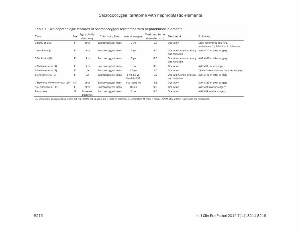
Table 1. Clinicopathologic features of sacrococcygeal teratomas with nephroblastic elements
Case Sex Age at initial discovery Chief complaint Age at surgery Maximum tumor
diameter (cm) Treatment Follow-up
1 Berry et al [3] F birth Sacrococcygeal mass 2 wo UA Operation Local recurrence and lung metastases 1 y later, lost to follow-up
2 Ward et al [7] F birth Sacrococcygeal mass 3 yo 8.0 Operation, chemotherapy and radiation
AWRM 12 m after surgery
3 Tebbi et al [8] F birth Sacrococcygeal mass 3 yo 8.0 Operation, chemotherapy and radiation
AWRM 46 m after surgery
4 Valdiserri et al [4] F birth Sacrococcygeal mass 3 do 9.0 Operation AWRM 6 y after surgery
5 Valdiserri et al [4] F UA Sacrococcygeal mass 1.5 yo 2.0 Operation Died of other diseases 3 y after surgery
6 Andrews et al [9] F UA Sacrococcygeal mass 1 wo-4.3 yo, the detail UA
UA Operation, chemotherapy and radiation
AWRM 46 m after surgery
7 Heerema-McKenney et al [10] NA birth Sacrococcygeal mass less than1 wo 3.8 Operation AWRM 25 m after surgery
8 Al-Adnani et al [11] F birth Sacrococcygeal mass 15 mo 9.0 Operation AWRM 9 m after surgery
9 our case M 24 weeks’ gestation
Sacrococcygeal mass 8 do 9.0 Operation AWRM 8 m after surgery
UA, unavailable; do, days old; wo, weeks old; mo, months old; yo, years old; y, years; m, months; cm, centimeters; M, male; F, female; AWRM, alive without recurrences and metastases.
Sacrococcygeal teratoma with nephroblastic elements
8216 Int J Clin Exp Pathol 2014;7(11):8211-8216
nephroblastoma was thought to be of meso-nephric origin. Emerson et al reported the nephroblastic components shared a clonal ori-gin with the teratoma in an interesting case of mature and immature teratoma with nephro-blastic components of the testis [12]. Yolk sac tumor can be found with sacrococcygeal tera-toma occasionally.Schiller-Duval bodies in yolk sac tumor mimicked the immature glomeruli.Serum AFP concentration is elevated in sacro-coccygeal teratoma with yolk sac tumor and positive immunoreactivity for AFP and negative immunoreactivity for WT-1 would also been shown in the tumor which is different from STWNE.
The standard therapy has not been established for STWNE for their scarity. Whether fetal STWNE need surgical interventions in utero or not are still unknown. STWNE generally behave indolently and metastases are very rare. Complete resection and close follow-up are optimal. 6 of the 9 STWNE cases received sur-gery alone and the other 3 cases were given comprehensive therapy including surgery, che-motherapy and radiation therapy because they were misdiagnosed as extrarenal nephroblas-tomas. 1 case died of other diseases 3 years after sugery, 1 recurred locally with lung metas-tases 1 year after sugery and was lost to follow-up, the left were all uneventful in the reported follow-up time. All the data suggest the progno-ses of STWNE are good.
In conclusion, we described a case of STWNE. This is the first case in Chinese. More data are needed to understand the biology and clinical behavior of this tumor.
Acknowledgements
This work was supported by the National Key Clinical Specialist Construction Programs of China (2014-2016).
Disclosure of conflict of interest
None.
Address correspondence to: Dr. Lian Chen, Department of Pathology, Children’s Hospital of Fudan University, Shanghai, China. Tel: (86)21-6493-1728; Fax: (86) 21-6493-1914; E-mail: [email protected]; Dr. Xianmin Xiao, Depart- ment of Surgery, Children’s Hospital of Fudan
University, Shanghai, China. E-mail: [email protected]
References
[1] Gucciardo L, Uyttebroek A, De Weyer I, Renard M, Claus F, Devlieger R, Lewi L, De Catte L, Deprest J. Prenatal assessment and manage-ment of sacrococcygeal teratoma. Prenat Diagn 2011; 31: 678-688.
[2] Mohta A, Sengar M, Neogi S, Khurana N. Gastric teratoma with predominant ne- phroblastic elements. Pediatr Surg Int 2010; 26: 923-925.
[3] Berry CL, Keeling J, Hilton C. Teratomata in in-fancy and childhood: a review of 91 cases. J Pathol 1969; 98: 241-252.
[4] Valdiserri RO, Yunis EJ. Sacrococcygeal terato-mas: a review of 68 cases. Cancer 1981; 48: 217-221.
[5] Ishida M, Hotta M, Ohta M, Taga T, Ohta S, Takeuchi Y, Okabe H. A case of retroperitoneal immature teratoma with nephroblastic compo-nents. J Pediatr Hematol Oncol 2012; 34: e22-25.
[6] Coli A, Angrisani B, Chiarello G, Massimi L, Novello M, Lauriola L. Ectopic immature renal tissue: clues for diagnosis and management. Int J Clin Exp Pathol 2012; 5: 977-981.
[7] Ward SP, Dehner LP. Sacrococcygealtera- toma with nephroblastoma (Wilm’s tumor): a variant of extragonadalteratoma in childhood. A histologic and ultrastructural study. Cancer 1974; 33: 1355-1363.
[8] Tebbi K, Ragab AH, Ternberg JL, Vietti TJ. An extrarenal Wilms’ tumor arising from a sacro-coccygeal teratoma. Clin Pediatr (Phila) 1974; 13: 1019-1021.
[9] Andrews PE, Kelalis PP, Haase GM. Extrarenal Wilms’ tumor: results of the National Wilms’ Tumor Study. J Pediatr Surg 1992; 27: 1181-1184.
[10] Heerema-McKenney A, Harrison MR, Bratton B, Farrell J, Zaloudek C. Congenital teratoma: a clinicopathologic study of 22 fetal and neona-tal tumors. Am J Surg Pathol 2005; 29: 29-38.
[11] Al-Adnani M, Walker J, Cohen M. Sacroco- ccygealteratoma with extensive nephrogenic differentiation: a rare finding not to be misdiag-nosed as yolk sac tumour. Histopathology 2009; 54: 764-765.
[12] Emerson RE, Ulbright TM, Zhang S, Foster RS, Eble JN, Cheng L. Nephroblastoma arising in a germ cell tumor of testicular origin. Am J Surg Pathol 2004; 28: 687-692.


![TERATOMA SACROCOCCÍGEO.Biblio [Sólo lectura] · En Ashcraft. Cirugía Pediátrica. 3ª Ed. Ediciones McGraw Hill. • Eagler RA, Pappo AS. Sacrococcygeal germ cell tumors. Uptodate](https://img.pdfslide.net/doc/110x75/5ff47bb66d819a1b74186614/teratoma-sacrococcgeobiblio-slo-lectura-en-ashcraft-ciruga-peditrica.jpg)