Embed Size (px)
Citation preview

Weaver: Electroporation of Biological Membranes from Multicellular to Nano Scales754
Electroporation of Biological Membranes fromMulticellular to Nano Scales
James C. WeaverHST Biomedical Engineering Center
Harvard-MIT Division of Health Sciences and TechnologyCambridge, MA 02139, USA
ABSTRACTElectroporation, widely used in research and applications, is briefly reviewed. Bothcell and artificial planar bilayer membranes exhibit dramatic changes if the trans-membrane voltage is raised to �0.2 to 1 V by various electric field pulses. Ionicand molecular transport increases by orders of magnitude, with both reversible andirreversible outcomes. Initially the term breakdown was used, but ion pair genera-tion of classic dielectric breakdown was ruled out. Instead, a stochastic pore hy-pothesis is consistent with features of electroporation in planar lipid membranes.There is a rapid, nonlinear conduction increase through a rapidly evolving porepopulation, and this causes the fast membrane discharge previously termed‘‘breakdown’’. Phenomena due to primary aqueous pores and secondary processessuch as heating and chemical exchange have been observed in planar bilayers, cellsingle systems encountered mainly in vitro, multicellular systems relevant to in vivoapplications, and possibly subcellular structures such as mitochondria. For mem-brane systems that approach nanoscales, modified behavior should occur becauseof conformational constraints, and deterministic processes may become more im-portant. Understanding electroporation is a subset of a general problem: obtaininga quantitative description of how electromagnetic field-altered changes in chemicalspecies within a biological system govern observed effects.
Index Terms — Electroporation, biological membranes, multicellular scale,nano scale membrane, bilayer membranes, cells, transmembrane voltage, ionictransport, molecular transport, lipid membranes, nonlinear conduction, mitochon-dria.
1 THE GENERAL PROBLEMVER the past two decades electroporationOresearch has been motivated by both applications and
w xmechanistic understanding 1�8 . But electroporation isonly part of the long standing scientific interest in the in-teraction of electric and magnetic fields with biologicalsystems. Phenomena caused by short exposures to strong
w xfields also include heating 9,10 , and extreme conforma-w xtional changes of voltage-gated channels 11,12 . Far less
dramatic effects occur with exposures to small fields.
The general problem includes animal navigation, en-dogenous fields and currents, drug delivery and othermedical interventions, and possible human health hazardsfrom environmental and occupational electromagnetic
w xfields 6,13�17 . These topics are all related. A unified un-derstanding of biological effects of electromagnetic fieldscan be sought by seeking quantitative understanding of
Manuscript recei®ed on 3 February 2003, in final form 1 July 2003.
the field-induced change in one or more biochemicalŽ .species ions and molecules in the context of a biologicalw xsystem 18 .
2 CONDITIONS FOR EFFECTSThere is long standing interest regarding conditions for
which electromagnetic field effects are expected in biolog-ical systems. Although particularly an issue for weak fields,it is also relevant for strong fields, particularly for tissueelectroporation in ®i®o. Briefly, the challenge is to under-stand how field induced changes from the field exposurecan stand out against changes due to other influences.
w x w xBoth physical 19,20 and chemical 21�24 changes can beconsidered, and thresholds estimated using signal-to-noise
Ž .ratio, SrN , criteria. Most physical assessments have em-phasized criteria based on the field-induced change inquantities such as the transmembrane voltage change, �Umw x w x19 , or on heating 25 .
However, significantly more restrictive constraints ariseif a generalized chemical noise is considered. For exam-
1070-9878rrrrr1rrrrr$17.00 � 2003 IEEE754

IEEE Transactions on Dielectrics and Electrical Insulation Vol. 10, No. 5; October 2003 755
ple, a threshold due to fundamental chemical noiseŽ .‘‘molecular shot noise’’ generates higher exposure
w xthresholds than obtained from thermal noise alone 21,22 .Moreover, a threshold estimate based on field-inducedchanges in chemistry naturally introduces the duration ofan exposure, t , into the analysis. Exposure time entersexp
because non-ionizing fields can only alter rates of chemi-cal transport and chemical reactions, and it is the field-altered accumulation of chemical amount that is consid-ered. Chemical-based assessments can also provide a basisfor predicting chemical changes that anticipate what canbe measured after the field exposure, and threshold esti-mates can include all significant sources of competing
w xchemical change 18 . This is highly relevant to electropo-ration experiments that measure cellular uptake or al-tered cellular function.
More specifically, a threshold exposure can be esti-Ž .mated using SrN in which the signal, S, is theCHEM
chemical change due to the field exposure, and the gener-alized chemical noise, N, is the variability in the quantityof the same chemical species due to all significant compet-ing chemical changes during the exposure. This includes
Ž .fundamental chemical noise molecular shot noise andalso changes in chemical amounts due to temperature
w xvariations, V 23,24 . Under in ®i®o conditions additionalchemical noise should be expected, due to normal physio-logical variations, tissue deformation by normal body ac-
w xtivity, and endogenous electric fields 18 .
The possible effects of strong field pulses can also beŽ .assessed using SrN . In the case of normal tissueCHEM
movement, there are impressive observations of molecularuptake due to transient cell membrane openings associ-
w xated solely with mechanical cell deformation 26�28 .However, electromechanical membrane deformation may
w xitself also be involved in electroporation 29�32 , particu-larly of suspended cells that are not mechanically con-strained by being bound to neighboring cells within a solidtissue. A further complication is electrically stimulated
Ž .movement of tissue, with two origins: 1 electrical stimu-Ž .lation of muscles, and 2 the general, passive deformation
of tissue under the influence of a strong electric field be-cause of the average dielectric properties of tissue. Inter-preting the contribution of electroporation and mechani-cal strain to cellular uptake should consider quantitativelythe relative contributions of ‘‘mechanical only mecha-nisms’’ and ‘‘strong field mechanisms’’ to uptake.
3 ELECTROPORATIONINTRODUCTION
According to experimental observations and the tran-w xsient aqueous pore mechanism hypothesis 1�5,7,33 , the
Ž .main features of electroporation involve 1 application ofŽ y6 y1 . Ž .short �10 to 10 s electrical pulses, 2 charging of
Ž .lipid bilayer membranes, 3 rapid, localized structural re-Ž .arrangements within the membrane, 4 transitions to wa-
ter-filled membrane structures which perforate the mem-Ž .brane ‘‘aqueous pathways’’, ‘‘pores’’ or ‘‘hydrophilic
Ž .pores’’, and 5 tremendous increase in ionic and molecu-lar transport through the membrane. Whether or not themembrane system fully recovers depends on many factors.
4 EARLY OBSERVATIONSEarly experiments exposing cell membranes to strong
electric fields involved diverse systems, ranging fromnerves to bacteria. In 1958 the nodes of Ranvier of nerves
w xwere reported to involve some type of ‘‘breakdown’’ 34 .Almost a decade later damaging effects of strong electricfields on microorganisms were reported suggesting non-
w xthermal membrane interactions 35�37 . Subsequent ex-periments showed that strong electric field pulses caused
w xmolecular transport across a biological membrane 38 .
Artificial planar bilayer membrane measurements pro-vided strong support for the transient aqueous pore hy-pothesis, initially reported in seven back-to-back papersw x39�45 , and then in a series of further experimental and
Ž w x .theoretical studies only some 46�48 cited here .
Further evidence for chemical transport through mem-branes involved experiments with red blood cellsw x31,49�54 . Erythrocytes were also used to demonstratethat DNA delivery into a cell is associated with dielectric
w xbreakdown of the cell membrane 55 but this did not in-volve the critical and important demonstration of trans-
w xfection, which was reported later 56 .
Well before humans carried out electroporation experi-ments, nature may have used electroporation due to light-ning strikes into the sea and land, to fuse or transfect mi-croorganisms. The fusion hypothesis was suggested firstw x57 . Recent experiments using artificial lightning strikes
w xinto soil show that microbial transfection can occur 58 . Itis therefore possible that electroporation has played a rolein evolution, before conjugation evolved to accelerate hor-izontal gene transfer.
5 PLANAR MEMBRANESPlanar membranes have the geometric advantage of
providing electrical and chemical access to both sides of amembrane, which is often of macroscopic size. The basicconstruction is a planar sheet of biomolecular lipid mem-
Žbrane or bilayer lipid membrane BLM; no membrane.proteins such as channels and enzymes in basic form
w x59,60 . To better approximate cell membranes membranemacromolecules can be integrated into BLMs, yielding re-constituted membranes.
Significant membrane tension exists in most planar bi-layer membranes. Application of a long, moderate trans-membrane voltage can create irreversible breakdownŽ .rupture . In contrast, application of much larger butshorter pulses cannot break the same membrane. Insteadthe large shorter pulses cause reversible electrical break-
1070-9878rrrrr1rrrrr$17.00 � 2003 IEEE 755
Weaver: Electroporation of Biological Membranes from Multicellular to Nano Scales756
Figure 1. Hypothetical structural rearrangements of a lipid bilayerw xmembrane 3 . A, Free volume fluctuation allowing entry of an un-
w x Ž . Žcharged molecule 74 dotted circle ; B, Dimple local membrane.compression and thinning ; C, Lateral fluctuation or ‘‘hydrophobic
w xpore’’, envisioned to be a precursor to a hydrophilic pore 39, 69 , andw xa possible route for water transport 75 ; D, Hydrophilic pore be-
w xlieved to dominate electroporation onset 39,69,76 ; E, Compositew xpore involving a membrane protein 3 ; F, ‘‘Foot-in-the-door’’ inter-
action based on insertion of a long, charged molecule into a hy-w xdrophilic pore while U is large 3 .m
down by creating a high conductance state that rapidlyw xdischarges the membrane before it can rupture 61�64 .
Initially deterministic mechanisms for membrane rup-w xture were sought 65�69 . A deformable membrane can
compress as U increases, eventually leading to instabil-m
ity, which has been interpreted as corresponding to irre-versible breakdown. But a relatively simple electrocom-
w xpression model 66 predicts instability at much too large aŽ .U �5 V if the mechanical properties of solvent-freem
bilayer membranes are used. Electrocompression modelsappear unable to explain reversible electrical breakdownfollowed by a slow membrane recovery.
Thin lipid bilayer membranes can behave like a fluid intwo dimensions, capable of supporting reversible molecu-
Ž .lar rearrangements Figure 1 . In 1975 two papers consid-Ž .ered pores ‘‘hydrophilic pores’’ in lipid membranes at Um
s0. One considering the possibility of bilayer lipid mem-w xbrane stability and spontaneous cell lysis 70 , and the
other osmotically-induced pores contributing to ‘‘flip-flop’’Žthe movement of membrane molecules from one side of
.the membrane to the other by moving along the inside ofw xa pore 71 . The hypothetical hydrophilic pore can be
thought of as an excitation of a lipid membrane.
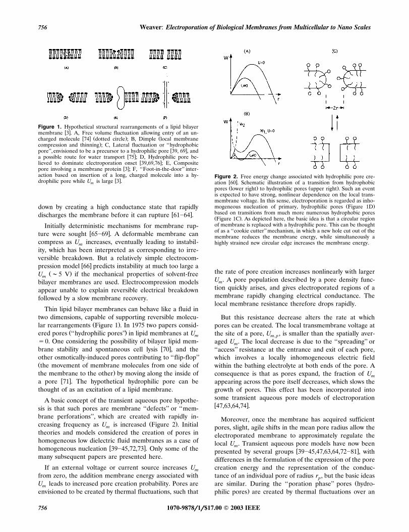
A basic concept of the transient aqueous pore hypothe-sis is that such pores are membrane ‘‘defects’’ or ‘‘mem-brane perforations’’, which are created with rapidly in-
Ž .creasing frequency as U is increased Figure 2 . Initialm
theories and models considered the creation of pores inhomogeneous low dielectric fluid membranes as a case of
w xhomogeneous nucleation 39�45,72,73 . Only some of themany subsequent papers are presented here.
If an external voltage or current source increases Um
from zero, the addition membrane energy associated withU leads to increased pore creation probability. Pores arem
envisioned to be created by thermal fluctuations, such that
Figure 2. Free energy change associated with hydrophilic pore cre-w xation 60 . Schematic illustration of a transition from hydrophobicŽ . Ž .pores lower right to hydrophilic pores upper right . Such an event
is expected to have strong, nonlinear dependence on the local trans-membrane voltage. In this sense, electroporation is regarded as inho-
Ž .mogeneous nucleation of primary, hydrophilic pores Figure 1Dbased on transitions from much more numerous hydrophobic poresŽ .Figure 1C . As depicted here, the basic idea is that a circular regionof membrane is replaced with a hydrophilic pore. This can be thoughtof as a ‘‘cookie cutter’’ mechanism, in which a new hole cut out of themembrane reduces the membrane energy, while simultaneously ahighly strained new circular edge increases the membrane energy.
the rate of pore creation increases nonlinearly with largerU . A pore population described by a pore density func-m
tion quickly arises, and gives electroporated regions of amembrane rapidly changing electrical conductance. Thelocal membrane resistance therefore drops rapidly.
But this resistance decrease alters the rate at whichpores can be created. The local transmembrane voltage atthe site of a pore, U , is smaller than the spatially aver-m, p
aged U . The local decrease is due to the ‘‘spreading’’ orm
‘‘access’’ resistance at the entrance and exit of each pore,which involves a locally inhomogeneous electric fieldwithin the bathing electrolyte at both ends of the pore. Aconsequence is that as pores expand, the fraction of Um
appearing across the pore itself decreases, which slows thegrowth of pores. This effect has been incorporated intosome transient aqueous pore models of electroporationw x47,63,64,74 .
Moreover, once the membrane has acquired sufficientpores, slight, agile shifts in the mean pore radius allow theelectroporated membrane to approximately regulate thelocal U . Transient aqueous pore models have now beenm
w xpresented by several groups 39�45,47,63,64,72�81 , withdifferences in the formulation of the expression of the porecreation energy and the representation of the conduc-tance of an individual pore of radius r , but the basic ideasp
Žare similar. During the ‘‘poration phase’’ pores hydro-.philic pores are created by thermal fluctuations over an
1070-9878rrrrr1rrrrr$17.00 � 2003 IEEE756

IEEE Transactions on Dielectrics and Electrical Insulation Vol. 10, No. 5; October 2003 757
Želectrically lowered barrier hydrophobic pore to hy-.drophilic pore transition , and a heterogeneous pore pop-
ulation develops, creating a nonlinear, hysteretic mem-brane conductance increase that interacts with externalconduction pathways.
Recovery of an electroporated membrane is much lesswell understood than the poration phase of electropora-tion. As the �U due to an applied E begins to slow andm e
then decrease, at some time the ionic conductance of thepore population begins to decrease. This can be regardedas the beginning of membrane ‘‘recovery’’ or ‘‘resealing’’.As the pore-expanding electrical interactions fade, partlythrough membrane discharge through the pores, and thelocal transmembrane voltage, U , begins to decrease,m,pore
pore expansion slows. According to the transient aqueouspore hypothesis, planar membrane recovery has two con-
Ž .tributions: 1 Diffusion in pore radius space, yielding aŽ .fluctuating return to a small primary pores on average,
Ž .and 2 Sudden, random transitions of these small poresŽ .to a pre-pore no pore state at this membrane location.
Significantly, measurements of fluctuations in electropo-w xrated membranes 48,82 are consistent with transient
w xaqueous pores 39,69 .
Recovery appears to depend significantly on tempera-ture. This is expected, because during most of the recov-ery phase U is small, and the originally strong electricalm
interactions are replaced by weaker interactions that de-pend on membrane and medium composition, with ther-mal fluctuations now of great importance. In a pure bi-layer lipid membrane a thermally activated process is hy-pothesized to govern the disappearance of hydrophilicpores.
6 CELL MEMBRANESCell membranes are much more complicated than arti-
ficial planar bilayer membranes, with respect to geometry,composition and the presence of active processes. A widevariety of cell studies have been carried out, almost allmotivated by introducing molecules or ions into the cell,with motivations that range from basic research to clinicalinterventions.
6.1 LOADING SUSPENDED CELLSThe great majority of applications involve placement of
Žan aqueous cell suspension cell-cell separation �cell. Ž .size into a small chamber a cuvette with two parallel
plane electrodes. DNA or other molecules to be loadedare provided in the suspension, outside the cells, and oneor more pulses applied. Typically one fraction of the cellpopulation takes up the desired molecule and survives, butmany cells may either die or not take up significantamounts of the molecule. In transfection protocols geneticmaterial is loaded, and if only a few loaded cells surviveand express the genetic material occur, the protocol is
usually viewed as a success, because such transfected cellsŽ .can be grown up amplified , and the overall result is a
clone that can be further expanded and fruitfully used inbiological research.
Loading of cells in suspension with microsecond to mil-Žlisecond pulses involves large electric fields field pulse in
the aqueous electrolyte of order E �105 V my1 fore.mammalian cells with characteristic size L �10 �m .cell
Bacteria are about an order of magnitude smaller, andrequire about a ten fold larger E .e
Quantitative determinations of the number of moleculestaken up are needed for comparison to models and theo-ries, and to help guide applications. Uptake that reaches
Žequilibrium with the external medium ‘‘equilibrium up-.take’’ occurs if the extent and duration of the increased
membrane permeability allows the intracellular chemicalconcentration to equilibrate with the external supply con-centration. Equilibrium uptake is readily understood, pro-vided the membrane openings are large enough to accom-modate the molecule, and the extra-cellular environmentis sufficiently large that it can be regarded as a reservoir.The effective partition coefficient for the intra- to extra-cellular environment can be included in determining
w xwhether equilibrium is achieved 83 .
Ž .Non-equilibrium uptake far fewer molecules taken upw x84 is more difficult to understand, particularly in thecases that the amount of uptake reaches a far-from-equi-librium plateau as the field pulse magnitude is increased.Plateau behavior has been observed for charged molecule
w xuptake 83,85,86 , which is qualitatively consistent with theapproximate plateau in U predicted by the transientm
w xaqueous pore mode 64 . Combined measurements of cellviability and molecular uptake can involve the stringenttest of clonal growth in which the fluorescence of micro-
w xcolonies is determined 87 .
6.2 LOADING INDIVIDUAL CELLSLoading of cells individually should provide more in-
sight and control. Theories and models could be bettertested against experimental results, and subsequent appli-cations improved. A flow-through microfabricated system
w xhas been demonstrated 88�90 in which individual cellsare transported by flow to a microhole capture site, thecell is temporarily held against the microhole edge, the
Žcell electrically measured and pulsed simultaneously with.optical measurement also possible , and then the cell is
released, with this cycle repeated serially in the flow-through device.
Alternatively, ultramicroelectrodes can be positionednear cells, and then pulsed with low voltages to preferen-tially electroporate a nearby region of cell membranesw x91,92 . The applied field is very heterogeneous, owing tothe use of two small electrode tips, but the cell can be
1070-9878rrrrr1rrrrr$17.00 � 2003 IEEE 757

Weaver: Electroporation of Biological Membranes from Multicellular to Nano Scales758
simultaneously patch clamped. Both single cell electropo-ration methods allow electrical behavior and fluorescentmolecule uptake to be measured.
6.3 MOLECULAR AND IONICTRANSPORT MECHANISMS
Movement of molecules and ions through electropo-rated membranes is believed to occur mainly by elec-trophoresis and diffusion. Minimum size hydrophilic poresŽ .Figure 1D have a large resistance due to the large Born
Ž .energy cost of ion and charged molecule insertion intow xthe pore and also due to hindered motion 47,63,64,69,80 .
Pores therefore tend to expand to create the small resis-tance needed to quickly discharge the membrane capaci-tance, an essential aspect of reversible electroporation.Creation of pores larger than r f1 nm also benefitsr,min
molecular transport. Neutral and charged molecules needto fit into a pore, or to expand the pore, to be transportedthrough the electroporated membrane. A broad, quantita-tive description of molecular transport has not yet beenachieved.
A contribution by electro-osmosis has been suggestedw x93 but the pore length along which coupling to flow can
Ž .occur is small �5 nm and electro-osmosis has not beenemphasized subsequently. Active uptake such as endocy-tosis may occur, but as a secondary transport processes
w xafter cells have been stimulated 94�96 .
6.4 ROLE OF RESTING POTENTIAL INUPTAKE
Ž .The resting transmembrane voltage ‘‘resting potential’’ ,U , is a candidate for contributing to observed asym-m,rest
metric electroporation responses in individual cellsw x97�101 . Another possible contribution has received lessattention: during the relatively long time of postpulsemembrane recovery, even a small U may generate am,rest
significant electrophoretic contribution to the transport ofw xions and charged molecules 102 .
For example, during reversible electroporation a volt-age-regulation effect due a variable voltage divider is pre-
w xdicted by the transient aqueous pore model 64 , with Um
regulated to �0.2 V. Qualitatively, a small U during themŽrecovery phase of duration � the characteristic lifetimemet
.of metastable pores can generate the same elec-trophoretic transport with a much smaller number of poresthan is present during the pulse. If the intra- and extracel-lular ion concentrations are not quickly erased, then themetabolically driven ion pump activity should not immedi-ately cease, and in spite of the electrical load of the rela-tively low resistance metastable pores, U will not be zerom
after the pulse.
An order of magnitude estimate is
U � tm ,ep expN fN 1Ž .p ,met p ,ep U ��m ,met met
where N is the characteristic number of metastablep,met
pores during the membrane recovery phase, and N isp,ep
the corresponding characteristic number of pores duringthe pulse. The transmembrane voltages are U f0.2 Vm,ep
Ž .and U f0.001 V an illustrative case . The pulse du-m,met
ration defines the approximate exposure time, typically oforder t f1 ms, and the metastable pore lifetime is typi-exp
cally � f1 to 10 s. Using the shorter lifetime and themet
assumed 1 mV, the postpulse pore fraction needs to beonly N rN f0.2. A 100 �s pulse would need onlyp,met p,ep
2% metastable pores. This order of magnitude estimatesuggests that postpulse endogenous electrophoresis byboth pre-existing ion concentration differences and resid-ual ion pump activity through a metastable pore popula-
w xtion should be further considered 102 .
6.5 FOOT-IN-THE-DOOR HYPOTHESISEnhanced uptake of small ions and molecules over a
Žprolonged time may occur if a long macromolecule e.g..unbranched dextrans, other polymers, DNA temporarily
w xreside with a fraction of their length inside a pore 3 . This‘‘foot-in-the-door’’ hypothesis assumes that the longmolecule interacts repulsively with the pore walls at short
Ždistances, tending therefore to hold the pore open inhibit.resealing until the long molecule leaves the pore. This is
relevant to diffusive and electrophoretic post pulse trans-port.
Existence of this effect is qualitatively consistent withthe altered transport of fluorescent-labeled dextrans into
w xcells 103 and of two small fluorescent molecules acrossw xhuman stratum corneum 104 , which is a barrier com-
prised of multilamellar lipid bilayers that separate com-Ž .partments of water and keratin corneocytes . A model for
w xpassive entry of DNA has been proposed 105 , and mightalso be examined to determine whether enhanced smallion and molecule transport should result.
6.6 MOLECULAR ELECTRO-INSERTIONINTO MEMBRANES
The surprising and striking demonstration of stable in-sertion of macromolecules into cell membranes may berelated to the foot-in-the-door hypothesis, and provides aconvincing demonstration that electroporation involvesmore than a permeability increase. An illustrative hypoth-esis is that an absorbed molecule is near the site of aforming pore, a combination of local electric fields andfluctuations cause one end of the molecule to enter thepore while the other end is bound, and the pore eventu-ally reseals around the hydrophobic portion of themolecule, resulting in electro-insertion. Recombinant CD4
1070-9878rrrrr1rrrrr$17.00 � 2003 IEEE758

IEEE Transactions on Dielectrics and Electrical Insulation Vol. 10, No. 5; October 2003 759
molecules can be stably inserted into red blood cell mem-branes, most with the desired inside-out orientationw x106�108 . Electro-insertion has been demonstrated by
w xothers using glycophorin 109 .
Electro-insertion should also be related to the possibil-ity of field-altered macromolecule protrusion, in the sensethat a macromolecule with a hydrophobic central region
Ž .of length � d y2 d and hydrophilic ends may re-m head
side stably within the membrane. Here d is the size ofhead
a phospholipid headgroup. If the hydrophilic ends arecharged, with different magnitude or distribution of chargeon each end, then a field-induced �U should cause am
w xshift in the molecule’s equilibrium position 110 . This mayprovide a mechanism for changing the protrusion of suchmolecules from the membrane, which in turn may alter
w xthe accessibility of binding sites 111 . Such conforma-tional changes that involve relative movement of themembrane and a membrane molecule deserve furtherconsideration.
6.7 CELL INACTIVATION AND KILLINGNon-thermal damage to cells can be used to inhibit or
w xeliminate undesired cells 35�37,112�117 and also plays aw xmajor role in electrical injury 10 . Two possibilities are
Ž .apparent: 1 some regions of a cell membrane behave asa planar membrane, and if the membrane tension is sig-nificant, rupture of the region’s membrane may occur, cre-ating a long lasting opening that leads to lethal biochemi-
Žcal imbalance similar in action to a pore-forming antibi-. Ž .otic , and 2 reversible membrane electroporation occurs,
but nevertheless creates a biochemical imbalance fromwhich the cell does not recover.
Electroporative delivery of a few hundred bleomycinmolecules from nanomolar concentrations outside a cell is
w xwell established to cause cell death 118�120 , and is animpressive example of non-thermal killing cells by chemi-cal entry from the extracellular medium. The non-thermalpulsing and extracellular medium conditions needed to killor inhibit cells by loss of biochemicals probably relies onsimultaneous depletion of several crucial ions andmolecules, and appears less well understood.
6.8 CELL MEMBRANE RECOVERYRecovery of cell membranes appears to be more com-
plicated than in artificial planar bilayer membranes, wherethermally activated hydrophilic pore destruction is ex-
w xpected 44,63,64,74�81,121 . Cell membrane tension is of-ten significantly smaller, but can also vary due to osmotic
Žchanges and external mechanical forces. If large e.g. �1. w x�m secondary pores arise 2,122 , then purely physical
recovery may occur in suspended cells or large vesiclesw x123,124 .
However, active processes may be important in livingcells. The idea that the cell membrane is a fixed barrier tomost lipid bilayer-impermeant ions and molecules has
w xbeen convincingly challenged 26�28 , particularly for cellsin ®i®o where motion and deformation are normal for mosttissues. Specifically, cell membrane ‘‘wounding’’ has beenshown to transiently compromise the membrane barrierfunction in many cells of tissues that experience move-ment. Further, a very effective and active membrane re-covery process is involved, preventing frequent membraneopenings from killing cells. This suggests that such activeprocesses may also be involved in some aspects of recov-ery or resealing of living cells that have been electropo-rated.
However, a basic feature of electroporation is the rapidŽcreation of many membrane defects of order 2 nm diam-
.eter , whereas cell wounding appears to involve muchŽ 2.larger areas of cell membrane e.g. �1 �m . It is there-
fore not yet clear how the recovery of cells from ‘‘wound-ing’’ is related to cell membrane recovery after electropo-ration.
7 ISOLATED CELL APPLICATIONSThe most widely used electroporation protocols are car-
ried out in ®itro with suspended cells that are widely sepa-rated. The following are examples of the diversity andflexibility of using strong electric field pulses to manipu-late isolated cells.
7.1 EX VIVO CELL TREATMENTCells can be removed from the body, electroporated,
and then re-introduced into the body. An interesting ex-ample consists of removing blood, supplying a smallmolecule that alters oxygen binding to hemoglobin, elec-troporating blood in a flow-through system, and then re-
w xturning the treated blood to the body 125 .
Some inhibition and killing processes lend themselveseasily to a flow mode of operation, as to some medically-
w xmotivated cell manipulations 88�90,114,125,126 . A fun-damental attribute of flowing a cell preparation throughan electroporation system is the straightforward scale-upthat is achieved by using progressively longer treatmenttimes.
7.2 CELL STIMULATIONA special case of cell loading involves the introduction
of externally supplied ions or molecules such that electro-poration creates a response similar to that of other agents.
ŽFor example, introduction of calcium ions ubiquitous in.most natural media creates ‘‘calcium puffs’’ within a
treated cell, such that the cell response mimics hormonalw xexposure 127 .
1070-9878rrrrr1rrrrr$17.00 � 2003 IEEE 759

Weaver: Electroporation of Biological Membranes from Multicellular to Nano Scales760
7.3 CELL INHIBITION AND KILLINGThe use of electrical pulses to temporarily halt, kill or
remove undesired cells is extremely attractive. Not sur-prisingly, since the early reports of non-thermal microbialkilling effects there has been considerable effort to under-stand and apply the use of strong electric fields to treatsystems that are considered to have contaminant cells. Al-though not fully established, the underlying concepts ap-pear to be very similar qualitatively to some of the con-cepts underlying electrical shock injury to humans. Specif-ically, although Joule heating generally leads to denatura-tion and inactivation, electroporation creates membrane
Ž .barrier compromise, undesired from the cell’s viewpointŽand transport in both directions loss of important bio-
.chemicals; entry of deleterious biochemicals . Heating andelectroporation-induced biochemical imbalances can bothbe used to combat undesirable cells, whether mammaliancancer cells or microbial pathogens.
Early work in the late 1960s set the stage for attackingw xmicroorganisms 35�37 . Since then many other investiga-
tors have looked at the use of electroporation to inhibitŽ . Ž .stun or kill purge undesired cells from aqueous envi-
w xronments 8,112�117,128 . Inhibition and killing can bothbe beneficial, but the general nature of the effect on the
Ž .cell or small multicellular pest is quite different. Inhibi-tion means arresting activity, not necessarily permanently,whereas killing is final. Stunning of pests can be ap-proached by using relatively short pulses that minimizethe energy demands of an overall treatment processw x25,112,115 .
7.4 ANTIBODY DELIVERY INTO CELLSThe general nature of electroporation can be combined
with the high specificity of antibodies to block specificmolecules within living cells. This was first demonstrated
w xin yeast 129 , and has been extended to other types ofw x y1cells 130,131 . The large size �150,000 g mol of anti-
body molecules and the ability to target specific intracellu-lar epitopes makes this method particularly impressive.
8 EXTRACELLULAR ENVIRONMENTBoth the composition and volume of the extracellular
medium should be important. Electroporative cell loadingis based on the premise that molecules introduced intothe extracellular medium can be delivered into cells. Buttransport can occur in both directions across a membrane,or on opposite sides of a cell. Clearly the chemical compo-sition of the extracellular medium is important. Loss ofcrucial intracellular biochemicals may also occur. If poresremain open long after the pulse ceases, and the extracel-lular volume is small compared to the intracelluar volume,then the net loss may be small. Some outward transportedmolecules may be transported back into the cell, or evenfrom one cell to a neighboring cell.
Figure 3. Illustration of the large difference in typical in ®itro andw xin ®i®o extracellular volumes 7,134 . Left: Widely separated sus-Ž .pended cells, typical of many in ®itro conditions. Right: Closely
spaced cells, typical of solid tissue in ®i®o.
The relative extracellular environment should thereforew xbe considered 7,132 . As noted with respect to non-ther-
mal cell inactivation and killing, while membrane recoveryis incomplete a cell may accumulate chemical stressthrough loss of ions and molecules into the extracellularfluid. This leads to the simple hypothesis that the ratio ofintra- to extracellular volume,
VextracellularR s 2Ž .volume Vintracellular
should be important. In ®itro electroporation conditionsusually involve R �1. For example, a suspensionvolume
of mammalian cells at 105 cells cmy3 has R �104volume
Ž y9 3.V �10 cm . In contrast, the extracellular volume1 cellŽof many solid tissues R �1, typically �0.1 Figurevolume
.3 .
This is a factor of 105 difference. It suggests that for thesame transmembrane voltage changes, in ®i®o tissue elec-troporation may lead to significantly less chemical stressand greater survival rates than in ®itro conditions.
9 MULTICELLULAR SYSTEMS ANDTISSUE
Tissue electroporation in ®i®o was first described in anw xearly demonstration of electrochemotherapy 133 . Elec-
troporation of a viable tissue model barrier system wasŽthen observed in ®itro, using viable frog skin experimen-
tally similar to planar bilayer membranes; a thin sheet of. w xtissue is used 134 that allowed electrodes to be placed
on both sides of the skin, and electrical behavior to bemeasured. Tissue electroporation can occur as an inciden-
w xtal consequence of cardiac defibrillation 135,136 , whichinvolves large tissue electric fields. However, most re-search of tissue electroporation has involved bulk solidtissue for local delivery of cancer drugs or for gene ther-apy, and is presently very active.
9.1 LOCAL DRUG DELIVERY TOTUMORS
Enhanced treatment of solid cancer tumors led to thefirst observation of tissue electroporation, which involved
1070-9878rrrrr1rrrrr$17.00 � 2003 IEEE760

IEEE Transactions on Dielectrics and Electrical Insulation Vol. 10, No. 5; October 2003 761
a ‘‘solid tissue’’, a multicellular system with many cells inclose proximity, with introduction of the anticancer drug
w xbleomycin into electroporated cells 133 . This was fol-Žlowed by a vigorous investigation of ECT electrochem-
.otherapy , leading to clinical trials and continuing vigor-ous research in treating electrically-accessible tumorsw x6,137,138 .
9.2 GENE THERAPYGene therapy by physical methods is of increasing in-
terest, because the use of viral vectors is viewed as posingrisk of immunological adverse reactions. Physical inter-vention based on tissue electroporation to introduce ge-netic material has therefore generated considerable inter-
w xest, and is described in detail elsewhere 6,120,139,140 .
9.3 SKIN ELECTROPORATIONHuman skin presents a highly effective barrier against
desiccation, mechanical injury, entry of infectious mi-croorganisms and toxic chemicals, and attempts at trans-
Ž .dermal drug delivery. The stratum corneum SC is theŽ .dead, outermost layer �20 pm-thick , and the site of
w xmost of the barrier function 141 . The SC barrier is usu-ally represented by a brick wall model, with keratin-filled
Ž .corneocytes ‘‘bricks’’ surrounded by multilamellar lipidŽ . w xbilayers ‘‘mortar’’ 141,142 .
Skin electroporation is based on the idea that �100lipid bilayer membranes must be penetrated by a moleculewhich moves through the SC to reach the viable epider-mis. If U �0.5 to 1 V electroporates single bilayers form
short pulses, then pulses which cause the transdermalvoltage to reach U �50 to 100 V should cause electro-skin
poration of the multilamellar bilayers within the SCw x143,144 . For U �5 V iontophoresis through pre-exist-skin
ing pathways occurs, and for 5�U �50 V electropora-skin
tion of the cell linings of sweat ducts and hair follicles setsw xin 145 . However, most of the skin is covered by the dead
SC, which should electroporate for U �50 V. Experi-skin
ments confirm this expectation. In ®i®o protocols usingtransdermal pulsing show that skin irritation and damage
w xis minimal 146 , consistent with the estimated tempera-w xture rise 147,148 .
A microporous keratin matrix within corneocytes blocksw xlarge molecule transport 149 . To overcome this block-
age, skin electroporation has been combined with benignw xmolecules which disrupt keratin 150 . Electroporation is
spontaneously concentrated at localized transport regionsŽ .LTRs located almost randomly over the skinw x149,151,152 . However, an electrically resistant mask withan array of microholes can restrict electroporation to pre-
w xdetermined sites 153 . The result is in situ creation ofw xmicroconduits 154,155 , which are SC-spanning openings
that can transport essentially any size molecule.
Figure 4. Didactic multicellular model of a portion of a tissue sub-w xjected to an electroporating pulse 165 . Here equipotentials areŽ .shown just as electroporation begins left and has reached its maxi-
mum extent for a 5 �s square 1100 Vrcm pulse with a realistic riseŽ .time right . This didactic model of a multicellular system has fifty
Žirregularly shaped cells with passive membrane interactions fixed re-. Žsistance and capacitance and active membrane interactions nonlin-
ear current source representing all the ion channels at each mem-brane site, and a hysteretic U -sensitive local membrane resistancem
.transition that approximates electroporation . Both weak and strongelectric field responses can therefore be represented in a single sys-tem model.
9.4 ELECTRICAL INJURYFor many years electrical shock injury was attributed to
Joule heating. A major advance occurred when it wasshown that electroporation can play an important role.Electroporation is particularly important for the larger,more ‘‘electrically susceptible’’ skeletal muscle and nervecells, and is involved in non-thermal, delayed damagew x156�158 .
A related important advance is the demonstration thatsurfactant molecules can be introduced in small amountsto seal electroporated cell membranes, providing the basis
w xfor a therapy after electrical shock 10,159 . The scientificinsight into injury and the new approach to therapy com-prise an important contribution to understanding electro-poration.
9.5 MULTICELLULAR THEORY ANDMODELS
Theoretical models and computer simulations have beenused to predict the tissue regions which should have elec-troporated cells, and therefore take up drug, based on acriterion of an average electric field magnitude, in combi-
Žnation with pulse temporal features pulse shape and du-.ration, number of pulses in a pulse sequence . Finite ele-
ment models for solid tissue have been used in which theŽ .computational volume elements voxels are much larger
w xthan the cells 160 . This is consistent with the widespreaduse of simulations to predict ‘‘dosimetry’’of low amplitudeelectric and magnetic fields on the spatial scale of order
w x Ž .mm 161 each mm-scale voxel has �106 cells .
A theoretical analysis of electrical impedance tomogra-Ž .phy EIT also treats tissue as a homogeneous medium,
1070-9878rrrrr1rrrrr$17.00 � 2003 IEEE 761

Weaver: Electroporation of Biological Membranes from Multicellular to Nano Scales762
but used a field-dependent switching transition that shuntsthe tissue voxel capacitance associated with cell mem-
w xbranes 162 . This feasibility study concludes that EITshould be able to detect the decreased tissue impedancecaused by electroporation.
A transport lattice modeling method has been de-scribed which allows multicellular models to be createdwith nonlinear interactions, including simple representa-
Ž .tions of local membrane electroporation Figure 4 . Thismethod for creating and solving models can be used toapproximate cells, multicellular systems and subcellularsystems, using local models that represent passive and ac-tive membrane processes. A didactic multicellular modelpredicts that electroporation should exhibit significantheterogeneity, but with invaginations and cell layers con-
w xtaining tight junctions as preferred sites 163 .
10 SUBCELLULAR SYSTEMSThe possibility of preferential interaction with subcellu-
lar, membrane-encased structures is based on the idea thatultrashort pulses may cause significant �U of subcellularm
structures while creating relatively small changes in thew xcell’s outer, plasma membrane 8,164�166 . Typical char-
acteristic times for charging mammalian cell plasma mem-branes are of order � �10y6 s. Most electroporationCHG
studies use pulse durations t �� , but have usuallypulse CHG
not analyzed the pulse rise time in accessing the response.The possibility of subcellular manipulations is based on
Ž y6 .shorter duration pulses ‘‘ultrashort pulses’’; t �10 s ,expw xso that the rise time is intrinsically fast 78,79,164,166 . An
intriguing hypothesis is that electroporation of subcellularmembranes may occur. If so, mitochondrial membraneelectroporation is a candidate mechanism for the apopto-sis caused by very large magnitude ultrashort pulsesw x165,166 .
Indeed, isolated mitochondria experiments provide evi-dence for an ability to electroporate mitochondrial mem-
w xbranes 167�170 . A feature of both Gram negative bacte-ria and mitochondria is their double membrane. The outermembrane is relatively leaky, a poor permeability barrierwith low electrical resistance, and also a low resting trans-membrane voltage. The inner membrane is the main bar-rier and has a large membrane resting transmembrane
w xvoltage, U f150 to 200 mV 171�173 . These valuesm,rest,in
are significantly larger than those of typical mammaliancell plasma membranes, viz. U f 60 to 90 mVm,restw x174,175 . If there are sufficiently large lipid regions withinmitochondrial membranes to allow hydrophilic pore cre-ation, then the larger U should require a smallerm,rest
field-induced �U to electroporate the inner mitochon-m
drial membrane.
The inner mitochondrial membrane is highly invagi-nated, with an area five to fifteen times that of the outer
w xmembrane 174 , so a proportionately larger capacitanceand longer charging time is expected. The inner mem-
w xbrane is also �75% protein 174 , and includes the mito-Ž .chondrial permeability transition pore complex MPTP
w x176 .
With this in mind, another possibility exists. It is knownthat biochemically-induced apoptosis involves the MPTPw x176 , which opens to allow nonspecific transport of smallmolecules up to 1,500 g moly1. Moreover, the mitochon-drial membrane voltage-dependent anion channel is im-
w xplicated in apoptosis 177 .
The alternative hypothesis is that ultrashort, large pulsespreferentially change the transmembrane voltage at mito-chondrial membrane sites, opening the MPTP. The innermembrane MPTP has been reported by several groups to
w xbe voltage sensitive 171,178�180 . The outer membrane isw xalso reported to have voltage sensitive channels 180 . The
response of cells to large, ultrashort pulses may thereforebe due to voltage gating of the MPTP and other channels,with both the inner and outer mitochondrial membranesinvolved. Patch clamp measurements might provide a test,using transmembrane voltage changes that are the sameas those created in situ at mitochondrial membranes bylarge, ultrashort pulses. The response of two membrane
Ž .systems could be compared: 1 reconstituted membranesŽ .with the MPTP and other mitochondrial channels, and 2
pure lipid bilayers. It is not clear whether a double mem-brane system would be required, because one membraneŽ .probably the inner may dominate the response.
For completeness it should be noted that other experi-ments with very pulses have also observed apoptosis, butdid not attribute this to interactions with subcellular
w xstructure membranes 182 .
11 SPATIALLY CONSTRAINEDMEMBRANES
The membrane conformational changes associated withelectroporation should be increasingly constrained as thesize of a lipid membrane region is decreased. Stochasticprocesses may be suppressed and deterministic deforma-tions may become more important. Membranes with highprotein content, and membrane interactions with the cy-toskeleton, may both result in restricted membrane re-gions that are capable of behaving like small planar bi-layer membranes. Consider a circular patch of lipid mem-brane. The minimum lipid patch radius capable of accom-modating a minimum size hydrophilic pore is
r f r qd r2f3.5 nm 3Ž .l i p ,min p ,min m
This suggests that nanoscale constraints should arise asthe spatial extent of a lipid region approaches the mem-
Ž .brane thicknesses d . Such membranes will have alteredm
electroporation processes, likely requiring a combination
1070-9878rrrrr1rrrrr$17.00 � 2003 IEEE762

IEEE Transactions on Dielectrics and Electrical Insulation Vol. 10, No. 5; October 2003 763
of larger U and longer times. This expectation has sev-m
eral origins.
Ž .First, a minimum membrane area volume is neededfor the molecular rearrangements that lead to formation
Ž Ž ..of a minimum size hydrophilic pore equation 3 . Sec-ond, the transition membrane conformational state thatexists briefly between the hydrophobic pore and hy-
Ž .drophilic pore Figures 1 and 2 may exceed the size esti-Ž .mated by equation 3 . Third, the original transient aque-
ous pore theory is based on thermal fluctuations and tran-sitions to local membrane conformations that have theirformation energy decreased as U is raised. If the mem-m
Ž .brane areas volumes are too small for traditional hy-drophilic pores, then deterministic deformations may stillallow creation of aqueous pathways through the mem-brane, but may require more energy and a larger U .m
Fourth, the Smoluchowski equation used in many tran-sient aqueous pore descriptions may be invalid for veryshort times. Based on measurements of small lipid vesiclesŽ . w x�0.3 �m diameter 183 , the characteristic time of poreformation is of order 10y6 s. For submicrosecond pulsesthe characteristic time for stochastic multimolecule rear-rangement to form a hydrophilic pore may therefore belonger than the time that large U exists, and determinis-m
tic processes may become important.
Voltage-gated specific ion channels, relatively nonspe-cific but voltage sensitive pore-forming proteins and tran-sient hydrophilic pores are all members of a general classof non-thermal mechanism that increase membrane bar-rier permeability through electrically induced conforma-tional changes. More than one interaction mechanism maytherefore be involved in the response of protein-richmembranes of Gram negative bacteria and mitochondriato electrical pulses of various magnitudes and durations.
Supported or tethered bilayer membranes may providea model system for investigation of electroporation in spa-tially constrained membranes. Significant spatial con-strains should arise because the membrane is anchored to
w xan underlying solid surface by chemical linkages 184 .These attachment sites should provide explicit conforma-tional constraints. If the average distance between sites is
Ž .less than r of equation 3 , hydrophilic pore forma-l i p,minŽtion should be inhibited. Supported bilayer membrane s-
.BLM can contain a significant number of pre-existingperforating defects that shunt the high resistance of the
w xlipid regions 185,186 . However, just as open membranechannels do not prevent cell membrane electroporation, itmay be possible to cause electroporation of tethered bi-layer membranes. It may also be possible to seal the pre-existing defects using surfactants, as demonstrated for
w xelectrically damaged cell membranes 159 . Electrostric-tion of supported lipid bilayers should be relevant to un-derstanding some aspects of electroconformational
w xchanges 187 . Finally, if the tethering molecules can beŽspatially constrained e.g. located only on a small scale
.Cartesian grid , then fluid lipid patches of defined spatialextent might be obtained, and used to experimentally in-vestigate spatially constrained electroporation.
12 SUMMARYIOLOGICAL transport barriers are often based onBone or more lipid bilayer membranes. Electric field
pulses that drive the transmembrane voltage to 0.2 to 1 VŽare hypothesized to create primary aqueous pathways hy-
.drophilic pores with minimum size of �1 nm. A dynamicinteraction involving both external membrane chargingand internal membrane discharging governs a rapidlyevolving pore population, which in turn controls electricalbehavior and molecular transport. Secondary phenomenacan arise from chemical imbalances and from cell stimula-tion.
In membranes with large areas of fluid lipid electropo-ration is believed to involve transient hydrophilic poresthat are created in the fluid lipid regions of a membrane,due to both electric field interactions and thermal fluctua-
Žtions. In spatially constrained membranes e.g. mitochon-drial membranes with �75% protein; supported or teth-
.ered membranes the stochastic model of hydrophilic poresmay not be applicable, and other membrane rearrange-ments may be involved in creating aqueous pathwaysthrough the membrane. Phenomena caused by ultrashort,very large field pulses may involve such electroporationmechanisms, or may involve conformational triggering ofmembrane macromolecular structures such as the mito-chondrial permeability transition pore complex.
ACKNOWLEDGMENTSThis work is supported by NIH grants RO1 AR 42921,
RO1-GM63857 and an AFOSRrDOD MURI grant onSubcellular Responses to Narrowband and Wideband Ra-dio Frequency Radiation, administered through Old Do-minion University.
REFERENCESw x1 E. Neumann, A. E. Sowers, and C. A. Jordan, editors, Electro-
poration and Electrofusion in Cell Biology, Plenum Press, NewYork, 1989.
w x2 D. C. Chang, B. M. Chassy, J. A. Saunders, and A. E. Sowers,editors, Guide to Electroporation and Electrofusion, AcademicPress, New York, 1992.
w x3 J. C. Weaver, ‘‘Electroporation: A General Phenomenon forManipulating Cells and Tissue’’, J. Cellular Biochem., Vol. 51,pp. 426�435, 1993.
w x4 J. C. Weaver and Y. A. Chizmadzhev, ‘‘Electroporation’’. In C.Polk and E. Postow, editors, Handbook of Biological Effects ofElectromagnetic Fields, CRC Press, Boca Raton, 2nd edition,1996, pp. 247�274.
w x5 U. Zimmermann, editor, The Effects of High Intensity ElectricField Pulses on Eukaryotic Cell Membranes: Fundamentals andApplications, CRC Press, Boca Raton, 1996.
w x6 M. J. Jaroszeski, R. Gilbert, and R. Heller, editors, ElectricallyMediated Deli®ery of Molecules to Cells: Electrochemotherapy,Electrogenetherapy and Transdermal Deli®ery by Electroporation,Humana Press, Totowa, 2000.
1070-9878rrrrr1rrrrr$17.00 � 2003 IEEE 763

Weaver: Electroporation of Biological Membranes from Multicellular to Nano Scales764
w x7 J. C. Weaver, ‘‘Electroporation of Cells and Tissues’’, IEEETrans. Plasma Sci., Vol. 28, pp. 24�33, 2000.
w x8 K. H. Schoenbach, S. Katsuki, R. H. Stark, E. S. Buescher, andS. J. Beebe, ‘‘Bioelectrics�New Applications for Pulsed PowerTechnology,’’ IEEE Trans. Plasma Sci., Vol. 30, pp. 293�300,2002.
w x9 R. C. Lee and M. S. Kolodney, ‘‘Electrical Injury Mechanisms:Dynamics of the Thermal Response’’,Plast. Reconstr. Surg., Vol.80, pp. 663�671, 1987.
w x10 R. C. Lee, D. Zhang, and J. Hannig, ‘‘Biophysical Injury Mecha-nisms in Electrical Shock Trauma’’, Ann. Rev. Biomedical Eng.,Vol. 2, pp. 477�509, 2000.
w x11 W. Chen and R. C. Lee, ‘‘Altered Ion Channel Conductanceand Ionic Selectivity Induced by Large Imposed Membrane Po-tential Pulses’’, Biophys., J., Vol. 67, pp. 603�612, 1994.
x12 W. Chen, Y. Han, Y. Chen, and D. Astumiam, ‘‘Electric Field-induced Functional Reductions in the Kq Channels Mainly Re-sulted from Supramembrane Potential-medicated Electrocon-formational Changes’’, Biophys. J., Vol. 75, pp. 196�206, 1998.
w x13 M. Blank and E. Findl, editors, Mechanistic Approaches to Inter-actions of Electromagnetic Fields with Li®ing Systems, PlenumPress, New York, 1987.
w x14 J. P. Reilly, Electrical Stimulation and Electropathology, Cam-bridge University Press, Cambridge, 1992.
w x15 W. Wiltschko and R. Wiltschko, Magnetic Orientation in Ani-mals, Springer Verlag, Berlin, 1995.
w x16 C. Polk and E. Postow, editors, CRC Handbook of Biological Ef-fects of Electromagnetic Fields, Second Edition, CRC Press, BocaRaton, 1996.
w x17 F. Bersani, editor, Electricity and Magnetism in Biology andMedicine, Plenum, New York, 1999.
w x18 J. C. Weaver, ‘‘Understanding Conditions for which BiologicalEffects of Nonionizing Electromagnetic Fields can be Expected’’,Bioelectrochemistry, Vol. 56, pp. 207�209, 2002.
w x19 J. C. Weaver and R. D. Astumian, ‘‘The Response of Cells toVery Weak Electric Fields: The Thermal Noise Limit’’, Science,Vol. 247, pp. 459�462, 1990.
w x20 R. K. Adair, ‘‘Constraints on Biological Effects of Weak Ex-tremely-low-frequency Electromagnetic Fields’’, Phys. Rev. A,Vol. 43, pp. 1039�104, 1991.
w x21 R. D. Astumian, J. C. Weaver, and R. K. Adair, ‘‘Rectificationand Signal Averaging of Weak Electric Fields by BiologicalCells’’, Proc. Nat. Acad. Sci., Vol. 92, pp. 3740�3743, 1995.
w x22 J. C. Weaver, T. E. Vaughan, R. K. Adair, and R. D. Astumian,‘‘Theoretical Limits on the Threshold for the Response of LongCells to Weak ELF Electric Fields due to Ionic and MolecularFlux Rectification’’, Biophys. J., Vol. 75, pp. 2251�2254, 1998.
w x23 J. C. Weaver, T. E. Vaughan, and G. T. Martin, ‘‘BiologicalEffects due to Weak Electric and Magnetic Fields: The Temper-ature Variation Threshold’’, Biophys. J., Vol. 76, pp. 3026�3030,1999.
w x24 J. C. Weaver, T. E. Vaughan, and R. D. Astumian, ‘‘Magneti-cally Sensitive Chemical Reactions Can Provide Biological Sens-ing of Small Field Differences’’, Nature, Vol. 405, pp. 707�709,2000.
w x25 K. R. Foster, ‘‘Thermal and Nonthermal Mechanisms of Inter-action of Radio-frequency Energy with Biological Systems’’,IEEE Trans. Plasma Sci., Vol. 28, pp. 15�23, 2000.
w x26 P. L. McNeil and S. Ito, ‘‘Molecular Traffic through PlasmaMembrane Disruptions of Cells, in ®i®o,’’J. Cell Sci., Vol. 96, pp.549�556, 1990.
w x27 P. L. McNeil, ‘‘Cell Wounding and Healing’’, Am. Sci., Vol. 79,pp. 222�235, 1991.
w x28 P. L. McNeil and M. Terasaki, ‘‘Coping with the Inevitable: HowCells Repair a Torn Surface Membrane’’, Nat. Cell Biol., Vol. 3,pp. 124�129, 2001.
w x29 E. Neumann and S. Kakorin, ‘‘Electrooptics of Membrane Elec-troporation and Vesicle Shape Deformation’’, Current OpinionColloid & Interface Science, Vol. 1, pp. 790�799, 1996.
w x30 E. Neumann, S. Kakorin, and K. Toensing, ‘‘Membrane Electro-poration and Electromechanical Deformation of Vesicles andCells’’, Faraday Discuss., Vol. 111, pp. 111�125, 1998.
w x31 V. L. Sukhorukov, H. Mussauer, and U. Zimmermann, ‘‘TheEffect of Electrical Deformation Forces on the Electroperme-abilization of Erythrocyte Membranes in Low- and High-con-ductivity Media’’, J. Membr. Biol., Vol. 163, pp. 235�245, 1998.
w x32 R. P. Joshi, Q. Hu, K. H. Schoenbach, and H. P. Hjalmarson,‘‘Theoretical Prediction of Electromechanical Deformation ofCells Subjected to High Voltages for Membrane Electropora-tion, Phys. Rev. E, Vol. 65, pp. 021913-1021913-10, 2002.
w x33 T. Y. Tsong, ‘‘Electroporation of Cell Membranes’’, BiophysicalJ., Vol. 60, pp. 297�306, 1991.
w x34 R. Stampfli, ‘‘Reversible Electrical Breakdown of the Excitable¨Membrane of a Ranvier Node’’, An. Acad. Brasil. Ciens., Vol.30, pp. 57�63, 1958.
w x35 A. J. H. Sale and A. Hamilton, ‘‘Effects of High Electric Fieldson Microorganisms: I. Killing of Bacteria and Yeasts’’, Biochem.Biophys. Acta, Vol. 148, pp. 781�788, 1967.
w x36 W. A. Hamilton and A. J. H. Sale, ‘‘Effects of High ElectricFields on Microorganisms: II. Killing of Bacteria and Yeasts’’,Biochim. Biophys. Acta, Vol. 148, pp. 7789�800, 1967.
w x37 A. J. H. Sale and W. A. Hamilton, ‘‘Effects of High ElectricFields on Microorganisms: III. Lysis of Erythrocytes and Proto-plasts’’, Biochim. Biophys. Acta, Vol. 163, pp. 37�43, 1968.
w x38 E. Neumann and K. Rosenheck, ‘‘Permeability Changes In-duced by Electric Impulses in Vesicular Membranes’’, J. Mem-brane Biol., Vol. 10, pp. 279�290, 1972.
w x39 I. G. Abidor, V. B. Arakelyan, L. V. Chernomordik, Yu. A.Chizmadzhev, V. F. Pastushenko, and M. R. Tarasevich, ‘‘Elec-tric Breakdown of Bilayer Membranes: I. The Main Experimen-tal Facts and Their Qualitative Discussion’’, Bioelectrochem.Bioenerget., Vol. 6, pp. 37�52, 1979.
w x40 V. F. Pastushenko, Yu. A. Chizmadzhev, and V. B. Arakelyan,‘‘Electric Breakdown of Bilayer Membranes: II. Calculation ofthe Membrane Lifetime in the Steady-state Diffusion Approxi-mation’’, Bioelectrochem. Bioenerget., Vol. 6, pp. 53�62, 1979.
w x41 Y. A. Chizmadzhev, V. B. Arakelyan, and V. F. Pastushenko,‘‘Electric Breakdown of Bilayer Membranes: III. Analysis ofPossible Mechanisms of Defect Origin’’, Bioelectrochem. Bioen-erget., Vol. 6, pp. 63�70, 1979.
w x42 V. F. Pastushenko, Y. A. Chizmadzhev, and V. B. Arakelyan,‘‘Electric Breakdown of Bilayer Membranes: IV. Considerationof the Kinetic Stage in the Case of the Single-defect Membrane’’,Bioelectrochem. Bioenerget., Vol. 6, pp. 71�79, 1979.
w x43 V. B. Arakelyan, Y. A. Chizmadzhev, and V. F. Pastushenko,‘‘Electric Breakdown of Bilayer Membranes: V. Considerationof the Kinetic Stage in the Case of the Membrane Containing anArbitrary Number of Defects’’,Bioelectrochem. Bioenerget., Vol.6, pp. 81�87, 1979.
w x44 V. F. Pastushenko, V. B. Arakelyan, and Y. A. Chizmadzhev,‘‘Electric Breakdown of Bilayer Membranes: VI. A StochasticTheory Taking into Account the Processes of Defect Formationand Death: Membrane Lifetime Distribution Function’’, Bioelec-trochem. Bioenerget., Vol. 6, pp. 89�95, 1979.
w x45 V. F. Pastushenko, V. B. Arakelyan, and Yu. A. Chizmadzhev,‘‘Electric Breakdown of Bilayer Membranes: VII. A StochasticTheory Taking into Account the Processes of Defect Formationand Death: Statistical Properties’’, Bioelectrochem. Bioenerget.,Vol. 6, pp. 97�104, 1979.
ww x46 L. V. Chernomordik, S. I. Sukharev, I. G. Abidor, and Y. A.Chizmadzhev, ‘‘The Study of the BLM Reversible ElectricalBreakdown Mechanism in the Presence of UO2q ’’, Bioelec-trochem. Bioenerget, Vol. 9, pp. 149�155, 1982.
w x47 V. F. Pastushenko and Y. A. Chizmadzhev, ‘‘Stabilization ofConducting Pores in BLM by Electric Current’’, Gen. Physiol.Biophys., Vol. 1, pp. 43�52, 1982.
w x48 K. C. Melikov, V. A. Frolov, A. Shcherbakov, A. V. Samsonov,and Y. A. Chizmadzhev, ‘‘Voltage-induced Nonconductive Pre-pores and Metastable Pores in Unmodified Planar Bilayer’’, Bio-phys. J., Vol. 80, pp. 1829�1836, 2001.
1070-9878rrrrr1rrrrr$17.00 � 2003 IEEE764

IEEE Transactions on Dielectrics and Electrical Insulation Vol. 10, No. 5; October 2003 765
w x49 U. Zimmermann, F. Riemann, and G. Pilwat, ‘‘Enzyme Loadingof Electrically Homogeneous Human Red Blood Cell GhostsPrepared by Dielectric Breakdown’’, Biochim. Biophys. Acta,Vol. 436, pp. 460�474, 1976.
w x50 K. Kinosita, Jr. and T. Y. Tsong, ‘‘Formation and Resealing ofPores of Controlled Sizes in Human Erythrocyte Membrane’’,Nature, Vol. 268, pp. 438�441, 1977.
w x51 K. Kinosita Jr. and T. Y. Tsong, ‘‘Survival of Sucrose-loadedErythrocytes in Circulation’’,Nature, Vol. 272, pp. 258�260, 1978.
w x52 J. Teissie and T. Y. Tsong, ‘‘Electric Field Induced TransientPores in Phospholipid Bilayer Vesicles’’, Biochemistry, Vol. 20,pp. 1548�1554, 1981.
w x53 U. Zimmermann, P. Scheurich, G. Pilwat, and R. Benz, ‘‘Cellswith Manipulated Functions: New Perspectives for Cell Biology,Medicine and Technology’’, Angew. Chem. Int. Ed. Eng., Vol.20, pp. 325�344, 1981.
w x54 E. H. Serpersu, K. Kinosita, Jr., and T. Y. Tsong, ‘‘Reversibleand Irreversible Modification of Erythrocyte Membrane Perme-ability by Electric Field’’, Biochem. Biophys. Acta, Vol. 812, pp.779�785, 1985.
w x55 D. Auer, G. Brandner, and W. Bodemer, ‘‘Dielectric Break-down of the Red Blood Cell Membrane and Uptake of SV 40DNA and Mammalian Cell RNA’’, Naturwiss., Vol. 63, p. 391,1976.
w x56 E. Neumann, M. Schaefer-Ridder, Y. Wang, and P. H. Hof-schneider, ‘‘Gene Transfer Into Mouse Lyoma Cells by Electro-poration in High Electric Fields’’, EMBO J., Vol. 1, pp. 841�845,1982.
w x57 U. Zimmermann and G. Kuppers, ‘‘Cell Fusion by Electromag-¨netic Waves and its Possible Relevance for Evolution’’, Natur-wissenschaften, Vol. 70, pp. 568�569, 1983.
w x58 S. Demaneche, F. Bertolla, F. Buret, R. Nalin, A. Sailland, P.´Auriol, T. M. Vogel, and P. Simonet, ‘‘Laboratory-scale Evi-dence for Lightning-mediated Gene Transfer in Soil’’, Appl.Environ. Microbiol., Vol. 67, pp. 3440�3444, 2001.
w x ( )59 H. T. Tien, Bilayer lipid membranes BLM : Theory and practice,Marcel Dekker, New York, 1974.
w x60 H. T. Tien, R. H Barish, L.-G. Gu, and A. L. Ottova, ‘‘Sup-ported Bilayer Lipid Membranes as Ion and Molecular Probes’’,Analyt. Sci., Vol. 14, pp. 3�18, 1998.
w x61 R. Benz, F. Beckers, and U. Zimmermann, ‘‘Reversible Electri-cal Breakdown of Lipid Bilayer Membranes: A Charge-pulseRelaxation Study’’, J. Membrane Bio., Vol. 48, pp. 181�204, 1979.
w x62 R. Benz and U. Zimmermann, ‘‘Relaxation Studies on CellMembranes and Lipid Bilayers in the High Electric Field Range’’,Bioelectrochem. and Bioenerg., Vol. 7, pp. 723�739, 1980.
w x63 A. Barnett and J. C. Weaver, ‘‘Electroporation: A Unified,Quantitative Theory of Reversible Electrical Breakdown andRupture’’, Bioelectrochem. and Bioenerg., Vol. 25, pp. 163�182,1991.
w x64 S. A. Freeman, M. A. Wang, and J. C. Weaver, ‘‘Theory ofElectroporation for a Planar Bilayer Membrane: Predictions ofthe Fractional Aqueous Area, Change in Capacitance andPore-pore Separation’’, Biophys. J., Vol. 67, pp. 42�56, 1994.
w x65 D. H. Michael, M. E. O’Neill, and J. C. Zuecher, ‘‘The Break-down of Electrical Insulation in a Planar Layer of InsulatingFluid by Electrocapillary Instability, J. Fluid Mech., Vol. 47, Vol.609�623, 1972.
w x66 J. M. Crowley, ‘‘Electrical Breakdown of Bimolecular LipidMembranes as an Electromechanical Instability’’, Biophysical J.,Vol. 13, pp. 711�724, 1973.
w x67 D. S. Dimitrov, ‘‘Electric Field-induced Breakdown of Lipid Bi-layers and Cell Membranes: a Thin Viscoelastic Film Model’’, J.Membrane Biol., Vol. 78, pp. 53�60, 1984.
w x68 D. S. Dimitrov and R. K. Jain, ‘‘Membrane Stability’’, Biochim.Biophys. Acta, Vol. 779, pp. 437�468, 1984.
w x69 J. C. Weaver and Y. A. Chizmadzhev, ‘‘Theory of Electropora-tion: A Review’’, Bioelectrochem. Bioenerget., Vol. 41, pp.135�160, 1996.
w x70 J. D. Litster, ‘‘Stability of Lipid Bilayers and Red Blood CellMembranes’’, Phys. Lett., Vol. 53A, pp. 193�194, 1975.
w x71 C. Taupin, M. Dvolaitzky, and C. Sauterey, ‘‘Osmotic PressureInduced Pores in Phospholipid Vesicles’’, Biochemistry, Vol. 14,pp. 4771�4775, 1975.
w x72 J. C. Weaver and R. A. Mintzer, ‘‘Decreased Bilayer Stabilitydue to Transmembrane Potentials’’, Phys. Lett., Vol. 86A, pp.57�59, 1981.
w x73 I. P. Sugar, ‘‘The Effects of External Fields on the Structure ofLipid Bilayers’’, J. Physiol. Paris, Vol. 77, pp. 1035�1042, 1981.
w x74 K. T. Powell and J. C. Weaver, ‘‘Transient Aqueous Pores inBilayer Membranes: A Statistical Theory’’, Bioelectrochem. Bio-electroenerg., Vol. 15, pp. 211�227, 1986.
w x75 R. W. Glaser, S. L. Leikin, L. V. Chernomordik, V. F. Pas-tushenko, and A. I. Sokirko, ‘‘Reversible Electrical Breakdownof Lipid Bilayers: Formation and Evolution of Pores’’, Biochim.Biophys. Acta, Vol. 940, pp. 275�287, 1988.
w x76 K. A. DeBruin and W. Krassowska, ‘‘Electroporation andShock-induced Transmembrane Potential in a Cardiac FiberDuring Defibrillation Strength Shocks’’, Ann. Biomed. Eng., Vol.26, pp. 584�596, 1998.
w x77 J. C. Neu and W. Krassowska, ‘‘Asymptotic Model of Electropo-ration’’, Phys. Rev. E, Vol. 59, pp. 3471�3482, 1999.
w x78 R. P. Joshi and K. H. Schoenbach, ‘‘Electroporation Dynamicsin Biological Cells Subjected to Ultrafast Electrical Pulses: aNumerical Simulation Study’’, Phys. Rev. E, Vol. 62, pp.1025�1033, 2000.
w x79 R. P. Joshi, Q. Hu, R. Aly, K. H. Schoenbach, and H. P. Hjal-marson, ‘‘Self-consistent Simulations of Electroporation Dynam-ics in Biological Cells Subjected to Ultrashort Pulses’’, Phys. Rev.E, Vol. 64, pp. 011913-1-011913-10, 2001.
w x80 R. P. Joshi, Q. Hu, K. H. Schoenbach, and H. P. Hjalmarson,‘‘Improved Energy Model for Membrane Electroporation in Bi-ological Cells Subjected to Electrical Pulses’’, Phys. Rev. E, Vol.62, pp. 041920-1-041920-8, 2002.
w x81 R. P. Joshi and K. H. Schoenbach, ‘‘Mechanism for MembraneElectroporation Irreversibility under Highintensity, UltrashortPulse Conditions’’, Phys. Rev. E, Vol. 66, pp. 052901-1-052901-4,2002.
w x82 A. Ridi, E. Scalas, and A. Gliozzi, ‘‘Noise Measurement in Bi-layer Lipid Membranes During Electroporation’’, Eur. Phys. J.E, Vol. 2, pp. 161�168, 2000.
w x83 E. A. Gift and J. C. Weaver, ‘‘Observation of Extremely Hetero-geneous Electroporative Molecular Uptake by SaccharomycesCere®isiae which Changes with Electric Field Pulse Amplitude’’,Biochim. Biophys. Acta, Vol. 1234, pp. 52�62, 1995.
w x84 D. C. Bartoletti, G. I. Harrison, and J. C. Weaver, ‘‘The Num-ber of Molecules Taken up by Electroporated Cells: Quantita-tive Determination’’, FEBS Letters, Vol. 256, pp. 4�10, 1989.
w x85 M. R. Prausnitz, B. S. Lau, C. D. Milano, S. Conner, R. Langer,and J. C. Weaver, ‘‘A Quantitative Study of ElectroporationShowing a Plateau in Net Molecular Transport’’, Biophys. J., Vol.65, pp. 414�422, 1993.
w x86 M. R. Prausnitz, C. D. Milano, J. A. Gimm, R. Langer, and J. C.Weaver, ‘‘Quantitative Study of Molecular Transport due toElectroporation: Uptake of Bovine Serum Albumin by HumanRed Blood Cell Ghosts’’, Biophys. J., Vol. 66, pp. 1522�1530,1994.
w x87 E. A. Gift and J. C. Weaver, ‘‘Simultaneous Quantitative Deter-mination of Electroporative Molecular Uptake and SubsquentCell Survival Using Gel Microdrops and Flow Cytometry’’, Cy-tometry, Vol. 39, pp. 243�249, 2000.
w x88 Y. Huang and B. Rubinsky, ‘‘Micro-electroporation: Improvingthe Efficiency and Understanding of Electrical Permeabilizationof Cells’’, Biomedical Microdevices, Vol. 2, pp. 145�150, 1999.
w x89 R. Davalos, Y. Huang, and B. Rubinsky, ‘‘Electroporation: Bio-electrochemical Mass Transfer at the Nano Scale’’, MicroscaleThermophys. Engr., Vol. 4, pp. 147�159, 2000.
w x90 Y. Huang and B. Rubinsky, ‘‘Microfabricated ElectroporationChip for Single Cell Membrane Permeabilization’’, Sensors andActuators A, Vol. 89, Vol. 242�249, 2001.
1070-9878rrrrr1rrrrr$17.00 � 2003 IEEE 765

Weaver: Electroporation of Biological Membranes from Multicellular to Nano Scales766
w x91 J. A. Lundqvist, F. Sahlin, M. A. Aberg, A. Stromberg, P. S.Eriksson, and O. Orwar, ‘‘Altering the Biochemical State of In-dividual Cultured Cells and Organelles with Ultramicroelec-trodes’’, Proc. Natl. Acad. Sci., Vol. 95, pp. 10356�10360, 1998.
w x92 F. Ryttsen, C. Farre, C. Brennan, S. G. Weber, K. Nolkrantz, K.´Jardemark, D. T. Chiu, and O. Orwar, ‘‘Characterization of Sin-gle-cell Electroporation by Using Patch-clamp and FluorescenceMicroscopy’’, Biophysical J., Vol. 79, pp. 1993�2001, 2000.
w x93 D. S. Dimitrov and A. E. Sowers, ‘‘Membrane Electroporation-fast Molecular Exchange by Electroosmosis’’, Biochim. Biophys.Acta, Vol. 1022, pp. 381�392, 1990.
w x94 L. V. Chernomordik, A. V. Sokolov, and V. G. Budker, ‘‘Elec-trostimulated Uptake of DNA by Liposomes’’,Biochim. Biophys.Acta, Vol. 1024, pp. 179�183, 1990.
w x95 M. P. Rols, P. Femenia, and J. Teissie, ‘‘Long-livedMacropinocytosis Takes Place in Electropermeabilized Mam-malian Cells’’, Biochem. Biophys. Res. Commun., Vol. 208, pp.26�35, 1995.
ˇw x96 S. Satkauskas, M. F. Bureau, A. Mahfoudi, and L. M. Mir, ‘‘SlowAccumulation of Plasmid in Muscle Cells: Supporting Evidencefor a Mechanism of DNA Uptake by Receptor-mediated Endo-cytosis’’, Mol. Ther., Vol. 4, pp. 317�323, 2001.
w x97 W. Mehrle, U. Zimmermann, and R. Hampp, ‘‘Evidence forAsymmetrical Uptake of Fluorescent Dyes Through Electro-per-meabilized Membranes of A®ena Meosphyll Protoplasts’’, FEBSLett., Vol. 185, pp. 89�94, 1985.
w x98 C. S. Djuzenova, U. Zimmermann, H. Frank, V. L. Sukhrukoov,E. Richter, and G. Fuhr, ‘‘Effect of Medium Conductivity andComposition on the Uptake of Propidium Iodide into Electrop-ermeabilized Myeloma Cells’’,Biochim. Biophys. Acta, Vol. 1284,pp. 143�152, 1996.
w x99 B. Gabriel and J. Teissie, ‘‘Time Courses of Mammalian CellElectropermeabilization Observed by Millisecond Imaging ofMembrane Property Changes During the Pulse’’,Biophys. J., Vol.76, pp. 2158�2165, 1999.
w x100 E. Tekle, R. D. Astumian, W. A. Fraiuf, and P. B. Chokk,‘‘Asymmetric Pore Distribution and Loss of Membrane Lipid inElectroporated DOPC Vesicles’’, Biophys. J., Vol. 81, pp.960�968, 2001.
w x101 M. Golzio, J. Teissie, and M. P. Rols, ‘‘Direct Visualization atthe Single-cell Level of Electrically Mediated Gene Delivery’’,Proc. Nat. Acad. Sci., Vol. 99, pp. 1292�1297, 2002.
w x102 J. C. Weaver and A. Barnett, Guide to Electroporation and Elec-trofusion, Ch. ‘‘Progress Towards a Theoretical Model of Elec-troporation Mechanism: Membrane Electrical Behavior andMolecular Transport’’, Academic Press, New York, pp. 91�117,1992.
w x103 S. I. Sukharev, V. A. Klenchin, S. M. Serov, L. V. Cher-nomordik, and Y. A. Chizmadzhev, ‘‘Electroporation and Elec-trophoretic DNA Transfer into Cells. The Effect of DNA Inter-action with Electropores’’, Biophys. J., Vol. 63, pp. 1320�1327,1992.
w x104 J. C. Weaver, R. Vanbever, T. E. Vaughan, and M. R. Praus-nitz, ‘‘Heparin Alters Transdermal Transport Associated withElectroporation’’, Biochem. Biophy. Res. Comm., Vol. 234, pp.637�640, 1997.
w x105 P.-G. de Gennes, ‘‘Passive Entry of a DNA Molecule into aSmall Pore’’, Proc. Nat. Acad. Sci., Vol. 96, pp. 7262�7264, 1999.
w x106 Y. Mouneimne, P.-F. Tosi, Y. Gazitt, and C. Nicolau, ‘‘Elec-tro-insertion of Xeno-glycophorin into the Red Blood CellMembrane’’, Biochem. Biophys. Res. Comm., Vol. 159, pp.34�40, 1989.
w x107 M. Zeira, P.-F. Tosi, Y. Mouneimne, J. Lazarte, L. Sneed, D.J. Volsky, and C. Nicolau, ‘‘Full-length CD4 Electro-inserted inthe Erythrocyte Membrane as a Long-lived Inhibitor of Infec-tion by Human Immunodeficiency Virus’’, Proc. Nat. Acad. Sci.,Vol. 88, pp. 4409�4413, 1991.
w x108 C. Nicolau, Y. Mouneimne, and P.-F. Tosi, ‘‘Electroinsertionof Proteins in the Plasma Membrane of Red Blood Cells’’, Ana-lytic. Biochem., Vol. 1993, pp. 1�10, 1993.
w x109 K. E. Ouagari, J. Teissie, and H. Benoist, ‘‘Glycophorin a Pro-tects K562 Cells from Natural Killer Cell Attack’’, J. Biol. Chem.,Vol. 270, pp. 26970�26975, 1995.
w x110 D. Mycue, J. Zahn, M. Zahn, and J. C. Weaver, ‘‘InteractionMechanism of Electric Fields with Cells: Alteration of BindingSite Access of Membrane Proteins’’, in M. Blank, editor, Elec-tricity and Magnetism in Biology and Medicine, San FranciscoPress, San Francisco, pp. 773�776, 1993.
w x111 M. Balazs, J. Matko, J. Szollosi, L. Matyus, M. J. Fulwyler, and´ ´ ¨ ´ ´S. Damjanovich, ‘‘Accessibility of Cell Surface Thiols in HumanLymphocytes is Altered by Ionophores or OKT-3 Antibody’’,Biochem. Biophys. Res. Comm., Vol. 140, pp. 999�1006, 1986.
w x112 K. H. Schoenbach, F. E. Peterkin, R. W. Alden III, and S. J.Beebe, ‘‘The Effect of Pulsed Electric Fields on Biological Cells:Experiments and Applications’’, IEEE Trans. Plasma Sci., Vol.23, 1997.
w x113 P. Lubicki and S. Jayaram, ‘‘High Voltage Pulse Applicationfor the Destruction of the Gram-negative Bacterium Yersinia en-terocolitic’’, Bioelectrochem. Bioenerget., Vol. 43, pp. 135�141,1997.
w x114 H. M. Eppich, R. Foxall, K. Gaynor, D. Dombkowski, N. Mirua,T. Cheng, S. Silva-Arrieta, R. H. Evans, J. A. Mangano, F. I.Preffer, and D. T. Scadden, ‘‘Pulsed Electric Fields for Selec-tion of Hematopoietic Cells and Depletion of Tumor Cell Con-taminants’’, Nat. Biotechnol., Vol. 18, pp. 882�887, 2000.
w x115 A. Abou-Ghazala and K. H. Schoenbach, ‘‘Biofouling Preven-tion with Pulsed Electric Fields’’, IEEE Trans. Plasma Sci., Vol.28, pp. 115�121, 2000.
w x116 H. M. Ulmer, V. Heinz, M. G. Ganzle, D. Knorr, and R. F.¨Vogel, ‘‘Effects of Pulsed Electric Fields on Inactivation andMetabolic Activity of Lactobacillus plantarum in Model Beer’’, J.Appl. Microbiol., Vol. 93, pp. 326�335, 2002.
w x117 H. H. Lado and A. E. Yousef, ‘‘Alternative Food-preservationTechnologies: Efficacy and Mechanisms’’, Microbes. Infect., Vol.4, pp. 443�440, 2002.
w x118 L. M. Mir, H. Banoun, and C. Paoletti, ‘‘Introduction of Defi-nite Amounts of Nonpermeant Molecules into Living Cells AfterElectropermeabilization: Direct Access to the Cytosol’’,Exp. CellRes., Vol. 175, pp. 15�25, 1988.
w x119 B. Poddevin, S. Orlowski, J. Belehradek Jr., and L. M. Mir,‘‘Very High Cytotoxicity of Bleomycin Introduced into the Cy-tosol of Cells in Culture’’, Biochem. Pharmacol., Vol. 42, Suppl.,pp. 567�575, 1991.
w x120 L. M. Mir, ‘‘Therapeutic Perspectives of in vivo Cell Electrop-ermeabilization’’, Bioelectrochemistry, Vol. 53, pp. 1�10, 2001.
w x121 M. Bier, W. Chen, T. R. Gowrishankar, R. D. Astumian, andR. C. Lee, ‘‘Resealing Dynamics of a Cell Membrane after Elec-troporation’’, Phys. Rev. E, Vol. 66, pp. 062905-1-062905-4, 2002.
w x122 D. C. Chang and T. S. Reese, ‘‘Changes in Membrane Struc-ture Induced by Electroporation as Revealed by Rapid-freezingElectron Microscopy’’, Biophysical J., Vol. 58, pp. 1�12, 1990.
w x123 O. Sandre, L. Moreaux, and F. Brochard-Wyart, ‘‘Dynamics ofTransient Pores in Stretched Vesicles’’, Proc. Nat. Acad. Sci.,Vol. 96, pp. 10591�10596, 1999.
w x124 F. Brochard-Wyart, P. G. de Gennes, and O. Sandre, ‘‘Tran-sient Pores in Stretched Vesicles: Role of Leak-out’’, Physica A,Vol. 278, pp. 32�51, 2000.
w x125 U. Broggemann, E. C. Roux, J. Hannig, and C. Nicolau,¨‘‘Low-oxygen-affinity Red Cells Produced in a Large Volume,Continuous-flow Electroporation System’’, Transfusion, Vol. 35,pp. 478�486, 1995.
w x126 S. Sixou and J. Teissie, ‘‘Specific Electropermeabilization ofLeucocytes in a Blood Sample and Application to Large VolumeSamples’’, Biochim. Biophys. Acta, Vol. 1028, pp. 154�160, 1990.
w x127 F. Bobanovic, M. D. Bootman, M. J. Berridege, N. A. Parkin-w 2q xson, and P. Lipp, ‘‘Elementary Ca Signals Generated byi
Electroporation Functionally Mimic Those Evoked by HormonalStimulation’’, FASEB J., Vol. 13, pp. 365�376, 1999.
w x128 T. C. Bakker Schut, B. G. de Grooth, and J. Greve, ‘‘A NewPrinciple of Cell Sorting by Using Selective Electroporation in a
1070-9878rrrrr1rrrrr$17.00 � 2003 IEEE766

IEEE Transactions on Dielectrics and Electrical Insulation Vol. 10, No. 5; October 2003 767
Modified Flow Cytometer’’, Cytometry, Vol. 11, pp. 659�666,1990.
w x129 I. Uno, K. Fukami, H. Kato, T. Takenawa, and T. Ishikawa,‘‘Essential Role for Phosphatidylinositol 4,5 Bisphosphate inYeast Cell Proliferation’’, Nature, Vol. 333, pp. 188�190, 1988.
w x130 D. L. Berglund and J. R. Starkey, Isolation of Viable TumorCells Following Introduction of Labelled Antibody to an Intra-cellular Oncogene Product Using Electroporation’’, J. Immunol.Methods, Vol. 125, pp. 79�87, 1989.
w x131 M. Rui, Y. Chen, Y. Zhang, and D. Ma, ‘‘Transfer of Anti-TFAR19 Monoclonal Antibody into HeLa Cells by in Situ Elec-troporation Can Inhibit Apoptosis’’, Life Sci., Vol. 71, pp.1771�1778, 2002.
w x132 J. C. Weaver, ‘‘Molecular Basis for Cell Membrane Electropo-ration’’, Ann. N. Y. Acad. Sci., Vol. 720, pp. 141�152, 1994.
w x133 M. Okino and H. Mohri, ‘‘Effects of a High-voltage ElectricalImpulse and an Anticancer Drug on in ®i®o Growing Tumors’’,Jpn. J. Cancer Res., Vol. 78, pp. 1319�1321, 1987.
w x134 K. T. Powell, A. W. Morgenthaler, and J. C. Weaver, ‘‘TissueElectroporation: Observation of Reversible Electrical Break-down in Viable Frog Skin’’, Biophys. J., Vol. 56, pp. 1163�1171,1989.
w x135 O. Tovar and L. Tung, ‘‘Electroporation of Cardiac Cell Mem-branes with Monophasic or Biphasic Rectangular Pulses’’, Pac-ing Clin. Electrophysiol., Vol. 14, pp. 1887�1892, 1991.
w x136 J. Gehl, T. Skovsgaard, and L. M. Mir, ‘‘Vascular Reactions toin vivo Electroporation: Characterization and Consequences forDrug and Gene Delivery’’, Biochim. Biophys. Acta, Vol. 1428,pp. 233�240, 1999.
w x137 L. M. Mir, S. Orlowski, Jr. J. Belehradek, and C. Paoletti, ‘‘InVi®o Potentiation of the Bleomycin Cytotoxicity by Local Elec-tric Pulses’’, Eur. J. Cancer, Vol. 27, pp. 68�72, 1991.
w x138 R. Heller, M. Jarozeski, J. Leo-Messina, R. Perrot, N. VanVoorhis, D. Reintgen, and R. Gilbert, ‘‘Treatment of B16 MouseMelanoma with the Combination of Electropermeabilization andChemotherapy’’, Bioelectrochem. Bioenerget., Vol. 36, pp.83�87, 1995.
w x139 R. Heller, M. Jaroszeski, A. Atkin, D. Moradpour, R. Gilbert,J. Wands, and C. Nicolau, ‘‘In Vivo Gene Electroinjection andExpression in Rat Liver’’, FEBS Lett., Vol. 389, pp. 225�228,1996.
w x140 L. M. Mir, M. F. Bureau, J. Gehl, R. Rangara, D. Rouy, J-M.Caillaud, P. Delaere, D. Brannellec, B. Schwarts, and D. Scher-man, ‘‘High-efficiency Gene Transfer into Skeletal Muscle Me-diated by Electric Pulses’’, Proc. Natl. Acad. Sci., Vol. 96, pp.4262�4267, 1999.
w x141 H. Schaefer and T. E. Redelmeier, Skin Barrier: Principles ofPercutaneous Absorption, Karger, Basel, 1996.
w x142 A. S. Michaels, S. K. Chandrasekaran, and J. E. Shaw, ‘‘DrugPermeation Through Human Skin: Theory and in vitro Experi-mental Measurements’’, AIChE J., Vol. 21, pp. 985�996, 1975.
w x143 M. R. Prausnitz, V. G. Bose, R. Langer, and J. C. Weaver,‘‘Electroporation of Mammalian Skin: A Mechanism to En-hance Transdermal Drug Delivery’’, Proc. Nat. Acad. Sci., Vol.90, pp. 10504�10508, 1993.
w x144 J. C. Weaver, T. E. Vaughan, and Y. Chizmadzhev, ‘‘Theory ofElectrical Creation of Aqueous Pathways Across Skin TransportBarriers’’, Advanced Drug Delivery Reviews, Vol. 35, pp. 21�39,1999.
w x145 Y. Chizmadzhev, A. V. Indenbom, P. I. Kuzmin, S. V. Gal-inchenko, J. C. Weaver, and R. Potts, ‘‘Electrical Properties ofSkin at Moderate Voltages: Contribution of AppendagealMacropores’’, Biophys. J., Vol. 74, pp. 843�856, 1998.
w x146 J. C. Weaver and R. Langer, ‘‘Electrochemical Creation ofLarge Aqueous Pathways: An Approach to Transdermal DrugDelivery’’, Progress in Dermatology, Vol. 33, pp. 1�10, 1999.
w x147 G. Martin, U. Pliquett, and J. C. Weaver, ‘‘Theoretical Analy-sis of Localized Heating in Human Skin Subjected to High Volt-age Pulses’’, Bioelectrochemistry, Vol. 57, pp. 55�64, 2002.
w x148 U. Pliquett, G. T. Martin, and J. C. Weaver, ‘‘ExperimentalDetection of the Temperature Rise within Human Stratum
Corneum during High Voltage Pulsing’’, Bioelectrochemistry,Vol. 57, pp. 65�72.
w x149 T. E. Zewert, U. F. Pliquett, R. Langer, and J. C. Weaver,‘‘Transdermal Transport of DNA Antisense Oligonucleotides byElectroporation’’, Biochem. Biophy. Res. Comm., Vol. 212, pp.286�292, 1995.
w x150 T. E. Zewert, U. Pliquett, R. Vanbever, R. Langer, and J. C.Weaver, ‘‘Creation of Transdermal Pathways for MacromoleculeTransport by Skin Electroporation and a Low Toxicity,Pathway-enlarging Molecule’’, Bioelectrochem. Bioenerget., Vol.49, pp. 11�20, 1999.
w x151 U. F. Pliquett, T. E. Zewert, T. Chen, R. Langer, and J. C.Weaver, ‘‘Imaging of Fluorescent Molecule and Small IonTransport Through Human Stratum Corneum During High-volt-age Pulsing: Localized Transport Regions are Involved’’, J. Bio-phys. Chem., Vol. 58, pp. 185�204, 1996.
w x152 M. R. Prausnitz, J. A. Gimm, R. H. Guy, R. Langer, J. C.Weaver, and C. Cullander, ‘‘Imaging Regions of TransportAcross Human Stratum Corneum During High Voltage and LowVoltage Exposures’’, J. Pharm. Sci., Vol. 85, pp. 1363�1370, 1996.
w x153 T. R. Gowrishankar, T. O. Herndon, T. E. Vaughan, and J. C.Weaver, ‘‘Spatially Constrained Localized Transport Regionsdue to Skin Electroporation’’, J. Controlled Release, Vol. 60, pp.101�110, 1999.
w x154 L. Ilic, T. R. Gowrishankar, T. E. Vaughan, T. O. Herndon,and J. C. Weaver, ‘‘Spatially Constrained Skin Electroporationwith Sodium Thiosulfate and Urea Creates Transdermal Micro-conduits’’, J. Control. Release, Vol. 61, pp. 185�202, 1999.
w x155 L. Ilic, T. R. Gowrishankar, T. E. Vaughan, T. O. Herndon,and J. C. Weaver, ‘‘Microfabrication of Individual 200 pm Di-ameter Microconduits using High Voltage Pulsing in SalicylicAcid and Benzoic Acid’’, J. Invest. Dermatol., Vol. 116, pp.40�49, 2001.
w x156 R. C. Lee and M. S. Kolodney, ‘‘Electrical Injury Mechanisms:Electrical Breakdown of Cell Membranes’’, Plast. Reconstr.Surg., Vol. 80, pp. 672�679, 1987.
w x157 D. C. Gaylor, K. Prakah-Asante, and R. C. Lee, ‘‘Significanceof Cell Size and Tissue Structure in Electrical Trauma’’,J. Theor.Biol., Vol. 133, pp. 223�237, 1988.
w x158 R. C. Lee, ‘‘Injury by Electrical Forces: Pathology, Manifesta-tions, and Therapy’’, Curr. Probl. Surg., Vol. 34, pp. 677�764,1997.
w x159 R. C. Lee, L. P. River, F.-S. Pan, L. Ji, and R. L. Wollmann,‘‘Surfactant Induced Sealing of Electropermeabilized SkeletalMuscle Membranes in ®i®o’’, Proc. Nat. Acad. Sci., Vol. 89, pp.4524�4528, 1992.
w x160 D. Miklavcic, K. Beravs, D. Semrov, M. Cemazar, F. Demsar,ˇˇand G. Sersa, ‘‘The Importance of Electric Field Distributionfor Effective in vivo Electroporation of Tissues’’, Biophys. J., Vol.74, pp. 2151�2158, 1998.
w x161 J. C. Lin and O. P. Gandhi, ‘‘Computational Methods for Pre-dicting Field Intensity’’, in C. Polk and E. Postow, editors,Handbook of Biological Effects of Electromagnetic Fields, CRCPress, Boca Raton, 2nd edition, pp. 337�402, 1996.
w x162 R. V. Davalos, B. Rubinsky, and D. M. Otten, ‘‘A FeasibilityStudy for Electrical Impedance Tomography as a Means toMonitor Tissue Electroporation for Molecular Medicine’’, IEEETrans. Biomed. Eng., Vol. 49, pp. 400�403, 2002.
w x163 T. R. Gowrishankar and J. C. Weaver, ‘‘An Approach to Elec-trical Modeling of Single and Multiple Cells’’, Proc. Nat. Acad.Sci., Vol. 100, pp. 3203�3208, 2003.
w x164 K. H. Schoenbach, S. J. Beebe, and E. S. Buescher, ‘‘Intracell-ular Effect of Ultrashort Pulses’’, Bioelectromagnetics, Vol. 22,pp. 440�448, 2001.
w x165 S. J. Beebe, P. M. Fox, L. J. Rec, K. Somers, R. H. Stark, andŽ .K. H. Schoenbach, ‘‘Nanosecond Pulsed Electric Field nsPEF
Effects on Cells and Tissues: Apoptosis Induction and TumorGrowth Inhibition’’, IEEE Trans. Plasma Sci., Vol. 30, pp.286�292, 2002.
w x166 S. J. Beebe, P. M. Fox, L. J. Rec, L. K. Willis, and K. H.Schoenbach, ‘‘Nanosecond, High Intensity Pulsed Electric FieldsInduce Apoptosis in Human Cells’’, FASEB J., Vol. 30, pp.286�292, 2003.
1070-9878rrrrr1rrrrr$17.00 � 2003 IEEE 767

Weaver: Electroporation of Biological Membranes from Multicellular to Nano Scales768
w x167 J. M. Collombet, V. C. Wheeler, F. Vogel, and C. Coutelle,‘‘Introduction of Plasmid DNA into Isolated Mitochondria byElectroporation, a Novel Approach Toward Gene Correction forMitochondrial Disorders’’, J. Biol. Chem., Vol. 21, pp. 5342�5347,1997.
w x168 A. M. Estevez, O. H. Thiemann, J. D. Alfonzo, and L. Simp-son, ‘‘T7 RNA Polymerase-driven Transcription in Mitochon-dria of Leishmania Tarentolae and Trypanosoma Brucei’’, Mol.Biochem. Parasitol., Vol. 103, pp. 251�259, 1999.
w x169 J. C. Farre and A. Araya, ‘‘Gene Expression in Isolated PlantMitochondria: High Fidelity of Transcription, Splicing and Edit-ing of a Transgene Product in Electroporated Organelles’’, Nu-cleic Acids Res., Vol. 29, pp. 2484�2491, 2001.
w x170 A. McGregor, R. Temperley, Z. M. Chrzanowska-Light-owlers, and R. N. Lightowlers, ‘‘Absence of Expression fromRNA Internalized into Electroporated Mammalian Mitochon-dria’’, Mol. Genet. Genomics, Vol. 5, pp. 721�729, 2001.
w x171 C. M. Palmeira and K. B. Wallace, ‘‘Benzoquinone Inhibits theVoltage-dependent Induction of the Mitochondrial PermeabilityTransition Caused by Redox-cycling Naphthoquinones’’, Toxicol.Appl. Pharmacol., Vol. 143, pp. 338�347, 1997.
w x172 N. Leducq, M.-C. Delmas-Beavieux, I. Bourdel-Marchasson, S.Dufour, J.-L. Gallis, P. Canioni, and P. Diolez, ‘‘MitochondrialPermeability Transition during Hypothermic to NormothermicReperfusion in Rat Liver Demonstrated by the Protective Effectof Cyclosporin’’, A. Biochem. J., Vol. 336, pp. 501�506, 1998.
w x173 I. Lee, E. Bender, S. Arnold, and B. Kadenbach, ‘‘New Controlof Mitochondrial Membrane Potential and ROS Formation�aHypothesis’’, Biol. Chem., Vol. 382, pp. 1629�1636, 2001.
w x174 J. Darnell, H. Locish, and D. Baltimore, Molecular Cell Biol-ogy, Scientific American Books, New York, 1986.
w x175 G. B. Benedek and F. M. H. Villars, Physics with Illustrati®eExamples from Medicine and Biology, 2nd Ed.: Volume 3, Electric-ity and Magnetism, AIP Press, New York, 2000.
w x176 A. P. Halestrap, G. P. McStay, and S. J. Clarke, ‘‘The Perme-ability Transition Pore Complex: Another View’’,Biochimie, Vol.84, pp. 153�166, 2002.
w x177 Y. Tsujimot and S. Shimizu, ‘‘The Voltage-dependent AnionChannel: an Essential Player in Apoptosis’’, Biochmie, Vol. 84,pp. 187�193, 2002.
w x178 V. Petronilli, A. Nicolli, P. Costantini, R. Colonna, and P.Bernardi, ‘‘Regulation of the Permeability Transition Pore, aVoltage-dependent Mitochondrial Channel Inhibited by Cy-closporin A’’, Biochim. Biophys. Acta, Vol. 1187, pp. 255�259,1994.
w x179 N. Brustovetsky, M. Tropschug, S. Heimpel, D. Heidkamper,¨and M. Klingenberg, ‘‘A Large Ca2q-dependent ChannelFormed by Recombinant ADPrATP Carrier from Neurosporacrassa Resembles the Mitochondrial Permeability TransitionPore’’, Biochemistry, Vol. 41, pp. 11804�11811, 2002.
w x180 C. Loupatatzis, G. Seitz, P. Schonfeld, F. Lang, and D. Siemen,‘‘Single-channel Currents of the Permeability Transition Porefrom the Inner Mitochondrial Membrane of Rat Liver and of aHuman Hepatoma Cell Line’’, Cell Physiol. Biochem., Vol. 12,pp. 269�278, 2002.
w x181 G. Bainbridge, I. Gokce, and J. H. Lakey, ‘‘Voltage Gating is aFundamental Feature of Porin and Toxin �-barrel MembraneChannels’’, FEBS Lett., Vol. 431, pp. 305�308, 1998.
w x182 F. Hofmann, L. H. Scheller, W. Strupp, U. Zimmermann, andC. Jassoy, ‘‘Electric Field Pulses Can Induce Apoptosis’’, J.Membr. Biol., Vol. 169, pp. 103�109, 1999.
w x183 S. Kakorin, S. P. Stoylov, and E. Neumann, ‘‘Electro-optics ofMembrane Electroporation in Diphenylhexatriene-doped LipidBilayer Vesicles’’, Biophys. Chem., Vol. 58, pp. 109�116, 1996.
w x184 W. Knoll, C. W. Frank, C. Heibel, R. Naumann, A.Offenhauser, J. Ruhe, E. K. Schmidt, W. W. Shen, and A. Sin-¨ ¨ner, ‘‘Functional Tethered Lipid Bilayers’’, Rev. Molec. Biotech.,Vol. 74, pp. 137�158, 2000.
w x185 P. Bassereau and F. Pincet, ‘‘Quantitative Analysis of Holes inSupported Bilayers Providing the Absorption Energy of Surfac-tants on Solid Substrate’’, Langmuir, Vol. 13, pp. 7003�7007,1997.
w x186 T. Cassier, A. Sinner, A. Offenhauser, and H. Mohwald, ‘‘Ho-¨ ¨mogeneity, Electrical Resistivity and Lateral Diffusion of LipidBilayers Coupled to Polyelectrolyte Multilayers’’, Colloids andSurfaces B: Biointerfaces, Vol. 15, pp. 215�225, 1999.
w x187 T. Hianik and A. Labajova, ‘‘Electrostriction of SupportedLipid Films at Presence of Cationic Surfactants, Sufactant-DNA
Ž .and DNA-Mg 2q Complexes’’,Bioelectrochemistry, Vol. 58, pp.97�105, 2002.
1070-9878rrrrr1rrrrr$17.00 � 2003 IEEE768