Embed Size (px)
Citation preview
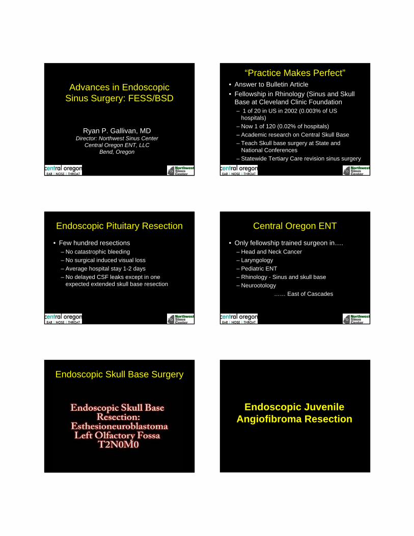
Advances in EndoscopicSinus Surgery: FESS/BSD
Ryan P. Gallivan, MDDirector: Northwest Sinus Center
Central Oregon ENT, LLCBend, Oregon
“Practice Makes Perfect”• Answer to Bulletin Article• Fellowship in Rhinology (Sinus and Skull
Base at Cleveland Clinic Foundation– 1 of 20 in US in 2002 (0.003% of US
hospitals)– Now 1 of 120 (0.02% of hospitals)– Academic research on Central Skull Base– Teach Skull base surgery at State and
National Conferences– Statewide Tertiary Care revision sinus surgery
Endoscopic Pituitary Resection
• Few hundred resections– No catastrophic bleeding– No surgical induced visual loss– Average hospital stay 1-2 days– No delayed CSF leaks except in one
expected extended skull base resection
Central Oregon ENT
• Only fellowship trained surgeon in….– Head and Neck Cancer – Laryngology– Pediatric ENT– Rhinology - Sinus and skull base – Neurootology
…… East of Cascades
Endoscopic Skull Base Surgery
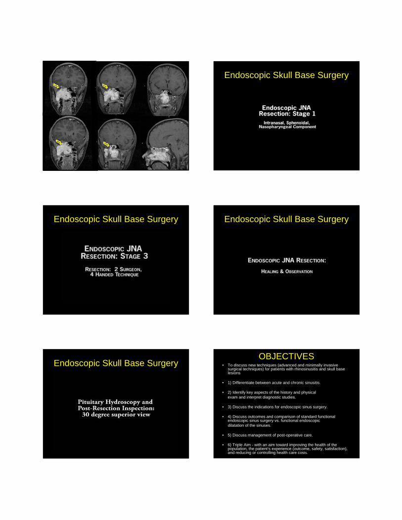
Endoscopic Juvenile Angiofibroma Resection
Endoscopic Skull Base Surgery
Endoscopic Skull Base Surgery Endoscopic Skull Base Surgery
Endoscopic Skull Base SurgeryOBJECTIVES
• To discuss new techniques (advanced and minimally invasive surgical techniques) for patients with rhinosinusitis and skull base lesions
• 1) Differentiate between acute and chronic sinusitis.
• 2) Identify key aspects of the history and physicalexam and interpret diagnostic studies.
• 3) Discuss the indications for endoscopic sinus surgery.
• 4) Discuss outcomes and comparison of standard functional endoscopic sinus surgery vs. functional endoscopicdilatation of the sinuses.
• 5) Discuss management of post-operative care.
• 6) Triple Aim - with an aim toward improving the health of the population, the patient’s experience (outcome, safety, satisfaction), and reducing or controlling health care costs.
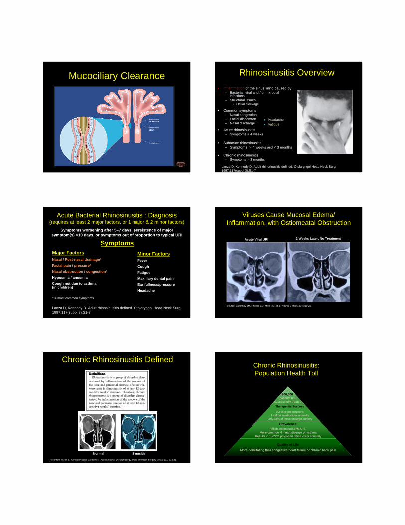
Mucociliary Clearance Rhinosinusitis Overview• Inflammation of the sinus lining caused by
– Bacterial, viral and / or microbial infections
– Structural issues• Ostial blockage
• Common symptoms– Nasal congestion– Facial discomfort– Nasal discharge
• Acute rhinosinusitis– Symptoms < 4 weeks
• Subacute rhinosinusitis– Symptoms > 4 weeks and < 3 months
• Chronic rhinosinusitis– Symptoms > 3 months
HeadacheFatigue
Lanza D, Kennedy D. Adult rhinosinusitis defined. Otolaryngol Head Neck Surg1997;117(suppl 3):S1-7
Acute Bacterial Rhinosinusitis : Diagnosis(requires at least 2 major factors, or 1 major & 2 minor factors)
Symptoms worsening after 5–7 days, persistence of major symptom(s) >10 days, or symptoms out of proportion to typical URI
SymptomsMajor FactorsNasal / Post-nasal drainage*Facial pain / pressure*Nasal obstruction / congestion*Hyposmia / anosmiaCough not due to asthma(in children)
Minor FactorsFeverCoughFatigueMaxillary dental painEar fullness/pressureHeadache
* = most common symptoms
Lanza D, Kennedy D. Adult rhinosinusitis defined. Otolaryngol Head Neck Surg1997;117(suppl 3):S1-7
Source: Gwaltney JM, Phillips CD, Miller RD, et al. N Engl J Med 1994;330:25.
Acute Viral URI 2 Weeks Later, No Treatment
Viruses Cause Mucosal Edema/Inflammation, with Ostiomeatal Obstruction
Chronic Rhinosinusitis Defined
Normal Sinusitis
Rosenfeld, RM et al. Clinical Practice Guidelines: Adult Sinusitis. Otolaryngology–Head and Neck Surgery (2007) 137, S1-S31
900,000patients not
successfully treatedTherapeutic Success
7M seek prescriptions1.4M fail medications annually
Only 35% of these undergo surgery
PrevalenceAfflicts estimated 37M U.S.
More common heart disease or asthmaResults in 18-22M physician office visits annually
Quality of LifeMore debilitating than congestive heart failure or chronic back pain
Chronic Rhinosinusitis: Population Health Toll
Hessler, J., et al. Clinical outcomes of chronic rhinosinusitis in response to medical therapy: Results of a prospective study. Am J Rhinol2007; 21(1): 10-18.
Common Medical Therapy for Acute andChronic Rhinosinusitis
Medical Treatment
(ENT)
Oral and/or intranasal antihistamines
Oral and/or intranasal steroids
Oral decongestants
Intranasal saline
Oral antihistamine/decongestants
Oral leukotriene blockers
Oral antibiotics
Medical therapy typically consists
of varying combinations of
these agents
Medical Treatment: Chronic Rhinosinusitis
Medical Treatment
(ENT)
Oral antibiotics based on endoscopic culture
Entended maximal medical therapy: 3-4 weeks of ABX +/- oral steroids
Nebulized Medications (Steroids, Antibiotics, Antifungals)
Nasal Surfactant (Pending Recall)
Medication in Vertex-Down Position (Polyps or hyposmia)Intranasal Pulmicort, Tobradex, Predforte,
Allergic Rhinitis Therapy
Aspirin desensitization (Per allergist for Aspirin Exacerbated Respiratory Disease)
No FDA approved medications for treatment of Chronic Rhinosinusitis…. .... But I also use
Structural Problems• Deviated nasal septum• Abnormal turbinates
– Hypertrophic, paradoxical, concha bullosa
• Hypertrophic adenoids• Evidence of eustachian tube
dysfunction
Mucosal Problems• Edema• Hyperemia• Purulence• Polyps or polypoid mucosa
Physical Examination of RS Patients
Enlarged, bluish-red inferior
turbinate of patient with allergic rhinitis
Septal Deviation Can Impinge on Ostiomeatal Region
Source: F. Netter. Collection of Ciba Geigy, 1989.
Diagnostic Nasal Endoscopy Can Be Performed with Rigid (0, 30, 70 degree, Rotatable) or Flexible
Endoscopes
Endoscopic Nasal Examination and Obtaining Culture for Diagnosis
Inferior Turbinate
Nasal Septum
Small culturette obtaining mucopussample from hiatus semilunaris
Sterile Sinus Secretion Collector
No prior surgeryAerobes – 75–100%
Coag- neg. Staphylococci Staph. AureusStrep. PneumoniaStrep. viridansH. InfluenzaCorynebacteriumMoraxella catarrhalis
No prior surgery
Anaerobes – 0–25%Fusobacterium sp.Provotella sp.Peptostreptococcus sp.Propionibacterium sp.
Prior Surgery
Pseudomonas sp.Klebsiella sp.Enterobacter sp.Coag- neg. StaphylococciS Aureus
Pathogenesis of CRS: Role of Bacteria
Source: Benninger M, Ferguson BJ, Hadley JA, et al. Otolaryngol Head Neck Surg 2003;129:S1-32.
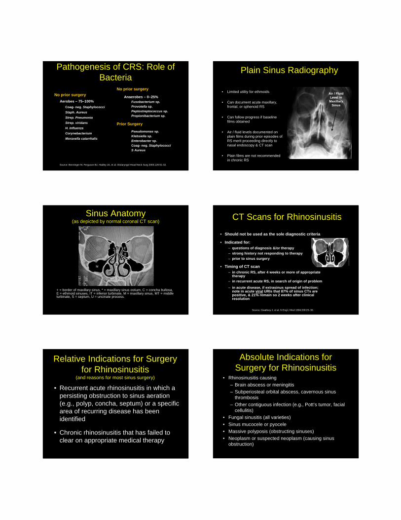
• Limited utility for ethmoids
• Can document acute maxillary, frontal, or sphenoid RS
• Can follow progress if baseline films obtained
• Air / fluid levels documented on plain films during prior episodes of RS merit proceeding directly to nasal endoscopy & CT scan
• Plain films are not recommended in chronic RS
Air / Fluid Level in Maxillary
Sinus
Plain Sinus Radiography
Sinus Anatomy(as depicted by normal coronal CT scan)
+ = border of maxillary sinus, * = maxillary sinus ostium, C = concha bullosa, E = ethmoid sinuses, IT = inferior turbinate, M = maxillary sinus, MT = middle turbinate, S = septum, U = uncinate process.
CT Scans for Rhinosinusitis
• Should not be used as the sole diagnostic criteria
• Indicated for: – questions of diagnosis &/or therapy– strong history not responding to therapy– prior to sinus surgery
• Timing of CT scan– in chronic RS, after 4 weeks or more of appropriate
therapy– in recurrent acute RS, in search of origin of problem
– in acute disease, if extrasinus spread of infection; note in acute viral URIs that 87% of sinus CTs are positive, & 21% remain so 2 weeks after clinical resolution
Source: Gwaltney J, et al. N Engl J Med 1994;330:25–30.
Relative Indications for Surgery for Rhinosinusitis
(and reasons for most sinus surgery)
• Recurrent acute rhinosinusitis in which a persisting obstruction to sinus aeration (e.g., polyp, concha, septum) or a specific area of recurring disease has been identified
• Chronic rhinosinusitis that has failed to clear on appropriate medical therapy
• Rhinosinusitis causing– Brain abscess or meningitis– Subperiosteal orbital abscess, cavernous sinus
thrombosis– Other contiguous infection (e.g., Pott’s tumor, facial
cellulitis)• Fungal sinusitis (all varieties) • Sinus mucocele or pyocele• Massive polyposis (obstructing sinuses)• Neoplasm or suspected neoplasm (causing sinus
obstruction)
Absolute Indications for Surgery for Rhinosinusitis
FESS/BSD : Definitions
FESS = Functional Endoscopic Sinus Surgery
FEDS = Functional Endoscopic Dilation of the Sinuses
=BSD(Balloon Sinus Dilatation)= BSP (Balloon Sinuplasty)
Preservation of Natural Ostium
• “The natural drainage and ventilation paths should be restored, the anatomy changed as little as possible, and the mucosa preserved to the greatest extent.”-Prof. Heinz Stammberger, M.D.
F.E.S.S. Endoscopic Diagnosis and Surgery of the Paranasal Sinuses and Anterior Skull Base, The Messerklinger Technique and Advanced Applications from the Graz School, 2003: pg. 22.
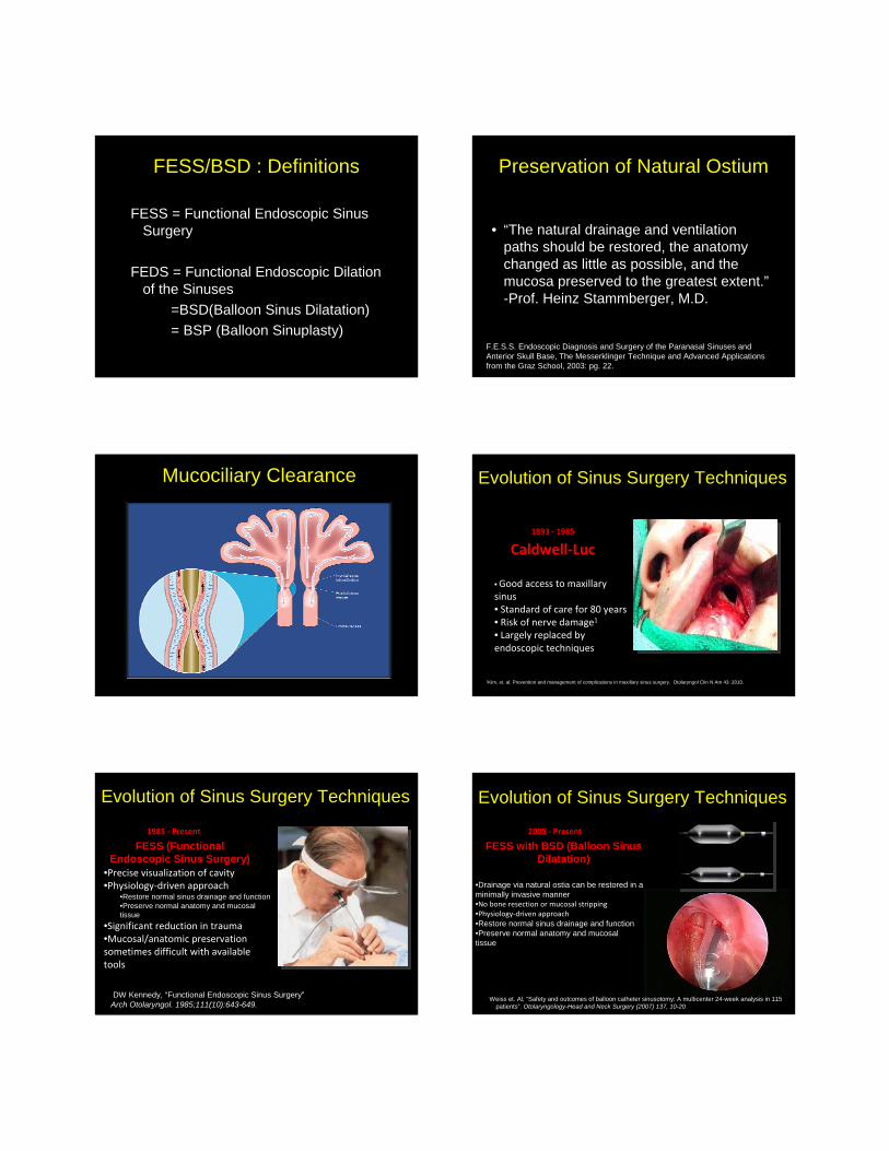
Mucociliary Clearance Evolution of Sinus Surgery Techniques
1893 ‐ 1985
Caldwell‐Luc
•Good access to maxillary sinus• Standard of care for 80 years• Risk of nerve damage1
• Largely replaced by endoscopic techniques
1Kim, et. al. Prevention and management of complications in maxillary sinus surgery. Otolaryngol Clin N Am 43: 2010.
Evolution of Sinus Surgery Techniques
1985 ‐ Present
•Precise visualization of cavity•Physiology‐driven approach
•Restore normal sinus drainage and function•Preserve normal anatomy and mucosal tissue
•Significant reduction in trauma•Mucosal/anatomic preservation sometimes difficult with available tools
FESS (Functional Endoscopic Sinus Surgery)
DW Kennedy, “Functional Endoscopic Sinus Surgery”Arch Otolaryngol. 1985;111(10):643-649.
Evolution of Sinus Surgery Techniques
2005 ‐ Present
•Drainage via natural ostia can be restored in a minimally invasive manner •No bone resection or mucosal stripping•Physiology‐driven approach•Restore normal sinus drainage and function•Preserve normal anatomy and mucosal tissue
FESS with BSD (Balloon Sinus Dilatation)
Weiss et. Al, “Safety and outcomes of balloon catheter sinusotomy: A multicenter 24-week analysis in 115 patients”. Otolaryngology-Head and Neck Surgery (2007) 137, 10-20
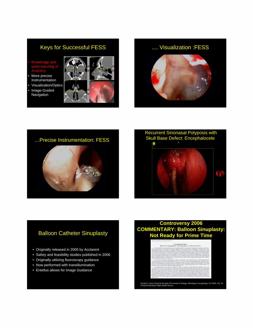
Keys for Successful FESS
• Knowledge and understanding of Anatomy
• More precise Instrumentation
• Visualization/Optics• Image-Guided
Navigation
.... Visualization :FESS
…Precise Instrumentation: FESSRecurrent Sinonasal Polyposis with Skull Base Defect: Encephalocele
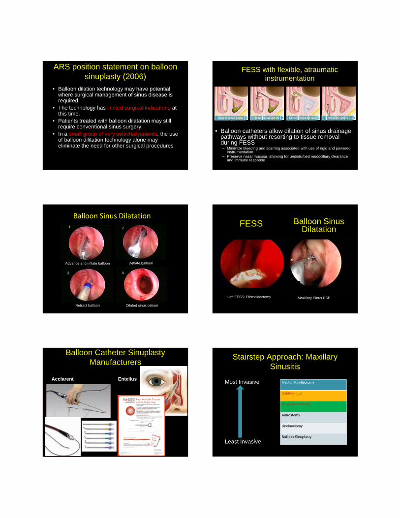
Balloon Catheter Sinuplasty
• Originally released in 2005 by Acclarent• Safety and feasibility studies published in 2006• Originally utilizing fluoroscopy guidance• Now performed with transillumination• Entellus allows for Image Guidance
Controversy 2006COMMENTARY: Balloon Sinuplasty:
Not Ready for Prime Time
Donald C Lanza; David W Kennedy.The Annals of Otology, Rhinology & Laryngology; Oct 2006; 115, 10;ProQuest Nursing & Allied Health Source
ARS position statement on balloon sinuplasty (2006)
• Balloon dilation technology may have potential where surgical management of sinus disease is required.
• The technology has limited surgical indications at this time.
• Patients treated with balloon dilatation may still require conventional sinus surgery.
• In a small group of very selected patients, the use of balloon dilitation technology alone may eliminate the need for other surgical procedures
FESS with flexible, atraumatic instrumentation
• Balloon catheters allow dilation of sinus drainage pathways without resorting to tissue removal during FESS– Minimize bleeding and scarring associated with use of rigid and powered
instrumentation– Preserve nasal mucosa, allowing for undisturbed mucociliary clearance
and immune response
Access sinus cavity Dilate natural ostium Directly irrigate sinus Remove system
Advance and inflate balloon Deflate balloon
Retract balloon Dilated sinus ostium
Balloon Sinus Dilatation1 2
3 4
FESS
Left FESS: Ethmoidectomy Maxillary Sinus BSP
Balloon Sinus Dilatation
Balloon Catheter Sinuplasty Manufacturers
Acclarent Entellus
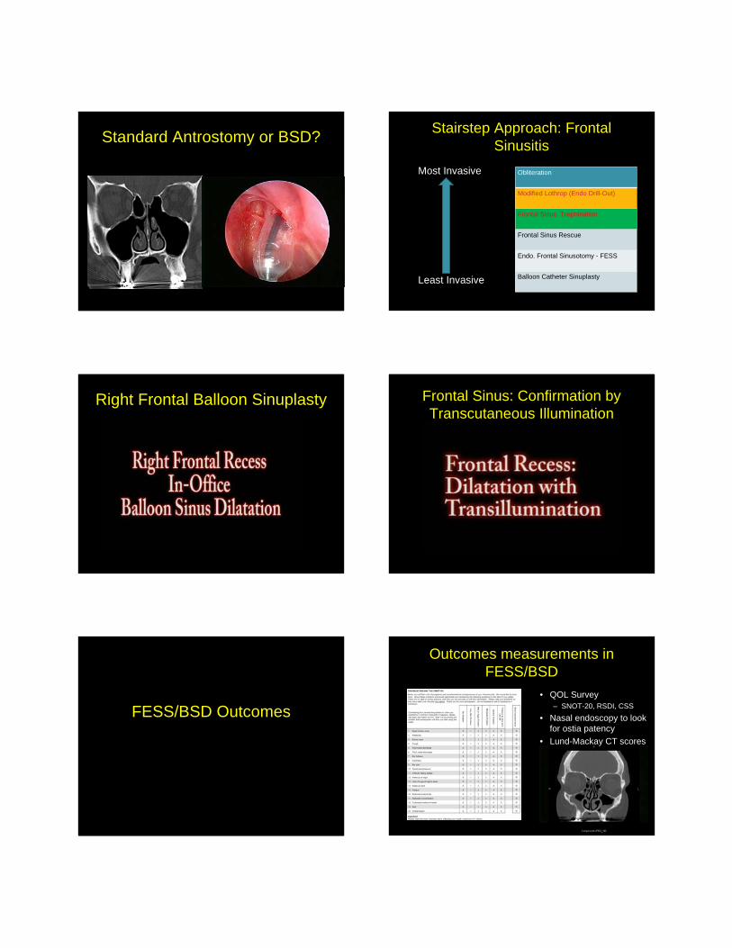
Stairstep Approach: Maxillary Sinusitis
Most Invasive
Least Invasive
Medial Maxillectomy
Caldwell-Luc
Mega-Antrostomy
Antrostomy
Uncinectomy
Balloon Sinuplasty
Standard Antrostomy or BSD? Stairstep Approach: Frontal Sinusitis
Most Invasive
Least Invasive
Obliteration
Modified Lothrop (Endo Drill-Out)
Frontal Sinus Trephination
Frontal Sinus Rescue
Endo. Frontal Sinusotomy - FESS
Balloon Catheter Sinuplasty
Right Frontal Balloon Sinuplasty
VKRFSIBSD2.wmvVKRFSIBSD2.wmv
Frontal Sinus: Confirmation by Transcutaneous Illumination
FESS/BSD Outcomes
Outcomes measurements in FESS/BSD
• QOL Survey – SNOT-20, RSDI, CSS
• Nasal endoscopy to look for ostia patency
• Lund-Mackay CT scores
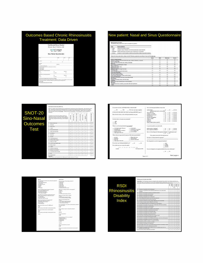
Outcomes Based Chronic Rhinosinusitis Treatment: Data Driven
New patient: Nasal and Sinus Questionnaire
SNOT-20Sino-NasalOutcomes
Test
RSDIRhinosinusitis
Disability Index
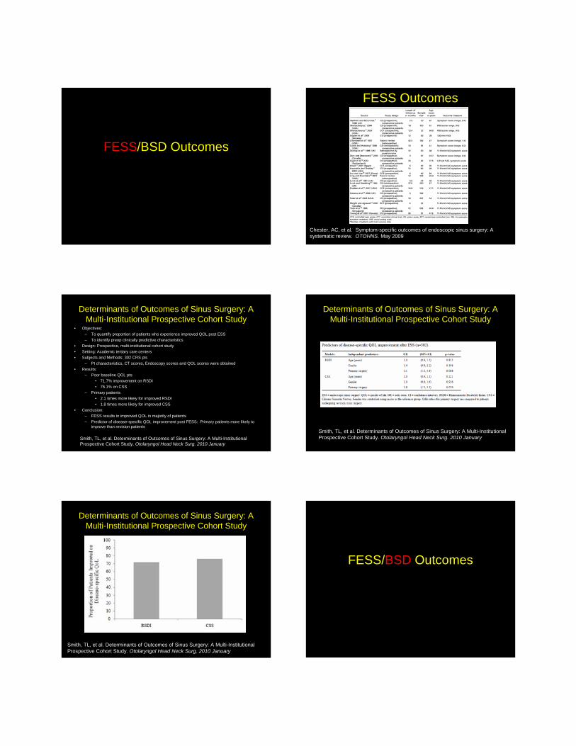
FESS/BSD Outcomes
FESS Outcomes
Chester, AC, et al. Symptom-specific outcomes of endoscopic sinus surgery: A systematic review. OTOHNS. May 2009
Determinants of Outcomes of Sinus Surgery: A Multi-Institutional Prospective Cohort Study
• Objectives: – To quantify proportion of patients who experience improved QOL post ESS– To identify preop clinically predictive characteristics
• Design: Prospective, multi-institutional cohort study• Setting: Academic tertiary care centers• Subjects and Methods: 302 CRS pts
– Pt characteristics, CT scores, Endoscopy scores and QOL scores were obtained• Results:
– Poor baseline QOL pts• 71.7% improvement on RSDI• 76.1% on CSS
– Primary patients• 2.1 times more likely for improved RSDI• 1.8 times more likely for improved CSS
• Conclusion: – FESS results in improved QOL in majority of patients– Predictor of disease-specific QOL improvement post FESS: Primary patients more likely to
improve than revision patients
Smith, TL, et al. Determinants of Outcomes of Sinus Surgery: A Multi-InstitutionalProspective Cohort Study. Otolaryngol Head Neck Surg. 2010 January
Determinants of Outcomes of Sinus Surgery: A Multi-Institutional Prospective Cohort Study
Smith, TL, et al. Determinants of Outcomes of Sinus Surgery: A Multi-InstitutionalProspective Cohort Study. Otolaryngol Head Neck Surg. 2010 January
Determinants of Outcomes of Sinus Surgery: A Multi-Institutional Prospective Cohort Study
Smith, TL, et al. Determinants of Outcomes of Sinus Surgery: A Multi-InstitutionalProspective Cohort Study. Otolaryngol Head Neck Surg. 2010 January
FESS/BSD Outcomes
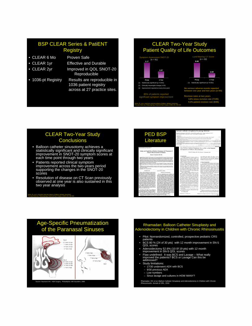
BSP CLEAR Series & PatiENT Registry
• CLEAR 6 Mo Proven Safe• CLEAR 1yr Effective and Durable • CLEAR 2yr Improved in QOL SNOT-20
Reproducible• 1036-pt Registry Results are reproducible in
1036 patient registry across at 27 practice sites.
CLEAR Two-Year StudyPatient Quality of Life Outcomes
Symptom Assessment SNOT-20(n = 61)
(1) Statistically significant (p <0.001)
(2) Clinically meaningful change (>0.8)
(3) Improvement reported at every time point
2.17
0.87
Preop 2 Year
Weiss, RL, et el. Long-term outcome analysis of balloon catheter sinusotomy: Two-year follow-up. Otolaryngology–Head and Neck Surgery (2008) 139, S38-S46.
85% of patients reported significant symptom improvement
Lund-MacKay CT Score(n = 32)
(1) Statistically significant (p <0.001)
9.66
2.69
Preop 2 Year
No serious adverse events reported between one year and two years (n=65)
Revision rates at two years3.6% sinus revision rate (7/195)9.2% patient revision rate (6/65)
CLEAR Two-Year StudyConclusions
• Balloon catheter sinusotomy achieves a statistically significant and clinically significant improvement in SNOT-20 symptom scores at each time point through two years
• Patients reported clinical symptom improvement across the two-years period supporting the changes in the SNOT-20 scores
• Resolution of disease on CT Scan previously observed at one year is also sustained in this two year analysis
Weiss, RL, et el. Long-term outcome analysis of balloon catheter sinusotomy: Two-year follow-up. Otolaryngology–Head and Neck Surgery (2008) 139, S38-S46.
PED BSP Literature
Age-Specific Pneumatization of the Paranasal Sinuses
Source: Naumann HH. H&N Surgery. Philadelphia: WB Saunders, 1980.
Rhamadan: Balloon Catheter Sinuplasty and Adenoidectomy in Children with Chronic Rhinosinusitis
• Pilot: Nonrandomized, controlled, prospective pediatric CRS patients
• BCS 80 % (24 of 30 pts) with 12 month improvement in SN-5 QOL scores
• Adenoidectomy 52.6% (10 0f 19 pts) with 12 month improvement in SN-5 QOL scores
• Flaw undefined: It was BCS and Lavage – What really improved the patients? BCS or Lavage Can this be addressed?
• Study limitations:– 17/30 underwent ADX with BCS– 9/30 previous ADX– Low numbers– Sinus lavage and cultures in HOW MANY?
Rhamadan, HH, et al. Balloon Catheter Sinuplasty and Adenoidectomy in Children with Chronic Rhinosinusitis. Annals of ORL, 2010
Outcome of adenoidectomy versus adenoidectomy with maxillary sinus wash for
chronic rhinosinusitis in children• OBJECTIVES: • To compare postoperative outcomes of adenoidectomy versus
adenoidectomy with maxillary sinus wash as surgical treatment of chronic rhinosinusitis (CRS) in children.
• STUDY DESIGN: retrospective review of prospectively collected data.• METHODS: CRS patients
– Adenoidectomy alone or adenoidectomy with a maxillary sinus wash. • RESULTS: 60 Children.
– 87.5% (28/32) wash/A improved SN-5 Scores – 60.7% (17/28) Adenoidectomy with improved SN-5 Scores – 93% improvement with Higher CT scores – 60% improvement with Higher CT scores
• CONCLUSIONS: Children with more severe sinus disease as evidenced by a high CT score had a higher success rate if a maxillary sinus wash was performed at the time of adenoidectomy. Children with a low CT score did not have that benefit.
Rhamadan HH, Cost JL. Outcome of adenoidectomy versus adenoidectomy with maxillary sinus wash for chronic rhinosinusitis in children. Laryngoscope. May, 2008.
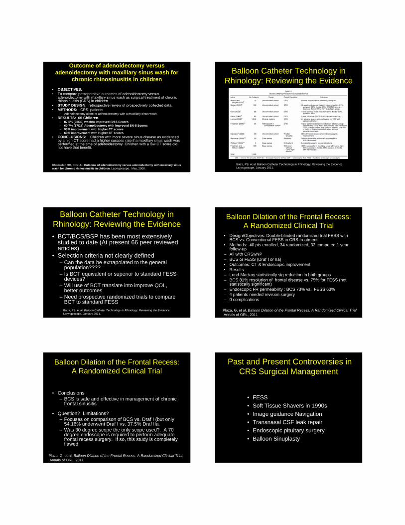
Balloon Catheter Technology in Rhinology: Reviewing the Evidence
Batra, PS, et al. Balloon Catheter Technology in Rhinology: Reviewing the Evidence.Laryngoscope, January 2011.
Balloon Catheter Technology in Rhinology: Reviewing the Evidence
Batra, PS, et al. Balloon Catheter Technology in Rhinology: Reviewing the Evidence.Laryngoscope, January 2011.
• BCT/BCS/BSP has been most extensively studied to date (At present 66 peer reviewed articles)
• Selection criteria not clearly defined – Can the data be extrapolated to the general
population????– Is BCT equivalent or superior to standard FESS
devices?– Will use of BCT translate into improve QOL,
better outcomes– Need prospective randomized trials to compare
BCT to standard FESS
Balloon Dilation of the Frontal Recess: A Randomized Clinical Trial
• Design/Objectives: Double-blinded randomized trial FESS with BCS vs. Conventional FESS in CRS treatment
• Methods: 40 pts enrolled, 34 randomized, 32 competed 1 year follow-up
– All with CRSwNP– BCS or FESS (Draf I or IIa) • Outcomes: CT & Endoscopic improvement• Results– Lund-Mackay statistically sig reduction in both groups– BCS 81% resolution of frontal disease vs. 75% for FESS (not
statistically significant)– Endoscopic FR permeability : BCS 73% vs. FESS 63%– 4 patients needed revision surgery– 0 complications
Plaza, G, et al. Balloon Dilation of the Frontal Recess: A Randomized Clinical Trial.Annals of ORL, 2011
Balloon Dilation of the Frontal Recess: A Randomized Clinical Trial
• Conclusions– BCS is safe and effective in management of chronic
frontal sinusitis
• Question? Limitations?– Focuses on comparison of BCS vs. Draf I (but only
54.16% underwent Draf I vs. 37.5% Draf IIa. – Was 30 degree scope the only scope used?. A 70
degree endoscope is required to perform adequate frontal recess surgery. If so, this study is completely flawed.
Plaza, G, et al. Balloon Dilation of the Frontal Recess: A Randomized Clinical Trial.Annals of ORL, 2011
Past and Present Controversies in CRS Surgical Management
• FESS • Soft Tissue Shavers in 1990s • Image guidance Navigation• Transnasal CSF leak repair • Endoscopic pituitary surgery• Balloon Sinuplasty
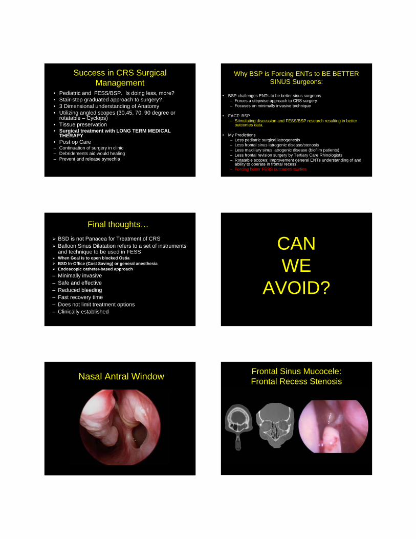
Success in CRS Surgical Management
• Pediatric and FESS/BSP. Is doing less, more?• Stair-step graduated approach to surgery?• 3 Dimensional understanding of Anatomy• Utilizing angled scopes (30,45, 70, 90 degree or
rotatable – Cyclops)• Tissue preservation• Surgical treatment with LONG TERM MEDICAL
THERAPY• Post op Care– Continuation of surgery in clinic– Debridements aid would healing– Prevent and release synechia
Why BSP is Forcing ENTs to BE BETTER SINUS Surgeons:
• BSP challenges ENTs to be better sinus surgeons– Forces a stepwise approach to CRS surgery– Focuses on minimally invasive technique
• FACT: BSP– Stimulating discussion and FESS/BSP research resulting in better
outcomes data.
• My Predictions– Less pediatric surgical iatrogenesis– Less frontal sinus iatrogenic disease/stenosis– Less maxillary sinus iatrogenic disease (biofilm patients)– Less frontal revision surgery by Tertiary Care Rhinologists– Rotatable scopes: Improvement general ENTs understanding of and
ability to operate in frontal recess – Forcing better FESS outcomes studies
Final thoughts…BSD is not Panacea for Treatment of CRSBalloon Sinus Dilatation refers to a set of instruments and technique to be used in FESSWhen Goal is to open blocked OstiaBSD In-Office (Cost Saving) or general anesthesiaEndoscopic catheter-based approach
– Minimally invasive– Safe and effective – Reduced bleeding– Fast recovery time – Does not limit treatment options– Clinically established
CAN WE
AVOID?
Nasal Antral Window Frontal Sinus Mucocele: Frontal Recess Stenosis
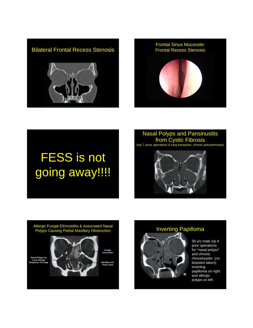
Bilateral Frontal Recess StenosisFrontal Sinus Mucocele: Frontal Recess Stenosis
FESS is not going away!!!!
Nasal Polyps and Pansinusitisfrom Cystic Fibrosis
(s/p 7 sinus operations & lung transplant, chronic pseudomonas)
Allergic Fungal Ethmoiditis & Associated Nasal Polyps Causing Partial Maxillary Obstruction
Fungal Concretion
Maxillary Air/ Fluid Level
Nasal Polyps As Local Allergic
Response To Fungi
Inverting Papilloma
36 y/o male s/p 4 prior operations for “nasal polyps”and chronic rhinosinusitis (no biopsies taken); inverting papilloma on right and allergic polyps on left
R
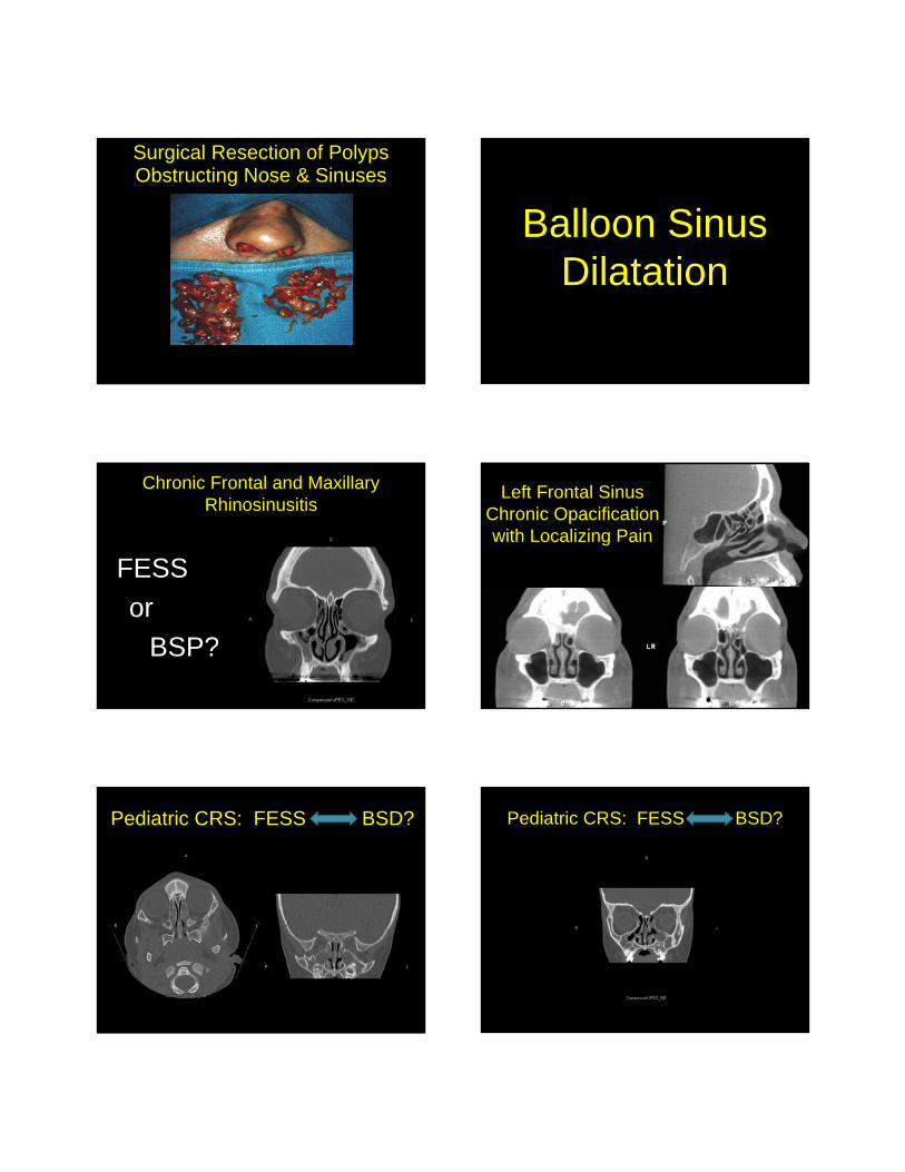
Surgical Resection of Polyps Obstructing Nose & Sinuses
Balloon Sinus Dilatation
Chronic Frontal and MaxillaryRhinosinusitis
FESS or
BSP?
Left Frontal Sinus Chronic Opacificationwith Localizing Pain
Pediatric CRS: FESS BSD? Pediatric CRS: FESS BSD?

Thank You