Embed Size (px)
Citation preview
S
O
Ec
AR
H
a
A
R
A
A
K
C
I
N
T
h2u
r e v b r a s o r t o p . 2 0 1 8;5 3(1):107–112
OCIEDADE BRASILEIRA DEORTOPEDIA E TRAUMATOLOGIA
www.rbo.org .br
riginal Article
pidemiology of cauda equina syndrome. Whathanged until 2015�
ndré Luiz Natálio Dias ∗, Fernando Flores de Araújo, Alexandre Fogaca Cristante,aphael Martus Marcon, Tarcísio Eloy Pessoa de Barros Filho, Olavo Biraghi Letaif
ospital das Clinicas, Faculdade de Medicina, Universidade de Sao Paulo (HCFMUSP), São Paulo, SP, Brazil
r t i c l e i n f o
rticle history:
eceived 1 February 2017
ccepted 2 March 2017
vailable online 6 December 2017
eywords:
auda equina
ntervertebral disc displacement
eurogenic urinary bladder
a b s t r a c t
Objective: The primary objective of this study was to analyze the characteristics and out-
comes of cases admitted to hospital with cauda equina syndrome (CES) at the Institute of
Orthopedics and Traumatology (IOT) from 2005 to 2015. Secondly, this article is a continua-
tion of the epidemiological work of the same base published in 2013, and will be important
for other comparative studies to a greater understanding of the disease and its epidemiology.
Methods: This was a retrospective study of the medical records of admissions due to CES at
IOT in the period 2005–2015 with diagnosis of CES and neuropathic bladder. The following
variables were analyzed: gender, age, etiology of the disease, topographic level of the injury,
time interval between injury and diagnosis, presence of neurogenic bladder, time interval
between diagnosis of the CES and surgery, and reversal of the deficit or of the neurogenic
bladder.
Results: Since this is a rare disease, with a low global incidence, it was not possible, just with
the current study to establish statistically significant correlations between the variables
and outcomes of the disease. However, this study demonstrates the shortcomings of the
Brazilian public health system, both with the initial management of these patients and the
need for urgent surgical treatment.
Conclusion: The study shows that despite well-defined basis for the conduct of CES, a higher
number of sequelae caused by the pathology is observed in Brazil. The delay in diagnosis
and, therefore, for definitive treatment, remains as the major cause for the high number of
sequelae. Level of evidence: 4, case series.
© 2017 Sociedade Brasileira de Ortopedia e Traumatologia. Published by Elsevier Editora
Ltda. This is an open access article under the CC BY-NC-ND license (http://
creativecommons.org/licenses/by-nc-nd/4.0/).
� Study conducted at the Laboratório de Investigacão Médica do Sistema Músculo-Esquelético (LIM 41), Departamento de Ortopedia eraumatologia, Faculdade de Medicina, Universidade de Sao Paulo (FMUSP), São Paulo, SP, Brazil.∗ Corresponding author.
E-mail: [email protected] (A.L. Dias).ttps://doi.org/10.1016/j.rboe.2017.11.006255-4971/© 2017 Sociedade Brasileira de Ortopedia e Traumatologia. Published by Elsevier Editora Ltda. This is an open access articlender the CC BY-NC-ND license (http://creativecommons.org/licenses/by-nc-nd/4.0/).
108 r e v b r a s o r t o p . 2 0 1 8;5 3(1):107–112
Epidemiologia da síndrome da cauda equina. O que mudou até 2015
Palavras-chave:
Cauda equina
Deslocamento do disco
intervertebral
Bexiga urinária neurogênica
r e s u m o
Objetivo: Analisar as características e os desfechos dos casos internados por síndrome
da cauda equina (SCE) no Instituto de Ortopedia e Traumatologia (IOT) da Faculdade de
Medicina da Universidade de São Paulo de 2005-2015. Secundariamente, este artigo é a
continuacão do trabalho epidemiológico de mesma base publicado em 2013 e servirá de
base para outros estudos comparativos com vistas a um entendimento maior da doenca e
de sua epidemiologia.
Métodos: Estudo retrospectivo dos prontuários das internacões por SCE no IOT de 2005 a 2015
com diagnósticos de SCE e bexiga neuropática. As seguintes variáveis foram analisadas:
sexo, idade, etiologia da doenca, nível topográfico da lesão, tempo de história da lesão até o
diagnóstico, presenca de bexiga neurogênica, tempo entre o diagnóstico da SCE e a cirurgia
e reversão do déficit ou da bexiga neurogênica.
Resultados: Por se tratar de uma doenca rara, com uma incidência global baixa, não foi
possível, somente com o estudo atual, estabelecer correlacões estatisticamente significa-
tivas entre as variáveis analisadas e os desfechos da doenca. Porém, este estudo continua
a evidenciar as deficiências do sistema público de saúde brasileiro, tanto no manejo inicial
desses pacientes quanto na necessidade de tratamento cirúrgico de urgência.
Conclusão: O trabalho mostra que, apesar de bem definidas as bases para conduta da SCE,
observa-se no Brasil um número maior de sequelas causadas pela patologia. O atraso no
diagnóstico e, a partir desse, no tratamento definitivo mantém-se como a causa para o alto
número de sequelas. Nível de evidência: 4, série de casos.
© 2017 Sociedade Brasileira de Ortopedia e Traumatologia. Publicado por Elsevier
Editora Ltda. Este e um artigo Open Access sob uma licenca CC BY-NC-ND (http://
Introduction
Cauda equina syndrome (CSE) is classically characterized bythe compression of the distal lumbar, sacral, and coccygealnerve roots at the end of the conus medullaris at the L1and L2 vertebral level.1 Although this disease has a low inci-dence in the population,2 ranging from 1:33,000 to 1:100,000inhabitants,3 its sequelae still generate high public healthcarecosts.
The clinical signs characteristic of the pathology are:severe back pain, often accompanied by sciatica; saddleanesthesia4,5; sphincter and sexual dysfunction; and lowerlimb weakness.6–8 The presence of all these signs simulta-neously is not required for diagnosis.6 The clinical historyand the neurological examination lead to the need for diag-nostic confirmation through complementary exams such ascomputed tomography (CT) and the gold standard, magneticresonance imaging (MRI).1 MRI is mandatory for determiningcompression topography and etiology.8
Among the causes of compression, the following arenoteworthy: extruded disc herniation5,9 (Figs. 1 and 2),tumor lesions10 (Fig. 3), vertebral fractures,11–13 canalstenoses,14 infections,13 surgical manipulation,15,16 spinalanesthesia,17 ankylosing spondylitis,18,19 and gunfirewounds.20 Some reports also indicate its onset afterchiropractic manipulation.21 This is an orthopedic emer-gency and its treatment of choice continues to be surgical
decompression,13,22–25 which, if performed as early aspossible,25–28 reduces neurological damage4,29,30 and improvesthe prognosis.26,27,31creativecommons.org/licenses/by-nc-nd/4.0/).
The primary objective of this study was to analyze, throughcross reference data, the characteristics of the cases andoutcome of patients admitted to hospital due to CES at the IOT-HC-FMUSP Spine Group. Secondarily, it will continue to serveas a basis for other comparative studies, aiming to achieve agreater understanding of the disease and its epidemiology.
Methods
This was a retrospective study of the medical records ofpatients admitted to hospital due to CES at the IOT from 2005to 2015. All records with diagnoses classified by the Interna-tional Classification of Diseases (ICD) with codes G83.4 andN31.0/N31.1/N31.2 (CES and neuropathic bladder, respectively)were investigated.
From these records, all those who presented spinal cordinjuries above the level of the T12 vertebra were excluded.Patients with confirmed CES diagnosis, detailed clinicalhistory and neurological examination at admission, andCT and MRI assessment of the lumbosacral spine wereincluded. The medical records of all cases included in thissample featured a well-documented report of the initialand postoperative physical examinations and of diseaseprogress.
The following variables were analyzed in the sample: gen-der, age, disease etiology, topographic level of the lesion, time
from injury to diagnosis, level of neurological deficit (con-sidered as being the last normal topographic level regardingstrength and sensitivity), presence of neurogenic bladder, time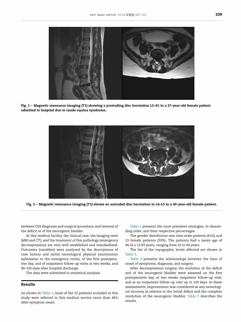
r e v b r a s o r t o p . 2 0 1 8;5 3(1):107–112 109
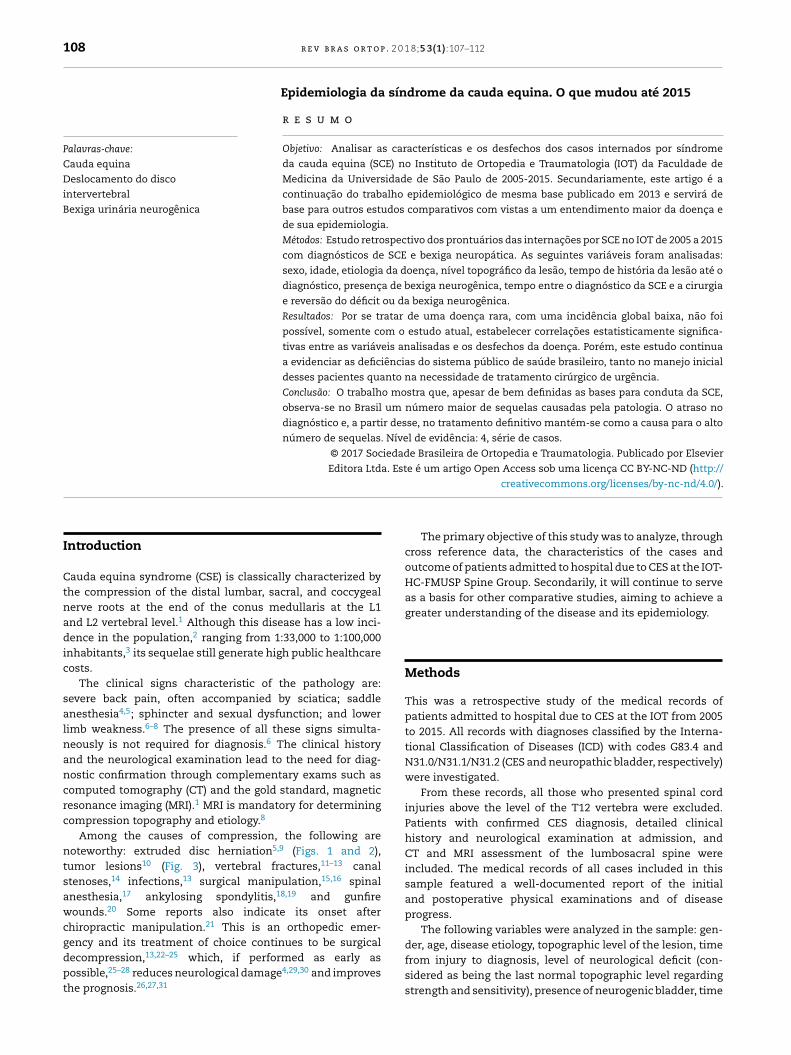
Fig. 1 – Magnetic resonance imaging (T1) showing a protruding disc herniation L5–S1 in a 37-year-old female patientadmitted to hospital due to cauda equina syndrome.
Fig. 2 – Magnetic resonance imaging (T1) shows an extruded disc herniation in L4–L5 in a 49-year-old female patient.
bt
(dOc(t9
R
Asa
etween CES diagnosis and surgical procedure, and reversal ofhe deficit or of the neurogenic bladder.
At this medical facility, the clinical care, the imaging testsMRI and CT), and the treatment of this pathology (emergencyecompression) are very well established and standardized.utcomes (variables) were analyzed by the descriptions ofase history and initial neurological physical examinationadmission to the emergency room), of the first postopera-ive day, and of outpatient follow-up visits at two weeks, and0–120 days after hospital discharge.
The data were submitted to statistical analysis.
esults
s shown in Table 1, most of the 22 patients included in thistudy were referred to this medical service more than 48 hfter symptom onset.
Table 2 presents the most prevalent etiologies, in descen-ding order, and their respective percentages.
The gender distribution was nine male patients (41%) and13 female patients (59%). The patients had a mean age of44.16 ± 12.83 years, ranging from 22 to 64 years.
The list of the topographic levels affected are shown inTable 3.
Table 4 presents the relationships between the time ofonset of symptoms, diagnosis, and surgery.
After decompression surgery, the evolution of the deficitand of the neurogenic bladder were assessed on the firstpostoperative day, at two weeks outpatient follow-up visit,and at an outpatient follow-up visit up to 120 days. In theseassessments, improvement was considered as any neurologi-
cal recovery in relation to the initial deficit and the completeresolution of the neurogenic bladder. Table 5 describes theresults.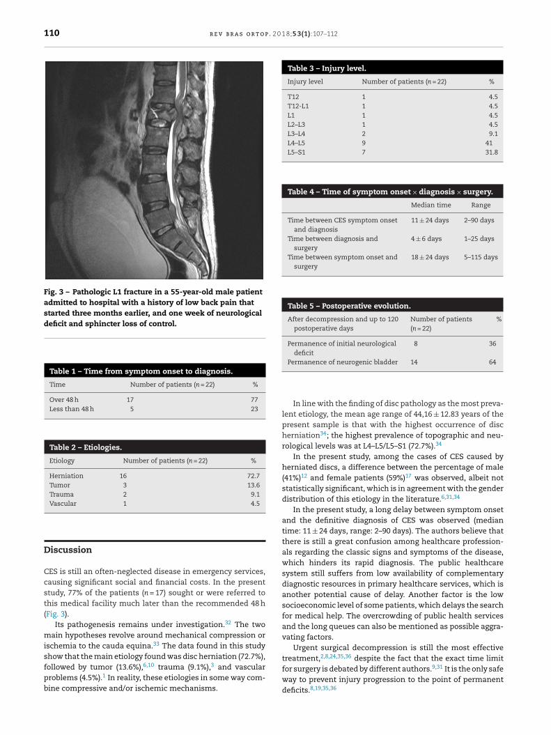
110 r e v b r a s o r t o p . 2 0 1 8;5 3(1):107–112
Fig. 3 – Pathologic L1 fracture in a 55-year-old male patientadmitted to hospital with a history of low back pain thatstarted three months earlier, and one week of neurologicaldeficit and sphincter loss of control.
Table 1 – Time from symptom onset to diagnosis.
Time Number of patients (n = 22) %
Over 48 h 17 77Less than 48 h 5 23
Table 2 – Etiologies.
Etiology Number of patients (n = 22) %
Herniation 16 72.7Tumor 3 13.6Trauma 2 9.1Vascular 1 4.5
Table 3 – Injury level.
Injury level Number of patients (n = 22) %
T12 1 4.5T12-L1 1 4.5L1 1 4.5L2–L3 1 4.5L3–L4 2 9.1L4–L5 9 41L5–S1 7 31.8
Table 4 – Time of symptom onset × diagnosis × surgery.
Median time Range
Time between CES symptom onsetand diagnosis
11 ± 24 days 2–90 days
Time between diagnosis andsurgery
4 ± 6 days 1–25 days
Time between symptom onset andsurgery
18 ± 24 days 5–115 days
Table 5 – Postoperative evolution.
After decompression and up to 120postoperative days
Number of patients(n = 22)
%
Permanence of initial neurologicaldeficit
8 36
Permanence of neurogenic bladder 14 64
Discussion
CES is still an often-neglected disease in emergency services,causing significant social and financial costs. In the presentstudy, 77% of the patients (n = 17) sought or were referred tothis medical facility much later than the recommended 48 h(Fig. 3).
Its pathogenesis remains under investigation.32 The twomain hypotheses revolve around mechanical compression orischemia to the cauda equina.33 The data found in this studyshow that the main etiology found was disc herniation (72.7%),followed by tumor (13.6%),6,10 trauma (9.1%),3 and vascular
problems (4.5%).1 In reality, these etiologies in some way com-bine compressive and/or ischemic mechanisms.In line with the finding of disc pathology as the most preva-lent etiology, the mean age range of 44,16 ± 12.83 years of thepresent sample is that with the highest occurrence of discherniation34; the highest prevalence of topographic and neu-rological levels was at L4–L5/L5–S1 (72.7%).34
In the present study, among the cases of CES caused byherniated discs, a difference between the percentage of male(41%)12 and female patients (59%)17 was observed, albeit notstatistically significant, which is in agreement with the genderdistribution of this etiology in the literature.6,31,34
In the present study, a long delay between symptom onsetand the definitive diagnosis of CES was observed (mediantime: 11 ± 24 days, range: 2–90 days). The authors believe thatthere is still a great confusion among healthcare profession-als regarding the classic signs and symptoms of the disease,which hinders its rapid diagnosis. The public healthcaresystem still suffers from low availability of complementarydiagnostic resources in primary healthcare services, which isanother potential cause of delay. Another factor is the lowsocioeconomic level of some patients, which delays the searchfor medical help. The overcrowding of public health servicesand the long queues can also be mentioned as possible aggra-vating factors.
Urgent surgical decompression is still the most effectivetreatment,2,8,24,35,36 despite the fact that the exact time limitfor surgery is debated by different authors.9,31 It is the only safe
way to prevent injury progression to the point of permanentdeficits.8,19,35,36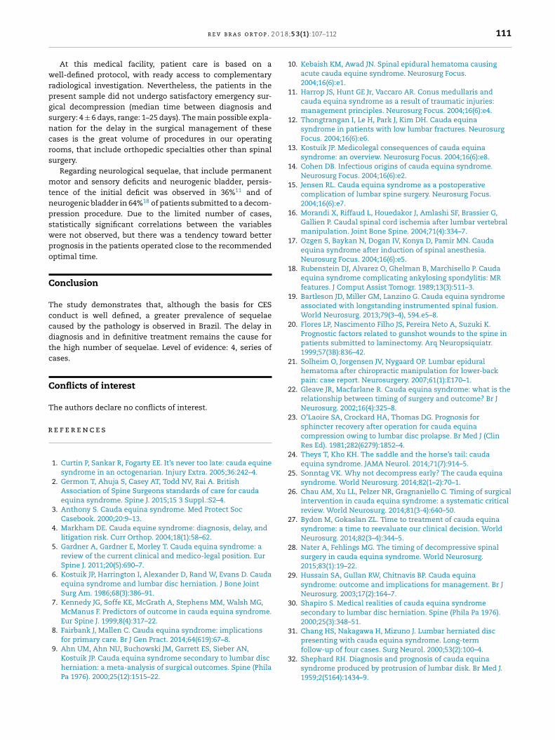
0 1 8
wrpgsncrs
mtnpswpo
C
Tccdtc
C
T
r
1
1
1
1
1
1
1
1
1
1
2
2
2
2
2
2
2
2
2
2
3
3
r e v b r a s o r t o p . 2
At this medical facility, patient care is based on aell-defined protocol, with ready access to complementary
adiological investigation. Nevertheless, the patients in theresent sample did not undergo satisfactory emergency sur-ical decompression (median time between diagnosis andurgery: 4 ± 6 days, range: 1–25 days). The main possible expla-ation for the delay in the surgical management of theseases is the great volume of procedures in our operatingooms, that include orthopedic specialties other than spinalurgery.
Regarding neurological sequelae, that include permanentotor and sensory deficits and neurogenic bladder, persis-
ence of the initial deficit was observed in 36%11 and ofeurogenic bladder in 64%18 of patients submitted to a decom-ression procedure. Due to the limited number of cases,tatistically significant correlations between the variablesere not observed, but there was a tendency toward betterrognosis in the patients operated close to the recommendedptimal time.
onclusion
he study demonstrates that, although the basis for CESonduct is well defined, a greater prevalence of sequelaeaused by the pathology is observed in Brazil. The delay iniagnosis and in definitive treatment remains the cause forhe high number of sequelae. Level of evidence: 4, series ofases.
onflicts of interest
he authors declare no conflicts of interest.
e f e r e n c e s
1. Curtin P, Sankar R, Fogarty EE. It’s never too late: cauda equinesyndrome in an octogenarian. Injury Extra. 2005;36:242–4.
2. Germon T, Ahuja S, Casey AT, Todd NV, Rai A. BritishAssociation of Spine Surgeons standards of care for caudaequina syndrome. Spine J. 2015;15 3 Suppl.:S2–4.
3. Anthony S. Cauda equina syndrome. Med Protect SocCasebook. 2000;20:9–13.
4. Markham DE. Cauda equine syndrome: diagnosis, delay, andlitigation risk. Curr Orthop. 2004;18(1):58–62.
5. Gardner A, Gardner E, Morley T. Cauda equina syndrome: areview of the current clinical and medico-legal position. EurSpine J. 2011;20(5):690–7.
6. Kostuik JP, Harrington I, Alexander D, Rand W, Evans D. Caudaequina syndrome and lumbar disc herniation. J Bone JointSurg Am. 1986;68(3):386–91.
7. Kennedy JG, Soffe KE, McGrath A, Stephens MM, Walsh MG,McManus F. Predictors of outcome in cauda equina syndrome.Eur Spine J. 1999;8(4):317–22.
8. Fairbank J, Mallen C. Cauda equina syndrome: implicationsfor primary care. Br J Gen Pract. 2014;64(619):67–8.
9. Ahn UM, Ahn NU, Buchowski JM, Garrett ES, Sieber AN,Kostuik JP. Cauda equina syndrome secondary to lumbar discherniation: a meta-analysis of surgical outcomes. Spine (PhilaPa 1976). 2000;25(12):1515–22.
3
;5 3(1):107–112 111
0. Kebaish KM, Awad JN. Spinal epidural hematoma causingacute cauda equine syndrome. Neurosurg Focus.2004;16(6):e1.
1. Harrop JS, Hunt GE Jr, Vaccaro AR. Conus medullaris andcauda equina syndrome as a result of traumatic injuries:management principles. Neurosurg Focus. 2004;16(6):e4.
2. Thongtrangan I, Le H, Park J, Kim DH. Cauda equinasyndrome in patients with low lumbar fractures. NeurosurgFocus. 2004;16(6):e6.
3. Kostuik JP. Medicolegal consequences of cauda equinasyndrome: an overview. Neurosurg Focus. 2004;16(6):e8.
4. Cohen DB. Infectious origins of cauda equina syndrome.Neurosurg Focus. 2004;16(6):e2.
5. Jensen RL. Cauda equina syndrome as a postoperativecomplication of lumbar spine surgery. Neurosurg Focus.2004;16(6):e7.
6. Morandi X, Riffaud L, Houedakor J, Amlashi SF, Brassier G,Gallien P. Caudal spinal cord ischemia after lumbar vertebralmanipulation. Joint Bone Spine. 2004;71(4):334–7.
7. Ozgen S, Baykan N, Dogan IV, Konya D, Pamir MN. Caudaequina syndrome after induction of spinal anesthesia.Neurosurg Focus. 2004;16(6):e5.
8. Rubenstein DJ, Alvarez O, Ghelman B, Marchisello P. Caudaequina syndrome complicating ankylosing spondylitis: MRfeatures. J Comput Assist Tomogr. 1989;13(3):511–3.
9. Bartleson JD, Miller GM, Lanzino G. Cauda equina syndromeassociated with longstanding instrumented spinal fusion.World Neurosurg. 2013;79(3–4), 594.e5–8.
0. Flores LP, Nascimento Filho JS, Pereira Neto A, Suzuki K.Prognostic factors related to gunshot wounds to the spine inpatients submitted to laminectomy. Arq Neuropsiquiatr.1999;57(3B):836–42.
1. Solheim O, Jorgensen JV, Nygaard OP. Lumbar epiduralhematoma after chiropractic manipulation for lower-backpain: case report. Neurosurgery. 2007;61(1):E170–1.
2. Gleave JR, Macfarlane R. Cauda equina syndrome: what is therelationship between timing of surgery and outcome? Br JNeurosurg. 2002;16(4):325–8.
3. O’Laoire SA, Crockard HA, Thomas DG. Prognosis forsphincter recovery after operation for cauda equinacompression owing to lumbar disc prolapse. Br Med J (ClinRes Ed). 1981;282(6279):1852–4.
4. Theys T, Kho KH. The saddle and the horse’s tail: caudaequina syndrome. JAMA Neurol. 2014;71(7):914–5.
5. Sonntag VK. Why not decompress early? The cauda equinasyndrome. World Neurosurg. 2014;82(1–2):70–1.
6. Chau AM, Xu LL, Pelzer NR, Gragnaniello C. Timing of surgicalintervention in cauda equina syndrome: a systematic criticalreview. World Neurosurg. 2014;81(3-4):640–50.
7. Bydon M, Gokaslan ZL. Time to treatment of cauda equinasyndrome: a time to reevaluate our clinical decision. WorldNeurosurg. 2014;82(3–4):344–5.
8. Nater A, Fehlings MG. The timing of decompressive spinalsurgery in cauda equina syndrome. World Neurosurg.2015;83(1):19–22.
9. Hussain SA, Gullan RW, Chitnavis BP. Cauda equinasyndrome: outcome and implications for management. Br JNeurosurg. 2003;17(2):164–7.
0. Shapiro S. Medical realities of cauda equina syndromesecondary to lumbar disc herniation. Spine (Phila Pa 1976).2000;25(3):348–51.
1. Chang HS, Nakagawa H, Mizuno J. Lumbar herniated discpresenting with cauda equina syndrome. Long-termfollow-up of four cases. Surg Neurol. 2000;53(2):100–4.
2. Shephard RH. Diagnosis and prognosis of cauda equina
syndrome produced by protrusion of lumbar disk. Br Med J.1959;2(5164):1434–9.
p . 2 0
3
3
3
Eur Spine J. 2007;16(12):2143–51.
112 r e v b r a s o r t o
3. Kohles SS, Kohles DA, Karp AP, Erlich VM, Polissar NL.Time-dependent surgical outcomes following cauda equinasyndrome diagnosis: comments on a meta-analysis. Spine
(Phila Pa 1976). 2004;29(11):1281–7.4. McCarthy MJ, Aylott CE, Grevitt MP, Hegarty J. Cauda equinasyndrome: factors affecting long-term functional andsphincteric outcome. Spine (Phila Pa 1976). 2007;32(2):207–16.
3
1 8;5 3(1):107–112
5. Qureshi A, Sell P. Cauda equina syndrome treated by surgicaldecompression: the influence of timing on surgical outcome.
6. Fuso FA, Dias AL, Letaif OB, Cristante AF, Marcon RM, deBarros TE. Epidemiological study of cauda equina syndrome.Acta Ortop Bras. 2013;21(3):159–62.