Embed Size (px)
Citation preview
University of Nebraska - LincolnDigitalCommons@University of Nebraska - Lincoln
Virology Papers Virology, Nebraska Center for
4-1-1999
Interferon Regulatory Factor 2 Represses theEpstein-Barr Virus BamHI Q Latency Promoter inType III LatencyLuwen ZhangUniversity of Nebraska-Lincoln, [email protected]
Joseph S. PaganoUniversity of North Carolina, Chapel Hill
Follow this and additional works at: http://digitalcommons.unl.edu/virologypub
Part of the Virology Commons
This Article is brought to you for free and open access by the Virology, Nebraska Center for at DigitalCommons@University of Nebraska - Lincoln. Ithas been accepted for inclusion in Virology Papers by an authorized administrator of DigitalCommons@University of Nebraska - Lincoln.
Zhang, Luwen and Pagano, Joseph S., "Interferon Regulatory Factor 2 Represses the Epstein-Barr Virus BamHI Q Latency Promoter inType III Latency" (1999). Virology Papers. 21.http://digitalcommons.unl.edu/virologypub/21

MOLECULAR AND CELLULAR BIOLOGY,0270-7306/99/$04.0010
Apr. 1999, p. 3216–3223 Vol. 19, No. 4
Copyright © 1999, American Society for Microbiology. All Rights Reserved.
Interferon Regulatory Factor 2 Represses the Epstein-BarrVirus BamHI Q Latency Promoter in Type III Latency
LUWEN ZHANG1* AND JOSEPH S. PAGANO1,2,3
Lineberger Comprehensive Cancer Center,1 Department of Medicine,2 and Department of Microbiology andImmunology,3 University of North Carolina, Chapel Hill, North Carolina 27599-7295
Received 23 July 1998/Returned for modification 19 October 1998/Accepted 14 January 1999
Epstein-Barr virus (EBV) nuclear antigen 1 (EBNA-1) is the essential protein for maintenance of the EBVepisome and establishment of latency. The BamHI Q promoter (Qp) is used for the transcription of EBNA-1mRNA in type I and type II latency, which are EBV infection states exemplified by Burkitt’s lymphoma andnasopharyngeal carcinoma. However, Qp is inactive in type III latency, and other promoters (the BamHI Cpromoter and/or the BamHI W promoter) are used for EBNA-1. The involvement of interferon regulatoryfactors (IRFs) in the regulation of Qp is suggested by the presence of an essential interferon-stimulatedresponse element (ISRE) in the promoter. In this work, expression of IRF-2 is shown to be inversely associatedwith Qp status, i.e., IRF-2 levels are high in type III latency (when Qp is inactive) and low in type I latency(when Qp is active). Also, IRF-2 is identified by electrophoretic mobility shift assay as the major proteinbinding to the Qp ISRE in type III latency. In transient transfection assays, IRF-2 represses the activity ofQp-reporter constructs. Overexpression of IRF-2 in a type I latency cell line did not activate the endogenousQp but marginally reduced the EBNA-1 mRNA level. Switching from type III latency (Qp inactive) to type IIlatency (Qp active), as produced by cell fusion, is directly associated with greatly reduced expression of IRF-2.These data strongly suggest that IRF-2 is a negative regulator of Qp and may contribute to the silencing of Qpin type III latency.
The biologic hallmark of Epstein-Barr virus (EBV) and itsusual interaction with B lymphocytes is latency. Three types oflatency, each having a distinct pattern of gene expression, havebeen described. Type I latency is exemplified by Burkitt’s lym-phoma (BL) tumors in vivo and earlier passages of culturedcell lines derived from BL biopsy specimens. Only EBV nu-clear antigen 1 (EBNA-1) is expressed in this form of latency.Several reports suggest that a type I-like form of latency existsin healthy carriers of EBV (4, 29, 38, 53). Interestingly, cells intype I latency can escape host immune surveillance becauseEBNA-1 can interfere with its peptide presentation on majorhistocompatibility complex class I molecules (25), which mightexplain the lifelong reservoir of virus in immunocompetent,seropositive persons. Type II latency is found in fusions be-tween lymphoblastoid cell lines (LCLs) and epithelial celllines, in nasopharyngeal carcinoma and in Hodgkin’s disease.EBNA-1, latent membrane protein 1 (LMP-1), LMP-2A, andLMP-2B are expressed in type II latency. Type III latency isrepresented by LCLs established after EBV infection of adultprimary B cells in vitro and by group III BL lines. Nine viralproteins are expressed, including six nuclear proteins (EBNA-1, EBNA-2, EBNA-3A, EBNA-3B, EBNA-3C, and EBNA-LP) and three integral membrane proteins (LMP-1, LMP-2A,and LMP-2B) (reviewed in references 22 and 40).
EBNA-1 is the sole protein needed for the replication of theEBV episome and maintenance of the latent infection state,events which are essential for cell immortalization (reviewed inreferences 22 and 40). The promoter usage for expression ofEBNA-1 differs in different types of latency. In type I and IIlatency, the BamHI Q promoter (Qp) is used for the transcrip-
tion of EBNA-1 mRNA. However, in type III latency Qp issilent, and the BamHI C promoter and/or the BamHI W pro-moter (C/Wp) are used (see Fig. 1A). The biological conse-quence of the Qp-to-C/Wp switch and the conversion to typeIII latency is the expression of the full spectrum of latencygenes (reviewed in reference 40), which confer enhanced cellsurvival, growth, and invasive potential (5, 12, 14, 19, 48, 54,59).
Since Qp usage not only is essential for the survival of thevirus in an immunocompetent host but also is associated withseveral tumors, understanding the regulation of Qp is essentialfor understanding the viral program in EBV-associated tu-mors. Both EBNA-1 and host factors are involved in the tran-scriptional regulation of Qp. The downstream element of Qp,the Q locus (see Fig. 3A), contains two binding sites for theEBNA-1 protein, which binds to them and acts in an autoreg-ulatory manner to repress Qp transcription (43, 50). However,E2F-1 overcomes EBNA-1-mediated repression of Qp in tran-sient transfection assays, and E2F-1 binds to the Q locus anddisplaces the binding of EBNA-1 (49), so that the promoter isregulated in a cell cycle-dependent manner (8).
An interferon-stimulated response element (ISRE) has beendiscovered and appears to be essential for the constitutiveactivity of Qp (see Fig. 3A) (32, 44, 61). Interferon regulatoryfactors (IRFs), which are a group of transcription factors withmultiple functions (reviewed in reference 30), could potentiallybind to the ISRE and regulate the activity of Qp. A newlyidentified IRF-7 has been implicated as a negative regulator ofQp (61). IRF-2, which is usually a repressor of transcription (3,9, 11, 15, 17, 18, 20, 27, 28, 35), has been reported to be a majorpositive regulator of Qp based on transient transfection assaysin IRF-2-null mouse embryonic fibroblasts (32, 44). The cellu-lar genes activated by IRF-2 that have been identified are thehistone H4 gene (55), as well as vascular cell adhesion mole-cule-1 (VCAM-1), which is highly cell specific (21).
* Corresponding author. Mailing address: Lineberger Comprehen-sive Cancer Center, University of North Carolina, Campus Box 7295,Chapel Hill, NC 27599. Phone: (919) 966-3036. Fax: (919) 966-9673.E-mail: [email protected].
3216

Perhaps the most intriguing aspect of Qp is how it is ren-dered inactive selectively in type III latency. Since any IRFcould potentially bind to the Qp ISRE, the possibility thatIRFs other than IRF-7 are involved in the inactivation of Qpwas raised. The data presented in this paper suggest that IRF-2acts as a constitutive repressor of Qp in type III latency.
MATERIALS AND METHODS
Cell culture. DG75 is an EBV-negative BL line (2). Sav I and Sav III, and KemI and Kem III, are paired EBV-positive BL lines that differ only in their latentinfection types (33, 41). CB95, X50-7, SFC-4, and KR-4 are LCLs (23, 34, 57).Jijoye is an EBV-positive type III BL line, and Akata is a type I BL line (51).FaDuHyg is a head-and-neck squamous carcinoma line (39). All these lines weremaintained in RPMI-1640 plus 10% fetal bovine serum (FBS). KH-1 and KH-2lines were derived from fusion of KR-4 (LCL) and HeLa (cervical carcinoma)cells (6). DKO is a mouse embryonic fibroblast line with targeted disruption ofboth the IRF-1 and the IRF-2 gene (28). KH-1, KH-2, HeLa, and DKO weremaintained in Dulbecco’s modified Eagle medium (DMEM) plus 10% FBS.
Plasmids and antibodies. IRF-2 and IRF-7A expression plasmids and IRF-7antibody have been described elsewhere (35, 61). pcDNA/CD4 is a human CD4expression plasmid (a gift from J. Ting). The b-galactosidase expression plasmid,pCMVb (6177-1), was purchased from Clontech, pQ1-CAT (2173 to 1116 rel-ative to the Qp start site) was obtained by removal of a HindIII-BamHI fragmentfrom pF2-CAT (49), while pQ2-CAT (2173 to 15) was made by cloning thecorresponding PCR fragment into pBS-CAT (10). The anti-IRF-1 (C-20) andanti-IRF-2 (C-19) antibodies were purchased from Santa Cruz Biotechnology,Inc. The anti-g-tubulin antibody (T-6557) was from Sigma. The Western blotanalysis with enhanced chemiluminescence (ECL) and protein assay were car-ried out essentially as described elsewhere (42, 61).
Transient transfection and isolation of transfected cells. For DG75, Akata,and Jijoye cells, 107 cells in 0.5 ml of medium were used for transfection with aBio-Rad Gene Pulser (at 320 V and 975 mF). For FaDuHyg cells, Superfecttransfection reagents were used according to the manufacturer’s recommenda-tions (Qiagen). For DKO cells in 60-mm culture dishes with 40 to 60% conflu-ence, 24 mg of Lipofectamine reagents (GIBCO BRL) in 400 ml of serum-freeOpti-MEM (GIBCO BRL) was mixed gently with another 400 ml of Opti-MEMcontaining 7.5 mg of DNA. These mixtures were then gently shaken at roomtemperature for 15 min, and 1 ml of serum-free medium was added beforeapplication to DKO cells, which were rinsed once with serum-free medium. After2 h of incubation in a 37°C incubator, the cells were rinsed again with completeDMEM. Two days after transfection, cells were collected for chloramphenicolacetyltransferase (CAT) assay or for isolation of transfected cells. The CAT andb-galactosidase assays were essentially the same as described elsewhere (24, 42,61). The CAT assay results were analyzed on a Molecular Dynamics Phosphor-Imager.
For isolation of transfected cells, cells were collected and CD4-positive cellswere enriched by use of anti-CD4-antibody-conjugated magnetic beads accord-ing to the manufacturer’s recommendation (Dynal, Inc.). These cells were usedfor the isolation of total RNA with the RNase kit (Qiagen).
RPA. RNase protection assays (RPAs) were performed with total RNA by useof the U.S. Biochemicals RPA kit. The hybridization temperature was 37°C. Theglyceraldehyde-3-phosphate dehydrogenase (GAPDH) probe was provided byU.S. Biochemicals, Inc. The EBNA-1 probe was generated by PCR with theExtend high-fidelity PCR system (Boehringer Mannheim) and specific primers.The sequence of primer A is 59-GCTCTAGATAATACGACTCACTATAGGGCGACAGACCCAAGCTTGGTACCGAGCTCGGATCCTGTCATAACAAGGTCCTTAATCGCA-39, and that of primer B is 59-GCTCTAGAGACTACCGACGAAGGAACTTGG-39. Primer A contains the T7 RNA polymerase pro-moter sequence to allow the transcription of the antisense EBNA-1 RNA probeand a spacer to provide separation between the unprotected and protected re-gions of the probe in the RPA. The protected region of EBNA-1 corresponds tonucleotides (nt) 109437 to 109704 of the EBV B95-8 strain. The purified PCRproduct was confirmed by enzymatic digestion and used directly for RNA probesynthesis by use of T7 RNA polymerase (Promega) and [a-32P]UTP (Amer-sham). The H4 histone probe was also generated by PCR with specific primersin a manner similar to that for EBNA-1. The sequences of primers used are59-GCTCTAGATTAAGCGGATCTCTGGCCTCAT-39 and 59-GCTCTAGATAATACGACTCACTATAGGGCGACAGACCCAAGCCTTGGTACCGAGCTCGGATCCCTAGCCTCCGAAGCCGTAGAGGGTTCTC-39. The protectedregion corresponds to nt 743 to 924 of the published sequence (36).
EMSA. Cell lysates were made and an electrophoretic mobility shift assay(EMSA) was performed essentially as described elsewhere (26, 37, 61). Whenantiserum was needed, 1 ml was added to the reaction mixture. The Qp ISREprobe is the same as F7/8, which has a 21-bp region from Qp (61).
RT-PCR for detection of Qp and C/Wp. C/Wp and Qp activities were detectedby reverse transcription-PCR (RT-PCR) with primer pairs which can distinguishthe use of C/Wp from that of Qp (53). After RT-PCR, the products wereseparated on a 2% agarose gel, transferred to a nitrocellulose membrane, andprobed with labeled primers for specific detection of either the C/Wp- or the Qp-derived PCR product. C/Wp activity was determined by the detection of both
EBNA-2 and EBNA-1 (Y3/U/K spliced form) mRNAs. Qp activity was deter-mined by detection of the Q/U/K spliced form of EBNA-1 mRNA. F promoter(Fp) activity was determined by detection of the F/U/K form of EBNA-1 mRNA(45) by use of induced Akata cDNA as a positive control. The F19 oligonucleo-tide (59-gctctagaGAGAGGAGGGGGATCCGGAG-39, corresponding to nt 62239to 62258) was used as a primer for the BamHI F region. (The lowercase lettersstand for the non-EBV sequences.) All the other primers and probes were thesame as reported elsewhere (53). With reference to the B95-8 genomic sequence,the genome coordinates for these primers and probes are 62440 to 62457, 107986to 107967, 14802 to 14822, 48583 to 48562, 67544 to 67563, 48397 to 48416, and47855 to 47904 (53).
RESULTS
Expression of IRF-2 is associated with EBV type III latency.The association of IRF-7 with EBV type III latency has beenestablished (61), and the levels of expression of other IRFswere investigated. Specifically, the expression of IRF-1 andIRF-2 was examined by Western blot analysis with specificantisera against IRF-1 or IRF-2 proteins. Sav I and Sav III, aswell as Kem I and Kem III, are paired, genetically identicalEBV-infected cell lines that differ only in their types of latency(33, 41). As shown in Fig. 1B, IRF-2 is expressed at muchhigher levels in type III latency (Fig. 1B, lanes 2 and 4) than intype I latency (lanes 1 and 3). IRF-2 is also expressed at higherlevels in SFC-4, CB95, X50-7, and Jijoye cells (all type III) thanin Akata cells (type I) (Fig. 1B, lanes 5 and 6; Fig. 3D, lanes 1and 2; also data not shown) (34, 51). In contrast, the IRF-1level is basically unchanged in type I and III cells. The types oflatency were confirmed by detection of LMP-1, EBNA-1, andEBNA-2 proteins. In all the cell lines tested, the expression of
FIG. 1. Correlation of Qp status and IRF-2 expression. (A) Schematic dia-gram of Qp status in EBV latency states. The arrows indicate that conversionscan happen naturally or be forced in cell culture. (B) IRF-2 is associated withtype III latency. Equal amounts of protein lysates were electrophoresed insodium dodecyl sulfate–10% polyacrylamide gel electrophoresis and stained withPonceau S Red after the transfer of protein to the membrane. Western blottingwith IRF-1 or IRF-2 antibodies was performed. Lanes 1 to 6, cell lysates fromSav I (type I), Sav III (type III), Kem I (type I), Kem III (type III), Akata (typeI), and CB95 (type III), respectively.
VOL. 19, 1999 IRF-2 REPRESSES EBV Qp 3217

IRF-2 is higher in type III cells than in type I cells. These datashow that a high level of expression of IRF-2 is associated withtype III latency and suggest that IRF-2 is a negative regulatorof Qp because Qp is inactive in type III latency.
IRF-2 is the major protein binding to the Qp ISRE in typeIII latency. Since the Qp ISRE is essential for Qp activity (50,61), the binding of various IRFs to Qp is likely to be informa-tive about their relative contributions to the regulation of Qp.An EMSA was used to examine the protein-binding patterngenerated with this element in type I and type III cell extracts.Lysates were prepared from Akata (type I) and CB95 (type III)cells, and equal amounts of protein were used with the QpISRE as the probe. There are clear differences in the protein-binding patterns with type I and III cell lysates (Fig. 2A). InCB95 lysates, a major band was detected that disappearedwhen the cold Qp ISRE competitor (1003) was used (Fig. 2A,lane 4) but not by competition with mutated or unrelatedprobes (data not shown). The protein in that band was iden-tified as IRF-2 by supershifting produced with an IRF-2 anti-body and not with IRF-7 or IRF-1 antibodies (Fig. 2A, lanes 5to 8). Almost identical results could be obtained when SFC-4(type III) lysate was used for supershift analysis. Under severalEMSA conditions tested, IRF-2 was always the major bindingprotein. As expected, IRF-2 binding was barely detectable withthe type I extracts (Fig. 2A, lanes 2 and 9); a large amount of
protein from Akata cells was required to detect any binding(data not shown). Similar results were obtained with the pairedcell lines, such as Sav I and Sav III (Fig. 2A, lanes 9 and 10).The clear difference in binding of IRF-2 produced with extractsfrom type I and III cells was consistent with the levels of IRF-2 detected in Western blot analysis (Fig. 1B).
That IRF-2 is the major protein binding to the Qp ISRE inEMSA suggests that IRF-2 has a high affinity for the Qp ISRE.This point is supported by a previous report that a sequenceidentical to the core sequence of the Qp ISRE could be se-lected randomly from an oligonucleotide pool by IRF-2 bind-ing (Fig. 2B) (52).
Interestingly, binding of IRF-1 and IRF-7 to Qp was hardlydetectable in this assay (Fig. 2A, lanes 6 and 7). Although IRF-7 is consistently associated with type III latency in all the celllines tested by Western blot analysis, and in vitro-made IRF-7binds to Qp, binding of IRF-7 was not detected with cell lysatesdespite extensive efforts (61). Because IRF-7 itself was clonedbased on its binding to the Qp ISRE in a yeast one-hybridsystem, it is conceivable that IRF-7 can bind to Qp with lowaffinity in vivo. Similarly, binding of IRF-1 is hard to detectwith cell lysates, although it can bind to Qp in vitro (data notshown). It seems likely that in the presence of IRF-2, otherIRFs compete with difficulty for binding to the Qp ISRE. Insupport of this notion, the IRF-1-Qp ISRE complex could beidentified in lysates of an IRF-2-null line (44), which againsuggests that IRF-2 has a higher affinity for Qp.
Overexpression of IRF-2 represses activity of Qp-reporterconstructs. The effect of IRF-2 on the activity of Qp was ex-amined by use of Qp-reporter constructs in transient transfec-tion assays. Since EBV can infect epithelial cells, the FaDuHygepithelial line (39), which has low endogenous expression ofboth IRF-2 and IRF-7, was chosen for reporter assays. Cotrans-fection of an IRF-2 expression plasmid with pQ2-CAT, a Qpreporter construct containing the ISRE sequence (Fig. 3A), re-sulted in a decrease in the constitutive activity of Qp (;80%[Fig. 3B]). As expected, IRF-7 also repressed pQ2-CAT activ-ity (Fig. 3B). Repression was also observed when pQ1-CAT(Fig. 3A) was used for this experiment (Fig. 3B). IRF-1 haslittle or no effect on these reporter constructs. No repressionby IRF-2 was observed when pFe3M (50), which has pointmutations in the ISRE that abolish binding of IRF-2, was usedas a reporter. Also, no repression was observed when pBS-CAT was used as a reporter, and IRF-2 could weakly activatea histone H4 reporter construct in this cell line (55) (data notshown). The data here indicate that IRF-2, as well as IRF-7,represses Qp activity.
Repression by IRF-2 of pQ2-CAT, which lacks the Q locus(Fig. 3A), could be detected consistently, although at low lev-els, in B-cell lines (e.g., DG75 cells); however, repression of thepQ1-CAT reporter construct, which contains the Q locus, washard to detect in B-cell lines as reported elsewhere (32) (datanot shown). These data suggest that the Q locus may contain apositive regulatory element(s) which overcomes the repressionof IRF-2. A likely candidate might be an E2F family mem-ber(s) (49). In support of this notion, the constitutive activity ofpQ1-CAT is about fourfold higher than that of pQ2-CAT (Fig.3B).
To address whether physiological levels of IRF-2 and IRF-7might inhibit Qp, Akata (type I) and Jijoye (type III) cell lineswere transfected with Qp-reporter constructs along with a b-galactosidase expression plasmid. Akata and Jijoye lines werechosen because they have similar transfection efficiencies, andexpression levels of both IRF-2 and IRF-7 are higher in Jijoyethan in Akata cells, as expected (Fig. 3D and data not shown).As shown in Fig. 3C, the constitutive activities of Qp-reporter
FIG. 2. IRF-2 is the major protein binding to the Qp ISRE in type III cells.(A) Patterns of binding to the Qp ISRE in type I and type III cells. The probewas Qp ISRE labeled with [a-32P]dCTP. Free probe (lane 1) and lysates fromAkata (lane 2), CB95 (lanes 3 to 8), Sav I (lane 9), and Sav III (lane 10) cells wereused for EMSA. Cold competitor (the Qp ISRE) was added in lane 4 at a100-fold molar excess over the hot probe. The following antibodies were added:preimmunization serum (lane 5), IRF-7 antiserum (lane 6), a-IRF-1 antiserum(lane 7), and a-IRF-2 antiserum (lane 8). Arrows with question mark indicate aband found primarily in type I cell lysates. (B) Sequence comparison between asequence selected randomly by IRF-2 binding and the Qp ISRE.
3218 ZHANG AND PAGANO MOL. CELL. BIOL.
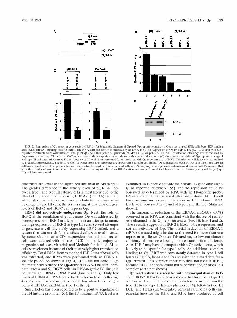
constructs are lower in the Jijoye cell line than in Akata cells.The greater difference in the activity levels of pQ1-CAT be-tween type I and type III latency cells is most likely due to theeffect of the additional repressor, EBNA-1 (Fig. 3A) (43, 50).Although other factors may also contribute to the lower activ-ity of Qp in type III cells, the results suggest that physiologicallevels of IRF-2 and IRF-7 can repress Qp.
IRF-2 did not activate endogenous Qp. Next, the role ofIRF-2 in the regulation of endogenous Qp was addressed byoverexpression of IRF-2 in a type I line in an attempt to mimicthe high expression of IRF-2 in type III cells. Several attemptsto generate a cell line stably expressing IRF-2 failed, and asystem that can enrich for transfected cells was used instead.By cotransfection of a CD4 expression plasmid, transfectedcells were selected with the use of CD4 antibody-conjugatedmagnetic beads (see Materials and Methods for details). Akatacells were chosen because of their relatively higher transfectionefficiency. Total RNA from vector and IRF-2-transfected cellswas extracted, and RPAs were performed with an EBNA-1-specific probe. As shown in Fig. 4, IRF-2 did not activate Qpbut marginally reduced the Qp-derived EBNA-1 mRNA (com-pare lanes 4 and 5). DG75 cells, an EBV-negative BL line, didnot show an EBNA-1 RNA band (lane 2 and 3). Only lowlevels of EBNA-1 mRNA could be detected in type I cells (Fig.4) (33), which is consistent with the low abundance of Qp-derived EBNA-1 mRNA in type I cells (8).
Since IRF-2 has been reported to be a positive regulator ofthe H4 histone promoter (55), the H4 histone mRNA level was
examined. IRF-2 could activate the histone H4 gene only slight-ly, as reported elsewhere (55), and no repression could beobserved as determined by RPA with an H4-specific probe.IRF-2 apparently has minimal effect on histone H4 in B-celllines because no obvious differences in H4 histone mRNAlevels were observed in a panel of type I and III lines (data notshown).
The amount of reduction of the EBNA-1 mRNA (;50%)observed in an RPA was consistent with the degree of repres-sion obtained in the Qp reporter assays (Fig. 3B, bars 1 and 2).These results suggest that IRF-2 is likely to be a repressor, andnot an activator, of Qp. The partial reduction of EBNA-1mRNA detected might be due to the need for more than onerepressor to silence Qp (see Discussion), to low enrichmentefficiency of transfected cells, or to cotransfection efficiency.Also, IRF-2 may have to compete with a Qp activator(s), whichis likely to be specific for type I cells. An additional complexbinding to Qp ISRE was consistently detected in type I celllysates (Fig. 2A, lanes 2 and 9) and might be a candidate for aQp activator. This complex apparently does not contain IRF-1,because IRF-1 antibody could not supershift and/or block thiscomplex (data not shown).
Qp reactivation is associated with down-regulation of IRF-2 and IRF-7. It has been clearly shown that fusion of a type IIIcell line with an epithelial cell line can force a switch from thetype III to the type II latency phenotype (6). KR-4 (a type IIILCL) and HeLa (EBV-negative cervical carcinoma cells) areparental lines for the KH-1 and KH-2 lines produced by cell
FIG. 3. Repression of Qp-reporter constructs by IRF-2. (A) Schematic diagram of Qp and Qp-reporter constructs. Open rectangle, ISRE; solid bars, E2F bindingsites; ovals, EBNA-1 binding sites (Q locus). The RNA start site for Qp is indicated by an arrow (46). (B) Repression of Qp by IRF-2. The pQ1-CAT and pQ2-CATreporter constructs were cotransfected with pCMVb and either pcDNA3 plasmids, pCMV-IRF-2, or pcDNA-IRF-7A. Transfection efficiency was normalized byb-galactosidase activity. The relative CAT activities from three experiments are shown with standard deviations. (C) Constitutive activities of Qp reporters in type Iand type III cell lines. Akata (type I) and Jijoye (type III) cell lines were used for transfection with Qp reporters and pCMVb. Transfection efficiency was normalizedby b-galactosidase activity. The relative CAT activities from four replicates are shown with standard deviations. (D) Endogenous levels of IRF-2 in type I and type IIIcell lines. Equal amounts of protein lysates were electrophoresed in sodium dodecyl sulfate–10% polyacrylamide gel electrophoresis and stained with Ponceau S Redafter the transfer of protein to the membrane. Western blotting with IRF-1 or IRF-2 antibodies was performed. Cell lysates from the Akata (type I) and Jijoye (typeIII) cell lines were used.
VOL. 19, 1999 IRF-2 REPRESSES EBV Qp 3219
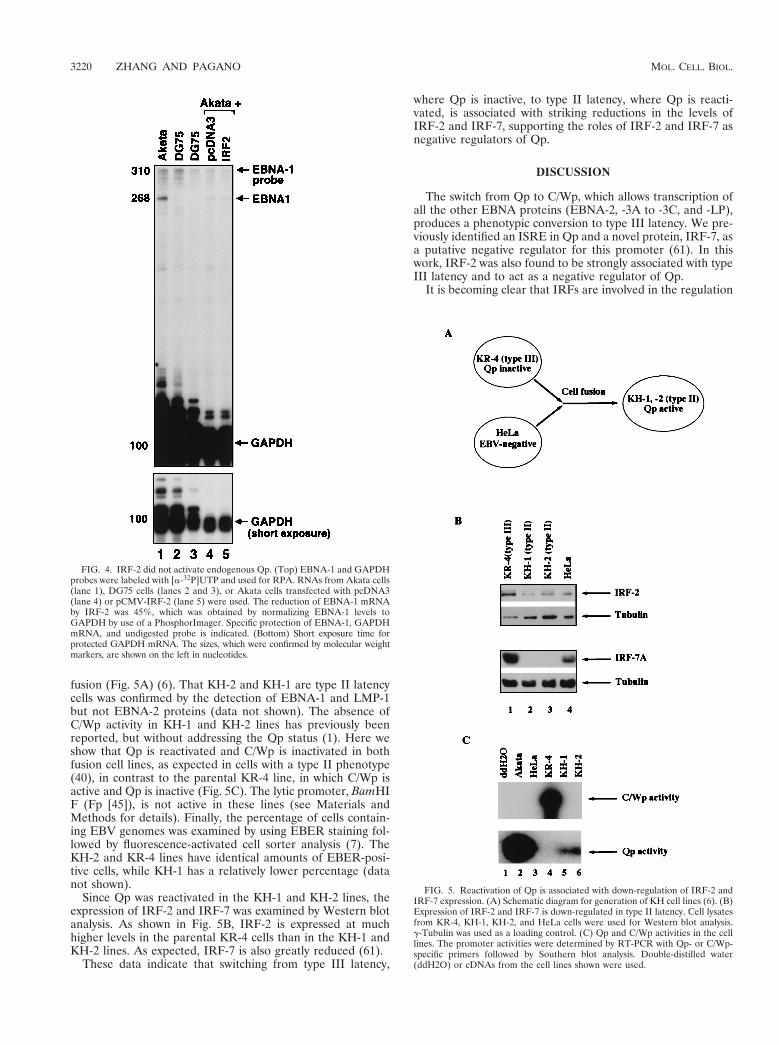
fusion (Fig. 5A) (6). That KH-2 and KH-1 are type II latencycells was confirmed by the detection of EBNA-1 and LMP-1but not EBNA-2 proteins (data not shown). The absence ofC/Wp activity in KH-1 and KH-2 lines has previously beenreported, but without addressing the Qp status (1). Here weshow that Qp is reactivated and C/Wp is inactivated in bothfusion cell lines, as expected in cells with a type II phenotype(40), in contrast to the parental KR-4 line, in which C/Wp isactive and Qp is inactive (Fig. 5C). The lytic promoter, BamHIF (Fp [45]), is not active in these lines (see Materials andMethods for details). Finally, the percentage of cells contain-ing EBV genomes was examined by using EBER staining fol-lowed by fluorescence-activated cell sorter analysis (7). TheKH-2 and KR-4 lines have identical amounts of EBER-posi-tive cells, while KH-1 has a relatively lower percentage (datanot shown).
Since Qp was reactivated in the KH-1 and KH-2 lines, theexpression of IRF-2 and IRF-7 was examined by Western blotanalysis. As shown in Fig. 5B, IRF-2 is expressed at muchhigher levels in the parental KR-4 cells than in the KH-1 andKH-2 lines. As expected, IRF-7 is also greatly reduced (61).
These data indicate that switching from type III latency,
where Qp is inactive, to type II latency, where Qp is reacti-vated, is associated with striking reductions in the levels ofIRF-2 and IRF-7, supporting the roles of IRF-2 and IRF-7 asnegative regulators of Qp.
DISCUSSION
The switch from Qp to C/Wp, which allows transcription ofall the other EBNA proteins (EBNA-2, -3A to -3C, and -LP),produces a phenotypic conversion to type III latency. We pre-viously identified an ISRE in Qp and a novel protein, IRF-7, asa putative negative regulator for this promoter (61). In thiswork, IRF-2 was also found to be strongly associated with typeIII latency and to act as a negative regulator of Qp.
It is becoming clear that IRFs are involved in the regulation
FIG. 4. IRF-2 did not activate endogenous Qp. (Top) EBNA-1 and GAPDHprobes were labeled with [a-32P]UTP and used for RPA. RNAs from Akata cells(lane 1), DG75 cells (lanes 2 and 3), or Akata cells transfected with pcDNA3(lane 4) or pCMV-IRF-2 (lane 5) were used. The reduction of EBNA-1 mRNAby IRF-2 was 45%, which was obtained by normalizing EBNA-1 levels toGAPDH by use of a PhosphorImager. Specific protection of EBNA-1, GAPDHmRNA, and undigested probe is indicated. (Bottom) Short exposure time forprotected GAPDH mRNA. The sizes, which were confirmed by molecular weightmarkers, are shown on the left in nucleotides.
FIG. 5. Reactivation of Qp is associated with down-regulation of IRF-2 andIRF-7 expression. (A) Schematic diagram for generation of KH cell lines (6). (B)Expression of IRF-2 and IRF-7 is down-regulated in type II latency. Cell lysatesfrom KR-4, KH-1, KH-2, and HeLa cells were used for Western blot analysis.g-Tubulin was used as a loading control. (C) Qp and C/Wp activities in the celllines. The promoter activities were determined by RT-PCR with Qp- or C/Wp-specific primers followed by Southern blot analysis. Double-distilled water(ddH2O) or cDNAs from the cell lines shown were used.
3220 ZHANG AND PAGANO MOL. CELL. BIOL.

of Qp. Although IRF-2 is reported to be a transactivator thatcan activate the histone H4 gene and VCAM-1 (21, 55), IRF-2 is usually a transcriptional repressor and an antagonist toIRF-1 (3, 9, 11, 15, 17, 18, 20, 27, 28, 35). It is interesting thatIRF-2 has been reported to be the major positive regulator ofQp in mouse fibroblasts (32, 44). However, the data presentedhere suggest strongly that IRF-2 is a negative regulator of Qp.First, IRF-2 expression is associated with EBV type III latency,where Qp is inactive (Fig. 1, 2, and 3D); second, overexpres-sion of IRF-2 in a type I line did not activate Qp but marginallyreduced the level of EBNA-1 mRNA derived from Qp (Fig. 4);third, IRF-2 is the major protein binding to Qp and appears tohave a high affinity for its ISRE (Fig. 2); fourth, IRF-2 re-presses the activity of Qp-reporter constructs in transienttransfection assays (Fig. 3); and fifth, Qp reactivation in type IIcells converted by cell fusion from type III cells, in which Qp isinactive, is associated with a sharp reduction in the expressionof IRF-2 (Fig. 5).
Previous reports that IRF-2 activates Qp may be due to themouse fibroblast lines that were used (32, 44). Indeed, IRF-2could activate our Qp-reporter constructs in a mouse IRF-2-null fibroblast line from the same source (Fig. 6). However,these results must be interpreted in relation to their biologicalrelevance. Selection of an appropriate line is especially impor-tant for IRF-2 research because IRF-2 possesses both a tran-scriptional repression domain and a latent activation domain(58). If the repressor domain of IRF-2 is not active in a givencell line, the latent activation domain may be activated, andIRF-2 becomes an activator. This scenario is documented inmuscle cells, where IRF-2 can activate the VCAM-1 promoter(21). Since EBV infects neither fibroblasts nor rodent cells, the
conclusion based solely on reporter assays conducted in mousefibroblasts raises questions about biological relevance. In con-trast, our results were obtained with human cell lines latentlyinfected or infectible with EBV.
Another explanation for the different results is that IRF-2might have a dual function in the regulation of Qp, i.e., it mightpositively regulate Qp in type I cells when the IRF-2 level islow and negatively regulate Qp in type III cells when expres-sion of IRF-2 is high. Data from transfections of Qp into anIRF-2-null line with high doses of IRF-2 may suggest such atendency (Fig. 6). This possibility needs to be rigorously ad-dressed. However, a dose-dependent repression of Qp by IRF-2 could be observed in biologically relevant lines, such asFaDuHyg and DG75 (data not shown).
Other than being a regulator of Qp, IRF-2 may also con-tribute to the transforming properties of EBV in type III la-tency. EBV can immortalize primary B cells and, at the sametime, establish type III latency. IRF-2 has oncogenic potentialbased on transformation assays (16, 31). It would be interestingto examine EBV-associated immunoblastic lymphomas (typeIII latency) directly to see if IRF-2 levels are elevated in thesemalignancies.
The inducer(s) of IRF-2 in type III cells is still unclear, al-though LMP-1 can stimulate the expression of IRF-7 (62). Fur-ther work needs to be done to identify the inducer(s) of IRF-2 intype III latency. Other possibilities are that the type I latencycells, in which IRF-2 and IRF-7 levels are low, are selected byEBV for infection; or that establishment of type I latency itselfinvolves the down-regulation of IRF-2 and IRF-7.
IRF-7 was cloned as a Qp-binding protein and subsequentlyinferred to be a negative regulator of Qp (61). This function isfurther supported by the fact that reactivation of Qp by fusionof type III cells with epithelial cells to produce cells with a typeII phenotype is associated with a striking reduction in the ex-pression of IRF-7 (Fig. 5). Other than being a Qp repressor, anadditional role of IRF-7, namely, virus-induced activation ofthe interferon-b gene, has just been reported (56).
It is interesting that both IRF-2 and IRF-7 are negativeregulators of Qp. There was no apparent synergistic effect be-tween these two factors in terms of Qp repression, and an ad-ditive effect was hard to detect when both were overexpressed(data not shown). Why would Qp use two repressors for a sin-gle ISRE site? One possible explanation is that IRF-2 andIRF-7 are apparently expressed at different times during thecell cycle (60). Therefore, IRF-2 and IRF-7 may functionallyrepress Qp in different phases of the cell cycle to keep Qpsilenced throughout the cycle.
Apart from IRF-2 and IRF-7, viral proteins definitely playan important role in the silencing of Qp. EBNA-1 can directlyrepress Qp activity (43, 50), and the higher levels of the proteinin type III latency may enhance its repressor effect (references8, 13, and 47 and our unpublished results). EBNA-2, which isresponsible for the increased expression of EBNA-1 in type IIIlatency, could be considered an indirect regulator of Qp.
It is clear that Qp regulation is complicated and that multi-ple factors, both viral and cellular, are involved. With IRF-2,IRF-7, and EBNA-1 all expressed at higher levels in type IIIlatency, it is reasonable to infer that Qp may be turned off bya combination of IRF-2, IRF-7, EBNA-1, and perhaps anotherrepressor(s), i.e., IRF-2 and IRF-7 may repress Qp through itsISRE while EBNA-1 acts through the Q locus (Fig. 3A). Incontrast, in type I latency, when Qp is active, all these factorsare expressed at much lower levels. The major positive regu-lator(s) of Qp operating through the ISRE is unidentified butapparently is associated with type I latency. A band whichseems to be specific for type I cells was detected by EMSA
FIG. 6. Activation of Qp-reporter constructs by IRF-2 in a mouse IRF-2-nullcell line. DKO, a mouse embryonic fibroblast line in which both IRF-1 and IRF-2 genes are disrupted (28), was used for transfection. The pQ1-CAT and pQ2-CAT reporter constructs were cotransfected with pcDNA3 plasmids or pCMV-IRF-2. The amounts of plasmids transfected are shown at the top. pCMVb wasalso cotransfected, and b-galactosidase activity was used to normalize transfec-tion efficiency. The relative CAT activities and standard deviations are shown.
VOL. 19, 1999 IRF-2 REPRESSES EBV Qp 3221

(Fig. 2) and was also noticed by other investigators (32); itmight be a candidate for such an activator.
Thus, we show that IRF-2, probably acting with IRF-7, andperhaps EBNA-1 as well, silences the promoter used in themost-restricted form of EBV latency, type I, with the indirectconsequence that the promoter for the least-restricted form ofEBV latency, type III, is used. It is likely that several IRFs,both positive and negative regulators, that have different af-finities for the type I promoter and are supplied at differenttimes in the cell cycle, govern the activity of this key, tightlyregulated EBV promoter.
ACKNOWLEDGMENTS
We thank Jenny Ting, Maria Masucci, Wanla Kulwichit, BernardWeissman, Patricia Vaughan, and Gary Stein for providing valuablereagents and/or help in this work. We thank T. Taniguchi for permis-sion to use the IRF knockout cell line. We also thank Shannon Kenneyand Nancy Raab-Traub for critical reading of the manuscript, MattDavenport and Val Zacny for editorial help, and Cyd Johnson fortechnical help.
This work was supported in part by grants from the National Insti-tute of Allergy and Infectious Diseases (AI 42372-01) and the NationalCancer Institute (CA 19014). L.Z. was supported by an NIH IndividualNational Research Service Award (5F 32 CA67433).
REFERENCES
1. Altiok, E., J. Minarovits, L. F. Hu, B. Contreras-Brodin, G. Klein, and I.Ernberg. 1992. Host-cell-phenotype-dependent control of the BCR2/BWR1promoter complex regulates the expression of Epstein-Barr virus nuclearantigens 2–6. Proc. Natl. Acad. Sci. USA 89:905–909.
2. Ben-Bassat, H., N. Goldblum, S. Mitrani, T. Goldblum, J. M. Yoffey, M. M.Cohen, Z. Bentwith, B. Ramot, E. Klein, and G. Klein. 1977. Establishmentin continuous culture of a new type of lymphocyte from a “Burkitt-like”malignant lymphoma (line D.G.-75). Int. J. Cancer 19:27–33.
3. Cha, Y., and A. B. Deisseroth. 1994. Human interferon regulatory factor 2gene. Intron-exon organization and functional analysis of 59-flanking region.J. Biol. Chem. 269:5279–5287.
4. Chen, F., J.-Z. Zou, L. di Renzo, G. Winberg, L.-F. Hu, E. Klein, G. Klein,and I. Ernberg. 1995. A subpopulation of normal B cells latently infectedwith Epstein-Barr virus resembles Burkitt lymphoma cells in expressingEBNA-1 but not EBNA-2 or LMP1. J. Virol. 69:3752–3758.
5. Cohen, J. I., F. Wang, J. Mannick, and E. Kieff. 1992. Epstein-Barr virusnuclear protein 2 is a key determinant of lymphocyte transformation. Proc.Natl. Acad. Sci. USA 86:9558–9562.
6. Contreras-Brodin, B. A., M. Anvret, S. Imreh, E. Altiok, G. Klein, and M. G.Masucci. 1991. B cell phenotype-dependent expression of the Epstein-Barrvirus nuclear antigens EBNA-2 to EBNA-6: studies with somatic cell hybrids.J. Gen. Virol. 72:3025–3033.
7. Crouch, J., D. Leitenberg, B. R. Smith, and J. G. Howe. 1997. Epstein-Barrvirus suspension cell assay using in situ hybridization and flow cytometry.Cytometry 29:50–57.
8. Davenport, M., and J. S. Pagano. Expression of EBNA-1 mRNA is regulatedby cell-cycle during Epstein-Barr virus type I latency. J. Virol, in press.
9. Drew, P. D., G. Franzoso, L. M. Carlson, W. E. Biddison, U. Siebenlist, andK. Ozato. 1995. Interferon regulatory factor-2 physically interacts with NF-kappa B in vitro and inhibits NF-kappa B induction of major histocompat-ibility class I and beta 2 microglobulin gene expression in transfected humanneuroblastoma cells. J. Neuroimmunol. 63:157–162.
10. Furnari, F. B., M. D. Adams, and J. S. Pagano. 1992. Regulation of theEpstein-Barr virus DNA polymerase gene. J. Virol. 66:2837–2845.
11. Garoufalis, E., I. Kwan, R. Lin, A. Mustafa, N. Pepin, A. Roulston, J.Lacoste, and J. Hiscott. 1994. Viral induction of the human beta interferonpromoter: modulation of transcription by NF-kB/rel proteins and interferonregulatory factors. J. Virol. 68:4707–4715.
12. Gregory, C. D., C. Dive, S. Henderson, C. A. Smith, G. T. Williams, J.Gordon, and A. B. Rickinson. 1991. Activation of Epstein-Barr virus latentgenes protects human B cells from death by apoptosis. Nature 349:612–614.
13. Gregory, C. D., M. Rowe, and A. B. Rickinson. 1990. Different Epstein-Barrvirus (EBV)-B cell interactions in phenotypically distinct clones of a Burkitt’slymphoma cell line. J. Gen. Virol. 71:1481–1495.
14. Hammerschmidt, W., and B. Sugden. 1989. Genetic analysis of immortaliz-ing functions of Epstein-Barr virus in human B lymphocytes. Nature 340:393–397.
15. Harada, H., T. Fujita, M. Miyamoto, Y. Kimura, M. Maruyama, A. Furia, T.Miyata, and T. Taniguchi. 1989. Structurally similar but functionally distinctfactors, IRF-1 and IRF-2, bind to the same regulatory elements of IFN and
IFN-inducible genes. Cell 58:729–739.16. Harada, H., M. Kitagawa, N. Tanaka, H. Yamamoto, K. Harada, M. Ishi-
hara, and T. Taniguchi. 1993. Anti-oncogenic and oncogenic potentials ofinterferon regulatory factors-1 and -2. Science 259:971–974.
17. Harada, H., E. Takahashi, S. Itoh, K. Harada, T. A. Hori, and T. Taniguchi.1994. Structure and regulation of the human interferon regulatory factor 1(IRF-1) and IRF-2 genes: implications for a gene network in the interferonsystem. Mol. Cell. Biol. 14:1500–1509.
18. Harada, H., K. Willison, J. Sakakibara, M. Miyamoto, T. Fujita, and T.Taniguchi. 1990. Absence of the type I IFN system in EC cells: transcrip-tional activator (IRF-1) and repressor (IRF-2) genes are developmentallyregulated. Cell 63:303–312.
19. Henderson, S., M. Rowe, C. Gregory, D. Croom-Carter, F. Wang, R. Long-necker, E. Kieff, and A. Rickinson. 1991. Induction of bcl-2 expression byEpstein-Barr virus latent membrane protein 1 protects infected B cells fromprogrammed cell death. Cell 65:1107–1115.
20. Horiuchi, M., G. Koike, T. Yamada, M. Mukoyama, M. Nakajima, and V. J.Dzau. 1995. The growth-dependent expression of angiotensin II type 2 re-ceptor is regulated by transcription factors interferon regulatory factor-1 and-2. J. Biol. Chem. 270:20225–20230.
21. Jesse, T. L., R. LaChance, M. F. Iademarco, and D. C. Dean. 1998. Inter-feron regulatory factor-2 is a transcriptional activator in muscle where itregulates expression of vascular cell adhesion molecule-1. J. Cell Biol. 140:1265–1276.
22. Kieff, E. 1995. Epstein-Barr virus and its replication, p. 2343–2396. In B. N.Fields, D. M. Knipe, and P. M. Howley (ed.), Virology, 3rd ed. Lippincott-Raven Publishers, Philadelphia, Pa.
23. Kozbor, D., A. E. Lagarde, and J. C. Roder. 1982. Human hybridomasconstructed with antigen-specific Epstein-Barr virus-transformed cell lines.Proc. Natl. Acad. Sci. USA 79:6651–6655.
24. Laimins, L. A., P. Gruss, R. Pozzatti, and G. G. Khoury. 1984. Character-ization of enhancer elements in the long terminal repeat of Moloney murinesarcoma virus. J. Virol. 49:183–189.
25. Levitskaya, J., M. Coram, V. Levitsky, S. Imreh, P. M. Steigerwald-Mullen,G. Klein, M. G. Kurilla, and M. G. Masucci. 1995. Inhibition of antigenprocessing by the internal repeat region of the Epstein-Barr virus nuclearantigen-1. Nature 375:685–688.
26. Levy, D. E., D. S. Kessler, R. Pine, N. Reich, and J. E. J. Darnell. 1988.Interferon-induced nuclear factors that bind a shared promoter elementcorrelate with positive and negative transcriptional control. Genes Dev. 2:383–393.
27. Lin, R., A. Mustafa, H. Nguyen, D. Gewert, and J. Hiscott. 1994. Mutationalanalysis of interferon (IFN) regulatory factors 1 and 2. Effects on the induc-tion of IFN-beta gene expression. J. Biol. Chem. 269:17542–17549.
28. Matsuyama, T., T. Kimura, M. Kitagawa, K. Pfeffer, T. Kawakami, N.Watanabe, T. M. Kundig, R. Amakawa, K. Kishihara, A. Wakeham, and T.Mak. 1993. Targeted disruption of IRF-1 or IRF-2 results in abnormal typeI IFN gene induction and aberrant lymphocyte development. Cell 75:83–97.
29. Miyashita, E. M., B. Yang, K. M. C. Lam, D. H. Crawford, and D. A.Thorley-Lawson. 1995. A novel form of Epstein-Barr virus latency in normalB cells in vivo. Cell 80:593–601.
30. Nguyen, H., J. Hiscott, and P. M. Pitha. 1997. The growing family of inter-feron regulatory factors. Cytokine Growth Factor Rev. 8:293–312.
31. Nguyen, H., A. Mustafa, J. Hiscott, and R. Lin. 1995. Transcription factorIRF-2 exerts its oncogenic phenotype through the DNA binding/transcrip-tion repression domain. Oncogene 11:537–544.
32. Nonkwelo, C., I. K. Ruf, and J. Sample. 1997. Interferon-independent and-induced regulation of Epstein-Barr virus EBNA-1 gene transcription inBurkitt lymphoma. J. Virol. 71:6887–6897.
33. Nonkwelo, C., J. Skinner, A. Bell, A. Rickinson, and J. Sample. 1996. Tran-scription start sites downstream of the Epstein-Barr virus (EBV) Fp pro-moter in early-passage Burkitt lymphoma cells define a fourth promoter forexpression of the EBV EBNA-1 protein. J. Virol. 70:623–627.
34. Pagano, J. S., G. Jimenez, N. S. Sung, N. Raab-Traub, and J. C. Lin. 1992.Epstein-Barr viral latency and cell immortalization as targets for antisenseoligomers. Ann. N. Y. Acad. Sci. 28:107–116.
35. Palombella, V. J., and T. Maniatis. 1992. Inducible processing of interferonregulatory factor-2. Mol. Cell. Biol. 12:3325–3336.
36. Pauli, U., S. Chrysogelos, G. Stein, J. Stein, and H. Nick. 1987. Protein-DNAinteractions in vivo upstream of a cell cycle-regulated human H4 histonegene. Science 236:1308–1311.
37. Pine, R. 1992. Constitutive expression of an ISGF2/IRF1 transgene leads tointerferon-independent activation of interferon-inducible genes and resis-tance to virus infection. J. Virol. 66:4470–4478.
38. Qu, L., and D. T. Rowe. 1992. Epstein-Barr virus latent gene expression inuncultured peripheral blood lymphocytes. J. Virol. 66:3715–3724.
39. Reiss, M., T. Munoz-Antonia, J. M. Cowan, P. C. Wilkins, Z. L. Zhou, andV. F. Vellucci. 1993. Resistance of human squamous carcinoma cells totransforming growth factor beta 1 is a recessive trait. Proc. Natl. Acad. Sci.USA 90:6280–6284.
40. Rickinson, A. B., and E. Kieff. 1995. Epstein-Barr virus, p. 2397–2446. InB. N. Fields, D. M. Knipe, and P. M. Howley (ed.), Virology, 3rd ed.
3222 ZHANG AND PAGANO MOL. CELL. BIOL.

Lippincott-Raven Publishers, Philadelphia, Pa.41. Rowe, M., R. Khanna, C. A. Jacob, V. Argaet, A. Kelly, S. Powis, M. Belich,
D. Croom-Carter, S. Lee, S. R. Burrows, J. Trowsdale, D. J. Moss, and A. B.Rickinson. 1995. Restoration of endogenous antigen processing in Burkitt’slymphoma cells by Epstein-Barr virus latent membrane protein-1: coordinateup-regulation of peptide transporters and HLA-class I antigen expression.Eur. J. Immunol. 25:1374–1384.
42. Sambrook, J., E. F. Fritsch, and T. Maniatis. 1989. Molecular cloning: alaboratory manual, 2nd ed. Cold Spring Harbor Laboratory, Cold SpringHarbor, N.Y.
43. Sample, J., E. Henson, and C. Sample. 1992. The Epstein-Barr virus nuclearprotein 1 promoter active in type I latency is autoregulated. J. Virol. 66:4654–4661.
44. Schaefer, B. C., E. Paulson, J. L. Strominger, and S. H. Speck. 1997. Con-stitutive activation of Epstein-Barr virus (EBV) nuclear antigen 1 genetranscription by IRF1 and IRF2 during restricted EBV latency. Mol. Cell.Biol. 17:873–886.
45. Schaefer, B. C., J. L. Strominger, and S. H. Speck. 1995. The Epstein-Barrvirus BamHI F promoter is an early lytic promoter: lack of correlation withEBNA 1 gene transcription in group 1 Burkitt’s lymphoma cell lines. J. Virol.69:5039–5047.
46. Schaefer, B. C., J. L. Strominger, and S. H. Speck. 1995. Redefining theEpstein-Barr virus-encoded nuclear antigen EBNA-1 gene promoter andtranscription initiation site in group I Burkitt lymphoma cell lines. Proc. Natl.Acad. Sci. USA 92:10565–10569.
47. Schaefer, B. C., J. L. Strominger, and S. H. Speck. 1997. Host-cell-deter-mined methylation of specific Epstein-Barr virus promoters regulates thechoice between distinct viral latency programs. Mol. Cell. Biol. 17:364–377.
48. Sinclair, A. J., I. Palmero, G. Peters, and P. J. Farrell. 1994. EBNA-2 andEBNA-LP cooperate to cause G0 to G1 transition during immortalization ofresting human B lymphocytes by Epstein-Barr virus. EMBO J. 13:3321–3328.
49. Sung, N. S., J. Wilson, M. Davenport, N. D. Sista, and J. S. Pagano. 1994.Reciprocal regulation of the Epstein-Barr virus BamHI-F promoter byEBNA-1 and an E2F transcription factor. Mol. Cell. Biol. 14:7144–7152.
50. Sung, N. S., J. Wilson, and J. S. Pagano. 1993. Characterization of cis-actingelements of the BamHI-F promoter of EBV, p. 239–242. In T. Tursz et al.
(ed.), The Epstein-Barr virus and associated diseases. INSERM/John LibbeyEurotext Limited, London, United Kingdom.
51. Takada, K. 1984. Cross-linking of cell surface immunoglobulins inducesEpstein-Barr virus in Burkitt’s lymphoma lines. Int. J. Cancer 33:27–32.
52. Tanaka, N., T. Kawakami, and T. Taniguchi. 1993. Recognition DNA se-quences of interferon regulatory factor 1 (IRF-1) and IRF-2, regulators ofcell growth and the interferon system. Mol. Cell. Biol. 13:4531–4538.
53. Tierney, R., N. Steven, L. Young, and A. Rickinson. 1994. Epstein-Barr viruslatency in blood mononuclear cells: analysis of viral gene transcription dur-ing primary infection and in the carrier state. J. Virol. 68:7374–7378.
54. Tomkinson, B., E. Robertson, and E. Kieff. 1993. Epstein-Barr virus nuclearproteins EBNA-3A and EBNA-3C are essential for B-lymphocyte growthtransformation. J. Virol. 67:2014–2025.
55. Vaughan, P. S., F. Aziz, A. J. van Wijnen, S. Wu, H. Harada, T. Taniguchi,K. J. Soprano, J. L. Stein, and G. S. Stein. 1995. Activation of a cell-cycle-regulated histone gene by the oncogenic transcription factor IRF-2. Nature377:362–365.
56. Wathelet, M. G., C. H. Lin, B. S. Parekh, L. V. Ronco, P. M. Howley, and T.Maniatis. 1998. Virus infection induces the assembly of coordinately acti-vated transcription factors on the IFN-beta enhancer in vivo. Mol. Cell 1:507–518.
57. Wilson, G., and G. Miller. 1979. Recovery of Epstein-Barr virus from non-producer neonatal human lymphoid cell transformants. Virology 95:351–358.
58. Yamamoto, H., M. S. Lamphier, T. Fujita, T. Taniguchi, and H. Harada.1994. The oncogenic transcription factor IRF-2 possesses a transcriptionalrepression and a latent activation domain. Oncogene 9:1423–1428.
59. Yoshizaki, T., H. Sato, M. Furukawa, and J. S. Pagano. 1998. The expressionof matrix metalloproteinase 9 is enhanced by Epstein-Barr virus latent mem-brane protein 1. Proc. Natl. Acad. Sci. USA 95:3621–3626.
60. Zhang, L., M. Davenport, and J. S. Pagano. Unpublished data.61. Zhang, L., and J. S. Pagano. 1997. IRF-7, a new interferon regulatory factor
associated with Epstein-Barr virus latency. Mol. Cell. Biol. 17:5748–5757.62. Zhang, L., and J. S. Pagano. Stimulation of the expression of interferon
regulatory factor 7 by Epstein-Barr virus latent membrane protein 1. Sub-mitted for publication.
VOL. 19, 1999 IRF-2 REPRESSES EBV Qp 3223