Embed Size (px)
Citation preview
2
İÇİNDEKİLER / CONTENTS
Sözlü Sunumlar / Oral Presentations .................................................................................................................................................................. 12
Kafes Kuşu Hastalıklarına Klinik Yaklaşım ............................................................................................................................... 13
Clinical Approach to Cage Bird Diseases ................................................................................................................................. 16
Davranış Problemlerine Genel Yaklaşım ve Doğru Tanı İçin İpuçları ....................................................................................... 18
Kompulsif Kuyruk Isıran Köpeklerin Serum Leptin ve Ghrelin Seviyeleri ve Serum Kortizol, Tiroid Hormonları, Lipidler,
Homosistein ve Folik Asit ile İlişkileri ........................................................................................................................................ 20
Hangisi Normal? Hangisi Anormal? .......................................................................................................................................... 22
Psikofarmakoloji ve Daha Fazlası ............................................................................................................................................ 24
Köpek ve Kedilerde Dermatozlarda Predispozan Nedenler..................................................................................................... 26
Predisposing Causes in Dog and Cat Dermatosis .................................................................................................................... 29
Comparison of stress parameters in cats with acute and chronic stress .................................................................................. 32
Köpeklerde Ebstein Anomali .................................................................................................................................................... 35
Ebstein Anomaly in Dogs ......................................................................................................................................................... 38
Hypothyroidism in dogs as a baseline of different clinical signs and laboratory results ............................................................ 39
Geçiş Dönemindeki Saanen Keçilerinde Metabolik Profillerin Değerlendirilmesi ...................................................................... 41
Evaluation Of Metabolic Profiles Of Saanen Goats İn The Transition Period ........................................................................... 42
Kanin Parvovirus ile Enfekte Köpeklerde Salya Proteomlarındaki Değişimler .......................................................................... 51
Changes Of Saliva Proteoms In Dogs With Canine Parvovirus................................................................................................ 63
Kırgız Cumhuriyetinde Veteriner Hekimliğin Tarihçesi ............................................................................................................. 64
History Of Veterinary Medicine In Kyrgyz Republıc .................................................................................................................. 69
Agresif Kedi ve Köpeklerde, İlaç Dışı Tamamlayıcı Tedavi Etkinliği-Klinik Gözlem .................................................................. 70
Complementary Nonpharmaceutical Therapy Efficiency in Canine and Feline Aggressive Behavior-Clinical Observation ..... 73
Koksidiyozis Tanısı Konulmuş Oğlaklarda Hemostasisin Tromboelastografik Olarak Değerlendirilmesi ................................. 76
Thromboelastographic Evaluation of Hemostasis in Kids Diagnosed with Coccidiosis ............................................................ 77
Bir Köpekte Ön Mitral Kapak Orifisi .......................................................................................................................................... 81
Anterior Mitral Valve Orifices In A Dog ..................................................................................................................................... 85
Neonatal Kuzularda Serum İnsülin Benzeri Büyüme Faktörü 1 (IGF-1) Üzerine Araştırmalar.................................................. 88
Investigations on İnsulin-Like Growth Factor -1 (IGF-1) İn Neonatal Lambs ............................................................................ 92
Bir Köpekte Granülositik Anaplasmosis Olgusu ....................................................................................................................... 96
Granulocytic Anaplasmosis Case In A Dog ............................................................................................................................ 102
Tüm Verileri ile Türkiye’de Bir Yaban Hayatı Kurtarma Merkezi Örneği; ................................................................................ 103
Kafkasrehab “2012-2018” ....................................................................................................................................................... 103
An Example Of Wildlife Rescue And Rehabilitatıon Centre in Turkey With All Data; ............................................................. 104
Kafkasrehab “2012-2018” ....................................................................................................................................................... 104
Bir Köpekte Çam Kese Böceği Setalarının Yenilmesine Bağlı Akut Eroziv Gastritis .............................................................. 105
Acute Erosive Gastritis Due to Pine Processionary Caterpillar Setae Ingestion in a Dog ...................................................... 106
Siddetli Sepsis ve Septik Şok Olan Köpeklerde Sistolik ve Diyastolik Fonksiyonun Değerlendirmesinde Doku Dopler
Görüntülemenin Prognostik Önemi ........................................................................................................................................ 107
Prognostic Importance of Tissue Doppler Imaging in Systolic and Diastolic Functions in Dogs with Severe Sepsis and Septic
Shock ..................................................................................................................................................................................... 108
3
Bir Kedide Aspergillus Flavus İle İlişkili Rinitis ........................................................................................................................ 109
Aspergillus Flavus in A Cat With Rhinitis ................................................................................................................................ 110
Parvoviral Enteritli Köpeklerde Bağırsak ve Kalp Hasarının Belirlenmesinde Bağırsak ve Kalbe Özgü Biyobelirteçlerin
Değerlendirilmesi .................................................................................................................................................................... 111
Assesment of Intestınal And Cardiac Biomarkers in Dogs Wıth Parvovıral Enterıtıs in Intestınal and Cardıac Damage ....... 112
Abdominal Distensiyonlu Sığırlarda Ultrasonografik Tanı....................................................................................................... 113
Ultrasonographic Diagnosis in Cattle with Abdominal Distension ........................................................................................... 114
Kalp Yetmezliği Olan Köpeklerde Benek Takibi Ekokardiyografi İle Radyal Global Gerilim ve Gerilim Hızlarının Belirlenmesi
............................................................................................................................................................................................... 115
Determination of Radial Global Strains and Strain Rates by Speckle-Tracking Echocardiography in Dogs with Heart Failure
............................................................................................................................................................................................... 116
Bir Danada Nervöz Koksidiyozis: Patogeneze Yeni Bir Bakış Açısı ....................................................................................... 117
Nervous Coccidiosis in a Calf: A New Aspect on the Pathogenesis ....................................................................................... 118
Basit İndigesyonlu Sığırlarda Serum Metilmalonik Asit ve B12 Vitamini Düzeylerinin Tanısal Önemi .................................... 119
Diagnostic Importance of Methylmalonic Acid and Vitamin B12 in Cattle with Simple Indigestion. ........................................ 120
Mitral Kapak Hastalıklı Köpeklerde Kalp Hızı Değişkenliği: Ön Çalışma ................................................................................ 121
A Preliminary Study of Heart Rate Variability in Dogs With Mitral Valve Disease .................................................................. 122
Bir Oğlakta Polioensefalomalazinin (PEM) İmmünohistokimyasal Yöntemle Tespiti: ............................................................. 123
Bir Olgu Sunumu .................................................................................................................................................................... 123
Detection of Polioencephalomalasi (PEM) in a Kid by Immunohistochemical Method: a Case Report .................................. 124
Bir Muhabbet Kuşunda Keratoakantom Olgusu ..................................................................................................................... 125
A Case of Keratoacanthoma in a Budgerigar ......................................................................................................................... 126
Diyarbakır Bölgesinde Diyareli Kedilerde Tritrichomonas Foetus’un İnsidansının Araştırılması ............................................. 127
An Investigation of the Incidence of Tritricomanas Fetus in Diarrhea Cats in Diyarbakir Region ........................................... 128
Neonatal Diyareli Buzağılarda Bağırsak Epitel Hasarının Belirlenmesinde Bağırsak Yağ Asidi Bağlayıcı Protein (I-FABP) ve
Trefoil Faktör 3 (TFF-3) Değerlendirilmesi .............................................................................................................................. 129
Evaluation Of İntestine-Fatty Acid Binding Protein (I-FABP) And Trefoil Factor 3 (TFF-3) in The Detection Of İntestinal
Epithelial Damage in Neonatal Calves With Diarrhea ............................................................................................................ 130
Farklı Enfeksiyöz Etkenlere Bağlı İshalli Neonatal Buzağılarda Kan Gaz Parametrelerindeki Değişimler .............................. 131
Changes in Blood Gas Parameters in The Neonatal Calves With Diarrhea Caused to Different Infectious Agents ............... 132
Pulmoner Adenomatozisli Koyunlarda Bazı Tümör Biyomarkırlarının (CEA, CA125 II, CA19-9, Ca 15-3 ve AFP-3) Seviyeleri
............................................................................................................................................................................................... 133
Some Tumor Biomarker Levels (CEA, CA125 II, CA19-9, Ca 15-3 And AFP-3) in Sheep With Pulmonary Adenomatosis ... 134
Canine Transmissible Venereal Tumor İn Unusual Locations Of The Body, Two Case Reports ........................................... 135
Van Kedisi’nde Görülen Sağırlığın İnsidansı ve Göz Renklerine Göre Dağılımı Üzerine Araştırmalar ................................... 138
Study on The Incidence of Deafness in Van Cats and Its Distrubution by Eye Colors ........................................................... 139
Pointer Irkı Bir Köpekte Mitral Kapak İlişkili Konjenital Hemokist Olgusu ............................................................................... 140
Mitral Valve Associated Congenital Hemocystıc Case In A Pointer ....................................................................................... 141
Köpeklerde Mitral Kapak Yetmezliğinde İki Boyutlu Benek Takibi Ekokardiyografi Yöntemiyle Belirlenen Radyal,
Sirkumferensiyal Gerilim ve Gerilim Hızı ve Torsiyonun Değerlendirilmesi ............................................................................ 142
Evaluation of Radial and Circumferential Strain and Strain Rate and Torsion Assessed by Speckle-Tracking
Echocardiography in Mitral Valve Insufficiency in Dogs ......................................................................................................... 143
Koksidiyozisli Buzağılarda Serum Beta-Defensin 1 Düzeyinin Araştırılması .......................................................................... 144
4
Investigation of Serum Beta-Defensin 1 Level in Calves With Coccidiosis ............................................................................. 145
Dermatophilus Congolensis ile Doğal Enfekte Koyunlarda Klinik, Hematolojik ve Bazı Biyokimyasal Bulgular ..................... 146
Clinical, Hematological and Some Biochemical Findings in Sheep Naturally Infected With Dermatophilus Congolensis ...... 147
Development of Specific Egg Yolk Immunoglobulins To Inhibit In-Vitro Growth Of Staphylococcus Aureus Causing Mastitis in
Bovines................................................................................................................................................................................... 148
Solunum Yolu Enfeksiyonu Olan Barınak Köpeklerinde Klinik Semptomlar Viral Ajan Varlığına Dair İpucu Verebilir mi? ..... 149
Can Clinical Symptoms Give Clue About The Presence of Viral Agents In Sheltered Dogs With Respiratory İnfection? ...... 150
Bir Oğlakta Tetanoz Olgusu ve İyileşme Süreci ..................................................................................................................... 151
Tetanus and Recovery Process in a Kid ................................................................................................................................ 155
Serological Survey Of Pestivirus Infection In Buffalo In Urmia, Iran ....................................................................................... 156
The First Survey Of Seroprevalence Of Toxoplasma Gondii In Horses Of West Azerbaijan Province, Iran ........................... 157
Ankara Bölgesinde Süt Sığırı İşletmeciliği Yapılan Çiftliklerde “Buzağılarda Pasif Transfer Yetmezliği” nin Değerlendirilmesi
............................................................................................................................................................................................... 158
Evaluation of “Passive Transfer Insufficiency in Calves” in The Farms Where Dairy Cattle Operations are Conducted in The
Ankara region ......................................................................................................................................................................... 159
Serum Immunoglobulin Responses Following Administration of Eps-Adjuvanted Vaccine For Pneumonic Pasteurellosis In
Goats ...................................................................................................................................................................................... 160
The Effect of Descurainia Sophia Seed Extract On Enzymatic Changes and Liver Damage Cause By Acetaminophen in Mice
............................................................................................................................................................................................... 161
Control of Ichthyophthirius Multifilis and Its Key Life Stage “Theronts” on Clarias Garipeinus Juveniles Using Aqaeous Leaves
Extract of Moringa Oleifera ..................................................................................................................................................... 162
Veteriner Kardiyolojide İki Boyutlu Benek Takibi Ekokardiyografi Kullanımı ........................................................................... 163
How To Use Two Dimensional Speckle Tracking Ecocardiography In Veterinary Cardiology ................................................ 168
İç Hastalıkları Kliniğine Getirilen Hastaların Retrospektif Olarak Değerlendirilmesi: 18198 Hastada Ankara Üniversitesi
Deneyimi ................................................................................................................................................................................ 169
A Retrospective Study of Veterinary Patients in Internal Medicine Clinic: Ankara University Experience on 18198 Animals . 170
Saha Çalışması İle Cryptosoporidium Enfeksiyonunun Ege Bölgesi Buzağılarında Anlık Dağılımının Tespiti ....................... 171
Field study regarding spatial distribution of cryptosoporidium infection among Eagean Region calves ................................. 173
Feline Gingivostomatitislerin Tedavisinde Hipokloröz’ün Klinik Başarısı ................................................................................ 174
Clinical Success of Hypochlorous in the Treatment of Feline Gingivostomatitis ..................................................................... 175
Neonatal İshalli Buzağıların Aksaray Bölgesinde Anlık Dağılımının Belirlenmesi ................................................................... 176
Determination of The İnstant Distribution of Calves With Neonatal Diarrhea in Aksaray Region ........................................... 177
Merada Beslenmeyen İneklerde İnterstitiel Anfizem Olguları ................................................................................................. 178
Interstitial Emphysema Cases in Nongrazing Cows ............................................................................................................... 179
Türkiye’de Kedilerde İlk Kalıtsal Polikistik Böbrek Hastalığı (PKD) ........................................................................................ 180
First Report of Hereditary Polycystic Kidney Disease (PKD) of Cats in Turkey ...................................................................... 181
Hayvan Sağlık Sigortası Yaptırma İsteğini Etkileyen Faktörler Sivas İli Örneği ...................................................................... 182
Factors Affecting The Request For Animal Health Insurance Sivas Province Sample ........................................................... 183
Serum Leptin And Ghrelin Levels And Their Relationship With Serum Cortisol, Thyroid Hormones, Lipids, Homocysteine And
Folic Acid İn Dogs With Compulsive Tail Chasing .................................................................................................................. 184
Geçiş Dönemindeki Sığırlarda Hastabaşı Bazı Metabolik Parametreler Arasındaki Korelasyonun Değerlendirilmesi* .......... 185
Correlation of Cowside Measured Some Metabolic Parameters in Transition Cattle* ............................................................ 186
Akut İshalli Neonatal Buzağılarda Plazma Sitrulin Konsantrasyonunun Değerlendirilmesi .................................................... 191
5
Evaluation of Plasma Citrulline Concentration in Neonatal Calves with Acute Diarrhea ........................................................ 192
Kedi ve Köpeklerde Yaşa Bağlı Değişimlere İlgili Hayvan Sahiplerinin Algılarının Değerlendirilmesi ..................................... 193
Evaluatıon of Perceptıons of Anımal Owners Related To Age-Related Changes In Cat And Dogs ....................................... 194
Atopik Dermatitli Köpeklerde Fekal Mikrobiyota Transplantasyonu ile Sağaltım: Derinin Altında, Bağırsakların İçerisinde
Gastroentero-Dermatolojiye Açılan Kapılar ............................................................................................................................ 195
Fecal Microbiota Transplantation Treatment in Dogs With Atopic Dermatitis: Doors Opening To Gastroentero-Dermatology
Under The Skin, İnside The Gut ............................................................................................................................................. 196
Demographic data ............................................................................................................................................................ 197
Kuzularda antigiardial amaçla silikon dioksit kullanımı ........................................................................................................... 199
Anti-giardial silicon dioxide usage in lambs ............................................................................................................................ 199
Poster Sunumlar / Poster Presentations ........................................................................................................................................................... 202
Holstein Neonatal Buzağılarda Probiyotik Katkısının Serum Leptin Düzeyleri Üzerine Etkisi................................................. 203
The Efficacy Of Probiotic Supplementation In Holstein Calves On Serum Leptin Levels ....................................................... 204
Neden Veteriner Dermatolojide kortikosteroidleri ya da diğer bazı immunosupresif moleküler ajanları kullanamazsınız?
Aslında kullanmamalısınız… .................................................................................................................................................. 205
Why you should not use corticosteroids or selected immunosupressive molecular agents? In real you can not. .................. 206
Kopay Irkı Bir Köpekte Dirofilariosis: Ekokardiyografi ile Pulmoner Arter’de Etkenin Saptanması ......................................... 207
Dirofilariasis in A Kopay Dog: Determination of Etiological Agent on Pulmonary Artery Via Echocardiography ..................... 208
İki Kısa Tüylü Evcil Kedide Dilate Kardiyomiyopati Olgusu ve Pimobendan ile Başarılı Kontrolü ........................................... 209
2 Case Reports of Dilated Cardiomyopathy in Two Short-Haired Domestic Cats and Successful Control with Pimobendan 210
Rottweiler Irkı Bir Köpekte Kutanöz Leishmaniazis Olgusu .................................................................................................... 211
Cutaneous Leishmaniasis Case in a Rottweiler Dog .............................................................................................................. 215
Patent Duktus Arteriozus - 4 Köpekte Olgu Raporu ............................................................................................................... 216
Patent Ductus Arterıosus - Case Report İn 4 Dogs ................................................................................................................ 217
Cor Triatriatum sinister - 3 Yaşlı Shih-Tzu Irkı Köpekte Vaka Raporu .................................................................................... 218
Cor Triatriatum sinister in 3-year-old Shih-Tzu Dog – Case Report ....................................................................................... 219
Koriza Gangrenoza Bovumun Baş Göz Formu Teşhis Edilen Sığırlarda Kan ve Bos Biyokimyası, Hematolojik Parametreler
İle Bos Sitolojisinin Değerlendirilmesi ..................................................................................................................................... 220
Evaluation Of Blood And Csf Biochemıstry, Csf Cytology And Hematological Parameters in Head Eye Form of Mcf
Determined Cattles ................................................................................................................................................................. 221
Sepsisli Neonatal Buzağılarda Artmış Serum Kardiyak Troponin-I Konsantrasyonu ve Kardiyak Enzim Aktiviteleri .............. 222
The Increased Cardiac Troponin-I Concentration and Cardiac Enzyme Activities in Neonatal Calves with Sepsis ............... 223
Streptococcus pluranimalium ile Doğal Enfekte Koyunlarda Serum Haptoglobin, Nitrik Oksit ve Malondialdehit Düzeylerinin
Değerlendirilmesi .................................................................................................................................................................... 224
The Assessment of Serum Haptoglobin, Nitric Oxide and Malondialdehyde Levels in Sheep Infected Naturally with
Streptococcus Pluranimalium ................................................................................................................................................. 225
Sığır Solunum Hastalıkları Kompleksli Buzağılarda Serum Demir ve Ferritin Düzeylerinin Değerlendirilmesi ....................... 226
The Evaluation of Serum Iron and Ferritin Levels in Calves with Bovine Respiratory Disease Complex ............................... 227
Bir Köpekte Septik Şoka Bağlı Gelişen Rabdomiyoliz Olgusu: Olgu Sunumu ........................................................................ 228
Rhabdomyolysis Triggered By Septic Shock İn A Dog: A Case Report ................................................................................. 229
Doramektin Zehirlenmesinin İntravenöz Lipid Emülsiyonu ile Tedavisi: Beş Amerikan Pitbull Terrier Yavrusu ...................... 230
Treatment of Doramectin Poisoning with Intravenous Lipid Emulsions: Five American Pitbull Terrier Puppies ..................... 231
Game Irkı Bir Köpekte Akut Salisilik Asit Toksikasyonu ......................................................................................................... 232
6
Acute Salicylic Acid Poisoning in a Game Dog ....................................................................................................................... 233
Klinik Olarak Sağlıklı Bir Kedide Akut Fulminant Pulmoner Ödem: Hipertrofik Kardiyomyopatili Asemptomatik Kedilerde
Preoperatif Kardiyak Kontrollerin Önemi ................................................................................................................................ 234
Acute Fulminant Pulmonary Edema in a Clinically Healthy Cat: Importance of Pre-operative Cardiac Controls in
Asymptomatic Cats with Hypertrophic Cardiomyopathy ......................................................................................................... 235
Bir Gümüş Martı’da (Larus michahelis) Kloakalite Bağlı Kloakal Tıkanıklık ............................................................................ 236
Cloacal Obstruction Due to Cloacolith in a Yellow-legged gull (Larus michahellis). ............................................................... 237
İki Köpekte Kronik Otoimmun Pododermatitis ........................................................................................................................ 238
Canine Chronic Otoimmune Pododermatitis in Two Dogs...................................................................................................... 240
Köpeklerde Serum Üre, Kreatinin ve Simetrik Dimetil Arginin (SDMA) Düzeyleri Arasındaki İlişkinin Belirlenmesi ................ 242
Relationships Between Symmetric Dimethylarginine (SDMA), Blood Urea Nitrogen And Creatinine Levels In Dogs ............ 249
Köpeklerde İki Farklı Trombositçe Zengin Plazma (PRP) Kit Sisteminin Özellikleri İle Terapötik Başarı Potansiyellerinin
Karşılaştırılması ...................................................................................................................................................................... 255
Comparison of Characteristics and Therapeutic Potentials of two Different Platelet Rich Plasma (PRP) Kit Systems in Dogs
............................................................................................................................................................................................... 262
Kuyruk Travmalı Bir Kedide Trombositçe Zengin Plazma (PRP) Uygulaması ........................................................................ 268
Platelet Rich Plasma (PRP) Application in a Cat With Tail Trauma ........................................................................................ 270
Bir Köpekte Enterococcus Faecalis’e Bağlı Gelişen Nadir Septik Perikardit Olgusu .............................................................. 272
A Rare Enterococcus Faecalis Associated Pericarditis Case İn A Dog .................................................................................. 273
Şap Hastalıklı Buzağılarda Serum Malondialdehit, Koenzim Q10 ve 8-hidroksi-2-Deoksiguanozin Seviyeleri....................... 274
Serum Malondialdehyde, Coenzyme Q10 and 8-hydroxy-2-deoxyguanosine Levels in Calves with Foot-and-Mouth Disease
............................................................................................................................................................................................... 275
Irak, Erbil Bölgesinde Kronik Solunum Sistemi Hastalığı Olan Koyunlarda Maedi-Visna Hastalığının Serolojik Olarak
Araştırılması ........................................................................................................................................................................... 276
Serological Investigation of Maedi-Visna in Sheep with Chronic Respiratory Disease in Erbil, Iraq ....................................... 277
Solunum Sistemi Hastalıklı Buzağılarda Farklı Oksijen Uygulama Yöntemlerinin Arteriyel ve Venöz Kan Gazları Üzerine
Etkileri..................................................................................................................................................................................... 278
Effects of Different Oxygen Application Methods on Arterial and Venous Blood Gases in Calves with Respiratory System
Disease .................................................................................................................................................................................. 279
Kedilerde Viburnum Opulus Un Antiürolitiatik Etkinliği - Ön Çalışma ..................................................................................... 280
Anti-Urolithiatıc Effectiveness of Viburnum Opulus In Cats - Preliminary Study ..................................................................... 283
Fasciola hepatica ile Doğal Enfekte Koyunlarda serum Amyloid A, Haptoglobin ve Fibrinogen Düzeylerinin Araştırılması .. 286
Investigation of serum Amyloid A, haptoglobin and fibrinogen levels in sheep infected with Fasciola hepatica ..................... 287
Nanobiyogümüş Kullanımının Akut, Subakut ve Subkronik Toksik Etkilerinin Rodent Modellerinde Araştırılması ................. 288
Investigation Of Acute Subacute And Subchronic Toxic Effects Of Nanobiosilver In The Rodent Model ............................... 289
Köpeklerde Viburnum Opulus'un Antiürolitiatik Etkinliği- Ön Çalışma .................................................................................... 290
Anti-Urolithiatic Effectiveness of Viburnum Opulus in Dogs - Preliminary Study .................................................................... 293
Bir Köpekte Otoimmun Poliglandüler Endokrinopati (Schmidt S Sendrom) ............................................................................ 296
Autoimmune Polyglandular Endocrınopathy In A Dog (Schmidt S Syndrome) ....................................................................... 297
Bir Buzağıda Kistik Urakal Kalıntı Sonucu Şekillenen Üretral Tıkanıklık Ve Hidronefroz Olgusu ........................................... 298
A Case Of Urethral Obstructıon And Hydronephrosıs Caused By A Cystıc Urachal Remnant In A Calf ................................ 299
Köpeklerde Atopik Dermatit Tedavisinde Prebiyotik, Probiyotik ve Postbiyotik Kullanımı ...................................................... 300
Prebiotic, Probiotic And Postbiotic Usage in The Treatment of Atopic Dermatitis in Dogs ..................................................... 301

7
Kriptosporodiozisli Buzağılarda D-Dimer Seviyelerinin Belirlenmesi ...................................................................................... 302
Determination of D-Dimer Levels in Calves with Cryptosporidiosis ........................................................................................ 303
Babesia canis ile Enfekte Köpeklerde Serum Amiloid-A, Haptoglobin, Seruloplazmin ve Albumin Seviyeleri* ...................... 304
The Serum Amyloid-A, Haptoglobin, Ceruloplasmin and Albumin Levels in the Dogs Which are Infected with Babesia Canis*
............................................................................................................................................................................................... 305
Neonatal İshalli Buzağılarda 25 (Oh) D3 Konsantrasyonlarının Araştırılması ........................................................................ 306
Evaluation of 25 (Oh) D3 Concentrations in Neonatal Calves with Diarrhea .......................................................................... 307
Buzağılarda Boynuzsuzlaştırma, Plazma Kortizol Ve Vitamin D3 Seviyeleri Arasındaki İlişkinin Belirlenmesi ....................... 308
Evaluation of Plasma Cortisol and Vitamin D3 Levels in Calves Undergoing Disbudding ...................................................... 309
Burdur ve Yöresinde Yaşayan Yaban Domuzların Mide ve Barsaklarında Bulunan Parazitler Üzerine Araştırma ................. 310
Burdur ve Yöresinde Yaşayan Yaban Domuzların Mide ve Barsaklarında Bulunan Parazitler Üzerine Araştırma ................. 311
Parvovirüs İle Enfekte Köpeklerde Serum C Reaktif Protein Değerlerinin Belirlenmesi ......................................................... 312
Determination of Serum C Reactive Protein Levels In Dogs Infected With Parvovirus .......................................................... 313
Köpeklerde Parvoviral Gastroenteritiste Rektal Yolla Enteroccocus faecium SF68 Probiyotik Sağaltımı ............................... 314
Enteroccocus faecium SF68 Probiotic Usage Via Rectal Route Against Parvoviral Gastroenteritis Among Dogs ................. 315
8
KONGRE DÜZENLEME KOMİTESİ / CONGRESS ORGANISATIONS COMMITTEE
Kongre Başkanı / Chair of Congress:
Doç. Dr. Buğrahan Bekir YAĞCI
Kongre Sekreteri / Secretaries of Congress:
Doç. Dr. Sibel YASA DURU
Üyeler / Members of Committee:
Doç. Dr. Naci ÖCAL
Doç. Dr. Serkal GAZYAĞCI
Araş. Gör. Erdal KARA
Araş. Gör. Yasin PARLATIR
DANIŞMA KURULU / ADVISORY BOARD
Zahid Tevfik AĞAOĞLU (Cumhuriyet Üniversitesi)
Alev AKDOĞAN KAYMAZ (İstanbul Üniversitesi)
Yakup AKGÜL (Yüzüncü Yıl Üniversitesi)
Gürbüz AKSOY (Harran Üniversitesi)
Hasan BATMAZ (Uludağ Üniversitesi)
Mehmet Kazım BÖRKÜ (Ankara Üniversitesi)
Mehmet ÇİTİL (Erciyes Üniversitesi)
Yusuf GÜL (Fırat Üniversitesi)
Aslan KALINBACAK (Ankara Üniversitesi)
Engin KENNERMAN (Uludağ Üniversitesi)
Arif KURTDEDE (Ankara Üniversitesi)
Mehmet MADEN (Selçuk Üniversitesi)
Mahmut OK (Selçuk Üniversitesi)
Serdar PAŞA (Adnan Menderes Üniversitesi)
Mutlu SEVİNÇ (Selçuk Üniversitesi)
Mehmet ŞAHAL (Ankara Üniversitesi)
Kürşat TURGUT (Yakın Doğu Üniversitesi)
9
KONGRE BİLİMSEL KOMİTESİ / CONGRESS SCIENTIFIC COMMITTEE
Abuzer ACAR (Kocatepe Üniversitesi, Afyon, TÜRKİYE / TURKEY) Zbigniew ADAMIAK (Warmia-Mazury University, Olsztyn, POLAND / POLONYA) Sinan AKTAŞ (Atatürk Üniversitesi, Ezurum, TÜRKİYE / TURKEY) Metin Koray ALBAY (Mehmet Akif Ersoy Üniversitesi, Burdur, TÜRKİYE / TURKEY) Arash ARAGHİ-SOOREH (Islamic Azad University, Urmia, IRAN / İRAN) Öznur ASLAN (Erciyes Üniversitesi, Kayseri, TÜRKİYE / TURKEY) Irena CELESKA (Cyril&Methodius Uni., MACEDONIA / MAKEDONYA) Turan CİVELEK (Afyon Kocatepe Üniversitesi, Afyon, TÜRKİYE / TURKEY) İlker ÇAMKERTEN (Aksaray Üniversitesi, Aksaray, TÜRKİYE / TURKEY) Cenker Çağrı CINGI (Kocatepe Üniversitesi, Afyon, TÜRKİYE / TURKEY) Ali Evren HAYDARDEDEOĞLU (Aksaray Üniversitesi, Aksaray, TÜRKİYE / TURKEY) Murat GÜZEL (Ondokuz Mayıs Üniversitesi, Samsun, TÜRKİYE / TURKEY) Askarbek TÜLÖBAYEV*, Nariste KADIRALİEVA* Hasan GÜZELBEKTEŞ *,** Hasan GÜZELBEKTEŞ (Kyrgyz-Turkish Manas University, Bishkek, KYRGYZSTAN / KIRGHIZISTAN) Hasan İÇEN (Dicle Üniversitesi, Diyarbakır, TÜRKİYE / TURKEY) Çağrı KARAKURUM (Mehmet Akif Ersoy Üniversitesi, Burdur, TÜRKİYE / TURKEY) Abdullah KAYAR (İstanbul Üniversitesi, İstanbul, TÜRKİYE / TURKEY) Muhammed KATICA (Sarajevo University, BOSNIA&HERZOGOVINA / BOSNA HERSEK) Ali Haydar KIRMIZIGÜL (Kafkas Üniversitesi, Kars TÜRKİYE / TURKEY)
10
Ömer KIZIL (Fırat Üniversitesi, Elazığ, TÜRKİYE / TURKEY) Lora Koenhemsi (İstanbul Üniversitesi, İstanbul, TÜRKİYE / TURKEY) Koycho KOEV (Stara Zagora University, BULGARİSTAN) Süleyman KOZAT (Yüzüncü Yıl Üniversitesi) Lazarin LAZAROV (Trakia University, Stara Zagora, BULGARIA / BULGARİSTAN) Mehmet Erman OR (İstanbul Üniversitesi, İstanbul, TÜRKİYE / TURKEY) Przemysław SOBIECH (Warmia-Mazury University, Olsztyn, POLAND / POLONYA) Askarbek TÜLÖBAYEV (Kyrgyz-Turkish Manas University, Bishkek, KYRGYZSTAN / KIRGIZİSTAN) Mutlu TEMİZEL (Uludağ Üniversitesi, Bursa, TÜRKİYE / TURKEY) Kerem URAL (Adnan Menderes Üniversitesi, Aydın, TÜRKİYE / TURKEY) Erdoğan UZLU (Kafkas Üniversitesi, Kars, TÜRKİYE / TURKEY) Karolina WRZESNİEWSKA (University of Life Sciences, Lublin, POLAND / POLONYA) Ebru YALÇIN (Uludağ Üniversitesi, Bursa, TÜRKİYE / TURKEY) Katarzyna ŻARCZYŃSKA (Warmia-Mazury University, Olsztyn, POLAND / POLONYA)
11
KONGRE ONUR KURULU / CONGRESS HONOR BOARD
Prof. Dr. Cemal Nadi AYTUĞ
Prof. Dr. Rauf CAN
Prof. Dr. Hikmet ÜNSÜREN
Prof. Dr. Hüseyin Yılmaz İMREN
Prof. Dr. Hüseyin TAN
Prof. Dr. Kemal YILMAZ
Prof. Dr. Mehmet Besim ÖZLEM
Prof. Dr. Veysi ASLAN
Prof. Dr. Nilüfer AYTUĞ
Prof. Dr. Abdulkadir UYSAL
Prof. Dr. Tamer DODURKA
Prof. Dr. Kemal IRMAK
Prof. Dr. Tarık BİLAL
12
Sözlü Sunumlar / Oral Presentations
13
Sözlü Sunu / Oral Presentation
(ÇAĞRILI TEBLİĞ) Tam Metin / Full Text
Kafes Kuşu Hastalıklarına Klinik Yaklaşım
Arif KURTDEDE
Ankara Üniversitesi Veteriner Fakültesi, İç Hastalıklar Anabilim Dalı, Ankara, Türkiye
Kafes kuşları anatomik ve fizyolojik olarak birbirlerine benzese de; doğal yaşam şartlarındaki farklılıklar kuşların
beslenme tarzlarında ve besin tercihlerinde farklılıklara yol açmıştır. Bu durum, kuşların başta gaga, kursak,
proventrikulus, ventrikulus, bağırsaklar ve kloaka olmak üzere, tüm vücut organlarında yapısal ve fizyolojik
değişikliklere neden olmuştur.
Kuş organlarında, yaşadıkları ortama göre gelişen yapısal ve fizyolojik özellikler, organların çeşitli hastalık etkenlerine
karşı duyarlıklarında da değişikliklere neden olmuştur. Ayrıca, kuşların yaşamları süresince karşılaştıkları
mikroorganizmalara karşı geliştirdikleri savunma mekanizmaları, kuş türlerinde hastalıklara karşı gelişen direnç
farklılığında rol oynayan diğer önemli bir faktördür.
Doğada kuşların gaga ve ağız yapıları, karşılaştıkları canlı-cansız gıda çeşitlerini gagayla tutma, parçalama ve onları
pozisyonlandırarak yutma yönünde gelişmiştir. Kuşlar doğal şartlarında çok farklı kaynaklardan tohum, bitki parçaları,
meyveler, canlı kurtçuklar, su canlıları, böcekler, sinekler ve diğer katı-sıvı materyalleri alırlar. Kuşların organları ve
metabolizmaları ve vücut hücre ve sıvılarının nitelikleri ve hastalık etkenlerine karşı savunma mekanizmaları, uzun
yıllar içinde, beslenme özellikleriyle uyumlu, hatta bunlara bağımlı hale gelmiştir.
Doğasından alınarak kafeslerde bakılmaya başlanan kuşlara nasıl bir besleme yapılacağı konusunda henüz kesin
ve yerleşik bilgiler bulunmamaktadır. Kuşların beslenme tarzı ile ilgili bilgiler ve mevcut uygulamalar; kuş yetiştiriciliği
ve satıcılığını yapan kişilerin, müşterilerine kuş satarken, birlikte sattıkları gıdalar ve verdikleri kısır bilgilerden
ibarettir. Örneğin kafes kuşlarının beslenmesi ile ilgili olarak genel olarak şu bilgiler verilmektedir: "Küçük psittakinler
(muhabbet kuşları) darı ile beslenirler; Büyük boyutlu psittakinler (afrika grisi papağanı) tuzsuz ve çiğ ay çekirdeği ve
fıstık ile beslenirler; Ötücü kuşlar (kanarya, bülbül, saka) ise kanarya tohumu ile beslenirler". Alışkanlıklara bağlı
olarak dillendirilen bu beslenme tarzının, kuşlarda yaşam süresince bazı sorunlara yol açtığı görülmektedir. Örneğin
muhabbet kuşlarında, vücut yağ miktarında artış ve xanthom, Afrika grisi papağanında hipokalsemi ve A
hipovitaminozis, bu tarz beslenmenin neden olduğu bozukluklara iki örnektir.
Kafes kuşlarında; kuş çeşidine göre değişmek üzere yapılması gereken beslenmede, hangi gıdaların ne miktarda
verileceği konusu tam anlaşılamamıştır. Kuş yemlerinin çeşitleri ve kuşlara verilecek miktarları kuş çeşidine göre
değişmektedir. Kafes kuşlarına hayvansal veya bitkisel kaynaklardan elde edilen karbonhidratlar, yağlar ve proteinler
gibi temel besin maddelerinin yanı sıra, A vitamini, D vitamini, kalsiyum ve iyot mutlaka verilmelidir.
Afrika grisi papağanlarında; A vitamini eksikliğinde alt gaga altında kese oluşumu, D vitamini ve kalsiyum
yetersizliğinde hipokalsemi belirtilerinin gelişmesi ve savunma sisteminde zayıflama; muhabbet kuşlarında;
kalsiyum yetersizliğinde kemik bütünlüğünde bozulma, iyot yetersizliğinde guatr hastalığının oluşumu örnek olarak
gösterilebilecek bozukluklardır.
Klinik gözlemlerimize göre; kafes kuşlarının kuş yetiştirici ve satıcılarının önerdikleri çeşitli tohumlarla, ay çekirdeğiyle
ve fıstıkla beslendikleri; kuşlara kalsiyum kaynağı olarak sunulan kalsiyum bloklarının ne derecede yararlı olduğunun
şüpheli olduğu; ayrıca kalsiyum, iyot, A vitamini ve D vitamini yetersizliklerinin kuş sahiplerince öneminin ve bunların
hangi kaynaklardan nasıl karşılanacağı konusunun çok iyi kavranmadığı görülmektedir.
Kafes kuşlarında enfeksiyöz hastalıkların çeşidi ve semptomları ile bunların kuş çeşitlerinde görülme sıklığı ve
patogenezisi farklılıklar göstermektedir. Örneğin muhabbet kuşlarında PBFD, kandidiyazis, aspergillozis, polyama
virüs enfeksiyonu, giardiyazis, ektoparziter enfeksiyonlar; Afrika grisi papağanlarında PBFD, psittakozis, tenya
14
enfeksiyonu; Kanaryalarda Circo virüs enfeksiyonu ve Pox virüs enfeksiyonu dikkati çekmektedir. Bu hastalıkların
tanısı genellikle etiyolojik olarak ortaya konulamamaktadır. Hastalıklar, hastalarda ortaya çıkan semptomlar,
hastalıkların klinik seyri, hastalığın bulaşıcılığı ile morbidite-mortaliteleri dikkate alınarak olası tanı şeklinde
tanınmaktadır.
Enfeksiyöz hastalıklara karşı amprik olarak bazı antibiyotikler kullanılmakta, kafes malzemeleri çeşitli
dezenfektanlarla (çamaşır suyu, benzalkonium hidroklorit, klorhekzidin glukonat gibi) temizlenmektedir. Hastalığın
yüksek mortalite-morbidite ile seyrettiği yetiştiriciliklerde; işletmedeki kuşlar tamamen boşaltılmakta, işletme
dezenfekte edilmekte ve işletme bir süreliğine kapatılmaktadır. Enfeksiyöz hastalıklara karşı koruyucu aşı
uygulaması bulunmamaktadır. Ektoparaziter ve endoparaziter enfeksiyonlardan şüphelenildiğinde antiparaziter
ilaçlar uygulanmaktadır. Mantar enfeksiyonlarında; kandidiyazise karşı (özellikle, başta kursak olmak üzere, diğer
organlarda) nistatin; aspergillozise karşı itrakonazol ve vorikonazol gibi antifungal ilaçlar kullanılmaktadır. Bu
uygulamaların sonuçlarının değerlendirilmesinde, genellikle hasta kuşta dışarıdan görülen klinik düzelme belirtileri
ve radiyografik olarak patolojik görüntülerdeki gerilemeler dikkate alınmaktadır.
A, D vitamini, iyot ve kalsiyum yetersizliğine bağlı kronik bozukluklar ile hipoglisemiye bağlı akut bozukluklar fazlaca
dikkate alınmayan bozukluklardır ve semptomatik sağaltım uygulamaları (vitamin-mineral-amino asit) kapsamında
sağaltılmaktadır. Deneyimlerimize göre bilinçli rutin uygulamalarla bu bozuklukların oluşması önlenebilir.
Kafes kuşlarında her türlü organ bozuklukları ve organlardaki neoplastik bozukluklar; karın içindeki organlarda
geliştiğinde karın genişlemesi ve solunum güçlüğü ortaya çıkar. Akciğer ve hava keselerinde olan bozukluklarda
solunum güçlüğü gözlenir. Kursak hastalıklarında; kursak genişlemesi ve tıkanması belirlenir. Baş bölgesi
bozukluklarında genel veya lokal nörolojik belirtiler ortaya çıkar. Karaciğer bozukluklarında, sindirim ile ilgili
bozukluklar ve vücut dokularında sarı renkli görünüm gözlenir. Böbrek bozukluklarında poliüri ve damlalarda bol ürat
atılımı ortaya çıkar. Pankreas bozukluklarında dışkı renginde açılma dikkati çeker.
Kafes kuşlarındaki bozuklukların tanısında; öncelikle kuş sahibinden anamnez bilgileri alınır "kuşun alındığı
yetiştiricilik, kuşların bakımı-beslemesi, kuştaki hastalığın başlaması, seyri, bulaşıcılığı ve oluşturduğu mortalite ile
ilgili ". Daha sonra hasta kuşta görülen belirtiler (kuşun tüneği kavraması, başın pozisyonu, kanatların duruşu, nefes
alış, kuyruğun hareketi, gözler, baş tüyleri ve vücut tüyleri, sere, burun delikleri, gaga, gözler, periorbital sinüsler)
gözlenir. Kafesin yapısı, kafesteki malzemelerin hijyenik durumu, kafesteki damlaların kontrolü ve kafesteki
malzemelere kanın bulaşıp bulaşmadığı incelenir.
Hastalığın tanısının konulmasında; hasta kuşun çeşidine göre, kuşun hastalıklara duyarlılığının bilinmesi, radyografik
görüntülerin değerlendirilmesi, mikrobiyolojik analiz bulguları ve patolojik bulgular önemli veriler sağlamaktadır. Kan
hücrelerinin sayımı ve kan biyokimyası verileri henüz yeterince değerlendirilmemektedir. Kuş hastalıklarının tanısı ve
sağaltımında klinik deneyim çok önemli bir yer tutmaktadır.
Kafes kuşlarının sağaltımındaki en önemli sorun, kuşlara ilaçların nasıl verileceğidir. İlaçların suya katılması; suyun
niteliğinin ne olduğunun bilinmemesi, ilacın sudaki dağılımı, ilacın suda eriyebilirliği, ilacın aktif maddesinin sudaki
aktivitesi, hasta kuşun suyu içme miktarı ve suyla ne kadar ilacın alınabileceği önemli sorunlardır. İlaçların kuşun
gagası yoluyla ağızdan verilmesi durumunda; ilacın tadı ve kuş tarafından kabul edilebilirliği, ilacın ne kadarının
yutulabildiği, kursağa giden ilacın ne kadarının geri çıkarıldığını bilmek güçtür. Ayrıca kuşlar ilk ilaç uygulamasından
sonraki uygulamalarda ilaç içmeyi reddedebilir. İlaçların kursak sondası ile kursağa ulaştırılması durumunda, ilacın
kursaktan ne kadarının geri çıkarılacağı sorularını akla getirmektedir.
Klinik gözlemlerimize göre; ilaçlar, küçük boyutlu psittakinlere (muhabbet kuşu, love bird, cockatiel gibi) gaga
aracılığıyla ağızdan verilebilir yada ilaçlar kursak sondası aracılığıyla kursak içi verilebilir. Bu ilaçlardan tadı kuş
tarafından kabul edilmeyenlerin bir kısmı (özellikle oral kullanım için hazırlanmamış olanları), regurgite edilmektedir.
Bu kuşlara ilaçlar suya eklenerek verilebilmektedir. Kas içi uygulamada göğüs kasları uygun bir uygulama yeridir,
fakat kuşlar kas içi uygulamaya ciddi reaksiyon göstermektedir. Deri altı uygulamada, arka ayak ile karın bölgesi
arası deri uygulama yapılabilir bir bölge olarak dikkati çekmiştir. Büyük boyutlu psittakinlere (Afrika grisi papağanı,
makav, kokadu ve benzeri boyuttakiler) ağızdan ilaç vermek neredeyse imkansızdır. İlaçlar suya katıldığında ilaçlı
15
suyun kuş tarafından red edildiği görülmüştür. Kas içi uygulamalar için göğüs kası kullanılabilirse de kas içi
uygulamalara ciddi reaksiyon gösterirler. Deri altı uygulamaları için arka bacak ile vücut arasındaki deri kısmı
kullanılabilmektedir. Ötücü kuşlara genellikle ilaçlar su içine katılarak verilmektedir.
Psittakinlerde sinüsler, solunum yolu ve hava keselerine ilişkin bozukluklarda, özellikle erken dönemde (seröz burun
akıntısı, periorbital şişlik), antibiyotikler (gentamisin veya kanamisin) 10 ml su içinde nebulizasyon şeklinde
uygulamasının etkili olduğu görülmüştür. Oksijen çadırı uygulamaları, özellikle solunum güçlüğü gösteren kuşlar için,
önemli ve etkili bir canlandırma uygulaması olduğu dikkati çekmiştir.
16
Sözlü Sunu / Oral Presentation
(ÇAĞRILI TEBLİĞ) Tam Metin / Full Text
Clinical Approach to Cage Bird Diseases
Arif KURTDEDE
Ankara University Faculty of Veterinary Medicine, Department of Internal Medicine, Ankara, Turkey
Although cage birds are anatomically and physiologically similar; differences in natural living conditions led to
differences in bird feeding styles and food preferences. This situation caused structural and physiological changes
in all body organs of birds, especially beak, crop, proventriculus, ventriculus, intestines and cloaca.
The structural and physiological characteristics of bird organs, depending on the environment they live in, have also
caused changes in the susceptibility of the organs to various disease agents. In addition, the defense mechanisms
developed by the birds against the microorganisms they encounter during their lives are another important factor that
plays a role in the differences in resistance to diseases in bird species.
The beak and mouth structures of birds in nature have developed in the direction of holding, splitting and swallowing
the kinds of live and inanimate food they encounter. Birds receive seeds, plant parts, fruits, live maggots, aquatic
organisms, insects, flies and other solid-liquid materials from many different sources in their natural conditions. The
organs and metabolisms of birds and the characteristics of body cells and fluids and defense mechanisms against
disease agents have become compatible with, or even dependent on, nutritional characteristics for many years.
There is no definite and established information on how to feed birds that are taken from their nature and started to
be kept in cages. Information on bird feeding style, and current practices; When selling birds to customers of bird
breeders and sellers, it consists of foods they sell together and vicious information they provide. For example, the
following information is generally given about the feeding of cage birds: "Small psittacins (budgerigars) are fed with
millet; Large psittacins (African gray parrot) are fed with salt-free and raw sunflower seeds and peanuts; Singing birds
(canary, finch) are fed with canary seed. It is seen that this feeding style, which is expressed depending on habits,
causes some problems in birds during life. For example, the increase in body fat in budgerigars and xanthom,
hypocalcemia in African gray parrots and A hypovitaminosis are two examples of such feeding-induced disorders.
In cage birds; It is not fully understood which foods will be given in what amount. The types and amount of bird feeds
vary according to the bird variety. In addition to basic nutrients such as carbohydrates, fats and proteins derived from
animal or vegetable sources, cage birds should be given vitamin A, vitamin D, calcium and iodine.
African gray parrots; Pouch formation under the lower beak in vitamin A deficiency, development of symptoms of
hypocalcemia in vitamin D and calcium deficiency and weakening of the defense system; in budgerigars; deterioration
of bone integrity in calcium insufficiency and the formation of goiter disease in iodine insufficiency are exemplary
disorders.
According to our clinical observations; cage birds are fed with various seeds, sunflower seeds and peanuts
recommended by bird breeders and sellers; it is questionable to what extent the calcium blocks provided to birds as
a source of calcium are useful; In addition, the importance of calcium, iodine, vitamin A and vitamin D deficiencies
by bird owners and the sources from which they can be met are not well understood.
The types and symptoms of infectious diseases in cage birds and their incidence and pathogenesis in bird species
vary. For example, in budgerigars PBFD, candidiasis, aspergillosis, polyama virus infection, giardiasis, ectoparasitic
infections; PBFD, psittacosis, tapeworm infection in African gray parrots; Circo virus infection and Pox virüs infection
are noteworthy in canaries. The diagnosis of these diseases is generally not established etiologically. Diseases are
recognized as a possible diagnosis by considering the symptoms, clinical course of the disease, contagiousness and
morbidity and mortality.
17
Some antibiotics are used empirically against infectious diseases and cage materials are cleaned with various disinfectants (such as bleach, benzalkonium hydrochloride, chlorhexidine gluconate). Disease progression with high mortality-morbidity in breeding; The birds in the farm are completely discharged, the farm is disinfected and the farm is closed for a while. There is no preventive vaccination against infectious diseases. Antiparasitic drugs are used when ectoparasitic and endoparasitic infections are suspected. Fungal infections; nystatin against candidiasis (especially in other organs, especially crops); antifungal drugs such as itraconazole and voriconazole are used against aspergillosis. In evaluating the results of these applications, signs of clinical improvement seen externally in the sick bird and regressions in pathological images radiographically are taken into consideration.
Chronic disorders due to vitamin A, vitamin D, iodine and calcium deficiency and acute disorders due to hypoglycemia are not considered much and are treated within the scope of symptomatic treatment applications (vitamin-mineral-amino acid). In our experience, these disorders can be prevented by conscious routine practices.
All kinds of organ disorders and neoplastic disorders in cage birds; When it develops in the organs in the abdomen, abdominal enlargement and breathing difficulties occur. Respiratory difficulty is observed in disorders of the lungs and air sacs. In crope diseases; The dilation and occlusion of the crop is determined. General or local neurological symptoms occur in head region disorders. In liver disorders, digestive disorders, and yellow tissues appear in the body tissues. In renal disorders, polyuria and abundant urate excretion occur in drops. In pancreatic disorders, fecal discoloration is noteworthy.
Diagnosis of disorders in cage birds; First of all, the anamnesis information from the bird owner is taken "about the breeding from which the bird is taken, the care-feeding of the birds, the onset of the disease in the bird, its course, contagiousness and the mortality it generates". Symptoms (bird grip, position of the head, wing position, breathing, tail movement, eyes, head feathers and body feathers, cere, nostrils, beak, eyes, periorbital sinuses) are then observed in the sick bird. The structure of the cage, the hygienic condition of the materials in the cage, the control of the drops in the cage and whether the blood in the cage is contaminated are examined.
In the diagnosis of the disease; According to the type of the bird, the sensitivity of the bird to disease, evaluation of radiographic images, microbiological analysis findings and pathological findings provide important data. Blood cell counting and blood biochemistry data are not yet adequately evaluated. Clinical experience plays an important role in the diagnosis and treatment of avian diseases.
The most important problem in the treatment of cage birds is how to give drugs to birds. Addition of drugs to water; not knowing what the nature of the water is, the distribution of the drug in water, the solubility of the drug, the activity of the active substance in the water, the amount of drinking water of the sick bird and how much medication can be taken with water. If drugs are given orally by the bird's beak; it is difficult to know the taste and acceptability of the drug, how much of the drug can be swallowed, and how much of the drug that goes to the crop is withdrawn. In addition, birds may refuse to take medication after the first medication. It raises the question of how much of the drug will be withdrawn from the crop if the medication is delivered to the crop with the crop probe.
According to our clinical observations; drugs may be given orally to small sized psittacins (budgerigar, love bird, cockatiel, etc.) or medicines may be given by means of crop probes. Some of these drugs (especially those not prepared for oral use), which are not accepted by the bird, are regurgitated. These birds can be given by adding drugs to water. In intramuscular administration, the chest muscles are a suitable site of application, but birds exhibit severe reaction to intramuscular administration. In subcutaneous administration, the skin between the hindfoot and the abdominal region was noted as a feasible region. It is almost impossible to administer oral medication to large psittachin (African gray parrot, macaw, cockatoo, etc.). When the drugs were added to the water, it was observed that the medicated water was rejected by the bird. Although chest muscle can be used for intramuscular administration, they show serious reaction to intramuscular administration. For subcutaneous administration, the skin between the hind limb and the body can be used. Songbirds are usually given by adding drugs to water. It has been shown to be effective in the nebulization of antibiotics (gentamycine or kanamycine) in 10 ml of water, especially in the early period (serous nasal discharge, periorbital swelling) in disorders of the sinuses, respiratory tract and air sac in psittacins. Oxygen tent applications, especially for birds with respiratory difficulties, has been noted to be an important and effective resuscitation application.
18
Sözlü Sunu / Oral Presentation
(ÇAĞRILI TEBLİĞ) Tam Metin / Full Text
Davranış Problemlerine Genel Yaklaşım ve Doğru Tanı İçin İpuçları
Prof.Dr.Ebru YALÇIN
Bursa Uludağ Üniversitesi Veteriner Fakültesi İç Hastalıkları AD,
Hayvan Hastanesi, Görükle Kampüsü, Nilüfer, Bursa
Kedi ve köpeklerde davranış problemleri yıllar geçtikçe daha da artmaktadır. Veteriner hekimler, bir davranış problemi
ihtimali ile karşılaştığında öncelikle bu problemin kökenine inmeli ve nedeni saptanmaya çalışmalıdır. Bir davranış
problemi için öncelikle,
Doğru anamnez alımı: Davranış anamnez formu doldurularak, problemli davranış hakkında oluş şekli, zamanı,
tekrar sayısı, yaşadığı çevre, aile durumu, son zamanlardaki değişiklikler ve stres oluşturabilecek sebepler
sorgulanır. Bu işlem, olay ile ilgili tüm ayrıntıları içerdiğinden yaklaşık 2-3 saati almaktadır. Kedi ya da köpeğin rutin
hayatı, mama, su kabı, dinlenme alanı ve kedinin kum kabının yeri, şekli, kum materyali vb pek çok konu üzerinde
durulmalı ve kayıt edilmelidir. Evdeki herhangi stres verici bir durum, sahiplerinin boşanması, evin çocuğunun
üniversiteye başka şehre gidişi, evdeki kedi/köpeğin ölümü, eve yeni bebek katılması ya da evden birinin ölümü, ev
değiştirme, evin dışına gelen diğer kedi ve köpeklerin sesi, kokusu, problemin şiddetlenmesine ve ortaya çıkmasına
neden olabilecektir.
İnspeksiyon: Muayene odasına girmeden ve girdikten sonra köpek/kedinin, sahibi ile ilişkisi gözlemlenmelidir.
Köpeğin/kedinin ismi bile sahibinin beklentisi hakkında bir ipucu verebilir. Sahibinden korkup korkmadığı, komutları
alma hızı ve ilişkisi dikkatlice gözlemlenmelidir. Sahibi ile sağlıklı iletişim kuramayan köpek/kedinin beden dili
incelenmeli ve yorumlanmalıdır.
Tam fiziksel muayene: Tüm hastalara tam genel muayene yapılmalı, hiç bir nokta eksik bırakılmamalıdır. Davranış
problemleri tanısı koyarken muhakkak ayrıntılı bir ayırıcı tanı listesi çıkarılmalı ve hastalıklar tek tek elimine
edilmelidir. Örneğin: Kabul edilmeyen ürinasyon ya da kum kabı aversiyonu, sistitis gibi üriner sistem problemleri ile
karıştırılabilir. Nedeni belirlenemeyen agresyonun sebebi beyin tümörü, ağrılı durumlar, menengitis,
hipo/hipertiroidism olabilir. Akral yalama dermatitisi olduğu düşünülen bir hasta atopi, alerjik dermatitis ya da
osteoarthritis olabilir. Dolayısıyla ayırıcı tanıda olabilecek tüm hastalıklar elimine edilmelidir.
Tanı testleri: Hastalıkların ayırıcı tanısı için, hemogram, serum biyokimyasal testler, tiroid testleri, hastalıklara özel
testler (tiroid ve diğer hormonlar, açlık ve tokluk safra asitleri) yapılmalı, radyoloji, ultrasonografi, ve gerekirse
tomografi çekilmelidir. Ayırıcı tanıdaki hastalıklar ya da davranış problemine eşlik eden medikal hastalıkların tanısı,
tedavinin başarısı açısından temel unsurdur. Örneğin: Agresyon/reaktivite gösteren hastalar için hemogram, serum
biyokimyasal testler, serebrospinal sıvı analizi ve tomografi muayeneleri yapılmalı, başına dokunulduğunda
reaksiyon gösteren bir hastaya kulak ve göz başta olmak üzere baş muayenesi yapılmalı, kuyruk ısırma gibi kompulsif
bir bozuklukta anal kese ve kalça displazisi için ortopedik muayene yapılmalı, cauda equina sendromu ve ağrıya yol
açacak hastalıklara dikkat edilmelidir. Kedilerde kabul edilmeyen ürinasyon problemleri, üriner sistem problemleri
ve kum kabı aversiyonu açısından takip edilmelidir. Aşırı yalanma şikayeti olan kedilerde alerji, atopi ve kronik ağrı
yaratan durumlar elimine edilmelidir. Doğru tanı konulmayan hiçbir hasta için akılcı ve uzun vadeli bir tedavi planı
oluşturulamayacağı akıldan çıkarılmamalıdır. Altında yatan medikal bir hastalığı, davranış problemlerinde kullanılan
ilaçlar ile tedavi edilmeye çalışıldığında sonuç başarısız olacak ve hasta sahibinin veteriner hekimine olan güveni
sarsılacak, hasta da sağlığına kavuşamamış olacaktır. Davranış problemi olan hastaların maalesef çok azı için tedavi
önerisinde bulunulmaktadır. Tüm dünyada davranış sorunlarının çözülemediği durumlarda kedi ve köpekler sahip
değiştirmekte ve maalesef barınağa/sokağa terkedilme ya da ötenazi edilmek zorunda kalmaktadırlar. Kedi/köpek ve
sahibi arasındaki bağın kopmaması için davranış olgularına doğru tanı konulmalı, tedavi başarılı şekilde yürütülmeli
ve soruna yol açan çevresel durumlar düzenlenmelidir.
19
Kaynaklar
1. Beaver BV. Canine Behavior Insight and Answers. 2nd edition. Saunders Elsevier, Canada: 133-136, 2009.
2. Casey R. Fear and stress. BSAVA Manual of Canine and Behavioral Medicine. D. F. Horwitz, D. S. Mills and
S. Heath. BSAVA, Quedgeley, Gloucester, UK: 150-161, 2002.
3. Dodman NH, Shuster L. Pharmacologic approaches to managing behavior problems in small animals. Vet
Med., Oct.: 960-969, 1994.
4. Dreschel NA. The biobehavioral effects of stress related to fear and anxiety in domestic canines. PhD thesis:
38-50, 2007.
5. Duffy DL, Kruger KA, Serpell JA. Evaluation of a behavioral assessment tool for dogs relinquished to shelters.
Preventive Veterinary Medicine 117: 601–9, 2014.
6. Overall KL. Clinical Behavioral Medicine for Small Animals. St. Louis, Mosby, 1997.
7. Landsberg G., Hunthausen W., Ackerman L. Handbook of behavior problems of the dog and cat,
Saunders, 2003.
20
Sözlü Sunu / Oral Presentation Tam Metin / Full Text
Kompulsif Kuyruk Isıran Köpeklerin Serum Leptin ve Ghrelin Seviyeleri ve Serum
Kortizol, Tiroid Hormonları, Lipidler, Homosistein ve Folik Asit ile İlişkileri
Ebru YALÇIN1, Zeki YILMAZ1, Yeşim OZARDA2
1 Bursa Uludağ Üniversitesi Veteriner Fakültesi İç Hastalıkları Anabilim Dalı, Bursa, Türkiye 2 Bursa Uludağ Üniversitesi Tıp Fakültesi Biyokimya Anabilim Dalı, Bursa, Türkiye
Bu çalışma, Kafkas Üniversitesi Veteriner Fakültesi Dergisi’nde 23 (2): 227-232, 2017 yayınlanmıştır.
Özet
Obsessif-kompulsif davranış bozukluğu insan ve hayvanlarda görülen nöropsikiatrik bir hastalıktır. Köpeklerde kuyruk
ısırma, ışık/gölge kovalama ve böğür emme en yaygın görülen problemlerdendir. İnsan ve hayvanlarda görülen
benzerlikler nedeni ile insanlar için bir model oluşturabilme ihtimali bulunmaktadır ve bu yüzden gelişmeler beşeri
hekimlik tarafından yakından izlenmektedir. Oluşan stresin hipotalamik-hipofiz-adrenal (HPA) sistemin
aktivasyonuna ve kortizolün yükselmesine neden olduğu bildirilmiştir. Leptin ve ghrelin enerji dengesi ile ilgili
hormonlardır. Leptin, anoreksijenik bir hormondur, gıda alımını suprese eder ve ağırlık kaybına neden olur. Ghrelin,
oroksijenik bir hormondur. Köpeklerde hiperkolesterolemi, serum leptin seviyesini artırır ve ghrelini azaltır, kortizol
tarafından etkilenen leptin ve ghrelinin strese karşı verilen cevabın düzenlenmesinde rol oynadığı düşünülmektedir.
İnsanlarda yapılan çalışmalarda bazı psikiyatrik hastalıkların folat, homosistein, lipidler ve tiroid fonksiyonları ile
ilişkisi bulunduğu bildirilmiştir.
Bu çalışmanın amacı, kompulsif kuyruk ısıran kopeklerde serum leptin ve ghrelin seviyeleri ve sirkule eden kortizol,
tiroid hormonları, lipidler, homosistein (Hcy) ve folik asit arasındaki ilişkiyi araştırmaktır. Çeşitli ağırlık, ırk, yaş ve her
iki cinsiyetten 15 kuyruk ısıran ve kontrol grubu olarak 15 sağlıklı köpek çalışmaya dahil edildi. Kuyruk ısırma tanısı,
davranış anamnez formu, klinik bulgular ve diğer medikal değerlendirmelerin sonuçlarına göre konuldu. Kompulsif
kuyruk ısıran kopeklerin hiçbirinde eşlik eden başka bir medikal hastalık bulunmamaktaydı. Kompulsif kuyruk ısıran
kopekler, kontrol grubundaki köpeklere göre yüksek leptin (8.3±0.9 ng/mL ve 1.7±0.2 ng/mL, P<0.001) ve düşük
ghrelin (74±7 pg/mL ve 144±41 pg/mL, P<0.05) seviyesine sahipti. Kontrol grubu ile karşılaştırıldığında kompulsif
kuyruk ısıran kopeklerin serum kortizol, lipid (kolesterol, fosfolipidler ve NEFA) ve Hcy seviyesi artmış (P<0.05),
bunun aksine serum folik asid seviyesi (P<0.001) azalmıştı. Serum ghrelin seviyesi, kolesterol (P<0.05) ile negatif
korelasyon gösterirken, serum leptin seviyesi, kolesterol, fT4 ve fosfolipidler (P<0.05) ile pozitif korelasyon
göstermekteydi.
Sonuçlar değerlendirildiğinde, serum leptin ve ghrelin seviyelerindeki değişikliklerin kompulsif kuyruk ısırmanın
patofizyolojik mekanizmasını anlamak için yeni bir perspektif getireceği düşünülmektedir. Her iki hormon da serum
lipid seviyeleri ve serbest T4 düzeyi ile ilişkili olabilir. Kuyruk ısıran köpeklerin total kolesterol ve LDL-kolesterol
düzeyleri, kontrol grubu köpeklerine göre daha yüksek bulunmuştur. VLDL-kolesterol ve trigliserid seviyeleri arasında
bir fark bulunamamıştır. Kuyruk ısıran köpeklerde serum fosfolipid ve NEFA düzeylerinde önemli artış bulunmuştur.
Fosfotidilkolin, primer bir fosfolipittir, myelin, hücre membranları ve beyin paranşiminde bulunur, insanlarda
nöropsikiatrik hastalıkların oluşumunda çok önemli bir role sahiptir. NEFA’nın yükselmesinin de, oksidatif stres ile
ilişkili olduğu düşünülmektedir. Kuyruk ısıran köpeklerde tiroid hormonlarından sadece T3 seviyesi yükselmiştir.
Homosistein, folat eksikliği için önemli bir belirteçtir, bu çalışmada homosistein seviyesinin yükseldiği, folik asit
seviyesinin düştüğü görülmüştür, bu durum insanlardakine benzerdir.
Sonuç olarak, köpeklerde kuyruk ısırmanın, insanlarda obsessif-kompulsif bozukluğa bir örnek oluşturabileceği,
stresi ifade eden kortizolün yükselmesine dayanarak, bu duruma stresin neden olabileceği, insanlardakine benzer
şekilde leptin, lipidler ve homosistein seviyesinin arttığı, ghrelin ve folik asit seviyesinin düştüğü sonuçlarına
ulaşılmıştır. Bu durum, insanlardaki obsessif-kompulsif bozukluklar ile köpeklerin kompulsif bozukluklarının benzer
etiyolojiye sahip olabileceği, tanı ve tedavide veteriner hekimlik ve beşeri hekimliğin birbirine yardımcı olabileceği
kanısına varılmıştır.
21
Kaynaklar
1. Hewson CJ, Luescher UA: Compulsive disorders in dogs. In, Voith VL,Borchelet PL (Eds): Readings in
Companion Animal Behavior. 153-158, Veterinary Learning Systems. Trenton, NJ, 1996.
2. Kluge M, Schüssler P, Künzel HE, Dresler M, Yassouridis A, Steiger A: Increased nocturnal secretion of
ACTH and cortisol in obsessive compulsive disorder. J Psyciatr Res, 4, 928-933, 2007.
3. Nibblett BM, Ketzis JK, Grigg EK: Comparison of stress experienced by cats examined in a clinic versus a
home setting. App Anim Beh Sci,173, 68-75, 2015.
4. Kawakami A, Okada N, Rokkaku K, Honda K, Ishibashi S, Onaka T: Leptin inhibits and ghrelin augments
hypothalamic noradrenaline release after stress. Stress, 11, 363-369, 2008.
5. Yilmaz Z, Ilcol YO, Golcu E: Serum leptin and ghrelin levels in response to methylprednisolone injection in
healthy dogs. Res Vet Sci, 82, 187-194, 2007.
6. Blom HJ, Smulders Y: Overview of homocysteine and folate metabolism with special references to
cardiovascular disease and neural tube defects. J Inherited Metab Dis, 34, 75-81, 2011.
7. Turksoy N, Bilici R, Yalciner A, Özdemir YO, Ornek I, Tufan AE, Kara A: Vitamin B12, folate, and
homocysteine levels in patients with obsessive-compulsive disorder. Neuropsychiat Dis Treat, 10, 1671-
1675, 2014.
8. Agargun, MY, Dulger H, Inci R, Kara H, Ozer EA, Sekeroglu MR, Besiroglu L: Serum lipid concentrations in
obsessive-compulsive disorder patients with and without panic attacks. Can J Psychiatry, 49, 776-778, 2004.
9. Yalcin E, Ilcol YO, Batmaz H: Serum lipid concentrations in dogs with tail chasing. J Small Anim Pract, 50,
133-135, 2009.
10. Overall KL, Dunham AE: Clinical features and outcome in dogs and cats with obsessive-compulsive disorder:
126 cases (1989-2000). J Am Vet Med Assoc, 221, 1445-1452, 2002.
11. Emul HM, Serteser M, Kurt E, Ozbulut O, Guler O, Gecici O: Ghrelin and leptin levels in patients with
obsessive-compulsive disorder. Prog Neuropsychopharmacol Biol Psychiatry, 31, 1270-1274, 2007.
22
Sözlü Sunu / Oral Presentation
(ÇAĞRILI TEBLİĞ) Tam Metin / Full Text
Hangisi Normal? Hangisi Anormal?
Prof.Dr.Ebru YALÇIN
Bursa Uludağ Üniversitesi Veteriner Fakültesi İç Hastalıkları AD,
Hayvan Hastanesi, Görükle Kampüsü, Nilüfer, Bursa
Veteriner hekimlikte davranış problemleri giderek artan bir öneme sahiptir. Her geçen gün çok sayıda veteriner hekim
pek çok davranış problemleri ve bununla bağlantılı medikal hastalıklar ile yüz yüze gelmektedir. Bir davranışın,
problem olup olmadığına karar vermeden önce normal ya da anormal bir davranış olup olmadığı konusunda doğru
tanı çalışmalarına özen gösterilmelidir. Örneğin hasta sahibinin davranış problemi olduğunu sanarak başvurduğu
durum köpeğin ya da kedinin normal davranışının bir parçası olabilir, tedavi etmek gerekmediği gibi her türlü
müdahale başka problemlere yol açması açısından sıkıntılı oluşturabilir. Örneğin: Doğru tırmalama tahtasına sahip
olmayan bir kedinin koltuğu tırmalaması bir davranış problemi değil, kedinin doğal ihtiyaçlarının doğru
karşılanmaması nedeni ile oluşan bir durumdur.
Veteriner hekimlerin kedi ve köpeğin normal beden dilini bilmeleri gereklidir ve bunu hasta sahiplerine de öğretip
kaygılı, ağrı duyan ya da reaktivite gösteren köpek ya da kedilerinin davranışlarını fark edebilmelerini sağlamalılardır.
Kaygılı bir kedinin beden dilini anlamayan hasta sahibinin, tedavi için veteriner hekime başvurması gecikebilir ya da
reaktif bir köpek, sahibini ya da bir çocuğu ısırabilir, tüm bu durumlar kedi ve köpeğin sahibi ile ilişkisinin bozulmasına
yol açacaktır.
Kaygılı bir köpekte yalanma, sık nefes alıp verme, kaşlarının çatık olması, esneme, yavaş hareket etme, etrafı
kolaçan etme, ileri geri yürüme ve yandan bakma-balina göz gibi belirtiler gözlenecektir. Benzer şekilde kaygılı bir
kedinin göz bebeklerinin büyüdüğü, kaşlarının çatık ve kulaklarının geriye doğru olduğu, bir objeye ilgisinin
sabitlendiği, saklandığı, kuyruğunu salladığı, tüylerini kabarttığı, aniden ve aşırı şekilde yalanmaya başladığını
görebiliriz.
Kedi ve köpekleri ile hasta sahiplerinin doğru bir iletişim içinde olması için beklentilerin gerçekçi olması gereklidir,
bu sağlanırsa her iki taraf içinde mutlu ve uyum içinde olan bir ortam sağlanabilir. Bunun için kedi ve köpeklerin temel
ihtiyaçları içgüdüleri ve yaratılışlarına uygun olarak düzenlenmeli, medikal ve davranışsal ihtiyaçları göz önüne
alınarak ev düzeni hazırlanmalıdır. Mama ve su kabı, kum kabı, evdeki kedi sayısından bir fazla olmalıdır. Mama
kapları ve su kapları ayrı olarak yerleştirilmeli, her gün temizlenmelidir. Mama ve su kabı sayısının az olması, doğada
yalnız bir avcı olan kedinin endişeli olmasına yol açacaktır ve yemek sırasında gereksiz gerginlik ve strese neden
olacaktır. Mamasını hızlı yemek zorunda olan bir kedi ya da köpekte, kronik gastiritis, gastrik ülser, gastrik dilatasyon
volvulus sendromu ve sindirim bozukluğuna bağlı enteritis ve mikrobiata dengesizliği ile karşılaşılabilecektir. Su
kabının uygun sayıda olmaması ve doğru yerde bulunmaması, kedini az su içmesine buna bağlı olarak alt üriner
sistem enfeksiyonları, sistitis ya da kristal problemlerine yol açacaktır. Kum kabının birden fazla kedi tarafından
kullanılması ve doğru şekilde temizlenmemesi, birbirlerinin feromonlarından rahatsız olan kedilerin yeterince idrar ve
dışkı yapmamalarına neden olacaktır, bu durum üriner ya da gastrointestinal sistem problemlerine neden olabileceği
gibi başta kabul edilmeyen ürinasyon ve daha pek çok davranış problemine yol açabilir. Tırmalama, kedilerin normal
davranış repertuarındadır ve tırmalama tahtası, her kedi için yeterli uzunlukta olmalıdır ve istediği zaman ulaşabilecek
mesafede olmalıdır. Her kedi ve köpek için dinlenme ve saklanma yeri olmalıdır, bu alanın olmaması strese neden
olur. Kedi ve köpek için oyun vazgeçilmez bir ihtiyaçtır ve sosyalizasyon ve çevresel zenginleştirmenin en önemli
parçasıdır. Hasta sahipleri ile kedi/köpeklerinin arasındaki bağı da kuvvetlendirici bir faktördür.
23
Sonuç olarak, davranışların ne kadarının normal olduğunu bilmek, bize anormal davranışlar hakkında bilgi verecektir.
Davranış problemlerinin oluşmaması için, kedi ve köpeklerin normal davranış repertuarlarını bilmek ve çevre
şartlarını buna göre hazırlamak gerektiği veteriner hekimler tarafından bilinmelidir.
Kaynaklar
1. Beaver BV. Canine Behavior Insight and Answers. 2nd edition. Saunders Elsevier, Canada: 133-136,
2009.
2. Casey R. Fear and stress. BSAVA Manual of Canine and Behavioral Medicine. D. F. Horwitz, D. S. Mills
and S. Heath. BSAVA, Quedgeley, Gloucester, UK: 150-161, 2002.
3. Dodman NH, Shuster L. Pharmacologic approaches to managing behavior problems in small animals.
Vet Med., Oct.: 960-969, 1994.
4. Dreschel NA. The biobehavioral effects of stress related to fear and anxiety in domestic canines. PhD
thesis: 38-50, 2007.
5. Duffy DL, Kruger KA, Serpell JA. Evaluation of a behavioral assessment tool for dogs relinquished to
shelters. Preventive Veterinary Medicine 117: 601–9, 2014.
6. Overall KL. Clinical Behavioral Medicine for Small Animals. St. Louis, Mosby, 1997.
7. Landsberg G., Hunthausen W., Ackerman L. Handbook of behavior problems of the dog and cat,
Saunders, 2003.
24
Sözlü Sunu / Oral Presentation
(ÇAĞRILI TEBLİĞ) Tam Metin / Full Text
Psikofarmakoloji ve Daha Fazlası
Prof.Dr.Ebru YALÇIN
Bursa Uludağ Üniversitesi Veteriner Fakültesi İç Hastalıkları AD,
Hayvan Hastanesi, Görükle Kampüsü, Nilüfer, Bursa
Davranış problemlerinin çözümünde farmakolojik ajanlar, gıda takviyeleri, özel bileşime sahip kuru mamalar, bitkisel
içerikli ürünler, feromonlar, duyarsızlaştırma ve çevresel değişiklikler kullanılabilir. Bu tedavi yöntemleri tek başına
kullanılabileceği gibi, kontraendikasyonu olmayan durumlarda iki ya da daha fazlası kombine de edilebilir. Hangi
olguda hangi tedavi seçeneği/seçeneklerin kullanılacağını veteriner hekim tarafından belirlemelidir.
İlaç tedavisi: Davranış problemlerinde kullanılan bir ilacın sedasyon oluşturmaması, köpeğin ya da kedinin doğru
davranışları oluşturması için uygun duygu durum oluşturması beklenir. İlaç, hastaya çabuk etkilemeli, yan etkileri az
olmalı, doz aralığı çok dar olmamalı, kolay uygulanabilir olmalı, tadı lezzetli olmalı ve ilaç verilirken ek bir stres
oluşturmamalıdır.
Davranış problemlerinde aşağıdaki ilaç gruplarından biri seçilebilir.
a. Benzodiazepinler (BZDs): Alprazolam, diazepam, midazolam, klonazepam ve gabapentin
b. Trisiklik antidepresanlar (TCAs): Amitriptilin, nortriptilin, klomipramin, imipramin ve doksepin
c. Selektif serotonin geri emilim inhibitorleri (SSRIs): Fluoksetin, paroksetin, fluvoksamin
d. Monoamine oksidaz inhibitorleri (MAOIs): Selegilin
e. Azapironlar: Buspiron
Gıda Takviyeleri:
a. Alfa kazozepin: Sütteki kazeinden elde edilen bu madde anne sütünde de bulunur ve doğal bir
benzodiazepin gibi işlem görerek hastanın kaygı durumunu azaltır. Bazı ürünlerde kolostrum kompleks
şeklinde de bulunabilir ve genellikle triptofan ya da başka bitkisel ürünler ile birlikte kombine halde piyasada
bulunur.
b. Triptofan: Serotonin prekürsörüdür. Serotonerjik yoldaki rolü ile memelilerde ruh hali ve davranışların
modülasyonunu destekleyen vazgeçilmez bir aminoasittir. Hayvanlarda kaygı, korku ve agresyonda etkili
olduğu bildirilmiştir.
c. Orta zincirli yağ asitleri: Epilepsili köpeklerde, dikkat eksikliği, hiperaktivite bozukluğu (DEHB) ile ilişkili
davranış problemlerinde, korku/kaygıda önemli bir iyileşme ve yaşlı köpeklerde bilişsel gelişime olumlu
etkileri olduğu görülmektedir. Günlük takviye olarak uygulanan orta zincirli yağ asitlerinin nöbet sıklığını ve
şiddetini azaltacağını, köpeklerde epilepsinin davranışsal komorbiditeleri ve köpeklerde bilişsel fonksiyon
yetmezliği olgularında kullanılabileceği bildirilmiştir.
d. Probiyotikler: Serotoninin çok büyük miktarının barsaklarda sentezlendiği göz önüne alındığında son
yıllarda beyin-barsak ekseni ile ilişkili giderek artan sayıda bilimsel çalışma, tedavi yöntemlerinin tekrar
gözden geçirilmesine neden olmuştur. Uygun beslenmeye ek olarak barsak florasının uygun şekilde
restorasyonu ve özellikle Bifidobacter kullanımının kaygı ve korku üzerine olumlu etkileri saptanmıştır. Fekal
analizlerin yapılarak dietin düzenlenmesi, barsak florasının restorasyonu sayesinde bazı olgularda ilaç ya da
besin takviyesi ile birlikte ya da tek başına kullanıldığında iyileşme sağlanabilmektedir.
25
Özel bileşime sahip kuru mamalar:
Bitkisel içerikli ürünler: İçeriğinde kaygıyı azaltıcı etkili bitkisel ürünler (motherwort, kediotu, melisa, nane, yaban
sümbülü, kekik, tatlı yonca, alıç, şerbetçiotu, sarı kantaron, ısırgan, şakayık kökü, papatya, valerian) ilaç
kullanılmasına gerek olmayan durumlarda tercih edilebilecek bir seçenektir. Bu ürünler günde 1-2 sefer tablet olarak
kullanılabileceği gibi, kedi otu, oğul otu, adaçayı ve fesleğen içeren ve elektrikli bir yayıcı ile kokusal bir uyaran olarak
etki edenler ürünler de bulunmaktadır. Günde en az 8-12 saat kullanılabilen bu ürünlerin insanlarda da benzer etki
yapabileceği unutulmamalıdır. Hem kedi hem de köpek için kullanılabilen bu tip ürünler yaklaşık olarak 60-80
metrekarelik bir alana etkimektedir. Kokusal uyaran kullanıldığında köpek ve kedi istemediğinde bu bölgeden
uzaklaşabileceği bir alan oluşturularak seçme şansı verilmesine dikkat edilmelidir.
Feromonlar: Hayvanlarda, türler içinde, bireyler arasında iletişimi sağlayan kokusal bir moleküldür. Yüz, çene,
kulakların altı, yanak ve ağız çevresinde, pati altlarında, dişilerde meme bezi civarında ve idrarda bol miktarda
bulunur. Kedi ve köpekler için türlere has olarak hazırlanmış yağa emdirilmiş koku molekülleri içeren diffüzer formu
ve spreyleri ticari olarak bulunmaktadır. Yer değişikliklerinde, eve yeni kedi ya da köpek eklendiğinde, eğ itim
sırasında, hastanelerde, kliniklerde, hospitalizasyon ünitelerinde ve kullanılabilir. Kediler için F3 yüz, kedi yatıştırıcı
ve parmak arası feromonu olmak üzere 3 farklı formu bulunmaktadır. Köpekler için tüm bu ticari formlara ek olarak
yavru köpekler için tasma formu da ticari olarak hazırlanmıştır.
Duyarsızlaştırma: Hastanın davranış problemine doğru tanı konulduktan sonra tek başına ya da başka bir yöntem
ile kombine olarak kullanılabilir. Ayrılık kaygısı, reaktivite ve kompulsif hastalarda tercih edilen yöntemlerdendir.
Çevresel değişiklikler: Hastanın anamnezi alındıktan sonra yaşadığı ev, çevre, aile yapısı ayrıntılı olarak
değerlendirilmeli, gerekirse ev ziyaretleri yapılmalıdır. Hasta sahibi tarafından önemsiz bir ayrıntı gibi görülebilen bir
durum problemin çözümü için bir ipucu olabilir. Evin içinde bir problem olarak tanımlanan durum ev dışına
çıkarıldığında çözülebilir. Sorun hasta sahiplerinin doğru zannettiği yanlış davranış kalıpları yüzünden oluşabilir, bu
yüzden evin, çevrenin ve sahiplerinin davranışlarının modifikasyonunun tedavinin başarısına katkısının
yadsınamayacağı unutulmamalıdır.
Kaynaklar
1. Berk BA, Packer RWA, Law TH, Wessmann A, Bathen-Nöthen A, Jokinen TS, Knebel A, Tipold A, Pelligand
L, Volk HA. A double-blinded randomised dietary supplement crossover trial design to investigate the short-
term influence of medium chain fatty acid (MCT) supplement on canine idiopathic epilepsy: study protocol.
BMC Veterinary Research 5: 181, 2019.
2. Gunn-Moore DA, Cameron ME. A pilot study using synthetic feline facial pheromone for the management of
feline idiopathic cystitis. Journal of Feline Medicine and Surgery 6: 133-138, 2004.
3. Jones S. The effects of pet remedy on the behaviour, heart rate and cortisol response of the domestic dog
(Canis familiaris), PhD thesis: 36-40, 2015.
4. Newberry RC. Environmental enrichment: Increasing the biological relevance of captive environments.
Applied Animal Behaviour Science 44 (2-4): 229-43, 1995.
5. Ortiz JG, Nieves-Natal J, Chavez P. Effects of Valeriana officinalis extracts on (3H) flunitrazepam binding,
synaptosomal (3H) GABA uptake, and hippocampal (3H) GABA release. Neurochemistry Research, 24:
1373-1778 1999.
6. Overall KL. Clinical Behavioral Medicine for Small Animals. St. Louis, Mosby, 1997.
7. Overall KL, Dunham AE. Clinical feature and outcome in dogs and with obsesive-compulsive disorder: 126
cases (1989-2000). JAVMA. 221: 1445-1452, 2002.
8. Toda M, Morimoto K. Effect of lavender aroma on salivary endocrinological stress markers. Archives of Oral
Biology 53: 964-968, 2008.
9. Wells DL, Egli JM. The influence of olfactory enrichment on the behaviour of black-footed cats, (Felis
nigripes). Applied Animal Behaviour Science 85: 107-119, 2004.
10. Landsberg G., Hunthausen W., Ackerman L. Handbook of behavior problems of the dog and cat, Saunders,
2003.
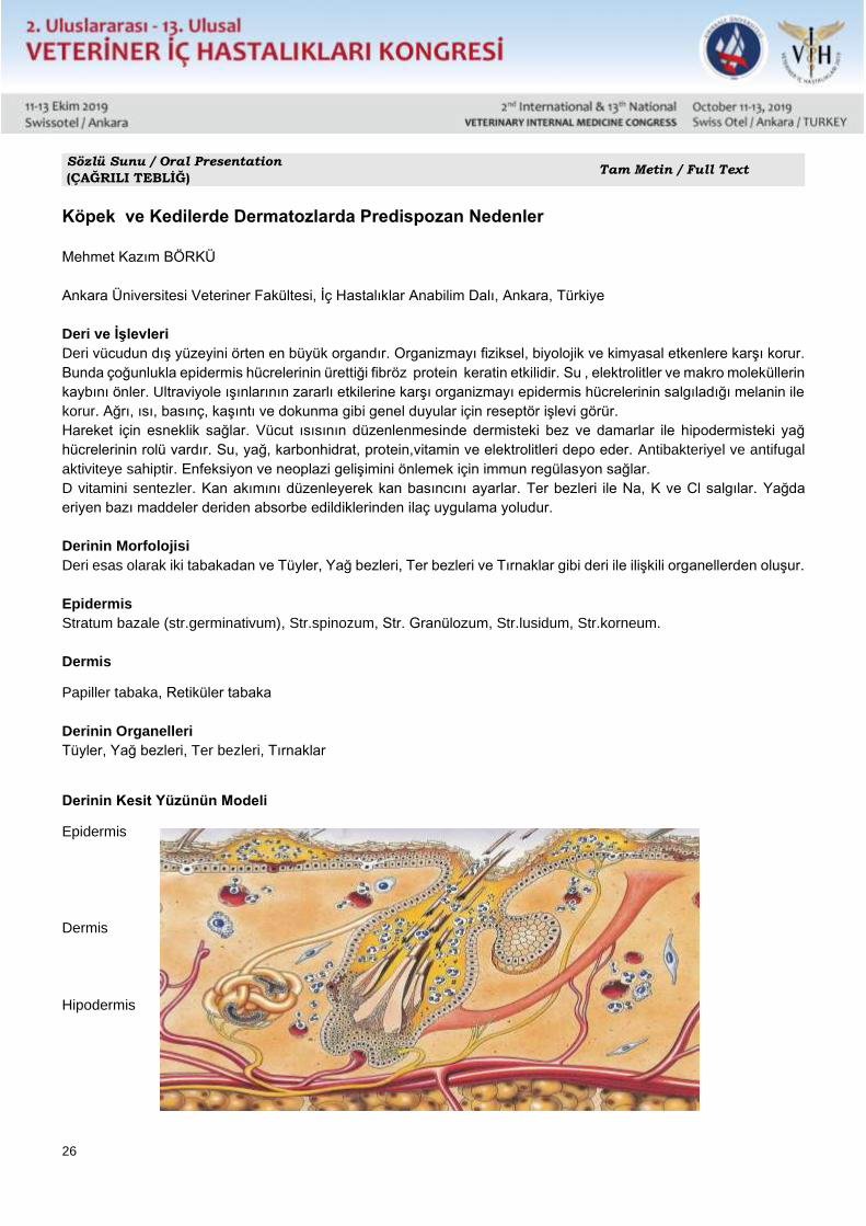
26
Sözlü Sunu / Oral Presentation
(ÇAĞRILI TEBLİĞ) Tam Metin / Full Text
Köpek ve Kedilerde Dermatozlarda Predispozan Nedenler
Mehmet Kazım BÖRKÜ
Ankara Üniversitesi Veteriner Fakültesi, İç Hastalıklar Anabilim Dalı, Ankara, Türkiye
Deri ve İşlevleri
Deri vücudun dış yüzeyini örten en büyük organdır. Organizmayı fiziksel, biyolojik ve kimyasal etkenlere karşı korur.
Bunda çoğunlukla epidermis hücrelerinin ürettiği fibröz protein keratin etkilidir. Su , elektrolitler ve makro moleküllerin
kaybını önler. Ultraviyole ışınlarının zararlı etkilerine karşı organizmayı epidermis hücrelerinin salgıladığı melanin ile
korur. Ağrı, ısı, basınç, kaşıntı ve dokunma gibi genel duyular için reseptör işlevi görür.
Hareket için esneklik sağlar. Vücut ısısının düzenlenmesinde dermisteki bez ve damarlar ile hipodermisteki yağ
hücrelerinin rolü vardır. Su, yağ, karbonhidrat, protein,vitamin ve elektrolitleri depo eder. Antibakteriyel ve antifugal
aktiviteye sahiptir. Enfeksiyon ve neoplazi gelişimini önlemek için immun regülasyon sağlar.
D vitamini sentezler. Kan akımını düzenleyerek kan basıncını ayarlar. Ter bezleri ile Na, K ve Cl salgılar. Yağda
eriyen bazı maddeler deriden absorbe edildiklerinden ilaç uygulama yoludur.
Derinin Morfolojisi
Deri esas olarak iki tabakadan ve Tüyler, Yağ bezleri, Ter bezleri ve Tırnaklar gibi deri ile ilişkili organellerden oluşur.
Epidermis
Stratum bazale (str.germinativum), Str.spinozum, Str. Granülozum, Str.lusidum, Str.korneum.
Dermis
Papiller tabaka, Retiküler tabaka
Derinin Organelleri
Tüyler, Yağ bezleri, Ter bezleri, Tırnaklar
Derinin Kesit Yüzünün Modeli
Epidermis
Dermis
Hipodermis
27
Dermatozlar
Dermatoz, derideki herhangi bir hastalığı tanımlayan ve geniş kapsamlı olan bir terimdir. Dermatitis ile
karıştırılmamalıdır. Dermatitis derinin yangılanmasını,irritasyonunu tanımlar.
Dermatozlar genel veteriner pratikte karşılaşılan hastalıkların büyük bir kısmını oluştururlar. Ancak dermatozların
nedenlerini belirlemek için, diğer sistemlere yerleşmiş olan hastalıklarda yapılan kadar araştırma yapılmamaktadır.
Kusan bir hasta, diagnostik testler için, hospitalize edilirken; kompleks hale gelmiş ciddi derecedeki dermatozlar bile,
ampirik ve semptomatik sağaltım uygulaması dışında laboratuar incelemesine tabi tutulmamaktadır.
Dermatozda predispozan faktörü araştırmak, spesifik tanıyı koymak, iyi bir tedavi ve prognoz için kapsamlı ve titiz
bir çalışma yapılmalıdır.
Dermatozlarda Predispozan Faktörler
İmmun sistemi doğrudan ve dolaylı olarak etkileyen hastalıklar ve faktörler
İmmunolojik etkiler
Hipersensitivite-Alerji: Atopi-atopik dermatitis,ürtiker ve anjioödem, kontakt hipersensitivite, gıda hipersensitivitesi-
gıda alerjisi-gıda intoleransı, pire ısırığı hipersensitivitesi, intestinal parazit hipersensitivitesi, hormonal
hipersensitivite, kutanöz ilaç erupsiyonları, stafilococcal hipersensitivite vs.
Otoimmun deri hastalıkları: Pemfigus foliakus,pemfigus vulgaris,pemfigus vegetans,pemfigus eritematozis,kanin
pemfigoid,sistemik lupus eritematozis,diskoid lupus eritematozis,soğuk aglütinin hastalığı,vs.
İmmun deri hastalıkları: Eritema multiforme,toksik epidermal nekrolizis,kutanöz vaskulitis,amiloidozis,köpeklerde
uveodermatolojik sendrom,kanin linear IgA dermatozis,vs.
Endokrin ve metabolik etkiler : Hipotroidizm,kedilerde hipertroidizm, hiperadrenokortisizm, hipofiz dwarfism,
köpeklerde büyüme hormonuna cevap veren dermatozlar, acromegaly,erkek ve dişi köpeklerde testosterona cevap
veren dermatozlar, erkek ve dişi köpeklerde östrojene cevap veren dermatozlar,köpeklerde adrenal seks hormonuna
cevap veren dermatozlar,diabetes mellitus,dişi köpeklerde hiperöstrojenizm, köpeklerde testis neoplazileri vs.
Neoplastik etkiler: Epitelial neoplasiler,mezanşimal tümörler,lenfosittik tümörler, histiosit kökenli tümörler,
melanositik neoplazmalar, neopastik olmayan tümörler,vs.
Paraziter etkiler: Otodectes cynotis, cheyletiellosis, demodicosis, sarcoptic mange, pire alejik dermatitis, myasis,
unicinariasis, pelodera dermatitis, dirofilariasis,vs.
Viral etkiler: Canine distemper, feline lökemi, dev hücreli dermatosis, feline immun yetmezlik, feline rinotrakeitis, vs.
Protozoer etkiler: Leishmaniasis,vs.
Nem etkisi: Yetersiz ventilasyon, ıslaklık, vs.
Travma etkisi: Yabancı cisim, ısırık,tırnak, vs ile derin yaralanmalar.
Lokalize ağrı etkisi: Özellikle kedilerin ağrılı bölgelerini çok fazla yalamaları.
Enjeksiyon etkisi: Enjeksiyon yapılan bölgede ilaç reaksiyonları, kontaminasyon, çeşitli dejeneratif bozukluklar
sonucu oluşan lokal deri reaksiyonları.
Hijyen etkisi: Ortamın çok pis ve kontamine olması.
Predispozan Faktörlerin Araştırılması
Anamnez
Bu bağlamda; hayvanın türü, ırkı, yaşı, cinsiyeti, kısırlaştırma durumu, aktivitesi, diyeti, iştahı, susaması, geçirdiği
ya da geçirmekte olduğu hastalıklar, daha önce uygulanan ya da uygulanmakta olan ilaçlar, deri problemlerinin neler
olduğu, problemlerin aniden mi çıktığı, lezyonun ilk nasıl başladığı ve lezyonun ilk görüldüğü yaş, lezyonun sürekli
mi, aralıklı mı, mevsimsel mi ortaya çıktığı, sahibinde de görülüp görülmediği, diğer hayvanlara da bulaşıp
bulaşmadığı, hayvanın hangi yaşta ve nereden alındığı, hayvanın hangi yörelerde de bulunduğu, aynı problemin,
aynı anneden doğan diğer hayvanlarda ve çevresindeki başka hayvanlarda dagörülüp görülmediği öğrenilmelidir.
Klinik muayene
Dermatolojik muayeneler (bakteriyel kültür, fungal kültür, biyopsi, alerji testi)
28
Sağaltım denemeleri
Genel testler (hematoloji,serum biyokimyasal analizi,endokrin analizler, spesifik antikor arama testleri,
stimulasyon testleri, ultrasonografi, radyografi, elektrokardiyografi, elektromyografi) yapılmalı ve predispozan
faktörler belirlenmelidir.
Dermatozlarda Derinin Rolü
Deri, immun sistemin bütünleyici ve aktif bileşeni olduğundan, pekçok hastalıkta, doğrudan ya da dolaylı olarak
etkilenmektedir.
Deri için fonksiyonel lenfoid bir organ tanımlaması yapmak yanlış olmaz.
Deride iki önemli immun sistem bulunur.
1-Selular sistem:Keratinositler,langerhans hücreleri,lenfositler,doku makrofajları,mast hücreleri,endotelial hücreler
ve granulositler.
2- Humoral sistem: İmmunglobulinler,komplement, fibrinolisin,sitokin,eikonosid, neuropeptid ve antimikrobiyel
peptitler.
Derideki yangısal ve yangısal olmayan hastalıklar bu iki sistemin etkileşimi ile ilgilidir.
Bu hastalıklar alerjik ( hipersensitivite) veya otoimmun olarak tanımlanırlar.
Dermatozlara Klinik Yaklaşım
Dermatozlu köpek ve kediler tedavi için veteriner kliniklerine getirildiklerinde etiyolojik ve predispozan faktörleri ortaya
çıkarabilmek için kapsamlı incelemelerin yapılması gerektiği halde; veterinerler sadece parazitolojik, bakteriyolojik,
fungal ve alerjen etkenler üzerinde yoğunlaşmaktadır. Bu klinik yaklaşımla veterinerler çoğunlukla staphylococlar,
fungal etkenler, demodeks ve çevresel alerjenleri belirlemektedirler. Dermatozlu hastalarda; etkene yönelik tedavi
başlatıldıktan sonra, son yıllarda çoğunlukla hipoalerjik bir diyet de önerilmektedir. Bu etkenlere karşı yapılan tedavi
denemelerinde, başlangıçta deri lezyonlarında bir gerileme görülmektedir. Dermatozun nedeni gıda alerjisi yada
intoleransı ise; önerilen gıda uygundığında deri lezyonları nüks etmemektedir. Ancak, sorun gıdaya bağlı değilse
ve çoğunlukla olduğu gibi, dermatozun oluşumunda başka predispozan nedenler varsa, kullanılan ilaçların etkileri
geçtikten sonra deri lezyonları nüks etmektedir.
Dermatozlarda nükslerin ortaya çıkmaması, deride koku, kepeklenme, kabuklanma, nem kaybı, kuruma, deri şeklinin
bozulması, elastikiyet kaybı, kalınlaşma, sertleşme, renk değişikliği, siyahlaşma ve kalıcı alopesinin oluşmaması
için; dermatozu olan bir hastaya müdahale edileceğinde; dermatolojik muayenelerin eşliğinde kapsamlı bir klinik
muayene, hematoloji, serum biyokimyasl muayeneler, görüntüleme teknikleri, serumda antikor arama, hormonal
stimulasyon testleri yapılarak predispozan neden belirlenmeli ve sağaltımda geç kalınmamalıdır. Sağaltım gecikirse
yangısel reaksiyonlar deri formasyonunu bozarak, sağlıksız deri görünümüne ve alopesiye zemin hazırlamaktadır.
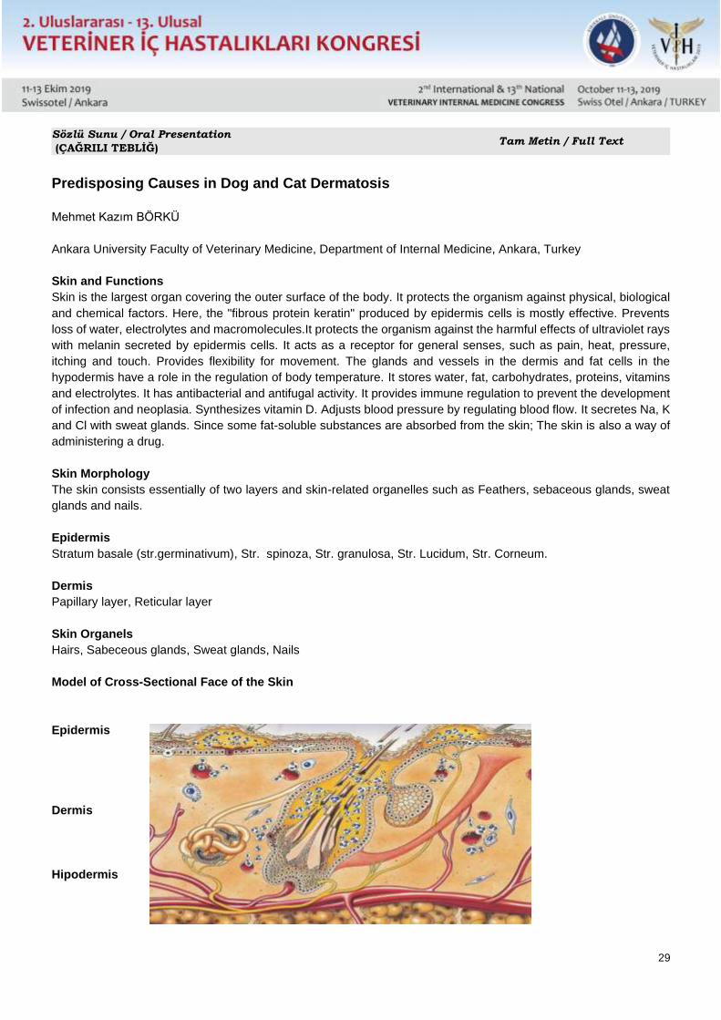
29
Sözlü Sunu / Oral Presentation
(ÇAĞRILI TEBLİĞ) Tam Metin / Full Text
Predisposing Causes in Dog and Cat Dermatosis
Mehmet Kazım BÖRKÜ
Ankara University Faculty of Veterinary Medicine, Department of Internal Medicine, Ankara, Turkey
Skin and Functions
Skin is the largest organ covering the outer surface of the body. It protects the organism against physical, biological
and chemical factors. Here, the "fibrous protein keratin" produced by epidermis cells is mostly effective. Prevents
loss of water, electrolytes and macromolecules.It protects the organism against the harmful effects of ultraviolet rays
with melanin secreted by epidermis cells. It acts as a receptor for general senses, such as pain, heat, pressure,
itching and touch. Provides flexibility for movement. The glands and vessels in the dermis and fat cells in the
hypodermis have a role in the regulation of body temperature. It stores water, fat, carbohydrates, proteins, vitamins
and electrolytes. It has antibacterial and antifugal activity. It provides immune regulation to prevent the development
of infection and neoplasia. Synthesizes vitamin D. Adjusts blood pressure by regulating blood flow. It secretes Na, K
and Cl with sweat glands. Since some fat-soluble substances are absorbed from the skin; The skin is also a way of
administering a drug.
Skin Morphology
The skin consists essentially of two layers and skin-related organelles such as Feathers, sebaceous glands, sweat
glands and nails.
Epidermis
Stratum basale (str.germinativum), Str. spinoza, Str. granulosa, Str. Lucidum, Str. Corneum.
Dermis
Papillary layer, Reticular layer
Skin Organels
Hairs, Sabeceous glands, Sweat glands, Nails
Model of Cross-Sectional Face of the Skin
Epidermis
Dermis
Hipodermis
30
Dermatoses
Dermatosis is a comprehensive term that describes any disease in the skin. It should not be confused with dermatitis.
Dermatitis defines inflammation and irritation of the skin.
Dermatoses constitute a large part of the diseases encountered in general veterinary practice. However, to determine
the causes of dermatoses, research has not been carried out as much as in other diseases.
When a vomiting patient is hospitalized for diagnostic tests; Even severe dermatoses that have become complex are
not subjected to laboratory examinations except for empirical and symptomatic treatment.
A thorough and rigorous study should be performed to investigate the predisposing factor in dermatosis, to make a
specific diagnosis and to provide a good treatment and prognosis.
Predisposant Factors in Dermatosis
Diseases and factors directly and indirectly affecting the immune system
Immunological effects
Hypersensitivity-Allergy: Atopy-atopic dermatitis, urticaria and angioedema;
Autoimmune skin diseases: Pemphigus foliacus, pemphigus vulgaris, pemphigus vegetans, pemphigus
erythematosus, canine pemphigoid, systemic lupus erythematosus, discoid lupus erythematosus, cold agglutinin
disease, etc.
Immune skin diseases: Erythema multiforme, toxic epidermal necrolysis, cutaneous vasculitis, amyloidosis,
uveodermatological syndrome in dogs, canine linear IgA dermatosis, etc.
Endocrine and metabolic effects: Hypothyroidism, hyperthyroidism in cats, hyperadrenocorticism, pituitary
dwarfism, dermatoses responsive to growth hormone in dogs, acromegaly, dermatoses responsive to testosterone
in male and female dogs, dermatosis responsive to estrogen in dogs, male and female dogs, diabetes mellitus,
hyperestrogenism in female dogs, testicular neoplasms in dogs, etc.
Neoplastic effects: Epithelial neoplasias, mesenchymal tumors, lymphocytic tumors, histiocyte tumors, melanocytic
neoplasms, non-neoplastic tumors, etc.
Parasitic effects: Otodectes cynotis, cheyletiellosis, demodicosis, sarcoptic mange, flea allergic dermatitis, myasis,
unicinariasis, pelodera dermatitis, dirofilariasis, etc.
Viral effects: Canine distemper, feline leukemia, giant cell dermatosis, feline immunodeficiency, feline rhinotrakeitis,
etc.
Protozoar effects: Leishmaniasis, etc.
Moisture effect: Inadequate ventilation, wetness, etc.
Trauma effect: Deep injuries with foreign body, bite, nail, etc.
Localized pain effect: Especially licking the painful areas of cats too much.
Injection effect: Local skin reactions caused by drug reactions, contamination, various degenerative disorders in
the injected area.
Effect of hygiene: Very dirty and contaminated environment.
Investigation of Predisposant Factors
History
In this context; animal species, breed, age, sex, neutering status, activity, diet, appetite, thirst, diseases that have
been or are undergoing, medications that have been or are being applied before, what are the skin problems, whether
the problems suddenly occur, how the lesion starts and the age at which the lesion was first seen, whether the lesion
appeared consistently, intermittently, seasonal, whether it was seen in its owner, whether it was transmitted to other
animals, at what age and where the animal was taken, in which regions the animal was present, the same problem
occurred in other animals of the same mother. and other animals in the vicinity.
Clinical examination
Dermatological examinations (bacterial culture, fungal culture, biopsy, allergy test)
31
Treatment trials
General tests (hematology, serum biochemical analysis, endocrine analysis, specific antibody search tests,
stimulation tests, ultrasonography, radiography, electrocardiography, electromyography) should be performed and
predisposing factors should be determined.
The Role of Skin in Dermatosis
Since skin is an integral and active component of the immune system, it is directly or indirectly affected in many
diseases.
It would not be wrong to define a functional lymphoid organ for the skin.
There are two important immune systems in the skin.
1-Cellular system: Keratinocytes, langerhans cells, lymphocytes, tissue macrophages, mast cells, endothelial cells
and granulocytes.
2- Humoral system: Immunoglobulins, complement, fibrinolysin, cytokine, eiconoside, neuropeptide and antimicrobial
peptides.
Inflammatory and non-inflammatory diseases of the skin are related to the interaction of these two systems.
These diseases are defined as allergic (hypersensitivity) or autoimmune.
Clinical Approach to Dermatosis
When dogs and cats with dermatosis are brought to veterinary clinics for treatment, extensive investigations are
needed to reveal the etiological and predisposing factors; veterinarians concentrate solely on parasitological,
bacteriological, fungal and allergen agents. With this clinical approach, veterinarians often identify staphylococci,
fungal agents, demodex and environmental allergens. In patients with dermatosis; After initiating treatment for the
causative agent, a hypoallergenic diet is often recommended in recent years. Trials against these factors show a
regression of skin lesions at baseline. If the cause of dermatosis is food allergy or intolerance; skin lesions do not
recur when the recommended food is appropriate. However, if the problem is not food-related and, as is often the
case, there are other predisposing causes in the formation of dermatosis, the skin lesions recur after the effects of
the drugs used.
In order to prevent recurrence of dermatoses, odor, dandruff, crusting, loss of moisture, drying, deformation of skin,
loss of elasticity, thickening, hardening, discoloration, blackening and permanent alopecia; when a patient with
dermatosis is treated; Dermatological examinations accompanied by a comprehensive clinical examination,
hematology, serum biochemical examinations, imaging techniques, antibody search in serum, hormonal stimulation
tests should be done to determine the predisposing cause and should not be late in treatment. If the treatment is
delayed, inflammatory reactions disrupt the formation of the skin and pave the way for unhealthy skin appearance
and alopecia.
32
Sözlü Sunu / Oral Presentation
(ÇAĞRILI TEBLİĞ) Tam Metin / Full Text
Comparison of stress parameters in cats with acute and chronic stress
Karolina Wrześniewska, Jacek Madany, Andrzej Milczak
Department and Clinic of Animal Internal Diseases, Faculty of Veterinary Medicine, University of Life Sciences in
Lublin, Głeboka 30, Poland
ABSTRACT
Domestic cats are exposed to a variety of stressful stimuli, which may have a negative effect on the cat’s welfare and
trigger a number of behavioral changes.
The aim of the study was to compare blood glucose, cortisol and catecholamines in cats with acute and chronic
stress. Thirty-four randomly sourced, mixed-breed, neutered cats (19 females and 15 males), aged 1 to 16 years
(mean age 4), which were classified as weakly tense and very tense with stress score. Twenty-one of them were
exposed to acute stress, while 13 to chronic stress. Healthy weakly tense and very tense cats exposed only to short-
term stressor had lower blood glucose (124 mg/dl) and higher cortisol level (4.9 +/- 3 µg/dl) and norepinephrine level
(0.24 +/- 0.19 ng/ml) than cats exposed to chronic stress, respectively, blood glucose values 127 mg/dl, cortisol 4.8
+/- 2.3 µg/dl and norepinephrine 0.13 +/- 0.06 ng/ml. Because of the non-normal distribution, epinephrine
concentrations were not used for further analysis. The number of evidence confirming the relationship between stress
and the development of diseases is still growing, which is why it is so important to look for biochemical parameters
that could be used to determine if a cat is under stress and help to determine its severity.
KEYWORDS: Stress score, cortisol, norepinephrine, epinephrine, cats
INTRODUCTION
The term ”stress” has been widely used in biology to describe a set of physiological and behavioral changes elicited
by noxious or unpleasant stimuli. Based on the concepts suggested by Langley, Cannon and Selye, the adrenal
response to stress reflects the activation of the sympathoadrenomedullary system (SAM) and hypothalamic-pituitary-
adrenocortical axis (HPA) (6, 9, 13, 15, 20).
In domestic animals, stressors can be divided into physical stressors, social stressors resulting from interaction with
individuals of the same species and stressors related to handling by humans.
Depending on their duration, stress is classified as acute or chronic (9).
When an animal experiences several stressors at the same time, the stress response will be much higher than if the
animal was exposed to only one stressor. Stress is very likely to reduce feed intake, and stress-related anorexia may
contribute to the development of potentially serious medical conditions. Stress also increases the risk of cats showing
urine marking and some forms of aggression, including redirected aggression. (19).
The aim of the study was to compare the levels of stress hormones: glucose, cortisol, norepinephrine and epinephrine
in weakly tense and very tense cats with acute and chronic stress, and to determine if these parameters can be used
to predict stress, determine its intensity and potential pain.
MATERIALS AND METHODS
Thirty-four randomly sourced, mixed-breed, neutered cats (19 females and 15 males), 1 to 16 years old (average
age 4 years), body condition scores ranged from 2 to 4 on a 5- point scale were classified to the study. Cats were
assigned as weakly tense and very tense according to the "Cat Stress Score" from Kassler & Turner, 1997. Twenty-
33
one cats were classified as healthy (14 females and 7 males) based on physical examination and morphological and
biochemical blood test (all values were of normal physiological ranges) and had negative feline immunodeficiency
virus antibody and feline leukemia virus antigen test results. Thirteen cats (5 females, 8 males) with non-adrenal
diseases, associated with transient hyperglycemia include urinary tract disease and viral infections.
Blood specimens were taken from jugular vein in sterile 2.0 ml syringes by 23 G needles. Due to the daily variability
of blood cortisol levels, blood samples were taken from 10 a.m. to 12.30 a.m. Immediately after collection, some
blood was placed in serum collection tubes to measure glucose and cortisol levels; for hormonal analysis 1 ml of
each sample was put into a sterile micro vacutainer, containing heparin and centrifuged for 15 minutes at room
temperature. Before determining hormone levels, plasma samples were stored at -80° C for 30 days. The cortisol
level test was performed on the IMMULITE 1000 apparatus, Siemens (Germany). Enzyme Conjugate - Cortisol
conjugated to horseradish peroxidase. The inter-assay CVs ranged between 6,5% and 7,7%. The limit of
quantification was 6,9 nmol/l. Epinephrine and norepinephrine - by specific enzyme test 2-cat (A-N) Research ELISA,
LDN, Germany in plasma. Sensitivity for epinephrine: 6,6 pg/ml and for norepinephrine 2,6 pg/ml. The hormonal
assays of epinephrine and norepinephrine were carried out by ELISA Reader Microplate (Molecular Devices Spectra
Max M2). Blood glucose level was quantified using the hexokinase enzymatic method on ABX Pentra 400 (Horiba
Medical, Poland). All data were expressed as median and range with MS Excel. The results in the text are expressed
as means +/- standard error of the mean.
RESULTS
Healthy cats weakly tense and very tense exposed only to short-term stressor, which was a visit to the doctor's office
and blood collection, had lower blood glucose (average 124 mg/dl) than cats exposed to long-term stressor, which
was a chronic and overlapping disease short-term stressor (average 127 mg/dl). Serum cortisol levels were higher
in healthy weakly tense and very tense cats exposed to short-term stressor (4.9 +/- 3 µg/dl) than in cats with chronic
diseases (4.8 +/- 2.3 µg/dl). The analysis showed that in healthy cats exposed to acute stress norepinephrine the
values were 0.24 +/- 0.19 ng/ml, while in cats with chronic diseases they equalled 0.13 +/- 0.06 ng/ml. In the group
of cats exposed to both short- and long-term stressor, there was a very wide individual variation in epinephrine
concentrations, due to the unusual distribution of values, epinephrine concentrations were not used for further
analysis.
DISCUSSION
Cortisol, catecholamines or blood glucose values during stressful situations in cats have been poorly documented
(4, 12, 16, 17, 25).
In our study, we showed that there is a relationship between the duration of a stress response and the
concentration of corticoids, catecholamines and blood glucose. We considered a short-acting stressor - a visit to
a veterinary clinic along with blood collection, and we considered cats with chronic diseases to be patients exposed
to long-term stress. The disease is a chronic stressor, and the mechanisms causing hyperglycemia may then be
completely different from those responsible for transient hyperglycemia. Diseases associated with transient
hyperglycemia include: urinary tract diseases, viral and bacterial infections, gastrointestinal diseases, cancer, renal
failure and cardiomyopathies. When hyperglycaemia is superimposed on disease-related hyperglycaemia during
stress associated with blood sampling, we will not avoid the increase of glucose levels more than when exposed
to a single stressor. Longer periods of stress can potentially cause higher glucose levels (17). As expected, cats
exposed to chronic stress presented higher glucose values than cats exposed to short-term stressor. It should be
noted that the blood glucose content showed a large inter-individual variability.
Changes in the content of catecholamines and steroid hormones of the adrenal cortex in the blood depend on the
type of stressor, the time and strength of its action and the psychophysical properties of the organism undergoing
such action.
The average values of cortisol levels in the group of healthy cats exposed to short-term stress were higher than
the values in the group of cats with chronic diseases, which may indirectly explain the research showing that during
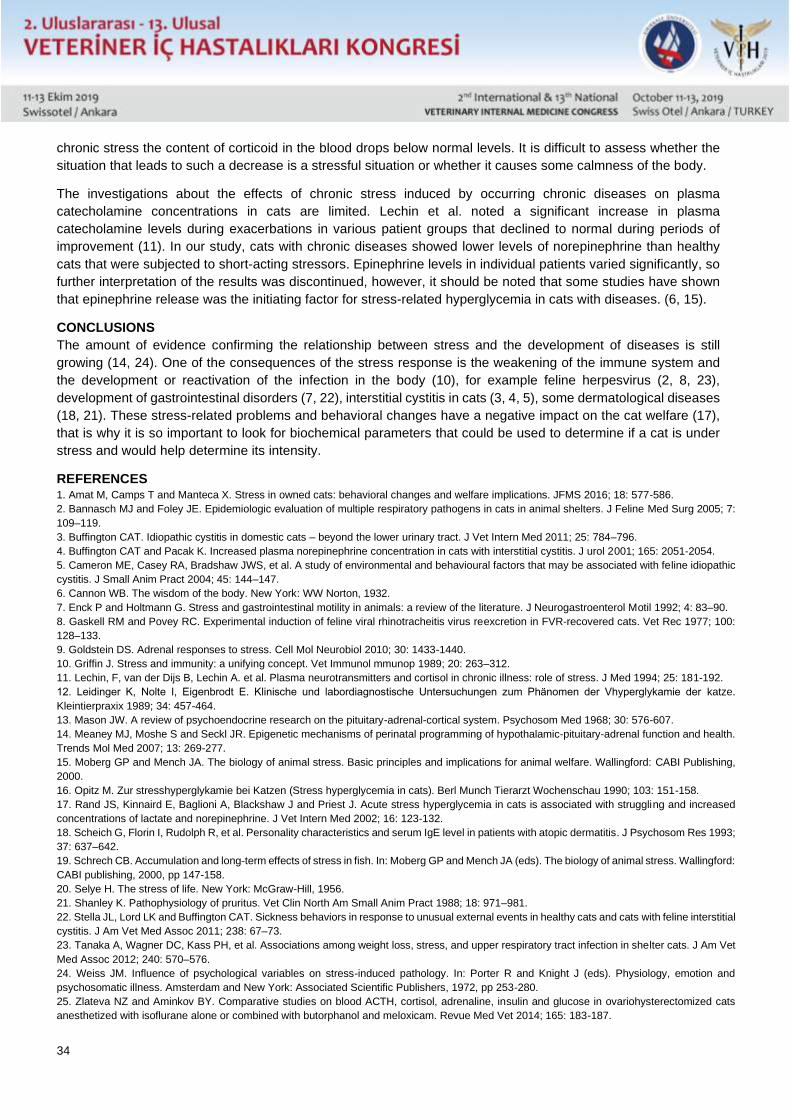
34
chronic stress the content of corticoid in the blood drops below normal levels. It is difficult to assess whether the
situation that leads to such a decrease is a stressful situation or whether it causes some calmness of the body.
The investigations about the effects of chronic stress induced by occurring chronic diseases on plasma
catecholamine concentrations in cats are limited. Lechin et al. noted a significant increase in plasma
catecholamine levels during exacerbations in various patient groups that declined to normal during periods of
improvement (11). In our study, cats with chronic diseases showed lower levels of norepinephrine than healthy
cats that were subjected to short-acting stressors. Epinephrine levels in individual patients varied significantly, so
further interpretation of the results was discontinued, however, it should be noted that some studies have shown
that epinephrine release was the initiating factor for stress-related hyperglycemia in cats with diseases. (6, 15).
CONCLUSIONS
The amount of evidence confirming the relationship between stress and the development of diseases is still
growing (14, 24). One of the consequences of the stress response is the weakening of the immune system and
the development or reactivation of the infection in the body (10), for example feline herpesvirus (2, 8, 23),
development of gastrointestinal disorders (7, 22), interstitial cystitis in cats (3, 4, 5), some dermatological diseases
(18, 21). These stress-related problems and behavioral changes have a negative impact on the cat welfare (17),
that is why it is so important to look for biochemical parameters that could be used to determine if a cat is under
stress and would help determine its intensity.
REFERENCES 1. Amat M, Camps T and Manteca X. Stress in owned cats: behavioral changes and welfare implications. JFMS 2016; 18: 577-586.
2. Bannasch MJ and Foley JE. Epidemiologic evaluation of multiple respiratory pathogens in cats in animal shelters. J Feline Med Surg 2005; 7:
109–119.
3. Buffington CAT. Idiopathic cystitis in domestic cats – beyond the lower urinary tract. J Vet Intern Med 2011; 25: 784–796.
4. Buffington CAT and Pacak K. Increased plasma norepinephrine concentration in cats with interstitial cystitis. J urol 2001; 165: 2051-2054.
5. Cameron ME, Casey RA, Bradshaw JWS, et al. A study of environmental and behavioural factors that may be associated with feline idiopathic
cystitis. J Small Anim Pract 2004; 45: 144–147.
6. Cannon WB. The wisdom of the body. New York: WW Norton, 1932.
7. Enck P and Holtmann G. Stress and gastrointestinal motility in animals: a review of the literature. J Neurogastroenterol Motil 1992; 4: 83–90.
8. Gaskell RM and Povey RC. Experimental induction of feline viral rhinotracheitis virus reexcretion in FVR-recovered cats. Vet Rec 1977; 100:
128–133.
9. Goldstein DS. Adrenal responses to stress. Cell Mol Neurobiol 2010; 30: 1433-1440.
10. Griffin J. Stress and immunity: a unifying concept. Vet Immunol mmunop 1989; 20: 263–312.
11. Lechin, F, van der Dijs B, Lechin A. et al. Plasma neurotransmitters and cortisol in chronic illness: role of stress. J Med 1994; 25: 181-192.
12. Leidinger K, Nolte I, Eigenbrodt E. Klinische und labordiagnostische Untersuchungen zum Phänomen der Vhyperglykamie der katze.
Kleintierpraxix 1989; 34: 457-464.
13. Mason JW. A review of psychoendocrine research on the pituitary-adrenal-cortical system. Psychosom Med 1968; 30: 576-607.
14. Meaney MJ, Moshe S and Seckl JR. Epigenetic mechanisms of perinatal programming of hypothalamic-pituitary-adrenal function and health.
Trends Mol Med 2007; 13: 269-277.
15. Moberg GP and Mench JA. The biology of animal stress. Basic principles and implications for animal welfare. Wallingford: CABI Publishing,
2000.
16. Opitz M. Zur stresshyperglykamie bei Katzen (Stress hyperglycemia in cats). Berl Munch Tierarzt Wochenschau 1990; 103: 151-158.
17. Rand JS, Kinnaird E, Baglioni A, Blackshaw J and Priest J. Acute stress hyperglycemia in cats is associated with struggling and increased
concentrations of lactate and norepinephrine. J Vet Intern Med 2002; 16: 123-132.
18. Scheich G, Florin I, Rudolph R, et al. Personality characteristics and serum IgE level in patients with atopic dermatitis. J Psychosom Res 1993;
37: 637–642.
19. Schrech CB. Accumulation and long-term effects of stress in fish. In: Moberg GP and Mench JA (eds). The biology of animal stress. Wallingford:
CABI publishing, 2000, pp 147-158.
20. Selye H. The stress of life. New York: McGraw-Hill, 1956.
21. Shanley K. Pathophysiology of pruritus. Vet Clin North Am Small Anim Pract 1988; 18: 971–981.
22. Stella JL, Lord LK and Buffington CAT. Sickness behaviors in response to unusual external events in healthy cats and cats with feline interstitial
cystitis. J Am Vet Med Assoc 2011; 238: 67–73.
23. Tanaka A, Wagner DC, Kass PH, et al. Associations among weight loss, stress, and upper respiratory tract infection in shelter cats. J Am Vet
Med Assoc 2012; 240: 570–576.
24. Weiss JM. Influence of psychological variables on stress-induced pathology. In: Porter R and Knight J (eds). Physiology, emotion and
psychosomatic illness. Amsterdam and New York: Associated Scientific Publishers, 1972, pp 253-280.
25. Zlateva NZ and Aminkov BY. Comparative studies on blood ACTH, cortisol, adrenaline, insulin and glucose in ovariohysterectomized cats
anesthetized with isoflurane alone or combined with butorphanol and meloxicam. Revue Med Vet 2014; 165: 183-187.
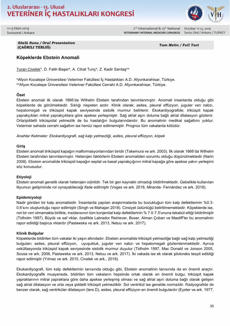
35
Sözlü Sunu / Oral Presentation
(ÇAĞRILI TEBLİĞ) Tam Metin / Full Text
Köpeklerde Ebstein Anomali
Turan Civelek*, D. Fatih Başer*, A. Cihat Tunç*, Z. Kadir Sarıtaş**
*Afyon Kocatepe Üniversitesi Veteriner Fakültesi İç Hastalıkları A.D. Afyonkarahisar, Türkiye.
**Afyon Kocatepe Üniversitesi Veteriner Fakültesi Cerrahi A.D. Afyonkarahisar, Türkiye.
Özet
Ebstein anomali ilk olarak 1866’da Wilhelm Ebstein tarafından tanımlanmıştır. Anomali insanlarda olduğu gibi
köpeklerde de görülmektedir. Sıklığı nispeten azdır. Klinik olarak; asites, pleural effüzyon, juguler ven nabzı,
hepatomegali ve triküspid kapak seviyesinde sistolik murmur belirlenir. Ekokardiyografide; triküspit kapak
yaprakçıkları mitral yaprakçıklara göre apekse yerleşmiştir. Sağ atrial aşırı doluma bağlı atrial dilatasyon gözlenir.
Orta/şiddetli triküspidal yetmezlik de bu hastalığın bulgularındandır. Bu anomalinin medikal sağaltımı yoktur.
Veteriner sahada cerrahi sağaltım ise henüz rapor edilmemiştir. Prognoz tüm vakalarda kötüdür.
Anahtar Kelimeler: Ekokardiyografi, sağ kalp yetmezliği, asites, pleural effüzyon, köpek
Giriş
Ebstein anomali tiriküspid kapağın malformasyonlarından biridir (Takemura ve ark. 2003). İlk olarak 1866’da Wilhelm
Ebstein tarafından tanımlanmıştır. Heterojen faktörlerin Ebstein anomaliden sorumlu olduğu düşünülmektedir (Narin
2008). Ebstein anomalide triküspid kapağın septal ve basal yaprakçığının mitral kapağa göre apekse yakın yerleşimi
söz konusudur.
Etiyoloji
Ebstein anomali genetik olarak heterojen orjinlidir. Tek bir gen kaynaklı olmadığı bildirilmektedir. Gebelikte kullanılan
lityumun gelişiminde rol oynayabileceği ifade edilmiştir (Voges ve ark. 2018, Miranda- Fernández ve ark. 2018).
Epidemiyoloji
Nadir görülen bir kalp anomalisidir. İnsanlarda yapılan araştırmalarda bu bozukluğun tüm kalp defektlerinin %0.3-
0.6’sını oluşturduğu rapor edilmiştir (Singh ve Mahajan 2018). Cinsiyet üstünlüğü belirtilmemektedir. Köpeklerde ise,
net bir veri olmamakla birlikte, insidansının tüm konjenital kalp defektlerinin % 7.0-7.5'ununa tekabül ettiği bildirilmiştir
(Tidholm 1997). Büyük ve saf ırklar, özellikle Labrador Retriever, Boxer, Alman Çoban ve Mastiff’ler bu anomalinin
rapor edildiği başlıca ırklardır (Paslawska ve ark. 2013, Netuu ve ark. 2017).
Klinik Bulgular
Köpeklerde bildirilen tüm vakalar iki yaşın altındadır. Ebstein anomalide triküspit yetmezliğe bağlı sağ kalp yetmezliği
bulguları; asites, pleural effüzyon, uyuşukluk, juguler ven nabzı ve hepatomegali gözlemlenmektedir. Ayrıca
oskültasyonda triküspid kapak seviyesinde sistolik murmur duyulur (Tidholm 1997, Mac Donald ve Jonson 2005,
Sousa ve ark. 2006, Paslawska ve ark. 2013, Netuu ve ark. 2017). İki vakada ise ek olarak şilotoraks tespit edildiği
rapor edilmiştir (Yılmaz ve ark. 2015, Civelek ve ark., 2019).
Ekokardiyografi, tüm kalp defektlerinin tanısında olduğu gibi, Ebstein anomalinin tanısında da en önemli araçtır.
Ekokardiyografik muayenede, bildirilen tüm vakaların hepsinde ortak olarak en önemli bulgu; triküspit kapak
yapraklarının mitral yapraklara göre daha apekse yerleşmiş olması ve sağ atrial aşırı doluma bağlı olarak gelişen
sağ atrial dilatasyon ve orta veya şiddetli triküspit yetmezliktir. Sol ventrikül ise genelde norma ldir. Radyografide de
benzer olarak, sağ ventriküler dilatasyon (ters D), asites, pleural effüzyon en önemli bulgulardır (Eyster ve ark. 1977,
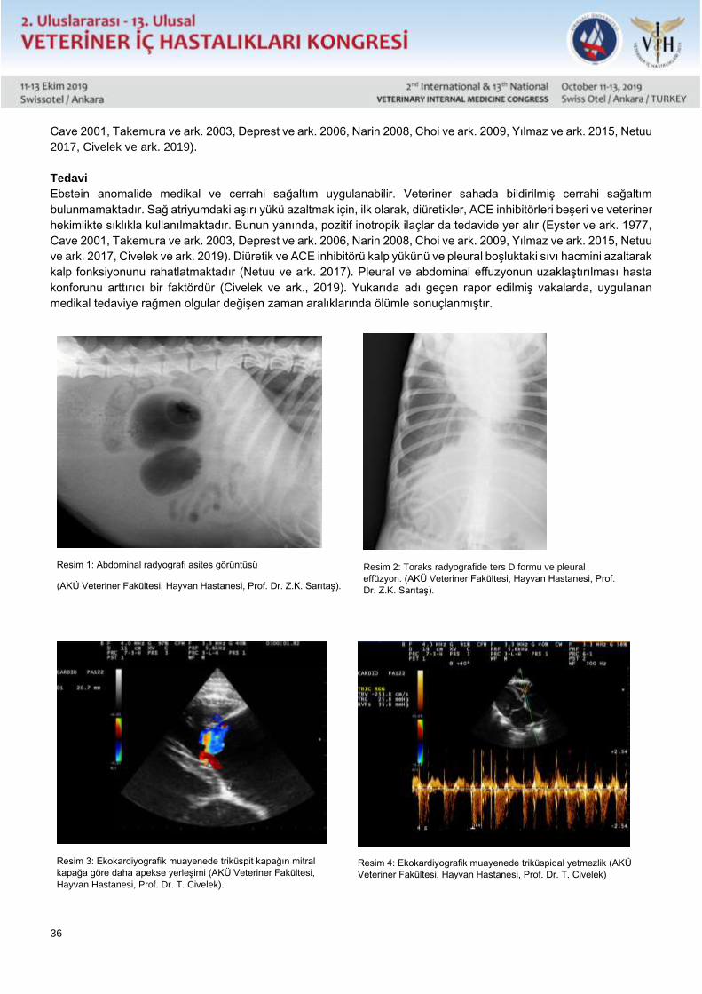
36
Cave 2001, Takemura ve ark. 2003, Deprest ve ark. 2006, Narin 2008, Choi ve ark. 2009, Yılmaz ve ark. 2015, Netuu
2017, Civelek ve ark. 2019).
Tedavi
Ebstein anomalide medikal ve cerrahi sağaltım uygulanabilir. Veteriner sahada bildirilmiş cerrahi sağaltım
bulunmamaktadır. Sağ atriyumdaki aşırı yükü azaltmak için, ilk olarak, diüretikler, ACE inhibitörleri beşeri ve veteriner
hekimlikte sıklıkla kullanılmaktadır. Bunun yanında, pozitif inotropik ilaçlar da tedavide yer alır (Eyster ve ark. 1977,
Cave 2001, Takemura ve ark. 2003, Deprest ve ark. 2006, Narin 2008, Choi ve ark. 2009, Yılmaz ve ark. 2015, Netuu
ve ark. 2017, Civelek ve ark. 2019). Diüretik ve ACE inhibitörü kalp yükünü ve pleural boşluktaki sıvı hacmini azaltarak
kalp fonksiyonunu rahatlatmaktadır (Netuu ve ark. 2017). Pleural ve abdominal effuzyonun uzaklaştırılması hasta
konforunu arttırıcı bir faktördür (Civelek ve ark., 2019). Yukarıda adı geçen rapor edilmiş vakalarda, uygulanan
medikal tedaviye rağmen olgular değişen zaman aralıklarında ölümle sonuçlanmıştır.
Resim 1: Abdominal radyografi asites görüntüsü
(AKÜ Veteriner Fakültesi, Hayvan Hastanesi, Prof. Dr. Z.K. Sarıtaş).
Resim 2: Toraks radyografide ters D formu ve pleural
effüzyon. (AKÜ Veteriner Fakültesi, Hayvan Hastanesi, Prof.
Dr. Z.K. Sarıtaş).
Resim 3: Ekokardiyografik muayenede triküspit kapağın mitral
kapağa göre daha apekse yerleşimi (AKÜ Veteriner Fakültesi,
Hayvan Hastanesi, Prof. Dr. T. Civelek).
Resim 4: Ekokardiyografik muayenede triküspidal yetmezlik (AKÜ
Veteriner Fakültesi, Hayvan Hastanesi, Prof. Dr. T. Civelek)
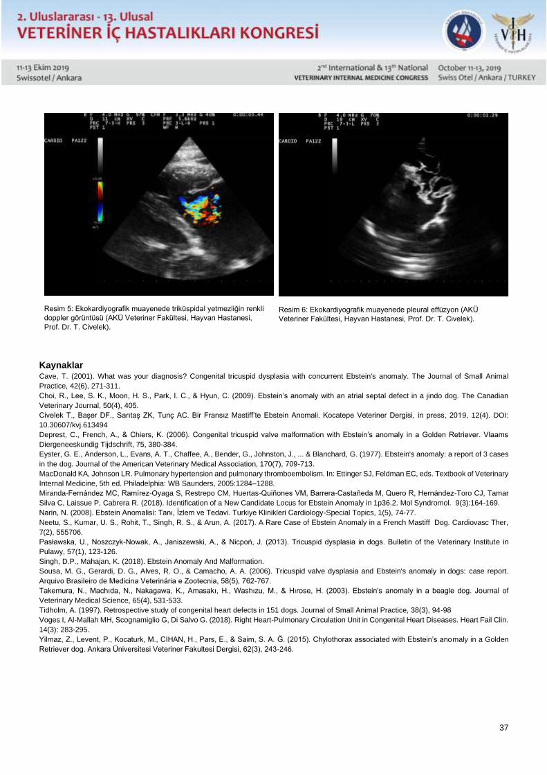
37
Resim 5: Ekokardiyografik muayenede triküspidal yetmezliğin renkli
doppler görüntüsü (AKÜ Veteriner Fakültesi, Hayvan Hastanesi,
Prof. Dr. T. Civelek).
Resim 6: Ekokardiyografik muayenede pleural effüzyon (AKÜ
Veteriner Fakültesi, Hayvan Hastanesi, Prof. Dr. T. Civelek).
Kaynaklar Cave, T. (2001). What was your diagnosis? Congenital tricuspid dysplasia with concurrent Ebstein's anomaly. The Journal of Small Animal
Practice, 42(6), 271-311.
Choi, R., Lee, S. K., Moon, H. S., Park, I. C., & Hyun, C. (2009). Ebstein’s anomaly with an atrial septal defect in a jindo dog. The Canadian
Veterinary Journal, 50(4), 405.
Civelek T., Başer DF., Sarıtaş ZK, Tunç AC. Bir Fransız Mastiff’te Ebstein Anomali. Kocatepe Veteriner Dergisi, in press, 2019, 12(4). DOI:
10.30607/kvj.613494
Deprest, C., French, A., & Chiers, K. (2006). Congenital tricuspid valve malformation with Ebstein’s anomaly in a Golden Retriever. Vlaams
Diergeneeskundig Tijdschrift, 75, 380-384.
Eyster, G. E., Anderson, L., Evans, A. T., Chaffee, A., Bender, G., Johnston, J., ... & Blanchard, G. (1977). Ebstein's anomaly: a report of 3 cases
in the dog. Journal of the American Veterinary Medical Association, 170(7), 709-713.
MacDonald KA, Johnson LR. Pulmonary hypertension and pulmonary thromboembolism. In: Ettinger SJ, Feldman EC, eds. Textbook of Veterinary
Internal Medicine, 5th ed. Philadelphia: WB Saunders, 2005:1284–1288.
Miranda-Fernández MC, Ramírez-Oyaga S, Restrepo CM, Huertas-Quiñones VM, Barrera-Castañeda M, Quero R, Hernández-Toro CJ, Tamar
Silva C, Laissue P, Cabrera R. (2018). Identification of a New Candidate Locus for Ebstein Anomaly in 1p36.2. Mol Syndromol. 9(3):164-169.
Narin, N. (2008). Ebstein Anomalisi: Tanı, İzlem ve Tedavi. Turkiye Klinikleri Cardiology-Special Topics, 1(5), 74-77.
Neetu, S., Kumar, U. S., Rohit, T., Singh, R. S., & Arun, A. (2017). A Rare Case of Ebstein Anomaly in a French Mastiff Dog. Cardiovasc Ther,
7(2), 555706.
Pasławska, U., Noszczyk-Nowak, A., Janiszewski, A., & Nicpoń, J. (2013). Tricuspid dysplasia in dogs. Bulletin of the Veterinary Institute in
Pulawy, 57(1), 123-126.
Singh, D.P., Mahajan, K. (2018). Ebstein Anomaly And Malformation.
Sousa, M. G., Gerardi, D. G., Alves, R. O., & Camacho, A. A. (2006). Tricuspid valve dysplasia and Ebstein's anomaly in dogs: case report.
Arquivo Brasileiro de Medicina Veterinária e Zootecnia, 58(5), 762-767.
Takemura, N., Machıda, N., Nakagawa, K., Amasakı, H., Washızu, M., & Hırose, H. (2003). Ebstein's anomaly in a beagle dog. Journal of
Veterinary Medical Science, 65(4), 531-533.
Tidholm, A. (1997). Retrospective study of congenital heart defects in 151 dogs. Journal of Small Animal Practice, 38(3), 94-98
Voges I, Al-Mallah MH, Scognamiglio G, Di Salvo G. (2018). Right Heart-Pulmonary Circulation Unit in Congenital Heart Diseases. Heart Fail Clin.
14(3): 283-295.
Yilmaz, Z., Levent, P., Kocaturk, M., CIHAN, H., Pars, E., & Saim, S. A. Ğ. (2015). Chylothorax associated with Ebstein’s anomaly in a Golden
Retriever dog. Ankara Üniversitesi Veteriner Fakultesi Dergisi, 62(3), 243-246.
38
Sözlü Sunu / Oral Presentation
(ÇAĞRILI TEBLİĞ) Tam Metin / Full Text
Ebstein Anomaly in Dogs
Turan Civelek*, D. Fatih Başer*, A. Cihat Tunç*, Z. Kadir Sarıtaş**
Afyon Kocatepe University Faculty of Veterinary Medicine Dep. of Internal Medicine Afyon, Turkey.
Afyon Kocatepe University Faculty of Veterinary Medicine Dep. of Surgery Afyon, Turkey.
The Ebstein anomaly was first described by Wilhelm Ebstein in 1866. Anomaly occurs in dogs as well as in humans.
Its frequency is relatively low. Clinically; ascites, pleural effusion, jugular vein pulse, hepatomegaly and systolic
murmur at tricuspid valve level are clinically determined. At echocardiography; tricuspid valve leaflets are located in
the apex relative to mitral leaflets. Atrial dilatation due to right atrial overfill is observed. Moderate to severe tricuspidal
insufficiency is one of the other symptom of this disease. There is no medical treatment of the anomally. Surgical
treatment in the veterinary field has not yet been reported. Prognosis is poor in all cases.
Keywords: Echocardiography, right heart failure, acids, pleural effusion, dog.
39
Sözlü Sunu / Oral Presentation Tam Metin / Full Text
Hypothyroidism in dogs as a baseline of different clinical signs and laboratory results
Irena Celeska, Elena Atanaskova Petrov
Department of pathophysiology, Faculty of Veterinary Medicine, University St. Cyril and Methodius
Department of internal medicine of small animals, Faculty of Veterinary Medicine, University St. Cyril and Methodius
Introduction:
Hypothyroidism is the most common endocrinopathy in dogs. It is clinical condition with decrease serum level
concentration of thyroid hormones, resulting with different clinical signs. As a common subclinical and clinical
condition, hypothyroidism can be congenital and acquired, as well as primary, secondary and tertiary (the last
mentioned is very rare). This clinical condition is sometimes hard for diagnosis, but it is very easy for treatment.
Aims: The main purpose of this clinical study is interpretation of serum concentration of fT4 in correlation with other
laboratory findings (hematology and biochemistry) and clinical manifestation. For that purposes 5 clinical cases will
be describe with history data, clinical manifestation and laboratory findings such as clinical condition after treatment
protocol.
Material and methods:
Clinical research was performed in 5 clinical cases admitted in the University clinic. According anamnestic data from
the owners, dogs presented different clinical signs, reported at the personal files of the patients. Hematology was
performed immediately after sampling on veterinary hematology analyzer Exigo (Sweden), as well as biochemistry
status: enzyme serum concentration (ALT, AST, ALKP), degradation profile (urea and creatinine), protein status
(albumin, total protein), lipid parameters (cholesterol, triglycerides), according manufacture instruction (Human,
Germany) with colorimetric procedure on automatized analyzer Chem Well 2910 (USA). Cytology (Diff Quick
staining) was performed only in cases with dermatological lesions. ELISA laboratory method was used for
measurement serum concentration of free thyroxin (fT4), on BDLS ELISA reader.
Results and Discussion:
First case was 6 year female mix breed neutered dog, with progressive skin lesion with hyperpigmentation
and episodes of epileptiphormic seizures. Hematology presented mild normocytic, normochromic anemia (5.09
1012/L), and biochemistry results presented only mild increase of ALT (69 U/L) and mild hyperlipidemia (cholesterol
6.89 mmol/l and triglycerides 2.05 mmol/l). Anticonvulsive therapy protocol (phenobarbital 1mg/kg) was not
responsive, epileptiformic seizures continue with the same frequency. Cytology finding show non-pruritic non-
inflammatory alopecia, with low cellularity of superficial cornified epithelial cells. Serum level
concentration fT4 presented severe hypothyroidism of 0.29 ng/dl. Initial treatment with levothyroxine started with
10 µcg/kg BID and after one month the treatment dose was increased of 22 µcg/kg BID. Clinical signs of epileptiformic
seizures disappeared and laboratory findings corrected within reference ranges. Skin lesions recurrence in irregular
intervals. After 5 years of treatment the patient developed hepatic fibrosis. Second case was 5 year male Samoyed,
with no specific clinical signs only with polyphagia and increased body condition score. Laboratory finding presented
only moderate elevation of degradation profile (urea 18.24 mmol/l and creatinine 174.54 µmol/l). Renal supportive
and protective protocol combined with intravenous fluid crystalloid therapy did not give satisfactory results. Serum
concentration of fT4 (0.51 ng/dl) show moderate decrease level. Initial treatment with levothyroxine 15 µcg/kg BID
give satisfactory results with correction of degradation profile, regular appetite and normal body condition score. Third
case was 10 year female Maltese, with history of chronic caught, collapse trachea, intolerance of physical exercise
and congestive heart failure with mitral dysplasia. Cardiotonic(0.25 mg/kg pimobendan BID) and diuretic (2 mg/kg
furosemide and spironolactone 1 mg/kg SID) treatment was not completely effective protocol, didn’t give satisfactory
results.There were intermittent remission of subcutaneous myxedema.Serum concentration of fT4 (0.34 ng/dl)
40
presented marked hypothyroidism. There is no evident dermatology lesions or alterations in laboratory results. Initial
treatment of levotiroxine in dose of 22 µcg/kg BID give satisfactory results. Fourth case is a 4 year
male cocker spaniel presented marked dermatology clinical signs with symmetric bilateral alopecia, “rat tail” and
superficial exfoliate dermatitis. Cytology findings present low cellularity of epithelial cells, intact inflammatory cells
predominant neutrophils, less small lymphocytes and telogen defluxion of hair. Dermatology cosmetically products,
topically used, was not effective treatment. Biochemistry lipid parameters (hypercholesterolemia 7.15 mmol/l
and hypertriglyceridemia 2.84 mmol/l) were indicators for measurement of fT4. Moderate serum concentration of
0.54 ng/dl indicate hypothyroidism.Treatment protocol of levothyroxine 15 µcg/kg BID together with dermal
supportive supplements give better condition of the skin and normal values of the lipid parameters. Fifth case: 5 year
male Labrador retriever, with bradycardia, myxedema, seborrhea sicca, obtain laboratory results have shown
increase serum level of ALT (105.3 U/L). Unclear clinical symptoms and elevated ALT were suspected indicators for
hypothyroidism. Serum concentration of fT4 of 0.61 ng/dl confirm diagnosis. Treatment with levothyroxine 20 µcg/kg
BID improve better condition of the patient.
Conclusion: Decreased serum concentration of fT4 can be used as baseline of many different
clinical conditions, non-responsive of regular conventional protocols. Clinical and subclinical hypothyroidism
correlated with the degree of clinical manifestation and altered laboratory results.
41
Sözlü Sunu / Oral Presentation Tam Metin / Full Text
Geçiş Dönemindeki Saanen Keçilerinde Metabolik Profillerin Değerlendirilmesi
Sezgin Şentürk*, Fatma Akkaya**, Zafer Mecitoğlu*, Sevim Kasap*, Selime Ertunç*, Çağrı Kandemir**
*Uludağ Üniversitesi Veteriner Fakültesi, İç Hastalıkları Anabilim Dalı, Bursa
**Ege Üniversitesi Ziraat Fakültesi Zootekni Bölümü, İzmir
Özet
Sunulan çalışmada geçiş dönemindeki sağlıklı, Saanen ırkı keçiler kullanılmıştır (n=30). Kan örnekleri, doğumdan
önceki 21., 14. ve 7. günlerde, doğum yaptığı günde ve doğum sonrasında 7., 14. ve 21. günlerde alınmıştır. Tüm
örneklerden, esterleşmemiş yağ asidi (NEFA), betahidroksi bütirik asit (BHBA), total protein (TP), albümin (ALB), kan
üre nitrojeni (BUN), kreatinin (CREA), aspartate amino transferaz (AST), gamma glutamil transferaz (GGT), sorbitol
dehidrojenaz (SDH), glukoz (GLU), kolesterol (CHOL), trigliserid (TG), kalsiyum (Ca+), fosfor (P) ve magneziyum
(Mg+) ölçümü yapılmıştır. Çalışma sürecinde, NEFA, SDH, CREA konsantrasyonlarının doğum ile birlikte yükseldiği,
CHOL ve TG'nin ise düştüğü belirlenmiştir. Parametreler arasında prepartum ve postpartum değerler
karşılaştırıldığında, NEFA, CHOL, GLU, TG ve Ca+ değerlerinin prepartum dönemde; BHBA, Mg+, ALB, GGT, AST,
TP, P, BUN, SDH değerlerinin ise postpartum dönemde yüksek olduğu (p <0.001) belirlenmiştir. Çalışmada elde
edilen veriler temelinde keçilerde yapılacak metabolik profil testleri; gebelik toksemisi, hipokalsemi, infertilite, düşük
süt verimi, metritis ve mastitis gibi hastalıkların tanı, önlem ve kontrolü için kullanılabilir.
Anahtar sözcükler: BHBA, NEFA, Gebelik Toksemisi, Saanen keçi, TP.
42
Sözlü Sunu / Oral Presentation Tam Metin / Full Text
Evaluation Of Metabolic Profiles Of Saanen Goats İn The Transition Period
Sezgin Şentürk*, Fatma Akkaya**, Zafer Mecitoğlu*, Sevim Kasap*, Selime Ertunç*, Çağrı Kandemir**
*Department of Internal Medicine, Faculty of Veterinary Medicine, Uludag University, Bursa
**Department of Zootechnics, Faculty of Agriculture, Ege University, İzmir
Abstract
Healthy Saanen goats (n=30) in periparturient period were used in the present study. Blood samples were collected
21, 14 and 7 days before parturition, at the time of birth and postpartum days 7, 14 and 21. Non-esterified fatty acids
(NEFA), betahydroxy butyric acid (BHBA), Total protein (TP), albumin (ALB), blood urea nitrogen (BUN), creatinine
(CREA) aspartate amino transferase (AST), gamma glutamyl transferase (GGT), sorbitol dehydrogenase (SDH),
glucose (GLU), cholesterol (CHOL), triglyceride (TG), calcium (Ca+), phosphorus (P), and magnesium (Mg+) levels
were evaluated. During the study period, NEFA, SDH, CREA levels increased and CHOL and TG levels decreased
at time of parturition. When the changes of parameters in prepartum and postpartum period were compared, the
concentrations of NEFA, CHOL, GLU, TG, Ca+ were higher (p <0.001) in prepartum period; however BHBA, Mg+,
ALB, GGT, AST, TP, P, BUN, SDH and CREA concentrations were detected to be higher (p <0.001) in postpartum
period. Metabolic Profile Test based on biochemical parameters evaluated in our study would be beneficial for
diagnosis, prevention and control of diseases such as pregnancy toxemia, hypocalcemia, infertility, milk production,
metritis, mastitis in goats.
Keywords: BHBA, NEFA, Pregnancy toxemia, Saanen goat, TP.
Introduction
Milk production levels in excess of the metabolic reserve capacity of the animal leads to many metabolic diseases in
periparturient period (Gilbert et al., 1998). In cattle, periparturient period is defined as the period including four weeks
before and four weeks after the parturition (Grummer, 1995; Civelek et al., 2006) while for goats it starts three weeks
before and ends three weeks after parturition. Metabolic profile test is widely used evaluate and take early preventive
measures in periparturient period problems of cattle. Unfortunately, in small ruminants these studies are mostly
conducted on sheep and studies conducted with goats are rare.
Metabolic profile test (MPT) is a quantitative test to typically performed to assist in the early diagnosis of many
metabolic diseases and to monitor animal health status on a flock basis (Ghergariu et al, 1984; Bogin et al., 1988;
Batmaz et al., 1992; Kida, 2002a, 2002b). MPT scores when used in conjunction with environment, nutrition, and
body condition evaluation could be employed for the assessment of yield characteristics, as well as in the
improvement of milk quality and quantity, elimination of fertility problems, and prevention of many subclinical
diseases. In sum, protection of herd health and reduction of possible economic losses could be achieved by regular
MPT implementation (Ghergariu et al, 1984; Bogin et al., 1988; Ivanov et al., 1993).
In order to evaluate energy metabolism in goats; parameters such as glucose (GLU), betahydroxy butyrate (BHBA),
non-esterified fatty acid (NEFA), and cholesterol (CHOL), for protein metabolism; total protein (TP), albumin (ALB),
globulin, and blood urea nitrogen (BUN) for liver function; aspartate aminotransferase (AST), gamma glutamyl
transferase (GGT), sorbitol dehydrogenase (SDH), triglyceride (TG); calcium (Ca), magnesium (Mg) and inorganic
phosphorus (P) for mineral metabolism could be evaluated (Borges et al., 1997; Kida, 2002a, 2002b). By measuring
these blood profiles, it is possible to comprehensively evaluate the nutritional status of animals, especially during the
transition period (Batmaz et al., 1992; Ivanov et al., 1993). MPT also allows detection of animals with fatty liver and
43
negative energy balance, which is highly important, as this could be an indication of other metabolic diseases (Aslan
et al., 1989; Aştı et al., 1989).
Although there have been many studies on metabolic profile testing in cattle, especially in the Holstein breed (Cozzi
et al., 2010; Ŝamanc et al., 2011), limited research has been conducted on high milk producing Saanen goats. This
gap in the extant knowledge has been motivated the present study, the aim of which is to obtain a preliminary
reference for the biochemical parameters and allow monitoring of changes in metabolic profiles of the Saanen goats
during the transition period.
Materials and Methods
Animal material: The study sample consisted of 30 dairy Saanen goats selected from a flock of 300 animals. All
animals included in the sample were fed with the same rations during the period, and their housing and environmental
conditions were identical. Routine clinical examinations (body temperature, pulsation and respiration rates, lymph
nodes, tracheal palpation, lung auscultation and percussion) were performed, and clinically healthy animals were
selected for inclusion in the study. The study was approved by Uludag University Animal Experiments Local Ethics
Committee (HADYEK), Bursa, Turkey (decision no: 2014-16/04).
Sample collection and evaluation: Blood samples were collected from the animals 21, 14, and 7 days before expected
parturition, at the time of parturition, as well as 7, 14 and 21 days postpartum. Blood was collected into plain tubes
and tubes containing EDTA six hours before and after the feeding. Samples collected into plain tubes were
centrifuged at 3000 rpm for 15 minutes and serum samples were immediately removed. Sera samples were stored
at -20°C until required for biochemical analysis.
Serum biochemical parameters: BHBA, NEFA, GLU, ALB, Ca+, P, Mg+, TP, BUN, CREA, CHOL, TG, GGT, AST,
and SDH levels were measured using blood samples collected in the periparturient period. Serum levels of albumin,
phosphorus, magnesium, AST, GGT, TP and BUN of in the serum samples were determined by Vet Scan- VS2(®
device, Abaxis, Inc.Union City, CA 94587) in Uludag University Veterinary Faculty Animal Hospital Central Laboratory
(Bursa-Turkey). Glucose, triglyceride, calcium, cholesterol and creatinine values were also evaluated in Uludag
University, Faculty of Medicine Central Laboratory (Bursa-Turkey), which is an accredited laboratory. BHBA levels
were measured using on farm ketone test kit and corresponding reading device. (Ketosite® BHBA test card,
Ketosite® instruments, Stanbio Laboratory Texas-USA). NEFA levels were determined spectrophotometrically in
Balikesir System Laboratory (Balıkesir, Turkey), using a commercial NEFA kit (NEFA-HR (2) Wako Chemicals
GmbH, Germany). SDH levels were measured in Istanbul Bilim Laboratory (Istanbul-Turkey).
Statistical analysis: All the parametric values required for the present study were statistically analyzed by one way
ANOVA test, using Sigma Plot 12 software. Significance of differences was determined by Tukey test and differences
between groups were established by Duncan test.
Results
The mean body temperature of the sample was 39.01 ± 0.5 °C, with the heart rate of 90.5 ± 0.6/min and respiratory
rate of 26.1 ± 0.7/min, based on the clinical examination of 30 goats used in the study. NEFA concentrations were
within normal range in the prepartum period, reaching the peak level (0.53 mmol/L) at the day of parturition, whereas
the lowest level (0.15 mmol/L) was detected 21 days postpartum (Table 1). The difference between prepartum and
postpartum NEFA levels was not statistically significant. However, NEFA levels were significantly higher on the day
of delivery when compared to prepartum levels (0.58 mmol/L) and gradually increased in the postpartum period,
reaching 0.85 mmol/L. The mean BHBA (as well as measured levels) concentrations were found to be within the
reference limits during the entire study period (Table 1). However, while the BHBA levels steadily decreased in the
periparturient period, increased significantly at the time of parturition (0.38 mmol/L) to reach the peak levels on day
21 (0.55 mmol/L) (Table 1).
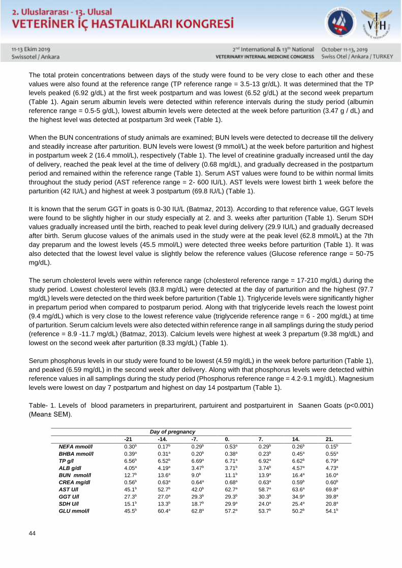
44
The total protein concentrations between days of the study were found to be very close to each other and these
values were also found at the reference range (TP reference range = 3.5-13 gr/dL). It was determined that the TP
levels peaked (6.92 g/dL) at the first week postpartum and was lowest (6.52 g/dL) at the second week prepartum
(Table 1). Again serum albumin levels were detected within reference intervals during the study period (albumin
reference range = 0.5-5 g/dL), lowest albumin levels were detected at the week before parturition (3.47 g / dL) and
the highest level was detected at postpartum 3rd week (Table 1).
When the BUN concentrations of study animals are examined; BUN levels were detected to decrease till the delivery
and steadily increase after parturition. BUN levels were lowest (9 mmol/L) at the week before parturition and highest
in postpartum week 2 (16.4 mmol/L), respectively (Table 1). The level of creatinine gradually increased until the day
of delivery, reached the peak level at the time of delivery (0.68 mg/dL), and gradually decreased in the postpartum
period and remained within the reference range (Table 1). Serum AST values were found to be within normal limits
throughout the study period (AST reference range = 2- 600 IU/L). AST levels were lowest birth 1 week before the
parturition (42 IU/L) and highest at week 3 postpartum (69.8 IU/L) (Table 1).
It is known that the serum GGT in goats is 0-30 IU/L (Batmaz, 2013). According to that reference value, GGT levels
were found to be slightly higher in our study especially at 2. and 3. weeks after parturition (Table 1). Serum SDH
values gradually increased until the birth, reached to peak level during delivery (29.9 IU/L) and gradually decreased
after birth. Serum glucose values of the animals used in the study were at the peak level (62.8 mmol/L) at the 7th
day preparum and the lowest levels (45.5 mmol/L) were detected three weeks before parturition (Table 1). It was
also detected that the lowest level value is slightly below the reference values (Glucose reference range = 50-75
mg/dL).
The serum cholesterol levels were within reference range (cholesterol reference range = 17-210 mg/dL) during the
study period. Lowest cholesterol levels (83.8 mg/dL) were detected at the day of parturition and the highest (97.7
mg/dL) levels were detected on the third week before parturition (Table 1). Triglyceride levels were significantly higher
in prepartum period when compared to postparum period. Along with that triglyceride levels reach the lowest point
(9.4 mg/dL) which is very close to the lowest reference value (triglyceride reference range = 6 - 200 mg/dL) at time
of parturition. Serum calcium levels were also detected within reference range in all samplings during the study period
(reference = 8.9 -11.7 mg/dL) (Batmaz, 2013). Calcium levels were highest at week 3 prepartum (9.38 mg/dL) and
lowest on the second week after parturition (8.33 mg/dL) (Table 1).
Serum phosphorus levels in our study were found to be lowest (4.59 mg/dL) in the week before parturition (Table 1),
and peaked (6.59 mg/dL) in the second week after delivery. Along with that phosphorus levels were detected within
reference values in all samplings during the study period (Phosphorus reference range = 4.2-9.1 mg/dL). Magnesium
levels were lowest on day 7 postpartum and highest on day 14 postpartum (Table 1).
Table- 1. Levels of blood parameters in preparturirent, partuirent and postpartuirent in Saanen Goats (p<0.001)
(Mean± SEM).
Day of pregnancy
-21 -14. -7. 0. 7. 14. 21.
NEFA mmol/l 0.30b 0.17b 0.29b 0.53a 0.29b 0.26b 0.15b
BHBA mmol/l 0.39a 0.31a 0.20b 0.38a 0.23b 0.45a 0.55a
TP g/l 6.56b 6.52b 6.69a 6.71a 6.92a 6.62b 6.79a
ALB g/dl 4.05a 4.19a 3.47b 3.71b 3.74b 4.57a 4.73a
BUN mmol/l 12.7b 13.6a 9.0b 11.1b 13.9a 16.4a 16.0a
CREA mg/dl 0.56b 0.63a 0.64a 0.68a 0.63a 0.59b 0.60b
AST U/l 45.1b 52.7b 42.0b 62.7a 58.7a 63.6a 69.8a
GGT U/l 27.3b 27.0a 29.3b 29.3b 30.3b 34.9a 39.8a
SDH U/l 15.1b 13.3b 18.7b 29.9a 24.0a 25.4a 20.8a
GLU mmol/l 45.5b 60.4a 62.8a 57.2a 53.7b 50.2b 54.1b
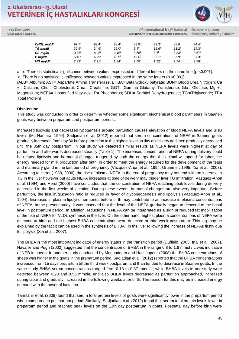
45
CHOL mg/dl 97.7a 92.4a 88.4b 83.8b 92.3a 88.6b 94.4a
TG mg/dl 32.9a 34.9a 36.0a 9.4b 15.6b 13.2b 14.3b
CA mg/dl 9.38a 8.98a 9.15a 8.68b 8.7b 8.33b 8.67b
P mg/dl 5.46a 6.29a 4.59b 4.66b 5.31a 6.59a 5.63a
MG mg/dl 2.23b 2.21a 1.94b 2.08a 1.92b 2.74a 2.56a
a, b: There is statistical significance between values expressed in different letters on the same line (p <0.001).
a: There is no statistical significance between values expressed in the same letters (p <0.001).
(ALB= Albumin; AST= Aspartate Amino Transferase; BHBA= Betahydroxy butyrate; BUN= Blood Urea Nitrogen; Ca
+= Calcium; Chol= Cholesterol; Crea= Creatinine; GGT= Gamma Glutamyl Transferase; Glu= Glucose; Mg +=
Magnesium; NEFA= Unsterified fatty acid; P= Phosphorus; SDH= Sorbitol Dehydrogenase; TG:=Triglyceride; TP=
Total Protein)
Discussion
This study was conducted in order to determine whether some significant biochemical blood parameters in Saanen
goats vary between prepartum and postpartum periods.
Increased lipolysis and decreased lypogenesis around parturition causes elevation of blood NEFA levels and BHB
levels (Mc Namara, 1994). Sadjadian et al. (2012) reported that serum concentrations of NEFA in Saanen goats
gradually increased from day 30 before parturition to the highest level on day of delivery and then gradually decreased
until the 35th day postpartum. In our study we detected similar results as NEFA levels were highest at day of
parturition and afterwards decreased steadily (Table 1). The increased concentration of NEFA during delivery could
be related lipolysis and hormonal changes triggered by both the energy that the animal will spend for labor, the
energy needed for milk production after birth, in order to meet the energy required for the development of the fetus
and mammary gland in the last period of pregnancy (Vazquez-Anon et al., 1994; Grummer, 1995; Xia et al., 2007).
According to Herdt (1988, 2000), the rise of plasma NEFA in the end of pregnancy may not end with an increase in
TG in the liver however but acute NEFA increases at time of delivery may trigger liver TG infiltration. Vazquez-Anon
et al. (1994) and Herdt (2000) have concluded that, the concentration of NEFA reaching peak levels during delivery
decreased in the first weeks of lactation. During these events, hormonal changes are also very important. Before
parturition, the insulin/glucagon ratio is reduced in favor of glyconeogenesis and lipolysis (Vazquez-Anon et al.,
1994). Increases in plasma lipolytic hormones before birth may contribute to an increase in plasma concentrations
of NEFA. In the present study, it was observed that the level of the NEFA gradually began to descend to the basal
level in postpartum period. In addition, reductions in NEFA can be interpreted as a sign of reduced fat mobilization
or the use of NEFA for VLDL synthesis in the liver. On the other hand, highest plasma concentrations of NEFA were
detected at birth and the highest BHBA concentrations were detected at third week postpartum. This lag may be
explained by the fact it can be used in the synthesis of BHBA in the liver following the increase of NEFAs firstly due
to lipolysis (Xia et al., 2007).
The BHBA is the most important indicator of energy status in the transition period (Duffield, 2003; Inal et al., 2007).
Navarre and Pugh (2002) suggested that the concentration of BHBA in the range 0.8 to 1.6 mmol / L was indicative
of NEB in sheep. In another study conducted by Moghaddam and Hassanpour (2008) the BHBA concentrations of
sheep was higher in the goats in the prepartum period. Sadjadian et al. (2012) reported that the BHBA concentrations
increased from 15 days prepartum till the third week postparum and than tended to decrease in Saanen goats. In the
same study BHBA serum concentrations ranged from 0.13 to 0.37 mmol/L; while BHBA levels in our study were
detected between 0.20 and 0.55 mmol/L and also BHBA levels decreased as parturition approached, increased
during labor and gradually increased in the following weeks after birth. The reason for this may be increased energy
demand with the onset of lactation.
Tanritanir et al. (2009) found that serum total protein levels of goats were significantly lower in the prepartum period
when compared to postpartum period. Similarly, Sadjadian et al. (2012) found that serum total protein levels lower in
prepartum period and reached peak levels on the 13th day postpartum in goats. Postnatal day before birth were
46
lower than those after postnatal birth. Our results were consistent with findings of mentioned studies, we were
determined that the total protein level reached 6.92 g/L on the 7 postpartum (Table 1). Serum total protein levels in
our study were lower before parturition than the postpartum period. This reduction may be due to the fact that protein
synthesis required for the development and growth of the fetus, is carried out using maternal amino acids and that
requirement is highest at the end of pregnancy.
Shetawi et al. (1992) conducted a study on pregnant sheep and found that with start of lactation, serum albumin
levels decreased and globulin increased. In the present study, albumin levels gradually decreased until parturition
and significantly increased 1 week after delivery (Table 1). As in humans (Ogbodo et al., 2012), the decrease in
albumin level may be associated with decreased albumin synthesis in pregancy, also and increased loss in urine in
pregnancy.
In a study conducted on Saanen goats (Sadjadian et al., 2012), blood urea nitrogen levels were found to be lowest
at birth and highest 21 days postpartum. However, statistically significant changes in BUN concentration were not
observed in our study. It is reported increased BUN levels in the postpartum cows and related this to the increase in
feed intake and dry matter consumption after birth (Bauchart, 1993). In accordance, BUN concentration in our study
were lower in the prepartum period, tended to increase after parturition, and the highest BUN concentration was
measured at day 14 postpartum (Table 1). The reason for lower BUN levels around parturition may be related to
reduced feed intake and dry matter consumption, associated with increased stress and hormonal changes. In the
present study, creatinine levels were found to increase slowly before birth, reached peak level at birth and then
gradually decreased after birth (Table 1). The increase in creatinine levels during labor may be attributed to the
hemodynamic effect of labor stress on glomerular filtration rate (GFR).
Ozyurtlu et al. (2007) reported that postpartum AST levels of sheep have increased significantly in postpartum period,
and that this increase may be due to changes related to the reproductive condition in the hormonal system. In a study
by Yokus et al. (2006) conducted on Sakız-Ivesi sheep, it is reported that the AST levels may change with the season
and also with the reproductive period. Tanritanir et al. (2009) reported that ALP activity increased significantly in the
postpartum period compared to the prenatal period in Kıl Goats, but a significant change in AST value was not
detected in the same study. AST activity is an important indicator for fatty liver disease in cows (Cebra et al., 1997;
Herdt, 2000, 2009). According to a study by Kaneko (1989), sensitivity of AST for detection of hepatic lipidosis in
cattle is estimated to be around 94%. Studies conducted on sheep by Taghipour et al. (2011) and a study conducted
on cattle by Seifi et al. (2007) also found a significantly higher AST concentration in the postpartum period, and this
elevation was explained to be indicative of fat infiltration in the liver due to NEB. Despite the fact that the postpartum
AST levels were higher than the prepartum levels in our study, the fact that the AST and BHBA levels in the pregnancy
were within the reference rate could indicate that the animals used in our study were not in the NEB (Table 1). On
the other hand, since a biopsy specimen from the liver must be taken in order to diagnose fatty infiltration in the liver,
no conclusions can be drawn about the fat infiltration of livers in animals used in our study.
It has been reported that serum GGT levels are high in negative energy balance associated fatty liver of ruminants,
and this parameter can be used as a diagnostic tool (Senturk, 2013). Indeed, elevated GGT activities may be
associated with the hepatobiliary system, and especially cholestasis. In addition, ALP, SDH and GLDH together with
GGT may indicate chronic liver failure (Senturk, 2013). Sevinc et al. (1999) found no statistically significant difference
in serum GGT levels between the 7th month of pregnancy and 2nd month after birth in a study conducted in cattle.
In the present study, it was determined that the serum GGT concentrations showed numerical increases, which did
not make a statistical difference when compared to the last week before birth and the first week after birth. This
increase in serum concentration of GGT may be related to increased fat infiltration and metabolic activity in the liver,
as seen in the elevation of AST enzyme.
SDH enzyme activity is very important in determining acute hepatocellular damage in ruminants. (Senturk, 2013).
SDH levels in our study gradually increased until birth and reached peak levels during delivery and then gradually
47
began to fall after birth (Table 1). Although this short-term rise in SDH reaching peak levels at birth strengthens the
likelihood of fatty liver, but this elevation coul also be related to hormonal changes during labor.
According to Khan et al. (2009), blood glucose concentrations are one of the most important parameters that can
reveal the nutritional status of animals during pregnancy. In a study conducted on Merino sheep, blood glucose levels
were measured as 3.0 mmol/L before flushing and 3.9 mmol/L after flushing (Venter and Greyling, 1994). Gurgoze
et al. (2009) assessed blood glucose levels in sheep during a transition period, according to which the lowest value
was 32.18 ± 1.60 mg/dL on day 120 of gestation and the highest value was 56.65 ± 3.68 on day 14 postpartum. It is
reported that blood glucose levels of pregnant goats are lower than those of non-pregnant animals (Mbassa and
Poulsen, 1991). Other reports indicate lower blood glucose levels in the last trimester of pregnancy in sheep (Balıkcı
et al., 2007). On the other hand in contrast to other studies, Al-Dewachi (1999) reported that plasma glucose levels
are increasing in pregnancy. In a study conducted on sheep by Charismiadou and Bizelis (2000), highest level of
glucose were measured near term.Vazquez-Annon et al. (1994) concluded that increased glucose at birth is due to
glyconeogenesis triggered by metabolic changes, especially hormonal changes (the effects of catecholamines and
glucocorticoids). Parallel to previous studies (Al-Dewachi, 1999; Charismiadou and Bizelis, 2000) blood glucose
levels reached peak levels during the parturition, gradually decreased in the postpartum period, and a statistically
significant difference between the postpartum glucose level and postpartum glucose level were detected in our study.
Decreased glucose levels at the beginning of lactation detected in our study are in accordance with Seifi et al. (2007),
and are probably caused by high milk yield.
Cholesterol levels are reported to decrease at the final stages of pregnancy (Azab and Abdel-Maksoud, 1999;
Krajničáková et al., 2003) however, Mbassa and Poulsan (1991) reported that plasma cholesterol levels increased
during lactation. Ozpinar et al. (1988) and Ozpinar and Ozpinar (1989) have reported that the level of cholesterol
decreases at the beginning of pregnancy and rises again when the after parturition. Similarly in the present study,
cholesterol levels decreased during pregnancy and signigicantly increased after parturition.
Fatty liver is associated with elevated serum fatty acids and decreased VLDL production. Assessment of cholesterol,
triglycerides and lipoproteins should be done promptly from the sera collected before feeding. Long-standing samples
at room temperature collected for triglyceride analysis could degradade and could lead to false high NEFA results.
Increased NEFA with triglyceride levels below 5 mg/dL could be indicative for hepatic lipidosis of cattle (Senturk,
2017). Findings in our study are similar to previous studies (Hamadeh et al., 1996; Balıkcı et al., 2007) that reported
an increase in serum triglyceride levels during the last months of pregnancy in sheep and goats. In contrast Obidike
et al. (2009) reported elevated triglyceride levels after parturition. In the present study, we determined that the
triglyceride level was significantly increased as the birthing approached, reaching peak level in the last week of
pregnancy, decreased at birth, and gradually increased again in the postpartum period (Table 1). Rukkwamsuk et al.
(1999) reported that the increase in triglyceride level during the last days of pregnancy and postnatal period could be
related with increased energy uptake in order to supplemet the energy requirement of the fetus by producing glycerol.
Increased triglyceride levels after birth may be caused by trglyceride synthesis of mammary glands for milk synthesis.
Since ruminants can tolerate low glucose levels as 40 mg/dL for many days, the excess glucose can easily be
converted into triglycerides (Obidike et al., 2009).
The pregnancy toxemia in small ruminants (ketosis in cattle) and calcium metabolism are in a relationship that directly
affect each other. Hypocalcemia can usually be detected at varying degrees in ketotic animals. Krajničáková et al.
(2003) reported that serum calcium levels began to fall in the near term and were at the lowest level (1.73 mmol / l)
on the third day postpartum and concluded that this decrease was a characteristic characteristic of the puerperal
period. In the present study, the lowest calcium levels were detected in the second week after birth, in accordance
with pervious reports (Krajničáková et al., 2003) this could probably related with the milk yield of the lactating animal
(Table 1).
48
Karapınar et al. (2007) reported a marked hypophosphatemia in ketotic animals, compared to healthy animals and
concluded that this could be related with appetite loss due to disease, as well as excessive loss of phosphorus in the
milk after parturition. The lowest phosphorus concentration in our study was detected one week before parturition,
slightly elevated at birth, and gradually increased after birth to reach prenatal levels (Table 1). The explanation of the
decreased phosphorus levels near term may be due to the fact that as the birth approaches, consumption of dry
matter decreases due to stress.
Magnesium ion is the most important of intracellular cations. Magnesium plays an important role in the cellular sodium
and potassium balance by regulating the activity of the Na, K, ATPase enzyme. Therefore, in hypomagnesemic
animals Na-K balance in the cell is distorted and severe stimulation occurs in the nerve conduction. Serum
magnesium level is generally low in winter months and subclinical hypomagnesaemia may occur during this period
(Senturk, 2017). In the present study, the magnesium concentration was found to be lowest in the last week before
and the first week after parturition (Table 1). It is possible to relate the decline in the last week near term to the
reduction of feed intake due to stress and the postpartum decline with hormonal adaptation or the onset of postpartum
hypocalcemia.
Conclusion
Diseases such as pregnancy toxemia, hypocalcemia, infertility, low milk yield, metritis, mastitis cause serious
economic loss in Saanen goats. It is necessary to conduct prenatal and postnatal metabolic profile testing in Saanen
goats susceptible to metabolic diseases such as pregnancy toxicity and ketosis due to their genetic characteristics.
In the present study, we aimed to investigate the presence of these diseases Saanen goats by metabolic profile test.
During the study, the values of NEFA, SDH and CREA increased and CHOL and TG values decreased with
parturition. When the values of 3 weeks before birth and 3 weeks after birth were compared, NEFA, CHOL, GLU,
TG, Ca+ levels were higher before and BHBA, Mg+, ALB, GGT, AST, TP, P, BUN, SDH and CREA were higher after
parturition. Not any of the animals used in the study suffered a metabolic or infectious diseases thus results of our
study may be considered as pioneering study for reference biochemical parameters for Saanen goats. On the other
hand, in order to clarify metabolic profiles changes during the transitional period in Saanen goats, it is necessary
conduct studies with higher study population also evaluating more detailed hormonal and biochemical parameters.
Acknowledgment
This work was supported by ULUDAG UNIVERSITY SCIENTIFIC RESEARCH PROJECTS UNIT (Grant no:
OUAP(V)-2015/28).
References Al-Dewachi OS (1999) Some biochemical constituents in the blood serum of pregnant Awassi ewes. Iraqi J Vet Sci 12: 275-279.
Aslan V, Aştı R, Nizamlıoglu M, Tekeli T, Basoğlu A, Demirci U (1989) Postpartum period diseases related to fatty liver syndrome. Eurasian J Vet
Sci 4(1): 43-51.
Aştı RN, Aslan V, Nizamlıoglu M, Demirci U, Tiftik AM, Gökçay Y (1989) Investigations on Fatty Liver Syndrome in Dairy Cows. Eurasian J Vet
Sci 4(1): 21-36.
Azab ME, Abdel-Maksoud HA (1999) Changes in some hematological and biochemical parameters during prepartum and postpartum periods in
female Baladi goats. Small Rumin Res 34: 77-85.
Balıkcı E, Yıldız A, Gurdogan F (2007) Blood metabolite concentrations during pregnancy and
postpartum in Akkaraman ewes. Small Rumin Res 67: 247–251.
Batmaz H, Mert N, Cetin M, Yavuz HM, Kennerman E (1992) Some hematological, biochemical
changes and clinical findings in cows in prepartum and postpartum period. Uludag Univ. Vet. Fak. Derg 1(11): 171-179.
Batmaz H (2013) Gebelik toksemisi, keçilerin ketozisi ve keçilerde karaciğer yağlanması. In: Koyun ve Keçilerin İç Hastalıkları. 1. baskı. Nobel
Tıp Kitabevleri LTD.ŞTİ. Bursa, Türkiye: pp 160-163.
Bauchart D (1993) Lipid absorption and transport in ruminants. J Dairy Sci 35: 229-232.
Bogin E, Seligman NG, Holzer Z, Avidar Y, Baram H (1988) Blood profile of a healthy beef herd grazing seasonal mediterranean range. J Vet
Med A 35: 270-276.
Borges JRJ, Grandy MC, Sıqueıra AJS (1997) A comparison of the blood levels of inorganic phosphorus, calcium, glucose, toplam protein, albumin
and globulins from jugular, mammary and coccygeal veins of lactating and non-lactating cows. Rev Bras Cienc Vet 4(2): 63-66.
Cebra CK, Garry FB, Getzy DM (1997) Hepatic lipidosis in anorectic, lactating Holstein cattle: A
retrospective study of serum biochemical abnormalities. J Vet Intern Med 11: 231-237.
49
Charismiadou MA, Bizelis JA (2000) Metabolic changes during the perinatal period in dairy sheep in relation to level of nutrition and breed. I. Late
pregnancy. Journal of animal physiology and animal nutrition 84: 61-72.
Civelek T, Sevinc M, Boydak M (2006) Serum apolipoprotein B100 concentrations in dairy cows with left sided displaced abomasum. Rev Med
Vet 157: 361-365.
Cozzi G, Ravarotto L, Gottardo F, Stefani AL, Contiero B, Moro L, Brscic M, Dalvit P (2011) Reference values for blood parameters in Holstein
dairy cows: Effects of parity, stage of lactation, and season of production. J Dairy Sci 94: 3895–3901.
Duffield T (2003) Minimizing subclinical metabolic diseases. Tri-State Dairy Nutrition Conference, Indiana: pp 43-55.
Gilbert RO, Shin ST, Guard CL, Erb HN (1998) Incidence of endometritis and effects on reproductive performance of dairy cows. Therio 49: 251
Ghergariu S, Rowlands GJ, Pop A, Danielescu N, Moldovalı NA (1984) A comparative study of
metabolic profiles obtained in dairy herds in Romania. Br Vet J 140(6): 600-608.
Grummer RR (1995) Impact of changes in organic nutrient metabolism on feeding the transition dairy cow. J Dairy Sci 73: 2820-2833.
Gurgoze S, Zonturlu AK, Ozyurtlu N, Içen H (2009) Investigation of some biochemical parametersand mineral substance during pregnancy and
postpartum period in Awassi ewes. J Fac Vet Med Uni Kafkas 15(6): 957-963.
Hamadeh ME, Bostedt H, Failing K (1996) Concentrations of metabolically relevant parameters in the blood of highly pregnant ewes. Berl Munch
Tierarztl Wochenschr 109: 81-86.
Herdt TH (1988) Fatty liver in dairy cows. Vet Clin North Am Food Anim Pract 4: 269-287.
Herdt TH (2000) Ruminant adaptation to negative energy balance: Influences on the etiology of ketosis and fatty liver. Vet Clin North Am Food
Anim Pract 16: 215-230.
Herdt TH, Gerloff BJ (2009) Ketosis. In: Anderson DE, Rings DM (eds). Current Veterinary Therapy: Food Animal Practice. Saunders Elsevier,
Missouri, USA: pp 141-144.
Inal ME, Atik U, Aksoy N, Haşimi A (2007) Ketoasidoz. In: Marks’ Basic Medical Biochemistry. 1st ed. Gunes Medicine Books, Ankara, Turkey:
pp 16-17.
Ivanov I, Damnjanovic Z, Radojicic S (1993) Disorders of the metabolism of macroelements during
terminal pregnancy and early lactation of cows. Vet Glasnik 47(4-5): 329-333.
Kaneko JJ (1989) Ketoasidozis. In: Clinical Biochemistry of Domestic Animals. 4th ed. Academic pres Inc., San Diego, California, USA: pp 74.
Karapınar T, Kızıl O, Kırbas A (2007) The investigation of serum phosphorus level in cows with
primary ketosis. Fırat Uni J Health Sci 21: 277-280.
Khan A, Faridi HAM, Ali M. (2009) Effects of cypermethrin on some clinico-hemato-biochemical and pathological parameters in male dwarf goat
(Capra hircus). Exp Toxicol Pathol 32: 151-160.
Kida K (2002a) Use of every ten-day criteria for metabolic profile test afer calving and dry off in dairy herds. J Vet Med Sci 64(11): 1003-10.
Kida K (2002b) The metabolic profile test: its practicability in assessing feding management and
periparturient diseases in high yielding commercial dairyherds. J Vet Med Sci 64(7): 557-63.
Krajničáková M, Kováč G, Kostecký M, Valocky I, Maraček I, Sutiaková I, Lenhardt L (2003) Selected clinico-biochemical parameters in the
puerperal period of goats. Bull Vet Inst Pulawy 47: 177-182.
Mbassa GK, Poulsen JS (1991) Influence of pregnancy, lactation and environment on some clinical
chemical reference values in Danish landrace dairy goats (Capra hircus) of different parity--II.
Plasma urea, creatinine, bilirubin, cholesterol, glucose and total serum proteins. Comp Biochem Physiol B 100(2): 423-31.
Mc Namara JP (1994) Lipid metabolism in adipose tissue during lactation: a model of a metabolic
restricted-fed system. J Nutr 124: 1383–1391.
Moghaddam G, Hassanpour A (2008) Comparison of blood serum glycose, bhba, blood urea nitrogen and calcium consantrations is pregnant and
lambed ewes. J Animal Veterinary Adv 7: 308-311.
Navarre CB, Pugh DG (2002) Liver fat in goats. In: Diseases of Liver in Sheep and Goat Medicine. 1st ed. WB Saunders, Philadephia, USA: pp
78-80.
Obidike IR, Aka LO, Okafor CI (2009) Time dependant peri-partum heamotological, biochemical and rectal temperature changes in west african
dwarf ewes. Small Rum Res 82: 53-57.
Ogbodo SO, Nwagha UI, Okaka ANC, Okeke AC, Chukwurah FE, Ezeonu PO (2012) Low levels of some nutritional parameters of pregnant
women in a rural community of south east Nigeria:
Implications for the attainment of the millennium developmental goal. Ann Med Health Sci Res 2(1):
49-55.
Ozpinar H, Schweigert FJ, Ozpinar A, Wierich M, Senel HS (1988) Aenderung der verteilung der
fettlöslichen vitamine auf die lipoproteinfraktionen bei saugkaelbern und kühen in abhaengigkeit
von der geburt. Berl Münch Tieraerztl Wschr 101: 383-387.
Ozpinar H, Ozpinar A (1989) Studies on the level of cholesterol and lipoprotein fractions in the serum of dairy cows. J Vet Univ Istanbul 15(2):
9¬19.
Ozyurtlu N, Gurgoze SY, Bademkıran S, Simsek A, Celik R (2007) Investigaiton of some biochemical parameters and mineral levels in pre- and
post-partum period of Awassi ewes. Fırat Uni J Health Sci 21(1): 33-36.
Rukkwamsuk T, Kruip TAM, Wensing T (1999) Relationship between overfeeding and overconditioning in the dry period and the problems of high
producing dairy cows during the postparturient period. Vet Quart 21: 71-77.
Sadjadian R, Seifi H, Mohri M (2012) Variations of energy biochemical metabolites in periparturient dairy saanen goats. Comp Clin Path 22: 449-
456.
Ŝamanc H, Kirovski D, Stojić V, Stojanovic D, Vujanac I, Prodanović R, Bojković-Kovaćević S (2011) Application of the metabolic profile test in
the prediction and diagnosis of fatty liver in Holstein
50
cows. Acta Vet Beograd 61(5-6): 543-553.
Seifi HA, Gorji-Dooz M, Mohri M (2007) Variations of energy-related biochemical metabolites during transition period in dairy cows. Comp Clin
Path 16: 253–258.
Senturk S (2013) Liver enzymes. In: Practical Clinical Laboratory Manual of Cattle. 1st ed. F. Ozsan
Publishing Inc., Bursa, Turkey: pp 44-49
Senturk S (2017) Evaluation of metabolic profile in lactation and dry animals. In: Practical Clinical
Laboratory Manual of Cattle, 2nd ed. F. Ozsan Publishing Inc., Bursa, Turkey: pp 258-293.
Sevinc M, Basoglu A, Birdane F (1999) Changes of metabolic profile during dry period and after
parturition in dairy cows. Tr J Vet Anim Sci 23: 475 - 478.
Shetawi MM, Daghash HA, Abdel ALL (1992) Effects of pregnancy and lactation on some biochemical components in the blood of Egyptian
coarse-wool ewes. Assiut Vet Med J 30: 64.
Taghipour B, Seifi HA, Mohri M (2011) Variations of energy related biochemical metabolites during periparturition period in fat-tailed baloochi
breed sheep. Iranian J Vet Res 56: 108-121.
Tanritanir P, Dede S, Ceylan E (2009) Changes in some macro minerals and biochemical parameters in female healthy Siirt hair goats before and
after parturation. J Anim Vet Adv 20: 13-18.
Vazquez-Anon M, Bertics S, Luck M, Grummer RR, Pinherio J (1994) Peripartum liver triglyceride and plasma metabolites in dairy cows. J Dairy
Sci 77: 1521-1528.
Venter JL, Greyling JPC (1994) Effect of different periods of flushing and synchronized mating on body weight, blood glucose and reproductive
performance in spring-mated ewes. Small Rum Res 13: 257-261.
Xia C, Wang Z, Li Y, Niu S, Xu C, Zhang C, Zhang H (2007) Effect of hypoglycemia on performances, metabolites, and hormones in periparturient
dairy cows. Agric Sci Chin 6(4): 505-512.
Yokus B, Cakir DU, Kanay Z (2006) Effects of seasonal and physiological variations on the serum
chemistry, vitamins and thyroid hormone concentrations in sheep. Transbound Emerg Dis 53: 271-
276.
51
Sözlü Sunu / Oral Presentation Tam Metin / Full Text
Kanin Parvovirus ile Enfekte Köpeklerde Salya Proteomlarındaki Değişimler
Lorena Franco-Martínez*, Asta Tvarijonaviciute*, Anita Horvatić**, Nicolas Guillemin**, José Joaquín Cerón*,
Damián Escribano*,***, David Eckersall**,****, Meriç Kocatürk*****, Zeki Yılmaz*****, Elsa Lamy******, Silvia
Martínez-Subiela*, Vladimir Mrljak**
*Interlab-UMU Disiplinlerarası Klinik Analiz Laboratuarı, Mare Nostrum Uluslararası Mükemmellik Yerel Kampüsü,
Murcia Üniversitesi, Espinardo, Murcia 30100, İspanya
** ERA Chair FP7, Uluslararası Hastalıkları Birimi, Zagreb Üniversitesi Veteriner Fakültesi , Heinzelova 55, 10 000
Zagreb, Hırvatistan
***Hayvan ve Gıda Bilimi Departmanı, Barcelona Autònoma Üniversitesi, 08193, Bellaterra, İspanya
**** Biyoçeşitlilik, Hayvan Sağlığı ve Karşılaştırmalı Hekimlik, Tıp, Veteriner Fakülteleri, Veteriner Fakültesi, Glasgow
Üniversitesi , Glasgow G611QH, Birleşik Krallık
*****Bursa Uludağ Üniversitesi Veteriner Fakültesi İç Hastalıkları AD, 16059, Bursa, Türkiye
******ICAAM - Akdeniz Tarım ve Çevre Bilimleri Enstütüsü, Evora Üniversitesi, Portekiz
Sunulan çalışmada parvovirus ile enfekte (CPV) köpeklerde salya proteom değişimleri yüksek verimli kantitatif
proteomik analizi ile değerlendirilmiştir. Sağlıklı ve parvoviral enteritisli (PVE) köpeklerden (tedavi sırasında hayatta
kalan ve hayatını kaybetmiş) alınan salya örneklerinden Tandem Kütle Spektrometri Analizi (TMT) gerçekleştirilmiştir.
Proteomik analizi 1516 peptid belirlemiş (niceliksel), 287 peptid (190 peptide karşılık) çalışılan gruplar arasında
anlamlı farklılık (oransal) göstermiştir. CPV enfekte köpeklerde hayatta kalanlar ve kalamayanlar karşılaştırıldığında
farklı 10 protein belirlenmiştir. Hayatta kalanlar ve kontrol grupları arasında farklı ifade edilen 90 proteinden hayatta
kalanlar grubunda en çok down-regule olanlar; cathelicidin antimicrobial peptide(CAMP), rho-GDP dissociation
inhibitor beta(ARHGDIB), apolipoprotein A-1(APO-A1), neutrophil elastase(ELANE), matrix metalloproteinase-
9(MMP9), EF-hand domain containing protein D2(EFHD2), CD177 antigen(CD177), plastin-2(LCP1), retinol binding
protein 4(RBP4) ve maltase-glucoamylase intestinal(MGAM) olmuştur. Hayatta kalan grupta ise epididymal–specific
lipocalin-9(LCN9), BPI fold-containing family B member 2(BPIFB2), lymphocyte antigen 6D(LY6D), desmoglein(-1
ve -3, DSG1, DSG3, sırasıyla), alpha-2-macrogloulin-like protein 1 (A2ML1), democollin-2 (DSC2), leucine-rich-alpa-
2-glycoprotein(LRG1), polymeric immunoglobulin receptor(PIGR) ve L-amino acid oxidase(IL4I1) en çok up-regule
olmuştur. Bunların yanında, hayatta kalan grup kontrol grubu karşılaştırıldığında CAMP ve ARHGDIB en çok down-
regule olmuş (-2.29 ve -2.26-kat daha az ifade edilmiş, sıra ile), LCN9 ve BPIFB2 en çok up-regule (2.28 ve 1.62-kat
daha yüksek) olarak belirlenmiştir. Hayatta kalamayanlar kontrol grubu karşılaştırıldığında, 90 protein yoğunluklarına
göre farklılıklar göstermiştir. En çok down-regule olan proteinler ARHGDIB, CAMP, APOA1, hemoglobin subunit beta
(HBB), RBP4, CD177, MMP9, vitamin D binding protein(GC) ve ELANE olarak belirlenmiştir. Diğer yandan,
immunoglobulin heavy chain(IGH), LCN9, clusterin(CLU), trefoil factor 1(TFF1), LY6D, olfactomedin 4(OLFM4),
LRG1, glyox-alase I(GLO1), BPIFB2 ve DSG3 ise en çok up-regule proteinler olarak belirlenmiştir. Bioinformatik
analizlerde salivanın PVE’de katalitik aktivite, bağlanma ve hastalığın gelişiminde önemli rol oynayan antimikrobiyal
humoral yanıtı gösteren birçok farklı yolağı yansıttığı belirlenmiştir. Sonuçlar PVE’de meydana gelen patofizyolojik
değişimleri yansıtan saliva proteinlerinin bu hastalıkta potansiyel biyobelirteç kaynağı olabileceğini göstermiştir.
Anahtar Kelimeler: Biyobelirteç, Kanin parvovirozis, Köpek, Etiketleme-temelli proteomik, Saliva
Giriş
Parvoviruslar birçok hayvan türü ve insanları da enfekte eden küçük, ikasohedral yapıda virsulardır. Parvovirus tip II
aşısız yetişkin ve yavrularda CPV ile sonuçlanan morbiditesi %100’e yaklaşan, mortalitesi %10-90’a varan hastalığa
neden olmaktadır (1). PVE’nin klinik sunumu klinik bulgular ve fiziksel muayene kapsamında hafiften ileri dereceye
52
kadar değişebilmektedir (2-3). Yavrularda en yaygın bulgular şiddetli kusma ve kanlı ishal, ateş, dehidrasyon ve
letarjidir (4). Hastalarda kardiyak hasar (5), sistemik infalamtuar yanıt sendromu (SIRS) ve sepsis tanımlanmıştır (6).
Köpeklerde saliva, leishmaniazis (7), helikobakter (8), stres değerlendirmeleri (9) gibi birçok durumun tanımında
biyolojik bir sıvı olarak yararlılığı çalışılmıştır. Saliva gibi örneklerin kullanımı invazif metodlara göre ağrısız oluşu,
kolay örneklenmesi ve örnekleme stresinin olmaması yönünden avantaj taşımaktadır. Saliva dışkı gibi diğer non-
invazif metodlara göre de üstünlük göstermektedir.
PVE durumlarında, dışkının ELISA testinde kullanımının sensitivitesinin zayıf olması (11) nedeni ile saliva hastalığın
monitorizasyonunda önemli bir rol oynayabilir (12). Bununla beraber PVE’de saliva bileşimi değişimleriyle ilgili rapor
edilen bir çalışma bulunmamaktadır. PVE’li köpeklerde saliva değişimlerinde olası biyobelirteçlerin belirlenmesi, PVE
hastalığı için bu biyolojik sıvının değerini arttırmaktadır.
Proteomik bilimi hücrelerin, organların ve organizmanın proteom içeriği üzerine odaklanmaktadır (13). Proteomik
teknikler erken tanıda, prognozda veya tedavinin izlenmesinde kullanılabilecek birçok biyolojik sıvının hastalık ile
ilişkili biyobelirteçlerin kullanımının keşvedilmei üzerine geniş bir alanda kullanılmaktadır (14, 15). Son yıllarda
izobarik etiketlemelerin kullanımı gibi yeni teknolojiler proteomik analizlerin sensitivitesini arttırmıştır (16-18). Tandem
Kütle Etiketlemeleri (TMT) farklı işaretlenen peptidlerin tanımlanması (19) ile herbir örnek işaretlenir ve rapor eden
iyon kütlelerindeki farklılıklara göre ayırt edilir (20, 21). Bununla beraber TMT teknolojisinin salivada kullanımı
konusunda literatürde eksiklikler mevcut olup, PVE’de diagnostik ve prognostik biyobelirteçlerin beirlenmesi için TMT
kullanımı ile ilişkili rapor bulunamamıştır.
Sunulan çalışmanın amacı, CPV ile ilişkili saliva proteinlerinin potansiyel değişimlerini değerlendirmektir. Bu çalışma
saliva bileşimini hastalıklarla ilişkili patofizyolojik değişimleri ortaya koyacak ve saliva örneklerinden PVE için
potansiyel biyobelirteçler ortaya konacaktır. Bu amaçla sağlıklı (kontrol) ve CPV ile enfekte (tedavi sırasında yaşayan
veya ölen) köpeklerden toplanan saliva örnekleri kullanılmıştır. Saliva örnekleri gruplar arasında değişkenlik gösteren
protein tanımlanmasına olacak sağlayan yüksek verimli TMT-temelli proteomik analiz kullanılmıştır. Farklı ifade
edilen proteinler protein etkileşim ağının oluşturulmasında bir başlangıç noktası oluşturacaktır.
Materyal ve Metot
Hayvan seçimi:
Bursa Uludağ Üniversitesi Veteriner Fakültesine kusma ve ishal şikayeti ile getirilen toplam 11 sahipli köpek
çalışmaya dahil edilmiştir (Tablo 1). Parvovirus enfeksiyonu klinik (akut kanlı ishal, kusma, anoreksi, dehidrasyon
gibi), hematolojik (lökopeni, nötropeni, lenfopeni vb), bulgular dahlinde kullanılan hızlı fekal diagnostik test (Anigen
Rapid PVE Test Kit, Animal Genetics, Inc., Suwon, Korea) kullanılarak yapılmıştır. Belirlenen klinik (beden sıcaklığı,
kalp ve solunum sayıları) ve hematolojik ( löykogram, eritrogram ve trombogram) bulguları hayvanın genel sağlık
durumunun belirlenmesinde kullanıldı. Başka bir enfeksiyon varlığı olan köpekler viral (Kanin Distemper, veya
coronavirus), paraziter (coccidiosis, giardiozis, askaridiozis vb) veya vektör aracılı hastalıklar (ehrlişyozis,
anaplazmozis, Lyme ve diroflariazis) için fekal testler, mikroskobik inceleme, ve serolojik diagnostik test (Anigen
Rapid Caniv-4, Bionote, Korea) kullanılarak ekarte edilmiştir. Köpekler klinik görünüm ve hastalığın gelişimine göre
3 gruba ayrılmıştır. 5 sağlıklı köpek kontrol grubunu oluşturmuştur. Virus klinik bulgular oluşmadan önce saçılmaya
başladığından, sağlıklı hayvanlar klinik, hematolojik ve serum biyokimyasallara ek olarak (Comprehensive Profile,
VetScan, Abaxis) fekal CPV testi ile taranmış, negatif sonuçlar çalışmaya katılmıştır. Test grubundakiler uygun
şekilde tedavi edilmiş (22), sıvı tedavisi (%5 Dextroz+dengeli elektrolit solüsyonu, Laktatlı Ringer ve Hetastarch),
anti-emektiker (metoklopramid: 0,2-0,4 mg/kg BID, sc; ranitidin: 2-4 mg/kg BID, im), parenteral antibiyotikler
(ampisilin 30 mg/kg BID+ amikasin 5-10 mg/kg BID+ metronidazol 25 mg kg BID) ve immun stimülanlar (imunex),
probiotik ve vitaminler (E ve C) kullanılmıştır.
PVE'li köpekler tedavi sırasında hayatta kalanlar ve kalamayanlar olarak iki gruba ayrılmıştır. Bunlardan 3 hasta
hayatta kalmış (yaş: 3.67±0.94 ay, 1 erkek ve 2 dişi) ve 3 hasta hayatını kaybetmiştir (yaş: 6.67±1.25 ay, 2 erkek ve
53
1 dişi). Bu nedenle köpekler klinik ve hematolojik bulgulara göre sınıflandırılmıştır (Tablo-1). Tedaviye rağmen 3
köpek 5 gün içinde hayatını kaybetmiştir (3,4 ve 5. günlerde).
Bütün prosedürler Bursa Uludağ Üniversitesi Hayvan Deneyleri Yerel Etik Kurulundan tarafından onaylanmıştır
(2014-05/06 sayı, 04.03.2014 tarih).
Saliva Örnekleme
Her köpekten ağız içerisine pamuk swaplar yerleştirilerek stimüle etmeksizin en az 0,5 ml saliva örneği alınmıştır.
Swaplar ıslandığında koruyucu kutularına (Salivette Saliva Collection Tube, V- Bottom, Sorstedt, Aktiengesellschaft
& Co, Nümbrecht, Germany) yerleştirilerek santrifüj edilmiş (3000×g, 10 dk, +4 ◦C), süpernatant - 80 ◦C'de analiz
için saklanmıştır (23). Dehidrasyon kaynaklı ağız kurluğu durumunda prosedür tekrarlanarak 0.5 ml örnek stimüle
edilmeksizin elde edilmiştir.
Saliva örneklerinin LC-MS/MS ile proteomik analizi:
Herbir örnekten 35 μg aseton-presipite protein redüksiyon, alkilasyon, digesyon ve işaretleme 6-plex TMT ajanları
kullanarak (Thermo Scientific) uygulanmıştır (24). Kısaca 35 μg örnek ve test standartları 200mM DTT ile indirgendi
(Sigma-Aldrich),
375mM iodoacetamid (Sigma- Aldrich) ve gece boyunca buz gibi soğuk asetonda (VWR, Pennsylvania, USA)
bekletildi. Örnekler santrifüj edilip aseton boşaltıldı. Pelletler 50 μl of 100mM TEAB buffer ile muamele edildi ve tripsin
(Promega) ile gece boyunca 37 °C'de (100 μg protein için 2.5 μg tripsin) parçalanmaya bırakıldı. TMT işaret
çözeltileri oda sıcaklığına getirildi, anhydrous acetonitrile LC–MS aşaması (Thermo Scientific) ile muamele edilerek
her örneğe eklendi. İşaretleme çözeltisi 1 saat oda sıcaklığında inkübe edildikten sonra 15 dakika boyunca 5%
hydroxylamine (Thermo Scientific) eklenerek baskılandı. Örnekler 6 μg' lık eşit miktarlarda olacak şekilde karıştırıldı
ve microplate üzerine yerleştirilerek LC–MS/MS analizi aşaması öncesinde - 20 °C' de bekletildi. LC−MS/MS analizi
Dionex Ultimate 3000 RSLS nano flow sistem (Dionex, Camberley, UK) ve Orbitrap Elite kütle spektrometresi
(Thermo Fisher Scientific) ile daha önce de belirtilen şekilde gerçekleştirildi (25).
İstatistiksel Analizler
Peptidlerin örnekteki yoğunluklarını karşılaştırmak adına gruplar arasında 3 karşılaştırmalı proteomik analizi
yapılarak (Kontrol-hayatta kalanlar, kontrol-hayatta kalamayanlar, hayatta kalanlar-hayatta kalamayanlar), elde
edilen veri logaritmik değişimler ve Student’s t-testi (tek yönlü, eşleştirilmemiş) kullanılmıştır.
Tüm olgularda, P < 0.05 değeri anlamlı kabul edilmiştir. Katlanarak değişimler (Fold change- FC) FC=log2 formülü
ile hesaplanmıştır (Grup 1 / Grup 2). İstatistikler RStudio (v1.0.143) (R Studio Team. RStudio ile gerçekleştirilmiştir
(26). “RStudio Team. RStudio: Integrated Development Environment for R [Internet]. Boston, MA: RStudio, Inc.;
2015. Adres: http://www.rstudio.com/,” 2015).
GO yolakları
Proteomik çalışmadan elde edilen veri Gen Ontoloji Analizi (GO) için kullanılmıştır. Kanin genleri tarafından farklı
ifade edilen proteinler insan ontologlarına Ensembl ontolog veri tabanı ve bu veri tabanının BioMart aracını
kullanılarak dönüştürülmüştür (www.ensembl. org). Elde edilen genler GO yolakları ile her iki durum (hayatta kalan
ve hayatta kalamayan) ve Cytoscape (v3.6.1) plug-in ClueGO (v2.5.0) [27,28] Homo sapiens üzerinden GO-biyolojik
süreçler değerlendirilmiştir (22/01/2018).
Her bir grup için gereksiz GO verilerini uzaklaştırmak adına REVIGO analizi (izin verilen benzerlik = 0.7, SimRel)
kullanılmıştır (29). Sonuç olarak, yolaklar ve iletişimleri Cytoscape ile radiyal düzeyde oluşturulmuştur.
Sonuçlar
Yüksek çözünürlüklü kantitatif proteomik analizi
54
Yüksek çözünürlüklü kantitatif proteomic analizi 11 seyreltilmemiş (non-depleted) kanin saliva örneğinden 1516 kanin
peptid tanımlamasına olanak sağlamıştır. Kontrol ve Hayatta kalanlar karşılaştırıldığında, 148 peptidin farklı ifade
edildiği ve bunların 90 tanesinin benzersiz protein olduğu gözlenmiştir. Kontrol ve Hayatta kalamayanlar
karşılaştırıldığında, 125 peptidin farklı ifade edildiği ve 90 tanesinin benzersiz protein olduğu gözlenmiştir. Hayatta
kalanlar ile Hayatta kalamayanlar karşılatırıldığında, 14 peptidin farklı ifade edildiği ve bunlar içerisinden 10 tanesinin
benzersiz protein olduğu gözlenmiştir. Bu nedenle, farklı gruplar arasında toplamda 190 protein tanımlanmıştır.
Hayatta kalanlar ve kontrol grupları arasında farklı ifade edilen 90 proteinden hayatta kalanlar grubunda en çok down-
regule olan proteinler; cathelicidin antimicrobial peptide (CAMP), rho-GDP dissociation inhibitor beta (ARHGDIB),
apolipoprotein A-1 (APO-A1), neutrophil elastase (ELANE), matrix metalloproteinase-9 (MMP9), EF-hand domain
containing protein D2 (EFHD2), CD177 antigen (CD177), plastin-2 (LCP1), retinol binding protein 4 (RBP4) ve
maltase-glucoamylase intestinal (MGAM) olmuştur. Hayatta kalan grupta ise epididymal – specific lipocalin-9 (LCN9),
BPI fold-containing family B member 2 (BPIFB2), lymphocyte antigen 6D (LY6D), desmoglein (-1 ve -3, DSG1 ve
DSG3, sırayla), alpha-2- macrogloulin-like protein 1 (A2ML1), democollin-2 (DSC2), leucine- rich-alpa-2-glycoprotein
(LRG1), polymeric immunoglobulin receptor (PIGR)ve L-amino acid oxidase (IL4I1) en çok up-regule olmuştur. Tüm
bunların içinde hayatta kalan grup ile control grubu karşılaştırıldığında CAMP ve ARHGDIB en çok down-regule
olmuşken (-2.29 ve -2.26-kat daha az ifade edilmiş, sıra ile), LCN9 ve BPIFB2 en çok up-regule (2.28 ve 1.62-kat
daha yüksek) olarak belirlenmiştir. Hayatta kalamayanlar ve kontrol grubu karşılaştırıldığında, 90 protein
yoğunluklarına göre farklılıklar göstermiştir. En çok down-regule olan proteinler ARHGDIB, CAMP, APOA1,
hemoglobin subunit beta (HBB), RBP4, CD177, MMP9, vitamin D binding protein (GC) ve ELANE olarak
belirlenmiştir. Diğer bir yandan, immunoglobulin heavy chain (IGH), LCN9, clusterin (CLU), trefoil factor 1 (TFF1),
LY6D, olfactomedin 4 (OLFM4), LRG1, glyox- alase I (GLO1), BPIFB2 ve DSG3 ise en çok up-regule proteinler
olarak belirlenmiştir.
Sonuç olarak, PVE’li iki grup köpek karşılaştırıldığında 10 protein farklı yoğunluk göstermiştir. Hayatta kalan grupta
Ezrin, allergen Dog 1, lactoperoxidase, L-lactate dehydrogenase A, cystatin-M ve macrophage-capping protein daha
yüksek belirlenirken, hastalıktan hayatını kaybeden köpeklerde alpha-actinin-1, peptidyl-prolyl cis-trans isomerase
A, ribosyldihy-dronicotinamide dehydrogenase ve lactoylglutathione lyase up-regule olmuştur.
Biyoinformatik
Cytoscape plug-in ClueGO programı ile GO analizi gerçekleştirilerek hayatta kalanlar ve kontroller arasında 34 GO
terimi belirlendi. Bunlar REVIGO kullanılarak mikatarına göre filtrelenmiş ve 12 ana gruba ayrılmıştır. Fonksiyonel
gruplar antimikrobiyal humoral yanıt, konakçı hücresine bakteri girişi, doku hemostazi, heterotipik hücre-hücre
adezyonu ve endoplazmik redikulum mannoz düzenleme şeklindedir (Figür 1).
Hayatta kalamayanlar ve kontol grubu karşılaştırıldığında, 42 GO terimi belirlenmiş ve 11 ana fonksiyonel gruba
ayrılmıştır; growth faktör uyarımına negatif hücresel yanıt regülasyonu, humoral yanıt, metal iyon homeostasis
değişimi, transition metal ion homeostasis, endoplazmik redikulum mannoz düzenleme ve glutatyon türevi
biyosentezi (Figür 2). Hayatta kalanlar ve hayatta kalamayanlar karşılaştırıldığında güvenilir bir GO analizi yapmak
adına ifade edilen yeterli protein belirlenememiştir.
Sonuç olarak, bu çalışmada REVIGO tarafından filtrelendikten sonra GO terimleri Cytoscape ile tasvir edilmiştir (Figür
3-6). Kontrol ve hayatta kalanlar arasındaki veri ağı analiz edildiğinde, birçok mekanizmaya katıla ve hastalık
oluşumunda sentral rol alan yara iyileşmesinde down regulasyon (fibrinogen FGA, FGB ve FGG genlerinin down
regulasyonu ile ilişkili) ve büyüme faktörü uyranına hücresel yanıtta negatif regulasyon (RPS27 A, UBC, UBB and
UBA52 genlerinden oluşan ubiquitin kompleks up regulasyonu) gibi odaklar belirlenmiştir.
Veri ağı hayatta kalamayanlar ve kontroller arasında aşırı ifade edilmiş olan UBS genini (ubiquitin C protein
sentezleyen) parvovirozisli hayatta kalamayan grupta birçok yolak ile merkezi bir rolü olduğu düşünülmektedir.
İlginç olarak hayatta kalamayan grupta ubiquitin C protein GO yolaklarında aşırı ifade edilmiş olduğu gözlenmiştir.
Çalışmada bu bulgu ilgili grupta ubiquitin kullanan tüm hücresel yolaklarda hayatta kalamama durumu ile pozitif
55
korelasyonlu olarak değerlendirilmiştir. Daha önce belirtilen veri ağında, parvoviruslu köpeklerin salivalarında
ibrinojen down regüle olmuştur. Kat değişim verisinin dönüştürülmesi yapılırken ve hayatta kalan ve hayatta
kalamayan gruplar arasında gözlenen yolaklar, elde edilen antimikrobiyal humoral yanıt ağı önemli noktaları
değerlendirilmiştir.
Antibakteriyel humoral yanıt, ekstarnal uyaranlara negatif regülasyon ve diğer organizma hücrelerini öldürme
antimikrobiyal humoral yanıt ile ilişkili olan 3 down reüle olmuş noktadır. Etkileşim ağı aynı zamanda merkez odağı
çevreleyen 14 proteini göstermiş, kontrollerle karşılaştırıldıklarında bunlardan 9 tanesi down-regüle olmuş (ELANE,
kininogen 1 [KNG-1], FGA, FGB, S100 calcium binding protein A8 [S100A8], S100 calcium binding protein A12
[S100A12], CAMP, lysozyme [LYZ] ve HRG) ve 5 tanesi up-regüle olmuştur (beta-2 microglobulin [B2M], deleted in
malignant brain tumours 1 [DMBT1], BPI fold-con- taining family B member 1 [BPIFB1], BPIFB2, and CLU).
TARTIŞMA
Bu makale salivada farklı ifade edilmiş proteinlerin tanımlanması ve PVE kaynaklı biyolojik rollerin değişiminin
tanımlanmasına odaklanmıştır. Bu çalışmada ilk defa sağlıklı ve parvovirozisli köpek salivalarında TMT teknolojisi
kullanılarak 190 farklı ifade edilmiş protein tanımlanmış, bunların içinden 10 tanesi hastalıktan hayatta kalanlar ve
hayatta kalamayanlar arasında farklı ifade edilmiştir. Proteinlerdeki bu farklılıklar, kanin parvoviroziste salivanın
birçok fizyolojik yolak tarafından etkilendiğini belirtmektedir.
Parvovirozisli köpek salivalarındaki protein ifade değişimlerinin hepsi yüksek çözünürlüklü kantitatif proteomik analizi
ile belirlenmiş, koagulasyon ve inflamasyon sistemleri ile yakından ilişkili olduğu görülmüştür (30). İnflamasyon
antikoagulan mekanizmaların baskılanmasını sağlarken, trombin gibi koagulasyon ürünleri inflamatuar yanıtların
harekete geçirilmesini sağlamaktadır (31). İmmun system enfeksiyona karşı koruma sağlarken, aktivasyonun aşırı
inflamatuar yanıt ve canlıya olası hasardan koruyacak şekilde düzenlenir (32). Parvovirozisli köpek saliva protein
değişimlerinden iki tanesi en fazla down-regule olmuştur. Hayatta kalan parvovirozisli köpeklerde en çok down regule
olan iki proteinin, cathelicidin antimicrobial peptide (CAMP) ve Rho-GDP dissociation inhibitor beta (ARHGDIB),
infalamtuar yanıta katılma ve enfeksiyonlara karşı koruma rolüne sahiptir. Cathelicidinler enfeksiyonlara karşı
korumada önemli bir rolü olan kısa katyonik antimikrobiyal peptid grubudur (33-35). CAMP’ların ana kaynağı
nötrofillerin aktivasyon sırasında salınan spesifik granülleridir (36-37) fakat epteliyal hücreler ve mukozal yüzeyler
tarafından da salınmaktadır (38). CAMP’lar enfeksiyon sırasında infalamtuar yanıtın modülasyonunda önemli rol alan
birçok tool-like reseptör aktivasyonunun regülasyonunda da görev aldığı bilinmektedir (39, 40). Antimikrobiyal
aktiviteye ek olarak, farelerde Escherichia coli enfeksiyonunda cathelidicin ekspresyon kaybı rapor edilmiştir (33,34).
Rho-GDP dissociation inhibitor beta (ARHGDIB) primer olarak hemapoietik hücrelerden salınır ve Rho-GTP-azları
regüle eden RhoGDI ailesine aittir (41). ARHGDIB insan immune yetmezlik virusu tip-1’de (HIV-1) azalma göstermiş,
replikasyon alanlarındaki azalma HIV-1 için biyobelirteç olarak kullanılmıştır (42). ARHGDIB salınımları metastaz
supresörü olarak nitelendirilmiş, ARHGDIB pozitif tümörlerde hastalıktan ari ve hastalık spesifik hayatta kalma oranı
ile insanlarda idrar kesesi kanserinde prognoz belirteci olarak (43). Diğer bir taraftan, hayatta kalan parvovirozisli
köpeklerde en fazla up-regule olan 2 protein lipocalin-9 (LCN9) ve BPI fold-containing family B member 2 (BPiFB2)
olarak bulunmuştur. Lipocalinler lipid taşıyan proteinler ailesinden, steroidleri, hormonları ve vitamin gibi metabolitleri
taşıma görevli bir proteindir (44). Aynı zamanda lipocalinler hücre büyümesi, metabolizması ve immun yanıtın
düzenlenmesinde rapor edilmiştir (45, 46). LCN9 hakkında az bilgi olmasına rağmen neutrophil gelatinase-
associated lipocalin (NGAL) insanlarda bakteriyel enfeksiyonla ilişkilendirilmiştir (47-49). BPiFB2 Gram-negatif
bakteriye karşı doğal immun yanıtın anahtar bileşeni olarak düşünülen bir protein ailesine ait olduğu düşünülmekte
(50), fakat fizyolojik fonksiyonları henüz bilinmemektedir (51). Buna rağmen, BPIFB2 üzerine yapılan bir çalışmada
BPIFB proteinlerinin sekretorik yolu ve Golgi kompleks morfolojisini kontol etmek surety ile enterovirus
enfeksiyonlarına katıldığını belirtmiştir (51).
Bu 4 proteine ek olarak, 86 diğer protein kontrollerle karşılaştırıldığında parvovirus enfekte köpeklerde anlamlı olarak
farklı ifade edilmiştir. Analiz ile, sunulmuş hücresel yolaklar gen ontoloji analizi ile belirlendiğinde katalitik aktivite,
bağlanma, taşıyıcı aktivite, antioksidant aktivite, yapısal molekül aktivitesi, reseptör aktivite ve dönüştürücü regulator
56
aktivite gibi diğer fizyopatolojik yolaklara katıldıkları gözlenebilir. Farklı ifade edilmiş olan genlerin %85’inden fazlası
katalitik aktivite ve bağlanma üzerine olduğundan tartışma bu proteinler üzerinden sürdürülecektir.
Katalitik aktiviteyle ilişkili proteinleri göz önünde tutarak, bazıları (apolipoprotein A-1 (APO-A1), neutrophil esterase
(ELANE), complement C3 (C3) and histidine-rich glycoprotein (HRG) down-regule olmuştur. APO-A1 insan ve diğer
memelilerde yüksek dansiteli lipoprotein kategorisinin ana bileşenidir (52) ve bu çalışmada APO-A1’in azalmış
düzeyleri belirlenmiş, bu da farklı leishmaniazis (53), kanin dilate kardiomiyopati (25) gibi kanin hastalıklarında
tanımlanmış olması APO-A1’in köpeklerde uygun bir biyobelirteç olabileceğini göstermektedir.
Kontrolle karşılaştırıldıklarında PVE ile enfekte köpeklerde her iki grupta da nötrofil esterazın azalmış salınımları
belirlenmiştir. Nötrofil elastaz nötrofillerden salınan önemli antmikrobiyal etkileri olan serin proteinaz ailesine ait bir
proteindir. Genindeki mutasyonlar (ELANE) köpek ve insanlarda herediter nötropenide tanımlanmıştır (54). Bir
kadında kronik parvovirozise bağlı rapor edilmiş ELANE gen mutasyonu rapor edilmiştir (55). Komplement C3’ün
aktivasyonu complement sistemin aktivasyonu için merkezi rol oynayarak patojenlere karşı ve humoral immun yanıtın
desteklenmesi için lokal immun yanıtı desteklemektedir (56). Artmış C3 salınımı kanin babezioziste de rapor edilmiştir
(57). Çalışmamızda, farklı hipotezler complement C3’ün PVE’li köpek salivasındaki down regulasyonu açıklayabilir.
Bunlar, proteinin complement kaskadını active etmek üzere protein yıkımlanmasındaki azalma veya mevcut immune
yetmezlik sonucunda oluşmaktadır (58). Histidine-rich glycoprotein (HRG) pıhtı regülasyonu, fibrinolizis ve
anjiyogenezis gibi durumlarda birçok odak ve fonksiyonu olan baskın plazma proteinlerindendir (59, 60). HRG’nin
antiinflamatuar ve patojen kontrolü roller rapor edilmiş; deneysel Streptococcus pyogenes ile deneysel enfeksiyon
sonrasında HRG-fakir fare vahşi tip fareye gore yüksek bakteriyel yük ve proinflamatuar sitokin yanıtı geliştirmiştir
(61).
Kontroller ve parvovirozisli köpek salivaları ile kontroller arasında farklı ifade edilen APO-A1, A2M, C3 ve LCN9 gibi
34 proteinin bağlanma aktivitesi mevcuttur.
Sağlıklı ve parvovirozisli köpek salivalarında farklı ifade edilen bağlanma aktiviteli diğer proteinler desmoglein-1 ve -
3(DSG1 ve DSG3, parvoviruslu köpeklerde sırasıyla 1.39 ve 1.43-kat daha yüksek)ve clusterinlerdir (CLU, 0.71-kat
yüksek). Desmogleinler kalsiyum bağlayan transmembran yapısında olan transmembran glikoproteinlerindendir (62).
İnsanlarda salya DSG1 ve DSG3 düzeyleri serum anti DSG titresiyle ilişkili olup, Pemphigus vulgaris tanısında uygun
bir belirteçtir (63). Clusteriner lipid metabolizması, doku rejenerasyonu, üreme, taşıma ve hücre apoptozisi gibi birçok
fizyopatolojik sürece dahil olan sekretirik glikoproteinlerdendir (64). Bu proteinler klasik olarak hücre ölümü belirteci
(65) olarak düşünülmektedir. Bununla beraber anti-apoptotik veya clusterinlerin komplement saldırılı karmaşık
fonksiyonlarından korunma gibi koruyucu roller de belirtilmiştir (66, 67).
Ortaya çıkan proteinlerin biyoinformatik analizi intrasellüler yolakları içeren verilerin anlaşılmasına olanak sağlamıştır.
Bu bulgular humoral ve hücresel yanıtların ve bakterinin konakçı hücresine girişindeki pozitif etki sürecine katkıda
bulunacaktır. Bu sonuçlar PVE'nin patojenitesi ile uyumlu olup, oral alım sonrasında kjöpeğin tonsiller lenfoid
dokusunda replike olup ardından kan dolaşımına yayılarak, hızlı replike olan hücreleri enfekte etmesi ve lenf
yumrularında ve nekroze intestinal kriptlerdeki lenfositlerin harcanması ile sonuçlanır. Bu mukozal intestinal lezyonlar
sekonder bakteriyel enfeksiyonara açık kılarak, sistemik inflamatuar yanıt sendromu (sırs), sepsis ve endotoksemi
oluşmasına zemin hazırlamaktadır (69-72).
Bioinformatik analizler saliva örneğinden PVE diagnozunda kullanışlı olabilacek protein ve yolağı aydınlatmıştır.
Örnek olarak, hayatta kalanlar ile kontrollerin karşılaştırılması ile kurulan iletişim ağı fibrinojen ve ubiquitin
komplekslerinin hastalığın gelişiminde önemli rollere sahip olduğunu göstermiştir. Bu çalışmada fibrinojenin down-
regulasyonu belirlenmiş, diğer çalışmalara zıt olarak parvovirozisli köpeklerde (73), leishmaniada (74) veya
babeziada (75) artmış fibrinojen oranları belirlenmiştir. Bununla beraber komplike olmamış babezioziste yüksek
fibrinojen oranları belirlenler SIRS veya çoklu organ yetmezlikli (MODS) olanlardır (75). Bu durum şu şekilde
açıklanabilir; köepklerde fibrinojen pozitif akut faz protein olarak tanımlanmasına ragmen, yaygın intravasküler
57
koagülasyon (DIC) ve hiperfibrinoliziste fibrinojen yıkımı ile fibrin monomerlerine dönüşmesi ile fibrinojen
konsantrasyonu düşmektedir (76, 77). Fibrinojenin devam eden azalması parvoviroziste olduğu gibi akut
kanamalarda da rapor edilmiştir (78).Diğer bir şekilde, ubiquitinler hücre siklusu kontrolü, apoptosis, DNA onarımı,
transkripsiyon regülasyonu, stress yanıtları ve 26S proteazom ile hücresel proteinlerin yıkımlanmasını odaklama gibi
birçok biyolojik fonksiyona katılmaktadır (79). Daha önceki raporlarda da belirtildiği gibi, viruslar sellüler proteinlerin
bozulmasını hedefleyen ubiquitin konjugasyonunu kullanmakta ve virus replikasyonunu etkilemektedir (80).
Çalışmanın kısıtlayan yönlerinden biri örneklem hacmidir. Bu durum PVE’li köpeklere bireyler arası farklılıklar
nedeniyle oluşabilecek protein konsantrasyon farklılıkları ile çalışmayı sınırlandırabilmektedir. Bu amaçla çalışmamız
pilot çalışma olarak düşünülebilir, ileri dönemde yapılacak daha geniş örneklem hacimli validasyon çalışmaları ile bu
bulgular teyit edilebilir. Saliva analizi üzerine kurulmuş olan protein ifadesindeki bu değişimler ve takiben
biyoinformatik analizi ile çalışma yapıldığından sistemikd değişmleri de yansıtan ileriki çalışmaların yapılmasına
ihtiyaç vardır. Bu değişimlere ragmen, sonuçlarımız PVE’nin saliva proteomunda değişimlere neden olabileceği ve
bu değişimlerin inflamatuar yanıt, koahulasyon sistemi değişimleri ve digger biyolojik süreçler ile beraber immun
yanıtı yansıtmaktadır.
Bu makale salivada farklı ifade edilmiş proteinlerin tanımlanmasına ve hastalıkla ilişkili proteinlerin biyolojik rollerinin
tanımlanmasına odaklanmıştır. İleri geniş çaplı klinik çalışmaların daha büyük örneklem hacimleri ile yapılması klinik
tedaviye katkı vermek, erken tanı ve diyagnostik yönlerden yararlılık adına gerekidir. Bu sonuçlar başarılı olduğunda,
bu proteinler hızlı tanı testlerinin geliştirilmesinde kullanılabilecektir.
Sonuçlar
TMT-temelli proteomik analizi sağlıklı ve PVE’li köpekler arasında farklı yopunluklarda olan 190 proteinin
tanımlanmasına olanar sağlamışr, bunların çoğunluğu daha önce parvovirosizte rapor edilmemiştir. Bu proteinler
koagülasyon, inflamasyon ve koruma mekanizmaları gibi birçok patofizyolojik sürece katılmakta ve bu durum da
hastalığın uygun biyobelirteci olma potansiyeli düşünülebilmektedir. Bu çalışma kanin parvovirozisin diyagnozunda
saliva proteomunun uygun bir biyolojik sıvı olduğunu göztermiştir.
Teşekkürler
LFM’nin predoktoral bağlantısı ‘FPU’ of University of Murcia, İspanya tarafından desteklenmiştir. AT ‘nin post-doktoral
işbirliği “Juan de la Cierva Incorporación” ve DE’nin post-doktoral işbirliği “Juan de la Cierva Formación” ve “Ministerio
de Economía y Competitividad”, İspanya tarafından desteklenmiştir. Bu çalışma Program for Research Groups
of Excellence of the Seneca Foundation, Murcia, Spain (grant 19894/GERM/15) fonu ve European Commission ERA
chair FP7 fonu tarafından desteklenmiştir (VetMedZg #621394).
Referanslar: 1.S. Nandi, M. Kumar, Canine parvovirus: current perspective, Indian J. Virol. 21 (1) (2010) 31–44.
2.V. Matijatko, V. Mrljak, I. Kiš, N. Kučer, J. Foršek, T. Živičnjak, Ž. Romić, Z. Šimec, J.J. Ceron, Evidence of an acute phase response in dogs
naturally infected with Babesia canis, Vet. Parasitol. 144 (2007) 242–250,
3.D. Panda, R.C. Patra, S. Nandi, D. Swarup, Oxidative stress indices in gastroenteritis in dogs with canine parvoviral infection, Res. Vet. Sci. 86
(2009) 36–42.
4.M. Kocaturk, A. Tvarijonaviciute, S. Martinez-Subiela, F. Tecles, O. Eralp, Z. Yilmaz, J.J. Ceron, Inflammatory and oxidative biomarkers of
disease severity in dogs with parvoviral enteritis, J. Small Anim. Pract. 56 (2015) 119–124.
5.J. Ford, L. McEndaffer, R. Renshaw, A. Molesan, K. Kelly, Parvovirus infection is associated with myocarditis and myocardial fibrosis in young
dogs, Vet. Pathol. 54 (2017) 964–971.
6.A.J. Mohr, A.L. Leisewitz, L.S. Jacobson, J.M. Steiner, C.G. Ruaux, Da Williams, Effect of early enteral nutrition on intestinal permeability,
intestinal protein loss, and outcome in dogs with severe parvoviral enteritis, J. Vet. Intern. Med. 17 (2003) 791–798.
7.A. Cantos-Barreda, D. Escribano, L.J. Bernal, J.J. Cerón, S. Martínez-Subiela, Quantification of anti-Leishmania antibodies in saliva of dogs,
Vet. Parasitol. 242 (2017) 54–58.
8.M. Jankowski, J. Spuzak, K. Kubiak, K. Glińska-Suchocka, M. Biernat, An evaluation of the usefulness of invasive and non-invasive methods
used to diagnose Helicobacter spp. Infections in dogs, Pol. J. Vet. Sci. 20 (2017) 491–499.
9.F. Di Nardo, L. Anfossi, L. Ozella, A. Saccani, C. Giovannoli, G. Spano, C. Baggiani, Validation of a qualitative immunochromatographic test for
the noninvasive as- sessment of stress in dogs, J. Chromatogr. B 1028 (2016) 192–198.
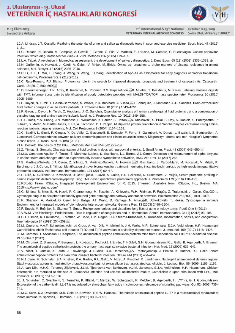
58
10.A. Lindsay, J.T. Costello, Realising the potential of urine and saliva as diagnostic tools in sport and exercise medicine, Sport. Med. 47 (2016)
1–21.
11.C. Desario, N. Decaro, M. Campolo, A. Cavalli, F. Cirone, G. Elia, V. Martella, E. Lorusso, M. Camero, C. Buonavoglia, Canine parvovirus
infection: which diag- nostic test for virus? J. Virol. Methods 126 (2005) 179–185.
12.L.A. Tabak, A revolution in biomedical assessment: the development of salivary diagnostics, J. Dent. Educ. 65 (12) (2001) 1335–1339.
13.N. Guillemin, A. Horvatić, J. Kuleš, A. Galan, V. Mrljak, M. Bhide, Omics ap- proaches to probe markers of disease resistance in animal
sciences, Mol. Biosyst. 12 (2016) 2036–2046.
14.H. Li, C. Li, H. Wu, T. Zhang, J. Wang, S. Wang, J. Chang, Identification of Apo-A1 as a biomarker for early diagnosis of bladder transitional
cell carcinoma, Proteome Sci. 9 (21) (2011).
15.C. Ruiz-Romero, F.J. Blanco, Proteomics role in the search for improved diagnosis, prognosis and treatment of osteoarthritis, Osteoarthr.
Cartil. 18 (2010) 500–509.
16.D. Baeumlisberger, T.N. Arrey, B. Rietschel, M. Rohmer, D.G. Papasotiriou,B. Mueller, T. Beckhaus, M. Karas, Labeling elastase digests
with TMT: informa- tional gain by identification of poorly detectable peptides with MALDI-TOF/TOF mass spectrometry, Proteomics 10 (2010)
3905–3909.
17.L. Dayon, N. Turck, T. García-Berrocoso, N. Walter, P.R. Burkhard, A. Vilalta,J. Sahuquillo, J. Montaner, J.-C. Sanchez, Brain extracellular
fluid protein changes in acute stroke patients, J. Proteome Res. 10 (2011) 1043–1051.
18.P. Giron, L. Dayon, N. Turck, C. Hoogland, J.-C. Sanchez, Quantitative analysis of human cerebrospinal fluid proteins using a combination of
cysteine tagging and amine-reactive isobaric labeling, J. Proteome Res. 10 (2011) 249–258.
19.P.L. Ross, Y.N. Huang, J.N. Marchese, B. Williamson, K. Parker, S. Hattan,N. Khainovski, S. Pillai, S. Dey, S. Daniels, S. Purkayastha, P.
Juhasz, S. Martin, M. Bartlet-Jones, F. He, A. Jacobson, D.J. Pappin, Multiplexed protein quantitation in Saccharomyces cerevisiae using amine-
reactive isobaric tagging reagents, Mol. Cell Proteomics 3 (2004) 1154–1169.
20.C. Baldini, L. Giusti, F. Ciregia, Y. Da Valle, C. Giacomelli, E. Donadio, F. Ferro, S. Galimberti, V. Donati, L. Bazzichi, S. Bombardieri, A.
Lucacchini, Correspondence between salivary proteomic pattern and clinical course in primary Sjögren syn- drome and non-Hodgkin’s lymphoma:
a case report, J. Transl. Med. 9 (188) (2011).
21.P. Beckett, The basics of 2D DIGE, Methods Mol. Biol. 854 (2012) 9–19.
22.Z. Yilmaz, S. Senturk, Characterisation of lipid profiles in dogs with parvoviral enteritis, J. Small Anim. Pract. 48 (2007) 643–650.
23.M.D. Contreras-Aguilar, F. Tecles, S. Martínez-Subiela, D. Escribano, L.J. Bernal, J.J. Cerón, Detection and measurement of alpha-amylase
in canine saliva and changes after an experimentally induced sympathetic activation, BMC Vet. Res. 13 (2017) 266.
24.S. Martínez-Subiela, J.J. Ceron, Z. Yilmaz, S. Martinez-Subiela, A. Horvatic,D. Escribano, L. Pardo-Marin, M. Kocaturk, V. Mrljak, R.
Burchmore, J.J. Ceron, Z. Yilmaz, Identification of novel biomarkers for treatment monitoring in canine leishmaniosis by high-resolution quantitative
proteomic analysis, Vet. Immunol. Immunopathol. 191 (2017) 60–67.
25.P. Bilić, N. Guillemin, A. Kovačević, B. Beer Ljubić, I. Jović, A. Galan, P.D. Eckersall, R. Burchmore, V. Mrljak, Serum proteome profiling in
canine idiopathic dilated cardiomyopathy using TMT-based quantitative proteomics approach, J. Proteomics 179 (2018) 110–121.
26.RStudio Team. RStudio, Integrated Development Environment for R, 2015. [Internet]. Available from: RStudio, Inc., Boston, MA,
2015http://www.rstudio. com/.
27.G. Bindea, B. Mlecnik, H. Hackl, P. Charoentong, M. Tosolini, A. Kirilovsky, W.H. Fridman, F. Pagès, Z. Trajanoski, J. Galon, ClueGO: a
Cytoscape plug-in to decipher functionally grouped gene ontology and pathway annotation networks, Bioinformatics 25 (2009) 1091–1093.
28.P. Shannon, A. Markiel, O. Ozier, N.S. Baliga, J.T. Wang, D. Ramage, N. Amin,B. Schwikowski, T. Ideker, Cytoscape: a software
Environment for integrated models of biomolecular interaction networks, Genome Res. 13 (2003) 2498–2504.
29.F. Supek, M. Bošnjak, N. Škunca, T. Šmuc, Revigo summarizes and visualizes long lists of gene ontology terms, PLoS One 6 (2011).
30.V.W.M. Van Hinsbergh, Endothelium - Role in regulation of coagulation and in- flammation, Semin. Immunopathol. 34 (1) (2012) 93–106.
31.C.T. Esmon, K. Fukudome, T. Mather, W. Bode, L.M. Regan, D.J. Stearns-Kurosawa, S. Kurosawa, Inflammation, sepsis, and coagulation,
Haematologica 84 (1999) 254–259.
32.M. Coorens, V.A.F. Schneider, A.M. de Groot, A. van Dijk, M. Meijerink, J.M. Wells, M.R. Scheenstra, E.J.A. Veldhuizen, H.P. Haagsman,
Cathelicidins inhibit Escherichia coli-Induced TLR2 and TLR4 activation in a viability-dependent manner, J. Immunol. 199 (2017) 1418–1428.
33.M. Chromek, I. Arvidsson, D. Karpman, The antimicrobial peptide cathelicidin protects mice from Escherichia coli O157:H7-Mediated disease,
PLoS One 7 (2012).
34.M. Chromek, Z. Slamová, P. Bergman, L. Kovács, L. Podracká, I. Ehrén, T. Hökfelt, G.H. Gudmundsson, R.L. Gallo, B. Agerberth, A. Brauner,
The antimicrobial peptide cathelicidin protects the urinary tract against invasive bacterial infection, Nat. Med. 12 (2006) 636–641.
35.V. Nizet, T. Ohtake, X. Lauth, J. Trowbridge, J. Rudisill, R.A. Dorschner,V. Pestonjamasp, J. Piraino, K. Huttner, R.L. Gallo, Innate
antimicrobial peptide protects the skin from invasive bacterial infection, Nature 414 (2001) 454–457.
36.N.J. Jann, M. Schmaler, S.A. Kristian, K.A. Radek, R.L. Gallo, V. Nizet, A. Peschel, R. Landmann, Neutrophil antimicrobial defense against
Staphylococcus aureus is mediated by phagolysosomal but not extracellular trap-associated cathelicidin, J. Leukoc. Biol. 86 (2009) 1159–1169.
37.A. van Dijk, M.H.G. Tersteeg-Zijderveld, J.L.M. Tjeerdsma-van Bokhoven, A.J.M. Jansman, E.J.A. Veldhuizen, H.P. Haagsman, Chicken
heterophils are recruited to the site of Salmonella infection and release antibacterial mature Cathelicidin-2 upon stimulation with LPS, Mol.
Immunol. 46 (2009) 1517–1526.
38. J. Schauber, C. Svanholm, S. Term??n, K. Iffland, T. Menzel, W. Scheppach, R. Melcher, B. Agerberth, H. L??hrs, G.H. Gudmundsson,
Expression of the cathe- licidin LL-37 is modulated by short chain fatty acids in colonocytes: relevance of signalling pathways, Gut 52 (2003) 735–
741.
39.M.G. Scott, D.J. Davidson, M.R. Gold, D. Bowdish, R.E.W. Hancock, The human antimicrobial peptide LL-37 is a multifunctional modulator of
innate immune re- sponses, J. Immunol. 169 (2002) 3883–3891.
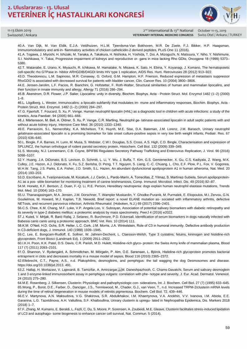
59
40.A. Van Dijk, M. Van Eldik, E.J.A. Veldhuizen, H.L.M. Tjeerdsma-Van Bokhoven, M.R. De Zoete, F.J. Bikker, H.P. Haagsman,
Immunomodulatory and anti-in- flammatory activities of chicken cathelicidin-2 derived peptides, PLoS One 11 (2016).
41.A. Togawa, J. Miyoshi, H. Ishizaki, M. Tanaka, A. Takakura, H. Nishioka, H. Yoshida, T. Doi, A. Mizoguchi, N. Matsuura, Y. Niho, Y. Nishimune,
S.I. Nishikawa, Y. Takai, Progressive impairment of kidneys and reproductive or- gans in mice lacking Rho GDIα, Oncogene 18 (1999) 5373–
5380.
42.T. Watanabe, E. Urano, K. Miyauchi, R. Ichikawa, M. Hamatake, N. Misawa, K. Sato, H. Ebina, Y. Koyanagi, J. Komano, The hematopoietic
cell-specific rho GTPase in- hibitor ARHGDIB/D4GDI limits HIV type 1 replication, AIDS Res. Hum. Retroviruses 28 (2012) 913–922.
43.D. Theodorescu, L.M. Sapinoso, M.R. Conaway, G. Oxford, G.M. Hampton, H.F. Frierson, Reduced expression of metastasis suppressor
RhoGDI2 is associated with decreased survival for patients with bladder cancer, Clin. Cancer Res. 10 (2004) 3800–3806.
44.E. Jensen-Jarolim, L.F. Pacios, R. Bianchini, G. Hofstetter, F. Roth-Walter, Structural similarities of human and mammalian lipocalins, and
their function in innate immunity and allergy, Allergy 71 (2016) 286–294.
45.B. Åkerstrom, D.R. Flower, J.P. Salier, Lipocalins: unity in diversity, Biochim. Biophys. Acta - Protein Struct. Mol. Enzymol 1482 (1–2) (2000)
1–8.
46.L. Lögdberg, L. Wester, Immunocalins: a lipocalin subfamily that modulates im- mune and inflammatory responses, Biochim. Biophys. Acta -
Protein Struct. Mol. Enzymol. 1482 (1–2) (2000) 284–297.
47.G. Fjaertoft, T. Foucard, S. Xu, P. Venge, Human neutrophil lipocalin (HNL) as a diagnostic tool in children with acute infections: a study of the
kinetics, Acta Paediatr. 94 (2005) 661–666.
48.J. Mårtensson, M. Bell, A. Oldner, S. Xu, P. Venge, C.R. Martling, Neutrophil ge- latinase-associated lipocalin in adult septic patients with and
without acute kidney injury, Intensive Care Med. 36 (2010) 1333–1340.
49.E. Parravicini, S.L. Nemerofsky, K.A. Michelson, T.K. Huynh, M.E. Sise, D.A. Bateman, J.M. Lorenz, J.M. Barasch, Urinary neutrophil
gelatinase-associated lipocalin is a promising biomarker for late onset culture-positive sepsis in very low birth weight infants, Pediatr. Res. 67
(2010) 636–640.
50.L. Bingle, F.A. Barnes, H. Lunn, M. Musa, S. Webster, C.W.I. Douglas, S.S. Cross, A.S. High, C.D. Bingle, Characterisation and expression of
SPLUNC2, the human orthologue of rodent parotid secretory protein, Histochem. Cell Biol. 132 (2009) 339–349.
51.S. Morosky, N.J. Lennemann, C.B. Coyne, BPIFB6 Regulates Secretory Pathway Trafficking and Enterovirus Replication, J. Virol. 90 (2016)
5098–5107.
52.Y. Huang, J.A. DiDonato, B.S. Levison, D. Schmitt, L. Li, Y. Wu, J. Buffa, T. Kim, G.S. Gerstenecker, X. Gu, C.S. Kadiyala, Z. Wang, M.K.
Culley, J.E. Hazen, A.J. Didonato, X. Fu, S.Z. Berisha, D. Peng, T.T. Nguyen, S. Liang, C.-C. Chuang, L. Cho, E.F. Plow, P.L. Fox, V. Gogonea,
W.H.W. Tang, J.S. Parks, E.A. Fisher, J.D. Smith, S.L. Hazen, An abundant dysfunctional apolipoprotein A1 in human atheroma, Nat. Med. 20
(2014) 193–203.
53.D. Escribano, A. Tvarijonaviciute, M. Kocaturk, J.J. Cerón, L. Pardo-Marín, A. Torrecillas, Z. Yilmaz, S. Martínez-Subiela, Serum apolipoprotein-
A1 as a pos- sible biomarker for monitoring treatment of canine leishmaniosis, Comp. Immunol. Microbiol. Infect. Dis. 49 (2016) 82–87.
54.M. Horwitz, K.F. Benson, Z. Duan, F.-Q. Li, R.E. Person, Hereditary neutropenia: dogs explain human neutrophil elastase mutations, Trends
Mol. Med. 10 (2004) 163–170.
55.U. Thanarajasingam, M.A. Jensen, J.M. Dorschner, T. Wampler Muskardin, Y. Ghodke-Puranik, M. Purmalek, E. Eliopoulos, M.I. Zervou, G.N.
Goulielmos, M. Howard, M.J. Kaplan, T.B. Niewold, Brief report: a novel ELANE mutation as- sociated with inflammatory arthritis, defective
NETosis, and recurrent parvovirus infection, Arthritis Rheumatol. (Hoboken, N.J.) 69 (2017) 2396–2401.
56.C.S. Chee, K.M. Chang, M.F. Loke, V.P. Angela Loo, V. Subrayan, Association of potential salivary biomarkers with diabetic retinopathy and
its severity in type-2 diabetes mellitus: a proteomic analysis by mass spectrometry, PeerJ 4 (2016) e2022.
57.J. Kuleš, V. Mrljak, R. Barić Rafaj, J. Selanec, R. Burchmore, P.D. Eckersall, Identification of serum biomarkers in dogs naturally infected with
Babesia canis canis using a proteomic approach, BMC Vet. Res. 10 (2014) 111.
58.K.M. O’Neil, H.D. Ochs, S.R. Heller, L.C. Cork, J.M. Morris, J.A. Winkelstein, Role of C3 in humoral immunity. Defective antibody production
in C3-deficient dogs, J. Immunol. 140 (1988) 1939–1945.
59.C. Lee, E. Bongcam-Rudloff, E. Sollner, W. Jahnen-Dechent, L. Claesson-Welsh, Type 3 cystatins; fetuins, kininogen and histidine-rich
glycoprotein, Front Biosci (Landmark Ed). 1 (2009) 2911–2922.
60.I.K.H. Poon, K.K. Patel, D.S. Davis, C.R. Parish, M.D. Hulett, Histidine-rich glyco- protein: the Swiss Army knife of mammalian plasma, Blood
117 (7) (2011) 2093–2101.
61.O. Shannon, V. Rydengård, A. Schmidtchen, M. Mörgelin, P. Alm, O.E. Sørensen, L. Björck, Histidine-rich glycoprotein promotes bacterial
entrapment in clots and decreases mortality in a mouse model of sepsis, Blood 116 (2010) 2365–2372.
62.Ellebrecht, C.T., Payne, A.S., n.d. Plakophilins, desmogleins, and pemphigus: the tail wagging the dog Desmosomes and disease.
https://doi.org/10.1038/jid.2013. 491.
63.Z. Hallaji, H. Mortazavi, V. Lajevardi, B. Tamizifar, A. Amirzargar,M. Daneshpazhooh, C. Chams-Davatchi, Serum and salivary desmoglein
1 and 3 enzyme-linked immunosorbent assay in pemphigus vulgaris: correlation with phe- notype and severity, J. Eur. Acad. Dermatol. Venereol.
24 (2010) 275–280.
64.M.E. Rosenberg, J. Silkensen, Clusterin: Physiologic and pathophysiologic con- siderations, Int. J. Biochem. Cell Biol. 27 (7) (1995) 633–645.
65.Wong, P., Borst, D.E., Farber, D., Danciger, J.S., Tenniswood, M., Chader, G.J., van Veen, T., n.d. Increased TRPM-2/clusterin mRNA levels
during the time of retinal degeneration in mouse models of retinitis pigmentosa. Biochem. Cell Biol. 72, 439–446.
66.E.V. Martynova, A.N. Maksudova, V.G. Shakirova, S.R. Abdulkhakov, I.M. Khaertynova, V.A. Anokhin, V.V. Ivanova, I.M. Abiola, E.E.
Garanina, L.G. Tazetdinova, A.H. Valiullina, S.F. Khaiboullina, Urinary clusterin is upregu- lated in Nephropathia Epidemica, Dis. Markers 2018
(2018) 1–7.
67.F. Zhang, M. Kumano, E. Beraldi, L. Fazli, C. Du, S. Moore, P. Sorensen, A. Zoubeidi, M.E. Gleave, Clusterin facilitates stress-induced lipidation
of LC3 and autophago- some biogenesis to enhance cancer cell survival, Nat. Commun. 5 (2014).
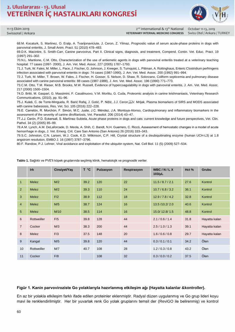
60
68.M. Kocaturk, S. Martinez, O. Eralp, A. Tvarijonaviciute, J. Ceron, Z. Yilmaz, Prognostic value of serum acute-phase proteins in dogs with
parvoviral enteritis, J. Small Anim. Pract. 51 (2010) 478–483.
69.D.K. Macintire, S. Smith-Carr, Canine parvovirus. Part II. Clinical signs, diagnosis, and treatment, Compend. Contin. Vet. Educ. Pract. 19
(1997) 291–302.
70.N.L. Mantione, C.M. Otto, Characterization of the use of antiemetic agents in dogs with parvoviral enteritis treated at a veterinary teaching
hospital: 77 cases (1997- 2000), J. Am. Vet. Med. Assoc. 227 (2005) 1787–1793.
71.J. Turk, W. Fales, M. Miller, L. Pace, J. Fischer, G. Johnson, J. Kreeger, S. Turnquist, L. Pittman, A. Rottinghaus, Enteric Clostridium perfringens
infection associated with parvoviral enteritis in dogs: 74 cases (1987-1990), J. Am. Vet. Med. Assoc. 200 (1992) 991–994.
72.J. Turk, M. Miller, T. Brown, W. Fales, J. Fischer, H. Gosser, S. Nelson, D. Shaw, R. Solorzano, Coliform septicemia and pulmonary disease
associated with canine parvoviral enteritis: 88 cases (1987-1988), J. Am. Vet. Med. Assoc. 196 (1990) 771–773.
73.C.M. Otto, T.M. Rieser, M.B. Brooks, M.W. Russell, Evidence of hypercoagulability in dogs with parvoviral enteritis, J. Am. Vet. Med. Assoc.
217 (2000) 1500–1504.
74.D. Britti, M. Gaspari, G. Massimini, F. Casalinuovo, V.M. Morittu, G. Cuda, Proteomic analysis in canine leishmaniasis, Veterinary Research
Communications, (2010), pp. 91–96.
75.J. Kuleš, C. de Torre-Minguela, R. Barić Rafaj, J. Gotić, P. Nižić, J.J. Ceron,V. Mrljak, Plasma biomarkers of SIRS and MODS associated
with canine babesiosis, Res. Vet. Sci. 105 (2016) 222–228.
76.E. Carretón, R. Morchón, F. Simón, M.C. Juste, J.C. Méndez, J.A. Montoya-Alonso, Cardiopulmonary and inflammatory biomarkers in the
assessment of the severity of canine dirofilariosis, Vet. Parasitol. 206 (2014) 43–47.
77.J.J. Cerón, P.D. Eckersall, S. Martínez-Subiela, Acute phase proteins in dogs and cats: current knowledge and future perspectives, Vet. Clin.
Pathol. 34 (2) (2005) 85–99.
78.A.M. Lynch, A.M. DeLaforcade, D. Meola, A. Shih, C. Bandt, N.H. Guerrero, C. Riccó, Assessment of hemostatic changes in a model of acute
hemorrhage in dogs, J. Vet. Emerg. Crit. Care San Antonio (San Antonio) 26 (2016) 333–343.
79.S.C. Johnston, C.N. Larsen, W.J. Cook, K.D. Wilkinson, C.P. Hill, Crystal structure of a deubiquitinating enzyme (human UCH-L3) at 1.8
angstrom resolution, EMBO J. 16 (1997) 3787–3796.
80.F. Randow, P.J. Lehner, Viral avoidance and exploitation of the ubiquitin system, Nat. Cell Biol. 11 (5) (2009) 527–534.
Tablo 1. Sağlıklı ve PVE'li köpek gruplarında seçilmiş klinik, hematolojik ve prognostik veriler.
Irk Cinsiyet/Yaş T °C Pulzasyon Respirasyon WBC / N / L X
103/μL
Hct % Grubu
1 Melez M/2 39.2 120 22 11.5 / 8.7 / 2.1 27.6 Kontrol
2 Melez M/2 39.3 110 24 10.7 / 6.8 / 3.2 36.1 Kontrol
3 Melez F/2 38.9 112 18 12.9 / 7.8 / 4.2 32.8 Kontrol
4 Melez M/5 38.7 124 16 13.5 /10.2/ 2.0 40.6 Kontrol
5 Melez M/10 38.5 114 16 15.0/ 12.8/ 1.5 48.8 Kontrol
6 Rottweiller F/5 39.8 128 44 2.1 / 0.6 / 1.4 31.8 Hayatta kalan
7 Cocker M/3 38.3 200 44 2.5 / 1.0 / 1.3 39.1 Hayatta kalan
8 Melez F/3 37.5 148 20 1.6 / 0.6 / 0.8 29.7 Hayatta kalan
9 Kangal M/5 39.8 120 44 0.3 / 0.1 / 0.1 34.2 Ölen
10 Rottweiller M/7 40.7 108 28 1.2 / 0.3 / 0.8 43.2 Ölen
11 Cocker F/8 108 32 0.3 / 0.0 / 0.2 37.5 Ölen
Figür 1. Kanin parvoviroziste Go yolaklarıyla hazırlanmış etkileşim ağı (Hayatta kalanlar &kontroller).
En az bir yolakla etkileşim farklı ifade edilen proteinler eklenmiştir. Radyal düzen uygulanmış ve Go grup lideri koyu
mavi ile renklendirilmiştir. Her bir yuvarlak renk Go yolak gruplarını temsil der (ReviGO ile belirenmiş) ve kontrol
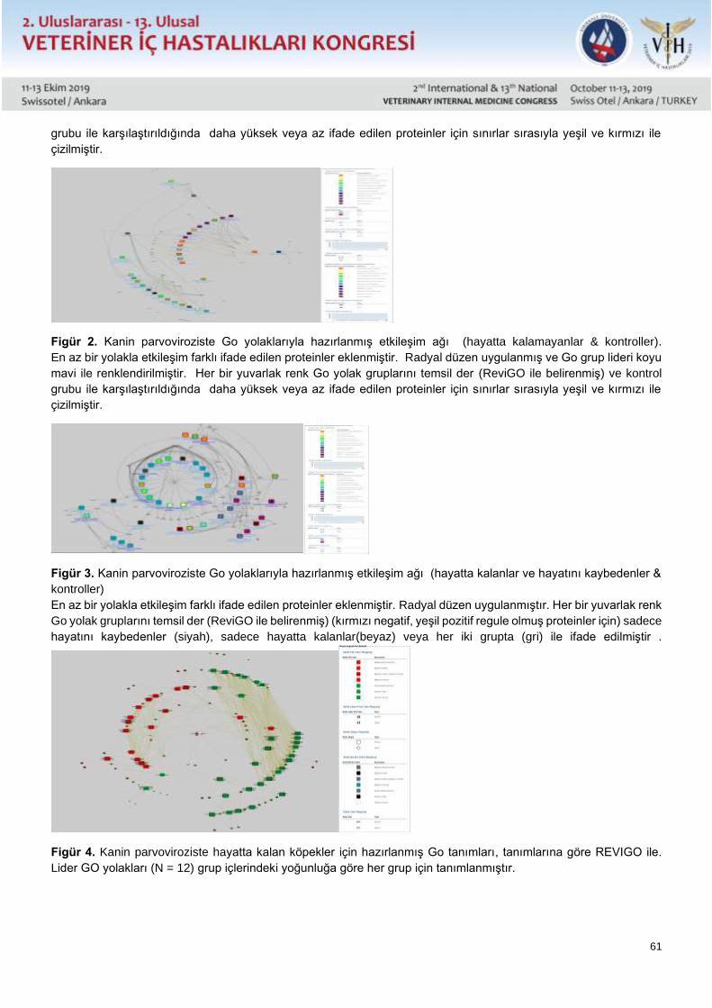
61
grubu ile karşılaştırıldığında daha yüksek veya az ifade edilen proteinler için sınırlar sırasıyla yeşil ve kırmızı ile
çizilmiştir.
Figür 2. Kanin parvoviroziste Go yolaklarıyla hazırlanmış etkileşim ağı (hayatta kalamayanlar & kontroller).
En az bir yolakla etkileşim farklı ifade edilen proteinler eklenmiştir. Radyal düzen uygulanmış ve Go grup lideri koyu
mavi ile renklendirilmiştir. Her bir yuvarlak renk Go yolak gruplarını temsil der (ReviGO ile belirenmiş) ve kontrol
grubu ile karşılaştırıldığında daha yüksek veya az ifade edilen proteinler için sınırlar sırasıyla yeşil ve kırmızı ile
çizilmiştir.
Figür 3. Kanin parvoviroziste Go yolaklarıyla hazırlanmış etkileşim ağı (hayatta kalanlar ve hayatını kaybedenler &
kontroller)
En az bir yolakla etkileşim farklı ifade edilen proteinler eklenmiştir. Radyal düzen uygulanmıştır. Her bir yuvarlak renk
Go yolak gruplarını temsil der (ReviGO ile belirenmiş) (kırmızı negatif, yeşil pozitif regule olmuş proteinler için) sadece
hayatını kaybedenler (siyah), sadece hayatta kalanlar(beyaz) veya her iki grupta (gri) ile ifade edilmiştir .
Figür 4. Kanin parvoviroziste hayatta kalan köpekler için hazırlanmış Go tanımları, tanımlarına göre REVIGO ile.
Lider GO yolakları (N = 12) grup içlerindeki yoğunluğa göre her grup için tanımlanmıştır.
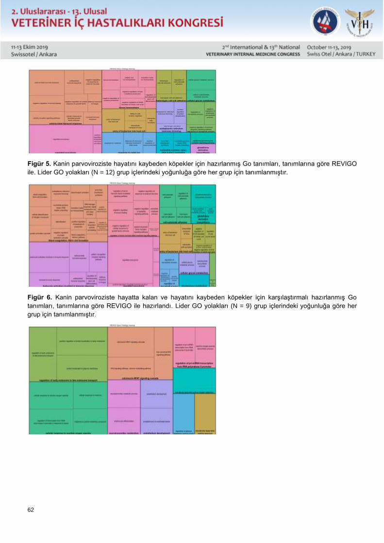
62
Figür 5. Kanin parvoviroziste hayatını kaybeden köpekler için hazırlanmış Go tanımları, tanımlarına göre REVIGO
ile. Lider GO yolakları (N = 12) grup içlerindeki yoğunluğa göre her grup için tanımlanmıştır.
Figür 6. Kanin parvoviroziste hayatta kalan ve hayatını kaybeden köpekler için karşılaştırmalı hazırlanmış Go
tanımları, tanımlarına göre REVIGO ile hazırlandı. Lider GO yolakları (N = 9) grup içlerindeki yoğunluğa göre her
grup için tanımlanmıştır.
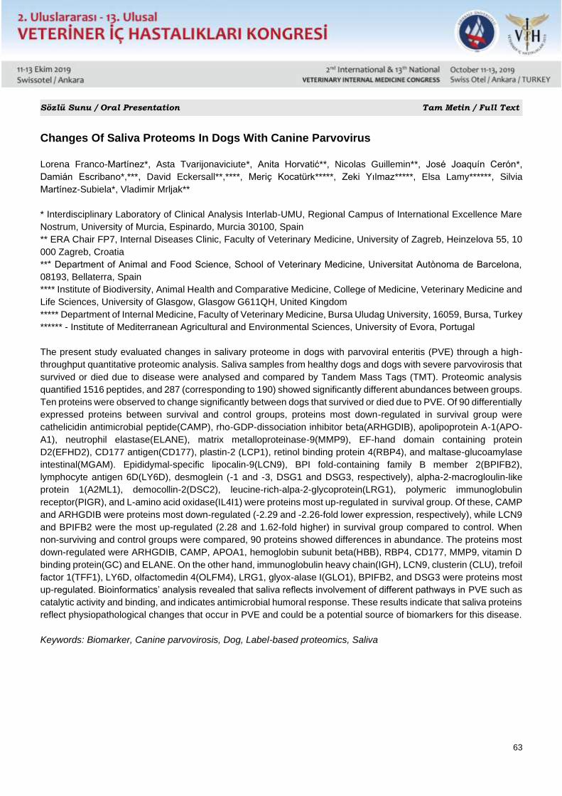
63
Sözlü Sunu / Oral Presentation Tam Metin / Full Text
Changes Of Saliva Proteoms In Dogs With Canine Parvovirus
Lorena Franco-Martínez*, Asta Tvarijonaviciute*, Anita Horvatić**, Nicolas Guillemin**, José Joaquín Cerón*,
Damián Escribano*,***, David Eckersall**,****, Meriç Kocatürk*****, Zeki Yılmaz*****, Elsa Lamy******, Silvia
Martínez-Subiela*, Vladimir Mrljak**
* Interdisciplinary Laboratory of Clinical Analysis Interlab-UMU, Regional Campus of International Excellence Mare
Nostrum, University of Murcia, Espinardo, Murcia 30100, Spain
** ERA Chair FP7, Internal Diseases Clinic, Faculty of Veterinary Medicine, University of Zagreb, Heinzelova 55, 10
000 Zagreb, Croatia
*** Department of Animal and Food Science, School of Veterinary Medicine, Universitat Autònoma de Barcelona,
08193, Bellaterra, Spain
**** Institute of Biodiversity, Animal Health and Comparative Medicine, College of Medicine, Veterinary Medicine and
Life Sciences, University of Glasgow, Glasgow G611QH, United Kingdom
***** Department of Internal Medicine, Faculty of Veterinary Medicine, Bursa Uludag University, 16059, Bursa, Turkey
****** - Institute of Mediterranean Agricultural and Environmental Sciences, University of Evora, Portugal
The present study evaluated changes in salivary proteome in dogs with parvoviral enteritis (PVE) through a high-
throughput quantitative proteomic analysis. Saliva samples from healthy dogs and dogs with severe parvovirosis that
survived or died due to disease were analysed and compared by Tandem Mass Tags (TMT). Proteomic analysis
quantified 1516 peptides, and 287 (corresponding to 190) showed significantly different abundances between groups.
Ten proteins were observed to change significantly between dogs that survived or died due to PVE. Of 90 differentially
expressed proteins between survival and control groups, proteins most down-regulated in survival group were
cathelicidin antimicrobial peptide(CAMP), rho-GDP-dissociation inhibitor beta(ARHGDIB), apolipoprotein A-1(APO-
A1), neutrophil elastase(ELANE), matrix metalloproteinase-9(MMP9), EF-hand domain containing protein
D2(EFHD2), CD177 antigen(CD177), plastin-2 (LCP1), retinol binding protein 4(RBP4), and maltase-glucoamylase
intestinal(MGAM). Epididymal-specific lipocalin-9(LCN9), BPI fold-containing family B member 2(BPIFB2),
lymphocyte antigen 6D(LY6D), desmoglein (-1 and -3, DSG1 and DSG3, respectively), alpha-2-macrogloulin-like
protein 1(A2ML1), democollin-2(DSC2), leucine-rich-alpa-2-glycoprotein(LRG1), polymeric immunoglobulin
receptor(PIGR), and L-amino acid oxidase(IL4I1) were proteins most up-regulated in survival group. Of these, CAMP
and ARHGDIB were proteins most down-regulated (-2.29 and -2.26-fold lower expression, respectively), while LCN9
and BPIFB2 were the most up-regulated (2.28 and 1.62-fold higher) in survival group compared to control. When
non-surviving and control groups were compared, 90 proteins showed differences in abundance. The proteins most
down-regulated were ARHGDIB, CAMP, APOA1, hemoglobin subunit beta(HBB), RBP4, CD177, MMP9, vitamin D
binding protein(GC) and ELANE. On the other hand, immunoglobulin heavy chain(IGH), LCN9, clusterin (CLU), trefoil
factor 1(TFF1), LY6D, olfactomedin 4(OLFM4), LRG1, glyox-alase I(GLO1), BPIFB2, and DSG3 were proteins most
up-regulated. Bioinformatics’ analysis revealed that saliva reflects involvement of different pathways in PVE such as
catalytic activity and binding, and indicates antimicrobial humoral response. These results indicate that saliva proteins
reflect physiopathological changes that occur in PVE and could be a potential source of biomarkers for this disease.
Keywords: Biomarker, Canine parvovirosis, Dog, Label-based proteomics, Saliva
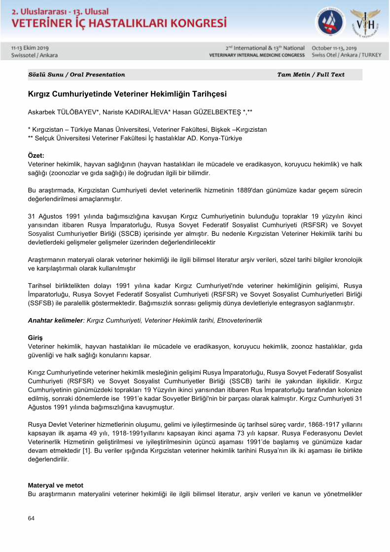
64
Sözlü Sunu / Oral Presentation Tam Metin / Full Text
Kırgız Cumhuriyetinde Veteriner Hekimliğin Tarihçesi
Askarbek TÜLÖBAYEV*, Nariste KADIRALİEVA* Hasan GÜZELBEKTEŞ *,**
* Kırgızistan – Türkiye Manas Üniversitesi, Veteriner Fakültesi, Bişkek –Kırgızistan
** Selçuk Üniversitesi Veteriner Fakültesi İç hastalıklar AD. Konya-Türkiye
Özet:
Veteriner hekimlik, hayvan sağlığının (hayvan hastalıkları ile mücadele ve eradikasyon, koruyucu hekimlik) ve halk
sağlığı (zoonozlar ve gıda sağlığı) ile doğrudan ilgili bir bilimdir.
Bu araştırmada, Kırgızistan Cumhuriyeti devlet veterinerlik hizmetinin 1889'dan günümüze kadar geçem sürecin
değerlendirilmesi amaçlanmıştır.
31 Ağustos 1991 yılında bağımsızlığına kavuşan Kırgız Cumhuriyetinin bulunduğu topraklar 19 yüzyılın ikinci
yarısından itibaren Rusya İmparatorluğu, Rusya Sovyet Federatif Sosyalist Cumhuriyeti (RSFSR) ve Sovyet
Sosyalist Cumhuriyetler Birliği (SSCB) içerisinde yer almıştır. Bu nedenle Kırgızistan Veteriner Hekimlik tarihi bu
devletlerdeki gelişmeler gelişmeler üzerinden değerlendirilecektir
Araştırmanın materyali olarak veteriner hekimliği ile ilgili bilimsel literatur arşiv verileri, sözel tarihi bilgiler kronolojik
ve karşılaştırmalı olarak kullanılmıştır
Tarihsel birliktelikten dolayı 1991 yılına kadar Kırgız Cumhuriyeti'nde veteriner hekimliğinin gelişimi, Rusya
İmparatorluğu, Rusya Sovyet Federatif Sosyalist Cumhuriyeti (RSFSR) ve Sovyet Sosyalist Cumhuriyetleri Birliği
(SSFSB) ile paralellik göstermektedir. Bağımsızlık sonrası gelişmiş dünya devletleriyle entegrasyon sağlanmıştır.
Anahtar kelimeler: Kırgız Cumhuriyeti, Veteriner Hekimlik tarihi, Etnoveterinerlik
Giriş
Veteriner hekimlik, hayvan hastalıkları ile mücadele ve eradikasyon, koruyucu hekimlik, zoonoz hastalıklar, gıda
güvenliği ve halk sağlığı konularını kapsar.
Kırıgz Cumhuriyetinde veteriner hekimlik mesleğinin gelişimi Rusya İmparatorluğu, Rusya Sovyet Federatif Sosyalist
Cumhuriyeti (RSFSR) ve Sovyet Sosyalist Cumhuriyetler Birliği (SSCB) tarihi ile yakından ilişkilidir. Kırgız
Cumhuriyetinin günümüzdeki toprakları 19 Yüzyılın ikinci yarısından itibaren Rus İmparatorluğu tarafından kolonize
edilmiş, sonraki dönemlerde ise 1991’e kadar Sovyetler Birliği'nin bir parçası olarak kalmıştır. Kırgız Cumhuriyeti 31
Ağustos 1991 yılında bağımsızlığına kavuşmuştur.
Rusya Devlet Veteriner hizmetlerinin oluşumu, gelimi ve iyileştirmesinde üç tarihsel süreç vardır, 1868-1917 yıllarını
kapsayan ilk aşama 49 yılı, 1918-1991yıllarını kapsayan ikinci aşama 73 yılı kapsar. Rusya Federasyonu Devlet
Veterinerlik Hizmetinin geliştirilmesi ve iyileştirilmesinin üçüncü aşaması 1991’de başlamış ve günümüze kadar
devam etmektedir [1]. Bu veriler ışığında Kırgızistan veteriner hekimlik tarihini Rusya’nın ilk iki aşaması ile birlikte
değerlendirilir.
Materyal ve metot
Bu araştırmanın materyalini veteriner hekimliği ile ilgili bilimsel literatur, arşiv verileri ve kanun ve yönetmelikler
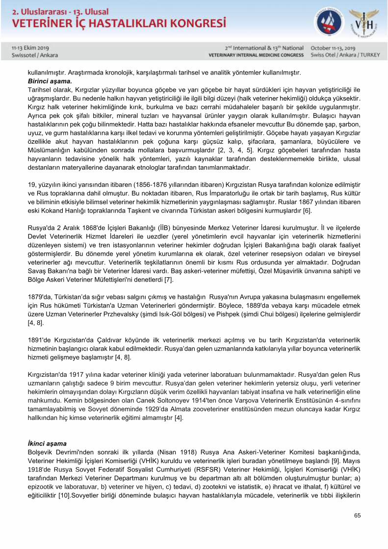
65
kullanılmıştır. Araştırmada kronolojik, karşılaştırmalı tarihsel ve analitik yöntemler kullanılmıştır.
Birinci aşama.
Tarihsel olarak, Kırgızlar yüzyıllar boyunca göçebe ve yarı göçebe bir hayat sürdükleri için hayvan yetiştiriciliği ile
uğraşmışlardır. Bu nedenle halkın hayvan yetiştiriciliği ile ilgili bilgi düzeyi (halk veteriner hekimliği) oldukça yüksektir.
Kırgız halk veteriner hekimliğinde kırık, burkulma ve bazı cerrahi müdahaleler başarılı bir şekilde uygulanmıştır.
Ayrıca pek çok şifalı bitkiler, mineral tuzları ve hayvansal ürünler yaygın olarak kullanılmıştır. Bulaşıcı hayvan
hastalıklarının pek çoğu bilinmektedir. Hatta bazı hastalıklar hakkında efsaneler mevcuttur Bu dönemde şap, şarbon,
uyuz, ve gurm hastalıklarına karşı ilkel tedavi ve korunma yöntemleri geliştirilmiştir. Göçebe hayatı yaşayan Kırgızlar
özellikle akut hayvan hastalıklarının pek çoğuna karşı güçsüz kalıp, şifacılara, şamanlara, büyücülere ve
Müslümanlığın kabülünden sonrada mollalara başvurmuşlardır [2, 3, 4, 5]. Kırgız göçebeleri tarafından hasta
hayvanların tedavisine yönelik halk yöntemleri, yazılı kaynaklar tarafından desteklenmemekle birlikte, ulusal
destanların materyallerine dayanarak etnologlar tarafından tanımlanmaktadır.
19, yüzyılın ikinci yarısından itibaren (1856-1876 yıllarından itibaren) Kırgızistan Rusya tarafından kolonize edilmiştir
ve Rus topraklarına dahil olmuştur. Bu noktadan itibaren, Rus İmparatorluğu ile ortak bir tarih başlamış, Rus kültür
ve biliminin etkisiyle bilimsel veteriner hekimlik hizmetlerinin yaygınlaşması sağlamıştır. Ruslar 1867 yılından itibaren
eski Kokand Hanlığı topraklarında Taşkent ve civarında Türkistan askeri bölgesini kurmuşlardır [6].
Rusya'da 2 Aralık 1868'de İçişleri Bakanlığı (İİB) bünyesinde Merkez Veteriner İdaresi kurulmuştur. İl ve ilçelerde
Devlet Veterinerlik Hizmet İdareleri ile uezdler (yerel yönetimlerin evcil hayvanlar için veterinerlik hizmetlerin i
düzenleyen sistemi) ve tren istasyonlarının veteriner hekimler doğrudan İçişleri Bakanlığına bağlı olarak faaliyet
göstermişlerdir. Bu dönemde yerel yönetim kurumlarına ek olarak, özel veteriner resepsiyon odaları ve bireysel
veterinerler ağı mevcuttur. Veterinerlik teşkilatlarının önemli bir kısmı Rus ordusunda yer almaktadır. Doğrudan
Savaş Bakanı'na bağlı bir Veteriner İdaresi vardı. Baş askeri-veteriner müfettişi, Özel Müşavirlik ünvanına sahipti ve
Bölge Askeri Veteriner Müfettişleri'ni denetlerdi [7].
1879'da, Türkistan’da sığır vebası salgını çıkmış ve hastalığın Rusya'nın Avrupa yakasına bulaşmasını engellemek
için Rus hükümeti Türkistan'a Uzman Veterinerleri göndermiştir. Böylece, 1889'da vebaya karşı mücadele etmek
üzere Uzman Veterinerler Przhevalsky (şimdi Isık-Göl bölgesi) ve Pishpek (şimdi Chui bölgesi) ilçelerine gelmişlerdir
[4, 8].
1891'de Kırgızistan'da Çaldıvar köyünde ilk veterinerlik merkezi açılmış ve bu tarih Kırgızistan'da veterinerlik
hizmetinin başlangıcı olarak kabul edilmektedir. Rusya’dan gelen uzmanlarında katkılarıyla yıllar boyunca veterinerlik
hizmeti gelişmeye başlamıştır [4, 8].
Kırgızistan'da 1917 yılına kadar veteriner kliniği yada veteriner laboratuarı bulunmamaktadır. Rusya'dan gelen Rus
uzmanların çalıştığı sadece 9 birim mevcuttur. Rusya’dan gelen veteriner hekimlerin yetersiz oluşu, yerli veteriner
hekimlerin olmayışından dolayı Kırgızların düşük verim özellikli hayvanları tabiyat insafına ve halk veterinerliğin eline
mahkumdu. Kemin bölgesinden olan Canek Soltonoyev 1914'ten önce Varşova Veterinerlik Enstitüsünün 4-sınıfını
tamamlayabilmiş ve Sovyet döneminde 1929’da Almata zooveteriner enstitüsünden mezun oluncaya kadar Kırgız
hallkından hiç kimse veterinerlik eğitimi almamıştır [4].
İkinci aşama
Bolşevik Devrimi'nden sonraki ilk yıllarda (Nisan 1918) Rusya Ana Askeri-Veteriner Komitesi başkanlığında,
Veteriner Hekimliği İçişleri Komiserliği (VHİK) kuruldu ve veterinerlik işleri buradan yönetilmeye başlandı [9]. Mayıs
1918'de Rusya Sovyet Federatif Sosyalist Cumhuriyeti (RSFSR) Veteriner Hekimliği, İçişleri Komiserliği (VHİK)
tarafından Merkezi Veteriner Departmanı kurulmuş ve bu departman altı alt bölümden oluşturulmuştur bunlar; a)
epizootik ve laboratuvar, b) veteriner ve hijyen, c) tedavi, d) zootekni ve istatistik, e) ihracat ve ithalat, f) kültürel ve
eğiticiliktir [10].Sovyetler birliği döneminde bulaşıcı hayvan hastalıklarıyla mücadele, veterinerlik ve tıbbi ilişkilerin
66
organizasyonu, ilk günlerden itibaren, et ve süt endüstrilerinde hijyen denetimine büyük önem verilmiştir. Sınırlarda,
tren istasyonlarında ve vapur marinalarında veteriner ve hijyen merkezleri ağı kurulmuştur. İllerde veterinerlik hijyen
denetim işleri veteriner bölüm başkanlıklarına verilmiştir [11]. 1920 yılın, haziranında RSFSR Halk Komiserleri
Konseyi, “Veteriner hekimlerin ve teknisyenlerin daha verimli kullanımına yönelik kanun çıkarılmıştır[12].
RSFSR’nin Kırgız Özerk Bölgesi’nde, 24 Ekim 1924 yılında Hijyen-epizootik ve veteriner-istatistik olmak üzere iki
bölümden oluşan Tarım genel müdürlüğü Veteriner Dairesi Başkanlığı kurulmuştur. Tüm illerde veterinerlik hizmeti
organize edilmiştir, bununla birlikte hayvanların yoğun geçtiği bölgelerde veteriner-hijyen merkezleri, Atbaşı’nda ise
sınır kordon koruma hattı kurulmuştur[13]. RSFSR’içerisindeki Kırgız Özerk Bölgesi 1 Şubat 1926’da, , Kırgız Özerk
Sosyalist Cumhuriyeti’ adını aldı. Veterinerlik bölümü dahil olmak üzere, Halk Cumhuriyeti Cumhuriyet Tarım
Komutanlığı kuruldu. Hayvan sayısının yoğun artışı nedeniyle, veterinerlik hizmet ağı genişletilmiştir. Mayıs 1927'de,
mera kurulu kapsamında mobil veteriner hizmetler oluşturulmuştur[14].
İlk kez Frunze şehrinde (şimdiki Bişkek) 1927-yılında veteriner-bakteriyoloji laboratuarı kurulmuş, sonrasında 1940
yılında bu laboratuar Kırgız Bilimsel – Araştırma Veteriner Deney istasyonu adını almıştır. Bu deney istasyonu 1956
yılında Kırgız Hayvancılık Bilimsel araştırma enstitüsüne dönüştürülmüştür. 1933 yılında Kırgız Zooveteriner
enstitüsünün kurulmuş ve 1937 yılında bu kurum Kırgız Tarım Enstütüsü olarak düzenlenmiştir [4, 15]. 1941 itibariyle,
500'den fazla uzmanın çalıştığı 281 kurum vardı. II. Dünya Savaşı sırasında uzmanların yarısından fazlası savaşa
gitti. Veterinerlik hizmet için gerekli olan ilaç, dezenfektan ve biyolojik preparatlarların yeterince sağlanmaması
sonucunda , daha önce elimine edilen pek çok hastalığın epizootik durumu yeniden kötüleşmiştir[15, 16].
Sovyetler Birliği döneminde, Devlet Veterinerlik Servisi sürekli olarak materyalini ve teknik tabanını genişletti,
güçlendirdi, geliştirdi. Veterinerlikle ilgili alet, ekipman, ilaç, dezenfektan, ve biyolojik maddeler geliştirilerek üretime
sokuldu. Böylece veteriner teşhis yöntemleri, hastalık önlemleri ve tedavi yöntemleri geliştirilmiştir.
Üçüncü aşama
Kırgız Cumhuriyeti Devlet Veterinerlik Teşkilatı'nın üçüncü gelişme aşaması 31 Ağustos 1991’den sonrasını kapsar:
SSCB'nin çöküşü, bağımsızlığın kazanılması ve pazar ekonomisine geçiş sürecinde veterinerlik hizmetleri neredeyse
tamamen çökmüş ve bulaşıcı hayvan hastalıkları kontrol edilemez bir hal almıştır. Bu durum, Kırgızistan Cumhuriyeti
Bakanlar Kurulunu 4 Mart 1991'de Tarım ve Gıda Bakanlığı veterinerlik müfettişliği ile Ana Veterinerlik İdaresi'ni
oluşturmaya zorlamıştır [17].
Kırgız Cumhuriyeti'nde ilk egemenlik yıllarında meydana gelen siyasi, sosyal ve ekonomik değişimler devlet
veterinerlik hizmetinin ve her veterinerlik uzmanının faaliyetlerini doğrudan etkilemiştir. Devlet veterinerlik servisinin
yapısı ve işlevleri değişmiş ve yeni bir veterinerlik hizmeti olan özel veteriner hekimlik ortaya çıkmıştır.
Sovyetler Birliği döneminde yürürlükte olan veterinerlik mevzuatı bağımsızlık sonrası ihtiyaca cevap veremez
konuma gelmiştir. Bu nedenle 6 Mart 1992 yılında Kırgız Cumhuriyeti Veteriner Hekimlik Mevzuatı (No. 806-XII) kabul
edilmiştir [18]. Bu kanun kabul ederken, veterinerlik ve sağlık önlemlerinin uygulanmasına yönelik bazı uluslararası
norm ve standartların gerekleri dikkate alınmamıştır. Kırgızistan Cumhuriyeti 1992 yılında Dünya Hayvan Sağlığı
Örgütü'ne (OIE) üye olmuştur [19]. Veteriner hekimliği alanında işbirliğini sürdürebilmek için 12 Mart 1993 tarihinde
Moskova'da Bağımsız Devletler Topluluğu (BDT) ülkeleri, veteriner hekimliği alanında işbirliği konusunda bir anlaşma
imzalamışlardır [20]. Kırgızistan 1998 yılında Dünya Ticaret Örgütü'ne (WTO) üye olmuştur. Bu bağlamda, veteriner
denetimindeki malların ülkeler arasındaki ticaretinde Veterinerlik mevzuatını uluslararası normlar ve standartlar ile
uyumlu hale getirmek için, 2 Haziran 1998'de, Kırgızistan Cumhuriyeti “Veteriner Hekimliği Kanunu” nda değişiklik
ve ilaveler yapılmıştır [21]. Kırgız Cumhuriyeti'nin veterinerlik hizmetini Avrupa Birliği, DTÖ ve OIE Karasal Hayvan
Sağlık Kanunu tarafından belirlenen standartlara uygun hale getirmek, veterinerlik hizmetlerini iyileştirmek amacıyla
Kırgızistan Cumhuriyeti Hükümeti tarafından 2008-2012 Veteriner Geliştirme Stratejik Planı onaylanmıştır (No 62,
25.11.2008) [22]. 18 Kasım 2010 tarihinde 277 Sayılı “Kırgız Cumhuriyeti Tarım Bakanlığı “Veteriner İlaçların Kontrol
ve Belgelendirme Merkezi Yönetmeliğinin Onaylanmasına Dair Kararı” (No277, 18.11.2010) kabul edilmiştir [23]. 12
67
Ocak 21012 yılında Kırgız Cumhuriyeti Hükümeti Devlet Hijyen, Veterinerlik ve Fitosanitasyon Güvenliği Müfettişliği
oluşturulmuştur. Bu birim Kırgızistan Cumhuriyeti'ndeki hayvansal gıdaların kalite kontrolünden sorumlu ana organ
haline gelmiştir [24, 25]. 5 Mart 2013 tarihinde, Kırgız Cumhuriyeti Hükümeti Altındaki Devlet Hijyen, Veterinerlik ve
Fitosanitasyon Güvenliği Müfettişliği, Kırgız Cumhuriyeti Veteriner ve Fitosanitasyon Güvenliği Denetleme Kurumuna
dönüştürülerek Hijyen güvenliği alanındaki gözetim görevi Sağlık Bakanlığına devredilmiştir [26]. 07 mayıs 2013
yılında Kırgız Cumhuriyeti Veteriner ve Fitosanitasyon Güvenliği Denetleme Kurumu bünyesine yer alan Devlet
Veteriner Departmanı ortadan kaldırılmıştır[27]. Kırgızistan Cumhuriyeti'nin Avrasya Ekonomik Birliği'ne (EAEU)
katılımı, Veterinerlik ve Fitosanitasyon güvenliğine olan taleplerin artışına neden olmuş, değişen şartlar ve acil olarak
veteriner hekimliği alanındaki yasal düzenlemelerin etkinliğini arttırma gereği göz önünde bulundurularak, 30 Aralık
2014 tarihinde, Veterinerlik alanındaki yasal ilişkileri düzenleyen ana yasal düzenleme olan Kırgız Cumhuriyeti
“Veteriner Hekimliği” Kanunu kabul edilmiştir[28].
Kırgızistan Cumhuriyeti "Veteriner Hekimliği" Kanunu uyarınca, Kırgızistan Cumhuriyeti'nin veteriner sistemi
aşağıdakileri içermektedir:
- Kırgız Cumhuriyeti hükümeti;
- Yetkili devlet veteriner otoritesi;
- Yetkileri dahilindeki yerel devlet idareleri ve delegasyon halindeki devlet yetkileri çerçevesinde yerel yönetimler;
- Kırgız Cumhuriyeti Veteriner Statuer Kurumu (Kırgız Cumhuriyeti Veteriner Odası);
- Veteriner alanında yasaların öngördüğü şekilde çalışan girişimcilik girişimciler.
Kırgız Cumhuriyeti Veteriner Statuer Kurumu (Veteriner Odası) 2015'ten itibaren özerk yönetim ilkeleriyle hareket
eden ve ülkedeki özel veterinerlik uygulamalarını düzenleyen bir organizasyon olmuştur. "Veteriner" Yasasına göre
Veteriner Odası, Kırgızistan Cumhuriyeti Hükümeti ve Veteriner Odası Şartı ile belirlenen şekilde veterinerlik
uygulamalarını düzenlemektedir (http://vsb.kg/ru/pages/istoriya-sozdaniya.html) [29].
Tartışma ve Sonuç
Yukarıda belirtildiği gibi 1889-1933 yy veteriner hizmetin Rusya ve Sovyet Sosyalist Cumhuriyetler Birliği (SSCB)
uzmanları temsil etmişlerdir. Benzer durum Türkiye, Çin, Moğolistan [30] ve eski Sovyetler Birliği'nin pek çok ülkesinin
veteriner tarihinin özelliğidir [9, 31]. Cumhuriyet döneminin başlarında Gazi Mustafa Kemal Atatürk’ün milleti çağdaş
medeniyetler seviyesine çıkarmayı hedeflemiş ve çok sayıda tanınmış uzman davet ederek Türkiye'de veterinerlik
ve hayvancılık alanlarına kayda değer katkılar sağlamışlardır[32].
Sovyetler Birliği döneminde Veteriner hekimliği tarihi ile ilgili materyaller temel olarak “Modern veteriner hekimliği
bülteni”, “Sosyalist inşaat alanında veteriner hekimi”, “Pratik veteriner hekimliği ve at yetiştiriciliği” dergilerinde
yayınlanırdı. Veterinerlik enstitülerinin bilimsel derlemelerinde de veteriner hekimliği tarihi ile ilgili makaleler
basılıyordu. 1954 yılında, Prof.Dr. Koropov tarafından ülkede veterinerlik mesleğinin tarihçesi üzerine ilk genelleştirici
çalışma olan "SSCB'de Veteriner Hekimliği Tarihi" çalışması yayınlanmıştır [31]. O yıllarda veteriner hekimliği tarihi
ile ilgili yayınlanmış eserlerin çoğu, tez çalışmalarıydı. 1960 yılında, Veteriner Tıbbı (Moskova) dergisinde tarihiyle
ilgili özel bir bölüm çıktı. Ancak yine de Sovyet döneminde, veteriner hekimliği tarihi yeterince incelenmemiştir.
Daha sonra, Veteriner Ansiklopedisi yayınlanmıştır[33], SSCB'de veteriner hekimliğinin tarihçesi, teorisi ve pratiğiyle
ilgili temel bilgiler içeren ilk bilimsel referans kitabı. 1970'lerde veteriner hekimlik tarihi hakkında birkaç makale
yayınlanmıştır [34, 35]. 1988'de SSCB'nin veteriner tarihini kapsayan “Veteriner Hekimliği Tarihi” [36] kitabı
yayınlanmıştır.
Tarihsel perspektifin gösterdiği gibi, 1991 yılına kadar Kırgız Cumhuriyeti'nde veteriner hekimliğinin oluşumu ve
gelişimi, Rusya İmparatorluğu, Rusya Sovyet Federatif Sosyalist Cumhuriyeti (RSFSR) ve Sovyet Sosyalist
Cumhuriyetleri Birliği (SSFSB) ve veteriner hekimliği gelişim tarihi ile yakından ilişkilidir. Bağımsızlık sonrası ülkedeki
özel veterinerlik uygulamalarının düzenlenmesi Veterinerlik Odası tarafından yapılmaktadır. Bu tür yasal veterinerlik
amaclı oluşturulan kuruluşlar birçok ülkede vardır, örneğin, Türk Veteriner Hekimleri Birliği ve Veteriner Odaları
68
bulunmaktadır [37]. Kırgızistan’da ise bu kurumu Kırgız Cumhuriyeti Veteriner ve Fitosanitasyon Güvenliği
Denetleme Kurumu temsil etmektedir.
Kaynaklar 1. Никитин И.Н. История ветеринарии. Учебник. – М.: КолосС, 2006.
2. Байбосунов А.А. Донаучные представления киргизов о природе. - Ф., 1990.
3. Солтоноев Б. (Кызыл) Кыргыз тарыхы: Тарыхый очерктер. – Бишкек, 1993.
4. Алдашев А.А. Этапы развития ветеринарии в Киргизии. – Ф.: Илим, 1989.
5. Тулобаев А.З., Салыков Р.С. Кыргызы / Народная ветеринария. М.: Наука, 2016. С. 456-462. (Народы и культуры).
6. Тугай Т.И. Представители Оренбургской губернской админиcтрации и краеведческих обществ в изучении Казахстана и
Средней Азии (втор. пол. XIX – нач. XX в.) // Вестник ОГУ №5 (141), май 2012.
7. Алексеева Л.В. Состояние ветеринарного дела в годы революции и гражданской войны // Вестник НВГУ. 2008. №1.
8. Жунушов А.Т. Организация и экономика ветеринарного дела. – Б.: 2002.
9. Никитин И.Н., Калугин В.И. История ветеринарии. М., 1988.
10. Коропов В.М. История ветеринарии в СССР. М., 1954.
11. Ветеринарная энциклопедия. М., 1968. Т. 1.
12. О правильном использовании ветеринарных врачей и ветфельдшеров и порядке их мобилизации: Постановление
Совнаркома РСФСР от 9 июня 1920 г. // Декреты советской власти / ред. О.Н. Зайцев [и др.]. – М.: Политиздат, 1983. – С. 220-
222.
13. ЦГА Кыргызской ССР, ф. 328, оп. 1, д. 760, л. 127.
14. ЦГА Кыргызской ССР, ф. 328, оп. 1, д. 1341, л. 29.
15. Жунушов А.Т. Ветеринарная служба на путях перестройки. - Б., 1991.
16. ЦГА Кыргызской ССР, ф. 1218, оп. 1, д. 7, л. 5.
17. Постановление Кабинета Министров Республики Кыргызстан от 4 марта 1991 года №64 «О схеме и структуре управления
Министерства сельского хозяйства и продовольствия Республики Кыргызстан» http://cbd.minjust.gov.kg/act/view/ru-
ru/39860?cl=ru-ru.
18. Закон Республики Кыргызстан «О ветеринарии». Принят Верховным Советом 6 марта 1992 года (№806-XII) (Ведомости
Верховного Совета Республики Кыргызстан, 1992 г., № 4/2, ст.177).
19. Всемирная организация охраны здоровья животных - Международное Эпизоотическое Бюро (OIE, МЭБ) (www.oie.int).
20. Соглашение о сотрудничестве в области ветеринарии от 12 марта 1993 года. г. Москва http://www.e-
cis.info/page.php?id=23038.
21. Закон Кыргызской Республики «О внесении изменений и дополнений в Закон «О ветеринарии» от 2 июня 1998 г. №73
http://cbd.minjust.gov.kg/act/view/ru-ru/81.
22. Стратегический план развития ветеринарной службы Кыргызской Республики на 2008-2012 годы
http://cbd.minjust.gov.kg/act/view/ru-ru/58899.
23. Постановление Правительства Кыргызской Республики от 18 ноября 2010 года №277 «Об утверждении Положения о Центре
по контролю и сертификации ветеринарных препаратов при Министерстве сельского хозяйства Кыргызской Республики».
"Эркин Тоо" от 26 ноября 2010 года N 108.
24. Постановление Правительства Кыргызской Республики от 12 января 2012 года № 12 http://cbd.minjust.gov.kg/act/view/ru-
ru/93378?cl=ru-ru.
25. Постановление Правительства Кыргызской Республики от 02 июня 2012 года №367 О внесении изменения и дополнения в
постановление Правительства Кыргызской Республики "Об организационных мерах в связи с реформой органов
исполнительной власти Кыргызской Республики" от 10 февраля 2012 года № 87 http://cbd.minjust.gov.kg/act/view/ru-ru/93271.
26. Постановление Правительства Кыргызской Республики от 05 марта 2013 года №109 «О функциональных и структурных
изменениях в системе государственных органов исполнительной власти Кыргызской Республики»
http://cbd.minjust.gov.kg/act/view/ru-ru/94215.
27. Постановление Правительства Кыргызской Республики от 07 мая 2013 года №256 «О внесении изменений и дополнения в
некоторые решения Правительства Кыргызской Республики» http://cbd.minjust.gov.kg/act/view/ru-ru/94347.
28. Закон Кыргызской Республики «О ветеринарии» от 30 декабря 2014 года №175 http://cbd.minjust.gov.kg/act/view/ru-ru/205396.
29. Ветеринарный статуарный орган Кыргызской Республики учреждение «Ветеринарная палата» (http://vsb.kg/ru/pages/istoriya-
sozdaniya.html).
30. Даваажав Б. Из истории развития ветеринарии в Монголии // Проблемы социально-экономического развития Сибири. 2017.
С.47-52.
31. Алексеева Л.В. Становление советской системы ветеринарной службы на Обь-Иртышском Севере (1917-1941 гг.):
Монография. – Нижневартовск, 2009.
32. Küçükaslan Ö, Yerlikaya N, Yiğit A: Contributions of foreign scientists to the Turkish veterinary medicine and animal husbandry in the
Early Republican Period (1923-1933). Kafkas Univ Vet Fak Derg, 24 (1): 45-51, 2018.
33. Ветеринарная энциклопедия / Гл. ред. К.И. Скрябин. М., 1968-1975. В 6-ти тт.
34. Джамбулатов М.М. К истории ветеринарии в Дагестане. Махачкала, 1971.
35. Ибрагимов Б.Г. История ветеринарии в Азербайджане. Баку, 1971.
36. Никитин И.Н., Калугин В.И. История ветеринарии. М., 1988.
37. Kızıltepe A: Türkiye’de Veteriner Hekim Odalarının Çalışmalarına Yönelik Bir İnceleme: Kars Bölgesi Veteriner Hekimler Odası
Örneği. Kafkas Univ Vet Fak Derg, 23 (5): 757-765, 2017.
69
Sözlü Sunu / Oral Presentation Tam Metin / Full Text
History Of Veterinary Medicine In Kyrgyz Republıc
Askarbek TÜLÖBAYEV*, Nariste KADIRALİEVA* Hasan GÜZELBEKTEŞ *,**
* Kyrgyz-Turkish Manas University, Faculty of Veteriner Medicine, Bishkek, Kyrgyzstan. ** Selcuk University, Faculty of Veteriner Medicine, Konya, Turkey
Abstract
Veterinary medicine is a science that is directly related to animal health (animal diseases and eradication, preventive
medicine) and public health (zoonoses and food health).
The aim of this study was to evaluated the historical process of the state veterinary service of the Kyrgyz Republic
from 1889 to the present day.
Today the territory of the Kyrgyz Republic has been part of the Russian Empire, Russian Soviet Federative Socialist
Republic (RSFSR) and the Union of Soviet Socialist Republics (USSR) since the second half of the 19th century.
Therefore, the history of Kyrgyzstan Veterinary Medicine will be evaluated based on the developments in these
states.
As the material of the research, archives of scientific literature, verbal historical information about veterinary medicine
were used chronologically and comparatively.
The development of veterinary medicine in the Kyrgyz Republic until 1991 due to historical association is in parallel
with the Russian Empire, the Russian Soviet Federative Socialist Republic (RSFSR) and the Union of Soviet Socialist
Republics (USSR). Integration with developed world states after independence
Keywords: Kyrgyz Republic, History of Veterinary Medicine, Etnoveterinerlik
70
Sözlü Sunu / Oral Presentation Tam Metin / Full Text
Agresif Kedi ve Köpeklerde, İlaç Dışı Tamamlayıcı Tedavi Etkinliği-Klinik Gözlem
Emre KÜLLÜK, Başar Ulaş SAYILKAN, Duygu DALĞIN, Yücel MERAL
Ondokuz Mayıs Üniversitesi Veteriner Fakültesi İç Hastalıkları Anabilim Dalı, Samsun
ÖZET
Köpek ve kedilerde agresyon, sıkça görülen, karmaşık ve tehlikeli bir davranıştır. Tedavinin insan sağlığı üzerine
etkileri vardır bu yüzden yüksek sorumluluk gerektirmektedir. Tedavi seçenekleri, trisiklik antidepresanlar ve
antipsikotiklerdir. Gözlem grubu insanlara karşı yüksek agresyon gösteren beş köpek (1-7 yaş, bir erkek ve 4 dişi) ve
2 kedi (3 ve 4 yaş sağlıklı, erkek ve dişi) den müteşekkildi. Ayrıca bir kedi ve iki köpeğin de kompulsif davranışları
vardı. Başlangıç olarak tedavide köpeklerde risperidon ve amitriptilin ve kedilerde amitriptilin ile başlandı. Klinik başarı
2 hafta içinde ve en zor vakada 4 hafta içinde elde edildi. Tedaviler 4 köpek ve 1 kedide Composure (Vetriscience),
diğerlerinde Kalmvet (VetExpert) ile idame ettirildi ve istikrarlı bir iyileşme ile sonuçlandı. Agresyon bozukluklarında
uzun süreli tedavilerin yan etkilerinden kaçınmak için, tamamlayıcı ilaç dışı preparasyonlar, tehlike riskinin ortadan
kaldırılmasından sonra dikkate alınmalıdır.
Anahtar kelimeler: Agresif davranış, Köpek, Kedi, İlaç Dışı, Tamamlayıcı
Giriş
Köpek ve kedilerde agresyon, sıkça görülen, karmaşık etiyolojili ve tehlikeli bir davranıştır. Agresyon probleminin
insan sağlığı ile etkileşimi nedeniyle, tedavi yüksek sorumluluk içermektedir. İnsana yönelik agresyon köpeklerde
tüm agresyon vakalarının %54-67 kadarını oluştururken, Amerika Birleşik devletlerinde, barınaklara terk edilen
kedilerin %27’sinin davranışsal nedenlerle bırakıldığı rapor edilmiştir (1-3).Köpek saldırılarının ortalama %2-4’ü
hastanede sonuçlanır (5). Hedef genellikle aile üyeleridir (4-8). Özellikle çocuklar kendilerini koruma açısından daha
zayıf olduklarından ve boy dezavantajı nedeniyle, yüz, boyun gibi hayati bölgelerden yara alma ihtimalleri daha
yüksektir (4). Kesin olarak, kedi ve köpeklerde agresivite vakalarında etyoloji, davranış modifikasyonu gibi
noktalardan çok daha önemli olan, saldırı davranışını etkin ve acil olarak ortadan kaldırmaktır.
Materyal ve Metotlar
Gözlem grubu, Ondokuz Mayıs Üniversitesi Veteriner Fakültesi Hayvan hastanesine agresivite problemleri ile
getirilen ve insanlara karşı yüksek agresyon gösteren beş köpek (1-7 yaş, bir erkek ve 4 dişi) ve 2 kedi (3 ve 4 yaş
sağlıklı, erkek ve dişi) den müteşekkildir. Bunların içinde bir kedi ve iki köpekte de kompulsif davranışlar tespit
edilmiştir. Başlangıç olarak tedavide köpeklerde risperidon (Risperdal, 1mg/m2) ve amitriptilin (Laroxyl, 1 - 4 mg/kg)
ve kedilerde amitriptilin (Laroxyl, 5 to 10 mg/kedi) ile başlandı. Agresyon giderildikten sonra tedrici olarak 4 köpek
ve 1 kedide Composure (Vetriscience), diğerlerinde Kalmvet (VetExpert)‘e geçiş yapıldı.
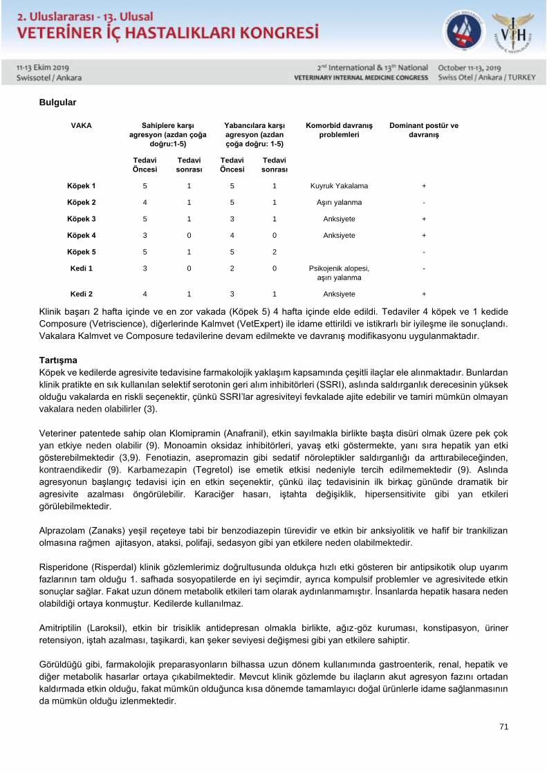
71
Bulgular
VAKA Sahiplere karşı
agresyon (azdan çoğa
doğru:1-5)
Yabancılara karşı
agresyon (azdan
çoğa doğru: 1-5)
Komorbid davranış
problemleri
Dominant postür ve
davranış
Tedavi
Öncesi
Tedavi
sonrası
Tedavi
Öncesi
Tedavi
sonrası
Köpek 1 5 1 5 1 Kuyruk Yakalama +
Köpek 2 4 1 5 1 Aşırı yalanma -
Köpek 3 5 1 3 1 Anksiyete +
Köpek 4 3 0 4 0 Anksiyete +
Köpek 5 5 1 5 2 -
Kedi 1 3 0 2 0 Psikojenik alopesi,
aşırı yalanma
-
Kedi 2 4 1 3 1 Anksiyete +
Klinik başarı 2 hafta içinde ve en zor vakada (Köpek 5) 4 hafta içinde elde edildi. Tedaviler 4 köpek ve 1 kedide
Composure (Vetriscience), diğerlerinde Kalmvet (VetExpert) ile idame ettirildi ve istikrarlı bir iyileşme ile sonuçlandı.
Vakalara Kalmvet ve Composure tedavilerine devam edilmekte ve davranış modifikasyonu uygulanmaktadır.
Tartışma
Köpek ve kedilerde agresivite tedavisine farmakolojik yaklaşım kapsamında çeşitli ilaçlar ele alınmaktadır. Bunlardan
klinik pratikte en sık kullanılan selektif serotonin geri alım inhibitörleri (SSRI), aslında saldırganlık derecesinin yüksek
olduğu vakalarda en riskli seçenektir, çünkü SSRI’lar agresiviteyi fevkalade ajite edebilir ve tamiri mümkün olmayan
vakalara neden olabilirler (3).
Veteriner patentede sahip olan Klomipramin (Anafranil), etkin sayılmakla birlikte başta disüri olmak üzere pek çok
yan etkiye neden olabilir (9). Monoamin oksidaz inhibitörleri, yavaş etki göstermekte, yanı sıra hepatik yan etki
gösterebilmektedir (3,9). Fenotiazin, asepromazin gibi sedatif nöroleptikler saldırganlığı da arttırabileceğinden,
kontraendikedir (9). Karbamezapin (Tegretol) ise emetik etkisi nedeniyle tercih edilmemektedir (9). Aslında
agresyonun başlangıç tedavisi için en etkin seçenektir, çünkü ilaç tedavisinin ilk birkaç gününde dramatik bir
agresivite azalması öngörülebilir. Karaciğer hasarı, iştahta değişiklik, hipersensitivite gibi yan etkileri
görülebilmektedir.
Alprazolam (Zanaks) yeşil reçeteye tabi bir benzodiazepin türevidir ve etkin bir anksiyolitik ve hafif bir trankilizan
olmasına rağmen ajitasyon, ataksi, polifaji, sedasyon gibi yan etkilere neden olabilmektedir.
Risperidone (Risperdal) klinik gözlemlerimiz doğrultusunda oldukça hızlı etki gösteren bir antipsikotik olup uyarım
fazlarının tam olduğu 1. safhada sosyopatilerde en iyi seçimdir, ayrıca kompulsif problemler ve agresivitede etkin
sonuçlar sağlar. Fakat uzun dönem metabolik etkileri tam olarak aydınlanmamıştır. İnsanlarda hepatik hasara neden
olabildiği ortaya konmuştur. Kedilerde kullanılmaz.
Amitriptilin (Laroksil), etkin bir trisiklik antidepresan olmakla birlikte, ağız-göz kuruması, konstipasyon, üriner
retensiyon, iştah azalması, taşikardi, kan şeker seviyesi değişmesi gibi yan etkilere sahiptir.
Görüldüğü gibi, farmakolojik preparasyonların bilhassa uzun dönem kullanımında gastroenterik, renal, hepatik ve
diğer metabolik hasarlar ortaya çıkabilmektedir. Mevcut klinik gözlemde bu ilaçların akut agresyon fazını ortadan
kaldırmada etkin olduğu, fakat mümkün olduğunca kısa dönemde tamamlayıcı doğal ürünlerle idame sağlanmasının
da mümkün olduğu izlenmektedir.
72
Sonuç
Agresyon bozukluklarında uzun süreli tedavilerin yan etkilerinden kaçınmak için, tamamlayıcı ilaç dışı
preparasyonlar, tehlike riskinin ortadan kaldırılmasından sonra dikkate alınmalıdır.
73
Sözlü Sunu / Oral Presentation Tam Metin / Full Text
Complementary Nonpharmaceutical Therapy Efficiency in Canine and Feline Aggressive
Behavior-Clinical Observation
Emre KULLUK*, Basar Ulas SAYILKAN, Duygu DALGIN, Yucel MERAL
Ondokuz Mayis University Department of Internal Medicine
Abstract
Aggressivity is a frequently observed, complex and dangerous behavior in dogs and cats, which involves risk of
human injury. Treatment also carries a human health impact bringing high responsibility. Treatment options include
tricyclic antidepressants and antipsychotics. Five dogs (1-7 years old, a male and 4 females, all intact) and 2 cats (3
and 4 years old intact male and female), were referred with high aggressivity to humans. One cat and two dogs also
had compulsive behavior Starting therapy included risperidone and amitriptyline in dogs and amitriptyline in cats.
Successful clinical responses were obtained in 2 weeks and 4 weeks in the most challenging case, and treatments
were continued with composure (Vetriscience) in 4 dogs and a cat, and Kalmvet (VetExpert), resulting with consistent
recovery. Complementary nonpharmaceuticals must be considered after annihilating risk of danger in order to avoid
side affects of long term treatments in aggressivity disorders.
Key Words: Aggressive Behavior, Dog, Cat, Nonpharmaceutical, Complementary
Introduction
Aggression in dogs and cats is a common and dangerous behavior with complex etiology. Due to the interaction of
the problem of aggression with human health, the treatment involves high responsibility. Human aggression accounts
for 54-67% of all cases of aggression in dogs, while in the United States, 27% of cats abandoned in shelters have
been reported to be left for behavioral reasons (1-3). On average 2-4% of dog attacks result in hospital( 5) and target
is usually family members (4-8). Children are more likely to get injured from vital areas such as the face and neck,
especially because children are weaker in protecting themselves and have the disadvantage of height (4). Certainly,
in cases of aggression in dogs and cats, the etiology and behavior modification are more important than the effective
and immediate elimination of attack behavior.
Materials and Methods
The observation group consisted of five dogs (1-7 years, 1 male and 4 female) and 2 cats (3 and 4 years old, healthy
male and female) who were brought to Ondokuz Mayıs University Veterinary Faculty Animal Hospital with aggressive
problems and showed high aggression against humans. Compulsive behaviors were detected in one cat and two
dogs. Initial treatment was started with risperidone (Risperdal, 1mg / m2) and amitriptyline (Laroxyl, 1 - 4 mg / kg) in
dogs and amitriptyline (Laroxyl, 5 to 10 mg / cat) in cats.
Gradually, 4 dogs and 1 cat were switched to Composure (Vetriscience) and the others to Kalmvet (VetExpert) after
the aggression was relieved.
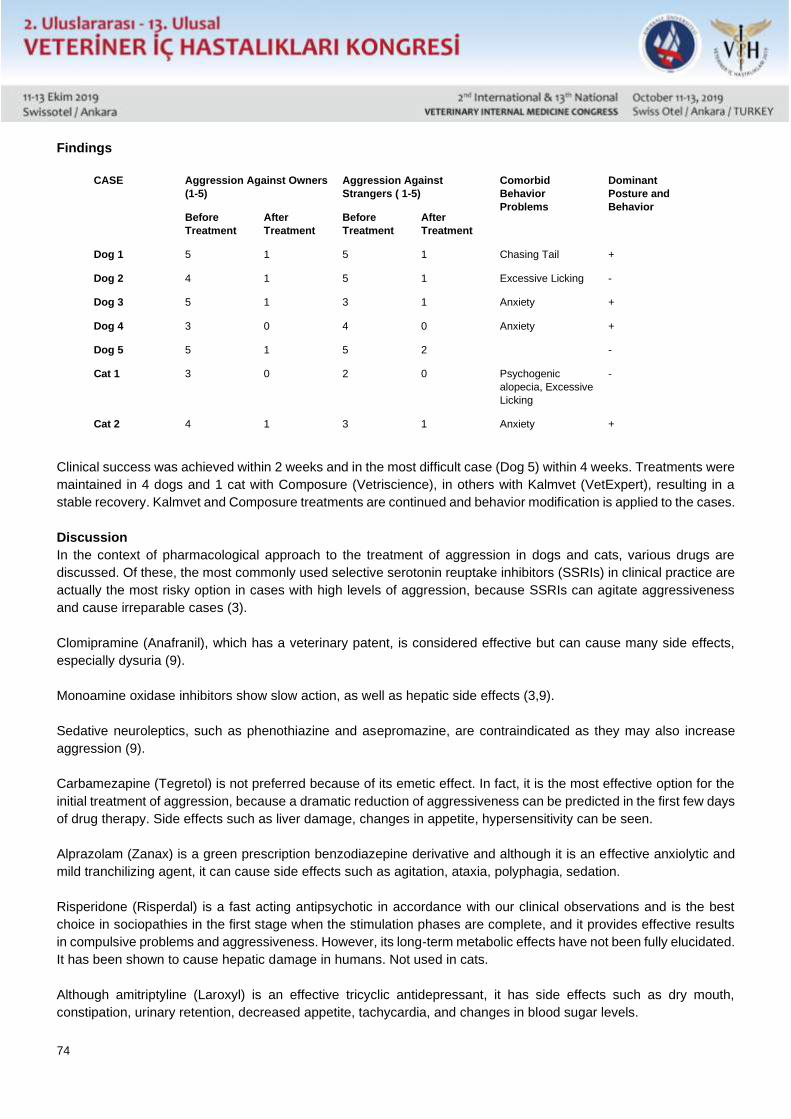
74
Findings
Clinical success was achieved within 2 weeks and in the most difficult case (Dog 5) within 4 weeks. Treatments were
maintained in 4 dogs and 1 cat with Composure (Vetriscience), in others with Kalmvet (VetExpert), resulting in a
stable recovery. Kalmvet and Composure treatments are continued and behavior modification is applied to the cases.
Discussion
In the context of pharmacological approach to the treatment of aggression in dogs and cats, various drugs are
discussed. Of these, the most commonly used selective serotonin reuptake inhibitors (SSRIs) in clinical practice are
actually the most risky option in cases with high levels of aggression, because SSRIs can agitate aggressiveness
and cause irreparable cases (3).
Clomipramine (Anafranil), which has a veterinary patent, is considered effective but can cause many side effects,
especially dysuria (9).
Monoamine oxidase inhibitors show slow action, as well as hepatic side effects (3,9).
Sedative neuroleptics, such as phenothiazine and asepromazine, are contraindicated as they may also increase
aggression (9).
Carbamezapine (Tegretol) is not preferred because of its emetic effect. In fact, it is the most effective option for the
initial treatment of aggression, because a dramatic reduction of aggressiveness can be predicted in the first few days
of drug therapy. Side effects such as liver damage, changes in appetite, hypersensitivity can be seen.
Alprazolam (Zanax) is a green prescription benzodiazepine derivative and although it is an effective anxiolytic and
mild tranchilizing agent, it can cause side effects such as agitation, ataxia, polyphagia, sedation.
Risperidone (Risperdal) is a fast acting antipsychotic in accordance with our clinical observations and is the best
choice in sociopathies in the first stage when the stimulation phases are complete, and it provides effective results
in compulsive problems and aggressiveness. However, its long-term metabolic effects have not been fully elucidated.
It has been shown to cause hepatic damage in humans. Not used in cats.
Although amitriptyline (Laroxyl) is an effective tricyclic antidepressant, it has side effects such as dry mouth,
constipation, urinary retention, decreased appetite, tachycardia, and changes in blood sugar levels.
CASE Aggression Against Owners
(1-5)
Aggression Against
Strangers ( 1-5)
Comorbid
Behavior
Problems
Dominant
Posture and
Behavior Before
Treatment
After
Treatment
Before
Treatment
After
Treatment
Dog 1 5 1 5 1 Chasing Tail +
Dog 2 4 1 5 1 Excessive Licking -
Dog 3 5 1 3 1 Anxiety +
Dog 4 3 0 4 0 Anxiety +
Dog 5 5 1 5 2 -
Cat 1 3 0 2 0 Psychogenic
alopecia, Excessive
Licking
-
Cat 2 4 1 3 1 Anxiety +
75
As can be seen, gastroenteric, renal, hepatic and other metabolic damage may occur especially in the long term use
of pharmacological preparations. Current clinical observation shows that these drugs are effective in eliminating the
acute aggression phase, but it is also possible to maintain complementary natural products as soon as possible.
Results
In order to avoid the side effects of long-term treatments in aggressive disorders, complementary non-drug
preparations should be considered after the elimination of the risk of danger.
References 1.Fatjo J, Amat M, Mariotti VM, Torre JLR, Manteca X: Analysis of 1040 cases of canine aggression in a referral practice in Spain. J Vet Behav,
2, 158-165, 2007.
2.Clarke NM, Fraser D: Animal control measures and their relationship to thereported incidence of dog bites in urban Canadian municipalities.
Can Vet J, 54, 145-149, 2013.
3.Landsberg GM, Hunthausen W, Ackerman L: Handbook of Behavior Problems of the Dog and Cat. 3rd edn., 166, 358-359, 420. Saunders,
Elsevier Health Sciences, Missuri, 2012
4.WHO: Animal bites. In, Media Centre, Fact Sheet. N373, February, 2013.
5.Duffy DL, Hsu Y, Serpell JA : Breed differences in canine aggression.Appl Anim Behav Sci, 114, 441-460, 2008.
6.Rodrigues R, Polo G, Castagna C, Presotto D, Baldini M, Pisciotta K, Lantzman M, Dias RA: Caracterização de casos de agressão canina em
Campinas, São Paulo, Brasil. Braz J Vet Res Anim Sci, 50, 233-237, 2013.
7.Reisner IR, Shofer FS, Nance ML: Behavioral assessment of child-directed canine aggression. Inj Prev, 13, 348-351, 2007.
8.Oliveira VR, Pereira P, Silva J, Miranda CF, Rodrigues KO, Rodrigues TO, Moreira EC: Mordedura cacina e atendimento antirra bico humano
em Minas Gerais. Arq Bras Med Vet Zoo, 64, 891-898, 2012.
9.Reisner,I. The use of medications in Canine behavior therapy. Todays Veterinary Practice,July, 2014, 63-69
10.Dalgın D, Meral Y, Çenesiz M: Risperidone as a promising treatment of choice in dog owner directed aggression. Kafkas Univ Vet Fak Derg,
23 (4): 629-634, 2017
76
Sözlü Sunu / Oral Presentation Tam Metin / Full Text
Koksidiyozis Tanısı Konulmuş Oğlaklarda Hemostasisin Tromboelastografik Olarak
Değerlendirilmesi
Oya ERALP İNAN
Eskisehir Osmangazi Üniversitesi, Tıbbi ve Cerrahi Deney Hayvanları Araştırma ve Uygulama Merkezi, Meşelik
Yerleşkesi, Eskişehir
Koksidiyozis tüm dünyada oğlak ve kuzularda yaygın olarak görülen protozoal bir hastalıktır. Keçilerde yaygın olarak
Eimeria arlongi ve E.hirci enfeksiyonlarına rastlanmaktadır. Bir aylık oğlaklar en yüksek hastalık insidansına sahiptir.
Çiftlik hayvanlarında enfeksiyöz hastalıkların oluşturabildiği sistemik enflamasyon sonucu yaygın damar içi
pıhtılaşma yüksek mortalite ile seyreden koagülasyon değişimlerindendir. Tromboelastografi (TEG), tam kanın ilk
pıhtı oluşumundan fibrinolizise kadar viskoelastik özelliklerini değerlendirmektedir. Bu çalışmada Koksidiyozis tanısı
konulmuş 10 Saanen oğlağında koagülasyonun tromboelastografik olarak değerlendirilmesi amaçlanmıştır.
Çalışmaya Uludağ Üniversitesi Veteriner Fakültesi Hayvan Sağlığı ve Hayvansal Üretim, Araştırma ve Uygulama
Merkezi Keçi Yetiştiriciliği Ünitesinden koksidiyozis tanısı konulmuş 10 oğlak ve yine aynı işletmeden kontrol grubu
olarak 10 sağlıklı oğlak dahil olmuştur. Tüm oğlaklardan kaolin aktive tromboelastografik analiz için sodyum sitratlı
kan örnekleri toplanmıştır. TEG analizinde 5 parametre değerlendirmeye alınmıştır: reaksiyon süresi (R), K süresi
(K), α açısı (α), maksimum amplitüt (MA) ve pıhtı gücü G (G). Koksidiyozis tanısı konulmuş oğlakların TEG sonuçları
sağlıklı kontrol grubu değerleriyle karşılaştırıldığında (ortalama R: 0,86 ± 0,11 ve K: 0,86 ± 0,11 mm) ortalama R
(6,6 ± 3,7 mm) ve K değerlerinde (4,0 ± 1,9 mm) artış (P < 0,05) ancak MA, α ve G değerlerinde (P < 0,001) düşüş
tespit edilmiştir. R süresi K süresi ile pozitif korelasyon (r=0,902, P = 0,01) gösterirken MA (r= -0,931, P<0,001), α
(r= -0,937, P = 0,007) ve G (r= -0,814, P = 0,048) ile negatif bir korelasyon göstermiştir. Bu şekilde sağlıklı oğlaklara
göre koksidiyozisli oğlakların pıhtılaşma süresinin uzadığı ve aynı zamanda pıhtı gücünün de düştüğü tespit
edilmiştir.
Bulgularımız koksidiyozis tanısı konulmuş oğlaklarda belirgin koagülasyon değişimlerinin oluştuğunu göstermiştir ve
klinik olguların değerlendirilmesinde koagülasyon komplikasyonlarının dikkate alınarak, ileri seviyede araştırmaların
tedaviye katkı sağlayabileceğine işaret etmektedir.
Anahtar Kelimeler: oğlak, koksidiyozis, koagülasyon, tromboelastografi
77
Sözlü Sunu / Oral Presentation Tam Metin / Full Text
Thromboelastographic Evaluation of Hemostasis in Kids Diagnosed with Coccidiosis
Oya ERALP INAN
Eskisehir Osmangazi University, Medical and Surgical Laboratory Animal Research and Practice Center, Meselik
Campus, Eskisehir
Coccidiosis is a worldwide protozoan disease especially common in kids and lambs. Eimeria arlongi and E.hirci
generally cause goats’ infections. Kids at the age of one month have the highest disease incidence. Disseminated
intravascular coagulation is one of the coagulation abnormalities with high mortality rates caused by inflammatory
response of infectious diseases in farm animals. Thromboelastography (TEG) evaluates the viscoelastic features
from the beginning of the first clot formation to fibrinolysis of whole blood. This study aimed to assess coagulation by
thromboelastography in 10 Saanen kids diagnosed with coccidiosis.
10 Saanen kids diagnosed with coccidiosis from the Goat Farm of the Faculty of Veterinary Medicine, Animal Health
and Production, Investigation and Practice Center, Uludag University and 10 healthy kids as control from this farm
were included in the study. For kaolin activated thromboelastographic evaluation citrated whole blood samples were
taken from all kids. 5 TEG parameters were included to evaluation: reaction time (R), K time (K), α angle (α),
maximum amplitude (MA) and clot firmness G (G). Mean R (6,6 ± 3,7 mm) and K values (4,0 ± 1,9 mm) in test
group increased (P < 0,05) but MA, AA and G values decreased (P < 0,001) compared to those of healthy controls
(0,86 ± 0,11 and 0,86 ± 0,11 mm, respectively. R time correlated positively with K time (r=0,902, P = 0,01) whereas
negatively with MA (r= - 0,931, P<0,001), AA (r= 0,937, P = 0,007) and G values (r= - 0,814, P = 0,048). The
prolongation of coagulation times and lesser firmness of the clot in kids with coccidiosis were detected.
Our results had shown that significant changes in coagulation can occur in kids with coccidiosis and in consideration
of coagulation complications in clinical cases further investigations can improve treatment options.
Key Words: kid, coccidiosis, coagulation, thromboelastography
Introduction
Coccidiosis is a worldwide seen protozoal disease in goat and kids, especially the 3-4 months of age are the most
affected ones. Different Eimeria types were reported that could cause disease in kids and goats (1). Poor hygenic
conditions and overcrowded herd size are mentioned in exposure and disease incidence due to the high risk of
contaminated water and food (1). The importance of the host breed, body condition score, age and immune status
are as much important as housing conditions during the disease course and outcome (1). Common clinical signs are
diarrhea (green-brown), which can be hemorrhagic but usually watery in goats, dehydration, abdominal pain,
tenesmus and weight loss. Dependent on the host and disease severity, morbidity rate can reach to %80 -%100 (2).
Severe diseases can lead to systemic inflammatory response syndrome (SIRS) or sepsis, which are the main causes
of disseminated intravascular coagulation (DIC), a common hemostatic disorder in large animal (3). Data about
hemostatic changes during diseases in goat kids are poor and reported parameters are in general global clotting
variables like activated partial thromboplastin time (aPTT) and prothrombin time (PT) as well as D-dimer, antithrombin
III (ATIII) and fibrinogen levels (3,4,5). Thromboelastography (TEG)is widely used in human and veterinary medicine
for years due to its prediction of primary, secondary and tertiary hemostasis in one sample measurement. The
purpose of this study was to evaluate the hemostatic status of 10 goat kids diagnosed with coccidiosis by TEG
evaluation.
78
Materials and Methods
A number of 20 Saanen goat kids from the Goat Farm (Faculty of Veterinary Medicine, Animal Health and Production,
Investigation and Practice Center, Uludag University, Bursa) were used. Two groups were designated. Control (n=10)
group included healthy kids and test group (n=10) included kids with coccidiosis, based on the clinical and laboratory
findings.
Hemostasis (coagulation status) was evaluated by TEG (TEG 5000, Hemostasis Analyzer System, Hemonetics,
USA) with anticoagulated (%3.8 9N sodium citrated 1.8ml, BD Vacutainer) whole blood samples.
Kaolin activated TEG was performed with recalcified citrated whole blood. According to the manufacturers’s
recommendations 1ml of citrated whole blood was placed in a silicate vial provided by the manufacturer which
contained kaolin, buffered stabilizers and a blend of phospholipids (Kaolin 1ml, Hemonetics, USA). Kaolin containing
vial were mixed gently for 5 times. In accordance to the standard manufacturer’s procedure pins and cups were
placed into the analyzer, which was pre-warmed to 39˚C (body temperature of patient). To each TEG cup 20µl of 0.2
molar calcium chloride and 340µl kaolin-activated citrated whole blood was added so that the total volume of 360µl
was reached. Five TEG variables were included: R, K, α, MA and G. R is the reaction time and is measured from the
beginning of the evaluation till a preset fibrin formation, in other words until the trace is 2 mm wide. The K-time (K)
begins at the end of R until the tracings diversion reached 20 mm. The α angle is closely related to the K value and
represents the rapidity of fibrin cross-linking. The maximum amplitude MA reflects the peak rigidity of the clot,
whereas the G value is an exponential reflection of MA (dynes per square mm), it is more indicative of small changes
(6).
TEG results were statistically compared by student t test between two groups and Shapiro-Wilk test was used as a
normality test.Pearson correlation was used to estimate a relation between parameters measured (SigmaStat,
GmBH, Erkrath, Germany).
Results
The mean results of TEG variables in 10 healthy kids and 10 kids with coccidiosis are shown in Table 1.
The R time correlated positively with K time (r=0,902, P = 0,01) whereas negatively with MA (r= - 0,931, P<0,001), α
(r= 0,937, P = 0,007) and G values (r= - 0,814, P = 0,048) in the disease group. The prolongation of coagulation
times and lesser firmness of the clot in kids with coccidiosis were detected.
Discussion
This study is the first evaluation of coagulation by thromboelastography in kids with coccidiosis. Reference intervals
for kaolin activated TEG are not available in literature, so there was no chance to compare our data with others of
the disease and healthy group. Reference interval studies in goats including global tests like aPTT, and PT etc. were
published, whereas coagulation differences in adult goats and kids were detected (4,5). Also coagulation data during
diseases in goats and kids is limited in literature. Sahinduran S. et al. (2012) evaluated coagulation variables like
aPTT, PT, fibrinogen and D-dimer to detect possible disorders in kids diagnosed with peste des petits
ruminants(PPR) (3). In our study a prolongation of the R and K time was detected in the disease group, not -, and
statistically significant respectively (P < 0,05), when compared with the healthy group.These extended results reflect
that the clot formation of a certain size/strength in kids with coccidiosis takes more time than in healthy ones. In this
study, the possible reason for prolongation of the R and K times might be due to anticoagulants and clotting factor
deficiencies, because of anticoagulants affecting fibrin polymerization and platelet function. Coagulation times in the
study mentioned before were also prolonged like in our study (3). Thrombocytopenia was reported in kids with PPR,
which could be considered one of the causes of the prolonged clotting time. The unavailability of the CBC in this
study makes it quite different to identify a reason. Distinctively no hemorrhage was seen in kids with coccidiosis, like
in kids with PPR (3). The time of the establishment of prolongations might have been at the time point of an overt
DIC phase, where thrombocytopenia is a consistent finding in dogs but not in cats and horses, which also may be
the case in kids (7). Impaired platelet function may be a reason, possibly due to inflammation, infection or fibrin
79
degradation products, which could be seen in patients in absence of thrombocytopenia with bleeding tendencies
(prolonged clotting time) (8). In literature, the diagnosis of overt DIC needs laboratory findings;2 or more test results
such as thrombocytopenia, prolonged global clotting times (aPTT, PT), low antithrombin, high D-dimer and/or
hypofibrinogenemia (7). It is mentioned that the minimum requirement for a diagnosis for DIC is an underlying primary
initiating disease, like in this study (7). The TEG parameters α, MA and G were statistically significant lower compared
to the healthy ones, which means a less firm clot is composed. α reflects the rapidity of the clot forming its’ maximum
strength in degrees. The lowered result of the α variable is affected like the K value, mentioned before and in this
study may be mostly related to fibrinogen. MA and G evaluations are primarily influenced by platelet aggregation
(platelet function and number) and, to a lesser extent, fibrinogen. Thus, the reason of the TEG results presented here
may show an impaired platelet function and hypofibrinogenemia due to consumption coagulopathy as a phase of
overt-DIC in response to coccidiosis. The results of this study were in compatible with a hypocoagulable state like in
the study of coagulation evaluation in kids with PPR (3). Positive correlationsin the disease group between the R and
K value depends on the common influences of these parameters, whereas both parameters reflect a part of the
thrombus generation. The R and K time both correlate negatively with α, MA and G values. In this case, the prolonged
clotting times with a weaker clot firmness could be considered as a sign of the consumption coagulopathy seen in
overt DIC.
Conclusion
To achieve specific pathophysiologic reasons in coagulation in kids with coccidiosis further coagulation assays like
aPTT, PT, fibrinogen, AT and D-dimer should be run together with TEG evaluations. This study indicated the influence
of coccidiosis in kids on coagulation and further investigations can contribute to treatment and prognosis.
Acknowledgements
Special thanks to Prof.Dr. Zeki Yılmaz and my former collegues at the Internal Medicine Department, Faculty of
Veterinary Medicine, Uludag University, Bursa, Turkey, whose contributions for this study were so important.
References 1.Khodakaram-Taft A., Hashemnia M. An overview of intestinal coccidiosis is sheep and goats. Revue Med. Vet. 2017; 167, 1-2, 9-20.
2.Chartier C., Paraud C.Coccidiosis due to Eimeria in sheep and goats a review. Small Ruminant Research.2012;103(1); 84-92.
3.Sahinduran S., Albay MK., Sezer K., Ozmen O., et al. Coagulation profile, heamatological and biochemical changes in kids naturally infected
with peste des petits ruminants. Trop Animal Health Prod. 2012; 44(3); 453-7.
4.Piccine G., Bertolucci C., Giannetto C., Guidice E. Clotting profiles in newborn maltese kids during the first week of life. J Vet Diagn Invest.
2008; 20(1); 114-8.
5.Zersen KM., Russell Moore A., Olver CS., Mathis JC. Reference intervals for coagulation variables in healthy adult domestic goats (Capra
aegagrushircus). Vet ClinPathol. 2018; 47(3); 396-399.
6.EralpInan O. Establishment of reference intervals for kaolin-activated TEG and the STA Compact Automated Analyzer for dogs and
coagulation response ina canine model of endotoxemia. Dissertation, VVB LaufersweilerVerlag. 2011; 27-30.
7.Stokol T. Disseminated intravascular coagulation, In: Weiss DJ.,Wardrop KJ. Eds. Schalm’s Veterinary Hematology. 6thEdt. Iowa: Blackwell
Publishing, 2010. Pp679-688.
8.Fry MM. Acquired platelet dysfunction. In: Weiss DJ.,Wardrop KJ. Eds. Schalm’s Veterinary Hematology. 6thEdt. Iowa: Blackwell Publishing,
2010. Pp626-631.
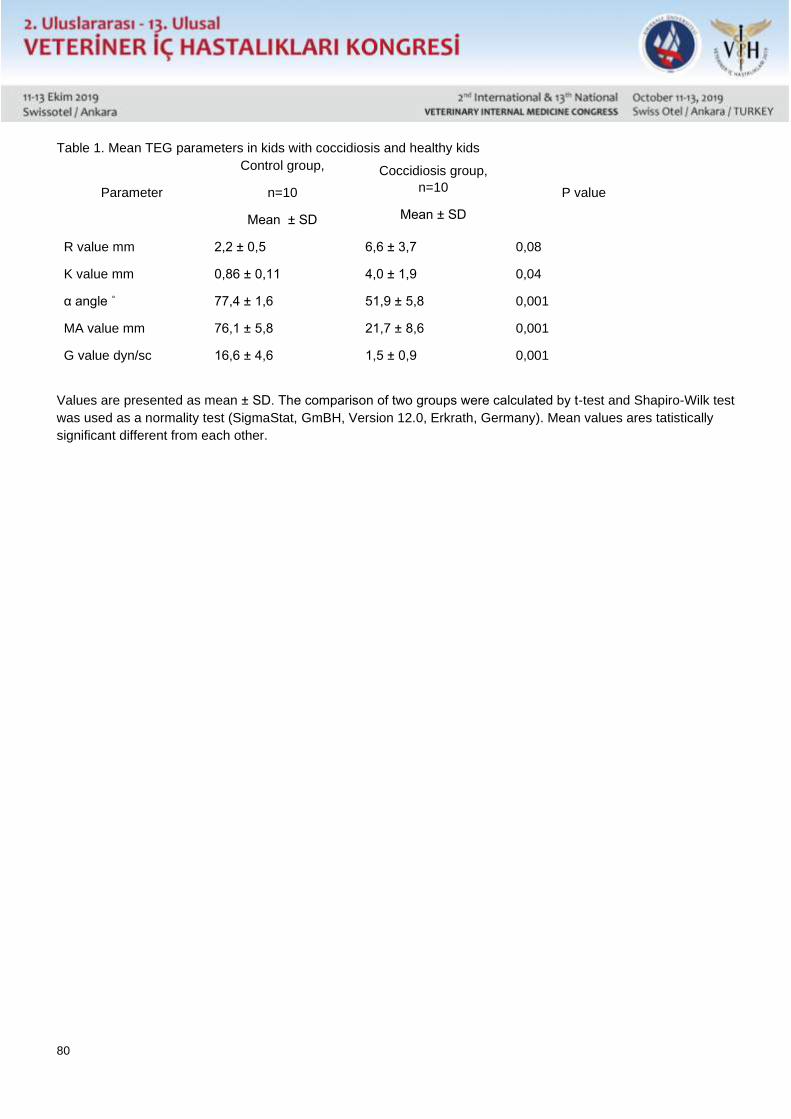
80
Table 1. Mean TEG parameters in kids with coccidiosis and healthy kids
Parameter
Control group,
n=10
Mean ± SD
Coccidiosis group,
n=10
Mean ± SD
P value
R value mm 2,2 ± 0,5 6,6 ± 3,7 0,08
K value mm 0,86 ± 0,11 4,0 ± 1,9 0,04
α angle ˚ 77,4 ± 1,6 51,9 ± 5,8 0,001
MA value mm 76,1 ± 5,8 21,7 ± 8,6 0,001
G value dyn/sc 16,6 ± 4,6 1,5 ± 0,9 0,001
Values are presented as mean ± SD. The comparison of two groups were calculated by t-test and Shapiro-Wilk test
was used as a normality test (SigmaStat, GmBH, Version 12.0, Erkrath, Germany). Mean values ares tatistically
significant different from each other.
81
Sözlü Sunu / Oral Presentation Tam Metin / Full Text
Bir Köpekte Ön Mitral Kapak Orifisi
Başar Ulaş Sayılkan, Yücel Meral, Emre Küllük, Duygu Dalgın
Ondokuz Mayıs Üniversitesi Veteriner Fakültesi İç Hastalıkları Anabilim Dalı, Samsun
Özet
Hastanemize epileptoik nöbet ve hipotiroidi şikayetleri ile getirilen 8 yaşlı, 33.5 kg canlı ağırlıkta Golden Retriever ırkı
köpeğe ait kardiyolojik inceleme sonucunda EKG’de bradikardi belirlendi.
Hastanın 2-D, M-Mode, PW, CW ve renkli doppler muayeneleri yapıldı. Hastanın 2-D rehberliğinde alınan M-Mode
görüntülemesi ile sol ventrikül hipokinetik ve mitral, trikuspital kapak hareketi kısıtlı olarak gözlemlendi. PW doppler
ve renkli doppler ile septal mitral kapak üzerinde 2 mm genişliğinde konjenital ön mitral kapak fissürü belirlendi. Bu
fissür ile LA’u 1/3 oranında kaplayan ve 2 m/sn olan regurgitant jetin varlığı ortaya konuldu. Qp/Qs: 1,7, PISA
hesaplamaları; VFR (mL/s), ERO (mm2) ve RVol (mL/atım) değerleri mitral regurgitasyon varmış gibi düşünülürek
hesaplandı ve orta altı düzeyde mitral regurgitasyona ait veriler elde edildi. Bu nedenle fissura ait herhangi bir operatif
sağaltım önerilmedi.
Mitral kapak fissurü nadir rastlanan bir konjenital patoloji olması ve bu konuda veteriner hekimlik alanında literatür
bulunmaması nedeniyle, verilerin ileride yapılacak bilimsel çalışmalara ışık tutabileceği düşüncesindeyiz.
Anahtar Kelimeler: Mitral Regürgitasyon, , Ekokardiyografi, Puls Dalga Doppler
Giriş
Mitral kapak yetmezlikleri hayvanlarda en sık görülen kalp rahatsızlıklarından biridir. Mitral regürgitasyon olarak
adlandırılan sol ventrikül kontraksiyonu sonucu kanın bir kısmının sol atriyuma geri dönmesi kalp yetmezliğini
meydana getirir. Mitral regürgitasyonun nedeni kalp kapakçıklarındaki üremeler kapakçıkların yetersiz kapanması,
korda tendinea yırtılmaları, aort stenozları, dilate kardiyomyopatiler, mitral kapakçık orifisleri olabilir.4 Bu olgunun
amacı, çok nadir rastlanan bir patoloji olan ve veteriner hekimliği alanında literatür bulunmayan mitral kapak
orifisinden elde edilen bilimsel verilerin karşılaşılan diğer vakalara ışık tutmasıdır.
Olgu
Hastanemize epileptoik nöbetler, hipotiroidi şikayetleri ile getirilen 8 yaşlı, 33,5 kg canlı ağırlıkta Golden Retriever ırkı
bir köpeğe ait kardiyolojik inceleme sonucunda oskultasyonda normal kalp sesleri ve normal pulmoner sesler
belirlendi. Femoral arter ve V. Jugularis dolgunluğu ve nabız kalitesi normal ve hastanın 5 mm/mV- 25 mm/sn ile
alınan EKG kaydında ritm normal olmakla birlikte bradikardi görüldü.
Hastanın 2-D, M-Mode, PW doppler, CW doppler ve Renkli doppler muayeneleri yapıldı. Hastanın 2-D ultrasonografi
rehberliğinde alınan M-Mode görüntülemesi ile sağ parasternal uzun aks, sağ parasternal kısa aks ve sol parasternal
4 ve 5 boşluk görüntüleme ile elde edilen ekokardiyografik veriler; LVIDd:4,23 cm, LVIDs:2,91 cm, LVPWd:1,66 cm,
LVPWs: 2,11 cm, IVSd: 1,94 cm, IVSs: 2,80 cm, Aod: 2,17, LAd: 3,55, LAd/Aod: 1.64, ESV: 32,57 ml, SV:47,20 ml,
EF:59,17 %,FS:%31.08, HR: 67 Bpm, CO: 3,089 L/min, MVEamp: 1,65 cm, MVAamp: 1,61 cm, MVDESlope: 48,16
cm/s, MVEFSlope: 7,15 cm/s, EPSS: 1,37 cm şeklindeydi. Sol ventrikül hipokinetik ve mitral, trikuspidal kapak
hareketi kısıtlı olarak gözlemlendi. PW doppler ve Renkli doppler ile septal mitral kapak üzerinde 2 mm genişliğinde
konjenital ön mitral kapak orifisi belirlendi. Bu orifis ile LA’u 1/3 oranında kaplayan ve hızı 3 m/sn olan bir
regurgitasyon jetin varlığı ortaya konuldu. LVOT ve RVOT genişliği normal ve Qp/Qs: 1.7, PISA hesaplamaları; VFR
(mL/s), ERO (mm2) ve RVol (mL/atım) değerleri mitral regurgitasyon varmış gibi düşünülerek hesaplandı ve orta altı
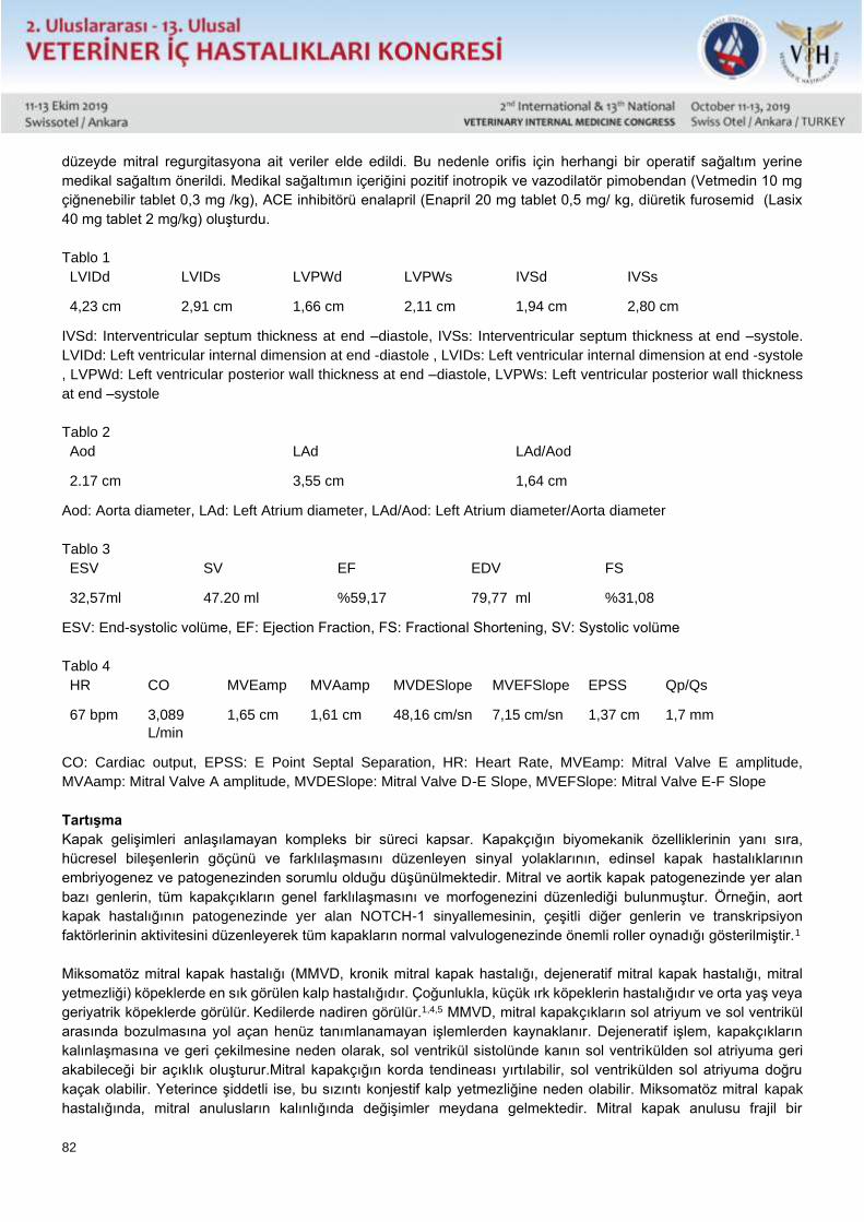
82
düzeyde mitral regurgitasyona ait veriler elde edildi. Bu nedenle orifis için herhangi bir operatif sağaltım yerine
medikal sağaltım önerildi. Medikal sağaltımın içeriğini pozitif inotropik ve vazodilatör pimobendan (Vetmedin 10 mg
çiğnenebilir tablet 0,3 mg /kg), ACE inhibitörü enalapril (Enapril 20 mg tablet 0,5 mg/ kg, diüretik furosemid (Lasix
40 mg tablet 2 mg/kg) oluşturdu.
Tablo 1
LVIDd LVIDs LVPWd LVPWs IVSd IVSs
4,23 cm 2,91 cm 1,66 cm 2,11 cm 1,94 cm 2,80 cm
IVSd: Interventricular septum thickness at end –diastole, IVSs: Interventricular septum thickness at end –systole.
LVIDd: Left ventricular internal dimension at end -diastole , LVIDs: Left ventricular internal dimension at end -systole
, LVPWd: Left ventricular posterior wall thickness at end –diastole, LVPWs: Left ventricular posterior wall thickness
at end –systole
Tablo 2
Aod LAd LAd/Aod
2.17 cm 3,55 cm 1,64 cm
Aod: Aorta diameter, LAd: Left Atrium diameter, LAd/Aod: Left Atrium diameter/Aorta diameter
Tablo 3
ESV SV EF EDV FS
32,57ml 47.20 ml %59,17 79,77 ml %31,08
ESV: End-systolic volüme, EF: Ejection Fraction, FS: Fractional Shortening, SV: Systolic volüme
Tablo 4
HR CO MVEamp MVAamp MVDESlope MVEFSlope EPSS Qp/Qs
67 bpm 3,089
L/min
1,65 cm 1,61 cm 48,16 cm/sn 7,15 cm/sn 1,37 cm 1,7 mm
CO: Cardiac output, EPSS: E Point Septal Separation, HR: Heart Rate, MVEamp: Mitral Valve E amplitude,
MVAamp: Mitral Valve A amplitude, MVDESlope: Mitral Valve D-E Slope, MVEFSlope: Mitral Valve E-F Slope
Tartışma
Kapak gelişimleri anlaşılamayan kompleks bir süreci kapsar. Kapakçığın biyomekanik özelliklerinin yanı sıra,
hücresel bileşenlerin göçünü ve farklılaşmasını düzenleyen sinyal yolaklarının, edinsel kapak hastalıklarının
embriyogenez ve patogenezinden sorumlu olduğu düşünülmektedir. Mitral ve aortik kapak patogenezinde yer alan
bazı genlerin, tüm kapakçıkların genel farklılaşmasını ve morfogenezini düzenlediği bulunmuştur. Örneğin, aort
kapak hastalığının patogenezinde yer alan NOTCH-1 sinyallemesinin, çeşitli diğer genlerin ve transkripsiyon
faktörlerinin aktivitesini düzenleyerek tüm kapakların normal valvulogenezinde önemli roller oynadığı gösterilmiştir.1
Miksomatöz mitral kapak hastalığı (MMVD, kronik mitral kapak hastalığı, dejeneratif mitral kapak hastalığı, mitral
yetmezliği) köpeklerde en sık görülen kalp hastalığıdır. Çoğunlukla, küçük ırk köpeklerin hastalığıdır ve orta yaş veya
geriyatrik köpeklerde görülür. Kedilerde nadiren görülür.1,4,5 MMVD, mitral kapakçıkların sol atriyum ve sol ventrikül
arasında bozulmasına yol açan henüz tanımlanamayan işlemlerden kaynaklanır. Dejeneratif işlem, kapakçıkların
kalınlaşmasına ve geri çekilmesine neden olarak, sol ventrikül sistolünde kanın sol ventrikülden sol atriyuma geri
akabileceği bir açıklık oluşturur.Mitral kapakçığın korda tendineası yırtılabilir, sol ventrikülden sol atriyuma doğru
kaçak olabilir. Yeterince şiddetli ise, bu sızıntı konjestif kalp yetmezliğine neden olabilir. Miksomatöz mitral kapak
hastalığında, mitral anulusların kalınlığında değişimler meydana gelmektedir. Mitral kapak anulusu frajil bir
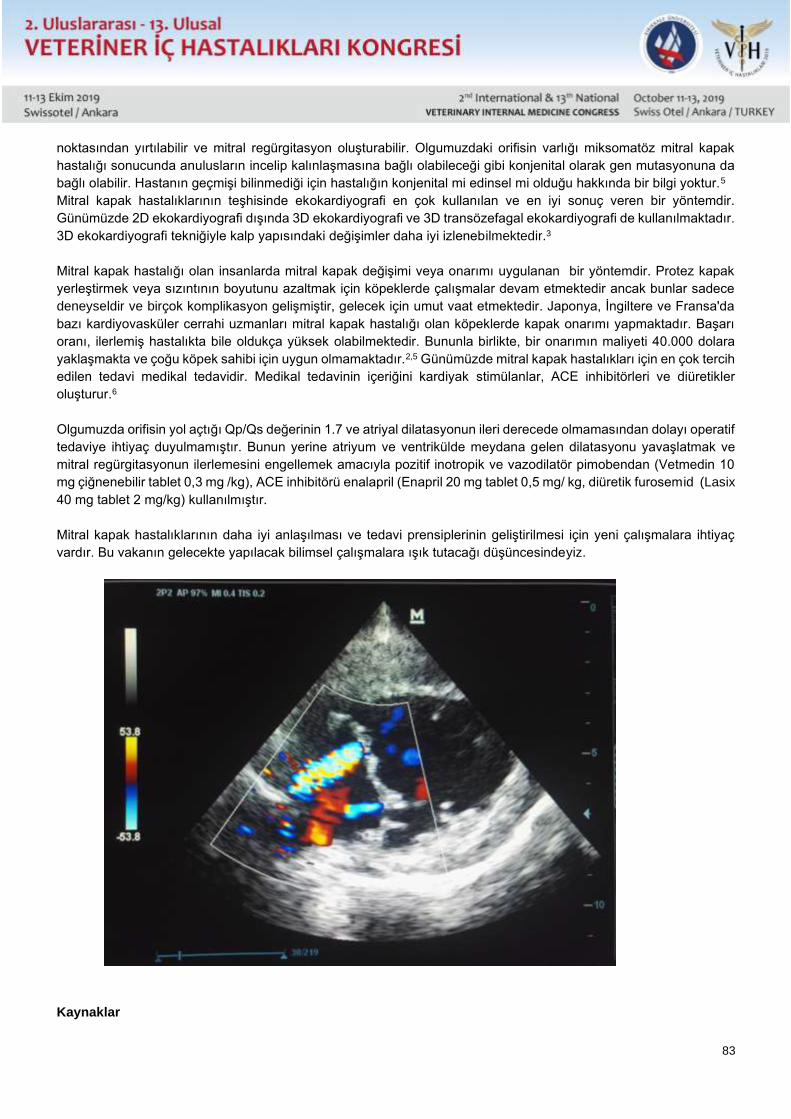
83
noktasından yırtılabilir ve mitral regürgitasyon oluşturabilir. Olgumuzdaki orifisin varlığı miksomatöz mitral kapak
hastalığı sonucunda anulusların incelip kalınlaşmasına bağlı olabileceği gibi konjenital olarak gen mutasyonuna da
bağlı olabilir. Hastanın geçmişi bilinmediği için hastalığın konjenital mi edinsel mi olduğu hakkında bir bilgi yoktur.5
Mitral kapak hastalıklarının teşhisinde ekokardiyografi en çok kullanılan ve en iyi sonuç veren bir yöntemdir.
Günümüzde 2D ekokardiyografi dışında 3D ekokardiyografi ve 3D transözefagal ekokardiyografi de kullanılmaktadır.
3D ekokardiyografi tekniğiyle kalp yapısındaki değişimler daha iyi izlenebilmektedir.3
Mitral kapak hastalığı olan insanlarda mitral kapak değişimi veya onarımı uygulanan bir yöntemdir. Protez kapak
yerleştirmek veya sızıntının boyutunu azaltmak için köpeklerde çalışmalar devam etmektedir ancak bunlar sadece
deneyseldir ve birçok komplikasyon gelişmiştir, gelecek için umut vaat etmektedir. Japonya, İngiltere ve Fransa'da
bazı kardiyovasküler cerrahi uzmanları mitral kapak hastalığı olan köpeklerde kapak onarımı yapmaktadır. Başarı
oranı, ilerlemiş hastalıkta bile oldukça yüksek olabilmektedir. Bununla birlikte, bir onarımın maliyeti 40.000 dolara
yaklaşmakta ve çoğu köpek sahibi için uygun olmamaktadır.2,5 Günümüzde mitral kapak hastalıkları için en çok tercih
edilen tedavi medikal tedavidir. Medikal tedavinin içeriğini kardiyak stimülanlar, ACE inhibitörleri ve diüretikler
oluşturur.6
Olgumuzda orifisin yol açtığı Qp/Qs değerinin 1.7 ve atriyal dilatasyonun ileri derecede olmamasından dolayı operatif
tedaviye ihtiyaç duyulmamıştır. Bunun yerine atriyum ve ventrikülde meydana gelen dilatasyonu yavaşlatmak ve
mitral regürgitasyonun ilerlemesini engellemek amacıyla pozitif inotropik ve vazodilatör pimobendan (Vetmedin 10
mg çiğnenebilir tablet 0,3 mg /kg), ACE inhibitörü enalapril (Enapril 20 mg tablet 0,5 mg/ kg, diüretik furosemid (Lasix
40 mg tablet 2 mg/kg) kullanılmıştır.
Mitral kapak hastalıklarının daha iyi anlaşılması ve tedavi prensiplerinin geliştirilmesi için yeni çalışmalara ihtiyaç
vardır. Bu vakanın gelecekte yapılacak bilimsel çalışmalara ışık tutacağı düşüncesindeyiz.
Kaynaklar
84
1-)Şaylık F, Mutluer FO, Tosu A, Selçuk M. Double orifice mitral valve and bicuspid aortic valve: pieces of the same single puzzle? Case Rep
Cardiol.2015; 2015: 305142.
2-)Kanemoto, I., Taguchi, D., Mihara, K., Kawase, K., Sato, K., Iwata, K., & Iwanaga, K. Long-term survival of two dogs after mitral valve plasty.
Veterinary Science Development. 2017; volume 7(1): 6335
3-)Müller S, Menciotti G, Borgarelli M. Anatomic regurgitant orifice area obtained using 3D-echocardiography as an indicator of severity of mitral
regurgitation in dogs with myxomatous mitral valve disease. J Vet Cardiol. 2017 Oct; 19(5): 433-440.
4-)Boswood A. Improving outcomes of myxomatous mitral valve disease in dogs. In practice. 2018; 40(Suppl 1):12-15
5-)https://veterinarypartner.vin.com/doc/?id=8526511&pid=19239
6-) Keene BW, Atkins CE, Bonagura JD, Fox PR, Häggström J, Fuentes VL, Oyama MA, Rush JE, Stepien R, Uechi M. ACVIM consensus
guidelines for the diagnosis and treatment of myxomatous mitral valve disease in dogs. J Vet Intern Med. 2019 May;33(3):1127-1140.
85
Sözlü Sunu / Oral Presentation Tam Metin / Full Text
Anterior Mitral Valve Orifices In A Dog
Basar Ulas Sayilkan, Yucel Meral, Emre Kulluk, Duygu Dalgin
Ondokuz Mayıs University Faculty of Veterinary Medicine, Department of Internal Medicine, Samsun
ABSTRACT
Bradycardia was detected in the ECG as a result of the cardiological examination of 33.5 kg weight Golden Retriever
dog which was brought to our hospital with epileptoic seizure and hypothyroidism.
2-D, M-Mode, PW, CW and color doppler examinations were performed. Left-ventricular hypokinetic and mitral and
tricuspital valve movements were observed with limited M-Mode imaging. Congenital anterior mitral valve fissure with
a width of 2 mm was determined on septal mitral valve by PW doppler and color doppler. With this fissure, the
presence of a regurgitant jet covering LA at a rate of 1/3 and 2 m / sec was demonstrated. Qp / Qs: 1,7, PISA
calculations; VFR (mL / s), ERO (mm2) and RVol (mL / pulse) values were calculated as if there were mitral
regurgitation and data were obtained for sub-moderate mitral regurgitation. Therefore, no operative treatment of
fissure was recommended.
Mitral valve fissure is a rare congenital pathology and there is no literature on veterinary medicine so we think that
the data may shed light on future scientific studies.
Keywords: Mitral Regurgitation, Echocardiography, Pulse Wave Doppler
Introduction
Mitral valve insufficiency is one of the most common cardiac disorders in animals. As a result of left ventricular
contraction called mitral regurgitation, the return of some of the blood to the left atrium causes heart failure. Mitral
regurgitation may be caused by heart valve regrowths, inadequate closure of the valves, rupture of the chorda
tendina, aortic stenosis, dilated cardiomyopathies, mitral valve orifices.4 The aim of this case is to shed light on other
cases encountered in the field of mitral valve orifice which is a very rare pathology and there is no literature on
veterinary medicine.
Case
The cardiological examination of a 33.5 kg live weight Golden Retriever dog aged 8 years, brought to our hospital
with epileptoic seizures and hypothyroidism, revealed normal heart sounds and normal pulmonary sounds during
auscultation. Femoral artery and V. Jugularis fullness and pulse quality were normal and the patient's ECG recordings
of 5 mm / mV-25 mm / sec showed bradycardia, although the rhythm was normal. 2-D, M-Mode, PW doppler, CW
doppler and Color doppler examinations were performed. Echocardiographic data obtained by right parasternal long
axis, right parasternal short axis and left parasternal 4 and 5 space imaging with M-Mode imaging of the patient under
the guidance of 2-D ultrasonography; LVIDd: 4.23 cm, LVIDs: 2.91 cm, LVPWd: 1.66 cm, LVPWs: 2.11 cm, IVSd:
1.94 cm, IVSs: 2.80 cm, Aod: 2.17, LAd: 3.55, LAd / Aod: 1.64, ESV: 32.57 ml, SV: 47.20 ml, EF: 59.17%, FS:
31.08%, HR: 67 Bpm, CO: 3.089 L / min, MVEamp: 1.65 cm, MVAamp: 1.61 cm, MVDESlope: 48.16 cm / s,
MVEFSlope: 7.15 cm / s, and EPSS: 1.37 cm. Left ventricular hypokinetic and mitral, tricuspidal valve movement
was observed to be limited.
Congenital anterior mitral valve orifice with a width of 2 mm was determined on the septal mitral valve by PW doppler
and color doppler. With this orifice, the presence of a regurgitation jet with a velocity of 3 m / sec covering 1/3 of LA
was demonstrated. LVOT and RVOT width normal and Qp / Qs: 1.7, PISA calculations; VFR (mL / s), ERO (mm2)
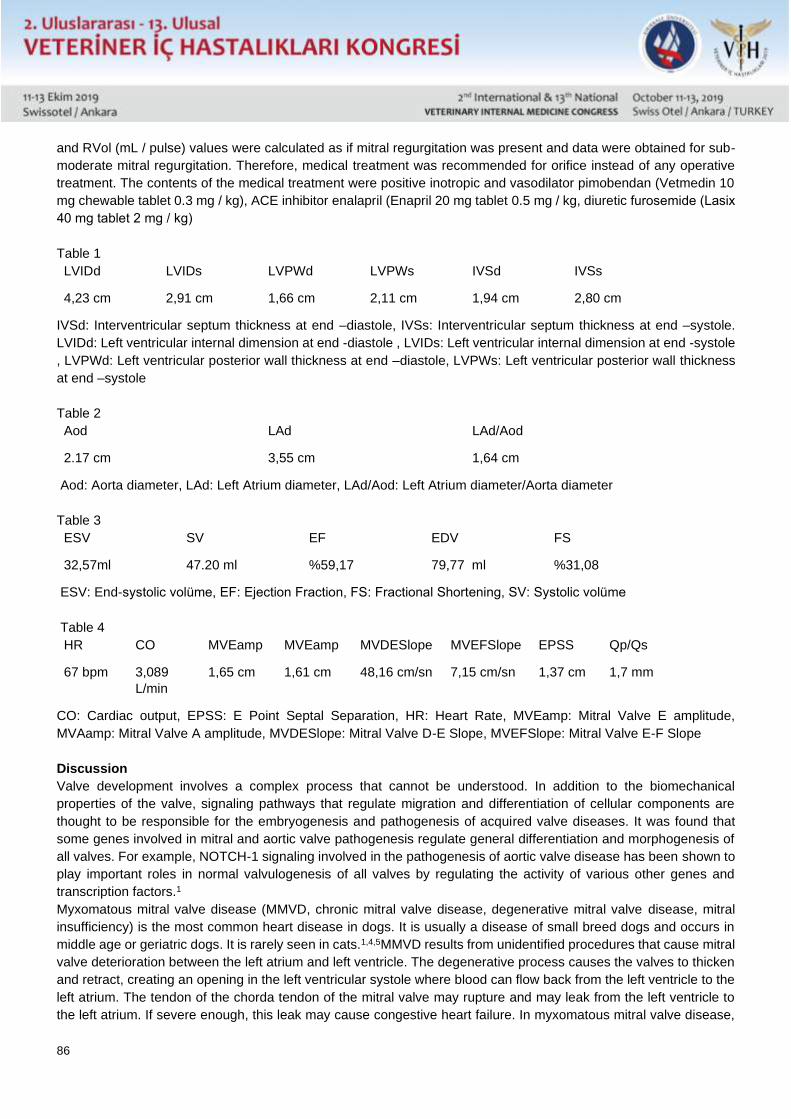
86
and RVol (mL / pulse) values were calculated as if mitral regurgitation was present and data were obtained for sub-
moderate mitral regurgitation. Therefore, medical treatment was recommended for orifice instead of any operative
treatment. The contents of the medical treatment were positive inotropic and vasodilator pimobendan (Vetmedin 10
mg chewable tablet 0.3 mg / kg), ACE inhibitor enalapril (Enapril 20 mg tablet 0.5 mg / kg, diuretic furosemide (Lasix
40 mg tablet 2 mg / kg)
Table 1
LVIDd LVIDs LVPWd LVPWs IVSd IVSs
4,23 cm 2,91 cm 1,66 cm 2,11 cm 1,94 cm 2,80 cm
IVSd: Interventricular septum thickness at end –diastole, IVSs: Interventricular septum thickness at end –systole.
LVIDd: Left ventricular internal dimension at end -diastole , LVIDs: Left ventricular internal dimension at end -systole
, LVPWd: Left ventricular posterior wall thickness at end –diastole, LVPWs: Left ventricular posterior wall thickness
at end –systole
Table 2
Aod LAd LAd/Aod
2.17 cm 3,55 cm 1,64 cm
Aod: Aorta diameter, LAd: Left Atrium diameter, LAd/Aod: Left Atrium diameter/Aorta diameter
Table 3
ESV SV EF EDV FS
32,57ml 47.20 ml %59,17 79,77 ml %31,08
ESV: End-systolic volüme, EF: Ejection Fraction, FS: Fractional Shortening, SV: Systolic volüme
Table 4
HR CO MVEamp MVEamp MVDESlope MVEFSlope EPSS Qp/Qs
67 bpm 3,089
L/min
1,65 cm 1,61 cm 48,16 cm/sn 7,15 cm/sn 1,37 cm 1,7 mm
CO: Cardiac output, EPSS: E Point Septal Separation, HR: Heart Rate, MVEamp: Mitral Valve E amplitude,
MVAamp: Mitral Valve A amplitude, MVDESlope: Mitral Valve D-E Slope, MVEFSlope: Mitral Valve E-F Slope
Discussion
Valve development involves a complex process that cannot be understood. In addition to the biomechanical
properties of the valve, signaling pathways that regulate migration and differentiation of cellular components are
thought to be responsible for the embryogenesis and pathogenesis of acquired valve diseases. It was found that
some genes involved in mitral and aortic valve pathogenesis regulate general differentiation and morphogenesis of
all valves. For example, NOTCH-1 signaling involved in the pathogenesis of aortic valve disease has been shown to
play important roles in normal valvulogenesis of all valves by regulating the activity of various other genes and
transcription factors.1
Myxomatous mitral valve disease (MMVD, chronic mitral valve disease, degenerative mitral valve disease, mitral
insufficiency) is the most common heart disease in dogs. It is usually a disease of small breed dogs and occurs in
middle age or geriatric dogs. It is rarely seen in cats.1,4,5MMVD results from unidentified procedures that cause mitral
valve deterioration between the left atrium and left ventricle. The degenerative process causes the valves to thicken
and retract, creating an opening in the left ventricular systole where blood can flow back from the left ventricle to the
left atrium. The tendon of the chorda tendon of the mitral valve may rupture and may leak from the left ventricle to
the left atrium. If severe enough, this leak may cause congestive heart failure. In myxomatous mitral valve disease,
87
changes in the thickness of the mitral annulus occur. The mitral valve annulus may rupture at a fragile point and
cause mitral regurgitation. The presence of orifice in our case may be due to thinning and thickening of the annulus
as a result of myxomatous mitral valve disease, or it may be due to gene mutation congenitally. Since the patient's
history is unknown, there is no information about whether the disease is congenital or acquired.5
Echocardiography is the most commonly used method in the diagnosis of mitral valve diseases. In addition to 2D
echocardiography, 3D echocardiography and 3D transesophageal echocardiography are also used. 3D
echocardiography can be used to better monitor the changes in heart structure.3
Mitral valve replacement or repair is a method in people with mitral valve disease. Studies are underway in dogs to
place prosthetic valves or reduce the size of the leak, but these are only experimental and many complications have
developed, promising for the future. In Japan, the UK and France, some cardiovascular surgeons perform valve
repair in dogs with mitral valve disease. The success rate can be quite high, even in advanced disease. However,
the cost of a repair is close to $ 40,000 and is not suitable for most dog owners.2,5
Today, medical treatment is the most preferred treatment for mitral valve diseases. The content of medical treatment
consists of cardiac stimulants, ACE inhibitors and diuretics.6
In our case, there was no need for operative treatment because the orifice-induced Qp / Qs value was 1.7 and atrial
dilatation was not advanced. Instead of positive inotropic and vasodilator pimobendan (Vetmedin 10 mg chewable
tablet 0.3 mg / kg), ACE inhibitor enalapril (Enapril 20 mg tablet 0.5 mg / kg, diuretic) furosemide (Lasix 40 mg tablet
2 mg / kg) was used.
Further studies are needed to better understand the mitral valve diseases and develop treatment principles. We
believe that this case will shed light on future scientific studies.
Resources 1-)Şaylık F, Mutluer FO, Tosu A, Selçuk M. Double orifice mitral valve and bicuspid aortic valve: pieces of the same single puzzle? Case Rep
Cardiol. 2015; 2015: 305142.
2-)Kanemoto, I., Taguchi, D., Mihara, K., Kawase, K., Sato, K., Iwata, K., & Iwanaga, K. Long-term survival of two dogs after mitral valve plasty.
Veterinary Science Development. 2017; volume 7(1): 6335
3-)Müller S, Menciotti G, Borgarelli M. Anatomic regurgitant orifice area obtained using 3D-echocardiography as an indicator of severity of mitral
regurgitation in dogs with myxomatous mitral valve disease. J Vet Cardiol. 2017 Oct; 19(5): 433-440.
4-)Boswood A. Improving outcomes of myxomatous mitral valve disease in dogs. In practice. 2018; 40(Suppl 1):12-15
5-)https://veterinarypartner.vin.com/doc/?id=8526511&pid=19239
6-) Keene BW, Atkins CE, Bonagura JD, Fox PR, Häggström J, Fuentes VL, Oyama MA, Rush JE, Stepien R, Uechi M. ACVIM consensus
guidelines for the diagnosis and treatment of myxomatous mitral valve disease in dogs. J Vet Intern Med. 2019 May;33(3):1127-1140.
88
Sözlü Sunu / Oral Presentation Tam Metin / Full Text
Neonatal Kuzularda Serum İnsülin Benzeri Büyüme Faktörü 1 (IGF-1) Üzerine
Araştırmalar
Ethem Mutlu Temizela, Fatih Kavukcub, Adil Ömer Karakuşc
Bursa Uludağ Üniversitesi, Veteriner Fakültesi, İç Hastalıkları Anabilim Dalı, Bursa – TÜRKİYE
aORCID: 0000-0002-4828-4116, bORCID: 0000-0002-3545-0883, cORCID: 0000-0002-6834-0808
Özet
Bu çalışmada neonatal kuzuların ikizlik durumunun, cinsiyetinin ve canlı ağırlıklarının serum IGF-1 ile olan ilişkisi
irdelenmiştir. Çalışma materyalini oluşturan toplam 32 kuzu cinsiyet ve çoklu doğum temelinde iki ayrı katogoride
incelendi. (14 dişi, 18 erkek ve 18 tek, 14 ikiz). Kuzuların 1., 7., 14., 21., 35., 49., ve 63. günlerde canlı ağırlıkları
ölçülerek klinik muayeneleri yapıldı. Belirtilen günlerde vena jugularisten kan numuneleri toplandı. Elde edilen
serumluk kan örneklerinden IGF-1 ölçümleri ELISA yöntemi ile yapıldı. Bu çalışmada cinsiyet temelinde canlı
ağırlıklar arasında istatistiksel olarak anlamlı farklar olmamakla birlikte, erkek kuzuların canlı ağırlığının dişi
kuzulardan daha yüksek olduğu bulundu. Ancak IGF-I düzeylerinin erkek hayvanlarda 14, 35 ve 49. günler haricinde
diğer ölçümlerde dişilerden daha yüksek olduğu bulundu. Korelasyon testi sonuçları, yenidoğan döneminin ilk 21
gününde istatistiksel olarak anlamlı bir korelasyon bulunmamasına rağmen, vücut ağırlığı ile IGF-I düzeyleri arasında
35, 49 ve 63. günlerde istatistiksel olarak anlamlı bir ilişki olduğunu göstermiştir. (p<0.05, 0.01, 0.001). Korelasyon
testi sonuçlarında, neonatal dönem sonrasında IGF-I ile canlı ağırlık arasında dişi ve ikiz kuzulardaki korelasyon
düzeyinin erkek ve tek kuzulara göre daha yüksek olduğu belirlendi. (p<0.001, p<0.01). Sonuç olarak, IGF-I’in
neonatal dönem sonrasında kuzularda canlı ağırlık kazancı üzerinde prediktif bir değeri olabileceği, neonatal
dönemde cinsiyet ve çoklu doğum faktörünün serum IGF-I düzeyi üzerinde etkisinin sınırlı düzeyde kaldığı ortaya
konmuştur.
Anahtar kelimeler: kuzu, igf-1, canlı ağırlık
Giriş
IGF-1, çoğunlukla karaciğer tarafından salgılanan ve GH varlığı ile birlikte çalışan bir hormondur (Trenkle, 1981).
IGF-1, canlı ağırlık artışından, iç organların gelişiminden ve diğer gelişimsel parametrelerden sorumlu bir hormondur.
Kuzuların cinsiyetinin ve ırkının IGF-1 konsantrasyonlarındaki farkın temel nedeni olmasının yanı sıra, beslenme,
doğan yavru sayısı ve sütten kesim yaşı gibi bir çok faktörün de konsantrasyonlardaki değişimlere neden olduğu
bildirilmektedir (Roberts ve ark., 1990; Medrano ve Bredford, 1991; Wylie ve ark., 1997).
Kuzularda IGF-1'in canlı ağırlık artışı (Blair ve ark., 1987), karkas kalitesi ve diğer verim parametreleri (Wylie ve ark.,
1997) üzerindeki etkileri ile ilgili çok sayıda çalışma olmasına rağmen, canlı ağırlık, cinsiyet ve doğan yavru sayısı
parametrelerinin IGF-1 ile ilişkisini bir arada dikkate alan bir çalışma sayısı sınırlıdır.
Bu çalışmada, IGF-I’in farklı günlerde tekrarlı ölçümleri yapılarak cinsiyet, çoklu doğum ve canlı ağırlığın IGF-1 ile
olan ilişkisinin irdelenmesi amaçlanmıştır.
Materyal ve Metod
Bu çalışmanın materyalini 14 adet dişi ve 18 adet erkek olmak üzere toplamda 32 adet kuzu oluşturdu. Kuzuların 1.,
7., 14., 21., 35., 49., ve 63. günlerde canlı ağırlık ölçümü, klinik muayene, cinsiyet, tek ve ikizlik durumları kayıt altına
alındı. Aynı zamanda belirtilen bu zamanlarda kan ve dışkı örnekleri toplandı (Tablo 1-2). Alınan serumluk kan
örneklerinden, IGF-1 ölçümleri ELISA yöntemi ile yapıldı. Elde edilen veriler temelinde; ikizlik/tekillik, cinsiyet ve canlı
ağırlık artışı ile IGF-1 açısından gruplar arasındaki istatistiki testler Sigma Stat paket program kullanılarak yapıldı.
89
Normal dağılım gösteren gruplarda tekrarlı ölçümler arasındaki istatistiki farkın ortaya konması için varyans analizi,
gruplar arasındaki farkların ortaya konması için t testi yapıldı. Gruplarda, IGF-I ve canlı ağırlık arasındaki korelasyon
analizleri pearson korelasyon testi ile yapıldı.
Bu çalışma Bursa Uludağ Üniversitesi Deney Hayvanları Yerel Etik Komitesi tarafından onaylanmıştır (2016-08).
Sonuçlar
Bu çalışmada cinsiyet temelinde canlı ağırlıklar arasında istatistiksel olarak önemli farklar olmamakla birlikte, erkek
kuzuların canlı ağırlığının dişi kuzulardan daha yüksek olduğu bulundu. Ancak istatistiki olarak önemli olmamakla
birlikte IGF-I düzeylerinin de erkek hayvanlarda 14, 35 ve 49. günler haricinde diğer ölçüm zamanlarında dişilerden
daha yüksek olduğu bulundu (Tablo 3).
İkiz ve tekil kuzuların oluşturduğu gruplara dair veriler Tablo 4’de sunulmuştur. Çoklu ve tek doğum temelinde yapılan
değerlendirmelerde, 1. (p <0.001), 21. (p <0.05), 35. (p <0.01), 49. (p <0.01) ve 63. (p <0.01) günlerde canlı ağırlıklar
arasında istatistiki olarak önemli farklar tespit edildi. İstatistiki olarak anlamlı olmamakla birlikte ikiz kuzların IGF-I
düzeylerinin tek olan kuzulardan daha yüksek olduğu tespit edildi.
IGF-I ile canlı ağırlık arasında özellikle 49. ve 63. Günlerde istatistiki olarak önemli derecede farklılık saptandı. Bu
farklılığın ikizlerde ve erkeklerde önem derecesinin, tek doğan kuzular ve ikiz doğan kuzulara göre daha yüksek
olduğu belirlendi. (Tablo 5)
Tartışma
IGF-1 konsantrasyonları, cinsiyet, ırk, doğan yavru sayısı, vücut ağırlığı, beslenme gibi çok çeşitli faktörler temelinde
farklılık gösterebilmektedir (Merimee ve ark., 1987; Blair ve ark., 1987; Breier ve ark. 1986). Roberts ve ark. (1989)
tarafından yapılan bir çalışmadan IGF-1 değerlerinin dişilere kıyasla erkek kuzularda daha yüksek bulunduğu
belirlenmiştir. Diğer taraftan, Luna ve ark. (1999) tarafından domuz yavruları üzerinde yapılan çalışmada, IGF-1
açısından erkek ve dişi domuzlar arasında bir fark tespit edilememiştir. Sunulan çalışmada ölçüm günlerinde canlı
ağırlık açısından istatistiksel farklar elde edilememesine rağmen erkek kuzuların dişilerden daha ağır olduğu tespit
edilmiştir. Bununla birlikte, IGF-1’in erkek kuzularda dişilere göre 14. 35. ve 49. günler haricinde daha yüksek
seyrettiği tespit edilmiştir. Bu durum, erkek hayvanlarla dişiler arasındaki canlı ağırlık farkına bağlı olarak beslenme
ilişkili olabileceğini düşündürmektedir.
Doğan yavru sayısının, canlı ağırlığa, anneden emilen süt miktarına ve IGF-1 konsantrasyonları üzerinde etkisi
olduğu bilinmektedir (Blair ve ark., 1987). Sunulan çalışmada tek doğan kuzuların canlı ağırlıkları kan alma
dönemlerinde ikiz kuzulara göre daha yüksek olmasına rağmen IGF-I düzeylerinin istatistiki olarak anlamlı olmamakla
birlikte ikiz kuzularda daha yüksek bulundu. Bu durum IGF-I düzeyi üzerinde beslenmenin haricinde birçok faktörün
etkili olabildiğini göstermektedir. Ayrıca annelerin sütlerindeki IGF-I düzelerinin belirlenmesiyle birlikte sütteki IGF-I
düzeyinin kuzulardaki düzey arasındaki etkileşimin irdelenmesinin kısmi olarak bu konuda daha sağlıklı bir yorum
yapılmasına imkan verecektir.
Sonuç olarak, neonatal dönemde serum IGF-I düzeyi ile cinsiyet ve çoklu doğum faktörünün tam olarak ilişkili
olmadığı ortaya konmuştur. Ayrıca IGF-I’in neonatal dönem sonrasında kuzularda canlı ağırlık kazancı üzerinde
prediktif bir değeri olabileceği kanısını uyandırmıştır.
Teşekkür
Bu çalışma Uludağ Üniversitesi Bilimsel Araştırma Projeler Birimi tarafından desteklenmiştir. Finansal desteklerinden
dolayı Uludağ Üniversitesi Bilimsel Projeler Birimine teşekkür ederiz.
Referanslar 1.Trenkle A. Endocrine regulation of energy metabolism in ruminants. Fed Proc, 1981, 40, 2536–2541.
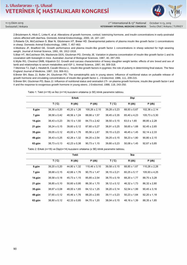
90
2.Brückmann A, Höck C, Linke K, et al. Alterations of growth hormone, cortisol, luteinizing hormone, and insulin concentrations in early-postnatal
calves affected with diarrhea. Domestic Animal Endocrinology, 2000, 18, 187–197.
3.Roberts CA, McCutcheon S. Blair N, Glückmann HT, Breier HD. Developmental patterns of plasma insulin like growth factor 1 concentrations
in sheep. Domestic Animal Endocrinology, 1990, 7, 457-463.
4.Medrano JF, Bradford GE. Growth performance and plasma insulin-like growth factor 1 concentrations in sheep selected for high weaning
weight. Journal of Animal Science, 1991, 69, 1912-1918.
5.Blair HT, McCutcheon SN, Mackenzie DDS, Gluckman PD, Ormsby JE. Variation in plasma concentration of insulin-like growth factor-1 and its
covariation with liveweight in mice. Australian Journal of Biological Sciences, 1987, 40, 287-293.
6.Wylie RG, Chestnut DMB, Kilpatrick DJ. Growth and carcass characteristics of heavy slaughter weight lambs: effects of sire breed and sex of
lamb and relationships to serum metabolites and IGF-1. Animal Science, 1997, 64, 309-318.
7.Merimee TJ, Zapf J, Hewlett B, Cavalli-Sforza LL. Insulin-like growth factors in pygmies: the role of puberty in determining final stature. The New
England Journal of Medicine, 1987, 316, 906-911.
8.Breier BH, Bass JJ, Butler JH, Gluckman PD. The somatotrophic axis in young steers: influence of nutritional status on pulsatile release of
growth hormone and circulating concentrations of insulin-like growth factor-1. J Endocrinol, 1986, 111, 209-215.
9.Breier BH, Gluckman PD, Bass JJ. Influence of nutritional status and oestradiol-17l~ on plasma growth hormone, insulin.like growth factor-I and
II and the response to exogenous growth hormone in young steers. J Endocrinol, 1988, 118, 243-250.
Tablo 1: Tekil (n=18) ve İkiz (n=14) kuzuların ortalama (± SE) klinik parametre tablosu.
Erkek Dişi
T (˚C) R (dk) P (dk) T (˚C) R (dk) P (dk)
0.gün 38,30 ± 0,20 45,20 ± 1,26 100,26 ± 2,10 39,28 ± 0,23 68,93 ± 0,67 102,36 ± 2,14
7.gün 38,58 ± 0,42 48,56 ± 1,24 88,66 ± 1,57 38,45 ± 0,35 65,40 ± 4,23 105,73 ± 5,30
14.gün 38,43 ± 0,23 39,13 ± 1,50 84,73 ± 2,42 38,50 ± 0,15 63,0 ± 1,65 88,85 ± 2,28
21.gün 38,24 ± 0,15 39,60 ± 0,12 87,60 ± 0,27 38,91 ± 0,25 58,65 ± 1,68 92,45 ± 2,65
35.gün 39,05 ± 0,12 45,00 ± 1,76 85,56 ± 1,87 39,15 ± 0,23 46,45 ± 1,45 92,14 ± 2,33
49.gün 38,43 ± 0,25 42,26 ± 1,32 84,20 ± 2,54 39,25 ± 0,15 58,23 ± 1,80 90,80 ± 2,15
63.gün 38,73 ± 0,13 42,23 ± 0,38 90,73 ± 1,15 39,80 ± 0,23 50,08 ± 1,45 92,67 ± 0,85
Tablo 2: Erkek (n=18) ve Dişi(n=14) kuzuların ortalama (± SE) klinik parametre tablosu.
Tekli İkiz
T (˚C) R (dk) P (dk) T (˚C) R (dk) P (dk)
0.gün 39,20 ± 0,20 44,80 ± 1,32 110,46 ± 3,10 39,58 ± 0,15 68,85 ± 1,67 110,26 ± 2,28
7.gün 38,68 ± 0,15 42,66 ± 1,76 90,75 ± 1,47 38,15 ± 0,21 60,25 ± 5,17 105,93 ± 4,25
14.gün 38,58 ± 0,18 45,73 ± 1,15 85,85 ± 2,54 38,70 ± 0,19 68,20 ± 1,77 88,70 ± 3,28
21.gün 38,85 ± 0,10 50,60 ± 0,95 86,30 ± 1,70 38,12 ± 0,12 46,32 ± 1,73 86,32 ± 2,80
35.gün 38,67 ± 0,08 48,00 ± 1,65 84,12 ± 1,25 38,20 ± 0,14 52,54 ± 1,99 90,45 ± 3,16
49.gün 37,65 ± 0,12 45,46 ± 1,76 88,20 ± 2,65 39,11 ± 0,23 50,23 ± 1,64 92,20 ± 1,18
63.gün 38,80 ± 0,12 42,33 ± 0,85 84,70 ± 1,20 38,04 ± 0,15 48,16 ± 1,39 88,35 ± 1,85
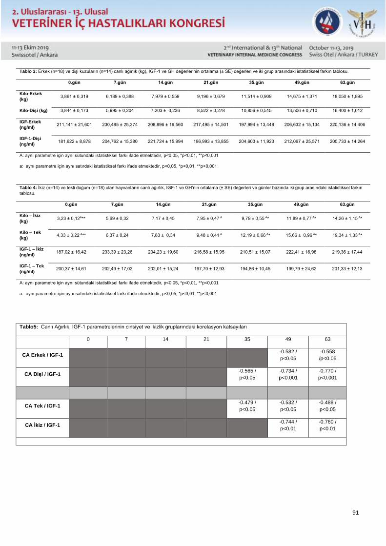
91
Tablo 3: Erkek (n=18) ve dişi kuzuların (n=14) canlı ağırlık (kg), IGF-1 ve GH değerlerinin ortalama (± SE) değerleri ve iki grup arasındaki istatistiksel farkın tablosu.
0.gün 7.gün 14.gün 21.gün 35.gün 49.gün 63.gün
Kilo-Erkek
(kg) 3,861 ± 0,319 6,189 ± 0,388 7,979 ± 0,559 9,196 ± 0,679 11,514 ± 0,909 14,675 ± 1,371 18,050 ± 1,895
Kilo-Dişi (kg) 3,844 ± 0,173 5,995 ± 0,204 7,203 ± 0,236 8,522 ± 0,278 10,856 ± 0,515 13,506 ± 0,710 16,400 ± 1,012
IGF-Erkek
(ng/ml) 211,141 ± 21,601 230,485 ± 25,374 208,896 ± 19,560 217,495 ± 14,501 197,994 ± 13,448 206,632 ± 15,134 220,136 ± 14,406
IGF-1-Dişi
(ng/ml) 181,622 ± 8,878 204,762 ± 15,380 221,724 ± 15,994 196,993 ± 13,855 204,603 ± 11,923 212,067 ± 25,571 200,733 ± 14,264
A: aynı parametre için aynı sütundaki istatistiksel farkı ifade etmektedir, p<0,05, *p<0,01, **p<0,001
a: aynı parametre için aynı satırdaki istatistiksel farkı ifade etmektedir, p<0,05, *p<0,01, **p<0,001
Tablo 4: İkiz (n=14) ve tekli doğum (n=18) olan hayvanların canlı ağırlık, IGF-1 ve GH’nin ortalama (± SE) değerleri ve günler bazında iki grup arasındaki istatistiksel farkın
tablosu.
0.gün 7.gün 14.gün 21.gün 35.gün 49.gün 63.gün
Kilo – İkiz
(kg) 3,23 ± 0,12A** 5,69 ± 0,32 7,17 ± 0,45 7,95 ± 0,47 A 9,79 ± 0,55 A* 11,89 ± 0,77 A* 14,26 ± 1,15 A*
Kilo – Tek
(kg) 4,33 ± 0,22 A** 6,37 ± 0,24 7,83 ± 0,34 9,48 ± 0,41 A 12,19 ± 0,66 A* 15,66 ± 0,96 A* 19,34 ± 1,33 A*
IGF-1 – İkiz
(ng/ml) 187,02 ± 16,42 233,39 ± 23,26 234,23 ± 19,60 216,58 ± 15,95 210,51 ± 15,07 222,41 ± 16,98 219,36 ± 17,44
IGF-1 – Tek
(ng/ml) 200,37 ± 14,61 202,49 ± 17,02 202,01 ± 15,24 197,70 ± 12,93 194,86 ± 10,45 199,79 ± 24,62 201,33 ± 12,13
A: aynı parametre için aynı sütundaki istatistiksel farkı ifade etmektedir, p<0,05, *p<0,01, **p<0,001
a: aynı parametre için aynı satırdaki istatistiksel farkı ifade etmektedir, p<0,05, *p<0,01, **p<0,001
Tablo5: Canlı Ağırlık, IGF-1 parametrelerinin cinsiyet ve ikizlik gruplarındaki korelasyon katsayıları
0 7 14 21 35 49 63
CA Erkek / IGF-1 -0.582 /
p<0.05
-0.558
/p<0.05
CA Dişi / IGF-1 -0.565 /
p<0.05
-0.734 /
p<0.001
-0.770 /
p<0.001
CA Tek / IGF-1 -0.479 /
p<0.05
-0.532 /
p<0.05
-0.488 /
p<0.05
CA İkiz / IGF-1 -0.744 /
p<0.01
-0.760 /
p<0.01
92
Sözlü Sunu / Oral Presentation Tam Metin / Full Text
Investigations on İnsulin-Like Growth Factor -1 (IGF-1) İn Neonatal Lambs
Ethem Mutlu Temizela, Fatih Kavukcub, Adil Ömer Karakuşc
Bursa Uludağ University, Faculty of Veterinary Medicine, Department of Internal Medicine, Bursa – TURKEY
aORCID: 0000-0002-4828-4116, bORCID: 0000-0002-3545-0883, cORCID: 0000-0002-6834-0808
Abstract
In this study, the relationships between sex and live weight of neonatal lambs along with serum IGF-1 was evaluated.
In this study, a total of 32 lambs were examined in two separate categories based on sex and multiple births (14
female, 18 male and 18 single, 14 twin). Blood samples were taken on the 1st, 7th, 14th, 21st, 35th, 49th and 63th
days, also clinical examinations and body weight measurements were performed on the stated days. Sex and litter
size of the newborn lambs were recorded. IGF-1 was measured using ELISA procedure. In this study, although there
was no statistically significant difference between live weights on the basis of sex, it was found that live weight of
male lambs was higher than female lambs. However, IGF-I levels were found to be higher in male animals than
females except for 14, 35 and 49 days. Correlation test results showed that while no statistically significant correlation
was found in the first 21 days of neonatal period, there was a statistically significant correlation between body weight
and IGF-I levels at 35, 49 and 63 days (p <0.05, 0.01, 0.001). Correlation test results showed that the correlation
between IGF-I and live weight was higher in female and twin lambs compared to male and single lambs after neonatal
period. In conclusion, it can be concluded that IGF-I may have a predictive value on live weight gain in lambs after
the neonatal period. However, the effect of sex and litter size on serum IGF-I levels in the neonatal period is limited.
Key words: lamb, igf-1, body weight
Introduction
IGF-1 is a hormone that is mainly secreted by the liver and works along with the presence of GH (Trenkle, 1981).
IGF-1 is responsible for the body weight gain, internal organ growth and other parameters in development. Both sex
and breed being the particular parameters in case of concentration difference for IGF-1, there are other factors such
as nutrition, litter size and weaning time that alters the serum IGF-1 values (Roberts et al., 1990; Medrano and
Bredford, 1991; Wylie et al., 1997).
Although there are many studies regarding the effects of IGF-1 on live weight gain (Blair et al., 1987), carcass quality
and other yield parameters (Wylie et al., 1997) in lambs, there is a limited number of studies that take into account
the relationship between live weight, sex, litter size and IGF-1.
The aim of this study was to investigate the relationship between IGF-1 and sex, litter size and live weight by repeated
measurements of IGF-I on different days.
Materials and Method
The study material consisted of 14 female and 18 male lambs in a total of 32 lambs. Live weight measurement,
clinical examination, sex, singleton and twins were recorded on 1st, 7th, 14th, 21st, 35th, 49th and 63th days. Blood
and fecal samples were also collected at same times (Table 1-2). IGF-1 was measured by ELISA method. Based on
the data obtained; the statistical tests between groups in terms of twins / singularity, sex and live weight gain and
IGF-1 were performed using Sigma Stat package program. Variance analysis was performed to reveal statistical
difference between repeated measures in groups with normal distribution, and t-test was performed to reveal
93
differences between groups. In groups, correlation analysis between IGF-I and live weight was performed with
Pearson correlation test.
This study was approved by the Animals Ethics Committee of Bursa Uludağ University in 2016 (2016-08).
Results
In this study, although there were no statistically significant differences between live weights and sex, it was found
that live weight of male lambs was higher than female lambs. However, although not statistically significant, IGF-I
levels were higher in male animals than females at other measurement times, except 14, 35 and 49 days. (Table 3)
Mean (± SE) and statistical significance values between groups categorized according to litter size (twin or single
birth) are presented in Table 2. In case of body weights on day 1 (p <0.001), 21 (p <0.05), 35 (p <0.01), 49 (p <0.01)
and 63 (p <0.01) measurements were statistically significant. (Table 4)
There was a statistically significant difference between IGF-I and body weight, especially on days 49 and 63. The
significance of this difference in twins and males was found to be higher than that of single-born lambs and twin-born
lambs. (Table 5)
Discussion
IGF-I levels express variations under numerous conditions including sex, litter size, body size, nutrition, etc. (Merimee
et al., 1987; Blair et al., 1987; Breier et al. 1986). In a study reported by Roberts et al. (1989), it was shown that IGF-
I levels was higher in male sheep than female sheep in postnatal period. On the other hand, in a study by Luna et al
(1999), it was found that there was no difference in IGF-I levels between castrated pigs and female piglets. In the
present study, although there was no statistical significance in live weight gain, it was found that live weight of male
lambs was higher than female lambs. However, IGF-I levels were found to be higher in male animals than male
females except 14th, 35th and 49th days. These finding suggests that it may be related to nutrition due to the
difference in live weight between male animals and females.
Litter size has effect on live weight and IGF-I, it also affects the amount of milk that is sucked by lambs (Blair et al.,
1987). In the present study, although body weights of single born lambs were higher than twin lambs during the blood
collection periods, IGF-I levels were found to be higher in twin lambs, although not statistically significant. This
suggests that many factors may have an effect on IGF-I levels. In addition, the determination of IGF-I levels in the
dam’s milk and the interaction of the IGF-I level in the milk to lamb’s serum IGF-1 will allow a more healthy
interpretation in this regard.In conclusion, it was found that gender difference and litter size is not fully associated
with IGF-I in neonatal. Furthermore, IGF-I may have a predictive value on live weight gain in lambs after neonatal
period.
Acknowledgement
This study was supported by scientific research projects unit of Uludag University. We would like to thank scientific
research projects unit of Uludag University for their financial support.
References 1.Trenkle A. Endocrine regulation of energy metabolism in ruminants. Fed Proc, 1981, 40, 2536–2541.
2.Brückmann A, Höck C, Linke K, et al. Alterations of growth hormone, cortisol, luteinizing hormone, and insulin concentrations in early-postnatal
calves affected with diarrhea. Domestic Animal Endocrinology, 2000, 18, 187–197.
3.Roberts CA, McCutcheon S. Blair N, Glückmann HT, Breier HD. Developmental patterns of plasma insulin like growth factor 1 concentrations
in sheep. Domestic Animal Endocrinology, 1990, 7, 457-463.
4.Medrano JF, Bradford GE. Growth performance and plasma insulin-like growth factor 1 concentrations in sheep selected for high weaning
weight. Journal of Animal Science, 1991, 69, 1912-1918.
5.Blair HT, McCutcheon SN, Mackenzie DDS, Gluckman PD, Ormsby JE. Variation in plasma concentration of insulin-like growth factor-1 and its
covariation with liveweight in mice. Australian Journal of Biological Sciences, 1987, 40, 287-293.
6.Wylie RG, Chestnut DMB, Kilpatrick DJ. Growth and carcass characteristics of heavy slaughter weight lambs: effects of sire breed and sex of
lamb and relationships to serum metabolites and IGF-1. Animal Science, 1997, 64, 309-318.
7.Merimee TJ, Zapf J, Hewlett B, Cavalli-Sforza LL. Insulin-like growth factors in pygmies: the role of puberty in determining final stature. The New
England Journal of Medicine, 1987, 316, 906-911.
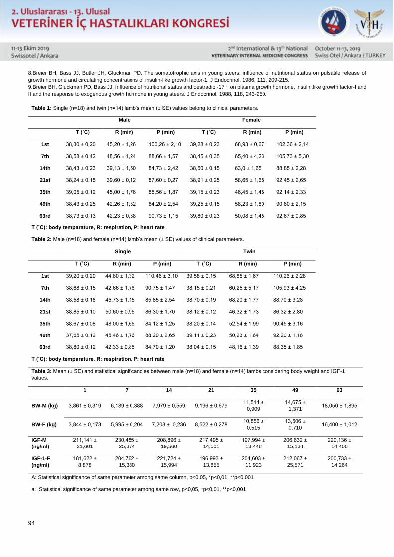
94
8.Breier BH, Bass JJ, Butler JH, Gluckman PD. The somatotrophic axis in young steers: influence of nutritional status on pulsatile release of
growth hormone and circulating concentrations of insulin-like growth factor-1. J Endocrinol, 1986, 111, 209-215.
9.Breier BH, Gluckman PD, Bass JJ. Influence of nutritional status and oestradiol-17l~ on plasma growth hormone, insulin.like growth factor-I and
II and the response to exogenous growth hormone in young steers. J Endocrinol, 1988, 118, 243-250.
Table 1: Single (n=18) and twin (n=14) lamb’s mean (± SE) values belong to clinical parameters.
Male Female
T (˚C) R (min) P (min) T (˚C) R (min) P (min)
1st 38,30 ± 0,20 45,20 ± 1,26 100,26 ± 2,10 39,28 ± 0,23 68,93 ± 0,67 102,36 ± 2,14
7th 38,58 ± 0,42 48,56 ± 1,24 88,66 ± 1,57 38,45 ± 0,35 65,40 ± 4,23 105,73 ± 5,30
14th 38,43 ± 0,23 39,13 ± 1,50 84,73 ± 2,42 38,50 ± 0,15 63,0 ± 1,65 88,85 ± 2,28
21st 38,24 ± 0,15 39,60 ± 0,12 87,60 ± 0,27 38,91 ± 0,25 58,65 ± 1,68 92,45 ± 2,65
35th 39,05 ± 0,12 45,00 ± 1,76 85,56 ± 1,87 39,15 ± 0,23 46,45 ± 1,45 92,14 ± 2,33
49th 38,43 ± 0,25 42,26 ± 1,32 84,20 ± 2,54 39,25 ± 0,15 58,23 ± 1,80 90,80 ± 2,15
63rd 38,73 ± 0,13 42,23 ± 0,38 90,73 ± 1,15 39,80 ± 0,23 50,08 ± 1,45 92,67 ± 0,85
T (˚C): body temparature, R: respiration, P: heart rate
Table 2: Male (n=18) and female (n=14) lamb’s mean (± SE) values of clinical parameters.
Single Twin
T (˚C) R (min) P (min) T (˚C) R (min) P (min)
1st 39,20 ± 0,20 44,80 ± 1,32 110,46 ± 3,10 39,58 ± 0,15 68,85 ± 1,67 110,26 ± 2,28
7th 38,68 ± 0,15 42,66 ± 1,76 90,75 ± 1,47 38,15 ± 0,21 60,25 ± 5,17 105,93 ± 4,25
14th 38,58 ± 0,18 45,73 ± 1,15 85,85 ± 2,54 38,70 ± 0,19 68,20 ± 1,77 88,70 ± 3,28
21st 38,85 ± 0,10 50,60 ± 0,95 86,30 ± 1,70 38,12 ± 0,12 46,32 ± 1,73 86,32 ± 2,80
35th 38,67 ± 0,08 48,00 ± 1,65 84,12 ± 1,25 38,20 ± 0,14 52,54 ± 1,99 90,45 ± 3,16
49th 37,65 ± 0,12 45,46 ± 1,76 88,20 ± 2,65 39,11 ± 0,23 50,23 ± 1,64 92,20 ± 1,18
63rd 38,80 ± 0,12 42,33 ± 0,85 84,70 ± 1,20 38,04 ± 0,15 48,16 ± 1,39 88,35 ± 1,85
T (˚C): body temparature, R: respiration, P: heart rate
Table 3: Mean (± SE) and statistical significancies between male (n=18) and female (n=14) lambs considering body weight and IGF-1
values.
1 7 14 21 35 49 63
BW-M (kg) 3,861 ± 0,319 6,189 ± 0,388 7,979 ± 0,559 9,196 ± 0,679 11,514 ±
0,909
14,675 ±
1,371 18,050 ± 1,895
BW-F (kg) 3,844 ± 0,173 5,995 ± 0,204 7,203 ± 0,236 8,522 ± 0,278 10,856 ±
0,515
13,506 ±
0,710 16,400 ± 1,012
IGF-M
(ng/ml)
211,141 ±
21,601
230,485 ±
25,374
208,896 ±
19,560
217,495 ±
14,501
197,994 ±
13,448
206,632 ±
15,134
220,136 ±
14,406
IGF-1-F
(ng/ml)
181,622 ±
8,878
204,762 ±
15,380
221,724 ±
15,994
196,993 ±
13,855
204,603 ±
11,923
212,067 ±
25,571
200,733 ±
14,264
A: Statistical significance of same parameter among same column, p<0,05, *p<0,01, **p<0,001
a: Statistical significance of same parameter among same row, p<0,05, *p<0,01, **p<0,001
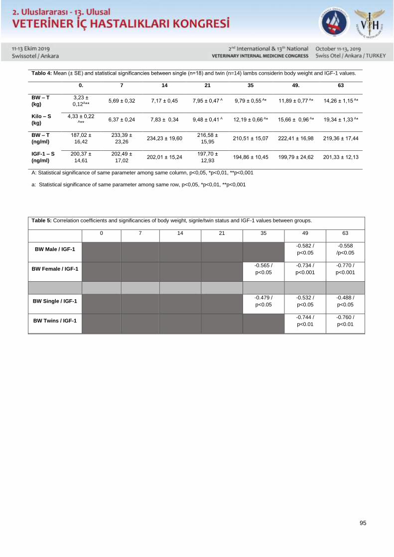
95
Tablo 4: Mean (± SE) and statistical significancies between single (n=18) and twin (n=14) lambs considerin body weight and IGF-1 values.
0. 7 14 21 35 49. 63
BW – T
(kg)
3,23 ±
0,12A** 5,69 ± 0,32 7,17 ± 0,45 7,95 ± 0,47 A 9,79 ± 0,55 A* 11,89 ± 0,77 A* 14,26 ± 1,15 A*
Kilo – S
(kg)
4,33 ± 0,22
A** 6,37 ± 0,24 7,83 ± 0,34 9,48 ± 0,41 A 12,19 ± 0,66 A* 15,66 ± 0,96 A* 19,34 ± 1,33 A*
BW – T
(ng/ml)
187,02 ±
16,42
233,39 ±
23,26 234,23 ± 19,60
216,58 ±
15,95 210,51 ± 15,07 222,41 ± 16,98 219,36 ± 17,44
IGF-1 – S
(ng/ml)
200,37 ±
14,61
202,49 ±
17,02 202,01 ± 15,24
197,70 ±
12,93 194,86 ± 10,45 199,79 ± 24,62 201,33 ± 12,13
A: Statistical significance of same parameter among same column, p<0,05, *p<0,01, **p<0,001
a: Statistical significance of same parameter among same row, p<0,05, *p<0,01, **p<0,001
Table 5: Correlation coefficients and significancies of body weight, signle/twin status and IGF-1 values between groups.
0 7 14 21 35 49 63
BW Male / IGF-1 -0.582 /
p<0.05
-0.558
/p<0.05
BW Female / IGF-1 -0.565 /
p<0.05
-0.734 /
p<0.001
-0.770 /
p<0.001
BW Single / IGF-1 -0.479 /
p<0.05
-0.532 /
p<0.05
-0.488 /
p<0.05
BW Twins / IGF-1 -0.744 /
p<0.01
-0.760 /
p<0.01
96
Sözlü Sunu / Oral Presentation Tam Metin / Full Text
Bir Köpekte Granülositik Anaplasmosis Olgusu
* Cenk Er, ** Handan Çetinkaya, *** Mehmet Erkan Avan, **** Armağan Erdem Ütük
* Çukurova Üniversitesi Ceyhan Veteriner Fakültesi Veterinerlik İç Hastalıkları Anabilim Dalı, Adana.
** İstanbul Üniversitesi Cerrahpaşa Veteriner Fakültesi Veterinerlik Parazitoloji Anabilim Dalı, İstanbul.
*** Deva Veteriner Tıp Merkezi, Adana.
**** Çukurova Üniversitesi Ceyhan Veteriner Fakültesi Veterinerlik Parazitoloji Anabilim Dalı, Adana.
Bu vaka sunumunun amacı; kronik burun kanaması, halsizlik ve yüksek ateş şikâyeti bulunan 10 yaşlı dişi bir köpekte
hastalığın etiyolojisini ortaya koymaktı. Bu amaçla hayvandan alınan kan örneği froti, immunokromatografi ve Nested-
PCR teknikleri ile incelenerek elde edilen veriler anamnez, klinik muayene ve hemogram bulguları ile birlikte
değerlendirildi. Hemogramda anemi, trombositopeni, löykositoz, nötrofili ve lenfositoz; serolojik olarak E.canis ve
A.platys pozitifliği tespit edilirken, Nested PCR sonucunda sadece A.phagocytophilum belirlendi. Bu çalışma ile,
köpeklerde klinik granülositik anaplasmosis konusunda az sayıda olan literatür bilgiye katkıda bulunulmuş; hızlı tanı
kitlerinin çapraz reaksiyon verme ihtimalinin olduğu, mevcut vakaların klinik semptomlar ve hemograma ilave olarak
serolojik ve moleküler tekniklerle birlikte değerlendirilmesinin gerekliliği vurgulanmıştır.
Anahtar kelimeler: Anaplasma phagocytophilum, Köpek, PCR, Serology, Turkey.
Giriş
Köpeklerin infeksiyöz hastalık etkenlerinden olan Ehrlichia canis, Anaplasma platys ve Anaplasma phagocytophilum,
Rickettsiales takımının, Anaplasmatacea eailesi içerisinde yer alan, zorunlu hücre içi mikroorganizmalardır (Dumler
ve ark 2001). Etkenler köpeklerde akut, sub-akut veya kronik seyirli, multisistemik ve kimi zaman da ölümcül
semptomlarla seyreden, zoonoz karakterli, kene kaynaklı hastalıklara sebep olur. Hastalığı atlatan köpeklerde sub-
klinik enfeksiyon uzun bir süre daha devam edebilir (Mylonakis ve Theodorou 2017). E. canis ve A. platys,
Rhipicephalus sanguineus (Liu ve ark 2016), A. phagocytophilum ise Ixode sricinus türü keneler aracılığıyla taşınır
(Dahmani ve ark 2015). Köpeklerde E. canis, Monositik Ehrlichiosis (CME); A. platys, Siklik Trombositopeni (CCT)
ve A.phagocytophilum ise Granülositik Anaplasmozis (CGA) adlı hastalıklara sebep olur (Kohn ve ark 2008,
Kaewmongkol ve ark 2017, Soares ve ark 2017). Enfekte hayvanlarda kilo kaybı, anoreksi, müköz membranlarda
solgunluk, yüksek ateş, letarji ve lenfadenopati gibi klinik bulguların yanı sıra hematolojik anormallikler; nadiren de
solunum ve sinir sistemi ile ilgili semptomlar görülebilir (Dantas-Torres 2008). Ehrlichia canis, A. platys ve A.
phagocytophilum aynı coğrafyada bulunabildiğinden mix-enfeksiyonlar yaygın olarak görülebilmekte ve bu durum
hastalığın klinik şiddetini artırmaktadır (Aktaş ve ark 2015). Bunun muhtemel nedeninin, E. canis’ in monositleri, A.
platys’ in trombositleri ve A. phagocytophilum’ un ise nötrofilleri ve daha az olarak da eozinofilleri aynı anda enfekte
etmesi olduğu düşünülmektedir (Pinyoowong ve ark 2008). Etkenlerin sebep olduğu hastalıkların klinik teşhisinde en
önemli hematolojik bulguların anemi, löykopeni, trombositopeni ve trombosit fonksiyon bozuklukları olduğu ve bu
parametrelerden tedaviye verilen cevabın değerlendirilmesinde yararlanılabileceği bildirilmiştir (Çetinkaya ve ark
2016). Diğer bazı araştırmacılar ise löykopeni bulgusunun tek başına CGA için tanımlayıcı olmadığını ve mix-
enfeksiyon olmayan vakalarda löykositoz görülme oranının daha yüksek olabileceğini bildirmişlerdir (Chirek ve ark
2018). Anaplasma phagocytophilum enfeksiyonlarının akut döneminde kandan yapılan frotilerde nötrofiller in
sitoplazmaları içerisinde ‘morula’ adı verilen inklüzyon cisimcikleri saptanabilir (Kohn ve ark 2011). Ancak
granülositler içerisindeki morulalar yalnızca hastalığın akut döneminde görülür ve bu nedenle kan frotisi ile her zaman
teşhis koymak mümkün değildir (Ulutaş ve ark 2007). Bu gibi durumlarda hastalığın tanısında spesifite ve sensitivitesi
daha yüksek olan serolojik ve moleküler tanı yöntemleri kullanılır (Kohn ve ark 2011).
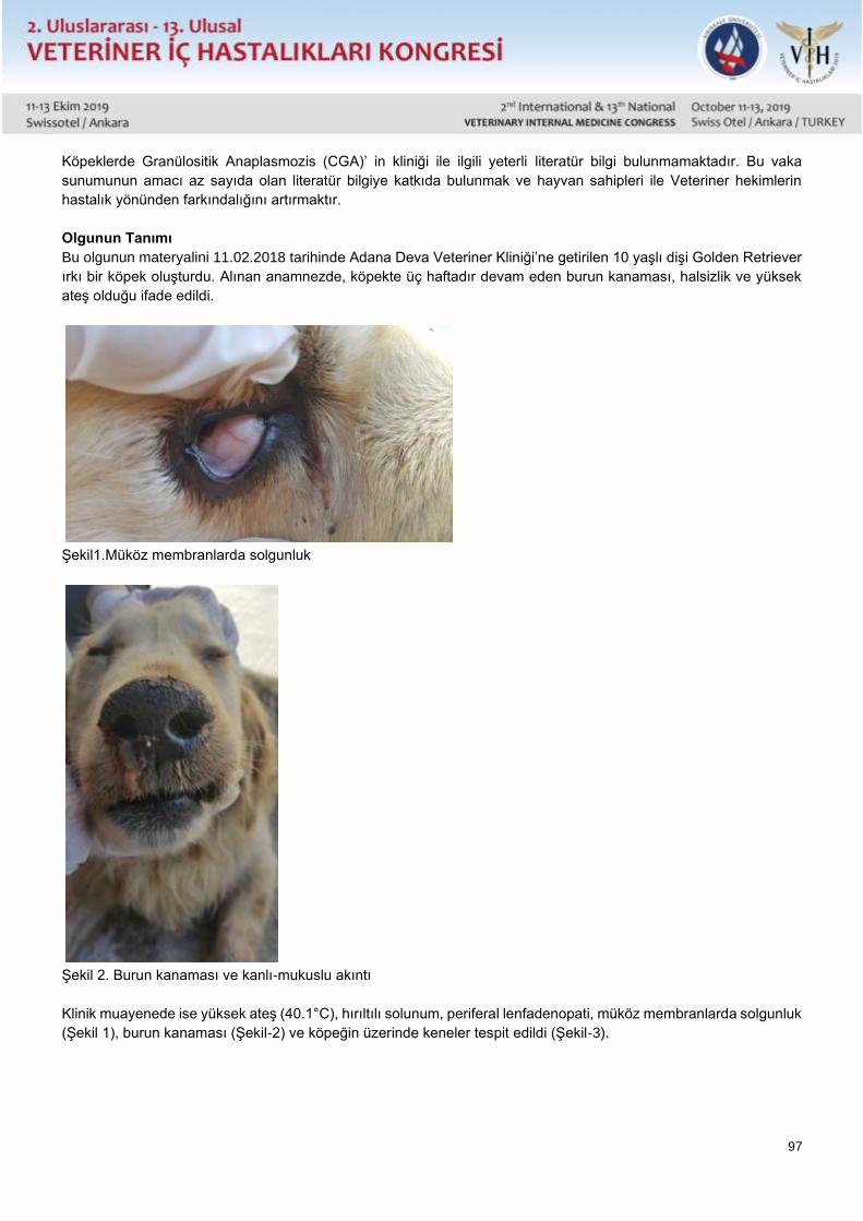
97
Köpeklerde Granülositik Anaplasmozis (CGA)’ in kliniği ile ilgili yeterli literatür bilgi bulunmamaktadır. Bu vaka
sunumunun amacı az sayıda olan literatür bilgiye katkıda bulunmak ve hayvan sahipleri ile Veteriner hekimlerin
hastalık yönünden farkındalığını artırmaktır.
Olgunun Tanımı
Bu olgunun materyalini 11.02.2018 tarihinde Adana Deva Veteriner Kliniği’ne getirilen 10 yaşlı dişi Golden Retriever
ırkı bir köpek oluşturdu. Alınan anamnezde, köpekte üç haftadır devam eden burun kanaması, halsizlik ve yüksek
ateş olduğu ifade edildi.
Şekil1.Müköz membranlarda solgunluk
Şekil 2. Burun kanaması ve kanlı-mukuslu akıntı
Klinik muayenede ise yüksek ateş (40.1°C), hırıltılı solunum, periferal lenfadenopati, müköz membranlarda solgunluk
(Şekil 1), burun kanaması (Şekil-2) ve köpeğin üzerinde keneler tespit edildi (Şekil-3).
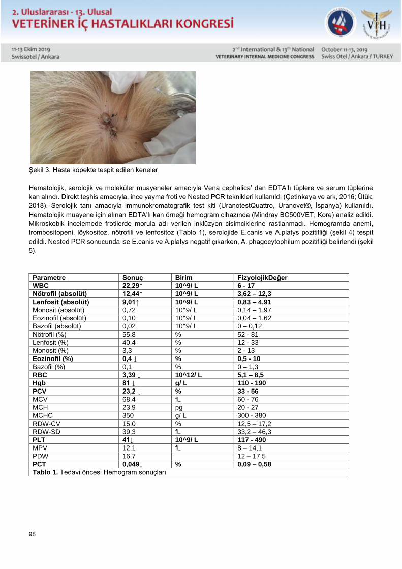
98
Şekil 3. Hasta köpekte tespit edilen keneler
Hematolojik, serolojik ve moleküler muayeneler amacıyla Vena cephalica’ dan EDTA’lı tüplere ve serum tüplerine
kan alındı. Direkt teşhis amacıyla, ince yayma froti ve Nested PCR teknikleri kullanıldı (Çetinkaya ve ark, 2016; Ütük,
2018). Serolojik tanı amacıyla immunokromatografik test kiti (UranotestQuattro, Uranovet®, İspanya) kullanıldı.
Hematolojik muayene için alınan EDTA’lı kan örneği hemogram cihazında (Mindray BC500VET, Kore) analiz edildi.
Mikroskobik incelemede frotilerde morula adı verilen inklüzyon cisimciklerine rastlanmadı. Hemogramda anemi,
trombositopeni, löykositoz, nötrofili ve lenfositoz (Tablo 1), serolojide E.canis ve A.platys pozitifliği (şekil 4) tespit
edildi. Nested PCR sonucunda ise E.canis ve A.platys negatif çıkarken, A. phagocytophilum pozitifliği belirlendi (şekil
5).
Parametre Sonuç Birim FizyolojikDeğer
WBC 22,29↑ 10^9/ L 6 - 17
Nötrofil (absolüt) 12,44↑ 10^9/ L 3,62 – 12,3
Lenfosit (absolüt) 9,01↑ 10^9/ L 0,83 – 4,91
Monosit (absolüt) 0,72 10^9/ L 0,14 – 1,97
Eozinofil (absolüt) 0,10 10^9/ L 0,04 – 1,62
Bazofil (absolüt) 0,02 10^9/ L 0 – 0,12
Nötrofil (%) 55,8 % 52 - 81
Lenfosit (%) 40,4 % 12 - 33
Monosit (%) 3,3 % 2 - 13
Eozinofil (%) 0,4 ↓ % 0,5 - 10
Bazofil (%) 0,1 % 0 – 1,3
RBC 3,39 ↓ 10^12/ L 5,1 – 8,5
Hgb 81 ↓ g/ L 110 - 190
PCV 23,2 ↓ % 33 - 56
MCV 68,4 fL 60 - 76
MCH 23,9 pg 20 - 27
MCHC 350 g/ L 300 - 380
RDW-CV 15,0 % 12,5 – 17,2
RDW-SD 39,3 fL 33,2 – 46,3
PLT 41↓ 10^9/ L 117 - 490
MPV 12,1 fL 8 – 14,1
PDW 16,7 12 – 17,5
PCT 0,049↓ % 0,09 – 0,58
Tablo 1. Tedavi öncesi Hemogram sonuçları
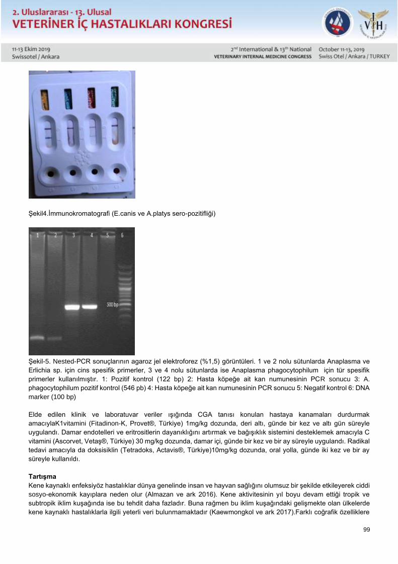
99
Şekil4.İmmunokromatografi (E.canis ve A.platys sero-pozitifliği)
Şekil-5. Nested-PCR sonuçlarının agaroz jel elektroforez (%1,5) görüntüleri. 1 ve 2 nolu sütunlarda Anaplasma ve
Erlichia sp. için cins spesifik primerler, 3 ve 4 nolu sütunlarda ise Anaplasma phagocytophilum için tür spesifik
primerler kullanılmıştır. 1: Pozitif kontrol (122 bp) 2: Hasta köpeğe ait kan numunesinin PCR sonucu 3: A.
phagocytophilum pozitif kontrol (546 pb) 4: Hasta köpeğe ait kan numunesinin PCR sonucu 5: Negatif kontrol 6: DNA
marker (100 bp)
Elde edilen klinik ve laboratuvar veriler ışığında CGA tanısı konulan hastaya kanamaları durdurmak
amacıylaK1vitamini (Fitadinon-K, Provet®, Türkiye) 1mg/kg dozunda, deri altı, günde bir kez ve altı gün süreyle
uygulandı. Damar endotelleri ve eritrositlerin dayanıklığını artırmak ve bağışıklık sistemini desteklemek amacıyla C
vitamini (Ascorvet, Vetaş®, Türkiye) 30 mg/kg dozunda, damar içi, günde bir kez ve bir ay süreyle uygulandı. Radikal
tedavi amacıyla da doksisiklin (Tetradoks, Actavis®, Türkiye)10mg/kg dozunda, oral yolla, günde iki kez ve bir ay
süreyle kullanıldı.
Tartışma
Kene kaynaklı enfeksiyöz hastalıklar dünya genelinde insan ve hayvan sağlığını olumsuz bir şekilde etkileyerek ciddi
sosyo-ekonomik kayıplara neden olur (Almazan ve ark 2016). Kene aktivitesinin yıl boyu devam ettiği tropik ve
subtropik iklim kuşağında ise bu tehdit daha fazladır. Buna rağmen bu iklim kuşağındaki gelişmekte olan ülkelerde
kene kaynaklı hastalıklarla ilgili yeterli veri bulunmamaktadır (Kaewmongkol ve ark 2017).Farklı coğrafik özelliklere
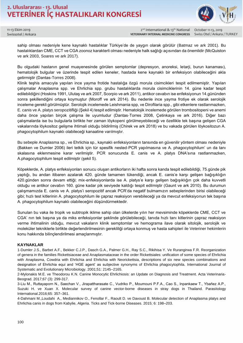
100
sahip olması nedeniyle kene kaynaklı hastalıklar Türkiye’de de yaygın olarak görülür (Batmaz ve ark 2001). Bu
hastalıklardan CME, CCT ve CGA zoonoz karakterli olması nedeniyle halk sağlığı açısından da önemlidir (McQuiston
ve ark 2003, Soares ve ark 2017).
Bu olgudaki hastanın genel muayenesinde görülen semptomlar (depresyon, anoreksi, letarji, burun kanaması),
hematolojik bulgular ve üzerinde tespit edilen keneler, hastada kene kaynaklı bir enfeksiyon olabileceğini akla
getirmiştir (Dantas-Torres 2008).
Klinik teşhis amacıyla yapılan ince yayma frotide hastalığa özgü morula cisimcikleri tespit edilmemiştir. Yapılan
çalışmalar Anaplasma spp. ve Ehrlichia spp. grubu hastalıklarda morula cisimciklerinin 14. güne kadar tespit
edilebildiğini (Hoskins 1991, Ulutaş ve ark 2007, Scorpio ve ark 2011), antikor cevabın ise enfeksiyonun 14.gününden
sonra şekillendiğini ortaya koymuştur (Moroff ve ark 2014). Bu nedenle ince yayma frotiye ek olarak serolojik
inceleme gerekli görülmüştür. Serolojik incelemede Leishmania spp, ve Dirofilaria spp., gibi etkenlere rastlanmazken,
E. canis ve A. platys seropozitifliği (Şekil 4) tespit edilmiştir. Hematolojik incelemede görülen trombositopeni ve anemi
daha önce yapılan birçok çalışma ile uyumludur (Dantas-Torres 2008, Çetinkaya ve ark 2016). Diğer bazı
çalışmalarda ise bu bulgularla birlikte her zaman löykopeni görülmeyebileceği ve özellikle tek başına gelişen CGA
vakalarında löykositoz gelişme ihtimali olduğu bildirilmiş (Chirek ve ark 2018) ve bu vakada görülen löykositozun A.
phagocytophilum kaynaklı olabileceği kanaatine varılmıştır.
Bu sebeple Anaplasma sp., ve Ehrlichia sp., kaynaklı enfeksiyonların tansında en güvenilir yöntem olması nedeniyle
(Bakken ve Dumler 2006) ileri tetkik için tür spesifik nested-PCR yapılmasına ve A. phagocytophilum’ un da tanı
skalasına eklenmesine karar verilmiştir. PCR sonucunda E. canis ve A. platys DNA’sına rastlanmazken,
A.phagocytophilum tespit edilmiştir (şekil 5).
Köpeklerde, A. platys enfeksiyonları sonucu oluşan antikorların iki hafta sonra kanda tespit edilebildiği, 75.günde pik
yaptığı, bu andan itibaren azalarak 420. günde tamamen tükendiği, ancak E. canis’e karşı gelişen bağışıklığın
420.günden sonra devam ettiği; mix-enfeksiyonlarda ise A. platys’e karşı gelişen bağışıklığın çok daha kuvvetli
olduğu ve antikor cevabın 160. güne kadar pik seviyede kaldığı tespit edilmiştir (Gaunt ve ark 2010). Bu durumun
çalışmamızda E. canis ve A. platys’i seropozitif ancak PCR’da negatif bulmamızın sebeplerinden birisi olabileceği
gibi; hızlı test kitlerinin A. phagocytophilum ile çapraz reaksiyon verebileceği ya da mevcut enfeksiyonun tek başına
A. phagocytophilum kaynaklı olabileceğini düşündürmektedir.
Sunulan bu vaka ile tropik ve subtropik iklime sahip olan ülkelerde yılın her mevsiminde köpeklerde CME, CCT ve
CGA’ nın tek başına ya da miks enfeksiyonlar şeklinde görülebileceği, tanıda hızlı tanı kitlerinin çapraz reaksiyon
verme ihtimalinin olduğu, mevcut vakaların klinik semptomlar ve hemograma ilave olarak sitolojik, serolojik ve
moleküler tekniklerle birlikte değerlendirilmesinin gerekliliği ortaya konmuş ve hasta sahipleri ile Veteriner hekimlerin
konu hakkında bilinçlendirilmesi amaçlanmıştır.
KAYNAKLAR
1-Dumler J.S., Barbet A.F., Bekker C.J.P., Dasch G.A., Palmer G.H., Ray S.C., Rikihisa Y. Ve Rurangirwa F.R. Reorganization
of genera in the families Rickettsiaceae and Anaplasmataceae in the order Rickettsiales: unification of some species of Ehrlichia
with Anaplasma, Cowdria with Ehrlichia and Ehrlichia with Neorickettsia, descriptions of six new species combinations and
designation of Ehrlichia equi and ‘HGE agent’ as subjective synonyms of Ehrlichia phagocytophila. International Journal of
Systematic and Evolutionary Microbiology. 2001;51: 2145–2165.
2-Mylonakis M.E. ve Theodorou K.N. Canine Monocytic Ehrlichiosis: an Update on Diagnosis and Treatment. Acta Veterinaria-
Beograd. 2017;67 (3): 299-317.
3-Liu M., Ruttayaporn N., Saechan V., Jirapattharasate C., Vudriko P., Moumouni P.F.A., Cao S., Inpankaew T., Ybañez A.P.,
Suzuki H. ve Xuan X. Molecular survey of canine vector-borne diseases in stray dogs in Thailand. Parasitology
International.2016;65: 357–361.
4-Dahmani M.,Loudahi A., Mediannikov O., Fenollar F., Raoult D. ve Davoust B. Molecular detection of Anaplasma platys and
Ehrlichia canis in dogs from Kabylie, Algeria. Ticks and Tick-borne Diseases. 2015; 6: 198–203.
101
5-Kohn B., Galke D., Beelitz P. ve Pfister K. Clinical Features of Canine Granulocytic Anaplasmosis in 18 Naturally Infected Dogs.
J Vet Intern Med. 2008; 22: 1289–1295.
6-Kaewmongkol G., Lukkana N., Yangtara S., Kaewmongkol S., Thengchaisri N., Sirinarumitr T., Jittapalapong S. Ve Fenwick
S.G. Association of Ehrlichiacanis, Hemotropic Mycoplasma spp. and Anaplasma platys and severe anemia in dogs in Thailand.
Veterinary Microbiology 2017; 201: 195–200.
7-Soares B., Ramos C.A., Pedroso T., Babo-Terra V., Cleveland H. ve De Araujo F. Molecular survey of Anaplasma platys and
Ehrlichia canis in dogs from Campo Grande, Mato Grosso do Sul, Brazil. Anais da Academia Brasileira de Ciências. 2017; 89(1):
301-306.
8-Dantas Torres F. Canine vector-borne diseases in Brazil. Parasites&Vectors.2008; 1: 25.
9-Aktaş M., Özübek S., Altay K., İpek N.D.S., Balkaya İ., Ütük A.E., Kırbaş A., Şimşek S. ve Dumanlı N. Molecular detection of
tick-borne rickettsial and protozoan pathogens in domestic dogs from Turkey. Parasites&Vectors. 2015; 8: 157.
10-Pinyoowong D., Jittapalapong S., Suksawat F., Stich R.W. ve Thamchaipenet A. Molecular characterization of Thai Ehrlichia
canis and Anaplasma platys strains detected in dogs. Infection, Genetics and Evolution. 2008; 8: 433–438.
11-Çetinkaya H., Matur E., Akyazı İ., Ekiz E.E., aydın L. ve Toparlak M. Serological and molecular investigation of Ehrlichia spp.
and Anaplasma spp. in ticks and blood of dogs, in the Thrace Region of Turkey. Ticks and Tick-borne Diseases 2016; 7: 706–
714.
12-Chirek A., Silaghi C., Pfister K. ve Kohn B. Granulocytic anaplasmosis in 63 dogs: clinical signs, laboratory results, therapy
and course of disease. Journal of Small Animal Practice.2018; 59: 112–120.
13-Ulutaş B., Bayramlı G.veKaragenç T. First Case of Anaplasma (Ehrlichia) platysInfection in a Dog in Turkey. Turk. J. Vet.
Anim. Sci. 2007; 31(4): 279-282
14-Kohn B., Silaghi C., Galke D., Arndt G. ve Pfister K. Infections with Anaplasma phagocytophilum in dogs in Germany. Research
in Veterinary Science 2011; 91: 71–76.
15-Ütük A.E. Protozoal Hastalıkların Laboratuvar Tanısı. Turkiye Klinikleri J Vet Sci Intern Med-Special Topics. 2018; 4(1): 17-22.
16-McQuiston J.H., McCall C.L. ve Nicholson W.L. Ehrlichiosis and related infections. JAVMA. 2003; 223 (12): 1750-1756.
17-Moroff S., Sokolchik I., Woodring T., Woodruff C., Atkinson B. ve Lappin M.R. Detection of antibodies against Anaplasma
phagocytophilum in dogs using an automated fluorescence based system. The Veterinary Journal. 2014; 202: 348–352.
18-Bakken J.S. ve Dumler J.S. Clinical Diagnosis and Treatment of Human Granulocytotropic Anaplasmosis. Ann. N.Y. Acad.
Sci. 2006; 1078: 236–247.
19-Gaunt S.D., Beall M.J., Stillman B.A., Lorentzen L., Diniz P.P.V.P., Chandrashekar R. ve Breitschwerdt E.B. Experimental
infection and co-infection of dogs with Anaplasma platys and Ehrlichia canis: hematologic, serologic and molecular findings.
Parasites & Vectors 2010, 3:33.
20-Almazan C., González-Álvarez V.H., Fernández de Mera I.G., Cabezas-Cruz A., Rodríguez-Martínez R. ve de la Fuente C.
Molecular identification and characterization of Anaplasma platys and Ehrlichiacanis in dogs in Mexico. Ticks and Tick-borne
Diseases. 2016; 7: 276–283.
21-Batmaz, H., Şentürk, S., Yilmaz, Z., Harrus, S., Nevo, E., Waner, T. Seroprevalence of Ehrlichia canis antibodies among dogs
in Turkey. Veterinary Record. 2001; 148: 665-666.
22-Hoskins, J.D. Ehrlichial diseases of dogs: diagnosis and treatment. Canine Pract. 1991; 16: 13-21.
102
Sözlü Sunu / Oral Presentation Özet / Abstract
Granulocytic Anaplasmosis Case In A Dog
* Cenk Er, ** Handan Çetinkaya, *** Mehmet Erkan Avan, **** Armağan Erdem Ütük
* Çukurova Üniversitesi Ceyhan Veteriner Fakültesi Veterinerlik İç Hastalıkları Anabilim Dalı, Adana.
** İstanbul Üniversitesi Cerrahpaşa Veteriner Fakültesi Veterinerlik Parazitoloji Anabilim Dalı, İstanbul.
*** Deva Veteriner Tıp Merkezi, Adana.
**** Çukurova Üniversitesi Ceyhan Veteriner Fakültesi Veterinerlik Parazitoloji Anabilim Dalı, Adana.
Abstract
The aim of this case report was to reveal the etiology of the disease in a 10-year-old female dog with chronic nose
bleed, weakness and high fever. For this purpose, the blood sample was collected from the animal and examined by
means of smear, immunochromatography and Nested-PCR techniques. Anemia, thrombocytopenia, leukococytosis,
neutrophilia and lymphocytosis were determined in hemogram. Ehrlichia canis and Anaplasma platys antibodies
were detected with serologically, whilst only A. phagocytophilum was detected in Nested-PCR. This study contributed
to a small number of literatures on the clinical granulocytic anaplasmosis in dogs; and it is also emphasized that rapid
diagnostic kits are likely to cross-react, and that existing cases should be evaluated together with clinical symptoms
and hemogram as well as with serological and molecular techniques.
KeyWords: Anaplasma phagocytophilum, Dog, PCR, Serology, Turkey
103
Sözlü Sunu / Oral Presentation Özet / Abstract
Tüm Verileri ile Türkiye’de Bir Yaban Hayatı Kurtarma Merkezi Örneği;
Kafkasrehab “2012-2018”
Erdoğan UZLU, Ekin Emre ERKILIÇ, Burhan ÖZBA, Mete CİHAN, Ayşegül ÇOBAN, Hatice Gizem BÜYÜKBAKİ,
Burak BÜYÜKBAKİ
Kafkas Üniversitesi Veteriner Fakültesi, Yaban Hayvan Hast. ve Ekoloji AD, Kars-Türkiye
Kafkas Üniversitesi Veteriner Fakültesi İç Hastalıkları Anabilim Dalı, Kars-Türkiye
Kafkas Üniversitesi Veteriner Fakültesi, Cerrahi AD, Kars-Türkiye
Kafkas Üniversitesi Veteriner Fakültesi, Cerrahi AD, Kars-Türkiye
Iğdır Üniversitesi Veteriner Fakültesi, Kuş Bilimi Uygulama ve Araştırma Mer., Iğdır-Türkiye
Kafkas Üniversitesi, ,Yaban Hayvan Kor.Kur.Rehab.Uyg ve Araş.Merkezi Kars-Türkiye
Kafkas Üniversitesi, ,Yaban Hayvan Kor.Kur.Rehab.Uyg ve Araş.Merkezi Kars-Türkiye
Ülkemiz Veteriner hekimliği eğitiminde yabani hayvan türleri ve ekoloji kavramı uzun yıllar önce akademik olarak bir
bilim dalına kavuşmuş ancak ülkemiz yükseköğretimini de etkileyen olağan üstü sosyal şartlar sebebi ile varlığı
sekteye uğramıştır. Bu alanda A.Ü. Veteriner Fakültesinde 1970’li yıllarda kurulmuş olan “Su Ürünleri, Balıkçılık ve
Av Hayvanları Kürsüsü” birçok akademik çalışmaya imza atmıştır. Kürsü hocalarından Prof.Dr.Zihni ERENÇİN’in “Av
hayvanları ve Av” isimli kitabı, ülkemiz Veteriner Hekimliği eğitiminde yabani hayvan türleri ve ekoloji kavramını
inceleyen ilk eser olma niteliğini taşımaktadır. Yakın geçmişten başlayarak bazı fakültelerdeki değerli
akademisyenlerin bireysel çabaları ile bu alanda sürdürülen bilimsel ve pratik çalışmalar günümüzde anabilim dalları
düzeyinde faaliyet şansı bulmaktadır.
Kafkas Üniversitesi, Veteriner Fakültesi Klinik Bilimleri bölümünde faaliyet gösteren ilgili anabilim dalının yanı sıra
üniversitede bu amaçla kurulan araştırma ve kurtarma merkezi de 7 yıldır resmi faaliyetlerde bulunmaktadır. Kafkas
Üniversitesi bünyesindeki "Yaban Hayatı Koruma, Kurtarma, Rehabilitsyon, Uygulama ve Araştırma Merkezi"
kurtarma ve rehabilitasyon faaliyetleri ile başta Kars ili olmak üzere Doğu ve Kuzeydoğu Anadolu’daki birçok il’e
destek vermektedir.
Bu çalışmada tamamı merkezin resmi kayıtlarından oluşan veriler sunulmuş ve bu amaçlı bir akademik merkezin
Veteriner hekimliğine ve yaban hayatına olan katkıları detaylı olarak ortaya konulmaya çalışılmıştır.
104
Sözlü Sunu / Oral Presentation Özet / Abstract
An Example Of Wildlife Rescue And Rehabilitatıon Centre in Turkey With All Data;
Kafkasrehab “2012-2018”
Erdoğan UZLU, Ekin Emre ERKILIÇ, Burhan ÖZBA, Mete CİHAN, Ayşegül ÇOBAN, Hatice Gizem BÜYÜKBAKİ,
Burak BÜYÜKBAKİ
Kafkas Üniversitesi Veteriner Fakültesi, Yaban Hayvan Hast. ve Ekoloji AD, Kars-Türkiye
Kafkas Üniversitesi Veteriner Fakültesi İç Hastalıkları Anabilim Dalı, Kars-Türkiye
Kafkas Üniversitesi Veteriner Fakültesi, Cerrahi AD, Kars-Türkiye
Kafkas Üniversitesi Veteriner Fakültesi, Cerrahi AD, Kars-Türkiye
Iğdır Üniversitesi Veteriner Fakültesi, Kuş Bilimi Uygulama ve Araştırma Mer., Iğdır-Türkiye
Kafkas Üniversitesi, ,Yaban Hayvan Kor.Kur.Rehab.Uyg ve Araş.Merkezi Kars-Türkiye
Kafkas Üniversitesi, ,Yaban Hayvan Kor.Kur.Rehab.Uyg ve Araş.Merkezi Kars-Türkiye
Many years ago, wild animal species and the term of “ecology “ reached to a status in an academic division in the
field of veterinary medicine education programme, but it’s existence was interrupted because of unusual social
conditions that also affected our country’s higher education system. By 1970s, “Department of Seafood, Fisheries
and Game (Hunting) Animals” was founded within the body of Ankara University Faculty of Veterinary Medicine and
succeed to many academic studies in this field. The book, “Game (Hunting) Animals and Hunting” published by Prof.
Dr. Zihni ERENÇİN in this department has a great importance as being the first academic work that handles the wild
animal species and the term of “ecology” in Veterinary Medicine Education of our country. Nowadays, scientific and
practical works sustained in this field with the personal efforts of valuable academics in some faculties, has
opportunity for activity at level of scientific departments.
Kafkas University Wildlife Rescue, Application and Rehabilitation Centre, that was founded for the purpose of wildlife
researches as well as the related department carrying out in Kafkas University, Faculty of Veterinary Medicine
Department of Clinical Sciences, is involved in official activities for seven years. The Centre provides rescue and
rehabilitation supports to particularly Kars and many provinces of Eastern-Northeastern Turkey.
In this study, all the data comprised of official records of the center were presented and it was aimed to present the
contributions of this academic Center to Veterinary Medicine and wildlife.
105
Sözlü Sunu / Oral Presentation Özet / Abstract
Bir Köpekte Çam Kese Böceği Setalarının Yenilmesine Bağlı Akut Eroziv Gastritis
Didem Pekmezci, Zeynep Nurselin Kot, Ümit Özcan
Ondokuz Mayıs Üniversitesi Veteriner Fakültesi İç Hastalıkları Anabilim Dalı, Samsun
Akdeniz ülkelerinde Thaumetopoea türü Çam Kese Böceği (ÇKB) ile temas; insan, köpek ve hatta kedilerde ciddi
lokal alerjik ve toksik reaksiyonlar sonucu acil müdahaleleri gerektirmektedir. Nisan 2018 tarihinde Veteriner Fakültesi
Eğitim-Uygulama ve Araştırma Hastanemize 4 aylık, erkek Pembroke Welsh Corgi kontrol altına alınamayan akut
kusma şikâyeti ile getirilmiştir. Hasta sahibinden ise kusma başlamadan hemen öncesinde köpeğin pamuk iplikleri
ile kaplanmış bir çam kozalağı ile oynadığı bilgisi alınmıştır. Klinik muayenede sürekli kusma belirtisi göstermesinden
başka herhangi bir anormallik tespit edilemeyen hastadan yabancı cisim alımı şüphesi ile direkt ve indirekt graf iler
alınmış ancak yabancı cisme rastlanılmamıştır. Her iki radyografi uygulamalarında ne gastrointestinal ne de diğer
sistemlere ait anormallikler tespit edilememiştir. Aynı zamanda hastanın tam kan ve tam biyokimya analizleri normal
olarak değerlendirilmiştir. Hastaya üst gastrointestinal sistem endoskopisi gerçekleştirilmiş olup orofarenksten piloris
sifinkterine kadar olan bölgelerde peteşiyel hemoraji ve erozyonlar tespit edilmiştir. Özefagoskopi ve gastroskopi
uygulamalarında herhangi bir yabancı cisme ya da ÇKB’ne rastlanılmamıştır. Sedasyonun etkilerinin zayıflaması
sonucunda hasta kusmaya devam etmiştir. Kusma içeriğinin mikroskobik muayenesinde ise çam polenlerine
rastlanılmıştır. Sonuçta “pamuk iplikleri”nin aslında ÇKB’ne ait koza parçalarının olduğu doğrulanarak hastada ÇKB
seta ingesyonu sonucunda şekillenen akut erozif gastritis teşhisi konulmuştur. İntravenöz antiemetik ve sıvı sağaltımı
ile oral gastrointestinal mukoza protektanlarının kullanımı ile birkaç gün diyet kısıtlaması kusma kesilene kadar
önerilmiştir. Birkaç gün sonrasında ise köpeğin iyi olduğu ve yemek yemeye başladığı gözlenmiştir. Yazarların
bilgisine göre daha önce hayvanlarda herhangi bir ÇKB seta ingesyonu bildirilmemiştir.
106
Sözlü Sunu / Oral Presentation Özet / Abstract
Acute Erosive Gastritis Due to Pine Processionary Caterpillar Setae Ingestion in a Dog
Didem Pekmezci, Zeynep Nurselin Kot, Umit Ozcan
Ondokuz Mayis University Faculty of Veterinary Medicine Department of Internal Medicine, Samsun
Direct contact with the Pine Processionary Caterpillar (PPC) Thaumetopoea species induces severe local allergic
and toxic reactions both in man, dogs even in cats in Mediterranean countries, with the necessitate of medical
intervention. Four months old, male, Pembroke Welsh Corgi, was presented to our Veterinary Teaching Hospital with
the complaints of acute persistent vomiting in April 2018. The owner informed that the dog had been playing with a
pine cone covered with cotton wool just before the persistent vomiting had started. Any abnormalities except
persistent vomiting were seen in the physical examination in the dog and initial direct and indirect radiographs were
taken for elucidating of a suspicious foreign body. Both radiographs were reflects any problem among the
gastrointestinal and other systems. Laboratory findings both whole blood count and serum biochemical profile were
also revealed as normal. Upper gastrointestinal system endoscopy was performed and multiple esophageal and
gastric petechial hemorrhages with erosions were started to be seen from the oropharaynx to the pylorus sphincter.
Any foreign body or PPC was seen in both esophagoscopy and gastroscopy. After wakening the effects of sedation
procedure the dog starts to vomit again. Pine pollens were detected during the microscopic examination of the
collected vomit content. Finally, we confirmed that the “cotton wool” on the pine cone was a nest of the PPC, and
acute erosive gastritis due to PPC setae ingestion was diagnosed. Treatment was started with intravenous
antiemetic and fluid supplementation with oral use of gastrointestinal mucosa protectants for several days with
restriction of both food and water consumption till the vomiting ended. The dog was being well after the second day
of treatment and started to eat. To the authors’ knowledge ingestion of PPC setae in animals have not been reported
before.
107
Sözlü Sunu / Oral Presentation Özet / Abstract
Siddetli Sepsis ve Septik Şok Olan Köpeklerde Sistolik ve Diyastolik Fonksiyonun
Değerlendirmesinde Doku Dopler Görüntülemenin Prognostik Önemi
*Mehmet Ege Ince DVM, PhD, *Kursad Turgut DVM, PhD, **Aybars Akar DVM, PhD, **Amir Naseri DVM, PhD,
**,*** Ismail Sen DVM, PhD, *Havva Süleymanoglu DVM, *Merve Ertan DVM
* Near East University, Faculty of Veterinary Medicine, Department of Internal Medicine, Nicosia, North Cyprus,
Turkey
** Selcuk University, Faculty of Veterinary Medicine, Department of Internal Medicine, Konya, Turkey
*** Department of Internal Medicine, Faculty of Veterinary Medicine, Kyrgyz Turkish Manas University, Bishkek,
Kyrgyzstan
Bu çalışmanın amacı, kanin parvovirus enfeksiyonuna bağlı şiddetli sepsis ve septikşok (SS/SS) gelişen köpeklerde
sol ventrikül (LV) sistolik ve diyastolik disfonksiyonlarının dağılımını ve bunların prognostic önemini belirlemekti.
Araştırmada SS/SS'li 20 köpek (deney grubu) ve 18 sağlıklı köpek (control grubu) kullanıldı. Sistolik ve diyastolik
disfonksiyon sırasıyla 3 (% 15) ve 14 (% 70) hasta köpekte, her iki tip disfonksiyon da 2 hasta köpekte (% 10) tesbit
edildi.. Bu köpekler hayatta kalanlar (Sv, n = 14) ve ölenler (non-Sv, n = 6) olarak iki gruba ayrıldı.
Bir sistolik fonksiyon indeksi olan nabızlı dalga doku Dopler (PW-TDI) septal mitral annulus sistolik hızı (LVS'),
Svvenon-Sv hayvanları ayırt etmek için ≥9.90 cm/sn eşik değerin de sırasıyla 83.3% (95% CI: 41.6-98.4) ve 83.3%
(95% CI: 59.8-94.8) değerleri ile yüksek sensitivite ve sipesifiteye sahipti. Bir diyastolik fonksiyon indeksi olan nabızlı
dalga doku Dopler (PW-TDI) septal mitral annulus erken diyastolik pik hızı (E '), Svve non-Sv hayvanları ayırt etmek
için ≤6.50 cm/sn eşik değerinde sırasıyla 100% (95% CI: 55.2-100) ve 100% (95% CI: 78.9-100) değerleri ile yüksek
sensitivite ve sipesifiteye sahipti. Diyastolik disfonksiyonun göstergesi olan E’nin iyi, bağımsız bir prognostic gösterge
olduğu belirlendi.
108
Sözlü Sunu / Oral Presentation Özet / Abstract
Prognostic Importance of Tissue Doppler Imaging in Systolic and Diastolic Functions in
Dogs with Severe Sepsis and Septic Shock
*Mehmet Ege Ince DVM, PhD, *Kursad Turgut DVM, PhD, **Aybars Akar DVM, PhD, **Amir Naseri DVM, PhD,
**,*** Ismail Sen DVM, PhD, *Havva Süleymanoglu DVM, *Merve Ertan DVM
* Near East University, Faculty of Veterinary Medicine, Department of Internal Medicine, Nicosia, North Cyprus,
Turkey
** Selcuk University, Faculty of Veterinary Medicine, Department of Internal Medicine, Konya, Turkey
*** Department of Internal Medicine, Faculty of Veterinary Medicine, Kyrgyz Turkish Manas University, Bishkek,
Kyrgyzstan
The goal of the study was to determine the distribution of left ventricle (LV) systolic and diastolic dysfunctions and
their prognostic value in canin parvovirus-infected dogs with severe sepsis and septic shock (SSandSS). In the study,
20 dogs with SSandSS (experimental group) and 18 healthy dogs (control group) were used as the materials. The
systolic and diastolic dysfunction was present in 3 (15%) and 14 (70%) patients, respectively, and both types of
dysfunction were present in 2 (10%) patients. The dogs were split into two groups as survivor (Sv) and non-survivor
(non-Sv) with the number of animals 14 and 6 according to the treatment response respectively.
The pulsed wave tissue Doppler (PW-TDI) septal mitral annulus systolic velocity (LVS’), an index of systolic
dysfunction, had the high sensitivity and specificity to differentiate Sv and non-Sv, with values of 83.3% (95% CI:
41.6-98.4) and 83.3% (95% CI: 59.8-94.8), respectively, at an optimum cut-off point of ≥9.90. The PW-TDI septal
early mitral annulus early-diastolic peak velocity (E’), an index of diastolic dysfunction, had the best sensitivity and
specificity to differentiate Sv and non-Sv, with values of 100% (95% CI: 55.2-100) and 100% (95% CI: 78.9-100),
respectively, at an optimum cut-off point of ≤6.50. Diastolic dysfunction determined by E’ was a good independent
predictor of outcome.
109
Sözlü Sunu / Oral Presentation Özet / Abstract
Bir Kedide Aspergillus Flavus İle İlişkili Rinitis
Pelin Fatoş POLAT, Adem ŞAHAN, Gürbüz AKSOY, Ayfer GÜLLÜ YÜCETEPE, Nihat YUMUŞAK
Harran Üniversitesi Veteriner Fakültesi İç Hastalıkları Ana bilim dalı, Şanlıurfa
Harran Üniversitesi Veteriner Fakültesi Mikrobiyoloji Ana bilim dalı, Şanlıurfa
Harran Üniversitesi Veteriner Fakültesi Patoloji Ana bilim dalı, Şanlıurfa
Bu olguda, 2 yaşında, tekir, dişi bir kedide saptanan nazal Aspergillus ile ilişkili rinitis bildirilmiştir. Hasta Harran
Üniversitesi Veteriner Fakültesi İç Hastalıkları kliniğine sık hapşırma, bazı hapşırmalarda burundan et parçası
şeklinde bir parçanın çıkması, mukopurulent ve kan ihtiva eden tek taraflı burun akıntısı şikayeti ile geldi. Klinik
şikayetlerin 4-5 aydır devam ettiği, daha önce özel bir klinikte 2 hafta süre ile Enrofloksasin ve B vitamin kompleksi
kullanıldığı ancak tedaviye yanıt alınamadığı bildirildi. Kliniğimizde tam kan sayımı, kan biyokimya yapıldı ve normal
sınırlar içerisinde tespit edildi ayrıca torasik radyografide anormal bir bulguya rastlanmadı. Etken teşhisi için
nazofarengeal swap kullanılarak alınan örnekler mikrobiyoloji ve patolojiye gönderildi. İdentifiye edilen Aspergillus
flavus etkeni ile sitopatololojik ve histopatolojik bulgular rinitis ve makroskobik bulguları destekler nitelikteydi. Hastaya
Sporanox 10 mg/ml oral solüsyon (Itrakanazol) 10 mg/kg dozunda günde bir kez olmak üzere tedaviye başlandı.
Tedaviye başlandıktan 4 hafta sonra klinik bulguların tamamen kaybolduğu gözlendi. Sonuç olarak, tedaviye 6 ay
kadar devam edilmesine karar verildi.
Anahtar Kelimeler : Fungal, Kedi, Aspergillus
110
Sözlü Sunu / Oral Presentation Özet / Abstract
Aspergillus Flavus in A Cat With Rhinitis
Pelin Fatoş POLAT, Adem ŞAHAN, Gürbüz AKSOY, Ayfer GÜLLÜ YÜCETEPE, Nihat YUMUŞAK
Harran University Faculty of Veterinary Medicine Department of Internal Medicine
Harran University Faculty of Veterinary Medicine Department of Microbiology
Harran University Faculty of Veterinary Medicine Department of Pathology
In this study, nasal Aspergillus related rhinitis was reported in a 2-year-old tabby female cat. The patient came to
Harran University Veterinary Faculty Internal Medicine Clinic with complaints of frequent sneezing, some sneezing
of a piece of meat from the nose, unilateral nasal discharge containing mucopurulent and blood. It was reported that
the clinical complaints lasted for 4-5 months and that Enrofloxacin and vitamin B complex was used for 2 weeks in
another clinic but there was no response to treatment. For the diagnosis, complete blood count, blood biochemistry
was performed in our clinic and within normal limits. Thoracic radiography showed no abnormal findings. The samples
were taken for microbiology and pathology by using nasopharyngeal swap for the diagnosis. Cytopathological and
histopathologic findings and rhinitis and macroscopic findings supported the Aspergillus flavus causative agent.
Sporanox 10 mg/ml oral solution (Itraconazole) was started at a dose of 10 mg/kg once daily. Clinical findings
disappeared 4 weeks after the treatment. As a result, it was decided to continue the treatment for 6 months.
Keywords: Fungal, Cat, Aspergillus
111
Sözlü Sunu / Oral Presentation Özet / Abstract
Parvoviral Enteritli Köpeklerde Bağırsak ve Kalp Hasarının Belirlenmesinde Bağırsak ve
Kalbe Özgü Biyobelirteçlerin Değerlendirilmesi
Erdem GÜLERSOY, Mahmut OK, Ramazan YILDIZ, Erman KORAL, Merve İDER, Mutlu SEVİNÇ, Aide
ZHUNUSHOVA
Selçuk Üniversitesi Veteriner Fakültesi İç Hastalıkları Anabilim Dalı, Konya
Mehmet Akif Ersoy Üniversitesi Veteriner Fakültesi İç Hastalıkları Anabilim Dalı, Burdur
Kırgız-Türk Manas Üniversitesi Veteriner Fakültesi, Bişkek
Bu çalışmanın amacı, parvoviral enteritli köpeklerde bağırsak ve kalp hasarının belirlenmesinde bağırsak ve kalple
ilişkili biyobelirteçleri değerlendirmektir.
Bu çalışmanın materyalini, Selçuk Üniversitesi Veteriner Fakültesi İç Hastalıkları Anabilim Dalı Küçük Hayvan
Kliniğine getirilen anoreksi, ateş, depresyon, letarji, kusma ve kanlı diyare bulgularına sahip 30 parvoviral enteritli
(deney grubu) ve 10 sağlıklı köpek (kontrol grubu) oluşturdu. Tüm köpeklerin yaşları 2 ila 6 ay arasındaydı.
Köpeklerde parvoviral enfeksiyon teşhisi dışkı parvovirus antijen testi ile kondu.
Tüm köpeklerin rutin klinik muayeneleri yapıldı. Vena sefalika’dan kan örnekleri alındı. Venöz kan gazı, hemogram
ve biyokimyasal ölçümler yapıldı. Biyokimyasal olarak bağırsak ve kalp hasarını belirlemek için bağırsak-yağ asidi
bağlayıcı protein (İ-FABP), trefoil faktör 3 (TFF3) ve Claudin-3 (CLND-3), kalp tipi-yağ asidi bağlayıcı protein (H-
FABP), kardiak troponin I (kTnI) ve kreatin kinaz miyokart binding (CK-MB) düzeyleri ölçüldü. Köpeklere standart
tedavi uygulandı. 25 parvoviral enteritli köpek iyileşirken, 5 köpek öldü.
Kan gazında pH, pO2, Na, HCO3 ve SatO2 düzeylerinde kontrol grubuna göre istatistiksel olarak önemli azalma
belirlendi. Parvoviral enteritli köpeklerde bağırsak ve kalp hasarını gösteren İ-FABP, TFF3, CK-MB ve kTnI
düzeylerinde kontrol grubuna göre önemli artış tespit edildi. Ayrıca hemogramda WBC düzeyinde kontrol grubuna
göre azalma, THR düzeyinde ise artış saptandı.
Sonuç olarak parvoviral enteritli köpeklerde bağırsak (İ-FABP, TFF3) ve kalp (CK-MB ve kTnI) hasarını gösteren
biyobelirteç düzeylerinde önemli artış belirlendi. Bağırsak hasarının belirlenmesinde İ-FABP ve TFF-3’ün, kalp
hasarının belirlenmesinde ise CK-MB ve kTnI’nin faydalı ve güvenilir biyobelirteç olduğu ortaya kondu.
112
Sözlü Sunu / Oral Presentation Özet / Abstract
Assesment of Intestınal And Cardiac Biomarkers in Dogs Wıth Parvovıral Enterıtıs in
Intestınal and Cardıac Damage
Erdem GULERSOY, Mahmut OK, Ramazan YILDIZ, Erman KORAL, Merve IDER, Mutlu SEVINC, Aide
ZHUNUSHOVA
Selcuk University Veterinary Faculty Department of Internal Medicine, Konya
Mehmet Akif Ersoy University Veterinary Faculty Department of Internal Medicine, Burdur
Kyrgyz-Turkish Manas University Veterinary Faculty, Bishkek
In the present study, the aim was to evaluate the intestinal and heart related biomarkers in the determination of
intestinal and cardiac damage in dogs with parvoviral enteritis.
The material of this study consisted of 10 healthy dogs (control group) and 30 dogs with parvoviral enteritis
(experimental group) with clinical findings of anorexia, fever, depression, lethargy, vomiting and bloody diarrhea
which were admitted to the Department of Internal Medicine, Faculty of Veterinary Medicine, Selcuk University. All
dogs were aged between 2 and 6 months. The diagnosis of parvoviral infection in dogs was made by stool parvovirus
antigen test.
Routine clinical examinations were performed to all dogs. Blood semples were taken from venous cephalica in the
all dogs. Venous haemogram and blood gas measurements were performed. To determine intestinal and cardiac
damage biochemically, intestinal-fatty acid-binding protein (I-FABP), trefoil factor 3 (TFF3) and Claudin-3 (CLND-3),
heart-type fatty acid-binding protein (H-FABP), cardiac troponin I (cTnI) and creatine kinase myocardial binding (CK-
MB) levels were measured. A standart treatment applied to dogs. 25 parvoviral dogs recovered, 5 parvoviral dogs
died.
A statistically significant decrease was determined in blood gas pH, pO2, Na, HCO3, and SatO2 levels compared to
the control group. In parvoviral dogs, significant increase was observed compared to the control group in the levels
of I-FABP, TFF3, CK-MB and cTnI which show intestinal and cardiac damage. Furthermore, in the haemogram a
decrease in WBC level and an increase in THR level was determined compared to the control group.
In conclusion, a significant increase was determined in the levels of biomarkers which show intestinal (I-FABP, TFF3)
and cardiac (CK-MB ve cTnI) damage in dogs with parvoviral enteritis. It was manifested that to determine intestinal
injury I-FABP and TFF-3, to determine cardiac injury CK-MB and cTnI were useful and reliable biomarkers.
113
Sözlü Sunu / Oral Presentation Özet / Abstract
Abdominal Distensiyonlu Sığırlarda Ultrasonografik Tanı
Ümit Özcan, Murat Güzel, Mehmet Tütüncü
Ondokuz Mayıs Üniversitesi, Veteriner Fakültesi, İç Hastalıkları Anabilim Dalı, Samsun
Bu çalışmanın amacı hastanemize gelen abdominal distensiyonlu sığırlarda ultrasonografinin tanıya katkısının
değerlendirilmesidir. Bu amaçla, 2018 yılından bu yana abdominal gerginliğin yanı sıra iştahsızlık, dışkıda azalma,
dışkının yokluğu, sancı, kilo kaybı şikayetleri ile Ondokuz Mayıs Üniversitesi, Veteriner Fakültesi, Hayvan
Hastanesine getirilen 33 sığırın (18 inek, 11 dana, 3 tosun, 1 düve) teşhisinde ultrasonografik muayeneden
yararlanıldı. Abdominal ultrasonografi 2,5-5 MHz konveks prob ile yapıldı. Yapılan muayenelerde; 19
Retikuloperitonitis travmatika (RPT), 4 ileus, 3 abomasum ülseri, 1 yavru zarları hidropsu, 1 invaginasyon, 1 ruminal
fistul, 2 retikuler perforasyon, 2 hastada ince bağırsakta infiltratif kitlesel lezyon belirlendi.
RPT teşhisi konulan 19 hayvanın (15 dişi, 4 erkek), tamamında kilo kaybı, 13’ünde ağrı deneylerine pozitif, 14’ünde
dışkı miktarında azalma tespit edildi. Ayrıca hayvanların tamamında gluteraldehit koagulasyon testi (GLA) testi
şiddetli pozitif olarak bulundu. İleus teşhis edilen 4 hayvanın (2 dişi, 2 erkek) 2’sinde kilo kaybı, sancı belirtileri ve
dışkılamanın olmadığı belirlendi. Abomazum ülseri, yavru zarlarının hidropsu, invaginasyon, ince barsaklarda
infiltrasyon, retiküler perforasyon ve ruminal fistül teşhis edilen toplam 10 hayvanda sancı, 2 hayvanda dışkının
çıkışının olmaması, 3 hayvanda dışkı miktarında azalma ve kilo kaybı olduğu belirlendi.
Sığırlarda abdominal distensiyon ile seyreden hastalıkların klinik muayene, hematoloji ve biyokimyasal
parametrelerle teşhisi ve ayırıcı tanısının konulması oldukça zordur. Çoğu zaman deneysel laporatomi
gerektirmektedir. Non-invaziv bir tanı yöntemi olan ultrasonografi son yıllarda sığırlarda abdominal hastalıkların
tanısında kullanımı giderek artmıştır. Bu çalışmada, abdominal hastalıklı sığırlarda ultrasonografinin ayrıcı tanı ve
prognoza katkı sağladığı belirlenmiştir.
Anahtar Kelimeler: Abdominal distensiyon, abdominal ultrasonografi, sığır
114
Sözlü Sunu / Oral Presentation Özet / Abstract
Ultrasonographic Diagnosis in Cattle with Abdominal Distension
Umit Ozcan, Murat Guzel, Mehmet Tutuncu
Ondokuz Mayıs University, Faculty of Veterinary Medicine, Department of Internal Medicine, Samsun
The aim of this study was to evaluate the contribution of ultrasonographic examination at diagnosis in abdominal
distention in cattle. For this purpose, ultrasonographic examination was performed in the diagnosis of 33 cattle (18
cows, 12 heifers, 3 bulls) brought to Ondokuz Mayıs University, Veterinary Faculty, Animal Hospital since 2018 with
complaints of anorexia, decreased stool, absence of stool, pain, weight loss in addition to abdominal distension.
Abdominal ultrasonography was performed with 2.5-5 MHz convex probe.Nineteen reticuloperitonitis traumatica
(RPT), 4 ileus, 3 abomasum ulcers, 1 hydrops allantois, 1 intussusception, 1 ruminal fistula, 2 reticular perforation
and 2 intestinal infiltrative mass lesions were detected in the examinations.
Of 19 animals (15 females, 4 males) diagnosed with RPT, all had weight loss, 13 had positive pain tests, and 14 had
decreased stool. In addition, glutaraldehyde coagulation test (GCT) test was found to be severe positive in all animals.
Two of 4 animals (2 females, 2 males) diagnosed with ileus were found to have weight loss, pain and no defecation.
Abomasum ulcer, hydrops allantois, intussusception, small intestine infiltration, reticular perforation and ruminal
fistula were diagnosed in 10 animals and all of them had pain, 2 of them had no defecation and 3 of these animals
had and weight lose symptoms.
Diagnosis and differential diagnosis of cattle with abdominal distension is difficult with clinical examination,
hematology and biochemical parameters. It often requires diagnostic laporatomy. Ultrasonography, which is a non-
invasive diagnostic method, has been increasingly used in the diagnosis of abdominal diseases in cattle in recent
years. In this study, it was determined that ultrasonography contributed to differential diagnosis and prognosis in
cattle with abdominal disease.
Keywords: Abdominal distension, abdominal ultrasonography, cattle
115
Sözlü Sunu / Oral Presentation Özet / Abstract
Kalp Yetmezliği Olan Köpeklerde Benek Takibi Ekokardiyografi İle Radyal Global Gerilim
ve Gerilim Hızlarının Belirlenmesi
Araş. Gör. Dr. Osman Safa TERZİ, Prof. Dr. Ebubekir Ceylan
Ankara Üniversitesi Veteriner Fakültesi İç Hastalıkları Anabilim Dalı
Kalp yetmezliği olan ve sağlıklı köpeklerde benek takibi ekokardiyografi yöntemi kullanılarak radyal sistolik
myokardiyal fonksiyonun belirlenmesi amaçlanmıştır. Sahipli 21 köpek bu çalışmaya dahil edilmiştir: 10 sağlıklı (Ng)
ve 11 kalp yetmezliği olan köpek (HFg). İki grupta da sol ventrikül kısa eksen görüntülemede benek takibi
ekokardiyografi ile sistol sırasında papiller kas seviyesinde radial gerilim ve gerilim hızları ölçülmüştür. Elde edilen
ham veriler retrospektif olarak bilgisayar yazılımı ile değerledirilmiştir. İki grup arasındaki zirve sistolik gerilim
açısından fark, tek yönlü ANOVA testi sonuçlarına göre istatistiksel olarak anlamlıydı (p = 0.0025). Benek takibi
ekokardiyografi tekniğinin, sol ventrikülün sistolik ve diyastolik fonksiyonlarını değerlendirmek için rutin klinik
uygulamalara dahil edilmesi veteriner kardiyoloji vakalarında erken teşhise yardımcı olacaktır.
Anahtar sözcükler: Köpek, kalp yetmezliği, benek takibi ekokardiyografi, gerilim, gerilim hızı.
116
Sözlü Sunu / Oral Presentation Özet / Abstract
Determination of Radial Global Strains and Strain Rates by Speckle-Tracking
Echocardiography in Dogs with Heart Failure
Res. Ass. Osman Safa Terzi DVM PhD, Prof. Ebubekir Ceylan DVM PhD.
Ankara University, Faculty of Veterinary Medicine, Internal Medicine Department
It is aimed to assess radial systolic myocardial function with speckle tracking echocardiography (STE) in healthy dogs
and in dogs diagnosed with hearth failure. Twenty one client-owned dogs were included in the study: 10 healthy (Ng)
and 11 diagnosed with heart failure (HFg). Speckle tracking echocardiography (STE) analysis of radial strain and
strain rate in systole was performed at the level of papillary muscle of the left ventricle. Raw records obtained and
were evaluated retrospectively by using computer software. The difference between the two groups in terms of peak
systolic strain was statistically significant according to the results of the one-way ANOVA test (p=0.0025). The
introduction of the speckle tracking echocardiography technique into routine clinical practice for evaluating left
ventricular systolic and diastolic functions will benefit early diagnosis in veterinary cardiology cases.
Keywords: Dog, heart failure, speckle tracking echocardiography, strain, strain rate.
117
Sözlü Sunu / Oral Presentation Özet / Abstract
Bir Danada Nervöz Koksidiyozis: Patogeneze Yeni Bir Bakış Açısı
Şükrü Değirmençay*, Selim Çomaklı**, Selçuk Özdemir***, Başak Hanedan*
*Atatürk Üniversitesi, Veteriner Fakültesi, İç Hastalıkları Anabilim Dalı, Erzurum
**Atatürk Üniversitesi, Veteriner Fakültesi, Patoloji Anabilim Dalı, Erzurum
***Atatürk Üniversitesi, Veteriner Fakültesi, Genetik Anabilim Dalı, Erzurum
Bu vaka raporunu opistotonus, hiperestezi, nistagmus, baş ve ekstremite kaslarında konvülsiyonlar, sulu kahverengi
renkte dışkı ve dışkıda yer yer kan görülmesi şikayetine sahip 9 aylık İsviçre Esmeri bir dana oluşturdu. Dışkının
parazitolojik muayenesinde Eimeria spp. ookistlerine rastlanıldı ve nervöz koksidiozis’ten şüphelenildi. Beyin dokusu
incelendiğinde, beyin parankimi ve beyin kılcallarında çok sayıda Eimeria merontlarına rastlandı. Beyin ve barsak
dokularında bulunan Eimeria etkenleri spesifik primerler kullanılarak qRT-PCR ile analiz edildi ve bu etkenlerin
Eimeria zuernii oldukları tespit edildi. Bu vaka raporu, dünyada ilk kez Eimeria zuernii’nin sığırların beyin dokusunda
yer aldığını ve beyine enterik kan damarı yoluyla gelebileceğini vurgulamakta ve böylelikle nervöz koksidiyozisin
patogenezisine önemli bir katkı sunmaktadır.
Anahtar Kelimeler: beyin, beyin damarları, Eimeria zuernii, düve, nervöz koksidiyozis

118
Sözlü Sunu / Oral Presentation Özet / Abstract
Nervous Coccidiosis in a Calf: A New Aspect on the Pathogenesis
Sukru Degirmencay*, Selim Comakli**, Selcuk Ozdemir***, Basak Hanedan*
*Department of Internal Medicine, Faculty of Veterinary Medicine, Ataturk University, Erzurum
**Department of Pathology, Faculty of Veterinary Medicine, Ataturk University, Erzurum
***Department of Genetics, Faculty of Veterinary Medicine, Atatürk University, Erzurum,
This case report consisted of a 9-month Swiss brown calf with the complaints of opisthotonus, hyperesthesia,
nystagmus, convulsions of the head and extremity muscles, watery brown faeces and mild blood in faeces. Eimeria
spp. oocysts were found in the parasitological examination of the faeces, and it was suspected of nervous coccidiosis.
When the brain tissue was examined, the numerous meronts of Eimeria were found in the brain parenchyma and
brain capillaries. Eimeria agents which were found in the brain and intestinal tissues were analyzed using specific
primers with qRT-PCR and it was revealed that these agents were Eimeria zuernii. This case report emphasizes that
for the first time in the world, Eimeria zuernii is located in the brain tissue of cattle and it could come to the brain
through the enteric blood vessel, thus this gives an important knowledge on the pathogenesis of nervous coccidiosis.
Keywords: brain, brain vessels, Eimeria zuernii, heifer, nervous coccidiosis
119
Sözlü Sunu / Oral Presentation Özet / Abstract
Basit İndigesyonlu Sığırlarda Serum Metilmalonik Asit ve B12 Vitamini Düzeylerinin
Tanısal Önemi
Vedat Baldaz, Abdullah Kaya
Siirt Üniversitesi Veteriner Fakültesi İç Hastalıkları Anabilim Dalı, Siirt
Van Yüzüncü Yıl Üniversitesi Veteriner Fakültesi İç Hastalıkları Anabilim Dalı, Van
Baldaz V. Basit İndigesyonlu Sığırlarda Serum Metilmalonik Asit ve B12 Vitamini Düzeylerinin Tanısal Önemi. Van
Yüzüncü Yıl Üniversitesi Sağlık Bilimleri Enstitüsü, Veteriner İç Hastalıkları Anabilim Dalı, Yüksek Lisans Tezi, Van,
2018. Basit indigesyonlu sığırlarda metilmalonik asit ve B12 vitamininin tanısal açıdan öneminin ortaya konulması
amaçlanmıştır. Bu amaçla 10 sağlıklı (kontrol grubu) ve 40 basit indigesyonlu olmak üzere toplam 50 farklı ırk ve
yaşta sığır kullanıldı. Sığırların indigesyonlu oldukları anamnez, klinik muayenesi ve rumen içerik muayenesi ile tespit
edildi. Her sığırdan MMA ve B12 vitamin düzeylerini saptamak için kan serum örnekleri ve rumen içerikleri alındı.
İştahsızlık, kilo kaybı, süt veriminde düşüş ve ön mide atonisi olan indigesyonlu grupta sağlıklı kontrol grubuna göre
metilmalonik asit arttığı (P<0.05) belirlendi. Vitamin B12 düzeyi indigesyon grubunda kontrol grubuna göre azaldığı
tespit edildi, fakat bu azalma istatistiki olarak (P>0.05) anlamlı bulunmadı. Hem indigesyonlu grupta hem de kontrol
grubunda MMA ve Vitamin B12 arasında negatif bir korelasyon olduğu saptandı. Sonuç olarak; indigesyonlu
sığırlarda Metilmalonik asit ve Vitamin B12 düzeylerinin belirlenmesi tanısal açıdan önemli olduğu belirlendi.
120
Sözlü Sunu / Oral Presentation Özet / Abstract
Diagnostic Importance of Methylmalonic Acid and Vitamin B12 in Cattle with Simple
Indigestion.
Vedat Baldaz, Abdullah Kaya
Siirt University, Faculty of Veterinary Medicine, Department of Veterinary Internal Medicine, Siirt
Van Yuzuncu Yil University, Faculty of Veterinary Medicine, Department of Veterinary Internal Medicine, Van
Baldaz V. Diagnostic Importance of Methylmalonic Acid and Vitamin B12 in Cattle with Simple Indigestion. Van
Yuzuncu Yil University, Institute of Health Sciences, Department of Veterinary Internal Medicine, Master Thesis, Van,
2018. The purpose of this thesis was to present the diagnostic importance of methylmalonic acid and vitamin B12 in
cattle with simple indigestion. For this purpose, a total of 50 cattle with different breed and ages, of which was 10
healthy (control group) and 40 with simple indigestion . The cattle were diagnosed as indigestion according to
anamnesis, clinical and ruminal content examination. Blood serum samples and ruminal contents were obtained from
every cattle in order to determine MMA and B12 levels. In indigestion group with weight loss, decrease in milk yield
and forestomach atony; methylmalonic acid levels were higher than the animals in control group (P<0.05). In
indigestion group, Vitamin B12 levels were determined lower than the control group, however this decrease was not
statistically significant (P>0.05). Negative correlation was determined between MMA and Vitamin B12 in both groups.
As a result; evaluating Methylmalonic acid and Vitamin B12 levels in cattle with indigetion was concluded to be
important.
121
Sözlü Sunu / Oral Presentation Özet / Abstract
Mitral Kapak Hastalıklı Köpeklerde Kalp Hızı Değişkenliği: Ön Çalışma
Ekrem Çağatay Çolakoğlu
Ankara Üniversitesi, Veteriner Fakültesi, Hayvan Hastanesi, İç Hastalıkları Anabilim Dalı, Ankara, Türkiye
Kalp hızı değişkenliği (HRV) normal kalp atımlarındaki dalgalanma aralıklarını ölçen bir yöntemdir. HRV ayrıca,
ortalama kalp atım sayısı çevresindeki kalp hızı dalgalanmalarını ifade etmektedir. HRV, sinus düğümü etkileyen
otonomik tonun değerlendirilmesini sağlamaktadır. Bu ön çalışmanın amacı mitral kapak hastalıklı stage B1
köpeklerde otonomik tonusun değerlendirilmesidir. Mitral kapak hastalıklı 5 stage B1 köpek çalışmaya dahil edildi.
Bu köpeklerden alınan radyografik ve ekokardiyografik görüntüler remodelingin eşlik ettiği ya da etmediği mitral kapak
hastalığını doğruladı. MEAN (ortalama NN interval), pNN50 ve RMSSD'yi kapsayan time domain HRV parametreleri,
24 saatlik holter kaydı kullanılarak analiz edildi. Stage B1 köpekler, stage C ile karşılaştırıldığında artmış HRV'ne
sahipti. Sonuç olarak; mitral kapak hastalığının geç dönemleri ile karşılaştırıldığında erken dönemlerinde HRV'nin
belirlenmesi (artmış HRV) otonomik fonksiyonun değerlendirilmesini sağlayabilir.
Anahtar Kelimeler: HRV, köpek, mitral

122
Sözlü Sunu / Oral Presentation Özet / Abstract
A Preliminary Study of Heart Rate Variability in Dogs With Mitral Valve Disease
Ekrem Cagatay Colakoglu
Ankara University, Faculty of Veterinary Medicine, Animal Hospital, Department of Internal Medicine, Ankara, Turkey
Heart rate variability (HRV) is a measure of fluctuations in the intervals between normal heart beats. It is also heart
rate fluctuations around the mean heart rate. HRV provides the evaluation of autonomic tone effecting the sinus
node. The purpose of this preliminary study to evaluate the autonomic tone in stage B1 dogs with mitral valve disease.
5 stage B1 dogs with mitral valve disease enrolled into the study. Radiographic and echocardiographic images of
these dogs confirmed the mitral valve disease with or without remodeling. Time domain HRV variables including
MEAN (average NN interval), pNN50 and RMSSD were analyzed over a 24 hour period by using holter monitoring.
Stage B1 dogs had increased HRV compared to stage C dogs. In conclusion, determination of HRV (increased HRV)
in early stages of mitral valve disease provides the evaluation of autonomic function compared to late stages.
Keywords: dog, HRV, mitral
123
Sözlü Sunu / Oral Presentation Özet / Abstract
Bir Oğlakta Polioensefalomalazinin (PEM) İmmünohistokimyasal Yöntemle Tespiti:
Bir Olgu Sunumu
Ahmet UYAR, Turan YAMAN
Hatay Mustafa Kemal Üniversitesi, Veteriner Fakültesi, Patoloji Anabilim Dalı, Hatay
Van Yüzüncü Yıl Üniversitesi, Veteriner Fakültesi, Patoloji Anabilim Dalı, Van
Bu rapor, 3 aylık bir oğlakta görülen Polioensefalomalazinin (PEM) klinik, histopatolojik ve immunohistokimyasal
bulgularını tarif eder. Klinik bulgular olarak körlük, koordinasyon bozukluğu, ataksi, opistotonus ve sırt üstü yatma
görüldü. Otopside, kafatası açıldığında belirgin bir hiperemi dikkati çekti. Bazı gyruslarda çökme, bazılarında ise
ödem kaynaklı şişlik olduğu tespit edildi. Gözlerde de opaklık vardı. Histopatolojik olarak, astrosit ve
oligodendrositlerde hidropik dejenerasyon tespit edildi. İmmünohistokimyasal olarak astrositlerin Glial fibril asidik
protein (GFAP) ile pozitif boyandığını görüldü. Olgu klinik ve patolojik (makroskobik, histopatolojik ve
immunohistokimyasal) bulgulara göre polioensefalomalazi olarak tanımlandı. Sonuçta, klinik olarak
polioensefalomalazili şüpheli hastaların immünohistokimyasal yöntemle de doğrulanabileceği kanısına varıldı.
Anahtar kelimeler: Polioensefalomalazi, GFAP, beyin, oğlak
124
Sözlü Sunu / Oral Presentation Özet / Abstract
Detection of Polioencephalomalasi (PEM) in a Kid by Immunohistochemical Method: a
Case Report
Ahmet Uyar, Turan Yaman
Hatay Mustafa Kemal University, Faculty of Veterinary Medicine, Department of Pathology, Hatay
Van Yüzüncü Yıl University, Faculty of Veterinary Medicine, Department of Pathology, Van
This report describes clinical, histopathological and immunohistochemical findings of Polioencephalomalacia (PEM)
in a 3-month-old kid. The clinical signs included blindness, incoordination, ataxia, opisthotonus and recumbency. At
necropsy, a marked hyperaemia was seen when the skull was opened. Some gyrus was found to collapse, while
others were found to be edema-induced swelling. There was also opacity in the eyes. Histopatologically, Hydropic
degeneration was detected at astrocytes and oligodentrocytes. Immunohistochemical studies showed positive
staining of the astrocytes with Glial fibrillary acidic protein (GFAP). The case was defined as polioencephalomalacia
by clinic and pathologic (gross, histopathology and immunohistochemistry). As a result, patients with clinically
suspected polioencephalomalacia can be confirmed by immunohistochemistry.
Key Words: Polioencephalomalacia, GFAP, brain, kid
125
Sözlü Sunu / Oral Presentation Özet / Abstract
Bir Muhabbet Kuşunda Keratoakantom Olgusu
Erhan Ayna, Ömer Faruk Keleş, Turan Yaman, Ahmet Uyar, Zabit Yener
Van Yüzüncü Yıl Üniversitesi, Veteriner Fakültesi, Patoloji Anabilim Dalı, Van
Hatay Mustafa Kemal Üniversitesi, Veteriner Fakültesi, Patoloji Anabilim Dalı, Hatay
Kliniğimize getirilen 4 yaşında erkek muhabbet kuşunun her iki kulağının gagasına oldukça yakın bölgesinde tümöral
kitleler görüldü. Kitleler anestezi altında total olarak ekstirpe edildi. Muhabbet kuşunda, operasyon sonrası
komplikasyonları önlemek amacıyla geniş spektrumlu antibiyotik kullanıldı. Postoperatif dönemde muhabbet kuşunun
kliniğimizde yapılan kontrollerinde operasyon bölgesinde herhangi bir komplikasyonun gözlenmediği ve sağlıklı bir
şekilde yaşamına devam ettiği görüldü. Alınan kitleler % 10’luk tamponlu formaldehit solüsyonuna konuldu.
Histopatolojik inceleme için Van Yüzüncü Yıl Üniversitesi Veteriner Fakültesi Patoloji Anabilim Dalına gönderildi.
Yapılan makroskobik incelemede tümoral kitlelerden sağ tarafta olanı 0.3x0.2x0.1 cm ve sol taraftaki ise 0.5x0.3x0.2
cm olarak kaydedildi. Kitlelerin oval, açık kırmızı renkli ve kısmen sert kıvamlı olduğu, ayrıca sol kitlenin ön yüzünde
krem renkli keratinize bir alanın bulunduğu dikkati çekti. Histopatolojik incelemede tümör parankiminin çok katlı
keratinize yassı epitel hücre kordonlarından oluştuğu görüldü. Hücre kordonlarının ortasında halka tarzında
keratinizasyon, çevresinde ise iyi diferensiye keratinositler gözlendi. Hücre kordonları bu görünümüyle kıl follikül
yapılarına benzemekteydi. Bu bulgular doğrultusunda vaka keratoakantom olarak değerlendirildi.
Anahtar kelimeler: Muhabbet kuşu, Keratoakantom
126
Sözlü Sunu / Oral Presentation Özet / Abstract
A Case of Keratoacanthoma in a Budgerigar
Erhan Ayna, Ömer Faruk Keleş, Turan Yaman, Ahmet Uyar, Zabit Yener
Van Yüzüncü Yıl University, Faculty of Veterinary Medicine, Department of Pathology, Van
Hatay Mustafa Kemal University, Faculty of Veterinary Medicine, Department of Pathology, Hatay
A 4-year-old male budgerigar was brought to our clinic and tumor masses were seen in the region close to the beak
of both ears. The masses were totally extirpated under anesthesia. In the budgerigar, broad-spectrum antibiotics
were used to prevent postoperative complications. When it was brought to our clinic for control, no complication was
observed in the operative region and continued to live healthily. The masses were placed in 10% buffered
formaldehyde solution. It was sent to Van Yüzüncü Yıl University, Faculty of Veterinary Medicine, Pathology
Department for histopathological examination. On the macroscopic examination, the right tumor was 0.3x0.2x0.1 cm
and the left side was 0.5x0.3x0.2 cm. The masses were oval, light red in color and partly hard in consistency, and a
cream colored keratinized area was observed on the front of the left mass. Histopathological examination revealed
that tumor parenchyma composed of stratified squamous keratinizing epithelial cell cords. There were ring-shaped
keratinization in the middle of these cell cords and well-differentiated keratinocytes around them. Based on these
findings, the case was evaluated as keratoacanthoma.
Key words: Keratoacanthoma, Budgerigar
127
Sözlü Sunu / Oral Presentation Özet / Abstract
Diyarbakır Bölgesinde Diyareli Kedilerde Tritrichomonas Foetus’un İnsidansının
Araştırılması
Ömer Faruk KATANALP, Akın KOÇHAN
Dicle Üniversitesi Veteriner Fakültesi İç Hastalıkları Anabilim Dalı, Diyarbakır
Bu çalışmada; Diyarbakır Bölgesinde kronik ve tekrarlayan diyareli kedilerde Tritrichomonas foetus (T. foetus)’un
insidasının ortaya konularak; hastalığın bölgedeki durumunun belirlenmesi, hasta kedilerin tedavilerinin yapılması ve
hastalığın profilaksisine katkıda bulunulması amaçlanmıştır. Çalışmanın materyalini Dicle Üniversitesi Veteriner
Fakültesi Prof. Dr. Servet SEKİN Polikliniği’ne getirilen ve klinik olarak tekrarlayan kronik diyare, anal bölgede ağrı,
iltahaplanma ve topaklanma tespit edilen ve hiçbir tedavi uygulanmayan 30 kedi oluşturdu. Hematolojik olarak kontrol
gurubu ile karşılaştırıldığında beyaz kan hücresi (WBC), granülosit, platelet (PLT), hematokrit (HCT), eritrositlerin
dağılım genişliği (RDW), platokrit (PCT), eozonofil yüzde miktarındaki artış ile lenfosit, trombosit dağılım genişliği
(PDW) ve eritrositlerdeki ortalama hemoglobin (MCH) değerlerindeki düşüş kontrol gurubu ile karşılaştırıldığında
istatistiksel önemli bulunmadı (p>0.005). Biyokimyasal olarak amilaz ve lipaz değerlerine bakıldı. Serum amilaz
değerindeki artış ile lipaz değerinde ise düşüş tespit edildi ancak kontrol gurubu ile karşılaştırıldığında istatistiksel
olarak önemli bulunmadı (p>0.005). Diyarbakır bölgesinde söz konusu 30 kediden alınan dışkı örnekleri Inpouch TF
Feline kültür test ve polimeraz zincir reaksiyonun (PZR) yöntemleriyle incelendi ve örneklerin hiçbirinde T. foetus
yönünden pozitiflik saptanmadı.
Sonuç olarak kronik ve tekrarlayan diyare olgularında T. foetus’un rolünün varlığı ile ilgili bir bulgu elde edilmemiş
olup daha verimli sonuç alınabilmesi için örnek sayısının artırılarak yeni çalışmaların yapılmasının faydalı olabileceği
kanaatine varıldı.
Anahtar Sözcükler: Diyare, İnsidans, Kedi, Tritrichomonas foetus
128
Sözlü Sunu / Oral Presentation Özet / Abstract
An Investigation of the Incidence of Tritricomanas Fetus in Diarrhea Cats in Diyarbakir
Region
Ömer Faruk KATANALP, Akın KOÇHAN
University of Dicle, Faculty of Veterinary, Department of İnternal Medicine, Diyarbakır,
In this study; The incidence of T. foetus in chronic and recurrent diarrhea cats in Diyarbakır Region was revealed;
The aim of this study is to determine the condition of the disease in the region, to treat the sick cats and to contribute
to the prophylaxis of the disease.
The material of the study was evaluated with 30 cats brought to Dicle University Faculty of Veterinary Medicine. Prof.
Dr. Servet SEKİN out patient clinic and with clinically recurrent chronic diarrhea, pain in the anal area, inflammation
and clumping without any previous treatment. WBC, granulocyte, PLT, HCT, RDW, PCT, eosonophil percentage
increase and decrease in lymphocyte, PDW and MCH values were not statistically significant when compared with
control group (p> 0.005). Biochemical amylase and lipase levels were measured. An increase in serum amylase and
a decrease in lipase values were detected, but it was not statistically significant when compared with the control
group (p> 0.005). Stool samples from 30 cats in Diyarbakır region were examined by Inpouch TF Feline culture test
and polymerase chain reaction (PCR) methods and none of the samples were positive for T. foetus.
In conclusion, there was no evidence of the role of T. foetus in chronic and recurrent diarrhea cases and In order to
obtain more efficient results, it was concluded that the number of samples could be increased and new studies could
be beneficial.Key Words: Cats, Diarrhea, Incidence, Tritrichomonas foetus
129
Sözlü Sunu / Oral Presentation Özet / Abstract
Neonatal Diyareli Buzağılarda Bağırsak Epitel Hasarının Belirlenmesinde Bağırsak Yağ
Asidi Bağlayıcı Protein (I-FABP) ve Trefoil Faktör 3 (TFF-3) Değerlendirilmesi
Mahmut OK, Ramazan Yıldız, Fatih Hatipoğlu, Nuri Baspinar, Merve İder, Kamil Üney, Alper Ertürk, Murat Kaan
Durgut, Funda Terzi
Selçuk Üniversitesi Veteriner Fakültesi İç Hastalıkları Anabilim Dalı, Konya
Mehmet Akif Ersoy Üniversitesi Veteriner Fakültesi İç Hastalıkları Anabilim Dalı, Burdur
Selçuk Üniversitesi Veteriner Fakültesi Patoloji Anabilim Dalı, Konya
Selçuk Üniversitesi Veteriner Fakültesi Biyokimya Anabilim Dalı, Konya
Selçuk Üniversitesi Veteriner Fakültesi İç Hastalıkları Anabilim Dalı, Konya
Selçuk Üniversitesi Veteriner Fakültesi Farmakoloji ve Toksikoloji Anabilim Dalı, Konya
Mustafa Kemal Üniversitesi Veteriner Fakültesi İç Hastalıkları Anabilim Dalı, Hatay
Selçuk Üniversitesi Veteriner Fakültesi İç Hastalıkları Anabilim Dalı, Konya
Kastamonu Üniversitesi Veteriner Fakültesi Patoloji Anabilim Dalı, Kastamonu
Bu çalışmanın amacı, farklı enfeksiyöz ajanların neden olduğu neonatal diyareli buzağılarda enterik epitel hasarın
varlığını belirlemek için bağırsak yağ asidi bağlayıcı protein (I-FABP) ve trefoil faktör 3 (TFF-3)'ün kullanılmasıdır.
Bu çalışmada sağlıklı (n = 10) ve ishalli (n = 30) neonatal buzağılar materyal olarak kullanıldı. Buzağılar 4 gruba
ayrıldı: Kontrol grubu (G-1, n = 10), E. Coli (G-2, n = 10), Rota-koronavirus (G-3, n = 10) ve kriptosporidium (G-4, n
= 10) diye 4 gruba ayrıldı. İshale neden olan enfeksiyöz ajanlar hızlı fekal antijen ve spesifik sığır ELISA testleri ile
tespit edildi. Buzağılardan 0., 24. ve 48. saatlerde venöz kan örnekleri toplandı. Serum örneklerinden, sığır spesifik
ELISA test kitleri kullanılarak I-FABP ve TFF-3 seviyeleri belirlendi.
Serum I-FABP ve TFF-3'ün konsantrasyonları, G2, G-3, G-4 grubunda G1 grubuna kıyasla anlamlı derecede yüksek
bulundu. E. coli grubunda (G2) ise en yüksek I-FABP ve TFF-3 seviyeleri belirlendi.
Sonuç olarak, I-FABP ve TFF-3’deki değişiklikler, çeşitli enfeksiyöz ajanlarda farklı derecede bağırsak hasarın
oluştuğunu göstermiştir. I-FABP ve TFF-3, bağırsak hasarının belirlenmesinde faydalı biyomarkırlar olarak bulundu.
Anahtar kelimeler: Buzağı, diyare, IFABP ve TFF-3
130
Sözlü Sunu / Oral Presentation Özet / Abstract
Evaluation Of İntestine-Fatty Acid Binding Protein (I-FABP) And Trefoil Factor 3 (TFF-3)
in The Detection Of İntestinal Epithelial Damage in Neonatal Calves With Diarrhea
Mahmut Ok, Ramazan Yıldız, Fatih Hatipoğlu, Nuri Başpınar, Merve İder, Kamil Üney, Alper Ertürk, Murat Kaan
Durgut, Funda Terzi
Department of İnternal Medicine, Faculty of Veterinary Medicine, University of Selçuk, Konya
Department of İnternal Medicine, Faculty of Veterinary Medicine, University of Mehmet Akif Ersoy, Burdur
Department of Pathology, Faculty of Veterinary Medicine, University of Selçuk, Konya
Department of Biochemistry, Faculty of Veterinary Medicine, University of Selçuk, Konya
Department of İnternal Medicine, Faculty of Veterinary Medicine, University of Selçuk, Konya
Department of Pharmacology and Toxicology, Faculty of Veterinary Medicine, University of Selçuk, Konya
Department of İnternal Medicine, Faculty of Veterinary Medicine, University of Mustafa Kemal, Hatay
Department of İnternal Medicine, Faculty of Veterinary Medicine, University of Selçuk, Konya
Department of pathology, Faculty of Veterinary Medicine, University of Kastamonu, Kastamonu
The aim of this study was to use intestine-fatty acid binding protein and trefoil factor 3 to determine the presence of
enteric epithelial damage caused by different infectious agents in the neonatal calves with diarrhea. In this study,
healthy (n = 10) and diarrhea (n = 30) neonatal calves were used as materials. The calves were allocated into 4
groups: Control group (G-1, n=10), calves infected with E. coli (G-2, n=10), with Rota-Coronavirus (G-3, n=10), with
Cryptosporidium (G-4, n=10). The infectious agents causing diarrhea in calves were detected by rapid fecal antigen
and specific bovine ELISA tests. Venous blood samples from calves were collected at 0., 24. and 48. hours. In the
serum samples, I-FABP and TFF-3 levels were determined using bovine specific ELISA test kits.
Serum I-FABP and TFF-3 levels were significantly higher in G-2, G-3, G-4 compared to those in G1. The highest
levels of I-FABP and TFF-3 were observed in E.coli group.
In conclusion, the changes of I-FABP and TFF-3 showed that in various infectious agents have different degree
intestinal damage. The I-FABP and TFF-3 were found to be useful biomarkers in the determination of intestinal
damage.
Keywords: Calf, diarrhea, I-FABP and, TFF-3
131
Sözlü Sunu / Oral Presentation Özet / Abstract
Farklı Enfeksiyöz Etkenlere Bağlı İshalli Neonatal Buzağılarda Kan Gaz
Parametrelerindeki Değişimler
Mahmut OK, Ramazan Yıldız, Fatih Hatipoğlu, Nuri Baspinar, Merve İder, Kamil Üney, Alper Ertürk, Murat Kaan
Durgut, Funda Terzi
Selçuk Üniversitesi Veteriner Fakültesi İç Hastalıkları Anabilim Dalı, Konya
Mehmet Akif Ersoy Üniversitesi Veteriner Fakültesi İç Hastalıkları Anabilim Dalı, Burdur
Selçuk Üniversitesi Veteriner Fakültesi Patoloji Anabilim Dalı, Konya
Selçuk Üniversitesi Veteriner Fakültesi Biyokimya Anabilim Dalı, Konya
Selçuk Üniversitesi Veteriner Fakültesi İç Hastalıkları Anabilim Dalı, Konya
Selçuk Üniversitesi Veteriner Fakültesi Farmakoloji ve Toksikoloji Anabilim Dalı, Konya
Mustafa Kemal Üniversitesi Veteriner Fakültesi İç Hastalıkları Anabilim Dalı, Hatay
Selçuk Üniversitesi Veteriner Fakültesi İç Hastalıkları Anabilim Dalı, Konya
Kastamonu Üniversitesi Veteriner Fakültesi Patoloji Anabilim Dalı, Kastamonu
Bu çalışmanın amacı, farklı enfeksiyöz etkenlere (bakteriyel, viral ve protozoal) bağlı şekillenen neonatal buzağı
ishallerinde venöz kan gaz değişimlerindeki farklılıkların ortaya koyulmasıdır. Bu araştırmada yaşları 2-20 gün
arasında olan 30 ishalli (Deneme grubu) ve 10 sağlıklı Holstein ırkı buzağı (kontrol grubu, G1) materyal olarak
kullanıldı. İshalli buzağılarda E. coli K 99 G2, rotavirus ve koronavirus’un G3 teşhisi dışkı hızlı antijen ve ELİSA
testleri, kriptosporidium’un G4 teşhisi hem dışkı hızlı antijen testi hem de dışkının mikroskobik muayenesi ile kondu.
Tüm buzağılardan vena jugularisten heparinli kan örnekleri alındı. Venöz kan örneklerinden 0., 24. ve 48. saatlerde
pH, pCO2, pO2, HCO3, BE, O2 SAT, laktat, Na ve K parametre düzeyleri kan gaz cihazında ölçüldü.
İshalli buzağıların pH, pO2, Na, HCO3, BE ve SatO2 düzeylerinde kontrol grubuna göre istatiksel olarak önemli
oranda düşük, laktat ve K düzeylerinin ise önemli oranda yüksek olduğu belirlendi. İstatiksel olarak en düşük pH, Na
ve HCO3 ve en yüksek K değerinin G3 grubunda olduğu tespi edildi. Tedavi sonrası 48. saatte yalnızca G3 grubunun
pH değerinin kontrol grubuna göre düşük olduğu görüldü. Gruplar arası O2 SAT ve pCO2 düzeyinde herhangi bir
istatiksel farklılık tespit edilmedi.
Sonuç olarak viral gruptaki kan gaz parametrelerindeki değişimlerin daha farklı olduğu görüldü. Viral etkenlerin dahil
olduğu ishallerin tedavi sürecinde asidemi ve hiperkalemi yönünden daha dikkatli değerlendirilmesi gerektiği tespit
edildi. Gruplar arasında yaşa bağlı değişimlerin kan gaz parametrelerini etkileyebileceği ise bu çalışmanın bir
sınırlaması olarak kabul edildi.
Anahtar kelimeler: Buzağı, diyare, kan gazı
132
Sözlü Sunu / Oral Presentation Özet / Abstract
Changes in Blood Gas Parameters in The Neonatal Calves With Diarrhea Caused to
Different Infectious Agents
Mahmut Ok, Ramazan Yıldız, Fatih Hatipoğlu, Nuri Başpınar, Merve İder, Kamil Üney, Alper Ertürk, Murat Kaan
Durgut, Funda Terzi
Department of İnternal Medicine, Faculty of Veterinary Medicine, University of Selçuk, Konya
Department of İnternal Medicine, Faculty of Veterinary Medicine, University of Mehmet Akif Ersoy, Burdur
Department of Pathology, Faculty of Veterinary Medicine, University of Selçuk, Konya
Department of Biochemistry, Faculty of Veterinary Medicine, University of Selçuk, Konya
Department of İnternal Medicine, Faculty of Veterinary Medicine, University of Selçuk, Konya
Department of Pharmacology and Toxicology, Faculty of Veterinary Medicine, University of Selçuk, Konya
Department of İnternal Medicine, Faculty of Veterinary Medicine, University of Mustafa Kemal, Hatay
Department of İnternal Medicine, Faculty of Veterinary Medicine, University of Selçuk, Konya
Department of pathology, Faculty of Veterinary Medicine, University of Kastamonu, Kastamonu
The aim of this study was to determine the differences in venous blood gas changes in neonatal diarrhea caused by
different infectious agents (bacterial, viral and protozoal). In this study, 30 diarrhea Holstein calves 30 (experimental)
groups and 10 healthy Holstein calves (Control group) aged between 2-20 days were used as a materials. Diagnosis
of E. coli K 99 G2, rotavirus and coronavirus G3 in diarrheal calves feces fast antigen and ELISA tests, diagnosis of
cryptosporidium G4 both of feces rapid antigen test and examined under microscope. Blood samples with heparin
of all calves were collected from the jugular vein. Venous pH, pCO2, pO2, BE, O2 SAT, lactate, and K parameter
levels were measured using blood gas analyzer at 0th,24th and 48th.
In blood gas, pH, pO2, Na, HCO3, BE and SatO2 levels were significantly decreased compared to the control group,
and lactate and K levels were increased. Statistically, the lowest pH, Na and HCO3 and the highest K value were
found in the G3 group. At the 48th after treatment, only the pH value of G3 group was lower than the control group.
No statistical difference was found between the groups in O2 SAT and pCO2 levels.
As a result, changes in blood gas parameters in the viral group were found to be different. It was determined that
diarrhea including viral agents should be evaluated more carefully in terms of acidemia and hyperkalemia during the
treatment process. It was accepted as a limitation of this study that age-related changes between groups could affect
blood gas parameters.
Keywords: Calf, diarrhea, blood gas
133
Sözlü Sunu / Oral Presentation Özet / Abstract
Pulmoner Adenomatozisli Koyunlarda Bazı Tümör Biyomarkırlarının (CEA, CA125 II,
CA19-9, Ca 15-3 ve AFP-3) Seviyeleri
Cumali ÖZKAN*, Serkan YILDIRIM**, Zübeyir HUYUT***, Mustafa ÖZBEK*1
*Van Yüzüncü Yıl Üniversitesi, Veteriner Fakültesi, İç Hastalıkları Anabilim Dalı, Van
**Ataturk Üniversitesi, Veteriner Fakültesi, Patoloji Anabilim Dalı, Erzurum
***Van Yüzüncü Yıl Üniversitesi, Tıp Fakültesi, Biyokimya Anabilim Dalı, Van
Koyunların pulmoner adenomatozisi (Ovine pulmonary adenomatosis, Jaagsiekte) kronik ve bulaşıcı bir
bronkoalveoler karsinoma olup, hastalığın etkeni Jaagsiekte Sheep Retrovirus’tür. Hastalığın kontrolü ve
eradikasyonu, etkili bir tedavi ve aşılama prosedürünün bulunmamasından dolayı oldukça zordur. Hastalık dünyanın
çeşitli bölgelerinde ve ülkemizde önemli ekonomik kayıplara sebep olmakta, bu nedenle son yıllarda hastalığın
kontrolü ve eradikasyonu ile ilgili çok sayıda çalışma yapılmaktadır. Bu çalışmada pulmoner adenomatozis li
koyunlarda, insanlarda kullanılan bazı tümör markırlarının (CEA, CA 125 II, CA 19-9, CA 15-3 and AFP-3)
seviyelerindeki değişimler ile bu değişimlerin hastalığın tanısındaki önemi değerlendirildi. Kesimden önce pulmoner
adenomatozis şüpheli bütün hayvanların klinik muayeneleri yapıldı ve kan örnekleri alındı. Kesim sonrası yapılan
makroskobik ve histopatolojik muayenelere göre hastalık pozitif olduğu tespit edilen hayvanların kan örnekleri yapılan
çalışmaya dahil edildi. 20 OPA pozitif ve 10 OPA negatif olmak üzere, toplam 30 koyun bu çalışmada kullanıldı. CEA
seviyeleri OPA pozitif ve kontrol grubunda istatistiksel olarak anlamlı bir farklılık göstermedi (p>0.05). Ancak, CA-19-
9 XR, CA-15-3, CA 125 II ve AFP seviyeleri kontrol grubundan daha yüksek tespit edildi ve bu artış istatistiksel olarak
anlamlıydı (p<0.05). OPA tespit edilen bütün hayvanlarda CA 125 II seviyeleri belirgin bir artış göstererek, 1 U/mL’den
daha yüksek tespit edildi. Sonuç olarak, pulmoner adenomatozisli koyunlarda serum karbonhidrat antijenleri (CA’lar)
ve AFP seviyelerinde belirgin bir artış olduğu, bu tümör markırlarının hastalığın teşhisine önemli bir katkı sağlayacağı
kanısına varıldı.
Bu çalışma Van Yüzüncü Yıl Üniversitesi Bilimsel Araştırma Projeleri Başkanlığı Tarafından “TAP-2019-8505” no’lu
proje olarak desteklenmiştir.
134
Sözlü Sunu / Oral Presentation Özet / Abstract
Some Tumor Biomarker Levels (CEA, CA125 II, CA19-9, Ca 15-3 And AFP-3) in Sheep
With Pulmonary Adenomatosis
Cumali ÖZKAN*, Serkan YILDIRIM**, Zübeyir HUYUT***, Mustafa ÖZBEK*
*Van Yuzuncu Yil University, Faculty of Veterinary Medicine, Department of Internal Medicine, Van
**Ataturk University, Faculty of Veterinary Medicine, Department of Pathology, Erzurum
**Van Yuzuncu Yil University, Faculty of Medicine, Department of Biochemistry, Van
Sheep pulmonary adenomatosis (Ovine pulmonary adenomatosis, Jaagsiekte) is a chronic and contagious
bronchoalveolar carcinoma, caused by Jaagsiekte Sheep Retrovirus. Control and eradication of the disease is quite
difficult, since effective treatment and vaccination procedure are not present. The disease leads to important
economic losses in several regions of the World and in our country, therefore studies are currently performed in order
to make control and eradication programmes. In this study, changes in levels of some human tumor markers (CEA,
CA 125 II, CA 19-9, CA 15-3 and AFP-3) as well as the importance of these changes in diagnosis of pulmonary
adenomatosis in sheep were investigated. Clinical examinations were performed and blood samples were obtained
from all animals with presumed pulmonary adenomatosis before slaughter. Following slaughter blood samples with
positive OPA results were included according to macroscopic and histopathological examinations. A total of 30
sheep; 20 sheep with positive and 10 with negative OPA results were used in this study. CEA levels were similar in
OPA and control group and there was not any statistical significance among them (p>0.05). However, CA-19-9 XR,
CA-15-3, CA 125 II and AFP levels were higher than control group and this increase was statistically significant
(p<0.05). In all animals with OPA, CA 125 II levels were remarkable and higher than 1 U/mL. As a result, it is
concluded that serum carbohydrate antigens (CAs) and AFP levels had a significant increase, and these studied
tumor markers is thought to make an important contribution for the diagnosis of OPA.
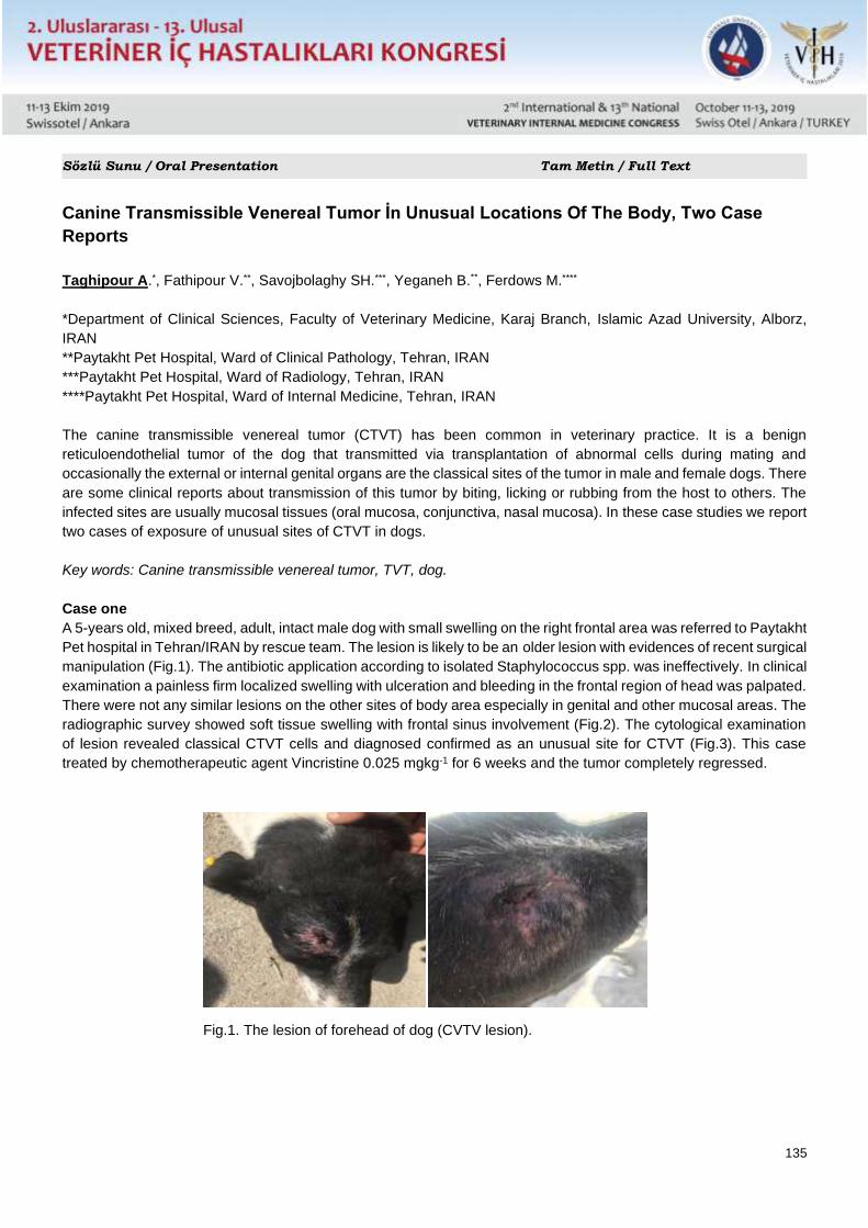
135
Sözlü Sunu / Oral Presentation Tam Metin / Full Text
Canine Transmissible Venereal Tumor İn Unusual Locations Of The Body, Two Case
Reports
Taghipour A.*, Fathipour V.**, Savojbolaghy SH.***, Yeganeh B.**, Ferdows M.****
*Department of Clinical Sciences, Faculty of Veterinary Medicine, Karaj Branch, Islamic Azad University, Alborz,
IRAN
**Paytakht Pet Hospital, Ward of Clinical Pathology, Tehran, IRAN
***Paytakht Pet Hospital, Ward of Radiology, Tehran, IRAN
****Paytakht Pet Hospital, Ward of Internal Medicine, Tehran, IRAN
The canine transmissible venereal tumor (CTVT) has been common in veterinary practice. It is a benign
reticuloendothelial tumor of the dog that transmitted via transplantation of abnormal cells during mating and
occasionally the external or internal genital organs are the classical sites of the tumor in male and female dogs. There
are some clinical reports about transmission of this tumor by biting, licking or rubbing from the host to others. The
infected sites are usually mucosal tissues (oral mucosa, conjunctiva, nasal mucosa). In these case studies we report
two cases of exposure of unusual sites of CTVT in dogs.
Key words: Canine transmissible venereal tumor, TVT, dog.
Case one
A 5-years old, mixed breed, adult, intact male dog with small swelling on the right frontal area was referred to Paytakht
Pet hospital in Tehran/IRAN by rescue team. The lesion is likely to be an older lesion with evidences of recent surgical
manipulation (Fig.1). The antibiotic application according to isolated Staphylococcus spp. was ineffectively. In clinical
examination a painless firm localized swelling with ulceration and bleeding in the frontal region of head was palpated.
There were not any similar lesions on the other sites of body area especially in genital and other mucosal areas. The
radiographic survey showed soft tissue swelling with frontal sinus involvement (Fig.2). The cytological examination
of lesion revealed classical CTVT cells and diagnosed confirmed as an unusual site for CTVT (Fig.3). This case
treated by chemotherapeutic agent Vincristine 0.025 mgkg-1 for 6 weeks and the tumor completely regressed.
Fig.1. The lesion of forehead of dog (CVTV lesion).
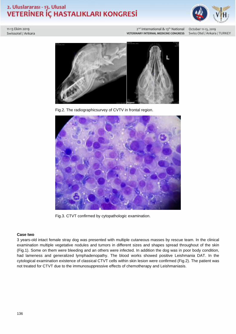
136
Fig.2. The radiographicsurvey of CVTV in frontal region.
Fig.3. CTVT confirmed by cytopathologic examination.
Case two
3 years-old intact female stray dog was presented with multiple cutaneous masses by rescue team. In the clinical
examination multiple vegetative nodules and tumors in different sizes and shapes spread throughout of the skin
(Fig.1). Some on them were bleeding and an others were infected. In addition the dog was in poor body condition,
had lameness and generalized lymphadenopathy. The blood works showed positive Leishmania DAT. In the
cytological examination existence of classical CTVT cells within skin lesion were confirmed (Fig.2). The patient was
not treated for CTVT due to the immunosuppressive effects of chemotherapy and Leishmaniasis.
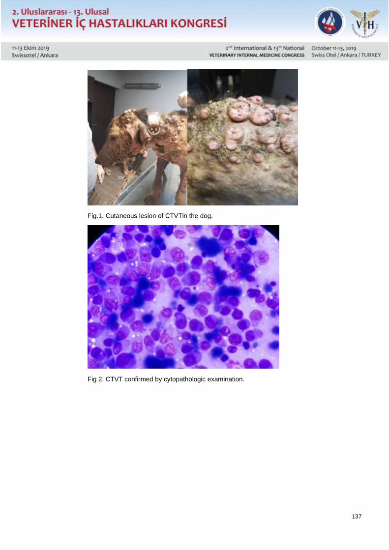
137
Fig.1. Cutaneous lesion of CTVTin the dog.
Fig 2. CTVT confirmed by cytopathologic examination.
138
Sözlü Sunu / Oral Presentation Özet / Abstract
Van Kedisi’nde Görülen Sağırlığın İnsidansı ve Göz Renklerine Göre Dağılımı Üzerine
Araştırmalar
Funda ALMAÇ ÇELİK, Abdullah KAYA
Van Yüzüncü Yıl Üniversitesi, Veteriner Fakültesi İç Hastalıkları Anabilim Dalı, Van
Bu çalışmada Van kedisinde görülen sağırlığın insidansı ve göz rengine göre dağılımının ortaya konulması
amaçlandı. Bu amaçla Van Yüzüncü Yıl Üniversitesi Van Kedisi Araştırma ve Uygulama Merkezi Müdürlüğü ve Van
İli sınırları içinde bulunan 2 ay ile 8 yaş arası her biri eşit sayıda olan 3 (üç) ayrı grup olmak üzere toplam 300 (üç
yüz) adet Van kedisine Otometrics ICS Chartr Ep 200 Klinik ABR cihazı ile işitme testi yapılmıştır. Çalışma sonucunda
Van kedileri’nde sağırlık oranı %14.33 olarak belirlenmiştir. Sağırlığın cinsiyet, tüy uzunluğu ile bir ilişkisinin olmadığı,
kafasında benek bulunan Van Kedilerinde sağırlığın bulunmadığı görülmüştür. Van kedilerinin duyma eşikleri baz
alınarak yapılan değerlendirme sonucunda Van Kedilerinin çoğunluğunun çok iyi işittiği (0-10 dB nHL) ortaya
konulmuştur. Van kedilerinde tek taraflı sağırlığın diğer beyaz kedi ırklarına göre çok daha düşük olduğu tespit
edilmiştir. Sonuç olarak Van kedilerinde görülen sağırlık oranının diğer beyaz kedi ırkları ile kıyaslandığında daha
düşük olduğu ortaya konulmuştur. Yapılan bu çalışma ile Van Kedisi Araştırma ve Uygulama Merkezi, Van ve
çevresinde ABR test sonuçlarına göre sağır olduğu tespit edilen kedilerin çiftleştirilmesinin önüne geçilecektir. İleride
yapılacak çalışmalarla Van kedilerindeki sağırlığın insidansının düşeceği düşünülmektedir. Bu çalışma Van
kedilerinde yapılan ilk sağırlık çalışması olduğundan ve ileride yapılacak olan çalışmalara ışık tutacağından
sunulmaya değer bulundu.
Bu çalışma Van Yüzüncü Yıl Üniversitesi Bilimsel Araştırma Projeleri Başkanlığı Tarafından “TAP-2019-8531” no’lu
proje olarak desteklenmiştir.
139
Sözlü Sunu / Oral Presentation Özet / Abstract
Study on The Incidence of Deafness in Van Cats and Its Distrubution by Eye Colors
Funda ALMAÇ CELİK, Abdullah KAYA
University of Van Yuzuncu Yil Van, Faculty of Veterinary Medicine, Departman of İnternal Diseases, Van
The objectives of this study are to determine the incidence of deafness in Van cats and its distribution according to
eye colors. A total of 300 (three hundred) cats that are present in Van Cat Research and Application Center and
avaliable in Van Province classified into 3 (three) equal groups, aging between 2 months to 8 years were performed
a hearing test with Otometrics ICS Chartr Ep 200 Clinical ABR. As a conseqeunce, deafness rate in these cats were
determined as 14.33%. Additionally there were no correlation observed on sex nor on hair length while the cats that
had spots on their heads were observed to have no deafness. Evaluation based on hearing treshold of Van cats
revelead that majority of them had well hearing. It was determined that one-sided deafness was significantly lower in
Van cats than in other white cat breeds. Therefore observation on deafness rate was found to be lower on Van cats
compared to the white cat breeds. By the means of this study, Van Cat Research and Application Center will prevent
the deaf cats mating in the region according to the conducted results of ABR test. It is concluded that with the future
studies incidence of deafness in Van cats will decrease. This study is the first deafness study conducted in Van cats
and it is worth to present because it will shed light on future studies.
140
Sözlü Sunu / Oral Presentation Özet / Abstract
Pointer Irkı Bir Köpekte Mitral Kapak İlişkili Konjenital Hemokist Olgusu
Osman Safa TERZİ, Kadir SEVİM, Arda Selin Çoşkan TUNÇ, Sevil ATALAY VURAL, Erfaneh PİRPANAHİ
Ankara Üniversitesi Veteriner Fakültesi İç Hastalıkları Anabilim Dalı
Ankara Üniversitesi Veteriner Fakültesi Patoloji Anabilim Dalı
Ankara Üniversitesi Veteriner Fakültesi Cerrahi Anabilim Dalı
Yedi yaşlı, kısırlaştırılmamış Pointer ırkı dişi köpek iştahsızlık, halsizlik, karında büyüme ve güç solunum şikayeti ile
Ankara Üniversitesi Veteriner Fakültesi Hayvan Hastanesi’ne getirildi. Abdomende genişleme, sol arka bacakta
ödem, mastitis benzeri meme ödemi ve zayıf vücut kondüsyonu fiziksel muayenede görüldü. Abdominal
ultrasonografide şiddetli asites belirlendi ve sıvıdan örnek alındı. Toraks radyografisinde kalpte büyüme ( VKS:14),
perikardiyal efüzyon ve pulmoner ödem belirlendi. Ekokardiyografik muayenesinde sol ventriküler dilatasyon ve
şiddetli sistolik disfonksiyon ( EF: %4.9, FS: %2.2 ) belirlendi. Doppler muayenesinde sol atriyal genişlemeye şiddetli
mitral regürgitasyonun eşlik ettiği görüldü. Mitral kapağın endokardiyal yüzeyinde homojen bir kitle belirlendi.
Postmortem muayenede kitlenin konjenital hemokist olduğu belirlendi. Bu olgu sunumu köpeklerde atrial
lokalizasyonlu ve valvüler kökenli kitlelerin konjenital hemokist olabileceğini vurgulamaktadır.
141
Sözlü Sunu / Oral Presentation Özet / Abstract
Mitral Valve Associated Congenital Hemocystıc Case In A Pointer
Kadir SEVİM, Arda Selin TUNÇ, Osman Safa TERZİ, Sevil ATALAY VURAL, Erfaneh PİRPANAHİ
Ankara University, Faculty of Veterinary Medicine, Internal Medicine Department
Ankara University, Faculty of Veterinary Medicine, Veterinary Pathology Department
Ankara University, Faculty of Veterinary Medicine, Veterinary Surgery Department
A 7-year-old, unspayed, female Pointer presented to the small animal clinic at Ankara University, Faculty of
Veterinary Medicine, with complaints as anorexia, weakness, abdominal enlargement and dispnea. Enlargement of
the abdomen, edema in the left hind limb, mastitis like mammary edema and weak body condition was seen during
physical examination. Severe ascites in abdominal cavity was revealed after abdominal ultrasonography.
Cardiomegaly ( VHS: 14) , pericardial effusion and pulmoner edema was determinated on thoracic radiographs.
Echocardiography was performed and severe left ventricular dilatation with highly decreased systolic function ( EF
4.9%, FS 2.2% ) revealed after M-mode examination. The left atrium was dilated and there was severe mitral
regurgitation based on color Doppler. Homogeneous mass was present on the endocardial surface of mitral
valve.Post mortem examination revealed that the mass was “congenital hemocyst”.This case report emphasizes that
atrial localization and valvular masses in dogs may be congenital hemocysts.
142
Sözlü Sunu / Oral Presentation Özet / Abstract
Köpeklerde Mitral Kapak Yetmezliğinde İki Boyutlu Benek Takibi Ekokardiyografi
Yöntemiyle Belirlenen Radyal, Sirkumferensiyal Gerilim ve Gerilim Hızı ve Torsiyonun
Değerlendirilmesi
Ayjamal RADGOHAR, Ebubekir CEYLAN
Pet Hospital, Ankara
Ankara Üniversitesi Veteriner Fakültesi İç Hastalıkları Anabilim Dalı, Ankara
Küçük ırk köpeklerde yaşlanmayla birlikte en sık görülen edinsel kapak hastalığı, mitral kapak hastalığıdır. Mitral
regürgitasyonun ilerlemesiyle sekonder olarak pulmoner arter hipertansiyonu gelişmektedir. Bunun sonucunda da
önce sol, sonra sağ taraflı konjestif kalp bozuklukları meydana gelmekte ve sonuçta tedaviye yanıt vermeyen
semptomlar nedeniyle hastalık ölüm ya da ötenazi ile sonuçlanmaktadır. Mitral kapak hastalığı olan köpeklerde
özellikle bu hastalığa yatkın ırklarda (Örneğin; Terrier) hastalığın erken tanısı, tedavinin başarı oranını
yükseltmektedir.
İki boyutlu benek takibi ekokardiyografi (2D Speckle Tracking Echocardiography, 2D-STE) miyokardiyal dokuyu
çevreleyen sınırlardan ve açı parametrelerinden bağımsızdır. Ayrıca 2D-STE ile, sol ventrikülün miyokardiyal
deformasyonları tüm olası segment pozisyonlarında ve üç farklı yönde (kısa eksende radyal ve sirkumferensiyel;
uzun eksende longitidunal) değerlendirebilmektedir. Miksömatöz kapak hastalığı (MMVD) bulunan köpeklerde, kalp
hücrelerindeki kronik volüm artışına bağlı değişiklikler sol ventrikülün şekli, boyutları ve fonksiyonunda yeni bir
yapılanmaya neden olmaktadır. Hastalığın çeşitli aşamalarında; kalp hücrelerinin sistolik ve diyastolik hızları,
esneyebilme ve gerilebilme yetenekleri ile kasılabilirliklerinde değişiklikler oluşmakta; kalp hastalığının şiddetine göre
sol ventrikülde eksantrik hipertrofi ya da dilatasyon şekillenebilmektedir.
Bu çalışmanın materyalini 10 adet mitral regürgitasyonu olan predispoze küçük ırk yetişkin köpek (<10 kg ve bir yaş
üstü) ve kontrol grubu olarak 10 adet sağlıklı, yetişkin ve hastalığa predispoze küçük ırk köpek oluşturdu.
Bu çalışmada mitral regürgitasyonu olan grupta pik sistolik global sirkumferensiyal kontrol grubuna göre daha yüksek
olarak saptandı (p<0,001). Hasta grubundaki pik sitolik ortalama radyal gerilim, kontrol grubuna göre daha yüksek
olarak saptandı (p=0,012). Parsiyal radyal gerilimde iki bölgede anlamlı fark tespit edilmiştir.
Bu çalışmada pik sistolik sirkumferensiyal, radyal gerilim hızı ve torsiyon değerlerinde kontrol ve mitral kapakçık
hastalığı olan gruplar arasında anlamlı fark bulunmamaktadır.
Benek takibi ekokardiyografi, miyokardın değerlendirilmesinde kesin veriler oluşturan yeni bir tekniktir. Bu tez
çalışması bu tekniğin pratik hekimlikte uygulanabilir bir teknik olduğunu göstermektedir.
143
Sözlü Sunu / Oral Presentation Özet / Abstract
Evaluation of Radial and Circumferential Strain and Strain Rate and Torsion Assessed by
Speckle-Tracking Echocardiography in Mitral Valve Insufficiency in Dogs
Ayjamal RADGOHAR, Ebubekir CEYLAN
*Pet Hospital, Ankara, Turkey
Ankara University Faculty of Veterinary Medicine, Internal Medicine Department, Ankara Turkey
Mitral valve disease (MVD) is the most common acquired valvular disease in small breed dogs with aging. Mitral
regurgitation worsening results in secondary to pulmonary arterial hypertension. In dogs early diagnose of MVD
increases the success rate of treatment.
2D Speckle Tracking Echocardiography (2D-STE) is relatively independent of cardiac translation, tethering and angle
parameters. Additionally, 2D-STE offers the opportunity to the assessment of myocardial deformations in all possible
segments of left ventricular myocardium and in 3 directions (radial and circumferential contraction in short-axis
planes, longitidunal contraction in long-axis planes). In dogs with myxomatous mitral valve disease (MMVD), left
ventricular’s shape, dimension and function lead to restructuring due to chronic volume overload in heart cells. At
various stages of disease; systolic and diastolic velocity of cardiac cells, ability to flexion, streching and contraction
alter and according to the severity of disease, eccentric left ventricular hypertrophy or dilatation may occur.
The material of this study consisted of 10 predisposed small breed adult dogs with mitral regurgitations (<10 kg and
over one year old) and 10 healthy, adult and predisposed small breed dogs as control group.
In this study, the peak systolic global circumferential strain of the group with mitral regurgitation was higher than that
of the control group (p<0.001). The peak systolic mean of the radial strain in the patient group was found higher than
the control group (p = 0.012). There was a significant difference between the two regions in the radial tension .
In this study, there was no significant difference between groups with peak systolic circumferential and radial strain
rate and torsion values of control and mitral valve disease groups.
Speckle tracking echocardiography is a new technique that creates exact data for the evaluation of myocardium. This
study shows that this technique can be applied in practical medicine.
144
Sözlü Sunu / Oral Presentation Özet / Abstract
Koksidiyozisli Buzağılarda Serum Beta-Defensin 1 Düzeyinin Araştırılması
Akın KOÇHAN
Dicle Üniversitesi Veteriner Fakültesi İç Hastalıkları Anabilim Dalı, Diyarbakır
Koksidiyozis, genç hayvanlarda hemorajik diyare, depresyon, zayıflama ve canlı ağırlık kaybının yanı sıra bazen de
ölümle sonuçlanabilen protozoer bir hastalıktır. Bu çalışmada; koksidiyozisli buzağılarda temel olarak deri ve
mukozadaki epitel hücreleri tarafından sentezlenen, antifungal, antibakteriyel, antiparaziter ve antiviral aktiviteleri
sahip bir antimikrobiyal peptid olan beta defensin 1 düzeyindeki değişikliklerin ortaya konulması amaçlandı.
Bu çalışmanın materyalini 15 günlük yaştan büyük, farklı cinsiyet ve ırktan, koksidiyozis tespit edilen 10 buzağı ile
sağlıklı 7 buzağı olmak üzere toplamda 17 buzağı oluşturdu. Beta-defensin-1düzeyinin belirlenmesi için hayvanların
vena jugularislerinden kan örnekleri alındı. Serum beta-defensin-1 düzeyleri ticari ELISA kiti kullanılarak belirlendi.
Hastalardaki beta-defensin 1 düzeyinde bir düşüş olduğu ancak kontrol gurubu ile karşılaştırıldığında, istatistiksel
olarak önemli olmadığı görüldü.
Bu araştırmanın sonucuna göre koksidiyozisli buzağılarda serum beta-defensin 1 düzeyinde önemli bir değişiklik
olmadığı saptandı. Daha etkin sonuçlar elde etmek için örnek sayısını artırarak yeni çalışmalar yapmanın faydalı
olacağına inanıyoruz.
Anahtar Kelimeler; Beta-defensin 1, Buzağı, Koksidiyozis
145
Sözlü Sunu / Oral Presentation Özet / Abstract
Investigation of Serum Beta-Defensin 1 Level in Calves With Coccidiosis
Akın KOÇHAN
University of Dicle, Faculty of Veterinary, Department of İnternal Medicine, Diyarbakır,
Coccidiosis is a protozoar disease that causes hemorrhagic diarrhea, depression, weight loss in young animals as
well as may result in death. The aim of this study was to determine the changes in the level of beta defensin 1, which
is an antimicrobial peptide with antifungal, antibacterial, antiparasitic and antiviral activities, mainly synthesized by
epithelial cells in the skin and mucosa of calves with coccidiosis.
The material of this study consisted of 17 calves, 10 calves with coccidiosis, and 7 healthy calves from different sexes
and races older than 15 days of age. Blood samples were taken from the vena jugularis of them for determination of
beta-defensin-1 level. Serum beta-defensin-1 levels were determined by using a commercial ELISA kit.
There was a decrease in the level of beta-defensin 1 in the calves with coccidiosis but it was not statistically significant
when compared with the control group.
According to the results of this study, there was no significant change in serum beta-defensin 1 level in calves with
coccidiosis. We believe that it will be beneficial to carry out new studies by increasing the number of samples in order
to obtain more and more efficient results.
Key Words; Beta-defensin 1, Calves, Coccidiosis,
146
Sözlü Sunu / Oral Presentation Özet / Abstract
Dermatophilus Congolensis ile Doğal Enfekte Koyunlarda Klinik, Hematolojik ve Bazı
Biyokimyasal Bulgular
Aynur ŞİMŞEK*, Akın KOÇHAN*, Simten YEŞİLMEN ALP**
*Dicle Üniversitesi Veteriner Fakültesi İç Hastalıkları Anabilim Dalı, Diyarbakır, TÜRKİYE
**Dicle Üniversitesi Veteriner Fakültesi, Mikrobiyoloji Anabilim Dalı, Diyarbakır
Dermatophilus congolensis’in neden olduğu dermatofilozis, insan ve hayvanları etkileyen akut, subakut veya kronik
seyirli bir eksudatif dermatitistir. Et, süt ve yünde verim kaybı, üreme kayıpları, deri ve yün kalitesinin bozulması,
tedavi maliyetleri ve ölüm nedeniyle hastalık ekonomik kayıplara neden olur.
Bu çalışmada Dermatophilus congolensis ile doğal enfekte olan koyunlarda klinik, hematolojik ve biyokimyasal
bulguların araştırılması amaçlandı.
Çalışmanın materyalini 200 başlık bir koyun sürüsündeki deri lezyonlu 35 koyun ve klinik olarak sağlıklı 10 koyun
(kontrol grubu) oluşturdu. Enfekte koyunlarda deride kabuklu lezyonlar, yapağıda topaklanma ve dökülme ile kaşıntı
görüldü. Klinik olarak dermatofilozisin tanısı konuldu ve hasta hayvanlardan alınan kabuk materyallerinden
mikrobiyolojik olarak tanı kesinleştirildi.
Grupların hematolojik parametreleri arasında önemli farklılık olmadığı tespit edildi. Biyokimyasal muayenede ise
hasta koyunlarda kontrol grubuna kıyasla serum alkalen fosfataz, alanin aminotransferaz, laktat dehidrogenaz, kan
üre nitrojen, total protein albumin ve kalsiyum seviyelerinin yüksek, kreatinin, sodyum ve potasyum seviyelerinin ise
düşük olduğu görüldü.
Sonuç olarak, Dermatophilus congolensis’in koyunlarda deri ve yapağı hasarına neden olduğu ve serum
biyokimyasal parametrelerde değişikliklere yol açabileceği kanısına varıldı.
Anahtar kelimeler: Dermatofilozis, hastalık, hayvan, serum.
147
Sözlü Sunu / Oral Presentation Özet / Abstract
Clinical, Hematological and Some Biochemical Findings in Sheep Naturally Infected With
Dermatophilus Congolensis
Aynur SIMSEK*, Akin KOCHAN*, Simten YESILMEN ALP**
*University of Dicle, Faculty of Veterinary Medicine, Department of Internal Medicine, Diyarbakir, TURKEY
**University of Dicle, Faculty of Veterinary Medicine, Department of Microbiology, Diyarbakir, TURKEY
Dermatophilosis caused by Dermatophilus congolensis, is an acute, subacute or chronic exudative dermatitis
affecting animals and humans. The disease leads to economic losses due to loss of meat, milk, fleece and
reproduction, deterioration of skin and fleece quality, treatment costs and death.
The aim of this study was to investigate the clinical, hematological and biochemical findings in sheep naturally
infected with Dermatophilus congolensis.
In this study, 35 sheep with skin lesions in a flock of 200 animals and 10 clinically healthy sheep (control group) were
included. Infected sheep showed scabby lesions, matted wool, alopecia, and pruritis. Clinical diagnosis of
dermatophilosis was made and confirmed bacteriologically from scab material.
There were no significant differences between groups in hematological parameters. In the biochemical examination,
of infected sheep, the concentrations of alkaline phosphatase, alanine aminotransferase, lactate dehydrogenase,
blood urea nitrogen, total protein, albumin and, calcium were significantly higher and the concentrations of creatinine,
sodium and potassium were significantly lower than the control group.
Inconclusion, Dermatophilus congolensis is cause of the damage of the skin and wool, and this disease may change
the levels of some biochemical parameters in the serum of sheep.
Keywords: Animal, dermatophilosis, disease, serum.
148
Sözlü Sunu / Oral Presentation Özet / Abstract
Development of Specific Egg Yolk Immunoglobulins To Inhibit In-Vitro Growth Of
Staphylococcus Aureus Causing Mastitis in Bovines
Tanveer Ahmad1*, Tajammal Hussain2, Sajjad-ur-Rahman2, Ghulam Muhammad2, Adnan Ayan3 Ejaz Ahmad1,
Muhammad Qamar Bilal4
1Department of Clinical Sciences, FVS, BZU, Multan, Pakistan 2Faculty of Veterinary Sciences, Agriculture University, Faisalabad 3Department of Genetics, Faculty of Veterinary Medicine, Van Yuzuncu Yil University, Van, Turkey 4Institute of Animal Sciences, Agriculture University, Faisalabad
*Corresponding Author: [email protected]
The present study was aimed to develop and evaluate specific egg yolk immunoglobulins (IgY) to inhibit growth of
Staphylococcus aureus (S. aureus) in-vitro. Thirty white leghorn layer birds were divided into three equal groups (A,
B and C) and received a dose of inactivated S. aureus 106 CFU/bird, 109 CFU/bird or 1012 CFU/bird at day 0, 7 and
21. Eggs from all birds were collected from day 7 to 50 post inoculation of S. aureus. IgY titer of egg yolk in all groups
was obtained through indirect hemagglutinin test (IHA). The IHA titers from day 7 to 21 with minimum GMT of 64,
78.8 and 97 and maximum GMT of 128, 157.6, and 194 were recorded in group A, B and C respectively. Similarly,
IHA titers from day 22 to 50 with minimum GMT of 97, 157.6 and 194 and maximum GMT of 256, 315, and 388 were
recorded in group A, B and C respectively. IgY was isolated though ammonium sulphate preparation method. The
minimum inhibitory concentration of IgY against S. aureus was estimated by using 50 µL of purified IgY and 50 µL of
S. aureus suspension (1ˣ 106) inoculated in micro titration plate along with 100 µL sterilized Luria-Bertani broth. The
highest dilution of 1:64 of test samples did not show growth of S. aureus. In conclusion, IgY can be used as alternative
therapeutic agent for controlling S. aureus mastitis in bovines.
Key word: Egg yolk Igy, Staphylococcus aureus, mastitis, bovine.
149
Sözlü Sunu / Oral Presentation Özet / Abstract
Solunum Yolu Enfeksiyonu Olan Barınak Köpeklerinde Klinik Semptomlar Viral Ajan
Varlığına Dair İpucu Verebilir mi?
Serkan İrfan KÖSE, Fırat DOĞAN
Hatay Mustafa Kemal Üniversitesi, Veteriner Fakültesi, İç Hastalıkları Anabilim Dalı, Hatay
Hatay Mustafa Kemal Üniversitesi, Veteriner Fakültesi, Viroloji Anabilim Dalı, Hatay
Sunulan çalışmada semptomatik olarak solunum yolu enfeksiyonu tespit edilen barınak köpeklerinde viral ajanların
klinik görünüme etkilerinin değerlendirilmesi amaçlandı.
Çalışmada 41 adet farklı ırk, yaş ve cinsiyette solunum yolu hastalığı belirtileri olan barınak köpeği kullanıldı.
Köpeklerin muayeneleri dokuz klinik parametre ile yapıldı ve skorlandı. Köpeklerden Bronkoalveolar Lavaj (BAL)
sıvısı manuel olarak alındı. Bronkoalveolar lavaj sıvısında moleküler yöntem ile viral ajan (Canine Distemper Virus-
CDV, Canine Adeno Virus tip2-CAV2, Canine Herpes Virus tip1-CnHV1, Canine Corona Virus-CCV, Canine
Influenza Virus-CIV) varlığı araştırıldı. Köpekler, BAL sıvısında viral ajan tespit edilenler (n:20) ve edilmeyenler (n:21)
olarak iki gruba ayrıldı ve klinik skorları karşılaştırıldı.
Viral ajan tespit edilen olguların (n:20) on üç tanesinde tek, yedi tanesinde ise iki ayrı viral etken belirlendi. Tekli
etken tespit edilen olgularda (n:13) CDV; iki ayrı etkenin tespit edildiği olguların (n:7) birinde CAV-2 + CDV, üçünde
CCV + CDV, üç tanesinde ise CnHV-1 + CDV belirlendi. Grupların klinik skorları karşılaştırıldığında; klinik durum,
mukoz membranlar, trakeal duyarlılık, öksürük skorları ile vücut ısısı, nabız ve solunum sayılarında fark
belirlenemezken (p>0.05), burun akıntısı (3.67±0,66) ve oskultasyon (4.00±0.00) skorlarındaki fark istatistiksel olarak
anlamlıydı (p<0.05).
Sonuç olarak barınak köpeklerinde CDV (20/20), CCV (3/20), CnHV-1 (3/20) ve CAV-2 (1/20) virüslerinin solunum
yolu enfeksiyonlarının nedenleri arasında yaygın olarak yer aldığı kanaatine varıldı. Ek olarak burun akıntısının
karakteri ve oskultasyonda duyulan seslerin viral etken varlığına yönelik ipucu verebileceği kanısına varılsa da bu
alanda daha kapsamlı çalışmalar yapılarak bu öngörünün desteklenmesi gerektiği düşünüldü.
Anahtar Kelimeler: Klinik Skorlama, Köpek, Solunum Yolu Hastalığı, Viral Etken
150
Sözlü Sunu / Oral Presentation Özet / Abstract
Can Clinical Symptoms Give Clue About The Presence of Viral Agents In Sheltered Dogs
With Respiratory İnfection?
Serkan İrfan KÖSE, Fırat DOĞAN
Hatay Mustafa Kemal University, Faculty of Veterinary Medicine, Department of Internal Medicine, Hatay
Hatay Mustafa Kemal University, Faculty of Veterinary Medicine, Department of Virology, Hatay
The aim of this study was to evaluate the effects of viral agents on clinical appearance in sheltered dogs with
respiratory disease determined as symptomatic.
In this study, 41 sheltered dogs in different breed, age and sex which were determined respiratory tract infection were
used. Examinations of all dogs were performed and scored with nine clinical parameters. Bronchoalveolar lavage
(BAL) fluid was obtained from all dogs by manual. The presence of viral agent (Canine Distemper Virus, Canine
Adeno Virus type2, Canine Herpes Virus type1, Canine Corona Virus, Canine Influenza Virus) in the BAL fluid was
investigated by molecular method. The dogs were divided into two groups as those with (n:20) and without (n:21)
viral agents in BAL fluid, and the clinical scores of groups were compared.
Of the viral agent detected cases (n: 20), thirteen of them had one and seven of them had two different viral agents.
In cases with single agent (n: 13), CDV was determined. In the cases (n:7) having two different agents; one of those
include CAV-2 + CDV, three of those contain CCV + CDV, and the last three of those were CnHV-1 + CDV. When
comparing the clinical scores of groups, there was no statistically difference except nasal discharge (3.67±0.66) and
auscultation (4.00±0.00) scores (p<0.05).
In the light of results, it was concluded that evaluated viruses are among the causes of respiratory infections in
sheltered dogs. Moreover, it was thought that even if the character of nasal discharge and the sounds heard during
auscultation could give clues about the presence of viral agents, this prediction should be supported by further studies
in this field.
Keywords: Canine, Clinical Scoring, Respiratory Tract Disease, Viral Agents
151
Sözlü Sunu / Oral Presentation Tam Metin / Full Text
Bir Oğlakta Tetanoz Olgusu ve İyileşme Süreci
Erdal Kara*, Mehmet Şahal**
* Kırıkkale Üniversitesi, Veteriner Fakültesi, İç Hastalıkları Anabilim Dalı, Kırıkkale, Türkiye
** Ankara Üniversitesi, Veteriner Fakültesi, İç Hastalıkları Anabilim Dalı, Ankara , Türkiye
Özet
Olgunun materyalini iştahsızlık, arka ayaklarda güçsüzlük ve konstipasyon şikayetiyle İç Hastalıkları kliniğine getirilen
45 günlük erkek oğlak oluşturdu. Yapılan muayenede hastanın vital parametrelerinin normal olduğu, hemogramında
sadece lökositoz (WBC 15 109/l) belirlendi. Fiziksel muayenede boyun kaslarında sertleşme ve kasılma, vücudun
tamamında kasılma, sertleşme ve yürüyüşünde tutukluk izlendi. Yürütülen hastanın çok uzun süre ayakta
kalamadığı, arka ayaklarda gözle görülür bir güçsüzlük olduğu, kısa süre sonra arka ayakları üzerine çöktüğü
görüldü. İlk gün extremitelere yapılan germe – bırakma hareketlerinde eklemlerin hareketli olduğu izlendi. Kulaklarda
dikleşme ve kuyruğun kalkık olması da dikkat çekici idi. Ayrıca sağ ön ayağın iç yüzünde humerus üzerinde 10 cm’lik
belirgin bir çizik tespit edildi. 24 saat sonra boynun tam extensiyon olduğu ve hiç hareket ettirilemediği, vücudun
tamamının şiddetli kasıldığı, extremitelerin hareket ettirilemediği ve çene kaslarının kilitlendiği görüldü. Klinik
tablonun tetanoz ile uyumlu olması sebebi ile tedavide hastaya yoğun penisilin uygulaması, trankilizan ve paranteral
besleme yapıldı. Tedavi süresince izole bir birimde hospitalize edilen hastada 15. günde çene kaslarında yumuşama
ve hareket başlarken 30 günlük tedavinin ardından total bir iyileşme gözlendi.
Anahtar Kelimeler: Oğlak, tetanoz, klinik bulgular, sağaltım
Giriş
Tetanoz hastalığına insan ve hayvanlarda Clostridium tetani (C. tetani) isimli Gram +, sporlu, zorunlu anaerop bir
basil sebep olmaktadır (Aydın et al., 2001; Doğan G et al., 2017). Etken toprakta, hayvan bağırsağında ve dışkıda
bolca bulunur (Aydın et al., 2001; Radostits et al., 2007). Bazı coğrafi bölgelerde daha yoğun olabilir, sporlar toprakta
aylarca canlı kalır ve yok edilemez. Hastalık bakteri sporlarının bütünlüğü bozulmuş dokudan girmesi ile oluşur (Aydın
et al., 2001). C. tetani vücuda yayılan bir bakteri değildir. Vücuda girdiği yara bölgesinde anaerobik ortamın
sağlanmasıyla hipoksemik dokularda ürer ve toksinleri aracılığı ile hastalık oluşturur (Ok ve Ertürk., 2019).
Tetanospazmin ve tetanolizin olmak üzere iki çok etkili nörotoksin ve hemolitik ekzotoksin salgılar. Bunlardan
tetanospazmin spazmlar ile karakterize klinik hastalık tablosundan sorumludur. Toksin motor nöronların
nöromusküler kavşağında nörotransmitter maddelerin salınımını engelleyerek etki gösterir ve sinir uçlarındaki
reseptörlere dönüşümsüz olarak bağlanır. Bağlanma olduktan sonra antitoksin uygulamalarının etkisi yoktur.
Antitoksin henüz toksin bağlanmamış nöronları koruyarak, hastalığın ilerlemesini durdurabilir, ancak geri
dönüştüremez (Atkinson W et al., 2006).
Tetanoz tüm memelileri etkilemesine karşın evcil hayvanlardan en duyarlı tür atlardır. Ruminantlar ve domuzlarda
oldukça duyarlıdır, karnivorlar ise etkene daha az duyarlıdır. Kanatlılar ise tetanozdan etkilenmezler. Hastalığın
generalize, sefalik, lokal ve neonatal olmak üzere 4 formu bulunmaktadır (Kuzucuoğlu T et al., 2011). At, ruminant
ve domuz gibi duyarlı hayvanlarda generalize tetanoz görülmesine karşın daha az duyarlı olan karnivorlarda lokal
tetanoz görülmektedir (Aydın N et al., 2001; Wernery U et al., 2004). Hastalığın inkübasyon periyodu ortalama 24
saat ile 2 hafta, oğlaklarda ve kuzularda ise daha kısadır (Aydın et al., 2001; Bostedt und Dedie, 2019).
Keçilerde tetanoz daha çok kastrasyon, boynuz kesme, kulak kesme, ısırılma, enjeksiyon yaraları, kırpma, kulak
küpesi takılması, tırnak yaralanması gibi manuplasyonlar ile muzipliğe bağlı derin nekrotik yaralanmalarda görülür
(Arfan Y et al., 2010). Bazı durumlarda yara odağı bulunamaz. Klinik belirtiler; kulaklarda dikleşme, kalkık kuyruk,
152
boyunda geriye doğru gerilme olması ve hareket ettirilememesi, genel kas sertliği, 3. göz kapağı prolapsusu, çenenin
kilitlenmesi (trismus), yeme ve su alımının azalması, disfaji ve aşırı duyarlılıktır. Larenks, farenks ve özefagus çizgili
kasları hastalıktan etkilenir. Extremiteler gergin, yürüyüş sert ve diktir. Ani irkiltiler kaslarda kramplara neden olur.
Hastalar 2-4 günde kötüleşir ve mekanik olarak solunum durması ile ölüme sürüklenir. Timpani ve konstipasyon yine
sıkça görülen belirtilerdendir. Solunum etkilenir. İlerleyen aşamada lateral yatış ve ölümle sonuçlanır (Arfan Y et al.,
2010; Imaad et al., 2017). Hastalığın tanısında patognomik olan klinik belirtiler ve anamnez bilgileri yeterlidir
(Upadhyay SR, et al., 2013; Altuğ N et al., 2017; Brook I, 2008; Ataro P et al., 2011, Bostedt und Dedie, 2019) .
Tetanoz basilinin dokularda mikroskobik tanısı doku örneklerinden üretme veya kan , yara sekreti veya dokulardan
toksin tanısı her zaman başarılı olmaz. Etken tetanoz olgularının sadece % 30 undan izole edilebilmiştir (Ataro P et
al., 2011). Fare deneyleri de teşhiste kullanılmaktadır (Altuğ N et al., 2017).
Oğlaklarda hastalığın prognozunun kötü olduğu bildirilmektedir (Bostedt ve Dedie, 2019). Tedavide öncelik toksinin
kaynağının ortadan kaldırılmasıdır. Bu amaçla yara bakımı ve etkene yönelik antibiyoterapi uygulanmaktadır. C
tetani olgularında penicilin türevleri ve metranidazole gibi anaerobik etkili antibiyotikler tercih edilmektedir (Kay G and
Knottenbelt DC, 2007; Tekin-Koruk S et al., 2013; Ok and Ertürk., 2019). Bir sonraki adım ise henüz nöronlara
bağlanmamış olan toksinin antitoksin ile inaktif hale getirilmesidir (Altuğ N et al., 2017). Tedavi esnasında hasta ses
ve ışık gibi uyaranlardan uzak tutularak izole bir ortama alınmalı, solunum güçlüğü var ise oksijen cihazına
bağlanmalıdır. Çene kaslarının kasıldığı dönemde paranteral besleme yada özefagus tüpü, rumen kanulü gibi
yöntemler ile hasta beslenmelidir. Kasılmaların azaltılması için asepromazin, Ksilazin ve diazepam tercih edilir (Altuğ
N et al., 2017; Ok and Ertürk., 2019).
Vaka Sunumu
Ankara Üniversitesi Veteriner Fakültesi İç Hastalıkları Anabilim Dalı Kliniğine evde beslenen, aşıları yapılmamış, 45
günlük dişi oğlak iştahsızlık, arka ayaklarda güçsüzlük, idrar çıkarımı ve dışkılamada zorlanma şikayetleriyle getirildi.
Klinik muayenede hastanın vücut ısısı 39 oC, kalp frekansı 102, solunum sayısı 22 kaydedilmiştir. Hemogramında
nötrofiler bir lökositoz (WBC 15 109/l) olduğu belirlendi. Fiziksel muayenede boyun kaslarında sertleşme ve kasılma,
vücudun tamamında esnekliğini yitirmiş bir tablo ile birlikte tutuk yürüme izlendi. Hastanın çok uzun süre ayakta
kalamadığı, arka ayaklarda gözle görülür bir güçsüzlük olduğu, kısa süre sonra arka ayakları üzerine çöktüğü
görüldü. Extremitelere yapılan germe – brakma hareketlerinde eklemlerin ilk gün hareketli olduğu izlendi. 24 saat
sonra kulaklarda dikleşme ve kuyruğun kalkık olması dikkat çekti. Ayrıca sağ ön ayağın iç yüzünde humerus üzerinde
10 cm lik yüzeysel bir çizik tespit edildi. Oğlağın göz muayenesinde 3. göz kapağı prolapsusu izlendi.
Alınan anamnez bilgileri ve yapılan ayrıntılı klinik muayene sonucunda patognomonik klinik tablonun tetanoz ile
uyumlu olabileceği şüphesi üzerine hasta hospitalize edilmiştir. Bir gün sonra hastanın vücudunun tamamıyla
kasılmış olduğu, ayağa kalkamadığı ve yan yatar pozisyonda olduğu görüldü. Hastanın boynu çok sert şekilde geriye
doğru kasılmış, kafa ve vücuy “ atlama sehbası “ diye tabir edilen pozisyonu almıştı. Hasta kaldırılarak hareket
ettiğinde heykel gibi hareketsiz kaldığı ve hiçbir eklemini oynatamadığı görüldü. Çalışmada alınan kan örneğinden
PCR yapılmış, analizindeki bazı teknik nedenlerden dolayı sonuç elde edilememiştir.
Klinik tablosu belirginleşen hastanın hergün rutin klinik kontrolleri yapılarak hastaya günde bir kez penisilin, iki kez
paranteral besleme ve 4 kez trankilizan verildi. 10 kg canlı ağırlığındaki oğlağa 15 gün süreyle diazepam (Diazem
amp., 0,5 ml, Deva İlaç) ve 50.000 U/kg dozunda penisilin ( Vetimisin, Vetaş) uygulaması, hospitalize edildiği süre
boyunca belirli aralıklar ile, vitamin uygulaması yapılmış, kasılmaların çözülmeye başlaması ile de fizik tedavi
uygulanmıştır.
Klinikte 50 gün hospitalize edilen hastanın ilk 15 gün sessiz ve gün ışığı almayan bir bölmede barındırılması
sağlanmış, takip eden günlerde normal bölmeye alınmıştır. Hasta hospitalize edildiği dönemde idrar çıkışı hiç
azalmamıştır. Katı gıda alımının olmadığı dönemde defekasyon azalmiş olmasına karşın tamamıyla durmamıştır.
Hastada orbitanın üzerinden, humerus eklemi üzerinden ve os coxada yatak yaraları şekillenmiştir. Hasta hospitalize
153
edilmesini takiben ilk 7 günlük sürede sadece paranteral beslenmiş, 7-10 günlerde burun özefagus sondasıyla
hastaya süt verilmiştir. Burun özefagus sondasının sürekli süt pıhtısıyla tıkanması sebebiyle 10-17. günler arasında
beslenme amacıyla hastaya özefagus tüpü uygulanmış ve süt ile beslenmeye bu yolla devam edilmiştir. 15. günde
çene kaslarını oynatabildiği farkedilmiş, 17. gün gevşeyen çene kaslarıyla birlikte oğlağa biberonla oral süt verilmiştir.
24. gün güçlenen çiğneme hareketleri oral katı gıda almasını sağlamıştır. Süt ile beslemede günde iki kez 250 ml süt
ile 150 ml su karıştırılarak verilmiş. Katı gıda olarak ise dut ve asma yaprağı adlipidum olarak yedirilmiştir.
Tartışma
Tetanoz tropik ve subtropik bölgelerde çiftlik hayvanlarında sık görülen bir hastalıktır. Çiftlik hayvanlarından atlar ile
birlikte koyun ve keçiler hastalığa en duyarlı türlerdir. Kuzu ve oğlaklarda tedavinin ümitsiz olduğu ve ekonomik
olmadığı bildirilmiştir (Ok ve Ertürk, 2019; Imaad ve ark., 2017; Bostedt ve Dedie, 2019).
Çiftlik hayvanlarına tetanoz etkeni daha çok kuyruk kesme, boynuz kesme, kastrasyon, göbek kordonu yarası, aşı
ve ilaç uygulamalarıyla bulaşmaktadır (Altuğ ve ark., 2017). Keçi ve oğlaklarda ise bunlara ilaveten hareketli mizaçları
sebebiyle yaramazlık yaraları diye tabir edilen vurma, çarpma, batma yaraları ve enjeksiyon yerleri de sıkça tetanoza
sebep olmaktadır (Yousuf ve ark, 2010). Bu vakada 45 günlük erkek oğlağın ilk fiziki muayenesinde sağ ön ayağın
iç yüzünde humerus üzerinde 10 cm’ lik yüzeysel bir çizik tespit edildi. Bu yaradan muhtemelen enfeksiyon etkeninin
vücuda girdiği düşünülmüştür.
Klinik muayende Ok ve Ertürk (2019), beden ısısı, solunum ve nabız frekanslarının artacağını bildirirken, Imaad ve
ark (2017) ise yetişkin bir tekede kastrasyona bağlı oluşan tetanoz olgusunda, bahsi geçen değerlerin fizyolojik
sınırlar içerisinde olduğunu bildirmişlerdir. Olguya konu olan oğlağın muayenesinde Imaad ve ark (2017) ve Bostedt
ve Dedie (2019) ile uyumlu olarak beden ısısı, solunum ve nabız frekanslarının normal sınırlarda olduğu tespit
edilmiştir.
Tetanoz olgularında hemogramda nötrofiler karakterde bir lökositoz gözlenmektedir (Ok ve Ertürk, 2019; Altuğ ve
ark., 2017; Parmer ve ark., 2015). Bu olguda da oğlaktan alınan kan örneğinde WBC 15 109/l olarak belirlenmiş ve
literatürler ile uyumlu bir hemogram hemogram ile karşılaşılmıştır.
Tetanozlu keçilerde dikkat çeken semptomlar arasında üçüncü göz kapağının prolapsusu, kuyruğun dik ve sert bir
hal alması, kulakların dikilmesi, tutuk yürüyüş, ön ayakların öne arka ayakların arkaya doğru tutularak gergin bir vücut
pozisyonu, ilerleyen olgularda yan yatış, yatan hastalarda boynu geriye doğru kasma ve tüm vücutta şekillenen,
germe-kasılma tablosu bulunmaktadır. Timpani ve konstipasyon gözlenebilir (Ok ve Ertürk, 2019; Imaad ve ark.,
2017; Parmer ve ark., 2015). Muayenesi yapılan oğlakta üçüncü göz kapağı prolapsusu dışında tüm semptomlar
değişik derecelerde tespit edilmiştir. Hastada belirli dönemlerde kostipasyon ve timpani gözlenmiştir.
Karakteristik patognomonik klinik tablo sebebi ile muayene bulgularına dayanarak tetanoz tanısı koyulması kolaydır
ancak kesin tanı için nekrotik yara odağından örnek alınarak bakteri izolasyonu ve kültürü yapılması gerekmekted ir
(Ok ve Ertürk, 2019). Bununla birlikte klinik tetanoz vakalarının ancak % 25 - 30’undan etken izole edilebilmiştir
(Ataro P et all., 2011; Dirksen G et all., 2006). Tetanoz vakalarında etken izolasyonunun genellikle başarısız olduğu,
bu nedenle klinik bulgulara göre yapılabileceği ifade edilmiştir (Altuğ ve ark., 2017). Bu olguda hastanın ilk
muayenesinde tetanoz odağı olabileceği düşünülen 10 cm lik yüzeysel bir çizik tespit edilmiş ancak teşhis klinik
tabloya göre yapılarak etken izolasyonu için laboratuvara gönderilen kan örneğinden neknik nedenlerden dolayı
sonuç alınamamıştır.
Tetanoz olgularında tedavi; yeni toksin salınımının engellenmesi ve dolaşımda serbest gezen toksinin bağlanması,
toksin kaynağı olan enfeksiyonun kontrol atına alınması, şiddetli kas spazmının giderilmesi ve bakım – beslemeden
oluşmaktadır (Ok ve Ertürk., 2019). Matt Ball 2012, keçilerde tetanoz olgularında ölümün hızlı şekillendiğini bildirilen,
Yousuf ve ark. 2010, takip ettikleri 10 keçiden antitoksin kullanılanlar da dahil prognozun kötü olduğunu, ortalama 5
günlük sürede ölümün şekillendiğini bildirmişlerdir. Radostis ve ark. 2007 genç ruminantlarda tetanoz vakalarının %
80 ölüm ile sonuçlandığını, tedavinin 10. Gününe kadar yaşatılabilen hastalarda kurtulma oranının arttığını
154
bildirmişlerdir. Imaad ve ark., 2017 5 günlük bir tedavi ile 16 aylık bir tekeyi iyileştirdiklerini bildiriken, Parmer ve ark.
2015, antitoksin de kullanılan 7 günlük bir tedavi sonucunda 2 keçide iyileşme sağlandığını bildirmişlerdir. Bu olguda
45 günlük genç bir oğlak olması, keçilerin tetanoza çok duyarlı olmaları, hastalık tablosunun çok hızlı ilerlemesi,
iyileşme şansının düşük olması ve tedavisinin ekonomik görülmemesi gibi bildirimlere rağmen hasta sahibinin hobi
amaçlı hayvan besliyor olması ve tedavide ısrarcı olması sebebiyle hasta klinikte hospitalize edilerek tedaviye alınmış
ve başarılı bir tedavide kullanılmamıştır. İlk muayenede antitoksin kullanımı uygun bulunmuşsada antitoksin temin
edilemediği için tedavide yer verilememiştir. Buna rağmen hastada iyileşme sağlanmıştır, ancak iyileşme Imaad ve
ark. 2017 ve Parmer ve ark. 2015’ in bildirdiği gibi kısa sürede gerçekleşmemiştir. Matball 2012 ve Yousuf ve ark.
2010 da tedavide başarısız olunmasının sebebinin tedaviye geç başlaması sonucu çok fazla toksin salgılanmış
olması ya da yara bölgesinin önemli olmuş olabileceği ileri sürülmektedir.
15 gün süre ile trankilizan ve antibiyoterapiye devam edilen hastaya 17. güne kadar paranteral besleme yapılmıştır.
24. günde katı gıda alımına başlayan hasta 30. günde ektremitelerini hareket ettirmeye başlamış ancak eklemlerde
oluşan osteo artroz sebebi ile yürümede zorlanmıştır. 20 gün süre ile fizik tedavi uygulanan hastanın ön ekstremiteleri
güçlenmiş ancak arka ekstremitelerdeki kas zayıflığı sebebi ile ortez uygulanarak taburcu edilmiştir. Ortez
uygulanmasından 3 hafta sonra oğlak yardımsız yürümeye başlamıştır. Hastalığa karşı çok duyarlı olan oğlaklarda
tedavinin düzenli takip edilmesi sonrasında sözkonusu ilaçlar ile başarılı bir iyileşme sağlanabileceği ortaya
koyulmuştur.
Kaynaklar
Altuğ N, Yüksek N, Karasu A, İlhan F, Ceylan E, Ekin İH, Arslan S. Diagnosis and treatment of umbilical cord-derived tetanus in
neonatal calves. Turk J Vet Anim Sci, 2017;42:317-322.
Arfan Y, Tanveer A, Muhammad Az, Rao ZA. Trials on Tetanus Treatment in Beetal Goats. Eg. J. of Sh. & G. Sc., 2010; 5(1);341-
347.
Ataro P, Mushatt D, Ahsan S. Tetanus: A Review. Southern Medical Journal, 2011;104(8):613-617.
Atkinson W, HamborskyJ, Mclntyre L, Wolfe S. Center for Disease Control and Prevention. Epidemiology and Prevention of
Vaccine Preventable Disease. (eds) 9th ed. Washington DC: Public Healty Foundation, 2006.
Aydın N, İzgür M, Diker S, Yardımcı H, Esendal Ö, Paracıkoğlu J, Akan M. Veteriner Mikrobiyoloji (Bakteriyel Hastalıklar) 2001.
Brook I. Current concepts in the managment of Clostridium tetani infection. Expert Rev Anti İnfect Ther. 2008;6(3):327-36.
Dirksen G, Gründer HD, Stöber M. Krankheiten des Rindes, 5. Auflage. 2016; syf.1068-1071.
Doğan G, Kayır S, Ekici AA, Aşıcı E. Tetanus Case with Mortal: A Case Reporte. Kocaeli Medical J 2017; 6 ; 2; 52-55.
Imaad R, Junaid A, Awais-ur-Rehman S, Ghulam M and Muhammad S. Tetanus In A Surgıcally Castrated Beetal Buck: A Case
Report. Matrix Science Pharma 2017; 1(2); 25-26.
Kay G and Knottenbelt DC. Original Article Tetanus in equids: A report of cases. Equine Vet. Educ., 2007; 19(2); 107-112.
Kuzucuoğlu T, İtal İ, Alatlı İ. Follow-up and Treatment of a severe Tetanus Case in the Intensive Care Unit. J Kartal TR
2011;22(1);45-48.
Matt Ball, Case Notes Tetanus in Goats. http://www.flockandherd.net.au/other/reader/tetanus-goats.html 16.00, 01.10.2019.
Ok M ve Ertürk A. Ruminantlarda Tetanoz (Kazıklı Humma). Ok M, editör. Ruminantlarda Klostridial Enfeksiyonlar . 1. Baskı.
Ankara: Türkiye Klinikleri; 2019; 10-14.
Parmar VL, Prasad A, Patel JS, Javia bb and Barad DB. Clinico – Therapeutic Management of Tetanus in Caprines. Intas Polivet,
2015; 16(2); 380-381.
Radostits OM, Gay CC, Hinchcliff KW, Constable PD. A Text Book of the Diseases of Cattle, Horses, Sheep, Pigs and
Goats.10th ed. Book Power; 2007. p.25-35.
Tekin – Koruk s, Şeyhanoğlu AS, Sarı T, İpekkan K. An Unvaccinated Tetanus Case That Recovered without Sequelae. Klimik
Dergisi, 2013; 26(1); 31-33.
Upadhyay SR, Hussain K, Singh R. Bovine neonatal tetanus:a case reprt. Buffalo Bull 2013;32:18-20.
Wernery U, Ul-Haq A, Joseph M, Kinne J. Tetanus in a Camel (Camelus Dromedarius) – A Case Report. Tropical Animal Health
and Production, 36(2004)217-224.
155
Sözlü Sunu / Oral Presentation Özet / Abstract
Tetanus and Recovery Process in a Kid
Erdal Kara*, Mehmet Şahal**
* Kırıkkale University, Veterinary Faculty, Internal Medicine Department, Kırıkkale, Turkey
** Ankara University, Veterinary Faculty, Internal Medicine Department, Kırıkkale, Turkey
Abstract
The case of the present study was a 45-day old male kid referred to our clinic due to anorexia, weakness on posterior
legs and constipation. The examination revealed that vital parameters of the patient were normal and leukocytosis
(WBC 15 109/l) was detected in the hemogram analysis. Stiffness and constriction on the neck muscles as well as
walking with a body condition that was completely inflexible were detected during physical exam. The patient was
forced to walk and he could not stand for a long period with a visible weakness on the posterior legs; and he kneeled
down on posterior legs after a short period. The joints were dynamic during tensing-releasing movements on the
limbs. Pricking up of the ears and raising of the tail were noticeable. Furthermore, a superficial scratch of 10 cm was
detected on the humerus inside part of the right anterior leg. After 24 hours, complete extension was detected on the
neck and it could not be moved; the body was severely constricted; extremities could not be moved and mandible
muscles were locked. Since the clinical presentation was consistent with tetanus, intense penicillin and sedative
medications as well as parenteral nutrition were implemented. The patient was hospitalized in an isolated unit during
the treatment period, motion started on mandible muscles at day 15 and total recovery was observed after a treatment
period for 30 days.
Key Words: Kid, tetanus, clinical findings, treatment
156
Sözlü Sunu / Oral Presentation Özet / Abstract
Serological Survey Of Pestivirus Infection In Buffalo In Urmia, Iran
Misagh Ahmadian*, .Arash Araghi-Sooreh**
* D.V.M. Graduate, Faculty of Veterinary Medicine, Urmia Branch, Islamic Azad University, Urmia, Iran.
** Associate Professor, Department of Clinical Sciences, Faculty of Veterinary Medicine, Urmia Branch, Islamic Azad
University, Urmia, Iran.
Pestiviruses within the Flaviviridae family are known to exist in ruminants worldwide. This study was aimed to
determine the seroprevalence of pestivirus infection in buffalo in Urmia, northwestern Iran. A total of 184 sera were
randomly taken from 109 male and 75 female buffaloes and tested for anti-pestivirus antibodies by competitive ELISA
method. The seroprevalence rate was 50.5% (95% CI: 43.3-57.7%). Seropositivity was significantly higher in female
buffaloes (P=0.033). Logistic regression showed that 0.578% of fluctuation of infection was justified by sex and
greater odds of infection in females was seen (OR: 1.783 and 95%CI: 0.906-3.15, P=0.094>0.05).This is the first
report of pestivirus infection in buffalo in northwest of Iran.
Key words: Pestivirus, ELISA, Buffalo, Iran
157
Sözlü Sunu / Oral Presentation Özet / Abstract
The First Survey Of Seroprevalence Of Toxoplasma Gondii In Horses Of West Azerbaijan
Province, Iran
Misagh Ahmadian*, .Arash Araghi-Sooreh**
* D.V.M. Graduate, Faculty of Veterinary Medicine, Urmia Branch, Islamic Azad University, Urmia, Iran.
** Associate Professor, Department of Clinical Sciences, Faculty of Veterinary Medicine, Urmia Branch, Islamic Azad
University, Urmia, Iran.
Toxoplasmosis is a worldwide zoonosis caused by Toxoplasma gondii, which can infect warm-blood
animals and humans. The objective of this study was to determine the seroprevalence of anti-T. gondii
antibodies in horses in west Azerbaijan province, northwestern Iran. A total of 184 healthy horses (115
female, 69 male), aged 2-9 years, from four different regions of province were sampled and tested for
antibodies to T. gondii using indirect ELISA test. Data were analyzed for effects of age, sex and region
on seropositivity. A total of 16 horses (8.7%, 95% CI: 4.6–12.8%) were positive for T. gondii antibodies.
There was a significant difference between age groups (P=0.024), but gender (P=0.589) and region
(P=0.790) present no association with seropositivity. Logestic regression showed that the odds of
disease was added with increase of age (OR:1.579 and 95% CI: 0.912–2.735, P> 0.05) and 0.457% of
fluctuation of disease was justified with age. The results indicated that horses are exposed to this
parasite in northwest of Iran.
Key words: Horse, Toxoplasma gondii, ELISA, Iran
158
Sözlü Sunu / Oral Presentation Özet / Abstract
Ankara Bölgesinde Süt Sığırı İşletmeciliği Yapılan Çiftliklerde “Buzağılarda Pasif Transfer
Yetmezliği” nin Değerlendirilmesi
Erdal Kara, Ebubekir Ceylan
Kırıkkale Üniversitesi, Veteriner Fakültesi, İç Hastalıkları Anabilim Dalı, Kırıkkale, Türkiye
Ankara Üniversitesi, Veteriner Fakültesi, İç Hastalıkları Anabilim Dalı, Ankara , Türkiye
Ruminantlarda makromoleküllerin yavruya geçişine müsaade etmeyen özel bir plasenta yapısı olması sebebi ile yeni
doğan buzağılar immun sistemleri oluşmamış olarak dünyaya gelirler. Kolostrum alan buzağılarda kolostral Ig’ler
hızla buzağının sistemik dolaşımına geçerek pasif immun transferi gerçekleştirir. Kaliteli bir kolostrumun 50 mg/ml
ve üzeri IgG konsantrasyonuna sahip olması istenir. Başarılı bir pasif immun transferde 24-48 saatlik yaştaki
buzağının kan serumu IgG konsantrasyonunun 10 mg/ml ve üzerinde olması istenir. IgG seviyesi bu eşik değerin
altında ise pasif transfer yetmezliği (PTY) şekillenmiş demektir. Bu çalışmada buzağı sağlığı açısından doğrudan
etkili olan PTY prevalansının Ankara ilinde üretim yapan sütçü sığır işletmelerindeki prevalansının belirlenmesi
amaçlanmıştır. Çalışmaya Ankara ilinde bulunan 9 işletme dahil edilmiştir. İşletmeler seçilirken; anaç hayvan
sayısının 100 baş ve üzeri olması, tam zamanlı veteriner hekim, teknisyen/tekniker istihdam edilmesi ve temel sürü
yönetim protokollerinin uygulanıyor olması aranmıştır. Çalışma kapsamında toplanan 400 kan serumundan 69
tanesinde, 363 kolostrumdan ise 44 tanesinde IgG seviyesi belirlenen referans değerin altında ölçülmüştür. Bu
sonuçlara göre Ankara bölgesinde üretim yapan sütçü işletmelerde PTY prevalansı %17.25, yetersiz kalitede
kolostrum üretme oranı ise %12.12 olarak bulunmuştur. Çalışma kapsamında toplanan çeşitli veriler
değerlendirildiğinde kolostrum kalitesinin PTY üzerine etkisi ve PTY’nin sütten kesilinceye kadar atlatılan hastalık
prevalansı üzerine etkileri önemli bulunmuştur. Buna göre kalitesiz kolostrum ile beslenen buzağılarda PTY görülme
oranı 15.422 kat artarken, PTY olan buzağılarda sütten kesilinceye kadarki dönemde hastalık atlatma sıklığı 1.6 kat
artmaktadır. Bu çalışma ülkemizde IgG seviyesinin direk ölçüldüğü metotlardan birisi ile yapılan ilk PTY prevalans
çalışması olması sebebi ile önemli bulunmuştur.
159
Sözlü Sunu / Oral Presentation Özet / Abstract
Evaluation of “Passive Transfer Insufficiency in Calves” in The Farms Where Dairy Cattle
Operations are Conducted in The Ankara region
Erdal Kara, Ebubekir Ceylan
Kırıkkale University, Veterinary Faculty, Internal Medicine Department, Kırıkkale, Turkey
Ankara University, Veterinary Faculty, Internal Medicine Department, Ankara, Turkey
Newborn calves come with an immature immune system since ruminants have a special placenta that does not allow
transition of macromolecules to the baby. Colostral IgGs are rapidly transferred to systemic circulation of the calf and
enable passive immune transfer in the calves which intake the colostrum. A qualified colostrum should include an
IgG concentration at and over 50 mg/ml. A successful passive immune transfer should provide IgG concentration at
and over 10 mg/ml in a 24- to 48-hour old calve. IgG levels below this threshold level cause passive transfer failure
(PTF). The aim of the present study was to determine the PTF prevalence which is directly effective on a calf health
in dairy cattle facilities in Ankara. Colostrum samples were obtained during first milking; blood samples were collected
from 24- to 48-hour old calves. The samples were stored in deep freezers at -20 oC until performance of analysis
procedures. IgG levels were measured through ELISA technique. IgG levels were measured below the reference in
69 of 400 blood samples and in 44 of 363 colostrum samples obtained in the study. According to these results, PTF
prevalence was found 17.25% and unqualified colostrum production rate was detected 12.12% in dairy cattle facilities
in Ankara. Evaluation of different data obtained in the study revealed that quality of colostrum was found significantly
effective on PTF, and PTF was also found significantly effective on disease prevalence until weaning. Accordingly,
PTF rate increased by 15.422-folds in the calves which are fed by unqualified colostrum and frequency of overcoming
a disease increases by 1.6 folds within the lactating period. The present study is important as being the first PTF
prevalence study where IgG levels were directly measured in our country.
160
Sözlü Sunu / Oral Presentation Özet / Abstract
Serum Immunoglobulin Responses Following Administration of Eps-Adjuvanted Vaccine
For Pneumonic Pasteurellosis In Goats
Mohd Effendy Abd Wahid*, Insyirah Husna Kamaradin**, Terengganu Suvik Assaw***
*School Of Fisheries And Aquaculture Sciences Universiti Malaysia Terengganu
**Institute Of Marine Biotechnology, Universiti Malaysia
***School Of Marine Sciences And Environment, Universiti Malaysia Terengganu
Pneumonic pasteurellosis is a common respiratory disease among small ruminants caused by a gram-negative
pathogenic bacterium, Mannheimia haemolytica A2. The commercially available vaccines for pneumonic
pasteurellosis are notably shown poor immunogenicity responses, or low protection level. This study was conducted
to measure the potential of exopolysaccharide (eps) extracted from indigenous microalgae species, Chlorella sp. as
vaccine adjuvant that can trigger the humoral response in both mucosal and systemic immunity against M.
haemolytica A2 infection. To evaluate the potential of Chlorella sp. eps as adjuvant for M. haemolytica A2 vaccine at
mucosal surfaces, eps-adjuvanted vaccine containing whole cell formalin-killed M. haemolytica A2 (adjuvant vaccine)
was prepared and evaluation was carried out to determine its effectiveness in protecting the goat against pneumonic
pasteurellosis. Nine male clinically healthy goats of 5-6 month age were divided into 3 groups; Group A was negative
control treated with phosphate buffered saline (PBS). Group B was positive control treated with whole cell of formalin-
killed M. haemolytica A2 vaccine, while Group C was treated with eps-adjuvanted vaccine containing whole cell
formalin-killed M. haemolytica A2. All goats were sprayed with 1 mL of samples via the intranasal route at two weeks
interval. Three weeks after the booster, all goats were challenged with live M. haemolytica A2 at 10^8 CFU/mL via
intratracheal injection. The serum was taken every week to determine the development pattern of IgM, IgG and IgA
production. Lung lavage samples were subjected to determine the development pattern of secreting IgA (SIgA)
antibody titer. One-way ANOVA analysis revealed that goats vaccinated with eps-adjuvanted formalin-killed M.
haemolytica A2 vaccine produced significantly (p<0.05) higher level of IgM, IgG and IgA at week 8, week 5 and week
3 and 4 respectively when compared with control groups. Not only that, the same group also produced the highest
antibody titer of IgA in the lung lavage of goat.
Keywords:pneumonic pasteurellosis, M haemolytica A2, exopolysaccharides, Chlorella sp, immunoglobulins
This study was supported by FRGS Grant provided by the Department of Higher Education Malaysia and JICA-
COSMOS Project
161
Sözlü Sunu / Oral Presentation Özet / Abstract
The Effect of Descurainia Sophia Seed Extract On Enzymatic Changes and Liver Damage
Cause By Acetaminophen in Mice
Maryam Iman*, Pedram Moshaee Nejad*, Hasti Iman**
*Baqiyatallah university
**Azad University
Paracetamol liver poisoning is still a major problem, Due to lack of medication for the treatment of liver diseases, the
need to find a new composition that is must be herbal and has less side effects is necessary. We investigated the
effects of Descurainia Sophia (DS) seed extract on acetaminophen-induced oxidative stress and liver toxicity in mice.
DS is a member of family Brassicaceae. DS can grow in most parts of the world especially in Iran. It is used for
treatment of many disease. Male Swiss albino mice were divided into 6 groups, the first group as control group
received NaCl 9%. The second group received paracetamol intraperitoneally (500mg/kg). The third group, including
four groups (T1=50, T2=100, T3=200 and T4=300 mg/kg, respectively) pretreated with different doses of Descurainia
sophia seed extract. 24 hours after the paracetamol administration, the mice were killed under mild anesthesia and
their blood samples were collected to determine ALT, AST and ALP, total bilirubin and MDA levels. Their livers were
also removed for histopathological examinations. Pretreatment of mice with Descurainia sophia seed extract,
significantly prevented the paracetamol induced elevation in the levels of serum ALT, AST and ALP, total bilirubin
and MDA (p<0.05). The results of histopathologic studies were consistent with the above findings. Descurainia sophia
seed extract able to protective the liver against acetaminophen induced hepatotoxicity.
Descurainia Sophia seed extract, liver, Oxidative Stress, Mice, Protective Effect.
162
Sözlü Sunu / Oral Presentation Özet / Abstract
Control of Ichthyophthirius Multifilis and Its Key Life Stage “Theronts” on Clarias
Garipeinus Juveniles Using Aqaeous Leaves Extract of Moringa Oleifera
Chika Bright Ikele*, Mgbenka Bernard Obialor **
*University of Nigeria
**Fihseries and Aquatic sciences, department of Zoology and Environmental Biology, Zoology University
Ichthyophthiriasis hampers aquaculture production and alter the host physiology growth. Currently due to the
negative impact of chemotherapy on human health and environment, there is an urgent need to discover both
effective and safe antiparasiticide against Ichthyophthirius multifilis. Aqueous leaves extract of Moringa oleifera (
ALEMO) seems at present to be very effective and less costly in controlling I. multifilis. In this study, anti-ich efficiency
of aqueous leaves extract of Moringa oleifera on Clarias gariepinus was investigated for 1h. Before and after
infestation, gills and skin below the dorsal fin were observed microscopically for I. multifilis abundance. After ALEMO
exposure, ALEMO-treated fish showed decreased infestation of I. multifilis in all investigated tissues than the
unexposed negative control fish. The minimum concentrations of ALEMO showed a positive anti-protozoal efficacy
in eliminating approximately 99% of the infective stage, theront, estimated at 95% confidence limit for concentration
as 0.247g/100ml. The viability of the infective theronts pretreated for 1h showed a slight re-infestation, prevalence
and intensity with I. multifilis against the non-treated group which is an indication that I. multifilis are less susceptible
to herbal therapy, Moringa oleifera. According to the results, 4,500mg/l ALEMO significantly eradicated the
ectoparasite and can be a good replacement for chemical treatments against I. multifilis.
Ichthyophthirius multifilis, Theronts, Moringa oleifera, antiparasitic potentials, Clarias gariepinus
163
Sözlü Sunu / Oral Presentation Tam Metin / Full Text
Veteriner Kardiyolojide İki Boyutlu Benek Takibi Ekokardiyografi Kullanımı
Araş. Gör. Dr. Osman Safa TERZİ, Veteriner Hekim Özel Öğr. Cansu TOKSÖZ
Ankara Üniversitesi Veteriner Fakültesi İç Hastalıkları Anabilim Dalı
ÖZET
İki boyutlu benek takibi ekokardiyografi, miyokard üzerinde kullanıcı tarafından belirlenen ultrasonografik noktaların
sistolik ve diyastolik olaylar sırasında takibini esas alan yeni bir tekniktir. Sağlıklı hayvanlarda sistol ve diyastol
sırasında kalbin bazisi apeks yönünde hareket edip tekrar eski haline geri dönmektedir. Bu hareketin seviyesi
özellikle sol ventrikülde ejeksyon fraksyonu ile ilişkilidir. Günümüzde veteriner kardiyoloji alanında sol ventrikülün
boşalma kapasitesi geleneksel yöntemlerden olan M mode ile belirlenebilmektedir. Bu yöntem interventriküler
septumun ve sol ventrikülün serbest duvarının yalnızca bir kısmının sistol ve diyastol sırasında ki hareketini ölçtüğü
için kısıtlı bir bilgi sağlamaktadır. Benek takibi ekokardiyografide ise strain ölçümü sayesinde kısa ve uzun eksende
sol ventrikülün kas hareketleri tamamıyla belirlenebilmektedir. Bu çalışma ile amaçlanan sağlıklı kedi (n=5) ve
köpeklerin (n=5) ekokardiyografik muayene kayıtlarında benek takibi ekokardiyografi tekniği ile kısa eksende radial
ve sirkümferensiyal yönde strain ve strain rate ölçümlerinin yapılarak, bu tekniğin video kayıtları aracılığı ile
tanıtılmasıdır.
GİRİŞ
İki boyutlu benek takibi ekokardiyografi (BTE) miyokard üzerinde kullanıcı tarafından belirlenen ultrasonografik
noktaların sistolik ve diyastolik olaylar sırasında takibini esas alan yeni bir tekniktir. Standart ekokardiyografide kedi
ve köpeklerde sol ventrikülün sistolik fonksyonunu belirleyebilmek için M mode verileri kullanılmaktadır (Chetboul,
Serres et al. 2007). M mode ekokardiyografide miyokard da yalnızca imlecin geçtiği alanın verileri değerlendirilirken,
BTE de hem bölgesel hem de global miyokard deformasyonları ortaya konabilmektedir (Morita, Nakamura et al.
2017).
Strain ve Strain Rate (Gerilim ve Gerilim Hızları)
Strain (St); bir miyokardiyal alanda belirlenen noktaların (ultrasonografik beneklerin) belli bir zaman içerisinde
meydana gelen yer değişiminin yüzdesini ifade eder. Bu sebeple strain yüzde ile ifade edilir. Negatif strain
miyokardiyal fibrillerde meydana gelen incelme ya da kısalmayı gösterirken pozitif strain uzama veya kalınlaşmayı
gösterir. Temel olarak strain kas liflerinin kontraksyon ve relaksasyon miktarını ölçer (Blessberger and Binder 2010).
Bu yer değiştirmenin zaman ile olan ilişkisini strain rate (SR) belirler (Pellerin, Sharma et al. 2003).
Deformasyon parametreleri spesifik yönlü vektörler sayesinde hesaplanabilmektedir. BTE de üç yönlü hareket takip
edilebilmektedir. Bunlar radial(radyal), longitudinal(uzunlamasına) ve sirkumferansiyel(çevresel) yönlerde
hareketlerdir. Uzunlamasına kontrasyon basisten apeks yönüne olan hareketi, radyal kontrasyon kısa eksende, uzun
eksen ve epikarda dik pozisyonda olup miyokardın kalınlaşması ve incelmesi hareketini, çevresel strain ise ralyal ve
uzun açıya dik olarak kısa eksenin çap değişimini ifade eder (Blessberger and Binder 2010).
Beşeri hekimlikte hem pediyatrik(Bussadori, Moreo et al. 2009) hem de erişkin (Hurlburt, Aurigemma et al. 2007,
Marwick, Leano et al. 2009) sağlıklı olgularda referans aralıklar belirlenmektedir. Veteriner hekimlik alanında da farklı
ırklarda sağlıklı köpeklerde benzer çalışmalar(Dickson, Shave et al. 2017, Morita, Nakamura et al. 2017, Dermlim,
Nakamura et al. 2019, Morita, Nakamura et al. 2019) vardır. Ülkemizde henüz sağlıklı hayvanlarda referans aralık
çalışması yapılmamıştır.
164
MATERYAL VE METOTLAR
• Hayvanlar
Çalışmadaki analizler Ankara Üniversitesi Veteriner Fakültesi Hayvan Hastanesine kardiyolojik muayene amacıyla
getirilen, kardiyolojik hastalığı olmayan kedi (n=5) ve köpeklerin (n=5) arasından görüntü kalitesi esası dikkate
alınarak seçilen görüntüler üzerinde yapılmıştır. Kedilerde ve köpeklerde cinsiyet dağılımı 2 erkek, 3 dişi şeklindeydi.
Yaş ortalaması kediler için 34 ay ± 5,54 köpekler için ise 37,2 ay ± 9,51’ dir.
• Benek Takibi Ekokardiyografi Muayenesi
o BTE Muayenesinde Hasta Pozisyonu
Muayeneler ayakta (Figür 1A), sağ lateral ve sol lateral pozisyonda yatarak yapılabilmektedir. Sıklıkla
konvansiyonel ekokardiyografide de olduğu gibi kısa eksen görüntülemelerde sağ parasternal, apikal uzun eksen
görüntülemelerde ise sol parasternal görüntüleme tercih edilir. Ayakta muayenelerde özel bir ekokardiyografi masası
gerekmezken yatarak yapılan muayenelerde veteriner hekimlikte kullanılan özel ekokardiyografi masaları tercih
edilmektedir (Figür 1D). Ekokardiyografik muayene masaları ultrason probu hastaya aşşağıdan yaklaştırıldığı için ya
normal muayene masalarının üzerinde bir platform şeklinde ya da kendinden delikli olabilmektedir (Figür 1B,1C) BTE
ölçümleri kullanıcının belirlediği ultrasonografik beneklerin takibi temeline dayandığı için görüntü kalitesinin ve kare
hızlarının (FPS) yüksek olması gerektiğinden hastanın posizyonu ve stabil duruşu bu muayenede son derece
önemlidir. Muayene sırasında hastaya eş zamanlı ekg propları da bağlandığı için muayene masalarının ekg uyumlu
olması tercih edilir.
o Kısa Eksen Görüntüleme
Ekokardiyografi cihazları içerisinde görüntüleme yazılımları farklılık göstermektedir. Bu derlemede hitachi arietta V60
model ultrason cihazında kullanılan ecoLAB DAS-RS1 yazılımı ile sol ventrikülde kısa ve uzun eksende yapılan
ölçümler anlatılacaktır.
Yapılacak ölçüm sol ventrikülde kısa eksende hangi seviyeden yapılacağı belirlendikten sonra (apeks, papiller kas,
basis) görüntüler kayıt altına alınır. Yazılım ard arda 8 kalp siklusu kaydetmektedir. Bu sikluslar arasında görüntü
kalitesi en net olan seçilir. Ekg verisinden faydalanılarak zirve sistolün olduğu bölgeye gelinir. Yazılımın 2 numaralı
analiz menüsünden kısa eksen seçilir. İşaretlemeler bu noktada başlar. Endokard sınırları dikkate alınarak ilgi çekici
noktalar (İÇN) sirküler şekilde belirlenir. Belilenen noktalar cihazın test tracking sekmesi sayesinde doğru takip
edilmeleri yönünden teste tabi tutulur. Doğru belirlenen noktalar sarı renkte kalır, hatalı belirlenen noktalar siklus
sırasında kırmızı renge döner. Bir siklus süresi içerisinde kırmızı nokta kalmayana kadar benekler teste tabi tutulur.
Ardından set EP sekmesi ile daha önceden belirlenen standarlarda epikarddaki benekler yazılım tarafından otamatik
yerleştirilir. Bu aşamadan sonra artık analiz başlatılabilir. Processing butonu ile analiz yapılabilir. Analizin ardından 6
numaralı görüntüleme menüsünden kısa eksende ki seviye belirlenir (basis, papiller kas, apeks). Analiz sonucunda
oluşan grafikte bir siklus sırasında aralarında 15 milisaniye olan her bir ana ait kalınlık, radyal strain, endokardiyal
sirkumferansiyel strain, epikardiyal sirkumferansiyel strain, ortalama sirkumferansiyel strain, radyal strain rate,
endokardiyal sirkumferansiyel strain rate, epikardiyal sirkumferansiyel strain rate, ortalama sirkumferansiyel strain
rate verileri bulunmaktadır.
o İstatistik Analizi
Yapılan ölçümlerin sonucunda elde edilen verilere tanımlayıcı istatistik testleri yapılarak, ortalamalar ve
standart sapmalar hesaplanmıştır.
165
BULGULAR
Kedilerin kısa eksen papiller kas seviyesinde yapılan ekokardiyografik muayeneleri sonucunda elde edilen
görüntülere St analizi yapıldı. Elde edilen veriler; Radial yönlü St (RSt) %24,47±1, radial yönlü SR (RSR) 0,29 s -
1±0,16, sirkümferansiyel yönlü St (SSt) ortalaması % -15,33±2,10, sirkümferansiyel yönlü SR (SSR) 0,05 s-1±0,22
(Tablo1) şeklindedir.
Köpeklerin kısa eksen papiller kas seviyesinde yapılan ekokardiyografik muayeneleri sonucunda elde edilen
görüntülere St analizi yapıldı. Elde edilen veriler; Radial yönlü St (RSt) %39,33±11,45, radial yönlü SR (RSR) -0,48s-
1±0,45, sirkümferansiyel yönlü St (SSt) ortalaması %-23,13±1,25, sirkümferansiyel yönlü SR (SSR) -0,64s-1±0,15
(Tablo2) şeklindedir.
TARTIŞMA
Bu çalışmayla köpek ve kedilerde ekokardiyografi muayenesi sonucu elde edilen görüntülerde BTE analizi yapıldı.
Analizin ülkemizde rutin ekokardiyografik muayeneler arasında olmaması dolayısıyla, her aşaması detaylı bir şekilde
aktarıldı. Ancak yalnızca kısa eksen papiller kas seviyesindeki görüntülere yapılan analizler sonucunda elde edilen
radial ve sirkümferansiyel yönlü St ve SR verileri paylaşıldı. Mevcut literatürde sol atrial BTE (Dermlim, Nakamura et
al. 2019), sağ ventriküler (Morita, Nakamura et al. 2019), sol ventriküler (Corda, Pinna Parpaglia et al. 2019)
çalışmalar da vardır. Burada tekniğin kullanım şekli anlatılmak hedeflendiği için pratik anlatım amacıyla yalnızca kısa
eksen görüntülerde ölçümler yapıldı. Aynı amaca yönelik hayvan sayısı da fazla tutulmadı. Bu iki durum çalışmanın
kısıtlamaları arasında sayılabilir.
Yapılan muayene neticesinde elde edilen veriler ışığında tekniğin rutinde kullanılabilir olacağı düşünülmektedir.
Dünya çapında yapılan çalışmalarda yalnızca kedi ve köpek gibi tür ayrımı yapılmadan spesifik olarak ırk çalışmaları
yapılmaktadır. 2017 yılında Pedro ve ark. yaptıkları çalışmada (Pedro, Stephenson et al. 2017) Danua ırkı köpeklerde
sağlıklı ve dilate kardiyomyopatili grupta BTE verilerini karşılaştırmıştır. Suzuki ve ark. (Suzuki, Matsumoto et al.
2013) yaptıkları çalışmada sağlıklı Beagle ırkı köpeklerde BTE verilerini değerlendirmiştir. Takano ve ark. Hipertrofik
kardiyomyopatisi olan ve olmayan kedilerde BTE verilerini karşılıklı değerlendirmiştir (Takano, Isogai et al. 2015).
Tüm bunların yanısıra özellikle referans aralıklarını belirlemeye yönelik birçok çalışma vardır (Visser, Scansen et al.
2015).
SONUÇ
BTE, doku doppler görüntülemede ki bir çok sınırlamanın olmadığı ve konvansiyonel ekokardiyografi görüntüleme
yöntemlerinden daha detaylı bilgi sağlaması yönüyle ön plana çıkmaktadır. Yazılım, cihaz, yüksek çözünürlükte
görüntü kaydı, tecrübeli uygulamacı gereksinimleri yönüyle M mode kadar kolay ulaşılabilir bir teknik değildir. Bu
yönlerinin dışında konvasiyonel ekokardiyografi ile karşılaştırıldığında muayene süresi açısından çok önemli bir fark
olmaması sebebi ile veteriner kardiyolojide rutin ekokardiyografik muayeneler arasına girebileceği düşünülmektedir.
KAYNAKLAR
Blessberger, H. and T. Binder (2010). "NON-invasive imaging: Two dimensional speckle tracking echocardiography: basic
principles." Heart 96(9): 716-722.
Bussadori, C., A. Moreo, M. Di Donato, B. De Chiara, D. Negura, E. Dall'Aglio, E. Lobiati, M. Chessa, C. Arcidiacono, J. S. Dua,
F. Mauri and M. Carminati (2009). "A new 2D-based method for myocardial velocity strain and strain rate quantification in a
normal adult and paediatric population: assessment of reference values." Cardiovasc Ultrasound 7: 8.
Chetboul, V., F. Serres, V. Gouni, R. Tissier and J. L. Pouchelon (2007). "Radial strain and strain rate by two-dimensional
speckle tracking echocardiography and the tissue velocity based technique in the dog." J Vet Cardiol 9(2): 69-81.
Corda, A., M. L. Pinna Parpaglia, G. Sotgiu, R. Zobba, P. Gomez Ochoa, J. Prieto Ramos and A. French (2019). "Use of 2-
dimensional speckle-tracking echocardiography to assess left ventricular systolic function in dogs with systemic inflammatory
response syndrome." J Vet Intern Med 33(2): 423-431.
Dermlim, A., K. Nakamura, T. Morita, T. Osuga, K. Nisa, K. Sasaoka, R. Leela-Arporn, N. Sasaki, H. Ohta and M. Takiguchi
(2019). "The Repeatability and Left Atrial Strain Analysis Obtained via Speckle Tracking Echocardiography in healthy Dogs." J
Vet Cardiol 23: 69-80.
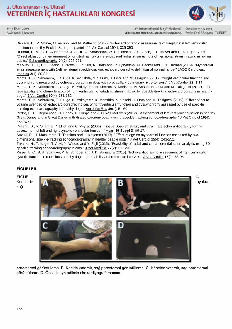
166
Dickson, D., R. Shave, M. Rishniw and M. Patteson (2017). "Echocardiographic assessments of longitudinal left ventricular
function in healthy English Springer spaniels." J Vet Cardiol 19(4): 339-350.
Hurlburt, H. M., G. P. Aurigemma, J. C. Hill, A. Narayanan, W. H. Gaasch, C. S. Vinch, T. E. Meyer and D. A. Tighe (2007).
"Direct ultrasound measurement of longitudinal, circumferential, and radial strain using 2-dimensional strain imaging in normal
adults." Echocardiography 24(7): 723-731.
Marwick, T. H., R. L. Leano, J. Brown, J. P. Sun, R. Hoffmann, P. Lysyansky, M. Becker and J. D. Thomas (2009). "Myocardial
strain measurement with 2-dimensional speckle-tracking echocardiography: definition of normal range." JACC Cardiovasc
Imaging 2(1): 80-84.
Morita, T., K. Nakamura, T. Osuga, K. Morishita, N. Sasaki, H. Ohta and M. Takiguchi (2019). "Right ventricular function and
dyssynchrony measured by echocardiography in dogs with precapillary pulmonary hypertension." J Vet Cardiol 23: 1-14.
Morita, T., K. Nakamura, T. Osuga, N. Yokoyama, N. Khoirun, K. Morishita, N. Sasaki, H. Ohta and M. Takiguchi (2017). "The
repeatability and characteristics of right ventricular longitudinal strain imaging by speckle-tracking echocardiography in healthy
dogs." J Vet Cardiol 19(4): 351-362.
Morita, T., K. Nakamura, T. Osuga, N. Yokoyama, K. Morishita, N. Sasaki, H. Ohta and M. Takiguchi (2019). "Effect of acute
volume overload on echocardiographic indices of right ventricular function and dyssynchrony assessed by use of speckle
tracking echocardiography in healthy dogs." Am J Vet Res 80(1): 51-60.
Pedro, B., H. Stephenson, C. Linney, P. Cripps and J. Dukes-McEwan (2017). "Assessment of left ventricular function in healthy
Great Danes and in Great Danes with dilated cardiomyopathy using speckle tracking echocardiography." J Vet Cardiol 19(4):
363-375.
Pellerin, D., R. Sharma, P. Elliott and C. Veyrat (2003). "Tissue Doppler, strain, and strain rate echocardiography for the
assessment of left and right systolic ventricular function." Heart 89 Suppl 3: iii9-17.
Suzuki, R., H. Matsumoto, T. Teshima and H. Koyama (2013). "Effect of age on myocardial function assessed by two-
dimensional speckle-tracking echocardiography in healthy beagle dogs." J Vet Cardiol 15(4): 243-252.
Takano, H., T. Isogai, T. Aoki, Y. Wakao and Y. Fujii (2015). "Feasibility of radial and circumferential strain analysis using 2D
speckle tracking echocardiography in cats." J Vet Med Sci 77(2): 193-201.
Visser, L. C., B. A. Scansen, K. E. Schober and J. D. Bonagura (2015). "Echocardiographic assessment of right ventricular
systolic function in conscious healthy dogs: repeatability and reference intervals." J Vet Cardiol 17(2): 83-96.
FİGÜRLER
FİGÜR 1. A.
Kedilerde ayakta,
sağ
parasternal görüntüleme. B. Kedide yatarak, sağ parasternal görüntüleme. C. Köpekte yatarak, sağ parasternal
görüntüleme. D. Özel dizayn edilmiş ekokardiyografi masası.
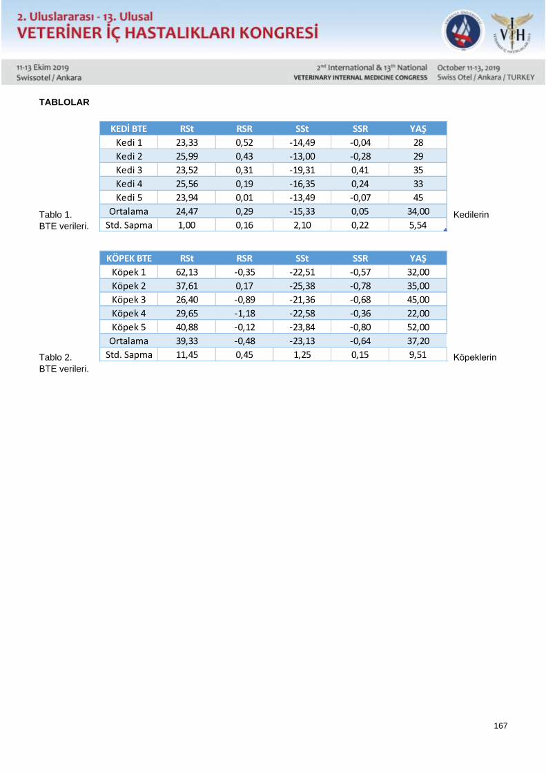
167
TABLOLAR
Tablo 1. Kedilerin
BTE verileri.
Tablo 2. Köpeklerin
BTE verileri.
KÖPEK BTE RSt RSR SSt SSR YAŞ
Köpek 1 62,13 -0,35 -22,51 -0,57 32,00
Köpek 2 37,61 0,17 -25,38 -0,78 35,00
Köpek 3 26,40 -0,89 -21,36 -0,68 45,00
Köpek 4 29,65 -1,18 -22,58 -0,36 22,00
Köpek 5 40,88 -0,12 -23,84 -0,80 52,00
Ortalama 39,33 -0,48 -23,13 -0,64 37,20
Std. Sapma 11,45 0,45 1,25 0,15 9,51
KEDİ BTE RSt RSR SSt SSR YAŞ
Kedi 1 23,33 0,52 -14,49 -0,04 28
Kedi 2 25,99 0,43 -13,00 -0,28 29
Kedi 3 23,52 0,31 -19,31 0,41 35
Kedi 4 25,56 0,19 -16,35 0,24 33
Kedi 5 23,94 0,01 -13,49 -0,07 45
Ortalama 24,47 0,29 -15,33 0,05 34,00
Std. Sapma 1,00 0,16 2,10 0,22 5,54
168
Sözlü Sunu / Oral Presentation Özet / Abstract
How To Use Two Dimensional Speckle Tracking Ecocardiography In Veterinary
Cardiology
Res. Ass. Osman Safa Terzi DVM PhD, Cansu Toksoz DVM
Ankara University, Faculty of Veterinary Medicine, Internal Medicine Department
Two-dimensional speckle monitoring echocardiography is a new technique based on the follow-up of user-defined
ultrasound points on the myocardium during systolic and diastolic events. During systole and diastole, heart moves
from base to apex and returns to its original point in healthy animals. The level of this movement is particularly
associated with ejection fraction in the left ventricle. Recently, in the veterinary cardiology field, the ejection capacity
of the left ventricle can be determined by the M mode. This method provides limited information as it measures the
movement of only a portion of interventricular septum and free wall in the left ventricle during systole and diastole. In
speckle tracking echocardiography, muscle movements of the left ventricle in the short and long axis can be
determined completely by strain measurement. The aim of this study is to introduce short axis radial and
circumferential strain and strain rate measurements by speckle tracking echocardiography technique in healthy cats
(n = 5) and dogs (n = 5) and to introduce this technique via cine-loops of these examinations.
169
Sözlü Sunu / Oral Presentation Özet / Abstract
İç Hastalıkları Kliniğine Getirilen Hastaların Retrospektif Olarak Değerlendirilmesi: 18198
Hastada Ankara Üniversitesi Deneyimi
Deniz Adıay, Nevra Keskin, Osman Safa Terzi, Ekrem Çağatay Çolakoğlu, Aslan Kalınbacak
Ankara Üniversitesi Veteriner Fakültesi İç Hastalıkları Anabilim Dalı, Ankara
Veteriner hekimliğe ilişkin sağlık problemleri ve çözümlerini daha iyi anlamak için araştırmacılar popülasyonların
sağlık durumu ve karakteristiklerine odaklanmaktadır. Bu bilgilerin eldesi; sadece klinik çalışmalara yardımcı olmak
amacıyla değil, aynı zamanda hekimin hastalarına yaklaşımında doğru veri toplamasının da önemini arttırmaktadır.
Bu çalışmada, Ankara Üniversitesi Veteriner Fakültesi Hayvan Hastanesi İç Hastalıkları Anabilim Dalı Kliniğine Mart
2018-Ağustos 2019 tarihleri arasında getirilmiş olan 18198 adet at, ruminant, kedi, köpek ve egzotik hayvana ait
veriler retrospektif olarak değerlendirildi.
18198 hayvan içinde, 13351 hayvana teşhis konulmuş ve tedavi edilmişti. Herhangi bir klinik şikayet ile getirilen
hayvanlar tüm hayvanların %35.33’ünü; rutin aşı ve antiparaziter uygulamalar ile kontrol ve sevk hastaları ise
%64,66’ını oluşturdu. En sık teşhis edilen hastalıklar enfeksiyöz ve paraziter hastalıklar (21%) olarak belirlendi. Bu
hastalıkları; gastrointestinal hastalıklar (19.54%), solunum sistemi hastalıkları (15.56%), üriner sistem hastalıkları
(12.14%), dermatolojik hastalıklar (11.17%), kardiyovasküler sistem hastalıkları (8.73%), onkolojik hastalıklar
(2.75%), karaciğer-pankreas hastalıkları (2.5%), endokrin hastalıklar (1.46%), hematolojik hastalıklar (1.25%),
otolojik hastalıklar (1.18%), toksikasyonlar (0.52%) ve diğerleri (2.14%) takip etti.
Ruminantlarda en sık karşılaşılan hastalıklar gastrointestinal sistem hastalıklarıyken; kedi ve köpeklerde ise sırasıyla
üriner sistem hastalıkları ve hemorajik gastroenteritis en sık karşılaşılan hastalıklar oldu. Egzotik hayvanlarda en sık
karşılaşılan hastalık muhabbet kuşlarında ve papağanlarda hava kesesi yangısıyken, tavşanlarda ise gastrointestinal
hastalıklar olarak kaydedildi.
Hastaların İç Hastalıkları kliniğine götürülmesine sebep olan birçok farklı bulgu olmasına rağmen, bizim
tecrübelerimize göre, veteriner hekimlerin sindirim ve üriner sistem muayenelerini ve testlerini, teşhiste ve tedavide
önemli rol oynaması sebebiyle daha detaylı yapması önerilmektedir. Verilerin değerlendirilmesinde hastane
otomasyon programının kullanılması veteriner fakültesi eğitim ve araştırma hastanelerine stratejik sağlayabilir.
Anahtar Kelimeler: veteriner, köpek, kedi, hastane
170
Sözlü Sunu / Oral Presentation Özet / Abstract
A Retrospective Study of Veterinary Patients in Internal Medicine Clinic: Ankara
University Experience on 18198 Animals
Deniz Adiay, Nevra Keskin, Osman Safa Terzi, Ekrem Cagatay Colakoglu, Aslan Kalinbacak
Ankara University Faculty of Veterinary Medicine Department of Internal Medicine, Ankara
In veterinary practise, several researchers focus on analysing the health status and population characteristics to
better understand the possible health problems and their solutions. Obtaining these data not only helps veterinarians
in their clinical practise, but also gains importance in terms of providing accurate information to physicians in their
communication with patients.
In this study, we retrospectively analysed the information of the caseload of internal medicine clinics of Ankara
University Veterinary Medical Teaching Hospital, from March 2018 to August 2019. The overall patient caseload was
18198, including horses, small and large ruminants, cats, dogs and exotic animals.
Of the 18198 animals, 13351 were diagnosed and treated. Medical complaints represented 35.33%, and wellness,
check up, control and dispatch patients represented 64.66% of diagnosed patients. The most common diagnosis
were infectious and parasitic diseases (21%). It was followed by gastrointesinal diseases (19.54%), respiratory
system diseases (15.56%), urinary system diseases (12.14%), dermatologic diseases (11.17%), cardiovascular
system diseases (8.73%), oncological disorders (2.75%), liver-pancreas diseases (2.5%), endocrine diseases
(1.46%), hematological diseases (1.25%), otologic diseases (1.18%), toxications (0.52%) and others (2.14%).
While the most common diseases among the large animals were related to gastrointestinal system disorders, it was
the urinary system diseases and haemorrhagic gastroenteritis in cats and dogs, respectively. The most prominent
diseases in exotic animals were aerosacculitis in budgerias and parrots, gastrointestinal disorders in rabbits.
Although there are several different diagnoses causing patients to be presented to the internal medicine clinics, in
our experience, veterinarians should make more detailed digestive and urinary system examinations and tests that
may play an important role in the diagnosis and treatment. Data obtained from the hospital software program may
also provide a strategy for the veterinary teaching hospitals.
Key Words: veterinary, dog, cat, hospital, caseload
171
Sözlü Sunu / Oral Presentation Özet / Abstract
Saha Çalışması İle Cryptosoporidium Enfeksiyonunun Ege Bölgesi Buzağılarında Anlık
Dağılımının Tespiti
Adnan AYAN*, Deniz ALIÇ URAL**, Elif TÜRK***, Kerem URAL***, Hasan ERDOĞAN***, Songül ERDOĞAN***,
Mehmet GÜLTEKİN***, Tahir ÖZALP***, Gizem GÜL***
* Van Yüzüncü Yıl Üniversitesi, Veteriner Fakültesi, Genetik Ana Bilim Dalı, Tuşba, Van, TÜRKİYE
** Aydın Adnan Menderes Üniversitesi, Veteriner Fakültesi, Fakülte Çiftliği, Işıklı, Aydın, TÜRKİYE
** Aydın Adnan Menderes Üniversitesi, Veteriner Fakültesi, İç Hastalıkları Ana Bilim Dalı, Işıklı, Aydın, TÜRKİYE
Neonatal buzağı ishalleri hayvancılık işletmelerinde buzağı ölümleri, ekonomik kaybın önde gelen olumsuz
umsurlarıdır. Neonatal buzağı ishallerinin etiyolojisinde bakteriler başta olmak üzere parazit ve viruslar
bulunmaktadır. Cryptosporidium parvum, rotavirüs ve koronavirüs, buzağı ishalin en önemli bulaşıcı nedenleri
arasında bulunmakta ve yaşamın ilk dört gününde buzağı ishallerinde sıklıkla rastlanmaktadır.C .parvum insanlarda
ve neonatal sığırlarda görülen gastrointestinal sistem (GIS) hastalıkları ile ilişkili protozon parazittir. Yenidoğan
buzağılar, çevrede bulunan oositlerle enfekte olmaktadır. Enfekte buzağılar en erken 2 günlük en geç de 14 günlük
yaşa kadar oositleri dökmeye devam ederler. Enterositlere invaze olan C. parvum enfeksiyonu şiddetli villus atrofisine
neden olurken mikrovilluslarda kaybı gibi bağırsak villuslarında değişiklikler yapmaktadır. Bu değişikliklere bağlı
olarak sindirilmemiş sütün fermantesyonu ve malabsorbsiyonunda bozulmalar sonucunda yetersiz beslenme ve ishal
görülmektedir. Aydın ilinde yapılan bu araştırmada ishal semptomu gösteren buzağılarda (n=119) cryptosporidium
ile monoenfekte ve koenfekte buzağıların belirlenmesi amaçlanmıştır. Bu kapsamda neonatal buzağılarda (n=84)
hızlı test kiti ile mono- ya da ko-enfeksiyon oranları belirlenmiştir. Yapılan hızlı test kiti ile hasta başı uygulamasında
monoenfeksiyon [Cryptosporidium spp. (n=23)] ya da koenfeksiyon [Cryptosporidium spp.+Corona virus (n=6), Rota
virus/Corona virus/Cryptosporidium spp. (n=2), Rota virus/Cryptosporidium spp. (n=5), Rota virus/Cryptosporidium
spp./Clostridium perfiringes (n=1), Cryptosporidium spp./Cl. perfiringes (n=10), E. coli/Cryptosporidium spp. (n=5)]
belirlendi Tüm bu sonuçlar saha koşullarında uygulanabilecek sağaltım protokellerinde değişim sağlayabilecek
niteliktedir.
Giriş
Neonatal ishal tüm dünyada buzağıların en önemli hastalıklarından biridir. (Holland, 1990). Buzağılarda yüksek
oranlarda mortalite ve morbiditeye sebep olan ciddi problemler görülebilir. (Cho ve Yoon 2014). Buzağı diyaresi ile
ilişkili major enterik patojenler; virusları (Bovine Rotavirus, Bovine Coronavirus, Bovine Viral Diarrhea Virus, Bovine
Torovirus, Bovine Norovirus, Nebovirus), bakterileri (Salmonella enterica, Escherichia coli, Clostridium perfringens),
Cryptosporidium parvum gibi protozoonu içerir (Papini ve ark. 1990). Cryptosporidium biyolojik monoksen özelliklere
sahiptir. Enfeksiyonlar çoğunlukla ağız yoluyla su ve yiyecekler vasıtasıyla ookistleri alarak gerçekleşir. (Sakarya ve
ark. 2010). Neonatal buzağılarda Cryptosporidiosis’ten kaynaklanan ekonomik kayıplar enfeksiyona sebep olan
Cryptosporidium’un tür/genotip’ine bağlıdır. (Asadpour ve ark. 2013).
Bu çalışmada Aydın ilinde ishal semptomu gösteren buzağılarda (n=119) cryptosporidium ile monoenfekte ve
koenfekte buzağıların belirlenmesi amaçlanmıştır.
Materyal ve Metot
Bu çalışmada Aydın ilinde ishal semptomu gösteren 119 adet buzağı dışkısı toplandı. Dışkı numuneleri manuel olarak
her bir hayvanın rektumundan tek kullanımlık lateks eldivenle alınarak dışkı kaplarına konuldu. Toplanan her numune
için hayvanın cinsiyeti ve yaşı kaydedildi. Daha sonra numuneler laboratuvara getirilerek inceleninceye kadar
+4°C’de saklandı. Daha sonra 84 adet neonatal buzağı dışkısına hızlı test kiti uygulanarak monoenfeksiyon ve
koenfeksiyon oranları belirlendi.
172
Bulgular
Hızlı test kiti uygulaması sonucunda 23 adet dışkı örneğinde Cryptosporidium spp. ile monoenfeksiyon görüldüğü ve
6 dışkı örneğinde Cryptosporidium spp.+Corona virüs, 2 dışkı örneğinde Rota virus/Corona virus/Cryptosporidium
spp., 5 dışkı örneğinde Rota virus/Cryptosporidium spp., 1 dışkı örneğinde Rota virus/Cryptosporidium
spp./Clostridium perfiringes, 10 dışkı örneğinde Cryptosporidium spp./Cl. perfiringes, 5 dışkı örneğinde E.
coli/Cryptosporidium spp. ile koenfeksiyon görüldüğü tespit edildi.
Tartışma
Neonatal buzağılarda diyare ciddi bir refah problemi ve mortalite, tedavi masrafları ve zayıf büyümeden, dolayı
ekonomik kayıpların sebebidir (Ammar ve ark. 2014). Kuliğ ve Coşkun, (2019) Sivas ve ilçelerindeki neonatal ishalli
138 adet buzağıdan 27’sinde (%20) çalışmalarında aranan enteropatojenlerden hiçbirine rastlanmadığını, 111 (%80)
tanesinde bir ya da daha fazla enteropatojen tespit edildiğini ve Buzağılardan 36 (%26) tanesinde E. coli, 30 (%22)
tanesinde Rotavirüs, 13 (%9) tanesinde Coronavirüs, 53 (%38) tanesinde Clostridium perfringens, 9 (%7) tanesinde
Cryptosporidium spp. tek ya da mix olarak belirlendiğini bildirmişlerdir. Al ve Balıkçı (2012)’de yaptıkları çalışmada
ishalli buzağıların 9’unda (%30) rotavirus, 4’ünde ( %13) coronavirus, 5’inde (%17) E. coli F5, 2’sinde (%6)
miksenfeksiyon, 10’unda (%33) diğer nedenlere bağlı ishal saptandığını ayrıca kullanılan ishalli buzağıların hiçbirinde
Cryptosporidium tespit edilmediğini bildirmiştir. Özalp ve Erdoğan, (2019) yapmış oldukları çalışmada buzağıların
%6’sının E. coli, %19’u Cryptosporidium sp., %13’ü Rotavirus, %6’sı Coronovirus, %6’sı Giardia ile mono-enfekte,
ko-enfekte buzağıların ise %11’i E. coli + Cryptosporidium sp., ve %22’sinin Rotavirüs + Cryptosporidium sp., ile
enfekte olduğunu ayrıca ve %17’sinin sağlıklı olduğunu bildirmiştir.
Araştırma kapsamında değerlendirilen buzağıların 23’ü Cryptosporidium spp. 6’sı dışkı örneğinde Cryptosporidium
spp. + Corona virüs, 2’si Rota virüs + Corona virüs + Cryptosporidium spp., 5’i Rota virüs + Cryptosporidium spp., 1’i
Rota virüs + Cryptosporidium spp. + Clostridium perfiringes, 10’u Cryptosporidium spp. + Cl. perfiringes, 5’i E. coli +
Cryptosporidium spp. ile enfekte olduğu belirlendi. Tüm bu sonuçlar saha koşullarında uygulanabilecek sağaltım
protokellerinde değişim sağlayabilecek niteliktedir.
References
Al M, Balıkçı E. 2012. Neonatal İshalli Buzağılarda Rotavirus, Coronavirus, E. coli K99 ve Cryptosporidium parvum'un Hızlı Test
Kitleri ile Teşhisi ve Enteropatojen ile Maternal İmmünite İlişkisi. F Ü Sağ Bil Vet Derg 26(2): 73-78.
Ammar SSM, Mokhtaria K, Tahar BB, Amar AA, Redha BA, Yuva B, Mohamed HS, Abdellatif N, Laid B. 2014. Prevalence of
rotavirus (GARV) and coronavirus (BCoV) associated with neonatal diarrhea in calves in western Algeria. Asian Pac J Trop
Biomed 4(1): S318-S322
Asadpour M, Razmi G, Mohhammadi G, Naghibi A. 2013. Prevalence and Molecular Identification of Cryptosporidium Spp. in Pre-
Weaned Dairy Calves in Mashhad Area, Khorasan Razavi Province, Iran. Iran J Parasitol 8(4): 601-607.
Cho YI, Yoon KJ. 2014. An overview of calf diarrhea – infectious etiology, diagnosis, and intervention. J Vet Sci 15:1-17.
Holland RE. 1990. Some infectious causes of diarrhea in young farm animals. Clin Microbiol Rev 3:345-375.
Küliğ CC, Coşkun A. 2019. Sivas ve İlçelerindeki Neonatal İshalli Buzağılarda E. coli, Cryptosporidium, Clostridium perfringens,
Rotavirüs ve Coronavirüs Prevalansı. Turk Vet J 1(2): 69-73
Özalp T, Erdoğan H. 2019. Evaluation of 25 (OH) D3 Concentrations in Neonatal Calves with Diarrhea. Kocatepe Vet J 12(3):
268-276
Papini R, Bonelli F, Montagnani M, Sgorbini M. 2018. Evaluation of three commercial rapid kits to detect Cryptosporidium parvum
in diarrhoeic calf stool. Ital. J Anim Sci 17(4): 1059-1064.
Sakarya Y, Kar S, Tanyuksel M, Karaer Z, Babur C, Vatansever Z. 2010. Detection of Cryptosporidium spp. in Humans and Calves
Through Nested PCR and Carbol Fuchsin Staining Methods in Ankara, Turkey. Kafkas Univ Vet Fak Derg 16 (6): 977-980.
Giriş
Materyal ve Metot
Bulgular
Tartışma
173
Sözlü Sunu / Oral Presentation Özet / Abstract
Field study regarding spatial distribution of cryptosoporidium infection among Eagean
Region calves
Adnan AYAN*, Elif TÜRK**, Kerem URAL**, Hasan ERDOĞAN**, Songül ERDOĞAN**, Mehmet GÜLTEKİN**,
Tahir ÖZALP**, Gizem GÜL**
* Van Yüzüncü Yıl Üniversitesi, Veteriner Fakültesi, Genetik Ana Bilim Dalı, Tuşba, Van, TÜRKİYE
** Aydın Adnan Menderes Üniversitesi, Veteriner Fakültesi, İç hastalıkları Ana Bilim Dalı, Işıklı, Aydın, TÜRKİYE
Calf deaths in livestock are the leading negative factors for economic loss. In the etiology of neonatal calf diarrhea,
parasites and viruses, especially bacteria, are present. Cryptosporidium parvum, rotavirus and coronavirus are
among the most important infectious causes of calf diarrhea and are frequently encountered in calf diarrhea in the
first four days of life. C.parvum is a protozone parasite associated with gastrointestinal system (GIS) diseases in
humans and neonatal cattle. Newborn calves are infected with oocytes in the environment. Infected calves continue
to shed oocytes until the age of 2 days on earliest and 14 days at the latest. Infection of C. parvum, which invades
enterocytes, causes severe villus atrophy, and changes in intestinal villi, such as loss of microvilli. Due to these
changes, malnutrition and malabsorption of undigested milk may lead to malnutrition and diarrhea. In this study
conducted in Aydın province, it was aimed to determine monoinfected and coinfected calves with cryptosporidium
associated with diarrhea symptoms (n = 119). In this context, mono- or co-infection rates were determined with the
rapid test kit in neonatal calves (n = 84). Monoinfection [Cryptosporidium spp. (n = 23)] or coinfection
[Cryptosporidium spp. + Corona virus (n = 6), Rota virus / Corona virus / Cryptosporidium spp. (n = 2), Rota virus /
Cryptosporidium spp. (n = 5), Rota virus / Cryptosporidium spp./Clostridium perfiringens (n = 1), Cryptosporidium
spp./Cl. perfiringes (n = 10), E. coli / Cryptosporidium spp. (n = 5)]. All of these results may alter the treatment
protocols that can be applied in the field conditions.
174
Sözlü Sunu / Oral Presentation Özet / Abstract
Feline Gingivostomatitislerin Tedavisinde Hipokloröz’ün Klinik Başarısı
Ali Evren HAYDARDEDEOĞLU, Müge BİRCAN AKKARTAL
Aksaray Üniversitesi Veteriner Fakültesi İç Hastalıkları Ana Bilim Dalı, Aksaray/Türkiye *Nova Veteriner Kliniği,
Balıkesir/Türkiye
Feline Gingivostomatitis (FGS) kedilerde yaygın görülen bir sendromdur. Farenks, bukkal mukoza, dil ve diş etinde
ağrılı, erozif lezyonlara yol açan yıkıcı, kronik bir kedi hastalığıdır. Bu hastalığın tedavisinde en önemli klinik yaklaşım
ağız temizliğidir. Bu amaçla FGS tespit edilen 20 kedi yavrusunda sağıltıma destek amaçla Hipokloroz asiti ağız
temizliği ve tedavi amacı ile kullanılmıştır. Hastaların klinik muayenesinde, şiddetli bir halitosis, oral disfaji,
hipersalivasyon ve yüksek ateş tespiti yapılmıştır. Hastalığın şiddetli enfeksiyonla seyrettiği vakalara (n=12) 7 gün
süreyle amoksisilin+klavulanik asit ile birlikte ve tüm hastalara sprey olarak günde 5 kez hipokloröz asit (%0,02 HOCl
Crystalin) enfekte akıntı ve kabuklar temizlendikten sonra sprey olarak uygulandı. Oral enfeksiyonu olmayan sadece
gingivostomatitis olan (n=8) vakalarda sadece hipokloröz asit uygulaması ve oral besleme solüsyonları ile tüm
hayvanların beslemesi yapıldı. Tüm vakalarda FeLV, FIV, Panlökopeni, FCV ve FHV yönünden hızlı test kitleri
yönünden teşhise yönelik taramalar yapılıp. Virolojik yönden negatif olan vakalar çalışmaya dahil edilmiştir.
Anahtar Kelimeler: Feline, Gingivostomatitis, Hipokloröz Asit
175
Sözlü Sunu / Oral Presentation Özet / Abstract
Clinical Success of Hypochlorous in the Treatment of Feline Gingivostomatitis
Ali Evren HAYDARDEDEOĞLU, Müge BİRCAN AKKARTAL
Aksaray University Faculty of Veterinary Medicine Department of Internal Medicine, Aksaray/Turkey *Nova
Veterinary Clinic, Balıkesir / Turkey
Feline Gingivostomatitis (FGS) is a common syndrome in cats. Pharynx is a devastating, chronic cat disease that
causes painful, erosive lesions on the buccal mucosa, tongue and gum. The most important clinical approach in the
treatment of this disease is oral hygiene. For this purpose, hypochlorous acid was used for oral cleaning and
treatment in 20 kittens with FGS. Clinical examination of the patients revealed severe halitosis, oral dysphagia,
hypersalivation and high fever. Clinical examination of the patients revealed severe halitosis, oral dysphagia,
hypersalivation and high fever. In cases with severe infection (n = 12), hypoxlorous acid (0.02% HOCl Crystalin) was
sprayed 5 times daily with amoxicillin + clavulanic acid and sprayed to all patients. In cases without gingivostomatitis
(n = 8) without oral infection, all animals were fed with hypochlorous acid application and oral feeding solutions. In
all cases FeLV, FIV, Panleukopenia, FCV and FHV for rapid test kits for diagnostic screening. Virological negative
cases were included in the study.
Keywords: Feline, Gingivostomatitis, Hypochlorous Acid
176
Sözlü Sunu / Oral Presentation Özet / Abstract
Neonatal İshalli Buzağıların Aksaray Bölgesinde Anlık Dağılımının Belirlenmesi
Ali Evren HAYDARDEDEOĞLU
Aksaray Üniversitesi Veteriner Fakültesi İç Hastalıkları Ana Bilim Dalı, Aksaray/Türkiye
Buzağı yetiştiriciliğinde en önemli sorunların başında gelmekte olan neonatal dönem hastalıkları ve bunların da en
mühimi buzağı ishalleri ciddi sıvı ve elektrolit kayıpları, gelişme geriliği ve sağaltım masrafları gibi bilinen ve öngörülen
sebepler dışında, son dönemde literatürde sözü edilen pıhtılaşma bozukluğu gibi göz ardı edilen nedenlerle de
mortaliteye sebebiyet vermektedir. Söz konusu ekonomik kayıpların buzağı ishallerine neden olan etmenlerin hızlı
bir şekilde teşhisinin yanı sıra oluşturdukları klinikopatolojik değişikliklerin belirlenmesiyle en aza indirilebileceği
belirtilmektedir. Aksaray ilinde, bulunan işletmelerinden Fakültemiz kliniklerine getirilen neonatal ishale ait klinik
bulguları mevcut buzağılara (n=en az 35) Bovid-5 katı fazlı immunokromatografi yöntemi ile çalışan hızlı test kitleri
aracılığıyla dışkı örneklerinden alınan numuneler ile tanı konulmuştur. Bu bağlamda olgular öncelikli olarak mono-
infeksiyona ilişkin en az 5 gruba, ko-infeksiyon durumlarına bağlı olarakta ilgili alt gruplara ayrılmıştır. İlgili ana
gruplara (her grupta n=en az 7) ayrılarak I. grup (E. coli ile infekte), II. grup (Rota virüs ile infekte), III. grup (Corona
virüs ile infekte), IV. grup (Cryptosporidium sp. ile infekte) ve V. grup (Giardia sp. ile infekte) hasta buzağılar yer
almıştır, kontrol olarak ayırılacak VI. grupta ise (n=en az 10) ise sağlıklı buzağılar yer alamıştır.
Son dönemde neonatal ishalli buzağılarda koagülasyon profilinde değişiklikler oluşabileceği yönünde sınırlı sayıda
çalışma literatürde yerini almaktadır. Anılan çalışmalarda etiyolojik nedenlere bakılmaksızın ve hastalık etkenleri
belirlenmeden yalnızca ishal baz alınarak değerlendirmeler yapılmıştır.
Aksaray bölgesinde ishalli buzağılar belirlenerek bahse konu test kitleri ile anlık mikrobiyel etkenlerin dağılımının
belirlenmesi amaçlanmıştır.
Anahtar Kelimeler: Buzağı, İshal, Neonatal, Anlık, Dağılım
Bu Çalışma Aksaray Üniversitesi BAP proje ofisi tarafından desteklenmiştir.
177
Sözlü Sunu / Oral Presentation Özet / Abstract
Determination of The İnstant Distribution of Calves With Neonatal Diarrhea in Aksaray
Region
Ali Evren HAYDARDEDEOĞLU
Aksaray University Faculty of Veterinary Medicine Department of Internal Medicine, Aksaray/Turkey
The most important problems in calf cultivation are neonatal diseases and the most important of them are calf
diarrhea, with serious fluid and electrolyte losses, growth retardation and treatment costs as well as the known and
prescribed reasons. İt gives rise to. It is stated that these economic losses can be minimized by the rapid identification
of the factors that cause calf diarrhea as well as by identifying the clinicopathological changes they create. The clinical
findings of neonatal diarrhea in Aksaray province, which were brought to our faculty clinics, were diagnosed with the
samples obtained from stool samples by means of rapid test kits with Bovid-5 solid phase immunochromatography
method. In this context, the cases were primarily divided into at least 5 groups related to mono-infection and related
sub-groups due to co-infection conditions. Group I (infected with E. Coli) was separated into the main groups (n = at
least 7 in each group), II. Group (infected with route virus), III. Group (infected with Corona virus), IV. Group (infected
with Cryptosporidium sp.) and group V (infected with Giardia sp.) patient calves were taken, as control VI. Healthy
calves were included in the group (n = 10).
Recently, a limited number of studies have been published in the literature in terms of changes in the coagulation
profile of calves with neonatal diarrhea. In the aforementioned studies, evaluations were made on the basis of only
diarrhea, regardless of etiologic factors and disease factors.
Diarrheal calves were determined in Aksaray region and it was aimed to determine the distribution of these microbial
agents with the test kits.
Keywords: Calf, Diarrhea, Neonatal, Instantaneous, Distribution
178
Sözlü Sunu / Oral Presentation Özet / Abstract
Merada Beslenmeyen İneklerde İnterstitiel Anfizem Olguları
Hasan Batmaz, İ.Taci Cangül
Bursa Uludağ Üniversitesi Veteriner Fakültesi, İç Hastalıkları AD, Bursa * Bursa Uludağ Üniversitesi Veteriner
Fakültesi, Patoloji AD, Bursa
Sığırlarda interstitiel anfizem (atipik interstitiel pneumoni) olguları genellikle gümrah meralarda otlayan sığırlarda
fazla alınan triptofanın rumende 3-metil indol’e dönüşmesi sonucu meydana gelir. Bu sunuda merada beslenmeyen
toplam 162 adet laktasyondaki Holştayn inekten 10’unda ortaya çıkan interstitiel anfizem olgularının sunulması
amaçlanmıştır. Yaklaşık 20 günlük sürede ineklerin birinde hafif, diğerlerinde orta-şiddetli düzeyde atipik pneumoni
dikkati çekmiştir. Yaşları 3-8 arasında olan ineklerde dikkati çeken en belirgin bozukluk, ağız açık ve hafif köpüklü bir
şekilde solunum güçlüğü olmasına rağmen belirgin ateş ve öksürük hemen hemen hiç olmamıştır. Hafif bir olgu
iyileşirken diğerlerinin hiçbiri tedaviye cevap vermemiştir. Ölen ilk hayvanın otopsisinde akciğerlerin belirgin hava ile
dolu olduğu ve adeta kostaların izinin olduğu dikkati çekmiştir. Hastalığın nedeninin araştırılması sonucu bu ineklerde
kuru yonca otunun yanısıra bu yıla özgü yaklaşık 1.5 aydır gümrah yonca silajının birlikte verilmesinden ileri
gelebileceği üzerinde durulmuştur. İnek başına günlük 5 kg kuru yonca ve 3 kg yonca silajı verildiği belirtilmiştir.
Yonca silajının yedirilmesi hemen durdurulmuş ve gruplarda süt veriminde yükselme gözlenmiş ve yonca silajı
verilmesi kesildikten yaklaşık 10 gün sonrasından itibaren yeni olgu çıkmamıştır. On olgudan hafif olan biri hastalığı
atlatmış, ancak diğer 9’u klinik belirtiler başladıktan sonraki 2-5 gün içinde ölümle sonuçlanmıştır. Hepsinin otopsi
bulgularında belirgin interstitiel anfizem gözlenmiştir. Kuru yonca ve yonca silajının triptofan düzeyleri ölçüldüğünde
miktarları sırasıyla 225 mg/100 g ve 153 mg/100 g saptanmış ve hayvanların aldıkları triptofan miktarının yüksek
olduğu görülmüştür.
Sonuç olarak laktasyondaki süt ineklerine kaba yem verilirken yonca gibi aşırı triptofan kaynaklarının fazla
yedirilmemesi, mera dışında da beslenmeye bağlı çok sayıda interstitiel anfizem olgularına rastlanabileceği
sonucuna varılmıştır.
Anahtar kelimeler: Sığır, interstitiel anfizem, yonca
179
Sözlü Sunu / Oral Presentation Özet / Abstract
Interstitial Emphysema Cases in Nongrazing Cows
Hasan Batmaz, İ.Taci Cangül
Bursa Uludağ University, Faculty of Veterinary Medicine, Department of Internal Medicine, Bursa-Turkey Bursa
Uludağ University, Faculty of Veterinary Medicine, Department of Pathology, Bursa-Turkey
Interstitial emphysema (atypical interstitial pneumonia) occurs in cattle which are grazed particulary in lush, rapidly
growing pastures after the ruminal degradation of L-tryptophan into 3-methylindole. This presentation is about
interstitial emhysema cases observed in 10 of 162 nongrazing lactating Holstein cows. In a period of around 20 days,
atypical pneumonia was observed in 10 cows, one case being mild and the rest being moderate-severe. The age of
the cows was between 3-8 years and the major clinical sign was dyspnea with open and frothy mouth; there were no
signs of fever or coughing. Only one case with mild clinical signs recovered, but the rest of the animals did not
respond to any treatment and died. Necropsy of the first animal revealed non-collapsing, air filled lungs with
impressions of costae. Feeding wih 5 kg of dehydrated alfalfa and 3 kg of lush alfalfa silage daily in the last 1.5
months was suggested as the cause of the disease.
Feeding with alfalfa silage was halted immediately; milk production of the cows increased and no further cases were
observed after 10 days. Only one animal recovered and the rest of the animals died within 2-5 days after the initiation
of the clinical signs. Prominent interstitial emphysema was observed during the necropsy of all animals. Tryptophan
levels of dehydrated alfalfa and lush alfalfa silage were measured as high as 225 mg/100 g and 153 mg/100 g.
As a conclusion we suggest that feeding of lactating cows with feedstuff containing high levels of tryptophan should
be avoided and that interstitial emphysema can also be observed in nongrazing lactating cows.
Keywords: Cattle, interstitial emphysema, alfalfa
180
Sözlü Sunu / Oral Presentation Özet / Abstract
Türkiye’de Kedilerde İlk Kalıtsal Polikistik Böbrek Hastalığı (PKD)
Nüket BİLGEN*, Merve BİŞKİN TÜRKMEN**, Yusuf ŞEN3, Mustafa Yenal AKKURT*, Özge Şebnem ÇILDIR*,
Sevim ISPARTA*, Zeynep BARS*, Bengi ÇINAR KUL*
* Genetik Anabilim Dalı, Veteriner Fakültesi, Ankara Üniversitesi, Ankara
**Patoloji Anabilim Dalı, Veteriner Fakültesi, Kırıkkale Üniversitesi, Kırıkkale
*** Cerrahi Anabilim Dalı, Veteriner Fakültesi, Ankara Üniversitesi, Ankara
Amaç: Bu çalışmanın amacı, Otozomal Dominant Polikistik Böbrek Hatalığı (ADPKD)'nın Anadolu Kedileri'ndeki
yaygınlığının ölçülmesi ve detaylı patofizyolojik değerlendirmesinin yapılmasıdır.
Materyal ve Metot: Farklı ırk ve yaşta sağlıklı 230 adet kedinin, PKD1 geninin 29 ekzonunda PCR-RFLP ile C→A
transversiyonu genotiplendirildi. Ek olarak, böbrek şikayeti olan dört kedide ultrason, serum-biyokimya ve idrar
analizi yapıldı. Böbrek yetmezliği nedeniyle ölen bir adet kedide ise nekropsi ve patolojik incelemeler gerçekleştirildi.
Bulgular: Böbrek şikayeti olan kedilerin ultrason analiz incelemelerinde böbreklerde kist oluşumu gözlendi ve bu
örnekler için C→A transversiyonu pozitiflik gösterdi. Nekropsi yapılan kedinin böbrek ve karaciğerinde değişen
boyutlarda çok sayıda kist görüldü. Histopatolojik incelemede, makroskobik kistlerin yanısıra, küçük mikrokistik
yapılara rastlandı. Kist çevresinde dejenere ve atrofik tubüller ile intersitisyel fibrozis yaygındı. Bu tubül epitellerinde,
dejenerasyon ve nekroz gözlendi. Karaciğerde ise, sınırlı sayıda mikroskobik kistler izlendi. Periportal alanlar
genişlemiş ve çok sayıda mononükleer hücreler içermekteydi. İmmunohistokimyasal analizler sonucunda; özellikle
dejenere tubül epitellerinde güçlü Caspase-3 ve TNF-α immunoreaktivitesi görüldü. Bcl-2 immunoreaktivitesi daha
çok normal görünümdeki tubül epitellerinde, TGF-β ekspresyonu ise fibrozis şekillenmiş alanlar ve tubül epitellerinde
tespit edildi. Sağlıklı kedilerin mutasyon taraması, nedensel mutasyon belirlenmedi.
Sonuç: Dejenere tubüllerde yüksek caspase-3 aktivitesi, böbrekte kistik sürecin etiyopatogenezinde apoptotik
sürecin aktif rol aldığını kanıtlarken, caspase-3 aktivitesinin TNF-alfa ekpresyonları ile ko-lokalizasyonu, apoptozisin
dışsal yol ile başlatılabileceğini göstermektedir. Yaygın intersitisyel fibrozis alanlarında, artmış TGF-beta
ekspresyonu tek olumsuz sonucun kist oluşumu değil, aynı zamanda böbrek fibrozisininde klinik böbrek
yetmezliğinde önemli bir unsur olabileceğini göstermektedir. PKD, uzun süre asemptomatik olabilir. Bu nedenle
yatkınlığı ve böbrek şikayeti olan kedilerde erken tanı için ultrason yapılmalıdır. Ayrıca genlerdeki mutasyonlar ve
hastalık prognozunun aydınlatılması, PKD'ye yönelik genetik kökenli tedavilerin geliştirilmesinde hayvan modeli
oluşturmaya katkı sağlayabileceği düşünülmektedir.
Anahtar Kelimeler: Anadolu kedileri, ADPKD , polikistik, böbrek
181
Sözlü Sunu / Oral Presentation Özet / Abstract
First Report of Hereditary Polycystic Kidney Disease (PKD) of Cats in Turkey
Nüket BİLGEN*, Merve BİŞKİN TÜRKMEN**, Yusuf ŞEN***, Mustafa Yenal AKKURT*, Özge Sebnem CILDIR*,
Sevim ISPARTA*, Zeynep BARS*, Bengi ÇINAR KUL*
* Department of Genetics Veterinary Faculty Ankara University, Ankara
** Department of Pathology Veterinary Faculty Kırıkkale University, Kırıkkale
*** Department of Surgery Veterinary Faculty Ankara University, Ankara
Aim:It is aimed to detect Autosomal Dominant Polycystic Kidney Disease (AD-PKD) prevalance in cats and to
establish pathophysiologic evaluation.
Material and Method:230 healthy cats in different ages, breeds were genotyped by PCR-RFLP for C→A transversion
mutation on 29th exon of PKD1 gene. In addition to these, 4 cats with renal symptoms were clinically examined and
they were also tested for PKD gene. One cat died during the study and it was forwarded to necropsy.
Results:Renal cysts were observed in ultrasound analysis of cats with renal symptoms, and C → A transversion was
positive for these samples. The cat underwent necropsy had multiple cysts were seen in the kidney and liver.
Histopathological examination revealed macroscopic cysts and small microcystic structures. Degenerated and
atrophic tubules and interstitial fibrosis were common around the cyst. A limited number of microscopic cysts were
observed in the liver. In immunohistochemical analysis, strong Caspase-3 and TNF-α immunoreactivity was observed
in the degenerated tubular epithelium. Bcl-2 was mostly expressed in normal tubular epithelium, whereas TGF-β was
expressed in fibrotic areas and tubules. None of the healthy cats carried the causative mutation.
Results:High caspase-3 activity in degenerated tubules proves that apoptotic process plays an active role in the
etiopathogenesis of cystic process in the kidney, whereas co-localization of caspase-3 activity with TNF-alpha
expressions suggests that apoptosis can be initiated by exogenous pathway. In diffuse interstitial fibrosis, increased
TGF-beta expression indicates that not only renal fibrosis may be an important factor in cyst formation but also in
clinical renal failure. PKD can be asymptomatic for a long time. Therefore, ultrasound should be performed for early
diagnosis in cats with predisposition and renal health issues. For elucidation of disease prognosis, mutations in genes
are thought to contribute to animal models in the development of genetic-based treatments for PKD.
Keywords:Anatolian cats,ADPKD,polycycstic,kidney
182
Sözlü Sunu / Oral Presentation Özet / Abstract
Hayvan Sağlık Sigortası Yaptırma İsteğini Etkileyen Faktörler Sivas İli Örneği
Özcan KARATAŞ, Zahid AĞAOĞLU
Sivas Cumhuriyet Üniversitesi Veteriner İç Hastalıkları Anabilim Dalı, Sivas
Bu çalışma, Sivas bölgesinde çiftçilerin hayvan hayat sigortası yaptırma isteklerini etkileyen faktörleri belirlemek
amacıyla yapılmıştır. Bu amaçla, farklı büyüklük ve kapasitede büyükbaş ve küçükbaş hayvan yetiştiriciliği yapan 50
işletme çalışmaya alınmıştır. Çalışmanın materyalini bölgede yetiştiricilik yapan ve hayvan hayat sigortası yaptırmış
50 çiftçi oluşturmuştur.
Verilerin toplanması Ocak-Haziran 2019 tarihleri arasında 30 sorudan oluşan anket yöntemi ile yapılmıştır. Anketler
bire bir görüşülerek gerçekleştirilmiştir. Elde edilen verilerin istatistiksel analizinde frekans analizi ve yüzde
hesaplamaları uygulanmıştır.
Araştırma sonuçlarına göre; çiftçilerin eğitim düzeyi, hayvanlarını güvence altına alma istekleri ve bankadan kredi
çekme avantajının hayvan hayat sigortası yaptırmalarında etkili faktörler olduğunu göstermiştir.
Anahtar kelimeler: Tarım sigortaları, karar verme, Sivas
183
Sözlü Sunu / Oral Presentation Özet / Abstract
Factors Affecting The Request For Animal Health Insurance Sivas Province Sample
Özcan KARATAŞ, Zahid AĞAOĞLU
Sivas Cumhuriyet Üniversitesi Veteriner İç Hastalıkları Anabilim Dalı, Sivas
This study was carried out in order to determine the factors affecting the farmers' desire to have animal life insurance
in Sivas region. For this purpose, 50 enterprises engaged in bovine and ovine breeding of different sizes and
capacities were included in the study. The material of the study consisted of 50 farmers engaged in husbandry and
animal life insurance in the region.
The data were collected between January and June 2019 using a questionnaire consisting of 30 questions.The
questionnaires were conducted by one-to-one interviews. In the statistical analysis of the data, frequency analysis
and percentage calculations were applied.
According to the results of the research; farmers' level of education, the desire to secure their animals and the
advantage of attracting credit from the bank were effective factors in taking out animal life insurance.
Key words: Agricultural insurance, decision making, Sivas
184
Sözlü Sunu / Oral Presentation Özet / Abstract
Serum Leptin And Ghrelin Levels And Their Relationship With Serum Cortisol, Thyroid
Hormones, Lipids, Homocysteine And Folic Acid İn Dogs With Compulsive Tail Chasing
Ebru YALCIN*, Zeki YILMAZ*, Yesim OZARDA**
*Department of Internal Medicine, Faculty of Veterinary Medicine, Uludag University,
**Department of Biochemistry, Medical School, Uludag University
The aim of this study was to investigate serum leptin and ghrelin levels and their relations with circulating cortisol,
thyroid hormones, lipids, homocysteine (Hcy) and folic acid in dogs with compulsive tail chasing (CTC). The material
of this study consists of fifteen dogs with CTC and 15 healthy controls of various weights, breeds, ages of both sexes
were enrolled in the study. CTC was diagnosed on the basis of the dog’s behavioral history, clinical signs, and results
of other medical assessments. None of the dogs were considered to have concurrent medical disease that would
account for CTC. Dogs with CTC had a higher leptin (8.3±0.9 ng/mL vs 1.7±0.2 ng/mL, P<0.001) and lower ghrelin
levels (74±7 pg/mL vs 144±41 pg/mL, P<0.05) than those of healthy controls. Serum cortisol, lipids (cholesterol,
phospholipids and NEFA) and Hcy levels increased (P<0.05), whereas serum folic acid decreased (P<0.001) in dogs
with CTC as compared with controls. Serum ghrelin correlated negatively with cholesterol (P<0.05), but serum leptin
correlated positively with cholesterol, fT4, and phospholipids (P<0.05). These results suggest that serum leptin and
ghrelin levels may bring up a new perspective on our understanding of the pathophysiological mechanisms
associated with CTC. Serum levels of both hormones may be associated with serum levels of lipids and free T4.
Keywords: Leptin, ghrelin, thyroid hormones, lipids, tail chasing, dog
185
Sözlü Sunu / Oral Presentation Özet / Abstract
Geçiş Dönemindeki Sığırlarda Hastabaşı Bazı Metabolik Parametreler Arasındaki
Korelasyonun Değerlendirilmesi*
Songül Erdoğan, Kerem Ural
Aydın Adnan Menderes Üniversitesi Veteriner Fakültesi İç Hastalıkları Anabilim Dalı, Aydın
Çalışmamızda geçiş dönemindeki sığırlarda BHBA, NEFA, Mg+2, Ca+2, laktat ve VKS’ nu içeren bazı metabolik
parametrelerin korelasyon ve varyasyonun belirlenmesi amaçlanmıştır. Her işletmede en az 12 hayvan olacak şekilde
toplamda 37 inekten geçiş döneminde haftalık olarak sabah beslemesinden 6-8 saat sonra V. Coccygea’ dan kan
örnekleri alındı. Tüm analizler, talimata uygun olarak optimum koşullarda çiftlik ortamında gerçekleştirildi.
Parametreler arasındaki olası ilişkileri ortaya çıkarmak için Pearson korelasyon testi kullanıldı. I. Çiftlikte Mg+2-NEFA
(0.55, p <0.05), BHBA-laktat (0.965, p<0.01), BHBA-VKS (0.703, p <0.05); I. Çiftlikte VKS-laktat (-0.672, p<0.05) ve
son olarak NEFA-VKS (0.772, p<0.01), Mg+2-VKS (-0.582, p<0.05) ile Mg+2-laktat (0.611, p<0.05). arasında ilişki
saptandı. Sonuç olarak, geçiş ineklerinde metabolik profili değerlendirmek için sıklıkla kullanılan parametreler
arasında anlamlı bir ilişki olabileceği ve daha geniş popülasyonlar ile farklı çalışmaların yapılması gerektiği
düşünüldü.
Anahtar Kelimeler: BHBA, Kalsiyum, Laktat, Magnezyum, NEFA.
* Bu çalışma Aydın Adnan Menderes Üniversitesi Bilim Araştırma Projeleri Ofisi (ADU-BAP) tarafından VTF-17045
proje numarası ile desteklenerek doktora tezinden özetlenmiştir.
186
Sözlü Sunu / Oral Presentation Özet / Abstract
Correlation of Cowside Measured Some Metabolic Parameters in Transition Cattle*
Songul Erdogan, Kerem Ural
Aydın Adnan Menderes University, Faculty of Veterinary Medicine, Department of Internal Medicine, Aydın
The objective of our study was to be describing the correlation and variation of some metabolic parameters included
BHBA, NEFA, Mg+2, Ca+2, lactate and BCS in transition cattle. Blood samples collected weekly from d 14 prepartum
to 14 postpartum for 37 cows were selected from 3 different dairy farm in which being at least 12 cases and from V.
coccygea 6-8 hours after early feeding. All analysis was performed farm condition with optimum temperature related
to instruction. Pearson Correlation test were used to reveal both the possible relationships between parameters.
Significant correlations between the parameters were found as Mg+2 and NEFA (0.55, p <0.05) in the I farm, BHBA-
lactate (0.965, p<0.01), BHBA-BCS (0.703, p <0.05) and BCS-lactate (-0.672, p<0.05) in the II farm and lastly NEFA-
BCS (0.772, p<0.01), Mg+2-BCS (-0.582, p<0.05) and Mg+2-lactate (0.611, p<0.05). in the III farm. In conclusion, it
was thought that there might be a significant relationship between the parameters often used to evaluate metabolic
profile in transition cows and different studies should be conducted with wider populations.
Keywords: BHBA, Calcium, Lactate, Magnesium, NEFA.
* This study was supported by Aydın Adnan Menderes University Sciencitic Research Projects Office (ADU-BAP)
with project number VTF-17045 and summarized from the doctoral thesis.
Introduction
Important physiological and metabolic alteration develop in transition period that affect performance, productive and
health of dairy cattle (1). Transition cattle coupled with negative energy balance (NEB) due to increased energy
demand including pregnancy, lactation despite decreased feed intake. As mostly non-esterified fatty acids (NEFA)
and beta-hydroxybutyric acid (BHBA) as indicators of NEB elevate in the blood when cattle fail to adaptation (2).
Besides these two phenomena generally evaluated together, potentially other indicators are prognosticate of NEB in
transition cattle associated diseases and metabolic adaptation (2-5). With this study we wanted to respond of
question that interpretation of the association between markers. Although relation of NEFA and BHBA for interpreting
of NEB resulted negative health and production is well known, there is no evidence that relation with other parameters
can be used together or interchangeably and all parameters mentioned above are sampled consecutively at a specific
time after feeding from the same cattle. The objective of our study was to described the correlation and variation of
some metabolic parameters included BHBA, NEFA, Mg+2, Ca+2, lactate and BCS in transition cattle.
Material and method
Animals and management
Three different farms in Aydın with 1) ≥250 cows, 2) freestall housing, 3) total mixed ration (TMR) feeding and/or 4)
dairy herd improvement programs were admitted to study as totally 37 cows having minimally 12 cows in each farm
with different breed, age and body weight. Farms management was determined with formed questionnaire by ourself
and farm visiting was performed weekly during the study. As a result of questionnaire performed by farm veterinarian
and manager, all farm doing TMR feeding twice a days after milking.
Blood analysis
Blood sampling were weekly performed at prepartum (-2nd and -1th week), postpartum (+1th and +2nd week) groups
and within the first 24 h at parturition group (0. d) by puncture of V. coccygea at 6 to 8 hours after early feeding.
187
Samples were taken to anticoagulant tubes with lithium-heparin and K3EDTA. Immediately after collection, blood
were centrifuged by hand-held device (Sprout, Healthrow Scientific, USA) to be used cow-side at 6000 rpm and
separated plasma.
Assay of parameters
BHBA level were analyzed with K3EDTA blood shortly after collected by cow-side handheld Vet TD-4235 ß-Keto
device (Antalya, Turkey) using amperometric method. The analyses of NEFA, Mg+2, Ca+2 and lactate were performed
with heparinized plasma by another veterinary cow-side device Vet Photometer 700 DP (Diaglobal, Germany;
distributor Genartek, Izmir, Turkey) using enzymatic colorimetric method. All analyses procedure was done in the
farm condition.
Body condition score
The classical BCS using a 5-point scale with 0.25 increments (1 to 5, thin to fat) were utilized (6) at prepartum,
parturition and postpartum groups and classified based on 1) 2<BCS<3, 2) 3< BCS <4 and 3) 4≥ BCS in itself for
statistic model.
Statistical analyses
Proper statistical methods were used to reveal both the possible relationships between parameters that calculated
using the Pearson Correlation test.
This study was presented as part of phD thesis of author and approved by Adnan Menderes University Local Ethical
Committee under certificate number: 64583101/2016/169.
Result
Relationship Between Biological Parameters
Correlations between the parameters for each farm were shown in Table 1. In the I farm, only correlations between
Mg+2 and NEFA was found significant (0.55, p <0.05). Similarly, correlations between the biochemical parameters
were numerically small and negative in the II farm, differently BHBA-lactate (0.965, p<0.01), BHBA-BCS (0.703, p
<0.05) and BCS-lactate (-0.672, p<0.05) correlation were found to be important. In the II farm, it can be said that
effect of BCS on lactate was negative and on BHBA was positive. And lastly the relations between the parameters
in the III farm were small and below zero, as in other farms and NEFA-BCS (0.772, p<0.01), Mg+2-BCS (-0.582,
p<0.05) and Mg+2-lactate (0.611, p<0.05) were determined.
Discussion
We aimed to described the correlation and variation of BHBA, NEFA, Mg+2, Ca+2, lactate and BCS sampled
consecutively at a specific time after feeding from the same cattle in transition period for each farm.
Lactate, endproduct of cellular glycolysis mediated with lactate dehydrogenase (7), is transported to liver and reused
for glucose synthesis during the gluconeogenesis stage and create energy for other tissues and organs (8). It was
stated that increased lactate dehydrogenase (9) as well as lactate levels associated with lameness (10). It was
pointed out that increased BHBA eventuated higher lactate dehydrogenase moreover prolonged lameness resulted
in more lactate production in tissue (9,10). Similarly, it was determined in our study that cases with lower BCS of II.
farm had an positive correlation between BHBA and BCS, lactate along with negative correlation between lactate
and BCS. Starting from these, lameness of cases with lowest BCS in the II farm might be associated with elevated
lactate levels. However, cattle with higher BCS has also related to developing risk of lameness (9). It can be said
that BCS was generally higher in Simmental breeding farm and considering lameness, determined lower lactate
levels of only 3 cattle in 2<BCS<3 group resulted from increased mean value due to the lack of cases. Taking into
consideration of both positions, it is evident that further research is needed with more cases to elucidate possible
relationship between lactate and VCS in detail.
188
In this study, considering Mg+2 correlation with other parameters, there was a relation with NEFA in the I farm (0.552,
p<0.05), lactate (0.611, p<0.05) and BCS (-0.582, p<0.05) in the III farm. Mg+2 is used in all physiological mechanisms
consumed ATP at the cellular level (11) similar to liver hepatocyte most of cytoplasmic ATPs being complex with
Mg+2 (12) in addition to most of Mg+2 in liver is cofactor of enzyme used in energy metabolism including TCA cycle,
glycolysis and gluconeogenesis (13). Magnesium deficiencies cause accumulation of triglycerides in the liver by
reducing the above-mentioned enzyme and protein synthesis involved in the oxidation of fatty acids (14). Although
there is no study the direct effect of Mg+2 on NED as well as the relationship with NEFA in transition cows, it is
possible to consider of a negative relationship between these two parameters in the light of all above information and
can be correlated with energy balance and/or stress. In contrast to these, it has been reported that Mg+2 levels
increase in acute hepatocellular injury due to possible leakage from damaged cell parallel to bilirubin (15). In transition
cattle Mg+2 was found to be dense (16) in hepatocyte related to mitochondrial damage. In another study indicated
that leakage of Mg+2 out of the cell as resulted of mitochondrial swelling related to oxidative stress in mice with
hypomagnesemia (17). It was notified that hepatic lipidosis was positively related to postpartum NEFA, BHBA and
total bilirubin (18). Although positive correlation between NEFA and Mg+2 in the I farm of our study may be explained
in the light of the above information, we thought that the possible complex relationship of Mg+2 with NEFA should be
elucidated by further studies.
It was stated that fed with magnesium sulphate had no effect on body condition in dry period nutrition (19), on the
other hand, adding to diet caused insulin resistance due to changing metabolic function resulted in obesity that had
a negative correlation with Mg+2 (20). In other study performed in transition cattle with hypomagnesemia to
investigated hormonal and metabolic alteration was indicated that increased glucagon/insulin ratio in parallel to
hypomagnesemia (21). High milk yield cows had an insulin resistance in transition period due to lactogenesis and
increased energy demand by NEB (22) and so insulin resistance is directly relevant in cattle with increased BCS
(23). In our study the negative interaction between Mg+2 and BCS in III farm can be explained with these. In fact
positive relation between BCS and NEFA of the same farm supports this thesis.
Considering to Mg+2 and lactate cooperation, hypomagnesemia could result in lactic acid production due to anaerobic
metabolism by interrupting oxidative phosphorylation (24). Mg+2 levels presented negative correlation with lactate
contrary to our result that observed to decreased from prepartum period to postpartum might be explained with heavy
anaerobic exercise cause to decreasing Mg+2 related to intercompartmental shifting and urine excretion and it return
to normal levels a few days after exercise (25) and with occurred anaerobic capacity as a result of decreased
fractional excretion of Mg+2 breaks of intracellular Mg+2 functions in nonpregnant lactation cows (26).
In conclusion, it was thought that there might be a significant relationship between the parameters often used to
evaluate metabolic profile in transition cows and different studies should be conducted with wider populations.
References 1. Herdt TH. Variability characteristics and test selection in herd-level nutritional metabolic profile testing. Veterinary Clinics of North America:
Food Animal Practice 2000, 16, 387-403.
2. Chapinal N, Carson ME, Duffield TF, Capel M, Godden S, Overton MW, Santos JE, LeBlanc SJ. The association of serum metabolites with
clinical disease during the transition period. Journal of Dairy Science 2011, 94(10), 4897-4903.
3. Goff JP. Mineral disorders of the transition period: origin and control. 24th Proceedings of the World Buiatrics Congress, 15-19 October 2006,
Nice, France.
4. Huzzey JM, Nydam DV, Grant RJ, Overton TR. Associations of prepartum plasma cortisol, haptoglobin, fecal cortisol metabolites, and non-
esterified fatty acids with postpartum health status in Holstein dairy cows. Journal of Dairy Science 2011, 94(12), 5878–5889.
5. Weaver SR, Laporta J, Moore SA, Hernandez LL. Serotonin and calcium homeostasis during the transition period. Domestic animal
endocrinology, 2016, 56, S147-S154.
6. Edmonson AJ, Lean IJ, Weaver LD, Farver T, Webster G. A body condition scoring chart for Holstein dairy cows. Journal of Dairy Science,
1989, 72(1), 68-78.
7. Rogatzki M, Ferguson B, Goodwin L, Gladden B. Lactate is always the end product of glycolysis. Frontiers in Neuroscience 2015, 9, 1-7.
8. Seal CJ, Reynolds CK. Nutritional implications of gastrointestinal and liver metabolism in ruminants. Nutrition Research Reviews 1993, 6, 185-
208.
189
9. Ristevski M, Toholj B, Cıncovıć M, Trojačanec P, Starıč J, Smolec O. Milk Production, Body Condition Score and Metabolic Parameters at the
Peak of Lactation as Risk Factors for Chronic Lameness in Dairy Cows. Kafkas Üniversitesi Veteriner Fakültesi Dergisi 2017, 23(5), 721-727.
10. Zhang G, Hailemariam D, Dervishi E, Deng Q, Goldansaz SA, Dunn SM, Ametaj BN. Alterations of innate immunity reactants in transition
dairy cows before clinical signs of lameness. Animals 2015, 5(3), 717-747.
11. Ağaoğlu ZT, Yüksek N, Başbuğan AGY. Magnezyum Metabolizması ve Bozuklukları. Turkiye Klinikleri Journal of Veterinary Sciences 2011,
2(2), 162-167.
12. Luthi D, Günzel D, McGuigan JA. Mg-ATP binding: its modification by spermine, the relevance to cytosolic Mg2+buffering, changes in the
intracellular ionized Mg2+concentration and the estimation of Mg2+by 31P-NMR. Experimental Physiology 1999, 84(2), 231-52.
13. Cowan JA. Structural and catalytic chemistry of magnesium-dependent enzymes. Biometals 2002, 15, 225-235.
14. Etwebi Z. Magnesium Regulation of Glucose and Fatty Acid Metabolism in HEPG2 Cells. Doctoral dissertation, Case Western Reserve
University, Cleveland 2011, 89.
15. Gowda P, Tembad MM. Study of serum magnesium in liver diseases. Journal of Evolutıon of Medıcal and Dental Scıences-Jemds 2015,
4(18), 3047-3056.
16. Bogucka K, Wojtczak L. Binding of magnesium by proteins of the mitochondrial intermembrane compartment. Biochemical and Biophysical
Research Communications 1976, 71(1), 161-167.
17. George GA, Heaton FW. Effect of magnesium deficiency on energy metabolism and protein synthesis by liver. International Journal of
Biochemistry, 1978, 9(6), 421-425.
18. Kirovski D, Sladojevic Z. Prediction and Diagnosis of Fatty Liver in Dairy Cows. Journal of Gastroenterology 2017, 3(1), 1005.
19. Stockdale CR. Effects of feeding magnesium sulfate to dry pregnant dairy cows with different body condition scores on intake in late gestation,
periparturient blood calcium concentrations and production in early lactation. Australian Journal of Experimental Agriculture 2004, 44(6), 539-546.
20. Huerta MG, Roemmich JN, Kington ML, Bovbjerg VE, Weltman AL, Holmes VF, Nadler JL. Magnesium deficiency is associated with insulin
resistance in obese children. Diabetes Care 2005, 28(5), 1175-1181.
21. Schimitt E, Pereira RA, Hoffmann D, Vendramin L, Lima ME, dos Santos Farofa T, Corrêa, MN. Hipomagnesemia subclínica em vacas leiteiras
durante o período de transição: ocorrências hormonais e metabólicas. Revista Brasileira de Ciência Veterinária 2016, 23(1-2).
22. Chalmeh A, Pourjafar M, Nazıfı S, Momenıfar F, Mohamadı M. Circulating Metabolic Profile of High Producing Holstein Dairy Cows.
Veterınarskı Arhıv 2014, 85(6), 621-633.
23. De Koster J, Opsomer G. Effect of body condition score on insulin sensitivity of precalving dairy cows. 15th International conference on
Production Diseases in Farm Animals, p67, 24-28 May 2013, Belgium.
24. Moskowitz A, Lee J, Donnino MW, Mark R, Celi L, Danziger J. The association between admission magnesium concentrations and lactic
acidosis in critical illness. Journal of Intensive Care Medicine 2016, 31(3), 187-192.
25. Deuster PA, Dolev E, Kyle SB, Anderson RA, Schoomaker EB. Magnesium homeostasis during high-intensity anaerobic exercise in men.
Journal of Applied Physiology 1987, 62(2), 545-550.
26. Schweigel M, Voigt J, Mohr E. Indication of intracellular magnesium deficiency in lactating dairy cows revealed by magnesium loading and
renal fractional excretion. Journal of Animal Physiology and Animal Nutrition 2009, 93(1), 105-112.
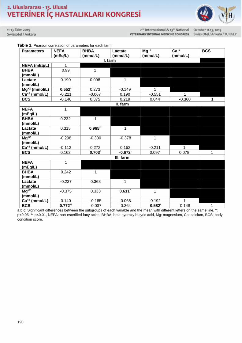
190
Table 1. Pearson correlation of parameters for each farm
Parameters NEFA (mEq/L)
BHBA (mmol/L)
Lactate (mmol/L)
Mg+2 (mmol/L)
Ca+2 (mmol/L)
BCS
I. farm
NEFA (mEq/L) 1
BHBA (mmol/L)
0.99 1
Lactate (mmol/L)
0.190 0.098 1
Mg+2 (mmol/L) 0.552* 0.273 -0.149 1
Ca+2 (mmol/L) -0.221 -0.067 0.190 -0.551 1
BCS -0.140 0.375 0.219 0.044 -0.360 1
II. farm
NEFA (mEq/L)
1
BHBA (mmol/L)
0.232 1
Lactate (mmol/L)
0.315 0.965** 1
Mg+2 (mmol/L)
-0.298 -0.300 -0.378 1
Ca+2 (mmol/L) -0.112 0.272 0.152 -0.211 1
BCS 0.162 0.703* -0.672* 0.097 0.078 1
III. farm
NEFA (mEq/L)
1
BHBA (mmol/L)
0.242 1
Lactate (mmol/L)
-0.237 0.368 1
Mg+2 (mmol/L)
-0.375 0.333 0.611* 1
Ca+2 (mmol/L) 0.140 -0.185 -0.068 -0.192 1
BCS 0.772** -0.037 -0.364 -0.582* -0.148 1 a.b.c: Significant differences between the subgroups of each variable and the mean with different letters on the same line, *:
p<0.05, ** p<0.01, NEFA: non-esterified fatty acids, BHBA: beta hydroxy butyric acid, Mg: magnesium, Ca: calcium, BCS: body
condition score.
191
Sözlü Sunu / Oral Presentation Özet / Abstract
Akut İshalli Neonatal Buzağılarda Plazma Sitrulin Konsantrasyonunun Değerlendirilmesi
Mehmet Gültekin, Hüseyin Voyvoda, Kerem Ural, Hasan Erdoğan, Canberk Balıkçı, Gamze Gültekin
Aydın Adnan Menderes Üniversitesi Veteriner Fakültesi İç Hastalıkları Anabilim Dalı, Aydın
Aydın Adnan Menderes Üniversitesi Veteriner Fakültesi Biyokimya Anabilim Dalı, Aydın
Plazma sitrülin konsantrasyonu özellikle insanlarda, fonksiyonel enterosit kütlesinin güvenilir bir belirteci olarak kabul
edilmektedir. Bu çalışmada sağlıklı ve akut ishalli neonatal buzağılarda plazma sitrulin konsantrasyonu ve ilişkili
parametrelerin değerlendirilmesi amaçlandı.
Çalışmada 10 sağlıklı, 60 akut ishalli olmak üzere her iki cinsiyetten toplam 70 neonatal (<28 günlük yaş) Holstein
ırkı buzağı kullanıldı. Klinik ve laboratuvar bulgular temelinde ishalli buzağılar etiyolojiye göre enfeksiyöz (n=50) ve
non-enfeksiyöz (n=10), ishalin şiddetine göre hafif (n=15), orta (n=25) ve şiddetli (n=20), sistemik yangısal yanıt
sendromu (SIRS) kriterlerine göre SIRS pozitif (n=27) ve SIRS negatif (n=33) olarak alt gruplarda değerlendirildi.
Sağlıklı buzağılarda parametrelerde yaşa bağlı değişikliklerin belirlenmesi amacıyla örneklemeler 2.,7.,14.,21. ve 28.
yaşam günlerinde, ishalli buzağılarda ise bir kez yapıldı. Dışkı örneklerinde etiyolojik değerlendirme; Cryptosporidium
parvum, Rotavirus, Coronavirus, E. coli K99 ve Giardia duodenalis antijenlerine yönelik hızlı test kiti, parazitolojik ve
mikrobiyolojik muayene ile gerçekleştirildi. Rutin laboratuvar analizler, tam kan sayımı, kan gazları ve biyokimyasal
parametreleri kapsadı. Plazma sitrulin konsantrasyonu yanında, sitrulin ile ilişkili arjinin, nitrik oksit ve amonyak
konsantrasyonları ölçüldü.
Sağlıklı buzağılar göre, akut ishalli buzağılarda plazma sitrulin ve arjinin konsantrasyonları düşük, kan amonyak ve
plazma nitrik oksit düzeyleri ise yüksek saptandı. Plazma sitrulin konsantrasyonundaki azalmaların etiyolojiye göre
enfeksiyöz, ishalin şiddetine göre hafif, orta ve şiddetli, SIRS değerlendirmesine göre ise SIRS pozitif gruplarda
olduğu belirlendi. Plazma arjinin konsantrasyonunun enfeksiyöz, şiddetli ve SIRS pozitif gruplarda azaldığı görüldü.
Nitrik oksit düzeyi enfeksiyöz, hafif, orta, şiddetli ve SIRS pozitif gruplarda yüksek bulundu. Amonyak düzeyindeki
artışların enfeksiyöz, şiddetli ve SIRS pozitif gruplarda olduğu ortaya konuldu.
Sonuç olarak, akut ishalli buzağılarda plazma sitrulin ve arjinin konsantrasyonlarında azalma ile kan amonyak
düzeyinde artış olduğu ilk defa belirlendi. Belirlenen değişikliklerin akut ishalli neonatal buzağılarda tanısal,
prognostik ve terapotik değere sahip olabileceği kanısına varıldı.
*Bu çalışma TÜBİTAK tarafından 3001 programı kapsamında desteklenen 116O014 no'lu projenin bir bölümüdür.
192
Sözlü Sunu / Oral Presentation Özet / Abstract
Evaluation of Plasma Citrulline Concentration in Neonatal Calves with Acute Diarrhea
Mehmet Gultekin, Huseyin Voyvoda, Kerem Ural, Hasan Erdogan, Canberk Balikci, Gamze Gultekin
Aydın Adnan Menderes University, Faculty of Veterinary, Department of Internal Medicine, Aydin, TURKEY
Aydın Adnan Menderes University, Faculty of Veterinary, Department of Biochemistry, Aydin, TURKEY
In this study, it was aimed to evaluate plasma citrulline concentration and related parameters in healthy and neonatal
calves with acute diarrhea.
A total of 70 neonatal (<28 days of age) Holstein calves of both sexes, comprising 60 with acute diarrhea and 10
healthy were enrolled in the study. On the basis of clinical and laboratory findings, calves with diarrhea was classified
as infectious and non-infectious according to etiology, mild, moderate and severe according to severity, systemic
inflammatory response syndrome (SIRS) positive and negative according to the criteria of the SIRS.
Etiological evaluation was provided with parasitological, microbiological examination and rapid test kit analysis
against antigens of Cryptosporidium parvum, Rotavirus, Coronavirus, E. coli K99 and Giardia duodenalis in stool
samples. Routine laboratory analysis covered by complete blood count, blood gases and serum biochemical
parameters measurement. Besides the concentration of plasma citrulline, arginine, nitric oxide and ammonia
concentrations were determined.
Plasma citrulline and arginine concentrations were found to be lower while concentrations of blood ammonia and
plasma nitric oxide concentrations were higher in calves with acute diarrhea. The reductions in plasma citrulline
concentrations were found to be in infectious, mild, moderate and severe diarrhea, and SIRS positive groups. Plasma
arginine concentration decreased in infectious, severe and SIRS positive groups. Nitric oxide levels were high in
infectious, mild, moderate, severe and SIRS positive groups. Increases in ammonia levels were found to be in
infectious, severe and SIRS positive groups.
As a result, the decreased concentrations of plasma citrulline and arginine, and the increase in blood ammonia level
were firstly determined in calves with acute diarrhea. It’s concluded that the changes in these variables might have
diagnostic, prognostic, and therapeutic value in diarrheic neonatal calves.
*This study is part of the project supported by TUBITAK 3001 programme (no. 116O014).
193
Sözlü Sunu / Oral Presentation Özet / Abstract
Kedi ve Köpeklerde Yaşa Bağlı Değişimlere İlgili Hayvan Sahiplerinin Algılarının
Değerlendirilmesi
Alev Akdoğan Kaymaz, Alper Bayrakal, Sinem Ülgen
İstanbul Üniversitesi-Cerrahpaşa Veteriner Fakültesi, İç hastalıklar Anabilim Dalı, Avcılar-İstanbul
Hayvanlarda yaşlanma; dolaşım bozuklukları, böbrek yetmezliği, mental bozukluklar vb. kronik hastalıkların
oluşumuna ve bu hastalıklara bağlı gelişen bazı davranış değişimlerine neden olabilmektedir. Günümüzde evlerde
beslenen kedi ve köpek sayısının giderek artması ve bu konudaki bilinçlenme ile insanlarda olduğu gibi hayvanlarda
da kaliteli bir şekilde yaşlanma büyük önem kazanmaktadır. Buradan yola çıkılarak planlanan bu çalışmada; çeşitli
sebeplerle kliniğimize getirilen toplam 900 geriatrik pet (421 kedi ve 479 köpek) sahibi ile yapılan bir anketle, bu
hayvanlarda yaşlanmayla birlikte sosyal ve doğal fonksiyonlar ile fiziksel ve mental sağlık durumları belirlenmiştir.
Diğer taraftan, bu süreçte hasta sahiplerinin evlerini paylaştığı hayvanlarının bakımı ve ruhsal açıdan yaşam
kalitesinin nasıl etkilenebileceği değerlendirilmiştir.
194
Sözlü Sunu / Oral Presentation Özet / Abstract
Evaluatıon of Perceptıons of Anımal Owners Related To Age-Related Changes In Cat And
Dogs
Alev Akdoğan Kaymaz, Alper Bayrakal, Sinem Ülgen
Istanbul Unıversity - Cerrahpasa, Faculty of Veterinary Medicine, Department of Internal Medicine, Avcılar- Istanbul
Aging in animals can cause the formation of chronical diseases such as circulatory disorders, renal failure, mental
disorders, etc. and developing behavioral changes due to these diseases. Nowadays, with the increasing number
of cats and dogs fed in the houses and with the awareness on this subject, aging in animals is gaining great
importance as well as in humans. In this study, which was planned based on this; a total of 900 geriatric pets (421
cats and 479 dogs) who were brought to our clinic for various reasons were identified in this questionnaire to
determine the social and natural functions and physical and mental health status of these animals with aging.
Besides, it was also determined how it influenced the pet owner of animal care and qualty of life.
195
Sözlü Sunu / Oral Presentation Özet / Abstract
Atopik Dermatitli Köpeklerde Fekal Mikrobiyota Transplantasyonu ile Sağaltım: Derinin
Altında, Bağırsakların İçerisinde Gastroentero-Dermatolojiye Açılan Kapılar
Kerem URAL
Aydın Adnan Menderes Üniversitesi, Veteriner Fakültesi, İç Hastalıkları Anabilim Dalı, Işıklı, Aydın
Alerjik hastalıklar gerek insan gerekse köpeklerde uzun müddettir heme gizemli hemde çekici konular arasında yer
almıştır. Atopik dermatit (Ad) insanlara benzer şekilde köpekler için de oldukça önemli, muhtemelen de en önde
gelen alerjik ve dermatolojik problemlerden birisidir. Güncel bildirimlerde köpeklerdeki Ad’in insanlarda aynı hastalık
için rol model olarak değerlendirilmektedir. Anılan hastalıkta öne sürülen hipotezlerden birisi de sızıntılı bağırsak
modeline eşlik eden intestinal permeabilite artışı ve bağırsak mikrobiyatasının bozulması olsa da, konuya ilişkin gerek
tanı gerekse sağaltım anlamında aradaki ilişkiye dair bilinmeyenler mevcuttur. Bu çalışmada Ad’li köpeklerde hastalık
aktivitesi ile bağırsak mikrobiyatası arasındaki ilişkinin fekal mikrobiyota transplantasyon sağaltım ile ortaya
konulması amaçlanmıştır. Çalışmada toplamda 7 Ad’li köpek (farklı yaş, ırk ve cinsiyetten) dahil edildi. Önceden
herhangi bir sağaltım uygulaması yapılmamış, Favrot kriterleri ve atopi ile uyumlu klinik bulgular gösteren (kaşıntı
vb.), alerjen-spesifik IgE düzeyinde artış şekillenmiş olgular CADESI-04 skorları ve mikrobiyota analizleri (16 s rRNA
genom tayini) ile belirlendikten fekal mikrobiyata transplantasyonu ile 15 gün boyunca sağaltıma alındı. Tüm
olgularda sağaltım sonrası Firmicutes:Bacteriodetes oranları, bağırsak mikrobiyomunda şekillenen değişimlerin yanı
sıra CADESI-04 skorlarında gerileme ile kaşıntı vd. klinik bulgularda tamamen gerileme olması Veteriner Dermatoloji
Alanına yönelik yeni bir sağaltım modülünün oluşmasına ait emareler olarak değerlendirilebilinir. Uygun motto ‘derinin
altında ama bağırsakların içerisinde’ söylemine eşlik edecek şekilde dermatoloji olgularının beyin-bağırsak-deri
ekseni ile sızıntılı bağırsak/sızıntılı deri gibi yeni yaklaşımlar doğrultusunda gastroenteroloji ile birlikte
değerlendirilmesi gerekliliği açıktır. Yeni hipotez gastroentero-dermatoloji olabilecektir.
196
Sözlü Sunu / Oral Presentation Özet / Abstract
Fecal Microbiota Transplantation Treatment in Dogs With Atopic Dermatitis: Doors
Opening To Gastroentero-Dermatology Under The Skin, İnside The Gut
Kerem URAL
Aydin Adnan Menderes University, Faculty of Veterinary, Department of Internal Medicine, Isikli, Aydin
Abstract
Allergy has long been a topic of mystery and fascination. Atopic dermatitis (Ad) mimicking simlarity to humanbeing,
as well as in dogs a very significant and probably one of the foremost dermatologic problems. Updated literature
suggested that Ad in dogs is a role model for the same disease in humans. Among hypothesis regarding the latter
disease, leaky gut syndrome associated intestinal permeability increase and disbiosis of gut microbiata, unknown
remains clear to those of both diagnosis and treatment. In the present project the aim was to evaluate the relationship
between disease activity among dogs with Ad within gut microbiota through fecal microbiota transplantation treatment
modality. In the present project a total of 7 dogs with Ad (among different age, breed and of both sexes) were
enrolled. Neither previous treatment application, clinical signs compatible with Favrot criteria and atopy, to those of
cases with increased allergen specific IgE, CADESI-04 scoring and microbiata analysis [16 s rRNA genom detection]
were subjected to fecal microbiota transplantation treatment for 15 days.Following treatment alterations among
Firmicutes:Bacteriodetes ratio, gut microbiome along within CADESI-04 scores and withdrawal of pruritus/other
relevant clinical findings should deemed circumstantial evidence of a novel therapeutical armamentarium against
Veterinary Dermatology. Given appropriate motto ‘under the skin but inside the gut’ appeal should accompany
interpretation of dermatology cases along with gastroenterology based on novel findings such as brain-gut-skin axis
with leaky gut/leaky skin. A new hypothesis might be gastroentero-dermatology.
Key words: FMT, canine atopy, microbiome
Introduction
The microbiome, nowadays renamed as a new organ, is one of the foremost activator for immune system
homeostasis (Bowe and Logan, 2011, Ali et al., 2014, Guo et al., 2013, Majamaa and Isolauri, 1996, Marsella et al.,
2001). Alterations in microbial composition/function, known as dysbiosis, in the skin and the gut have recently been
linked to alterations in immune responses and to the development of skin diseases, such as atopic dermatitis (AD)
(Ali et al., 2014, Guo et al., 2013, Majamaa and Isolauri, 1996, Marsella et al., 2001). Atopic dermatitis (AD) has
been linked to changes in intestinal permeability (Guo et al., 2013, Suchodolski et al. 2009), thus probably to
microbiota composition. There are limited therapeutical options for CAD (Marsella and Girolomoni, 2009, Favrot et
al., 2010, which prompted me search for a safe and effective long term therapeutical armamentarium for CAD. The
aim of this field study was to analyze efficacy of a commercially available FMT capsule in CAD.
Material and methods
The case series enrolled with written owner consent and comperative evaluation involved pretreatment clinical
biomarkers with values on two subsequent re-evaluations at day 0 and 28, thereafter completion of treatment.
Research era / Inclusion criteria
A total of 4 dogs with pruritic dermatoses (Hill et al., 2007) were initially diagnosed with a presumed Cad based on a)
history, b) clinical findings, c)lab work (through skin pathology) (Marsella and Girolomoni, 2009, Favrot et al., 2010).
Full clinical assessments were performed on days 0, and 28 at the Faculty Clinic. The Canine Atopic Dermatitis
Extent and Severity Index version 4 (CADESI-04) itenarary was used to score the clinical findings involving erythema
(acute), lichenification (chronic), excoriations/alopecia and pruritus. The owners assessed pruritus via a 10 cm visual
197
analog scale (VAS) involving descriptors (Cosgrove at al, 2013) similar to another study (Hill et al., 2007). The owners
were asked to complete a questionnaire for the number of treatment applications, side effects, or other necessary
information.
In vitro allergen detection Polycheck Canine Allergy Test (Polycheck, Allergy test, Gmbh, Germany; distributer RDA Group, Istanbul) an vitro
test which detects allergen-specific IgE in dog serum) was used (Ural et al., 2018) to detect underlying etiology as
previously described (Bizikova et al., 2015, Ural et al., 2018).
Trial protocol
The present veterinary surgeon (K.U. along with his pHD students applied oral FMT capsules at initial referral
following completion of analysis protocole (Animal Biome Gut Restoration System FMT capsules, Animal Biome,
Oakland, California) nationally distributed by RDA Group, İstanbul, Turkey. The latter capsules were adviced to be
applied twice daily for 4 weeks perorally.
Microbiome testing
This procedure briefly included 16 S rRNA gene (505F/816 R) (Pichler et al., 2018) generation using one step PCR
(Comeau et al, 2017, Pichler et al., 2018), iv) alpha and beta diversity analysis (Callahan et al., 2016), involving
100% operational taxonomic units (OTUs) classified against SILVA reference database (Quast et al. 2013). All of the
procedures were completed at Animal Biome Laboratory at Oakland, USA by experted and specialized people
(approximately 58 dollar for each sampling).
Results
Demographic data
A total of 7 dogs were enrolled which were intact male. At initial referral age ranged from 3 to 6 years, weighing 12 to 38 kg. None of the dogs were withdrawn during study period, nor side effects were recorded during treatment. No previous antibiotic treatment or any biological compound usage were evident. Plasma cortisol and total and free thyroxine values were checked for all cases, which determined reference ranges.
Gut composition
Comperative evaluation of gut microbiota analysis indicated changes in Firmicutes:Bacteriodetes ratio among 4 dogs
enrolled before [case I: 126.44, case II: 1.09, Case III: 0.88, case IV:1.35] and after completion of treatment [ 0.39
,0.66, 0.67, 3.85, respectively]
Discussion
The present study suggested that FMT capsules administration appears to be a highly effective treatment for Cad.
Changes in both pruritus and clinical lesions, as detected by VAS pruritus scores and CADESI-04, respectively,
showed significant importance. All 7 dogs achieved decreases in both CADESI-04 with a clinical improvement in this
study. The CADESI-04 and pruritus scores were highly correlated (data not shown due to small sample size),
suggesting both that the commercially available FMT capsules caused alterations among clinical lesions and that the
improvement was, to the present authors’ knowledge, relevant to the efficacy, which might be due to i) restoration of
gut microbiota (swifting disbiosis (as previously described by Watanabe and Nansawa, 2003) to eubiosis.
Therefore it should not be unwise to draw suggestion that Ad might be reflection of gut disbiosis and elevated
intestinal permeability (Craig, 2016). Hence manuplation of gut microbiota to those of dogs with Ad in the present
study by use of oral FMT capsules and clinical recovery might all support those findings.
Referances Cosgrove SB, Wren JA, Cleaver DM, Martin DD, Walsh KF, Jessica A Harfst JA,Follis SL, King VL, Boucher JF, Michael R Stegemann MR
(2013). Efficacy and safety of oclacitinib for the control of pruritus and associated skin lesions in dogs with canine allergic dermatitis. Vet Dermatol
24(5): 479–e114.
Hill PB, Lau P, Rybnicek J (2007). Development of an owner‐assessed scale to measure the severity of pruritus in dogs. Vet Dermatol 18(5) :301-
308.
Ural K, Erdoğan H, Gültekin M (2018). Allergen specific IgE determination by in vitro allergy test in head and facial feline dermatitis: A pilot study.
Ankara Üniv Vet Fak Derg 65: 379-386.
198
Bizikova P, Santoro D, Marsella R (2015). Review: Clinical and histological manifestations of canine atopic dermatitis. Vet Dermatol 26(2): 79.
Pichler R, Fritz J, Tulchiner G (2018). Increased accuracy of a novel mRNA‐based urine test for bladder cancer surveillance. BJU Int 121(1): 29-
37.
Comeau AM, Douglas GM, Langille MG (2017). Microbiome helper: a custom and streamlined workflow for microbiome research. MSystems 2(1):
e00127-16.
Callahan BJ, McMurdie PJ, Rosen MJ (2016). DADA2: high-resolution sample inference from Illumina amplicon data. Nat methods 13(7): 581.
Quast S, Berger A, Eberle J (2013). ROS-dependent phosphorylation of Bax by wortmannin sensitizes melanoma cells for TRAIL-induced
apoptosis. Cell Death Dis 4(10): e839.
Watanabe S, Nansawa Y (2003). Differences in fecal microflora between patients with atopic dermatitis and healthy control subjects. J Allergy
Clin Immunol 111: 587– 591.
Marsella R, Olivry T, Carlotti DN (2001). International task force on canine atopic dermatitis: Current evidence of skin barrier dysfunction in human
and canine atopic dermatitis. Vet Dermatol 22: 239– 248.
Majamaa H, Isolauri E (1996). Evaluation of the gut mucosal barrier: evidence for increased antigen transfer in children with atopic eczema. J
Allergy Clin Immunol 97(4): 985-990.
Guo S, Al‐Sadi R, Siad HM (2013). Lipopolysaccharide causes an increase in intestinal tight junction permeability in vitro and in vivo by inducing
enterocyte membrane expression and localization of TLR‐4 and CD14. Am J Pathol 182: 375– 387.
Bowe WP, Logan AC (2011). Acne vulgaris, probiotics and the gut-brain-skin axis-back to the future?. Gut pathog 3(1): 1.
Ali IA, Foolad N, Sivamani RK (2014). Considering the Gut‐Skin Axis for Dermatological Diseases. Austin J Dermatol 1: 1024.
Craig JM (2016). Atopic dermatitis and the intestinal microbiota in humans and dogs. J Vet Med Sci 2(2): 95-105.
Suchodolski JS, Dowd SE, Westermarck E (2009). The effect of the macrolide antibiotic tylosin on microbial diversity in the canine small intestine
as demonstrated by massive parallel 16S rRNA gene sequencing. BMC Microbiol 9: 210.
Smit JJ, Folkerts G, Nijkamp FP (2004). Mycobacteria, genes and the ‘hygiene hypothesis’ Curr Opin Allergy. Clin Immunol 4(1): 57–62.
Watanabe S, Narisawa Y, Arase S (2003). Differences in fecal microflora between patients with atopic dermatitis and healthy control subjects. J
Allergy Clin Immunol 111(3): 587–591.
Favrot C, Steffan J, Seewald W (2010). A prospective study on the clinical features of chronic canine atopic dermatitis and its diagnosis. Vet
dermatol 21(1): 23-31.
Marsella R, Girolomoni G (2009). Canine models of atopic dermatitis: a useful tool with untapped potential. J Invest Dermatol 129(10): 2351-
2357.
199
Sözlü Sunu / Oral Presentation Özet / Abstract
Kuzularda antigiardial amaçla silikon dioksit kullanımı
Anti-giardial silicon dioxide usage in lambs
Deniz ALIÇ URAL, Songül ERDOĞAN
Aydin Adnan Menderes University, Faculty of Veterinary, Faculty Farm, Isikli, Aydin
Aydin Adnan Menderes University, Faculty of Veterinary, Department of Internal Medicine, Isikli, Aydin
Abstract The purpose of this field study was to detect the therapeutic efficacy of rectal silicon dioxide in lambs with giardiasis. A total of 13lambs naturally infected with G. duodenalis at the age of 21 to 44 days were involved. Diagnosis was based on rapid diagnostic test kits. Lambs with giardiasis were randomly assigned into two groups (n=7 in treatment and n=6 in control), group I [1 g/kg silicon dioxide for 7 days or group II [were left as control without any treatment]. Therapeutic efficacy of silicon dioxide was determined by microscopical examination of faecal samples obtained on days 0 and 7. Cyst counts o day 0 (140750-299050) for group I was diminished on day 7 after therapy (declined to 0 to 150 cyst per gram of feces). In conclusion, it may be safey suggested that rectal administration for 7 days effectively reduct cyst excretion with antiparasitic efficacy among lambs with giardiasis.
Key Words: Lamb, giardiasis, silikon dioksit
Özet
Bu saha çalışmasında giardiazis ile enfekte kuzularda rektal yolla uygulanan silikon dioksitin sağaltım etkinliğinin
araştırılmasıdır. Bu amaçla 21-44 günlük yaş aralığındaki G. duodenalis ile doğal enfekte 13 kuzu çalışmaya dahil
edildi. Tanıda hızlı tanı test kitlerinden yararlanıldı. Giardiazis ile enfekte kuzular grup I (n= 7, 1 g/kg silikon dioksit
ile 7 gün boyunca tedavi edilen) ve grup II (n=6, sağaltım uygulanmayan kontrol grubu) şeklinde rastgele 2 farklı
gruba ayrıldı. Silikon dioksitin sağaltım etkinliği 0. ve 7. günlerde gerçekleştirilen dışkı örnkelerinin mikroskobik
incelemesiyle belirlendi. Grup I’ de 0. gün kist sayısı (140750-299050) sağaltım sonrası 7. günde 0-150 kist sayısı/gr
dışkı şeklinde azalma gösterdi. Sonuç olarak 7 gün süreyle rektal uygulanan silikon dioksitin, giardiazili kuzularda
antiparaziter etkinliği ile kist atılımını etkili bir şekilde azalttığı söylenebilinir.
Anahtar sözcükler: kuzu, giardiazis, silikon dioksit
Giriş
Global paraziter/protozoer etmenler arasında yer alan Giardia duodenalis (Giardia intestinalis ya da diğer bilinen adı
ile Giardia lamblia) Ege Bölgesinde çiftlik hayvanlarında anlık dağılım incelendiğinde sıklıkla tespit edilen enfeksiyöz
ajanlar arasındadır. Giardiazis; ishal, abdominal ağrı ve diğer gastrointestinal bulguların kilo kayıplarından sorumlu
olabilmektedir. Etmen aralıklı kist saçılımı nedeni ile asemptomatik vakalarda gelişim geriliğine (Prado ve ark., 2005),
çiftlik hayvanlarında da klinik ve ekonomik öneme haizdir (Olson ve ark., 2004; Aloisio ve ark., 2006; Thomson ve
ark., 2008). G. duodenalis ile enfekte olabilen gevişgetiren hayvanlar çoğu zaman subklinik seyir izlemekte, büyüme
oranında ciddi negatif değişim, yem dönüştürme etkinliğinde farklılaşma, persistent ishal bulgular
gözlemlenebilmektedir (Lalle ve ark., 2005). Koyunlar, su kaynaklarının kirlenmesi açısından potansiyel tehdittir
(Robertson, 2009). Anılan enfeksiyon zoonotik karakterde olup, koyun benzeri küçük gevişgetiren hayvanlar
insanlarda bulaş için rezervuardır (Ey ve ark., 1997; Robertson, 2009). Giardiazisli hayvanların olası potansiyel risk
sebebi ile insanlar ya da başka hayvanlar için rezervuar oldularından sağaltılmalıdır (Geurden ve ark., 2011).
200
Materyal ve Metot
Aydın ili Bozdoğan ilçesinde mevcut özel bir işletmede ishal bulunan, her iki cinsiyetten 21-44 günlük yaştaki 13 kuzu
değerlendirildi. İshalle ilişkisi bulunan bazı hastalıkların teşhisine yönelik hızlı tanısal test kitleri (Anigen Rapid Bovid-
5 Ag Test Kit, Bionote Lab.) ile E. coli K99 sp., G. duodenalis, Rotavirus Coronavirus ve Cryptosporidium sp. ve
antijenleri arandı. Bilgi onam formu dahil edildi.
Gruplandırma
Giardiazisli kuzular her grupta n=7 olarak rastgele I. gruba [1 g/kg dozda 7 gün oral yolla silikon dioksit (Zoosorb
toz) uygulanırken], II. gruba [ilaç uygulaması yapılmaksızın kontrol grubu] ayırıldı. Klinoptilolit etken maddesini içeren
mikronize toz halindeki preparat 1’e 2 oranında sulandırılarak orogastrik sonda aracılığıyla verildi. Uygulama
esnasında çalışmada yer alan araştırmacıların tamamı ilgili analiz günlerde işletmeye bizzat iştirak etmiş, aktif rol
dağılımı teşkil etmişlerdir.
Dışkı analizleri
Çalışmanın ilk aşamasında ve gruplara rastgele dağılım öncesi dışkıda Giardia sp. ait kist ve trofozoitlerinin
saptanması için rektumdan direkt alınan dışkı numuneleri iki ince yayma froti hazırlanarak Giemsa boyama ile
incelendi. Kuzularda silikon dioksit sağaltımının etkinliği 0. ve 7. günlerde elde edilen dışkı numunelerinin mikroskobik
muayenesiyle tespit edildi. Kuzulardan elde edilen dışkı numuneleri potasyum dikromat çözeltisi aracılığı ile
mikrosokopik muayene aşamasına kadar +4°C’ de tutuldu. Müteakip 1.18 dansite çinko sülfat flotasyon ile muamele
edilerek ×40 veya ×100 büyütmede gram dışkı kist sayısına bakıldı.
Bulgular Gruplara dağılım gerçekleştirilmeden hemen önce her 2 araştırmacı [multidisipliner olarak Ziraat Mühendisliği, Hayvan Yetiştirme ve Veteriner Hekimliği İç Hastalıkları alanlarında uzman ekip] gerçekleştirilen analizlerde tüm olgularda ishale eşlik eden sulu kıvamda, sarı renkte dışkılama tespit edildi. Çalışmanın multidisipliner teşkili nedeni ile silikon dioksitin hayvan yetiştirme sahasında kullanımına yönelik Dr. (Yüksek) Ziraat Mühendisi ile yine alanında ve geviş getiren hayvanlarda çalışmaları mevcut Veteriner Hekim (Dahiliye Ana Bilim Dalından) görev almışlardır. Bir haftalık süre zarfında dışkı analizlerinde farklı bir patojen görülmemiştir. Sağaltım grubunun teşkil eden kuzuların hepsinde 7. günde dışkı kıvamının normale döndüğü, buna karşın kontrol grubu kuzularının ishal tablosunun süregeldiği belirlendi. Sağaltım grubundaki kuzularda klinoptilolit uygulamasına ilişkin herhangi bir yan etki gözlemlenmedi. Kontrol grubundaki kuzularda mortalite görülmemesi olumlu karşılandı. Çalışma öncesi 0. gün sırası ile grup I ila grup II’de kist sayımları (140750-299050) karşılık (170980-301050) iken, silikon dioksit sağaltımı sonrası grup I kuzularda belirgin bir azalma (0 ile 150 arası) mevcut iken, kontrol gurubunda halen yüksek kist sayımları (190000-271000) saptandı.
Tartışma Silisyum dioksit veya silika, gerek oksijen gerekse silisyum içeren kimyasal bileşime sahiptir. İşlenmiş gıda, ilaç ya da kozmetik ürünlerinde yaygın olarak kullanıma sahip olup, ruminantlarda metabolizması uzun yıllar evvel irdelenmiştir ((Bailey, 1981). Bu çalışmada silikon dioksitin kuzularda doğal yolla oluşan giardiazise karşı terapötik etkinliği, güncel literatüre mühim katkı sağlayabilir. Bununda ötesinde saha veteriner hekimlerinin arayışlarına yönelik yeni bir sağaltım modülü olabilir. Ekonomik olması, çalışma süresince kullanıldığı dozda herhangi bir yan etkiye neden olmayışı olumlu yönleri ile silikon dioksitin antiparaziter amaçla giardiazise karşı kullanımını teşvik edebilir. Aynı araştırmacı grubunun bir kısmının, daha önceden buzağılarda (Alıç Ural ve ark., 2016) ve oğlaklarda (Alıç Ural ve ark., 2017) yine giardiazise yönelik olarak klinoptilolit ile etkinlik sağladığı bildirimleri, sözü edilen bu çalışmada 7 günde saptanan etkinlik giardiaziste aralıklı kist saçılımına karşın önemli bir saha tecrübesi sağlayabilir.
Referanslar Alıç Ural D, Aysul N, Gültekin M (2016). Buzağılarda Oral Yolla Klinoptilolit Uygulamasının Doğal Yolla Oluşan Giardiazis’e Karşı Etkinliği. Kocatepe Vet J 9(4): 288- 293. Alıç Ural D, Erdoğan H, Toplu S., Ayan A (2017). Oğlaklarda Giardiazis Kontrolüne Yönelik Oral Klinoptilolit Uygulaması Kocatepe Vet J 10(3): 158-163 Aloisio F, Filippini G, Antenucci P, Lepri E, Pezzotti G, Cacciò SM (2006). Severe weight loss in lambs infected with Giardia duodenalis assemblage B. Vet Parasitol 142: 154-158. Bailey CB (1981). Silica metabolism and silica urolithiasis in ruminants. A review. Can J Anim Sci 61: 219-235.
201
Ey PL, Mansouri M, Kulda J, Nohynkova E, Monis PT, Andrews RH (1997). Genetic analysis of Giardia from hoofed farm animals reveals artiodactyls-specific and potentially zoonotic genotypes. J Eukaryot Microbiol 44: 625-635. Geurden T, Pohleb H, Sarrea C, Dreesena L, Vercruyssea J, Claerebouta E (2011). The effcacy of a treatment with fenbendazole against an experimental Giardia duodenalis infection in lambs. Small Rum Res 96(2-3): 211-215. Geurden T, Vercruysse J, Claerebout E (2009). Is Giardia a signifcant pathogen in production animals?. Exp Parasitol 124: 98-106. Lalle M, Pozio E, Capelli G, Bruschi F, Crotti D, Cacció SM (2005). Genetic heterogeneity at the ß-giardin locus among human and animal isolates of Giardia duodenalis and identification of potentially zoonotic subgenotypes. Int J Parasitol 35: 207-213. Olson ME, O’Handley RM, Ralston BJ, McAllister TA, Thompson RC (2004). Update on Cryptosporidium and Giardia infections in cattle. Trends Parasitol 20: 185-191. Prado MS, Cairncross S, Strina A, Barreto ML, Oliveira- Assis AM, Rego S (2005) Asymptomatic giardiasis and growth in young children; a longitudinal study in Salvador, Brazil. Parasitol 131: 51-56. Robertson LJ (2009). Giardia and Cryptosporidium infections in sheep and goats: A review of the potential for transmission to humans via environmental contamination. Epidemiol Infect 137: 913-921. Thompson RC, Palmer CS, O’Handley R (2008). The public health and clinical significance of Giardia and Cryptosporidium in domestic animals. Vet J 177: 18-25
202
Poster Sunumlar / Poster Presentations
203
Poster Sunu / Poster Presentation Özet / Abstract
Holstein Neonatal Buzağılarda Probiyotik Katkısının Serum Leptin Düzeyleri Üzerine
Etkisi
Deniz ALIÇ URAL, Songül ERDOĞAN, Hasan ERDOĞAN, Kerem URAL, Elif TÜRK
1.Adnan Menderes Üniversitesi, Veteriner Fakültesi, Işıklı, Aydın
2.Adnan Menderes Üniversitesi, Veteriner Fakültesi İç Hastalıkları Ana Bilim Dalı, Işıklı, Aydın
Probiyotik takviyesinin, yenidoğan buzağılarda leptin seviyeleri üzerine etkenliğinin araştırıldığı bu çalışmada bir
hipotez olarak probiyotik katkısı ile anılan hormonu üzerine yararlı etkinlik ile muhtemelen hayvanların büyümesi ve
geleceği üzerinde olumlu etkileri olacabileceği düşündüldü. Toplam 21 yenidoğan Holstein buzağı, rasgele seçilmiş
ve 3 gruba ayrılmıştır [Grup I (probiyotik + kolostrum) (n = 7); doğumdan itibaren (0. gün) probiyotik ve kolostrum,
grup II (probiyotik + süt) (n = 7) 3. günden itibaren probiyotik, grup III (kolostrum + süt, kontrol) (n = 7); doğumdan
sonra probiyotik uygulaması olmadan sadece kolostrum ve süt uuygulandı]. I gruptaki buzağılarda, leptin
konsantrasyonlarının 0. günden (probiyotik uygulamadan önce) 14. güne kadar düşme eğilimi gösterirken, 28. günde,
14. günün aksine değerleri sırasıyla,0.72 ± 0.11'e den 0.74 ± 0.11 yükselmiştir. Öte yandan probiyotik +süt verilen
(doğumdan sonraki 3. günden itibaren) II. gruptaki buzağılar, sırasıyla 0. günden 7. güne, 0.60 ± 0.10'a karşı 0.85±
0.30 arasında ılımlı bir artış göstermiştir. Daha sonra, leptin seviyelerinde 14. güne (0.63 ±0.14) kadar bir düşüş
olmuş, ardından 28. günde (0.78 ± 0.19) artmıştır. Kontrol olarak bırakılan III gruptaki buzağılarda, çalışma boyunca
leptin düzeylerinde artış olduğunu gösterilmiştir [0. günde 0.69 ± 0.10 - 28. günde 0.92 ± 0.12]. Gruplar arasında
leptin değerlerine ilişkin istatistiksel olarak anlamlı bir değişiklik 14. günde belirginleşmiştir (p = 0.016). Probiyotik
takviyesinin leptin konsantrasyonları üzerinde artan azalan etkinliği bizim bir sonraki/müteakip çalışmamızın
amacıyla doğru orantılı olarak farklı probiyotik suşlarının daha büyük buzağı popülasyonları ile karşılaştırılabilir
etkinliğine yönelik olacaktır.
Bu çalışma, Adnan Menderes Üniversitesi Bilimsel Araştırma Projeleri Birimi (ADU-BAP) tarafından Proje No: VTF-
18029 ile finanse edilmiştir.
Anahtar Kelimeler: Buzağı, yenidoğan, propiyotik, leptin
204
Poster Sunu / Poster Presentation Özet / Abstract
The Efficacy Of Probiotic Supplementation In Holstein Calves On Serum Leptin Levels
Deniz ALIÇ URAL, Songül ERDOĞAN, Hasan ERDOĞAN, Kerem URAL, Elif TÜRK
1. Adnan Menderes University, Faculty of Veterinary, Işıklı, Aydın
2. Adnan Menderes University, Faculty of Veterinary, Department of Internal Medicine, Işıklı, Aydın
The study aimed at assessing the effects of probiotic administration on leptin levels among neonatal calves in
which we hypothesized that probiotic supplementation would have a beneficial effect on the latter hormone,
which would probably have influencing effects on growth and future of the animals. A total of 21 neonatal
Holstein calves randomly selected and classified into 3 groups [Group I (probiotic+colostrum) (n=7); applicated
probiotic and colostrum at parturiency (0th day), group II (probiotic+milk) (n=7) commenced probiotic as from
3th days, group III (colostrum+milk, control) (n=7); comprised without probiotic only given colostrum and milk
following parturiency]. Group I calves showed presented a tendency of decreased leptin concentrations through
day 0 (prior to probiotic application) to day 14, whereas day 28 values were elevated in contrast to day 14, 0.74
0.11vs.0.72 0.11, respectively. On the other hand group II calves, were receiving probiotic+milk (as from 3th
day following parturiency) presented a moderate increase from day 0 to day 7, 0.60 0.10 vs. 0.85 0.30,
respectively. Afterwards there was a decrease in leptin levels through day 14 (0.63 0.14) which then were
increased on day 28 (0.78 0.19). Group III of calves which were left as control, showed increased leptin levels
throughout the study [0.69 0.10 on day 0 to 0.92 0.12 on day 28]. A statisitically significant alteration
regarding leptin values among groups were evident on day 14 (p=0.016). A waxing and wanning effect of
probiotic supplementation was evident to those of leptin concentrations, in which the purpose of our
next/subsequent study would be towards comparable efficacy of different probiotic strains with a larger calf
population.
This study was funded by Adnan Menderes University, Research Funding Unit (ADU-BAP) with project no:
VTF-18029
Key words: calves, neonatal, probiotic, leptin
205
Poster Sunu / Poster Presentation Özet / Abstract
Neden Veteriner Dermatolojide kortikosteroidleri ya da diğer bazı immunosupresif
moleküler ajanları kullanamazsınız? Aslında kullanmamalısınız…
Kerem URAL, Gizem GÜL, Deniz Sude ATEŞ, Gözde BAĞCI, Halil ibrahim ADAK, Pelin Kandemir, Elif TÜRK
Adnan Menderes Üniversitesi Veteriner Fakültesi İç Hastalıkları Ana Bilim Dalı Aydın
Farklı bir çok deri rahatsızlığının günümüzde psikojenik stres ile ilişkide olduğu bilinmektedir. Bununla birlikte stres
yanıt ile dermatolojik problemlerin başlangıcı arasında ki kuvvetli bağlantı hastalığa bağlı değişiklik göstermektedir.
Son 2 asırdır bilindiği üzere deri ‘iyi niyetli’ (bona fide) endokrin bir organ olarak steriodogenezis kapasitesine sahip,
çevresel stresan faktörlerle ilişkide bir özelliğe sahiptir. Strese maruz kalan derinin deoksikortikosteron (DOC),
18(OH)DOC, kortikosteron ve kortizol vb. kortikosteroidleri ürettiği (sekresyonunu yaptığı) önemli literatür eşiliğinde
iyi bilinmektedir, daha net ifade ile bilinmelidir. Stres altında yanıt olarak oluşan endokrin cevap santral sinir sisteminin
kontrolünde olsa da, lokal olarak doku-spesifik cevapta şekillenmektedir. Tüm bunları destekler mahiyette derinin
endokrin bir organ olabileceğine dair konsept tanımlanmıştır. Atopik dermatitli köpeklerde kıllarda yüksek oranda
kortizol saptanması (hal böyle iken neden kullanılır ki?), insanlarda deride CRH-ACTH kortizol üretiminin epidermal,
dermal bileşenler ile kıl foliküllerini içine alması ile açıklanabilir.Bu makalenin yazarlarına göre akut ya da kronik
dermatozlarda kortikosteroidal uygulamalar yukarıda ki literatür bilgileri ve kendi kişisel gözlemleri doğrultusunda,
yersiz/gereksiz ve zarar veren uygulamalardır. Siz hiç immunsupresif uygulama ile düzelen/nüks şekillenmeyen
dermatoloji vakası gördünüz mü? Biz görmedik, çünkü kullanmadık, kullanmayacağız. Kullanan çok meslektaşımızı
gördük, yetti. Özdemir Asaf’ın ‘Çağırdığım balık yemi çıkar dedi, oltayı görmeden gelmem’ dizesi hatırlanır ise,
dermatolojide immunosupresif uygulamaların balığa yem atmaktan öteye geçmeyeceği öne sürülebilinir.
Kullanmamanız gerektiğini daha detaylı olarak açıklayıp, kanıtlayacağız.
206
Poster Sunu / Poster Presentation Özet / Abstract
Why you should not use corticosteroids or selected immunosupressive molecular
agents? In real you can not.
Kerem URAL, Gizem GÜL, Deniz Sude ATEŞ, Gözde BAĞCI, Halil ibrahim ADAK, Pelin Kandemir, Elif TÜRK
Adnan Menderes University Faculty of Veterinary Department of Internal Medicine Aydın
It has been well recognized that several different skin disorders are currently associated with psychogenic stress.
Besides, the powerfull link between the stress response and the onset of dermatological problems varies depending
on the disease. As it has been known for the last 2 centuries, the skin as a ‘bona fide’ endocrine organ capable of
stereidogenesis , has a fetaure of relationship with the to environmental stressor factors. The skin exposed under
stress produces (secretion) corticosteroids such as deoxycorticosterone (DOC), 18 (OH) DOC, corticosterone and
cortisol, etc. with well known in the significant literature, better defined as must be known. Although the endocrine
response in response to stress is controlled by the central nervous system, localized tissue-specific response might
also exist. All contributing the concept that the skin may be an endocrine organ has been defined. In dogs with atopic
dermatitis, the presence of high levels of cortisol in the hair (if this is the condition why it is used?) might be explained
by the fact that CRH-ACTH cortisol production in the skin involves epidermal, dermal components and hair follicles
in humans. According to the authors of this article, corticosteroidal applications in acute or chronic dermatoses are
misplaced/unnecessary and damaging applications according to the literature information and their personal
observations above. Have you ever seen a case of dermatology that recover/does not relapse with
immunosuppressive administration? We didn't see it because we didn't use it, we won't. We have seen many
colleagues who used it, which was enough. If someone remember Özdemir Asaf's poem ‘said the fish i called, off
the feed, i do not come without seeing fish rod’, it can be claimed that immunosuppressive applications in dermatology
will not go beyond feeding fish. We will prove and explain in more detail even if you should aware using it.
207
Poster Sunu / Poster Presentation Özet / Abstract
Kopay Irkı Bir Köpekte Dirofilariosis: Ekokardiyografi ile Pulmoner Arter’de Etkenin
Saptanması
Gözde Bağcı, Kerem Ural, Canberk Balıkçı
Adnan Menderes Üniversitesi Veteriner Fakültesi İç Hastalıkları Anabilim Dalı, Aydın
Sunum Yapacak Kişi: Gözde Bağcı
Özet
Dirofilaria immitis ile enfekte köpeklerde şekillenen Kalp Kurdu Hastalığı, kardiyovasküler ve dolaşım bozukluklarına
neden olmaktadır. Yetişkin dirofilarial etkenler sıklıkla pulmoner arter ve sağ ventrikülde bulunsa da ektopik olarak
beyin, omurilik, karaciğer, epidural boşluk, gözde vitreus/anterior çember, ve periton boşluğu gibi diğer bölgelere göç
edebilmektedir. Solunum güçlüğü, öksürük, hareket etmede isteksizlik ve karın bölgesinde şişkinlik şikayetleri ile
Adnan Menderes Üniversitesi Veteriner Fakültesi İç Hastalıkları Anabilim Dalı Küçük Hayvan Kliniğine getirilen 6
yaşlı erkek kopay ırkı köpekte Snap 4Dx Plus test kiti ile Dirofilaria immitis antijenleri saptandıktan sonra yapılan
ekokardiyofik muayene ile etkenler pulmoner arterde saptanmıştır.
Anahtar Kelimeler: Dirofilaria immitis, Ekokardiyografi, Pulmoner arter, Köpek
208
Poster Sunu / Poster Presentation Özet / Abstract
Dirofilariasis in A Kopay Dog: Determination of Etiological Agent on Pulmonary Artery
Via Echocardiography
Gözde Bağcı, Kerem Ural, Canberk Balıkçı
Adnan Menderes University Veterinary Faculty Department of Internal Diseases, Aydın
Abstract
Cardiac disease, which might exist in dogs infected with Dirofilaria immitis, causes cardiovascular and circulatoric
disorders. Although adult dirofilarial agents are often found in the pulmonary artery and right ventricle, they could
migrate to other localizations such as the brain, spinal cord, liver, epidural space, vitreous / anterior chamber, and
peritoneal cavity. A 6-year-old male Kopay dog referred to the Adnan Menderes University Veterinary Faculty
Department of Internal Diseases, Small Animal Clinic with dyspnea, coughing, exercise intolerance and abdominal
distention was detected within the Dirofilaria immitis antigens by use of Snap 4Dx Plus test kit, was found to have
etiological agents in the pulmonary artery via echocardiographic examination.
Key Words: Dirofilaria immitis, Echocardiography, Pulmonary artery, Dog
209
Poster Sunu / Poster Presentation Özet / Abstract
İki Kısa Tüylü Evcil Kedide Dilate Kardiyomiyopati Olgusu ve Pimobendan ile Başarılı
Kontrolü
Hadi ALİHOSSEİNİ, İsmail Özkaptan, Ozan Mutlu
Terapist Veteriner Polikliniği, İstanbul, Türkiye
ÖZET: 1987’e öncesine göre DCM, kedilerde en sık teşhis edilen Dilate Kardiyomiyopati (DCM), primer miyokard
yetmezliği ile karakterize bir hastalığıdır. Kedilerde primer idiyopatik DCM’nin yaygın olmamasına rağmen taurin
eksikliği ile ilişkili sekonder DCM arasıra rapor edilmektedir.
Materyal ve Metod: Bu betimsel çalışmada farklı yaşl ve cinsiyetten iki Kısa Tüylü Evcil Kedide acil fulminant
pulmoner ödem şikayeti ile kardiyoloji servisine sev edildi. Toraks radyografisinde genişlemiş kardiyak silüheti, mixed
yaygın plevral efüzyon ve kaudal loplarda patchy pulmoner ödem deseni saptandı. Ekokardiyografik incelemede; Sol
atriyum diltasyonu ve sol atriyum aort oranının 1.6’nın üzerinde artış, interventrikler septum ve sol ventrikül arka
duvarlar çaplarında düşüş (<6mm), sol ventrikül (LV) lumeninin sistolik çapı>11mm, diyastolik çapı>16mm’nin
üzerinde kaydedildi. LV’nin fraksiyonel yüzdelerinde de belirgin düşüş (FS<%30, EF<%45) saptandı. Kedilerin serum
Taurin düzeylerinde düşüş izlenmiştir. Sağaltımda Pimobendan 0,25mg/kg, bid, PO ile Furosemide 2mg/kg, bid, PO
ve %95’in üzerinde nemli oksijen tedavisi başlatıldı.
Sonuç: Kedilerin genel durumlarında hızlıca iyileşme, akciğer ödemin giderilmesi ve 2. ve 4. Haftada kontrollerinde
Ekokardiyografik inclemelerinde iyileşmeye yönelik ölçümler kaydedildi. Yan etki olarak herhangi bir bulguya
rastlanmadı. Hastaların 3. ve 6. Ay klinik takip muayenelerinde hastaların kilo almaları ve genel durumlarının iyi
derecede oldukları saptandı.
Anahtar kelemeler: Kedi, Dilate Kardiyomiyopati ve Pimobendan.
210
Poster Sunu / Poster Presentation Özet / Abstract
2 Case Reports of Dilated Cardiomyopathy in Two Short-Haired Domestic Cats and
Successful Control with Pimobendan
Hadi ALİHOSSEİNİ, İsmail Özkaptan, Ozan Mutlu
Terapist Veteriner Polikliniği - Istanbul / TURKEY
SUMMARY: Before to 1987, DCM is the most commonly diagnosed dilated cardiomyopathy (DCM) in cats, a disease
characterized by primary myocardial failure. Although primary idiopathic DCM is not common in cats, secondary DCM
associated with taurine deficiency is occasionally reported.
Material and Methods: In this descriptive study, two short-haired domestic Cats of different age and sexes were
referred to the cardiology service with the complaint of emergency fulminant pulmonary edema. Thorax radiography
revealed enlarged cardiac silhouette, mixed diffuse pleural effusion and patchy pulmonary edema pattern in caudal
lobes. In echocardiographic examination; Left atrial dilatation and increased left atrial aortic ratio increased
(LA/Ao>1.6), decrease in interventricular septum and left ventricular posterior wall diyastolik diameters (<6mm),
increase in left ventricular (LV) lumen systolic diameter> 11mm, diastolic diameter> 16mm. There was also a
remarkable decrease in the fractional percentages of LV (FS <30%, EF <45%). A decrease in serum Taurine levels
was observed in cats. Treatment with Pimobendan 0.25 mg / kg, bid, PO and Furosemide 2 mg / kg, bid, PO and
95% humidified oxygen therapy was initiated.
Conclusion: Measurements showed rapid improvement in general condition of cats, disappearances of pulmonary
edema and amelioration in the echocardiographic criterias at the next 2 and 4 weeks were recorded. No side effects
were observed. In the 3rd and 6th month clinical follow-up examinations, it was found that the patients gained weight
and their general condition was better.
Key words: Cat, Dilated Cardiomyopathy and Pimobendan.
211
Poster Sunu / Poster Presentation Tam Metin / Full Text
Rottweiler Irkı Bir Köpekte Kutanöz Leishmaniazis Olgusu
Cenk Er*, Mehmet Erkan Avan**
* Çukurova Üniversitesi Ceyhan Veteriner Fakültesi, Veterinerlik İç Hastalıkları Anabilim Dalı, Adana.
** Deva Veteriner Tıp Merkezi, Adana.
Bu olgu sunumunda, 9 aylık Rottweiler ırkı bir köpekte Leishmania sp. kökenli kutanöz leishmaniazisin teşhisi ve
tedavisi değerlendirildi. Klinik ve laboratuvar muayeneler ışığında Leishmania sp. antikoru pozitif bulunan bu olgu,
kutanöz leishmaniazis olarak tanımlandı. Hasta immun aktivasyon amacıyla aralıklı levamizol (2,5 mg/kg, SC, 3 gün
ilaç, 3 gün ara, 3 gün ilaç) ve allopurinol (10 mg/kg, PO, 6 ay) ile altı ay boyunca tedavi edildi. Sonuç olarak,
Leishmania sp. kökenli kutanöz leishmaniazis olgularında tedavi süresinin uzun tutulması gerektiği ve 3 gün ilaç, 3
gün ara, 3 gün ilaç şeklinde yapılan aralıklı levamizol tedavisinin immun aktivasyonda başarılı olduğu ve herhangi bir
komplikasyona neden olmaksızın allopurinol tedavisi ile birlikte uygulandığında tedavinin başarı şansını artırdığı
değerlendirildi.
Anahtar sözcükler:Köpek, Leishmania sp., leishmaniazis, levamizol.
Giriş
Leishmaniazis hastalığı tüm dünyada yaygın olarak görülen ve çok çeşitli memeli türlerini etkileyen, vektör kaynaklı,
zoonoz karakterli, protozoal bir hastalıktır.Leishmania türlerinin ana vektörleri Phlebotomus ve Lutzomyia türü dişi
kum sinekleridir (Sasani ve ark 2016). Hastalık, dünyanın 98 ülkesinde görülür ve zoonotik potansiyeli nedeniyle
insan ve hayvan sağlığı için önemlidir. Hastalığın visseral, kutanöz ve mukokutanöz olmak üzere üç klinik formu
vardır. Dünya çapında yaklaşık 310 milyon insan hastalık riski altındadır.Her yıl ortalama 300.000 viseral ve 1 milyon
kutanöz leishmaniasis vakası meydana gelmekte ve viseral leishmaniazis sonucu 20bin ile 40bin arasında insan
hayatını kaybetmektedir (Ütük ve ark 2018). Leishmania infantum, Türkiye'de köpek leishmaniasis'in (CanL) etiyolojik
ajanıdır ve Ege ile Akdeniz bölgelerinde endemiktir (Ütük ve ark 2018). Köpek leishmaniazis’inde görülen klinik
bulgular non-spesifik ve multi-sistemiktir. Lenfadenomegali, kilo kaybı, anoreksi, letarji, anemi, splenomegali, poliüri,
polidipsi, ateş, kusma, ishal, dermatitis, alopesi, blepharitis, konjuktivitis, keratokonjuktivitis, anterior uveitis,
endophtalmitis, epistaxis, nörolojik ve vasküleranomaliler ve topallık CanL’de görülen başlıca klinik belirtilerdir (Ahuja
1993, Roze 2003). Hastalık teşhisinde parazitolojik (sitoloji, hücre kültürü, immunohistokimya), serolojik
(Immunokromotografik, ELISA, IFAT, DAT, rK39 temelli hızlı test kitleri) ve moleküler (konvansiyonel PCR, Nested
PCR, Real time PCR) teşhis metodlarından yararlanılır (Akhoundi ve ark 2017). Tedavide ise amaç parazitin
üremesini durdurmak, yeterli immun cevabın oluşmasını sağlayarak organ hasarlarının tamirini sağlamak ve
hastalığın nüks etmesini önlemektir (Oliva ve ark 2010). İmmun cevabın aktivasyonu amacıyla antihelmintik bir ajan
olan levamizolün düşük dozlarda kullanımı önerilmektedir (Sampson ve Lui 1976). Levamizolün 2,5 mg/kg dozunda
aralıklı kullanımının monosit kaynaklı dendritik hücrelerin (DC) ve T hücrelerinin aktivasyonunu ve olgunlaşmasını
artırdığı, böylece bağışıklığın güçlenmesinde etkili olduğu bilinmektedir (Chen ve ark 2007). Bu sayede hem tedavi
etkinliğinin artacağı hem de tedavi sonrası nükslerin görülme oranının azalacağı; antimonial bileşiklerin kullanımının
mümkün olmadığı durumlarda veteriner hekimliğinde alternatif bir tedavi yöntemi olarak denenebileceği
öngörülmektedir.
Olgunun Tanımı
Bu olguda bir köpekteki kutanöz leishmaniazis değerlendirildi. Olgunun materyalini 20.04.2018 tarihinde Adana Deva
Veteriner Kliniği’ne deride kabuklanma, tüylerde dökülme ve halsizlik şikayetiylegetirilen 9 aylık, dişi, Rottweiler ırkı
bir köpek oluşturdu. Klinik muayene ve gözlemlerde baş bölgesinde (kulaklarda, göz etrafında, ağız çevresinde,
burunda) ve arka bacaklarda deride kabuklanmalar, pul pul dökülmeler ve allopesi belirlendi (Şekil 1-2-3).
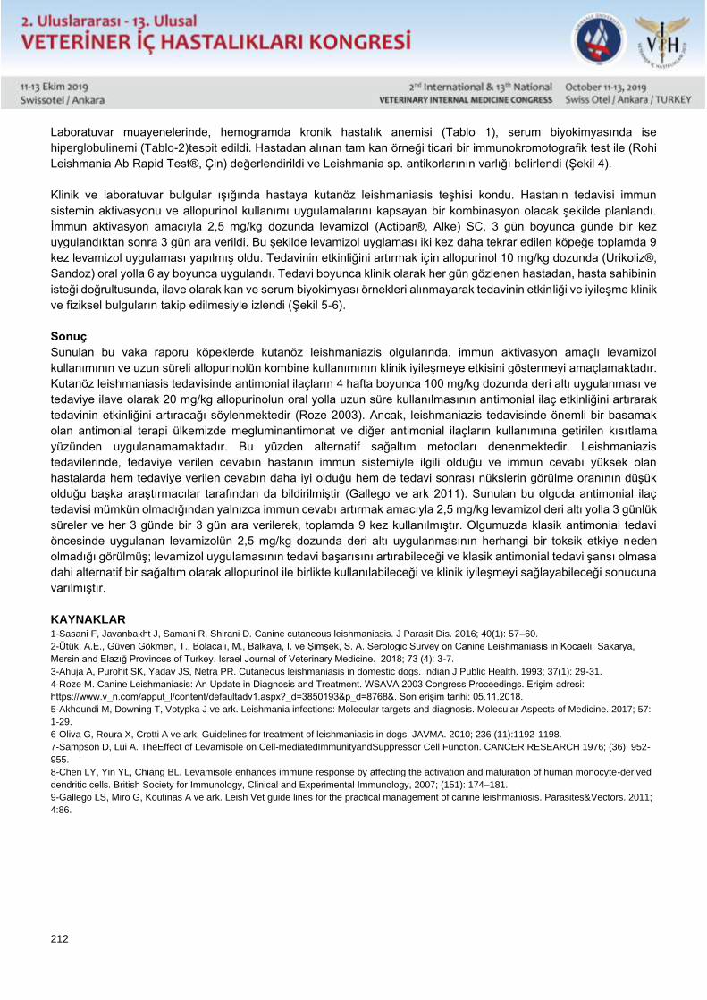
212
Laboratuvar muayenelerinde, hemogramda kronik hastalık anemisi (Tablo 1), serum biyokimyasında ise
hiperglobulinemi (Tablo-2)tespit edildi. Hastadan alınan tam kan örneği ticari bir immunokromotografik test ile (Rohi
Leishmania Ab Rapid Test®, Çin) değerlendirildi ve Leishmania sp. antikorlarının varlığı belirlendi (Şekil 4).
Klinik ve laboratuvar bulgular ışığında hastaya kutanöz leishmaniasis teşhisi kondu. Hastanın tedavisi immun
sistemin aktivasyonu ve allopurinol kullanımı uygulamalarını kapsayan bir kombinasyon olacak şekilde planlandı.
İmmun aktivasyon amacıyla 2,5 mg/kg dozunda levamizol (Actipar®, Alke) SC, 3 gün boyunca günde bir kez
uygulandıktan sonra 3 gün ara verildi. Bu şekilde levamizol uyglaması iki kez daha tekrar edilen köpeğe toplamda 9
kez levamizol uygulaması yapılmış oldu. Tedavinin etkinliğini artırmak için allopurinol 10 mg/kg dozunda (Urikoliz®,
Sandoz) oral yolla 6 ay boyunca uygulandı. Tedavi boyunca klinik olarak her gün gözlenen hastadan, hasta sahibinin
isteği doğrultusunda, ilave olarak kan ve serum biyokimyası örnekleri alınmayarak tedavinin etkinliği ve iyileşme klinik
ve fiziksel bulguların takip edilmesiyle izlendi (Şekil 5-6).
Sonuç
Sunulan bu vaka raporu köpeklerde kutanöz leishmaniazis olgularında, immun aktivasyon amaçlı levamizol
kullanımının ve uzun süreli allopurinolün kombine kullanımının klinik iyileşmeye etkisini göstermeyi amaçlamaktadır.
Kutanöz leishmaniasis tedavisinde antimonial ilaçların 4 hafta boyunca 100 mg/kg dozunda deri altı uygulanması ve
tedaviye ilave olarak 20 mg/kg allopurinolun oral yolla uzun süre kullanılmasının antimonial ilaç etkinliğini artırarak
tedavinin etkinliğini artıracağı söylenmektedir (Roze 2003). Ancak, leishmaniazis tedavisinde önemli bir basamak
olan antimonial terapi ülkemizde megluminantimonat ve diğer antimonial ilaçların kullanımına getirilen kısıtlama
yüzünden uygulanamamaktadır. Bu yüzden alternatif sağaltım metodları denenmektedir. Leishmaniazis
tedavilerinde, tedaviye verilen cevabın hastanın immun sistemiyle ilgili olduğu ve immun cevabı yüksek olan
hastalarda hem tedaviye verilen cevabın daha iyi olduğu hem de tedavi sonrası nükslerin görülme oranının düşük
olduğu başka araştırmacılar tarafından da bildirilmiştir (Gallego ve ark 2011). Sunulan bu olguda antimonial ilaç
tedavisi mümkün olmadığından yalnızca immun cevabı artırmak amacıyla 2,5 mg/kg levamizol deri altı yolla 3 günlük
süreler ve her 3 günde bir 3 gün ara verilerek, toplamda 9 kez kullanılmıştır. Olgumuzda klasik antimonial tedavi
öncesinde uygulanan levamizolün 2,5 mg/kg dozunda deri altı uygulanmasının herhangi bir toksik etkiye neden
olmadığı görülmüş; levamizol uygulamasının tedavi başarısını artırabileceği ve klasik antimonial tedavi şansı olmasa
dahi alternatif bir sağaltım olarak allopurinol ile birlikte kullanılabileceği ve klinik iyileşmeyi sağlayabileceği sonucuna
varılmıştır.
KAYNAKLAR 1-Sasani F, Javanbakht J, Samani R, Shirani D. Canine cutaneous leishmaniasis. J Parasit Dis. 2016; 40(1): 57–60.
2-Ütük, A.E., Güven Gökmen, T., Bolacalı, M., Balkaya, I. ve Şimşek, S. A. Serologic Survey on Canine Leishmaniasis in Kocaeli, Sakarya,
Mersin and Elazığ Provinces of Turkey. Israel Journal of Veterinary Medicine. 2018; 73 (4): 3-7.
3-Ahuja A, Purohit SK, Yadav JS, Netra PR. Cutaneous leishmaniasis in domestic dogs. Indian J Public Health. 1993; 37(1): 29-31.
4-Roze M. Canine Leishmaniasis: An Update in Diagnosis and Treatment. WSAVA 2003 Congress Proceedings. Erişim adresi:
https://www.v_n.com/apput_l/content/defaultadv1.aspx?_d=3850193&p_d=8768&. Son erişim tarihi: 05.11.2018.
5-Akhoundi M, Downing T, Votypka J ve ark. Leishmania infections: Molecular targets and diagnosis. Molecular Aspects of Medicine. 2017; 57:
1-29.
6-Oliva G, Roura X, Crotti A ve ark. Guidelines for treatment of leishmaniasis in dogs. JAVMA. 2010; 236 (11):1192-1198.
7-Sampson D, Lui A. TheEffect of Levamisole on Cell-mediatedImmunityandSuppressor Cell Function. CANCER RESEARCH 1976; (36): 952-
955.
8-Chen LY, Yin YL, Chiang BL. Levamisole enhances immune response by affecting the activation and maturation of human monocyte-derived
dendritic cells. British Society for Immunology, Clinical and Experimental Immunology, 2007; (151): 174–181.
9-Gallego LS, Miro G, Koutinas A ve ark. Leish Vet guide lines for the practical management of canine leishmaniosis. Parasites&Vectors. 2011;
4:86.
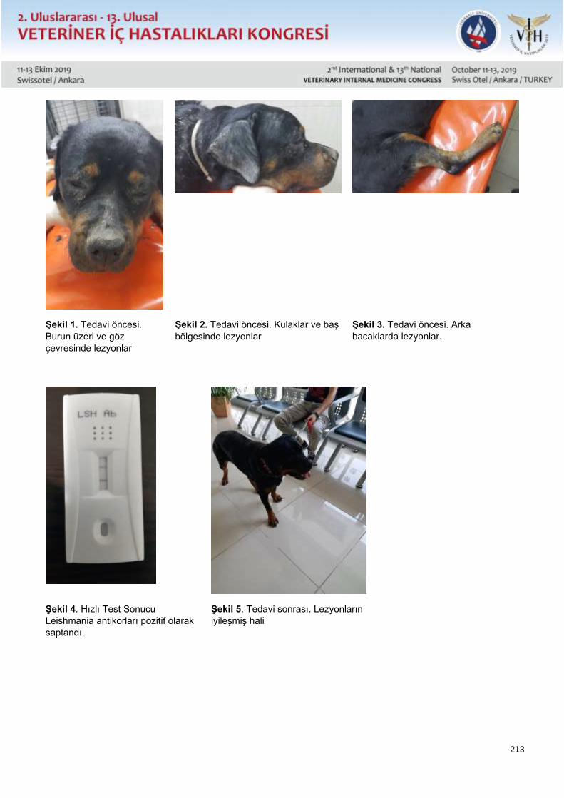
213
Şekil 1. Tedavi öncesi.
Burun üzeri ve göz
çevresinde lezyonlar
Şekil 2. Tedavi öncesi. Kulaklar ve baş
bölgesinde lezyonlar
Şekil 3. Tedavi öncesi. Arka
bacaklarda lezyonlar.
Şekil 4. Hızlı Test Sonucu
Leishmania antikorları pozitif olarak
saptandı.
Şekil 5. Tedavi sonrası. Lezyonların
iyileşmiş hali
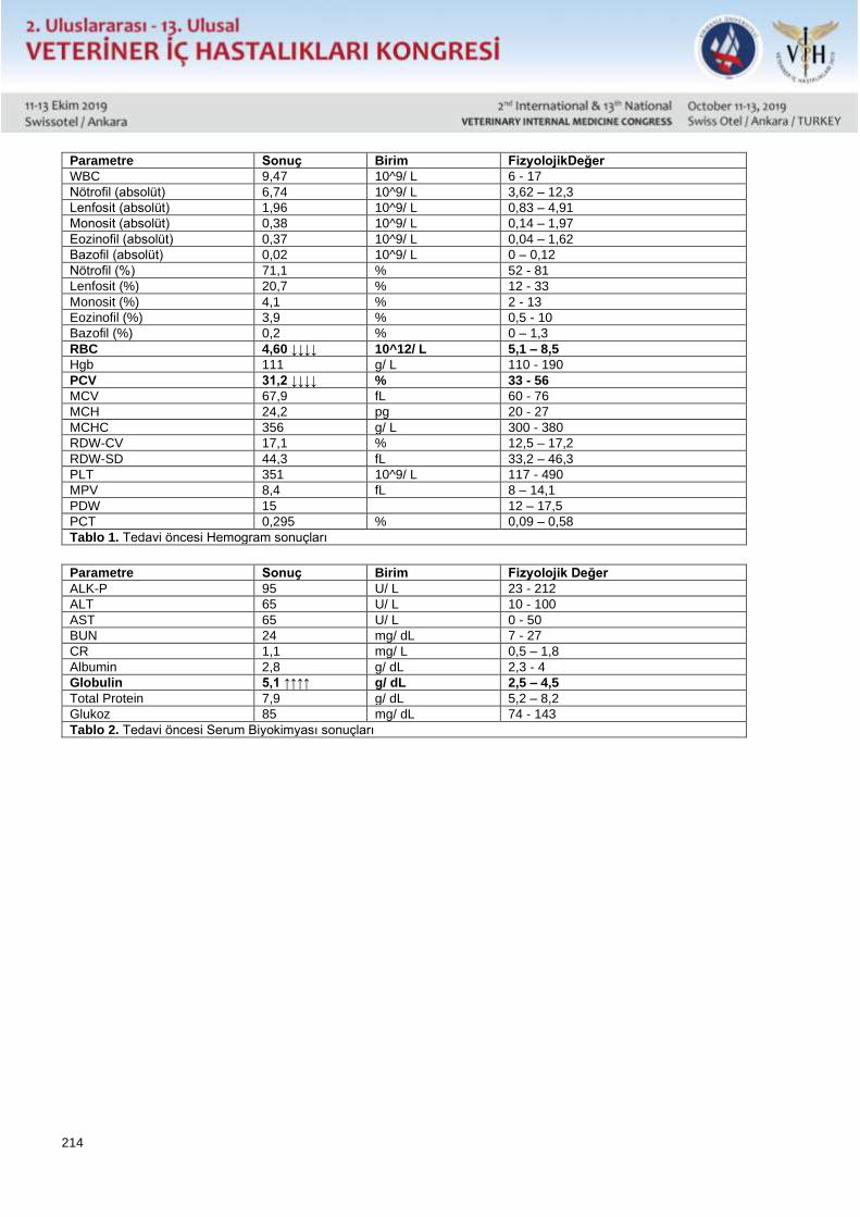
214
Parametre Sonuç Birim FizyolojikDeğer
WBC 9,47 10^9/ L 6 - 17
Nötrofil (absolüt) 6,74 10^9/ L 3,62 – 12,3
Lenfosit (absolüt) 1,96 10^9/ L 0,83 – 4,91
Monosit (absolüt) 0,38 10^9/ L 0,14 – 1,97
Eozinofil (absolüt) 0,37 10^9/ L 0,04 – 1,62
Bazofil (absolüt) 0,02 10^9/ L 0 – 0,12
Nötrofil (%) 71,1 % 52 - 81
Lenfosit (%) 20,7 % 12 - 33
Monosit (%) 4,1 % 2 - 13
Eozinofil (%) 3,9 % 0,5 - 10
Bazofil (%) 0,2 % 0 – 1,3
RBC 4,60 ↓↓↓↓ 10^12/ L 5,1 – 8,5
Hgb 111 g/ L 110 - 190
PCV 31,2 ↓↓↓↓ % 33 - 56
MCV 67,9 fL 60 - 76
MCH 24,2 pg 20 - 27
MCHC 356 g/ L 300 - 380
RDW-CV 17,1 % 12,5 – 17,2
RDW-SD 44,3 fL 33,2 – 46,3
PLT 351 10^9/ L 117 - 490
MPV 8,4 fL 8 – 14,1
PDW 15 12 – 17,5
PCT 0,295 % 0,09 – 0,58
Tablo 1. Tedavi öncesi Hemogram sonuçları
Parametre Sonuç Birim Fizyolojik Değer
ALK-P 95 U/ L 23 - 212
ALT 65 U/ L 10 - 100
AST 65 U/ L 0 - 50
BUN 24 mg/ dL 7 - 27
CR 1,1 mg/ L 0,5 – 1,8
Albumin 2,8 g/ dL 2,3 - 4
Globulin 5,1 ↑↑↑↑ g/ dL 2,5 – 4,5
Total Protein 7,9 g/ dL 5,2 – 8,2
Glukoz 85 mg/ dL 74 - 143
Tablo 2. Tedavi öncesi Serum Biyokimyası sonuçları
215
Poster Sunu / Poster Presentation Özet / Abstract
Cutaneous Leishmaniasis Case in a Rottweiler Dog
Cenk Er*, Mehmet Erkan Avan**
* Çukurova Üniversitesi Ceyhan Veteriner Fakültesi, Veterinerlik İç Hastalıkları Anabilim Dalı, Adana
** Deva Veteriner Tıp Merkezi, Adana.
In this casereport, the diagnosis and treatment of cutaneous leishmaniasis, originated from Leishmania sp. in a 9
month old Rottweiler dog was evaluated.In the light of clinical and laboratory examinations, Leishmania sp. antibody
was positive and it was defined as cutaneous leishmaniasis. The dog was treated for six months with intermittant
levamizol therapy for immun activation (2,5 mg/kg, SC, 3 days medicine, 3 days free, 3 days medicine, 3 days free,
3 days medicine) and allopurinol (10 mg/kg, PO, 6 months). Consequently it is evaluated that, treatment period for
cutaneous leishmaniazis should be longer, and intermittent levamizol therapy to activate immun system, combined
with allopurinol, could improve treatment success without any complications.
Keywords:Dog, Leishmaniasp., leishmaniazis, levamizol.

216
Poster Sunu / Poster Presentation Özet / Abstract
Patent Duktus Arteriozus - 4 Köpekte Olgu Raporu
Hadi ALİHOSSEİNİ, İsmail Özkaptan, Ozan Mutlu
Terapist Veteriner Polikliniği - İstanbul/ Türkiye
Oksijenli kanı aort içine doğru çekmeye başaran fetal dolaşımdaki ductus arteriozus doğumdan kısa bir süre sonra
bazı kedi ve köpeklerde kanal ligamentum arteriyumu üreten fibröz kontraktür kanalı kapatmakta başarısız olup ve
açık kanal aortadan pulmoner artere doğru Şant oluşur.
Materyal ve Metod: Farklı yaş (2 – 14 ay), ırk (Cavalier king charlse spaneil, Pomeranian, melez ırk ve Maltese
Terrier) ve cinsiyetlerden; Dişi (n=3), Erkek (n=1) solunum distresi olan dört köpeğin genel muayenelerinde uzamış
kapiler dolma zamanı, hafif siyantoki mukozal membranlar, precordiyal thrill, oskültasyonda taşikardi (>120/dk) ile
göğüs kafesinin 2. ve 4. interkostal aralıklarında (Sağ>Sol) devamlı (machinary veya continuous) üfürüm sesi ve
femural arter palpasyonunda şiddetli ve sıçrayıcı nabız saptandı. EKG değerlendirilmesinde sinüs taşikardi, II, III
derivasyonlarında uzun R dalgası ve derin Q dalgası dikkat çekti. Toraks Radyografilerde aortik kemerde genişleme,
pulmoner arter ve sol atriyum bölümünde dilatasyon, sağ kranyal lobar arterin genişlemesi, kardiyomegali ile
pulmoner ödem saptandı. Kesin tanı için Ekokiardiyografik incelemesinde sol atriyum ve so ventrikülde (LV)
dilatasyon, volüm overolad ve artmış LV fraksiyonel yüzdeleri kaydedildi. Diyastol sırasında soldan sağa doğru kan
akışı Doppler ekokardiyografi ile açık duktus tespit edildi. Qp/Qs oranı 3 köpekte birin altında, bir köpekte ise 1.6
olarak şantın sağdan sola revers şeklinde olduğu saptandı. Revers şantı olan köpekte pulmoner arter basıncının
25mmHg üzerinde olduğundan Pulomner arter hipertansiyon varlığı tespit edildi.
Sonuç: PDA tanısı olan köpeklerde Cerrahi yöntemle duktus ligatüre edildi. Operasyon sonrası 1. ve 2. hafta
Ekokardiyografik değerlendirilmelerde kardiyak hemodinami kriterlerin iyileşmeye yönelik tespiti ve hastanın klinik
olarak stabilizasyonu sağlandı. Revers şantı olan köpekte ise operasyon sonrası pulmoner arter hipertansiyonu için
Sildenafil 0.5mg/kg, bid, PO olarak sağaltım rejimine ilave edildi.
Anahtar kelemeler: köpek, PDA, pulmoner hipertansiyon.
217
Poster Sunu / Poster Presentation Özet / Abstract
Patent Ductus Arterıosus - Case Report İn 4 Dogs
Hadi ALİHOSSEİNİ, İsmail Özkaptan, Ozan Mutlu
Terapist Veterinary Polyclnic - Istanbul / TURKEY
Fetal circulatory ductus arteriosus, which manages to draw oxygenated blood into the aorta, fails to close the fibrous
contracture channel that produces the canal ligamentum artery in some cats and dogs shortly after birth, and shunts
from the open canal aorta to the pulmonary artery.
Material and Methods: Four dogs of different ages (2 – 14 mo) , Breeds (CKCS, Pomeranian, mongrel ve Maltese
Terrier) and sexes; Female (n=3), Male (n=1) with respiratory distress were referred. In the clinical examination;
prolonged CRT, mild cyanotic mucosal membrane with precordial thrill in palpation were determinded. In the
auscultation tachycardia (> 120bpm), a continous (machinary) murmur were detected between the 2nd–4th
intercostal spaces (Right> Left). A rebounding pulse in femural artery palpation was remarkable. In the ECG tracing
long R wave and deep Q wave in leads II and III were noted. Thorax radiographs revealed bulging of the aortic arch
with dilatation of the main pulmonary artery trunk, dilatation of right cranial lobar artery and pulmonary edema. The
2D Echocardiographic examinations dilatation and volume overload of the left ventrivular (LV) and increased LV
fractional percentages were detected. Diagnosis was confirmed by diastolic turbulent blood flow from aorta to the
pulmonary trunk in the Color Doppler Echocardiographic imaging. According to the Qp/Qs ratio, the shunt was found
to be less than one in 3 dogs and 1.6 in one dog (Reverse PDA). Pulmonary arterial hypertension was detected in
one dog with reversed shunt because the pulmonary artery pressure was above 25mmHg.
Conclusion: Ductus ligation was performed surgically for PDA dogs. Post-surgical serial Echocardiographic
evaluations showed improvement of cardiac hemodynamic criteria in following days. In the dog with reversed shunt,
Sildenafil 0.5mg / kg, bid, PO was added to the treatment regimen for postoperative pulmonary artery hypertension.
Key words: dog, PDA, pulmonary hypertension.
218
Poster Sunu / Poster Presentation Özet / Abstract
Cor Triatriatum sinister - 3 Yaşlı Shih-Tzu Irkı Köpekte Vaka Raporu
Hadi ALİHOSSEİNİ, İsmail Özkaptan, Ozan Mutlu
Terapist Veteriner Polikliniği, İstanbul, Türkiye
Cor triatriatum, kalbin 3 atriyal boşluğu olan nadir görülen bir doğmasal kalp hastalıktır. Cor triatriatum sinister (CTS)
ve dexter (CTD), sırasıyla fibröz bir membranla iki bölmeye bölünmüş sol veya sağ atriyumdan oluşur.
Materyal ve Metod: Solunum distresi olan ve arasıra taşipne şikayeti ile bilinen 3 yaşlı Erkek Shih-tzu ırkı bir köpek
merkezimize sevk edildi. Genel muayenede pembe mukozal membranlar ve normal kapiler dolma zamanı saptandı.
Prekordiyal palpasyonu normal, oskültasyonda ise taşikardi (145 v/dk), split S1 sesi, femural arter palpasyonunda
kalp atımları ile uyumlu güçlü ve değişken nabız saptandı. Kan değerleri ve akciğer radyografisi normal olarak
değerlendirildi. Kesin tanı Ekokardiyografik muayenesi RPLAX ve LAP4C görünümlerinde sol atriyumun (LA) bir
hiperekoik bant ile 2 kamaraya bölünmesi saptandı. Doppler Ekokardiyografik değerlendirmesinde mitral
regürgitasyona rastlanmadı. Hastanın sistolik kan basıncı 124mmHg olarak hesaplandı. Sol ventrikülün sistolik ve
diyastolik fonksiyonları normal olarak değerlendirildi. Doku Doppler görüntülemesinde septal myokardiyumun hızında
düşüş (E/E’:9.48), lateral duvarın hızı normal oranlarda (E/E’:6.51) olarak kaydedildi. Transmitral inflow
değerlendirilmesinde herhangi bir LV diyastolik yetmezliğine rastlanmadı.
Anahtar kelemeler: Cor Triatriatum sinister, ekokardiyografi, köpek.
219
Poster Sunu / Poster Presentation Özet / Abstract
Cor Triatriatum sinister in 3-year-old Shih-Tzu Dog – Case Report
Hadi ALİHOSSEİNİ, İsmail Özkaptan, Ozan Mutlu
Terapist Veterinary Polyclinic - Istanbul / TURKEY
Cor triatriatum is a rare congenital heart disease with 3 atrial cavities of the heart. The cor triatriatum sinister (CTS)
and dexter (CTD) consist of a left or right atrium divided into two compartments by a fibrous membrane, respectively.
Material and Methods: A 3-year-old male Shih-tzu breed with respiratory distress and trransient tachypnea was
referred to our center. In the clinical examination pinky mucosal membranes, normal capillary filling time and
precordial palpation were revealed. Tachycardia (145bpm) with split S1 sound was auscultated. A variable and strong
femoral pulse synchronus to the heart beats were palpated. The blood works and chest radiography were normal.
The main diagosis confirmed by Echocardiographic examination which showed that the left atrium (LA) was divided
into 2 chambers with a hyperechoic band on both RPLAX and LAP4C views. Mitral regurgitation was not found. The
systolic blood pressure of the patient was calculated as 124mmHg. The left ventricular systolic and diastolic functions
were normal. Tissue Doppler imaging showed a decrease in the velocity of the septal myocardium (E/E’:9.48) and a
normal rate of lateral wall velocity (E/E’:6.51). No LV diastolic function abnormality was detected in the of transmitral
inflow inspection too.
Key words: Cor Triatriatum sinister, Echocardiography, dog.
220
Poster Sunu / Poster Presentation Özet / Abstract
Koriza Gangrenoza Bovumun Baş Göz Formu Teşhis Edilen Sığırlarda Kan ve Bos
Biyokimyası, Hematolojik Parametreler İle Bos Sitolojisinin Değerlendirilmesi
Erdoğan UZLU, Ekin Emre ERKILIÇ, Yasemen ADALI, Metin ÖĞÜN, Kezban CAN ŞAHNA, Nilhan ERYEĞEN, Celal
Şahin ERMUTLU, Hüseyin Avni EROĞLU, Hasan ABAYLI, Ali Haydar KIRMIZIGÜL
Kafkas Üniversitesi Veteriner Fakültesi İç Hastalıkları Anabilim Dalı, Kars
Kafkas Üniversitesi Veteriner Fakültesi İç Hastalıkları Anabilim Dalı, Kars
Çanakkale On Sekiz Mart Üniversitesi Tıp Fakültesi Tıbbi Patoloji Anabilim Dalı, Çanakkale
Kafkas Üniversitesi Tıp Fakültesi Biyokimya Anabilim Dalı,Kars
Fırat Üniversitesi Veteriner Fakültesi Viroloji Anabilim Dalı, Elazığ
Kafkas Üniversitesi Tıp Fakültesi Tıbbi Patoloji Anabilim Dalı,Kars
Kafkas Üniversitesi Veteriner Fakültesi Cerrahi Anabilim Dalı, Kars
Çanakkale On Sekiz Mart Üniversitesi Tıp Fakültesi Fizyoloji Anabilim Dalı, Çanakkale
Fırat Üniversitesi Veteriner Fakültesi Viroloji Anabilim Dalı, Elazığ
Kafkas Üniversitesi Veteriner Fakültesi İç Hastalıkları Anabilim Dalı, Kars
Bu çalışmanın amacı Koriza Gangrenoza Bovum (CGB) ile enfekte sığırlarda beyin omurilik sıvısı (BOS) ve
serumdaki biyokimyasal değişiklikler ile kandaki hematolojik değişikliklerin belirlenmesi ve BOS’un sitolojik olarak
incelenmesidir. Bu amaçla CGB’nin baş-göz formu teşhis edilen toplam 22 ve klinik olarak sağlıklı 10 sığır
değerlendirildi. Tüm sığırlardan kan ve BOS örnekleri alındı.
Serumda AST, üre, glukoz, CK (P <0.05), LDH düzeyleri (P <0.01) CGB’li hayvanlarda sağlıklılara göre yüksek olarak
belirlenirken, CGB’li hayvanlarda ALT, ALP, kolesterol (P <0.05), Ca, total protein (P <0.01), Mg, albumin ve Fe
seviyelerinin (P<0.001) sağlıklılara göre düşük olduğu tespit edildi.
CGB’li hayvanların BOS analizinde Ca (P <0.01) ve total protein düzeyleri (P <0.001) sağlıklılara göre yüksek olarak
bulunurken, glikoz (P<0.05) seviyeleri düşük olarak belirlendi. Hematolojik olarak bazı parametrelerde de gruplar
arasında farklılıklar bulundu.
BOS’un sitolojik olarak değerlendirilmesinde polimorfonükleer lökositler, lenfositler, eritrositler, makrofajlar ve plazma
hücrelerinin varlığı belirlendi.
Sonuç olarak, CGB'nin özellikle BOS'taki biyokimyasal ve sitolojik incelemeleri ile hematolojik sonuçlarını inceleyen
sınırlı sayıda çalışma bulunduğundan, çalışmamızın sonuçlarının sığırlarda sinir sistemine etki eden viral hastalıklar
ile ilgili gelecekteki çalışmalar için önemli olabileceği sonucuna varıldı.
221
Poster Sunu / Poster Presentation Özet / Abstract
Evaluation Of Blood And Csf Biochemıstry, Csf Cytology And Hematological Parameters
in Head Eye Form of Mcf Determined Cattles
Erdoğan UZLU, Ekin Emre ERKILIÇ, Yasemen ADALI, Metin ÖĞÜN, Kezban CAN ŞAHNA, Nilhan ERYEĞEN, Celal
Şahin ERMUTLU, Hüseyin Avni EROĞLU, Hasan ABAYLI, Ali Haydar KIRMIZIGÜL
Kafkas Üniversitesi Veteriner Fakültesi İç Hastalıkları Anabilim Dalı, Kars
Kafkas Üniversitesi Veteriner Fakültesi İç Hastalıkları Anabilim Dalı, Kars
Çanakkale On Sekiz Mart Üniversitesi Tıp Fakültesi Tıbbi Patoloji Anabilim Dalı, Çanakkale
Kafkas Üniversitesi Tıp Fakültesi Biyokimya Anabilim Dalı,Kars
Fırat Üniversitesi Veteriner Fakültesi Viroloji Anabilim Dalı, Elazığ
Kafkas Üniversitesi Tıp Fakültesi Tıbbi Patoloji Anabilim Dalı,Kars
Kafkas Üniversitesi Veteriner Fakültesi Cerrahi Anabilim Dalı, Kars
Çanakkale On Sekiz Mart Üniversitesi Tıp Fakültesi Fizyoloji Anabilim Dalı, Çanakkale
Fırat Üniversitesi Veteriner Fakültesi Viroloji Anabilim Dalı, Elazığ
Kafkas Üniversitesi Veteriner Fakültesi İç Hastalıkları Anabilim Dalı, Kars
The aims of this study were to determine biochemical changes in cerebro spinal fluid (CSF) and sera, hematological
changes in blood and cytological investigation of CSF in cattle with Malignant Catarrhal Fever (MCF). For this purpose
22 cattle which diagnosed "head-eye form" of MCF and clinically healthy 10 cattle were evaluated. Blood and
cerebrospinal fluid (CSF) were taken from all evaluated cattle. In sera, AST, urea, glucose, CK (P<0.05), LDH levels
(P<0.01) were found be high, ALT, ALP, cholesterol (P<0.05), Ca, total protein (P<0.01) and Mg, albumine and Fe
levels (P<0.001) were found to be low in MCF group than control group. In CSF, Ca (P<0.01) and total protein levels
(P<0.001) were found be high, glucose levels (P<0.05) was found be low in MCF group than control group. In
haematologically, some parameters were determined different between groups too. In cytologically results on CSF,
polymorphonuclear leucocytes, lymphocytes, erytrocytes, macrophages and plasma cells were determined in
cytological slides.
In conclusion, since there are a limited number of studies examining biochemical, cytologic and hematological results
of MCF especially in CSF, the results from our study are thought to be important for future studies which viral diseases
effects to the nervous system of cattles.
222
Poster Sunu / Poster Presentation Özet / Abstract
Sepsisli Neonatal Buzağılarda Artmış Serum Kardiyak Troponin-I Konsantrasyonu ve
Kardiyak Enzim Aktiviteleri
Akın Kırbaş*, Şükrü Değirmençay*, Ayten Aşkın Kılınç**, Muhammed Sertaç Eroğlu*
*Atatürk Üniversitesi Veteriner Fakültesi İç Hastalıkları Anabilim Dalı, Erzurum
**Tarım ve Orman Bakanlığı, Tavukçuluk Araştırma Enstitüsü, Ankara
Amaç: Bu çalışmada sepsisli neonatal buzağılarda serum kardiyak troponin-I (cTnI) konsantrasyonunun ve kardiyak
enzim aktivitelerinin (kreatin kinaz-MB [CK-MB], laktat dehidrogenaz [LDH] ve aspartat aminotransferaz [AST])
değerlendirilmesi amaçlandı.
Materyal ve Metot: Çalışmanın hayvan materyalini yaşları 1-10 gün arasında olan 15 adet sepsisli neonatal buzağı
(Sepsis grubu, SG) ile yaşları 1-10 gün arasında olan 10 adet sağlıklı neonatal buzağı (Kontrol grubu, KG) oluşturdu.
Hematolojik ve biyokimyasal analizler için hayvanların vena jugularisinden antikoagülanlı ve antikoagülansız tüplere
kan örnekleri alındı. Hematolojik muayeneler hemogram cihazında yapıldı. Biyokimyasal analizlerden cTnI
konsantrasyonu ticari immunoassay sistemi one step sandwich yöntemi ile belirlenirken CK-MB, LDH ve AST
aktiviteleri ile üre ve kreatinin konsantrasyonları ticari kitler vasıtası ile otoanalizör kullanılarak tespit edildi.
Bulgular: SG’nin total lökosit (WBC) ve nötrofil (NEU) sayılarının KG’den önemli düzeyde yüksek olduğu gözlendi
(P<0.001). SG’nin cTnI konsantrasyonu (P<0.001), CK-MB, LDH ve AST aktiviteleri (P<0.05) ile üre ve kreatinin (Cr)
konsantrasyonları (P<0.001) KG’den önemli düzeyde yüksekti. Miyokard hasarını göstermedeki duyarlılık değerleri
cTnI:% 100, CK-MB:% 66,7, LDH ve AST: %53,3 olarak tespit edildi. ROC eğrisi altında kalan alan cTnI, CK-MB,
LDH ve AST için sırasıyla 0.98, 0.76, 0.69 ve 0,80 olarak belirlendi.
Sonuç: Bu çalışma, sepsisli neonatal buzağılarda miyokard hasarının tespitinde cTnI konsantrasyonunun genel
tanısal performansının, CK-MB, LDH ve AST aktivitelerinden daha üstün olduğunu ve sepsisin prognozunun
belirlenmesinde önemli bir belirteç olabileciğini ortaya koymuştur.
Anahtar Kelimeler: Buzağı, Kardiyak enzimler, Kardiyak troponin-I, Miyokardiyal hasar, Neonatal sepsis
223
Poster Sunu / Poster Presentation Özet / Abstract
The Increased Cardiac Troponin-I Concentration and Cardiac Enzyme Activities in
Neonatal Calves with Sepsis
Akın Kırbas, Sukru Degirmencay, Ayten Askın Kılınc, Muhammed Sertac Eroglu
*Department of Internal Medicine, Faculty of Veterinary Medicine, Ataturk University, Erzurum
**Directorate of Poultry Research Institute, Ministry of Agriculture and Forestry, Ankara
Aim: The aim of this study was to evaluate serum cardiac troponin-I (cTnI) concentration and cardiac enzyme
activities (creatine kinase-MB [CK-MB], lactate dehydrogenase [LDH] and aspartate aminotransferase [AST] in
neonatal calves with sepsis.
Material and Method: The animal material of the study consisted of 15 neonatal calves with sepsis (Sepsis group,
SG) aged between 1-10 days and 10 healthy neonatal calves (Control group, CG) aged 1-10 days. Blood samples
were taken from the vena jugularis of animals in anticoagulant and coagulant tubes for haematological and
biochemical analyses. Haematological examinations were performed on the hemogram device. CK-MB, LDH and
AST activities and urea and creatinine concentrations were determined using autoanalyser via commercial kits while
cTnI concentration was determined by the one-step sandwich method of commercial immunoassay system.
Results: Total leukocyte (WBC) and neutrophil (NEU) counts of SG were significantly higher than CG (P<0.001). The
cTnI concentration (P<0.001), CK-MB, LDH and AST activities (P<0.05), urea and creatinine (Cr) concentrations
(P<0.001) of SG were significantly higher than CG. Sensitivity values for myocardial damage were cTnI: 100%, CK-
MB: 66.7%, LDH and AST: 53.3%. The areas under the ROC curve were 0.98, 0.76, 0.69 and 0.80 for cTnI, CK-MB,
LDH and AST, respectively.
Conclusion: This study demonstrated that the overall diagnostic performance of cTnI concentration is superior to CK-
MB, LDH, and AST activities in the detection of myocardial injury in neonatal calves with sepsis and may be an
important marker for the prognosis of sepsis.
Keywords: Calf, Cardiac enzymes, Cardiac troponin-I, Myocardial injury, Neonatal sepsis
224
Poster Sunu / Poster Presentation Özet / Abstract
Streptococcus pluranimalium ile Doğal Enfekte Koyunlarda Serum Haptoglobin, Nitrik
Oksit ve Malondialdehit Düzeylerinin Değerlendirilmesi
Akın Kırbaş,* Ramazan Kaman,* Oğuz Merhan,** Kadir Bozukluhan,*** Şükrü Değirmençay,*
Muhammed Sertaç Eroğlu*
*Atatürk Üniversitesi, Veteriner Fakültesi, İç Hastalıkları Anabilim Dalı, Erzurum
**Kafkas Üniversitesi, Veteriner Fakültesi, Biyokimya Anabilim Dalı, Kars
***Kafkas Üniversitesi, Kars Meslek Yüksekokulu, Veteriner Sağlık Bölümü, Kars
Amaç: Bu çalışmada Streptococcus pluranimalium ile doğal enfekte koyunlarda serum haptoglobin (Hp), nitrik oksit
(NO) ve malondialdehit (MDA) düzeylerinin değerlendirilmesi amaçlanmıştır.
Materyal ve Metot: Çalışmanın hayvan materyalini, yaşları 4-6 arasında olan 60 adet Streptococcus pluranimalium
ile enfekte koyun (Hasta grup, HG) ile yaşları 4-6 arasında olan 15 adet sağlıklı koyun (Kontrol grubu, KG) oluşturdu.
Biyokimyasal analizler için vakumlu tüplere hayvanların vena jugularislerinden kan örnekleri alındı. Serum
örneklerinde Hp ve NO düzeyleri spektrofotometrik yöntemle, MDA düzeyleri ise tiyobarbiturik asit reaksiyonu ile
belirlendi.
Bulgular: HG’nin Hp (0,17±0,04 g/L), MDA (1,67±0,31 μmol/L) (P<0.01) ve NO (31,14±12,49 μmol/L) (P<0.05)
düzeylerinin KG’den önemli düzeyde yüksek olduğu belirlendi.
Sonuç: Streptococcus pluranimalium ile doğal enfekte koyunlarda Hp, MDA ve NO düzeylerinin yüksek olmasının
enfeksiyonun patogenezi ve prognozunda önemli bir yer tutabileceği kanaatine varılmıştır.
Anahtar Kelimeler: Haptoglobin, Koyun, Malondialdehit, Nitrik oksit, Streptococcus pluranimalium
225
Poster Sunu / Poster Presentation Özet / Abstract
The Assessment of Serum Haptoglobin, Nitric Oxide and Malondialdehyde Levels in
Sheep Infected Naturally with Streptococcus Pluranimalium
Akın Kırbas,* Ramazan Kaman,* Oguz Merhan,** Kadir Bozukluhan,*** Sukru Degirmencay,*
Muhammed Sertac Eroglu*
*Department of Internal Medicine, Faculty of Veterinary Medicine, Ataturk University, Erzurum
**Department of Biochmesitry, Faculty of Veterinary Medicine, Kafkas University, Kars
***Department of Veterinary Health, Kars School of Higher Vocational Education, Kafkas University, Kars
Aim: The aim of this study was to evaluate serum haptoglobin (Hp), nitric oxide (NO) and malondialdehyde (MDA)
levels in naturally infected sheep with Streptococcus pluranimalium.
Material and Method: The animal material of the study consisted of 60 sheep infected with Streptococcus
pluranimalium (patient group) aged between 4-6 years and 15 healthy sheep (control group) aged 4-6 years. Blood
samples were taken from the vena jugularis of animals in vacuum tubes for biochemical analysis. Serum Hp and NO
levels were determined by spectrophotometric method and MDA levels were determined by the thiobarbituric acid
reaction.
Results: Hp (0.17±0.04 g/L), MDA (1.67±0.31 μmol/L) (P<0.01) and NO (31.14±12.49 μmol/L) (P<0.05) levels of the
patient group were found to be significantly higher than the control group.
Conclusion: It was concluded that the high levels of Hp, MDA and NO in naturally infected sheep with Streptococcus
pluranimalium may play an important role in the pathogenesis and prognosis of infection.
Keywords: Haptoglobin, Sheep, Malondialdehyde, Nitric oxide, Streptococcus pluranimalium
226
Poster Sunu / Poster Presentation Özet / Abstract
Sığır Solunum Hastalıkları Kompleksli Buzağılarda Serum Demir ve Ferritin Düzeylerinin
Değerlendirilmesi
Şükrü Değirmencay,* Akın Kırbaş,* Hakan Aydın**, Ömer Aydın*, M. Sinan Aktaş*, Ramazan Kaman*
*Atatürk Üniversitesi Veteriner Fakültesi İç Hastalıkları Anabilim Dalı, Erzurum, ** Atatürk Üniversitesi Veteriner
Fakültesi Viroloji Anabilim Dalı, Erzurum
Amaç: Bu çalışmada sığır solunum hastalıkları kompleksli buzağılarda serum demir (Fe), ferritin (Ferr), Transferrin
ve doymamış demir bağlama kapasitesi (DDBK) ile hematolojik parametrelerin değerlendirilmesi amaçlandı.
Materyal ve Metot: Çalışmanın materyalini ortalama 1-3 aylık yaşta olan 15 adet sığır solunum sistemi hastalıkları
kompleksli buzağı (hasta grup, Grup I) ile ortalama 1-3 aylık yaşta olan 10 adet sağlıklı buzağı (kontrol grubu, Grup
II) oluşturdu. Hayvanların vena jugularis externa’sından hematolojik ve biyokimyasal analizler için kan örnekleri alındı.
Serum örneklerinde, Fe, Ferr, Transferrin ve DDBK ticari kitlerle otoanalizatör kullanılarak belirlendi. Hematolojik
analizler kan sayım cihazında yapıldı. Etiyolojik tanı için alınan nazal swap örneklerinde PCR yöntemi kullanılarak
virolojik analizler yapıldı.
Bulgular: Biyokimyasal muayenede, Grup I’in serum Fe ve Ferr düzeyleri Grup II’den yüksek bulundu (P<0.001).
Hematolojik bulgulardan Grup I’in nötrofil (NEU) ve eozinofil (EOS) sayıları Grup II’den yüksek bulunurken (P<0.001),
ortalama eritrosit hemoglobini (MCH) ve ortalama eritrosit hemoglobin konsantrasyonu (MCHc) düşük bulundu
(P<0.001). Serum Fe konsantrasyonu, NEU (P<0.01) ve EOS (P<0.05) ile negatif korelasyon gösterirken MCH ve
MCHc ile pozitif korelasyon gösterdi (P<0.01). Serum Ferr konsantrasyonu NEU ile pozitif korelasyon gösterdi
(P<0.05). Nazal swap örneklerinin virolojik analizleri sonucu buzağıların % 26.6’sında bovine respiratory synctycial
virüs (BRSV), % 13.3’ünde bovine viral diare virüsü (BVDV) ve %13.3’ünde BRSV+BVDV saptandı.
Sonuç: Bu çalışmada sığır solunum hastalıkları kompleksli buzağılarda serum Fe konsantrasyonların azaldığı, serum
Ferr düzeylerinin ise arttığı tespit edildi. Sonuç olarak serum Fe ve Ferr düzeylerinin sığır solunum hastalıkları
kompleksli buzağılarda hastalığın patogenezinde rol oynayabileceği kanısına varıldı.
Anahtar Kelimeler: Buzağı, Demir, Ferritin, Sığır Solunum Hastalıkları Kompleksi
227
Poster Sunu / Poster Presentation Özet / Abstract
The Evaluation of Serum Iron and Ferritin Levels in Calves with Bovine Respiratory
Disease Complex
Sukru Degirmencay,* Akin Kirbas,* Hakan Aydin**, Omer Aydin*, M. Sinan Aktas*, Ramazan Kaman*
*Department of Internal Medicine, Faculty of Veterinary Medicine, Ataturk University, Erzurum **Department of
Virology, Faculty of Veterinary Medicine, Ataturk University, Erzurum
Aim: This study aimed to evaluate serum iron (Fe), ferritin (Ferr), transferrin and unsaturated iron-binding capacity
(UIBC) and haematological parameters in calves with bovine respiratory disease complex.
Material and Method: The material of the study consisted of 15 calves, aged 1-3 months with bovine respiratory
disease complex (Patient group, Group I) and 10 healthy calves aged 1-3 months (Control group, Group II). Blood
samples were taken from the vena jugularis externa of the animals for haematological and biochemical analyses. In
serum samples, Fe, Ferr, Transferrin and UIBC were determined by commercial kits using autoanalyser.
Haematological analyses were performed on blood counting device. In the etiologic diagnosis, virological analysis of
nasal swab samples was performed using the PCR method.
Results: In a biochemical examination, serum Fe and Ferr levels of Group I were higher than Group II (P <0.001).
According to the hematologic findings, neutrophil (NEU) and eosinophil (EOS) numbers of Group I were higher than
Group II (P <0.001) and mean erythrocyte haemoglobin (MCH) and mean erythrocyte haemoglobin concentration
(MCHc) were lower (P <0.001). Serum Fe concentration was negatively correlated with NEU (P <0.01) and EOS (P
<0.05), but positively correlated with MCH and MCHc (P <0.01). Serum Ferr concentration was positively correlated
with NEU (P <0.05). Virological analysis of nasal swab samples revealed bovine respiratory syncytial virus (BRSV)
in 26.6%, bovine viral diarrhoea virus (BVDV) in 13.3% and BRSV + BVDV in 13.3% of calves.
Conclusion: In this study, it was found that serum Fe concentrations decreased and serum Ferr levels increased in
calves with bovine respiratory disease complex. As a result, it was concluded that serum Fe and Ferr levels may play
a role in the pathogenesis of bovine respiratory disease complex in calves.
Keywords: Calf, Iron, Ferritin, Bovine Respiratory Diseases Complex
228
Poster Sunu / Poster Presentation Özet / Abstract
Bir Köpekte Septik Şoka Bağlı Gelişen Rabdomiyoliz Olgusu: Olgu Sunumu Havva Süleymanoglu1, Merve Ertan1, MehmetEge Ince1, Kursad Turgut1 Yakın Doğu Üniversitesi, Veteriner Hekimliği Fakültesi, İç Hastalıkları Bölümü, Lefkoşa, Kuzey Kıbrıs, Türkiye Sunum Yapacak Kişi: Havva Suleymanoglu Rabdomiyoliz çizgili kasların akut nekrozudur. Rabdomiyolizin şiddeti asemptomatik yüksek CK seviyesi ve hayvanların yaşamını etkileyebilecek çok yüksek CK seviyesi arasında değişebilir. 13 yaşlı, erkek, Pekinez ırkı kopek hastanemize solunum güçlüğü, halsizlik, şiddetli kas ağrısı, kusma ve güçsüzlük şikayetleri ile başvurdu. Hastanemizde yapılan ilk muayenede, köpeğin bilincinin kapalı, ağrılı ve dehidrasyon (8-10 %) ile birlikte letarji olduğu belirlendi. Köpeğin ayakta duramadığı, sternumun üzerinde abdominal pozisyonu dayattığı, abdomen bölgesinde ve kaslarda ağrısı olduğu tespit edildi. Mucosal membranlarda siyanoz, kapillar dolum süresinde uzama, dört bacaktada myotatik reflekslerde azalma ve nabız siddetinde azalma belirlendi. Serum biyokimyası analzi sonucu çoklu organ yetmezliği tesbit edildi. Bir bolus sıvı resüsitasyonuna rağmen hipotansiyonun (sistolikkanbasıncı (SBP): 83 mmHg (referansaralığı: >90 mmHg), ortalama arter basıncı (MAP): 63 mmHg (referans aralığı: >65mmHg) devam etmesi sonucunda septik şok tanısı konuldu. Ekokardiyografik muayenede, ejeksiyon fraksiyonu (EF) %82 ve stroke volume (SV) 12.74 ml olarak tespit edildi. Hastanın hospitalizasyondaki 1.gün CK seviyesi13.690 UI/l olurken, 2. gün 31.587 UI/l değerine yükseldi. 4. ve 9. günlerdeki CK değerleri sırasıyla 15.796 UI/l ve 1.064UI/l olarak ölçüldü. Agresif şok tedavisi ve rabdomiyeliz sonucu gelişen komplikasyonlara yönelik yapılan tüm tedaviye rağmen hastada ileri miyalji ve şiddetli solunum yetmezliğine bağlı 9. günde ölüm gerçekleşti. Sonuç olarak, septik şokla birlikte nadir görülen şiddetli rabdomiyoliz olgusuna tanık olduk. Bu olağan dışı olgu sepsis ile birlikte gelişen rabdomiyoliz olgularına katkıda bulunabilir.
229
Poster Sunu / Poster Presentation Özet / Abstract
Rhabdomyolysis Triggered By Septic Shock İn A Dog: A Case Report Havva Suleymanoglu, Merve Ertan, Mehmet Ege Ince, Kursad Turgut Near East University, Faculty of Veterinary Medicine, Department of Internal Medicine, Nicosia, North Cyprus, Turkey Rhabdomyolysis is acute necrosis of striated muscle. It ranges in severity from an asymptomatic elevation of CK levels in blood, to severe life-threatening cases associated with very high CK levels. A 13-year-old Pekinese male dog was admitted to our department with a history of respiratory distress, fatigue, severe myalgias, vomiting and generalized severe weakness. On the initial examination, the dog was uncounciness and had pain, and was in a stuporous state, with dehydration (8-10 %). He was unable to stand, lying on his sternum position, pain of the abdominal cavity and muscles was noted. There were cyanotic mucosal membranes and prolonged capillary refill time. The dog had a decrease in myotatic reflexes in all 4 limbs. Pulse was very weak. There was a variety organ dysfunction. The diagnosis of septic shock was based on the presence of severe sepsis and hypotension (systolic blood pressure (SBP): 83 mmHg (reference range: >90 mmHg), mean arterial pressure (MAP): 63mmHg (reference range: >65mmHg)) that could not be reversed with fluid resuscitation. The echocardiographic findings included a normal LV systolic function with ejection fraction (EF) (82 per cent) and stroke volume (SV) (12.74 ml). CK level was very high (13.690 UI/l) at the 1st day of hospitalizations and went up to 31.587 UI/l at the 2nd day of hospitalizations. CK values at the 4rd and 9th days were 15.796 UI/l and 1.064 UI/l, respectively. Despite aggressive shock treatment and adequate treatment of secondary infections and the complication of rhabdomyolysis (azotemia and liver failure), the patient developed a progressive myalgias, progressive respiratory failure and low compliance resulting in death on day 9. In conclusion, we describe an unusual case of severe rhabdomyolysis associated with septic shock. This unusual case may further add to the understanding of rhabdomyolysis with sepsis.
230
Poster Sunu / Poster Presentation Özet / Abstract
Doramektin Zehirlenmesinin İntravenöz Lipid Emülsiyonu ile Tedavisi: Beş Amerikan
Pitbull Terrier Yavrusu
Ümit ÖZCAN, Çağatay ESİN, Murat GÜZEL
Ondokuz Mayıs Üniversitesi, Veteriner Fakültesi İç Hastalıkları Anabilim Dalı
Bu olgunun materyalini deri altı doramektin (0,5 ml, 5mg/kg) uygulanan beş adet üç günlük (1kg) bull terrier yavrusu
oluşturdu. Anemnezde enjeksiyondan 6 saat sonra titreme, hızlı solunum ve inleme şikayetlerinin başladığı öğrenildi.
18 saat sonra hastanemize getirilen yavrularda tremor, bilinç kaybı, inleme, midriasis ve taşipne tespit edildi. Beden
ısılarının ortalama 34,0°C, solunum sayılarının ortalama 22/dak, nabız sayılarının ortalama 213/dak olduğu belirlendi.
Anamnez ve klinik bulgular doğrultusunda doramektin zehirlenmesi teşhisi konuldu. Tedavide köpeklere 1,5 ml/kg
bolus ve 0,25 ml/kg infuzyon şeklinde 6 saat süreyle damar içi yolla İntravenöz lipid emülsiyonları uygulandı.
Uygulamadan 30 dakika sonra semptomlar ortadan kalktı. Tedavinin 5. saatinde yavrulardan biri öldü. Tedavi 6 saat
sonra kesilip hastalar taburcu edildi.
Son yıllarda veteriner hekimlikte ivermektin, permetrin, verapamil ve lokal anestezik toksikasyonlarında intravenöz
lipid emülsiyonları (İLE) yaygın olarak kullanılmaktadır. Bu olgu sunumuyla diğer avermektin grubuı ilaçlarda olduğu
gibi doremektin zehirlenmesi tedavisinde de non-spesifik antidot olarak İLE’nın kullanılabileceği belirlenmiştir.
Anahtar Kelimeler: Köpek, İntravenöz lipid emilsiyonları, Doramektin, Zehirlenme
231
Poster Sunu / Poster Presentation Özet / Abstract
Treatment of Doramectin Poisoning with Intravenous Lipid Emulsions: Five American
Pitbull Terrier Puppies
Umit OZCAN, Cagatay ESIN, Murat GUZEL
Ondokuz Mayıs University, Faculty of Veterinary Medicine, Deparment of Internal Medicine
The material of this case consisted of five three-day (1kg) bull terrier puppies treated with subcutaneous doramectin
(0,5ml, 5mg/kg). It was learned that tremor, rapid breathing, groaning complaints had started 6 hours after the
injection. The puppies brought to our hospital 18 hours later had tremor, unconsciousness, groaning, midriasis,
tachypnea. The mean body temperature was 34,0°C, respiratory rate was 22/min and pulse rate was 213/min. Base
on their anamnesis and clinical findings, doramectin poisoning was diagnosed. In treatment, intravenous lipid
emulsions were administrated intravenously for 6 hours in the form of bolus 1,5ml/kg and infusion of 0,25ml/kg.
Symptoms disappeared 30 minutes after administration. One of puppies died at fifth hour of treatment. The treatment
was finished after 6 hours and were discharged.
In recent years, intravenous lipid emulsions have been used in veterinary medicine for ivermectin, permethrin,
verapamil and local anesthetic toxcicities. In this case report, ILE can be used as non-specific antidote for the
treatment of doramectin intoxications as in other avermectin drugs.
Key word: Dog, Intravenous lipid emulsions, Doramectin, Toxication
232
Poster Sunu / Poster Presentation Özet / Abstract
Game Irkı Bir Köpekte Akut Salisilik Asit Toksikasyonu
Şükrü Değirmençay*, K. Emre Yanar*, M. Sertaç Eroğlu*, Akın Kırbaş*
Atatürk Üniversitesi, Veteriner Fakültesi, İç Hastalıkları Anabilim Dalı, Erzurum
Salisilik asit, asetilsalisilik asit’in (Aspirin) bir metaboliti olup aspirinin anti-inflamatuar etkisinden sorumludur.
Antiplatelet etki aspirine spesifik olmasına rağmen salisilik asit ile aspirin benzer özellikleri taşımaktadır. Bu vaka
raporunda bir köpekte şekillenen akut salisilik asit toksikasyonunu bildirmek amaçlandı. Bu olgunun materyalini
ayağa kalkamama, halsizlik, taşipne ve yüksek ateş şikayetlerine sahip 11.5 aylık ve 20 kg canlı ağırlığındaki Game
ırkı erkek bir köpek oluşturdu. Anamnezde köpeğin hastaneye gelmeden 6 saat önce salisilik asit içeren 80 gr’lık
“Biyoteknik Salisil” merhemin yaklaşık olarak yarısını yediği ve 3 saat sonra klinik bulguların şekillenmeye başladığı
öğrenildi. 2000 mg/kg dozda salisilik asit tüketimine denk gelen bu durum sonucu köpekte salisilik asit toksikasyonu
şekillendiği anlaşıldı. Köpeğin klinik muayenesinde; hipertermi (39.9 C), taşipne (56/dk), konjuktival mukozalarda
hafif solgunluk, halsizlik, yürümeye karşı isteksizlik ve arka bacaklarda parezis olduğu saptandı. Hemogram
bulgularında eritrosit, hemoglobin ve hematokrit parametrelerinin bazal seviyede olduğu, nötrofilik lökositozis
şekillendiği, trombosit sayısının ise normal olduğu gözlendi. Tedavide Laktatlı Ringer solüsyonu (40 ml/kg/gün dozda,
iv., 3 gün), ranitidin (2 mg/kg, günde 2 kez, iv., 7 gün) ve sülfadoksin-trimetoprim (500 mg/kg sülfadoksin+100 mg/kg
trimetoprim, günde bir kez, im., 3 gün.) uygulandı. Tedavi ilerledikçe hayvanın yürümeye başladığı, genel durumunun
iyiye gittiği, lökosit düzeylerinin normal seviyelere geldiği, eritrosit, hemoglobin ve hematokrit düzeylerinde ise artış
olduğu kaydedildi. Sonuç olarak bu vaka bildirimi ile köpeklerde oluşan salisilik asit toksikasyonunun aspirin
toksikasyonu ile benzer fakat daha hafif klinik bulgulara sahip olduğu ve aspirin toksikasyonunda uygulanan tedavi
protokollerinin salisilik asit toksikasyonunda da uygulanabileceği kanısına varılmıştır.
233
Poster Sunu / Poster Presentation Özet / Abstract
Acute Salicylic Acid Poisoning in a Game Dog
Sukru Degirmencay*, K. Emre Yanar*, M. Sertac Eroglu*, Akın Kırbas*
Department of Internal Medicine, Faculty of Veterinary Medicine, Ataturk University, Erzurum
Salicylic acid is a metabolite of acetylsalicylic acid (Aspirin) and is responsible for the anti-inflammatory effect of
aspirin. Although the antiplatelet effect is specific to aspirin, salicylic acid and aspirin have similar properties. In this
case report, it was aimed to report acute salicylic acid intoxication in a dog. The material of this case consisted of an
11.5-month-old and 20 kg live weight Game dog male with complaints of inability to stand up, fatigue, tachypnea and
high fever. In the anamnesis, it was learned that the dog ate approximately half of the 80 grams of “Biyoteknik Salisil”
ointment containing salicylic acid 6 hours before coming to the hospital and the clinical symptoms started to take
shape 3 hours later. Salicylic acid toxicity was observed in the dog as a result of this situation which corresponds to
salicylic acid consumption at 2000 mg/kg dose. Clinical examination of the dog revealed hyperthermia (39.9 C),
tachypnea (56/min), mild wilt in the conjunctival mucosa, weakness, reluctance to walk, and paresis on the hind legs.
In the hemogram findings, erythrocyte, haemoglobin and hematocrit parameters were at the baseline level,
neutrophilic leukocytosis was observed and thrombocyte count was normal. Lactated Ringer solution (40 ml/kg/day,
iv., 3 days), Ranitidine (2 mg/kg, twice daily, iv., 7 days) and sulfadoxine-trimethoprim (500 mg/kg sulfadoxin+100
mg/kg trimethoprim, once daily, im., 3 days) was implemented. As the treatment proceeded, it was noticed that the
dog started to walk, general medical condition bettered, the level of leucocyte turned to the normal level and the
levels of erythrocyte, haemoglobin, and hematocrit increased. In conclusion, it was concluded that salicylic acid
toxicity in dogs had similar but milder clinical findings with aspirin toxicity and that treatment protocols applied in
aspirin toxicity could be applied in salicylic acid toxicity.
234
Poster Sunu / Poster Presentation Özet / Abstract
Klinik Olarak Sağlıklı Bir Kedide Akut Fulminant Pulmoner Ödem: Hipertrofik
Kardiyomyopatili Asemptomatik Kedilerde Preoperatif Kardiyak Kontrollerin Önemi
Ekrem Çağatay Çolakoğlu
Ankara Üniversitesi, Veteriner Fakültesi, Hayvan Hastanesi, İç Hastalıkları Anabilim Dalı, Ankara, Türkiye
Yüksek mortalite ile karakterize akut fulminant pulmoner ödem kedi acillerinden biridir. Burada sunulan olgu, klinik
olarak sağlıklı bir kedide akut fulminant pulmoner ödemin yönetimini yansıtmaktadır. 1.5 yaşlı British Shorthair ırkı
kedi hastanemiz kardiyoloji birimine cerrahi sonrası başlayan taşipne hikayesi ile getirildi. Fiziksel muayene;
oskültasyonda bilateral olarak maskelenmiş kalp sesleri ve şiddetli taşipneyi (solunum sayısı: 85/dk) ortaya koydu.
Torasik grafilerde kranial ve kaudal akciğer alanlarında lober sign bulgusunun eşlik ettiği şiddetli pulmoner ödem
dikkati çekti. Ekokardiyografi, hipertrofik kardiyomyopati ve perikardial efüzyonu doğruladı. Hospitalizasyon
ünitesindeki 3 günlük takip süresince; oksijenasyon, furosemid (1 mg/kg, IM, BID), lisinopril (0.5 mg/kg, PO, BID) ve
heparin (220 IU/kg, SC, q4h) uygulaması yapıldı. Üçüncü günün sonunda iyileşme torasik grafiler ve ekokardiyografi
ile doğrulandı ve hasta taburcu edildi. Tedavi evde benazepril, hidroklortiazid ve asetil salisilik asit ile sürdürüldü.
Anahtar Kelimeler: anestezi, cerrahi, hipertrofi kardiyomyopati, kedi, pulmoner ödem
235
Poster Sunu / Poster Presentation Özet / Abstract
Acute Fulminant Pulmonary Edema in a Clinically Healthy Cat: Importance of Pre-
operative Cardiac Controls in Asymptomatic Cats with Hypertrophic Cardiomyopathy
Ekrem Cagatay Colakoglu
Ankara University, Faculty of Veterinary Medicine, Animal Hospital, Department of Internal Medicine, Ankara, Turkey
Acute fulminant pulmonary edema characterized by high mortality is an emergency situation of cats. The case
presented here reflects the management of acute fulminant pulmonary edema in a clinically healthy cat. A 1.5 year-
old British Shorthair cat with a history of tachypnea following the surgery referred to Cardiology Unit of Small Animal
Hospital. Physical examination revealed bilaterally muffled heart sounds on auscultation and severe tachypnea
(Respiratory rate: 85/min) with caudal crackles. Thoracic radiographs revealed pulmonary edema with lober sign on
cranial and caudal lung fields. Echocardiographic exam confirmed hypertrophic cardiomyopathy with pericardial
effusion. During 3 day follow-up in hospitalization unit, oxygenation, furosemid (1 mg/kg, IM, BID), lisinopril (0.5
mg/kg, PO, BID) and heparin (220 IU/kg, SC, q4h) were initiated. Recovery was confirmed with thoracic radiographs
and echocardiography at the end of the third day and cat was discharged from the hospital. Therapy at home was
maintained with benazepril, hydrochlorothiazide and acetyl salisilic acid.
Keywords: Anesthesia, cat, hypertrophic cardiomyopathy, pulmonary edema, surgery
236
Poster Sunu / Poster Presentation Özet / Abstract
Bir Gümüş Martı’da (Larus michahelis) Kloakalite Bağlı Kloakal Tıkanıklık
Kübra Gerbaga Özsemir, Ümit Özcan
*Ondokuz Mayıs Üniversitesi, Veteriner Fakültesi, İç Hastalıkları Anabilim Dalı, Samsun
610 gram ağırlığında, cinsiyeti bilinmeyen bir gümüş martı (Larus michahellis) İstanbul Üniverstiesi-Cerahpaşa,
Hayvan Hastanesi Acil Servisi’ne 2 gündür kusma, iştah kaybı ve yürüyememe öyküsüyle getirildi. Fiziksel
muayenede kuşun vücut kondüsyon skoru 3/5 olarak değerlendirildi, palpasyonla muayenede abdominal gerginlik ve
kolakal tıkanıklık belirlendi. Radyografik muayenede kolaka bölgesinde yumuşak doku dansitesi görüldü. Kloaka
taşları manuel olarak uzaklaştırıldı (12 parça) ve taşlar en küçük 6 mm en büyük 15 mm olarak ölçüldü. Kloaka taşları
sert, pürüzlü yüzeylidir ve dışkı materyali içerir. Kuşlarda nadiren görülür ve patogenezi belirsizdir. Bu çalışma bir
Gümüş Martıda (Larus michahellis) kloakolit ile bildirilen ilk kloakal tıkanıklık vakasıdır.
Anahtar kelimeler: gümüş martı, Larus michahellis, tıkanıklık, kanatlı
237
Poster Sunu / Poster Presentation Özet / Abstract
Cloacal Obstruction Due to Cloacolith in a Yellow-legged gull (Larus michahellis).
Kubra Gerbaga Ozsemir, Umit Ozcan
*Ondokuz Mayıs University, Faculty of Veterinary Medicine, Department of Internal Medicine, Samsun
An adult Yellow-legged gull (Larus michahellis) weighing 610 g and unknown gender was presented to the
Emergency Service of the Animal Hospital of Istanbul University-Cerrahpaşa with a 2 day history of vomiting,
inappetence and inability to walk. On physical examination bird’s body condition score was evaluated 3/5 and
abdominal distention and cloacal obstruction were found on palpation. On radiographic examination was observed
soft tissue radiodensity in the region of cloaca. Cloacaliths were removed manually (12 pieces) and measaured as
the smallest 6 mm and the largest 15 mm. Cloacoliths are firm, rough-surfaced and they also contain fecal material.
Cloacalith is uncommon in birds, and the pathogenesis is unclear. This study is the first reported case of cloacal
obstruction with cloacolith in a Yellow-legged gull (Larus michahellis).
Keywords: Yellow-legged gull, Larus michahellis, obstruction, avian
238
Poster Sunu / Poster Presentation Tam Metin / Full Text
İki Köpekte Kronik Otoimmun Pododermatitis
Başar Ulaş Sayılkan*, Emre Küllük*, Ümit Özcan*, Merve Gizem Sezener**, Yücel Meral*, Arzu Fındık**, Duygu
Dalgın*
*Ondokuz Mayıs Üniversitesi Veteriner Fakültesi İç Hastalıkları Anabilim Dalı, Samsun
**Ondokuz Mayıs Üniversitesi Veteriner Fakültesi Mikrobiyoloji Anabilim Dalı, Samsun
Özet
Kronik ve ilerleyici pododermatit semptomları olan sekiz yaşında, erkek Labrador Retriever ve dört yaşında bir Alman
Çoban köpeği olmak üzere iki köpek Ondokuz Mayıs Üniversitesi Hayvan Hastanesi’ne başvurdu. Alman Çoban
Köpeği öncesinde beş ay boyunca dış merkezde tedavi edilmiş, başarı sağlanamamıştı ve hayvanda hareket etmede
isteksizlik vardı. Labrador Retriever, klinik olarak daha iyi bir durumdaydı fakat topallık mevcuttu. Yaygın klinik
bulgular; ödem, paronişi, ülserasyon, komedon, akıntı, seroprulent eksudat ve pruritus idi. Her iki durumda da tüm
ayaklar etkilenmişti. Fiziksel, mikroskobik ve rutin laboratuvar testleri eşlik eden bir hastalığı ya da fungal bir
hastalığın mevcudiyetini ortaya koymadı. Eksudatların mikrobiyolojik incelemesi, Alman Çoban köpeğinde
teikoplanine ve Labrador Retriever köpeğinde gentamisin duyarlı MRSA tutulumu olduğunu gösterdi. Antinükleer
Antikor Testleri yüksek pozitif titreler ortaya çıkardı. Prednisolon tedavisi ile birlikte duyarlı olan antibiyotiğin
kullanılması iyileşmeyle sonuçlandı. Köpeklerde kronik pododermatit olgularında dirençli bakteri ve immun müdahale
göz önüne alınmalıdır.
Anahtar Kelimeler: Köpek, Pododermatit, Otoimmun, Rezistant Bakteri
Giriş
Pododermatitis, ayak derisinin irinli yangısı olarak adlandırılır. Köpeklerde bir ya da daha fazla ayağın etkilendiği
yaygın görülen bu durum özellikle interdigital bölgelerde ve tırnaklarda yangı semptomlarına yol açar. Lezyonlar
iyileşebilir, artıp azalabilir ve bazen kalıcı olabilir. Bazen tek bir nedenle şekillenebileceği gibi multifaktöriyal
nedenlerden kaynaklanabilir. Paraziter hastalıklar, yabancı cisim batmaları, allerjiler, sistemik enfeksiyonlar,
hormonal hastalıklar sonucu oluşabilir.1,2 Demodex pododermatiti köpeklerde yaygındır.5 Pododermatitis bir hastalık
değildir, lokal bir travmanın ya da sistemik bir hastalığın sonucu oluşan klinik bulgudur. Etkilenen ayaklarda pruritus,
eritem, ödem, nodül oluşumları, paronişi, alopesi, ülserasyon, komedon, serosanguinöz ya da seropurulent akıntı
şekillenebilir. Ayaklar şişkin ve ağrılıdır.1,2,3 Bu olgunun amacı, sağaltımda başarı sağlanamayan kronik olan
vakalarda antibiyogram sonucu duyarlı antibiyotik seçiminin ve immun müdahalenin önemini belirtmek ve gelecekte
yapılacak bilimsel çalışmalara yardımcı olmaktır.
Olgu
Kronik ve ilerleyici pododermatit semptomları olan sekiz yaşında bir Labrador Retriever ve dört yaşında bir Alman
Çoban köpeği olmak üzere iki köpek Ondokuz Mayıs Üniversitesi Hayvan Hastanesi’ne başvurdu. Alman Çoban
Köpeği öncesinde beş ay boyunca dış merkezde tedavi edilmiş, başarı sağlanamamıştı ve hayvanda hareket etmede
isteksizlik vardı. Labrador Retriever, klinik olarak daha iyi bir durumdaydı fakat topallık mevcuttu. Yaygın klinik
bulgular; ödem, paronişi, ülserasyon, komedon, akıntı, seroprulent eksudat ve pruritus idi. Her iki durumda da tüm
ayaklar etkilenmişti. Fiziksel, mikroskobik ve rutin laboratuvar testleri eşlik eden bir hastalığı ya da fungal bir
hastalığın mevcudiyetini ortaya koymadı. Eksudatların mikrobiyolojik incelemesi, Alman Çoban köpeğinde
teikoplanin ve Labrador Retriever köpeğinde gentamisin duyarlı MRSA tutulumu olduğunu gösterdi. Antinükleer
Antikor Testleri yüksek pozitif titreler ortaya çıkardı. Prednisolon tedavisi ile birlikte duyarlı olan antibiyotiğin
kullanılması iyileşmeyle sonuçlandı. Köpeklerde kronik pododermatit olgularında dirençli bakteri ve immun müdahale
göz önüne alınmalıdır.
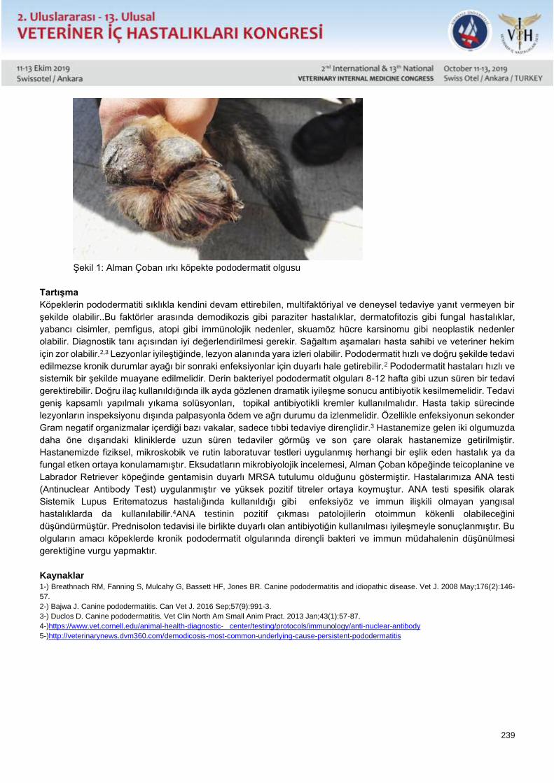
239
Şekil 1: Alman Çoban ırkı köpekte pododermatit olgusu
Tartışma
Köpeklerin pododermatiti sıklıkla kendini devam ettirebilen, multifaktöriyal ve deneysel tedaviye yanıt vermeyen bir
şekilde olabilir..Bu faktörler arasında demodikozis gibi paraziter hastalıklar, dermatofitozis gibi fungal hastalıklar,
yabancı cisimler, pemfigus, atopi gibi immünolojik nedenler, skuamöz hücre karsinomu gibi neoplastik nedenler
olabilir. Diagnostik tanı açısından iyi değerlendirilmesi gerekir. Sağaltım aşamaları hasta sahibi ve veteriner hekim
için zor olabilir.2,3 Lezyonlar iyileştiğinde, lezyon alanında yara izleri olabilir. Pododermatit hızlı ve doğru şekilde tedavi
edilmezse kronik durumlar ayağı bir sonraki enfeksiyonlar için duyarlı hale getirebilir.2 Pododermatit hastaları hızlı ve
sistemik bir şekilde muayane edilmelidir. Derin bakteriyel pododermatit olguları 8-12 hafta gibi uzun süren bir tedavi
gerektirebilir. Doğru ilaç kullanıldığında ilk ayda gözlenen dramatik iyileşme sonucu antibiyotik kesilmemelidir. Tedavi
geniş kapsamlı yapılmalı yıkama solüsyonları, topikal antibiyotikli kremler kullanılmalıdır. Hasta takip sürecinde
lezyonların inspeksiyonu dışında palpasyonla ödem ve ağrı durumu da izlenmelidir. Özellikle enfeksiyonun sekonder
Gram negatif organizmalar içerdiği bazı vakalar, sadece tıbbi tedaviye dirençlidir.3 Hastanemize gelen iki olgumuzda
daha öne dışarıdaki kliniklerde uzun süren tedaviler görmüş ve son çare olarak hastanemize getirilmiştir.
Hastanemizde fiziksel, mikroskobik ve rutin laboratuvar testleri uygulanmış herhangi bir eşlik eden hastalık ya da
fungal etken ortaya konulamamıştır. Eksudatların mikrobiyolojik incelemesi, Alman Çoban köpeğinde teicoplanine ve
Labrador Retriever köpeğinde gentamisin duyarlı MRSA tutulumu olduğunu göstermiştir. Hastalarımıza ANA testi
(Antinuclear Antibody Test) uygulanmıştır ve yüksek pozitif titreler ortaya koymuştur. ANA testi spesifik olarak
Sistemik Lupus Eritematozus hastalığında kullanıldığı gibi enfeksiyöz ve immun ilişkili olmayan yangısal
hastalıklarda da kullanılabilir.4ANA testinin pozitif çıkması patolojilerin otoimmun kökenli olabileceğini
düşündürmüştür. Prednisolon tedavisi ile birlikte duyarlı olan antibiyotiğin kullanılması iyileşmeyle sonuçlanmıştır. Bu
olguların amacı köpeklerde kronik pododermatit olgularında dirençli bakteri ve immun müdahalenin düşünülmesi
gerektiğine vurgu yapmaktır.
Kaynaklar 1-) Breathnach RM, Fanning S, Mulcahy G, Bassett HF, Jones BR. Canine pododermatitis and idiopathic disease. Vet J. 2008 May;176(2):146-
57.
2-) Bajwa J. Canine pododermatitis. Can Vet J. 2016 Sep;57(9):991-3.
3-) Duclos D. Canine pododermatitis. Vet Clin North Am Small Anim Pract. 2013 Jan;43(1):57-87.
4-)https://www.vet.cornell.edu/animal-health-diagnostic- center/testing/protocols/immunology/anti-nuclear-antibody
5-)http://veterinarynews.dvm360.com/demodicosis-most-common-underlying-cause-persistent-pododermatitis
240
Poster Sunu / Poster Presentation Tam Metin / Full Text
Canine Chronic Otoimmune Pododermatitis in Two Dogs
Basar Ulas Sayılkan*, Emre Kulluk*, Umit Ozcan*, Merve Gizem Sezener**, Yucel Meral*, Arzu Fındık**, Duygu
Dalgın*
*Ondokuz Mayıs University, Faculty of Veterinary Medicine, Department of Internal Medicine, Samsun
** Ondokuz Mayıs University, Faculty of Veterinary Medicine, Department of Microbiology, Samsun
Abstract
Two dogs; an 8 years old male Labrador retriever and a 4 years old German shepherd were referred to Ondokuz
Mayıs University Animal Hospital with chronic and progressive pododermatitis symptoms. German Shepherd was
treated for the former 5 months conventionally, without any recovery and the symtoms worsened resulting with
reluctance to move. Labrador referred with a better clinical status, but lameness was present. Common clinical signs
were edema, paronychia, ulceration, comedones, draining seropurulent exudate, pruritis. In both cases all paws were
affected. Physical, microscopic and routine laboratory examinations did not present and concomitant disorders or
fungal intervention. Microbiological examination of exudates revealed MRSA involvement, sensitive to teicoplamine
in the German shepherd and gentamicine in Labrador. Antinuclear antibody tests revealed high positive titers.
Prednisolone therapy together with the sensitive antimicrobial resulted in recovery. Resistant bacteria and immune
intervention must be considered in chronic pododermatitis cases in dogs.
Key Words: Dog, Pododermatitis, Otoimmune, Resistant bacteria
Introduction
Pododermatitis is called a purulent inflammation of the foot skin. It is common in dogs that one or more feet are
affected, especially in interdigital regions and nails, causing inflammation symptoms. Lesions may heal, increase or
decrease, and can sometimes be permanent. Sometimes it can be formed for one reason or it can be caused by
multifactorial reasons. Parasitic diseases may occur as a result of stinging foreign bodies, allergies, systemic
infections and hormonal diseases.1,2 Demodex pododermatitis is common in dogs.5Pododermatitis is not a disease
but a clinical manifestation of local trauma or systemic disease. Pruritus, erythema, edema, nodule formation,
paronychia, alopecia, ulceration, comedon, serosanguinous or seropurulent discharge may occur in the affected feet.
Feet are swollen and painful.1,2,3The aim of this case report is to emphasize the importance of antibiotic selection
and immune intervention in antibiotic cases and to help future scientific studies.
Case
Two dogs, an eight-year-old Labrador Retriever and a four-year-old German Shepherd dog with chronic and
progressive pododermatitis symptoms, applied to Ondokuz Mayıs University Animal Hospital. Prior to the German
Shepherd Dog, he had been treated in an external center for five months without success, and the animal was
reluctant to move. Labrador Retriever was clinically better off, but there was lameness. Common clinical findings;
edema, paronychia, ulceration, comedon, discharge, seroprulent exudate and pruritus. In both cases all the feet were
affected. Physical, microscopic and routine laboratory tests did not reveal the presence of a concomitant disease or
a fungal disease. Microbiological examination of exudates,Teicoplanin in the German Shepherd and Labrador
Retriever showed gentamicin-sensitive MRSA uptake. Antinuclear Antibody Tests revealed high positive titers. The
use of the antibiotic, which was sensitive with prednisolone treatment, resulted in improvement. Resistant bacteria
and immune intervention should be considered in patients with chronic pododermatitis in dogs.
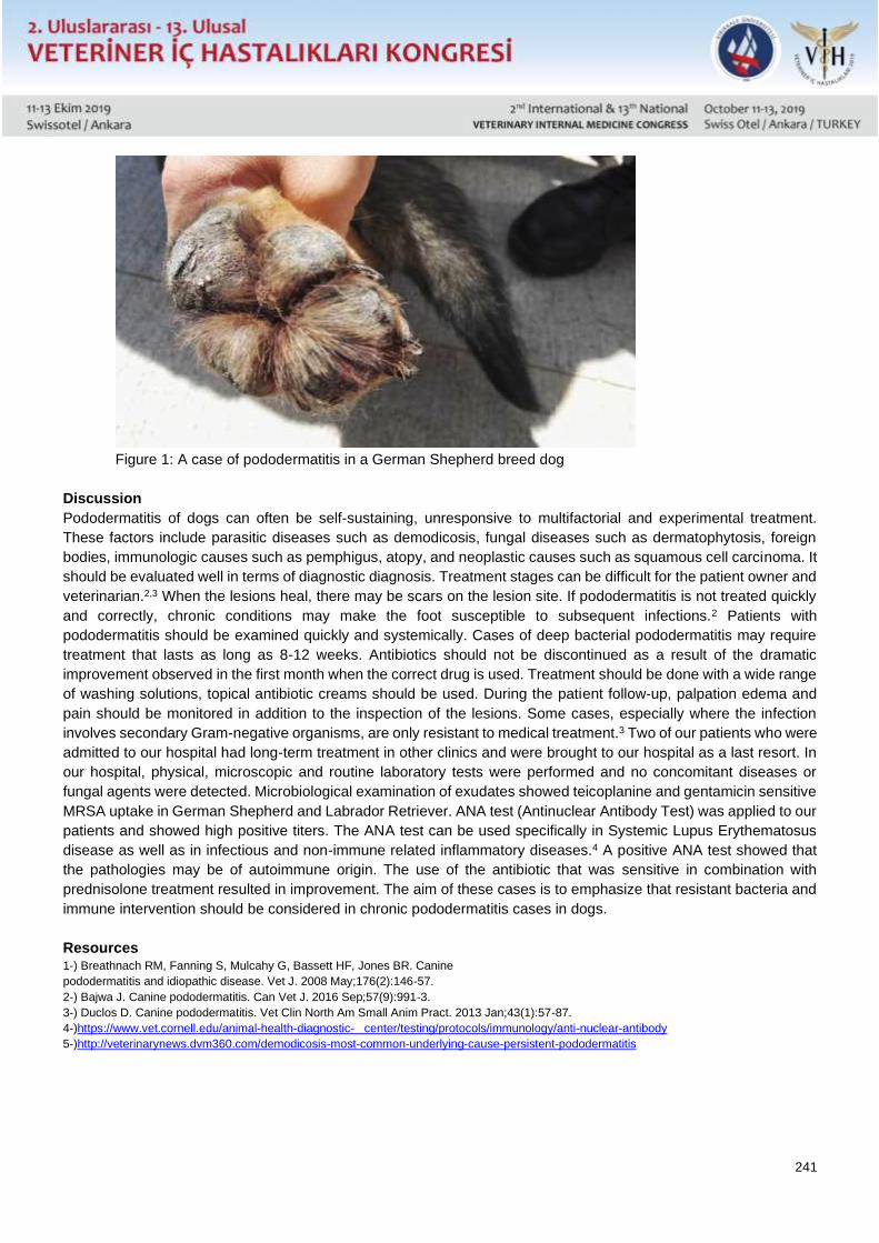
241
Figure 1: A case of pododermatitis in a German Shepherd breed dog
Discussion
Pododermatitis of dogs can often be self-sustaining, unresponsive to multifactorial and experimental treatment.
These factors include parasitic diseases such as demodicosis, fungal diseases such as dermatophytosis, foreign
bodies, immunologic causes such as pemphigus, atopy, and neoplastic causes such as squamous cell carcinoma. It
should be evaluated well in terms of diagnostic diagnosis. Treatment stages can be difficult for the patient owner and
veterinarian.2,3 When the lesions heal, there may be scars on the lesion site. If pododermatitis is not treated quickly
and correctly, chronic conditions may make the foot susceptible to subsequent infections.2 Patients with
pododermatitis should be examined quickly and systemically. Cases of deep bacterial pododermatitis may require
treatment that lasts as long as 8-12 weeks. Antibiotics should not be discontinued as a result of the dramatic
improvement observed in the first month when the correct drug is used. Treatment should be done with a wide range
of washing solutions, topical antibiotic creams should be used. During the patient follow-up, palpation edema and
pain should be monitored in addition to the inspection of the lesions. Some cases, especially where the infection
involves secondary Gram-negative organisms, are only resistant to medical treatment.3 Two of our patients who were
admitted to our hospital had long-term treatment in other clinics and were brought to our hospital as a last resort. In
our hospital, physical, microscopic and routine laboratory tests were performed and no concomitant diseases or
fungal agents were detected. Microbiological examination of exudates showed teicoplanine and gentamicin sensitive
MRSA uptake in German Shepherd and Labrador Retriever. ANA test (Antinuclear Antibody Test) was applied to our
patients and showed high positive titers. The ANA test can be used specifically in Systemic Lupus Erythematosus
disease as well as in infectious and non-immune related inflammatory diseases.4 A positive ANA test showed that
the pathologies may be of autoimmune origin. The use of the antibiotic that was sensitive in combination with
prednisolone treatment resulted in improvement. The aim of these cases is to emphasize that resistant bacteria and
immune intervention should be considered in chronic pododermatitis cases in dogs.
Resources 1-) Breathnach RM, Fanning S, Mulcahy G, Bassett HF, Jones BR. Canine
pododermatitis and idiopathic disease. Vet J. 2008 May;176(2):146-57.
2-) Bajwa J. Canine pododermatitis. Can Vet J. 2016 Sep;57(9):991-3.
3-) Duclos D. Canine pododermatitis. Vet Clin North Am Small Anim Pract. 2013 Jan;43(1):57-87.
4-)https://www.vet.cornell.edu/animal-health-diagnostic- center/testing/protocols/immunology/anti-nuclear-antibody
5-)http://veterinarynews.dvm360.com/demodicosis-most-common-underlying-cause-persistent-pododermatitis
242
Poster Sunu / Poster Presentation Tam Metin / Full Text
Köpeklerde Serum Üre, Kreatinin ve Simetrik Dimetil Arginin (SDMA) Düzeyleri
Arasındaki İlişkinin Belirlenmesi
Yücel Meral*, Duygu Dalgın*, Sena Çenesiz**, Emre Küllük*, Başar Ulaş Sayılkan*
*Ondokuz Mayıs Üniversitesi Veteriner Fakültesi İç Hastalıkları Anabilim Dalı, Samsun
**Ondokuz Mayıs Üniversitesi Veteriner Fakültesi Biyokimya Anabilim Dalı, Samsun
Özet
Böbrek yetmezliği, küçük hayvan hekimliğinde sık karşılaşılan bir hastalıktır. Böbrek yetmezliğinde erken tanı,
veteriner hekimlikte nefrodiyaliz imkanı olmadığından ayrı bir önem taşımaktadır, fakat hayvanların klinik rahatsızlığı
yansıtmadaki yetersizlikten dolayı genellikle hastalık başlangıçta teşhis edilememektedir. Böbrek hasarı tanısında
klinik pratikte kullanılan diagnostik parametreler serum üre ve kreatinin düzeyleri olup, serum üre düzeyi farklı
metabolik ve iatrojenik durumlardan etkilenebildiğinden, böbrek spesifik olarak dikkate alınan majör parametre
kreatinin düzeyi olmaktadır. Fakat kreatinin düzeyi hastada ancak böbrek hasarı ileri düzeyde gerçekleştikten sonra
yükseldiğinden, tedavi için önemli bir süreç kaybedilmiş ve teşhis çok geç konulabilmiş olmaktadır. Son yıllarda
yapılan çalışmalar, böbrek fonksiyonuna spesifik bir biyobelirteç olan Serum Simetrik Dimetil Arginin (SDMA)
düzeyinin, serum üre ve kreatinine oranla böbrek yetmezliğini çok daha erken belirlediğini ortaya koymuştur. Bu
çalışmada rutin klinik muayeneler sırasında biyokimyasal panel çalışılan 50 adet köpekte, ayrıca SDMA düzeyleri de
çalışılmış ve veriler doğrultusunda köpeklerde serum üre, kreatinin ve SDMA düzeyleri arasındaki bir korelasyon
olmadığı ortaya konmuş olunmasının yanı sıra, böbrek yetmezliği ön tanısı almayan ve bu yönde klinik şikayeti
olmayan, ayrıca üre ve kreatinin düzeylerinin böbrek yetmezliğini işaret etmediği deneklerin %14’ünde, SDMA
sonuçları böbrek yetmezliğinin başladığını belirlenmiştir. Konvansiyonel prosedürde böbrek yetmezliği tanısı
konulmayacak olan bu denekler, SDMA sonuçları doğrultusunda tedavi altına alınmışlardır. Çok çarpıcı olan bu oran,
SDMA belirtecinin diagnostik etkinliğini ortaya koymakta olup, erken teşhis, terapötik başarı şansı, tedavi giderleri ve
hayvan refahı açısından mutlaka klinik pratiğe yerleşmesi gerekliliğini vurgulamaktadır.
Anahtar Kelimeler: Köpek, Simetrik dimetil arginin (SDMA), Böbrek yetmezliği
Giriş
Böbrek yetmezliği, küçük hayvan hekimliğinde sık karşılaşılan bir hastalıktır. Böbrek yetmezliğinde erken tanı,
veteriner hekimlikte nefrodiyaliz imkanı olmadığından ayrı bir önem taşımaktadır, fakat hayvanların klinik rahatsızlığı
yansıtmadaki yetersizlikten dolayı genellikle hastalık başlangıçta teşhis edilememektedir.
Oysa akut böbrek yetmezliği için tedavi şansı mümkün iken, hadise kronikleştikten sonra tedavinin mümkün
olmamasının yanı sıra, hayvan sahibi için duygusal ve maliyetli, hayvan için ise özellikle ürenin yakıcı etkilerinden
dolayı son derece acılı bir süreç ölüme kadar devam etmektedir. Böbrek hasarı tanısında klinik pratikte kullanılan
diagnostik parametreler serum üre ve kreatinin düzeyleri olup, serum üre düzeyi farklı metabolik ve iatrojenik
durumlardan etkilenebildiğinden, böbrek spesifik olarak dikkate alınan majör parametre kreatinin düzeyi olmaktadır.
Fakat kreatinin düzeyi hastada ancak böbrek hasarı ileri düzeyde gerçekleştikten sonra yükseldiğinden, tedavi için
önemli bir süreç kaybedilmiş ve teşhis çok geç konulabilmiş olmaktadır. Bunun yanı sıra tedavi etkinliğinin
izlenmesinde de kreatinin hareket aralığı çok kısıtlı olduğundan, ancak üre bir yol gösterici olabilmekte, fakat başka
faktörlerden de etkilenebildiği için güvenilirliği sınırlı olmaktadır. Ayrıca kreatinin düzeyi kas kitlesiyle doğrudan ilgili
bir belirteç olduğundan, hastalık etkisiyle kilo verildikçe, bu düzeyde pek bir değişiklik olmamakta, bu durum da
yanıltıcı olmaktadır.
243
Son yıllarda yapılan çalışmalar, böbrek fonksiyonuna spesifik bir biyobelirteç olan Serum Simetrik Dimetil Arginin
(SDMA) düzeyinin, serum üre ve kreatinine oranla böbrek yetmezliğini çok daha erken belirlediğini ortaya koymuştur.
Sağlıklı köpeklerde SDMA referans değerleri ortaya konmuş olmakla birlikte, klinik pratikte kullanılan kan üre,
kreatinin değerleri ve SDMA düzeyi arasındaki ilişkiye odaklanmış çalışma sayısı sınırlıdır.
Genel Bilgiler
Köpeklerde böbrek yetmezliği sık tanı alan bir hastalık olup, akut ve kronik olarak iki kategoride değerlendirilir. Akut
böbrek yetmezliği, böbreklerde oluşan hasar sonucu süzme, metabolik ve endokrin fonksiyonlarda, dolayısıyla
glomerüler filtrasyon hızında azalma olarak tanımlanırken, tedavi edilmezse kronik böbrek yetmezliği ile
sonuçlanır(1). Kronik böbrek yetmezliği (KBY), böbrekte yapısal ve fonksiyonel kayıpla karakterize, geri dönüşümsüz
ve progresif seyirli bir hastalıktır(1,2). Klinik olarak belirgin hale gelmeden önce hastalık aylar, bazen de yıllar boyu
seyrini sürdürür. Geriatrik köpeklerde %10 oranında, genel olarak küçük hayvan popülasyonu çerçevesinde ise %1-
3 insidans bildirilmektedir (3). Hastalık köpeklerde en sık görülen renal bozukluktur (2,3,4).
Böbrek yemezliğinin prognozu büyük ölçüde erken tanı ile alakalıdır. Güncel klinik pratikte kullanılan tanısal
parametreler, serum üre ve kreatinin düzeyi ile sınırlı kalmaktadır. Oysa bu parametrelerin hastalığı yansıtabilmesi
için böbrek hasarının oldukça ilerlemiş olması gerekmektedir. Klasik literatürde serum kreatinin düzeyinin, renal
fonksiyonun %75’i kaybolmadan yükselmediği kabul edilmektedir (4). Son yıllarda yapılan çalışmalar, böbrek
fonksiyonuna spesifik bir biyobelirteç olan Serum Simetrik Dimetil Arginin (SDMA) düzeyinin, serum üre ve kreatinine
oranla böbrek yetmezliğini çok daha erken belirlediğini ortaya koymuştur(5-9). SDMA böbrekler tarafından atılır ve
glomerüler filtrasyon hızını çok daha doğru şekilde yansıtır, hatta böbrek fonksiyonunun %25 düzeyinde kaybında
bile yükseldiği rapor edilmiştir (5-9). Bir çalışmada KBYli kedilerde SDMA in kreatininden 17 ay önce ve glomerüler
filtrasyon hızı %40 düzeyinde iken yükseldiği, SDMA %100 sensitivite ve %91 spesifite gösterirken, kreatininin %17
sensitivite gösterdiği bildirilmiştir (8). Yine KBYli köpeklerde SDMA’in kreatinine oranla ortalama 9.5 ay önce
yükseldiği ortaya konmuştur (6).
Ayrıca kreatininin aksine SDMA’in vücut ağırlığıyla da bir korelasyonu bulunmadığından, kilo kaybı ile karakterize
KBYde kilo kaybı nedeniyle dejenerasyonla orantılı seyretmeyen kreatinin yanıltıcı etkisi de ortadan kalkmaktadır (9).
Bu nedenle SDMA’in sistolik kan basıncı ve kreatinin düzeyiyle birlikte izlenmesinin, KBYnin seyrini, sağlıklı ve duyarlı
biçimde ortaya koyacağı öngörülmektedir (10,11). Sağlıklı köpeklerde SDMA referans değerleri ortaya konmuş
olmakla birlikte (12), klinik pratikte kullanılan kan üre, kreatinin değerleri ve SDMA düzeyi arasındaki korelasyona
odaklanmış çalışma sayısı sınırlıdır.
Bu nedenle köpeklerde üre, kreatinin ile SDMA düzeyleri arasındaki ilişkinin ortaya konmasını amaçlayan bu çalışma,
konuya önemli katkı sağlayacaktır. Ayrıca yurtdışında rutin klinik diagnoz spektrumuna girmekte olan bu böbrek
spesifik biyobelirtecin, üniversitemiz Hayvan Hastanesini başlangıç alarak ülkemizde de küçük hayvan klinik
pratiğinde yerini alması öngörülmektedir. SDMA in rutin kullanımı ile, köpeklerde sık rastlanan bir renal bozukluk olan
böbrek yetmezliğinin erken teşhisi mümkün olacak, bu sayede tedavi başarısı artacak ve vakaların kronik böbrek
yetmezliği noktasına varmadan tedavi şansı ortaya çıkacaktır. Bilindiği üzere, kronik böbrek yetmezliği beşeri
hekimlikte diyalizle hayatın idamesinin sağlandığı ve ancak böbrek nakliyle şifa bulabilen bir hastalıktır. Veteriner
hekimlikte ise, kronik böbrek yetmezlikli hastalarda ancak son raddeye kadar semptomatik ve maliyetli bir tedavi
uygulanabilmekte, ileri dönemde ise diyaliz imkanı bulunmadığından ötenazi uygulanmak durumunda kalınmaktadır.
Bu nedenle diagnostik süreçte SDMA kullanımı hem hayvan refahı, hem de ekonomik açıdan önemi katkı
sağlayacaktır.
Gereç ve Yöntem
Çalışmada, OMÜ Veteriner Fakültesi Hayvan Hastanesi kliniklerine başvuran 50 adet sahipli köpek kullanılmıştır. Ön
muayene sonucunda kan üre ve kreatinin düzeylerini de içermek üzere serum biyokimyasal profillerinin
incelenmesine karar verilen hastaların, protokol numaraları, anamnez bilgileri, yaş, tür, ırk özellikleri, hasta sahibi
bilgileri kayıt altına alınmıştır.
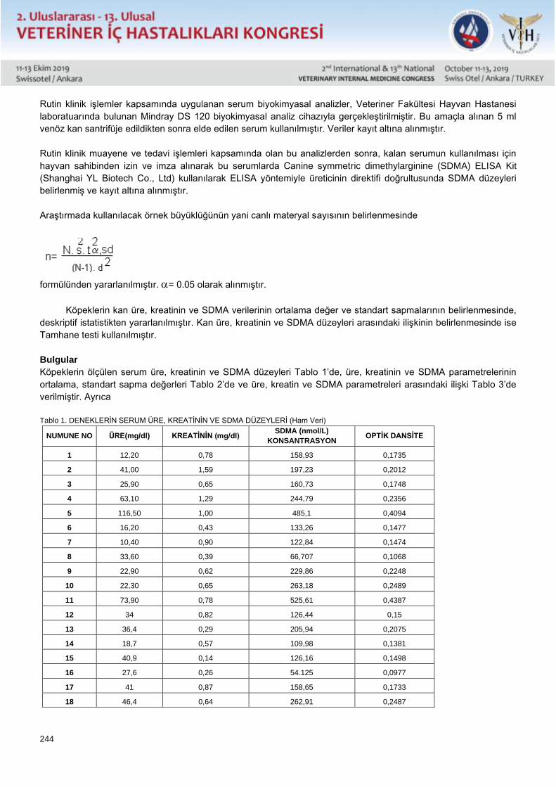
244
Rutin klinik işlemler kapsamında uygulanan serum biyokimyasal analizler, Veteriner Fakültesi Hayvan Hastanesi
laboratuarında bulunan Mindray DS 120 biyokimyasal analiz cihazıyla gerçekleştirilmiştir. Bu amaçla alınan 5 ml
venöz kan santrifüje edildikten sonra elde edilen serum kullanılmıştır. Veriler kayıt altına alınmıştır.
Rutin klinik muayene ve tedavi işlemleri kapsamında olan bu analizlerden sonra, kalan serumun kullanılması için
hayvan sahibinden izin ve imza alınarak bu serumlarda Canine symmetric dimethylarginine (SDMA) ELISA Kit
(Shanghai YL Biotech Co., Ltd) kullanılarak ELISA yöntemiyle üreticinin direktifi doğrultusunda SDMA düzeyleri
belirlenmiş ve kayıt altına alınmıştır.
Araştırmada kullanılacak örnek büyüklüğünün yani canlı materyal sayısının belirlenmesinde
formülünden yararlanılmıştır. = 0.05 olarak alınmıştır.
Köpeklerin kan üre, kreatinin ve SDMA verilerinin ortalama değer ve standart sapmalarının belirlenmesinde,
deskriptif istatistikten yararlanılmıştır. Kan üre, kreatinin ve SDMA düzeyleri arasındaki ilişkinin belirlenmesinde ise
Tamhane testi kullanılmıştır.
Bulgular
Köpeklerin ölçülen serum üre, kreatinin ve SDMA düzeyleri Tablo 1’de, üre, kreatinin ve SDMA parametrelerinin
ortalama, standart sapma değerleri Tablo 2’de ve üre, kreatin ve SDMA parametreleri arasındaki ilişki Tablo 3’de
verilmiştir. Ayrıca
Tablo 1. DENEKLERİN SERUM ÜRE, KREATİNİN VE SDMA DÜZEYLERİ (Ham Veri)
NUMUNE NO ÜRE(mg/dl) KREATİNİN (mg/dl) SDMA (nmol/L)
KONSANTRASYON OPTİK DANSİTE
1 12,20 0,78 158,93 0,1735
2 41,00 1,59 197,23 0,2012
3 25,90 0,65 160,73 0,1748
4 63,10 1,29 244,79 0,2356
5 116,50 1,00 485,1 0,4094
6 16,20 0,43 133,26 0,1477
7 10,40 0,90 122,84 0,1474
8 33,60 0,39 66,707 0,1068
9 22,90 0,62 229,86 0,2248
10 22,30 0,65 263,18 0,2489
11 73,90 0,78 525,61 0,4387
12 34 0,82 126,44 0,15
13 36,4 0,29 205,94 0,2075
14 18,7 0,57 109,98 0,1381
15 40,9 0,14 126,16 0,1498
16 27,6 0,26 54.125 0,0977
17 41 0,87 158,65 0,1733
18 46,4 0,64 262,91 0,2487
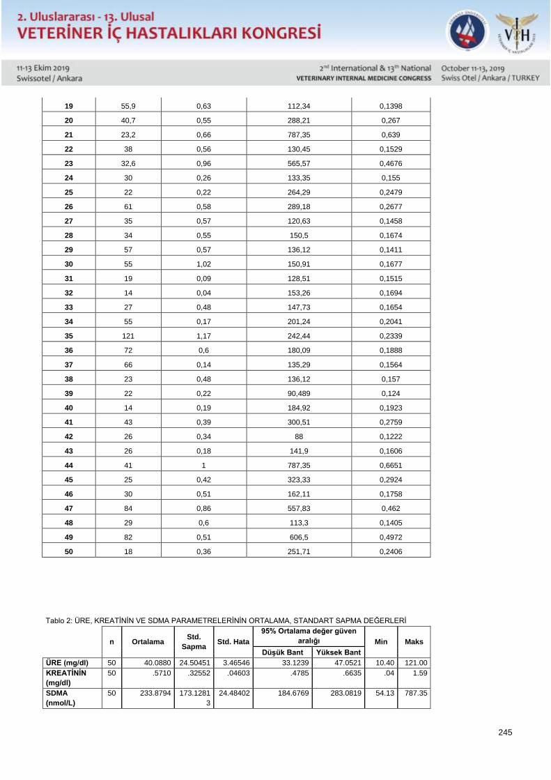
245
19 55,9 0,63 112,34 0,1398
20 40,7 0,55 288,21 0,267
21 23,2 0,66 787,35 0,639
22 38 0,56 130,45 0,1529
23 32,6 0,96 565,57 0,4676
24 30 0,26 133,35 0,155
25 22 0,22 264,29 0,2479
26 61 0,58 289,18 0,2677
27 35 0,57 120,63 0,1458
28 34 0,55 150,5 0,1674
29 57 0,57 136,12 0,1411
30 55 1,02 150,91 0,1677
31 19 0,09 128,51 0,1515
32 14 0,04 153,26 0,1694
33 27 0,48 147,73 0,1654
34 55 0,17 201,24 0,2041
35 121 1,17 242,44 0,2339
36 72 0,6 180,09 0,1888
37 66 0,14 135,29 0,1564
38 23 0,48 136,12 0,157
39 22 0,22 90,489 0,124
40 14 0,19 184,92 0,1923
41 43 0,39 300,51 0,2759
42 26 0,34 88 0,1222
43 26 0,18 141,9 0,1606
44 41 1 787,35 0,6651
45 25 0,42 323,33 0,2924
46 30 0,51 162,11 0,1758
47 84 0,86 557,83 0,462
48 29 0,6 113,3 0,1405
49 82 0,51 606,5 0,4972
50 18 0,36 251,71 0,2406
Tablo 2: ÜRE, KREATİNİN VE SDMA PARAMETRELERİNİN ORTALAMA, STANDART SAPMA DEĞERLERİ
n Ortalama Std.
Sapma Std. Hata
95% Ortalama değer güven
aralığı Min Maks
Düşük Bant Yüksek Bant
ÜRE (mg/dl) 50 40.0880 24.50451 3.46546 33.1239 47.0521 10.40 121.00
KREATİNİN
(mg/dl)
50 .5710 .32552 .04603 .4785 .6635 .04 1.59
SDMA
(nmol/L)
50 233.8794 173.1281
3
24.48402 184.6769 283.0819 54.13 787.35
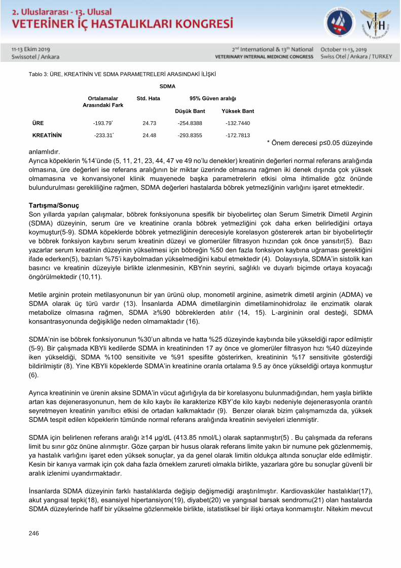
246
Tablo 3: ÜRE, KREATİNİN VE SDMA PARAMETRELERİ ARASINDAKİ İLİŞKİ
* Önem derecesi p≤0.05 düzeyinde
anlamlıdır.
Ayrıca köpeklerin %14’ünde (5, 11, 21, 23, 44, 47 ve 49 no’lu denekler) kreatinin değerleri normal referans aralığında
olmasına, üre değerleri ise referans aralığının bir miktar üzerinde olmasına rağmen iki denek dışında çok yüksek
olmamasına ve konvansiyonel klinik muayenede başka parametrelerin etkisi olma ihtimalide göz önünde
bulundurulması gerekliliğine rağmen, SDMA değerleri hastalarda böbrek yetmezliğinin varlığını işaret etmektedir.
Tartışma/Sonuç
Son yıllarda yapılan çalışmalar, böbrek fonksiyonuna spesifik bir biyobelirteç olan Serum Simetrik Dimetil Arginin
(SDMA) düzeyinin, serum üre ve kreatinine oranla böbrek yetmezliğini çok daha erken belirlediğini ortaya
koymuştur(5-9). SDMA köpeklerde böbrek yetmezliğinin derecesiyle korelasyon göstererek artan bir biyobelirteçtir
ve böbrek fonksiyon kaybını serum kreatinin düzeyi ve glomerüler filtrasyon hızından çok önce yansıtır(5). Bazı
yazarlar serum kreatinin düzeyinin yükselmesi için böbreğin %50 den fazla fonksiyon kaybına uğraması gerektiğini
ifade ederken(5), bazıları %75’i kaybolmadan yükselmediğini kabul etmektedir (4). Dolayısıyla, SDMA’in sistolik kan
basıncı ve kreatinin düzeyiyle birlikte izlenmesinin, KBYnin seyrini, sağlıklı ve duyarlı biçimde ortaya koyacağı
öngörülmektedir (10,11).
Metile arginin protein metilasyonunun bir yan ürünü olup, monometil arginine, asimetrik dimetil arginin (ADMA) ve
SDMA olarak üç türü vardır (13). İnsanlarda ADMA dimetilarginin dimetilaminohidrolaz ile enzimatik olarak
metabolize olmasına rağmen, SDMA ≥%90 böbreklerden atılır (14, 15). L-argininin oral desteği, SDMA
konsantrasyonunda değişikliğe neden olmamaktadır (16).
SDMA’nin ise böbrek fonksiyonunun %30’un altında ve hatta %25 düzeyinde kaybında bile yükseldiği rapor edilmiştir
(5-9). Bir çalışmada KBYli kedilerde SDMA in kreatininden 17 ay önce ve glomerüler filtrasyon hızı %40 düzeyinde
iken yükseldiği, SDMA %100 sensitivite ve %91 spesifite gösterirken, kreatininin %17 sensitivite gösterdiği
bildirilmiştir (8). Yine KBYli köpeklerde SDMA’in kreatinine oranla ortalama 9.5 ay önce yükseldiği ortaya konmuştur
(6).
Ayrıca kreatininin ve ürenin aksine SDMA’in vücut ağırlığıyla da bir korelasyonu bulunmadığından, hem yaşla birlikte
artan kas dejenerasyonunun, hem de kilo kaybı ile karakterize KBY’de kilo kaybı nedeniyle dejenerasyonla orantılı
seyretmeyen kreatinin yanıltıcı etkisi de ortadan kalkmaktadır (9). Benzer olarak bizim çalışmamızda da, yüksek
SDMA tespit edilen köpeklerin tümünde normal referans aralığında kreatinin seviyeleri izlenmiştir.
SDMA için belirlenen referans aralığı ≥14 μg/dL (413.85 nmol/L) olarak saptanmıştır(5) . Bu çalışmada da referans
limit bu sınır göz önüne alınmıştır. Göze çarpan bir husus olarak referans limite yakın bir numune pek gözlenmemiş,
ya hastalık varlığını işaret eden yüksek sonuçlar, ya da genel olarak limitin oldukça altında sonuçlar elde edilmiştir.
Kesin bir kanıya varmak için çok daha fazla örneklem zarureti olmakla birlikte, yazarlara göre bu sonuçlar güvenli bir
aralık izlenimi uyandırmaktadır.
İnsanlarda SDMA düzeyinin farklı hastalıklarda değişip değişmediği araştırılmıştır. Kardiovasküler hastalıklar(17),
akut yangısal tepki(18), esansiyel hipertansiyon(19), diyabet(20) ve yangısal barsak sendromu(21) olan hastalarda
SDMA düzeylerinde hafif bir yükselme gözlenmekle birlikte, istatistiksel bir ilişki ortaya konmamıştır. Nitekim mevcut
SDMA
Ortalamalar
Arasındaki Fark
Std. Hata 95% Güven aralığı
Düşük Bant Yüksek Bant
ÜRE -193.79* 24.73 -254.8388 -132.7440
KREATİNİN -233.31* 24.48 -293.8355 -172.7813
247
çalışmada da deneklerin hepsi klinik bir şikayetle getirildikleri ve farklı problemler yaşadıkları halde, bu klinik
bozukluklar ile SDMA düzeyleri arasında bir ilişki bulunmamıştır.
Köpeklerde SDMA düzeyleri ile yaş, ırk, cinsiyet veya diyet arasında bir bağlantı saptanmamıştır (9,22). Benzer
olarak mevcut çalışmada da, farklı yaş, ırk ve cinsten, farklı diyetlerle beslenen köpeklerde herhangi bir ilişki
saptanmamıştır.
Sonuç olarak köpeklerde kan üre, kreatinin değerleri ile SDMA düzeyi arasında bir korelasyon bulunmamış, fakat
deneklerin %14’ünde kreatinin düzeyi normal referans limitler içinde seyrettiği halde, yüksek SDMA düzeyleri
saptanmıştır. Konvansiyonel prosedürde böbrek yetmezliği tanısı konulmayacak olan bu denekler, SDMA sonuçları
doğrultusunda tedavi altına alınmışlardır. Çok çarpıcı olan bu oran, SDMA belirtecinin diagnostik etkinliğini ortaya
koymakta olup, erken teşhis, terapötik başarı şansı, tedavi giderleri ve hayvan refahı açısından mutlaka klinik pratiğe
yerleşmesi gerekliliğini vurgulamaktadır.
Kaynaklar 1.Blackwell S., O'reılly D.S., Reıd D. (2011): Plasma dimethylarginines during the acute inflammatory response. Eur J Clin Invest. 41, 635–
641.https://onlinelibrary.wiley.com/doi/pdf/10.1111/j.1365-2362.2010.02451.x
2.Brown S.A. (2018): Renal dysfunction in small animals. In: The Merck Veterinary Manual,
www.merckmanuals.com/vet/urinary_system/noninfectious_diseases_ (1.12. 2018)
3.Grouer F.G. (2009): Urinary tract disorders. In: Small Animal Internal Medicine Nelson, R.W., C.G. Couto, (Ed) Saunders Elsevier, St. Louis,
pp. 489.
https://www.scribd.com/document/318210604/NELSON-R-W-COUTO-C-G-Small-animal-internal-medicine-4-ed-St-Louis-Mosby-2008-pdf
4. Hall JA., Yerramilli M., Obare E., Yerramilli M., Almes K., Jewell DE. (2016): Serum concentrations of symmetric dimethylarginine and creatinine
in dogs with naturally occurring chronic kidney disease. J Vet Intern Med. 30, 794–802.https://onlinelibrary.wiley.com/doi/full/10.1111/jvim.13942
5. Hall JA., Yerramilli M., Obare E., Yerramilli M., Melendez LD., Jewell DE. (2015): Relationship between lean body mass and serum renal
biomarkers in healthy dogs. J Vet Intern Med. 29, 808–814.
https://onlinelibrary.wiley.com/doi/full/10.1111/jvim.12607
6. Hall JA., Yerramilli M., Obare E., Yerramilli M., Yu S., Jewell DE. (2014): Comparison of serum concentrations of symmetric dimethylarginine
and creatinine as kidney function biomarkers in healthy geriatric cats fed reduced protein foods enriched with fish oil, L-carnitine, and medium-
chain triglycerides. Vet J. 202, 588–596.
https://www.sciencedirect.com/science/article/pii/S1090023314004341?via%3Dihub
7.IRIS (2018) International Renal Society, IRIS staging of CKD. http://www.iris-kidney.com/guidelines/staging.html
9. Kielstein JT., Böger RH., Bode-Böger SM., Frölich JC., Haller H., Ritz E., Fliser D. (2002): Marked increase of asymmetric dimethylarginine in
patients with incipient primary chronic renal disease. J Am Soc Nephrol. 13, 170–176.https://jasn.asnjournals.org/content/13/1/170.long
10.Krzyzanowska K., Mittermayer F., N. Shnawa. (2007): Asymmetrical dimethylarginine is related to renal function, chronic inflammation and
macroangiopathy in patients with Type 2 diabetes and albuminuria. Diabet Med. 24, 81–86.https://onlinelibrary.wiley.com/doi/abs/10.1111/j.1464-
5491.2007.02018.x
11.Meinitzer A., Kielstein JT., Pilz S., Drechsler C., Ritz E., Boehm BO., Winkelmann BR, März W. (2011): Symmetrical and asymmetrical
dimethylarginine as predictors for mortality in patients referred for coronary angiography: The Ludwigshafen Risk and Cardiovascular Health study.
Clin Chem. 57, 112–121.
http://clinchem.aaccjnls.org/content/57/1/112.long
12.Moesgaard SG., Holte AV., Mogensen T., Mølbak J., Kristensen AT., Jensen AL., Teerlink T., Reynolds AJ., Olsen LH. (2007): Effects of
breed, gender, exercise and white-coat effect on markers of endothelial function in dogs. Res Vet Sci. 82, 409–415.
https://www.science.ku.dk/english/research/phd/student/phdtheses/?pure=en%2Fpublications%2Feffects-of-breed-gender-exercise-and-
whitecoat-effect-on-markers-of-endothelial-function-in-dogs(3a550280-a1c2-11dd-b6ae-000ea68e967b)%2Fexport.html
13.Nabity MB., Lees GE., Boggess MM., Yerramilli M., Obare E., Yerramilli M., Rakitin A., Aguiar J., Relford R. (2015): Symmetric dimethylarginine
assay validation, stability, and evaluation as a marker for early detection of chronic kidney disease in dogs. J Vet Intern Med. 9, 1036–1044.
https://onlinelibrary.wiley.com/doi/full/10.1111/jvim.12835
14.Owczarek D., Cibor D., Mach T. (2010). Asymmetric dimethylarginine (ADMA), symmetric dimethylarginine (SDMA), arginine, and 8-iso-
prostaglandin F2alpha (8-iso-PGF2alpha) level in patients with inflammatory bowel diseases. Inflamm Bowel Dis. 16, 52–
57.https://www.ncbi.nlm.nih.gov/pubmed/19575355
15.Relford R, Robertson J., Clements C. (2016): Symmetric Dimethylarginine: Improving the Diagnosis and Staging of Chronic Kidney Disease in
Small Animals. Veterinary Clinics of North America: Small Animal Practice. 46, 941–960.
https://www.sciencedirect.com/science/article/pii/S0195561616300468?via%3Dihub
16.Rentko V., Nabıty M., Yerramıllı M. (2013): Determination of serum symmetric dimethylarginine reference limit in clinically healthy dogs. J Vet
Intern Med. 27, 750.
https://www.researchgate.net/publication/306123124_Determination_of_serum_symmetric_dimethylarginine_reference_limit_in_clinically_health
y_dogs_abstract
248
17.Roudebush P., Polzin DJ., Adams LG., Towell TL., Forrester SD. (2010): An evidence-based review of therapies for canine chronic kidney
disease. Journal of Small Animal Practice. 51, 244–252.
https://onlinelibrary.wiley.com/doi/full/10.1111/j.1748-5827.2010.00932.x
18. Rytlewski K., Olszanecki R., Korbut R., Zdebski Z. (2005): Effects of prolonged oral supplementation with L-arginine on blood pressure and
nitric oxide synthesis in preeclampsia. Eur J Clin Invest. 35, 32–37.
https://onlinelibrary.wiley.com/doi/abs/10.1111/j.1365-2362.2005.01445.x
19. Schwedhelm E, Böger RH. (2011): The role of asymmetric and symmetric dimethylarginines in renal disease. Nat Rev Nephrol. 7, 275–
285.https://www.nature.com/articles/nrneph.2011.31
20.Shaw DH., S. Ihle (2007): Urinary tract diseases and fluid and electrolyte disorders. Nelson R., N.G. Couto (2007): Small Animal Internal
Medicine. Lippincott Williams-Wilkins. pp. 99.
21. Wang D., Strandgaard S., Iversen J., Wilcox CS. (2009): Asymmetric dimethylarginine, oxidative stress, and vascular nitric oxide synthase in
essential hypertension. Am J Physiol Regul Integr Comp Physiol. 296, 195–200.
https://www.physiology.org/doi/full/10.1152/ajpregu.90506.2008?url_ver=Z39.88-
2003&rfr_id=ori%3Arid%3Acrossref.org&rfr_dat=cr_pub%3Dpubmed&
22. Hall JA., Yerramilli M., Obare E., Yerramilli M., Jewell DE. (2014): Comparison of serum concentrations of symmetric dimethylarginine and
creatinine as kidney function biomarkers in cats with chronic kidney disease. J Vet Intern Med. 28, 1676–
1683.https://onlinelibrary.wiley.com/doi/full/10.1111/jvim.12445
23.Yerramıllı M., Obare E., Jewell DE., Hall JA. (2014): Symmetric dimethylarginine (SDMA) increases earlier than serum creatinine in dogs with
chronic kidney disease (CKD). J Vet Intern Med. 28, 1084–1085.
https://www.researchgate.net/publication/306153696_Symmetric_dimethylarginine_SDMA_increases_earlier_than_serum_creatinine_in_dogs_
with_chronic_kidney_disease_CKD
249
Poster Sunu / Poster Presentation Tam Metin / Full Text
Relationships Between Symmetric Dimethylarginine (SDMA), Blood Urea Nitrogen And
Creatinine Levels In Dogs
Yucel Meral*, Duygu Dalgin*, Sena Cenesiz**, Emre Kulluk*, Basar Ulas Sayilkan*
*Ondokuz Mayıs University, Faculty of Veterinary Medicine, Department of Internal Medicine, Samsun
** Ondokuz Mayıs University, Faculty of Veterinary Medicine, Department of Biochemistry, Samsun
*Correspondence:
Res. Ast. Başar Ulaş SAYILKAN, Ondokuz Mayıs University, Faculty of Veterinary Medicine, Department of Internal
Medicine, Atakum, 55139, Samsun, Turkey.
This research had been supported by Ondokuz Mayıs University Scientific Research Projects comission with
reference number PYO.VET.1901.17.014.
Abstract
Renal failure is a frequently encountered disorder of the dogs and cats. Eraly diagnosis in renal failure is especially
important in small animal practice because of absence of nephrodialysis possibilities, but due to the incapability of
animals reflecting clinical suffering, generally delayed diagnosis are achieved. Blood urea and creatinin levels are
the conventional diagnostic parameters in renal failure, but due to multiple effective factors on urea levels, creatinin
is accepted as the major nephro spesific diagnostic tool. Alas, creatinin levels elevate only after advanced renal
failure, therefore a significant period for the therapy onset is mislayed. Current research indicates that serum symetric
dimethly arginine (SDMA) is a spesific biomarker of renal insufficiency, displaying the disorder much more earlier
that both of the conventionel tools. In the present study, SDMA levels are determined in 50 dogs in which serum
biochemical panels were operated. Obtained results displayed an absence of a correlation between serum urea,
creatinin and SDMA levels, but more important indicated renal insufficiency in 14% of the dogs which had none
related clinical suffering. Also their urea and creatinin results did not yield renal failure. These dogs which would
have not get renal failure diagnosis in conventional procedure were started therapy at once. This striking ratio
displayes the diagnostic efficiency of SDMA and also emphasizes the requirement of routine SDMA usage in clinical
practice on behalf of early diagnosis, theurapeutic success, therapy expanses and animal welfare.
Key words: Dog, Symmetric dimethyl arginine (SDMA), Renal insufficiency
Introduction
Renal failure in dogs is a frequently diagnosed disorder and evaluated in two categories; Acute renal failure (ARF) is
defined as reduction in glomerular filtration rate and metabolic and endocrine functions, due to renal damage and if
not treated results with chronic renal failure (Shaw and Ihle, 2007). Chronic renal failure (CRF) is a progressive and
irreversibl disorder characterized by structural and functional loss of the kidneys (Shaw and Ihle, 2007; Roudebush
et al., 2010). The condition may sustain for years before becoming clinically apparent. Reported incidence in geriatric
dogs is 10%, where in general pet population is 1-3% (Brown, 2017). CRF is the most frequent renal disorder of dogs
(Grouer, 2009; Roudebush et al., 2010; Brown, 2017).
Prognosis of renal failure is largely correlated with the time of diagnosis. Current diagnostic and prognostic
parameters are restricted with serum creatinine level, systolic blood pressure, and blood urea level in clinical practice
(IRIS, 2018). In fact, these parameters reflect renal damage only on advanced stages. Literature states that serum
creatinine level increases after75% of renal function is lost (Brown, 2017). Last studies demonstrated that, a kidney
spesific biomarker, Serum Symmetric Dimethyl Arginine (SDMA) levels determines renal failure much more earlier
250
that serum urea and creatinine levels (Yerramıllı And Jewell, 2014; Hall et al., 2014; Hall et al.,2015; Hall et al.,2016;
Nabıty et al., 2016). SDMA is excreted by the kidneys, so precisely reflects glomerular filtration rate, as well is
reported to increase even in 25% renal function loss (Yerramıllı And Jewell, 2014; Hall et al., 2014; Hall et al.,2015;
Nabıty et al., 2015; Hall et al.,2016).
Furthermore, unlike creatinine level which is in correlation with body weight, SDMA is independent with body
condition, so more accurate monitoring of the disease progression can be achieved in CRF, characterized by
progressive weight loss (Hall et al.,2015). Therefore, the projection for a sensitive and earlier diagnosis plus accurate
monitoring in CRF includes SDMA determination together with systolic blood pressure and serum creatinine level
(Yerramıllı et al., 2014; Relford et al., 2016; Irıs, 2018). Reference values for SDMA in healthy dogs are reported
(Rentko et al., 2013), but studies focusing on relationship between serum SDMA and urea and creatinine levels are
limited. The aim of this study is to determine the SDMA levels in healthy dogs together with urea and creatinine
levels, and demonstrate any possible relationships if present.
Materials and methods
Ethical Approval
This study was approved by the Ondokuz Mayıs University Animal Studies Local Ethic Committee with 68489742-
604.01.02-E.6791 approval number.
Animals
In the study, 50 dogs from both gender and various ages referring to University Animal Hospital with various complaint
were included. Routine clinical examination were performed. Blood specimen for biochemical analyses were
collected from the patients and history, age, species, gender and owner information were recorded.
Serum Analysis
Serum specimen were obtained from 5 ml of venous blood after centrifugation for each patient. Routine serum
biochemical profile (total protein, globuline, liver and pancreatic enzymes, urea and creatinine levels were analysed
with Mindray DS 120 biochemical analyser according to the manufacturers instructions.
Serum SDMA levels were analysed with Canine symmetric dimethylarginine (SDMA) ELISA Kit (Shanghai YL Biotech
Co., Ltd) with ELISA method according to the manufacturers instructions.
Statistical Analysis
In comparision of serum urea, creatinine and SDMA levels descriptive analysis and Tamhane test for relationship
were used.
Results and discussion
Recorded serum urea, creatinine and SDMA levels of dogs are presented in Table 1, mean and standart deviations
of the parameters are presented in Table 2 and correlation between the parameters are presented in Table 3.
Table 1. Serum urea, creatinine and SDMA levels
Table 2. Mean and standart deviations of serum urea, creatinine and SDMA levels
Table 3. Relationshıp between serum urea, creatinine and SDMA levels
Another remarkable point is in 14% of the dogs (dog 5, 11, 21, 23, 44, 47 and 49) creatinine values were in normal
and urea levels -which also may be affected by other interventions- were slightly above the reference levels, SDMA
levels indicated renal failure presence.
Current research indicates that SDMA, a kidney spesific biomarker, diagnoses renal failure much earlier than serum
urea and creatinine biomarkers. (Hall et al., 2014; Yerramıllı And Jewell, 2014; Hall et al., 2015; Nabıty et al., 2015;
Hall et al.,2016). SDMA is in correlation with the stage of renal failure in dogs and reflects renal function lost and
determines the degree of functional failure much more earlier than serum creatinine level and glomerular filtration
251
late (Nabity et al., 2015). Some authers state that creatinine level start to elevate only after 50% renal function lost
(Grouer, 2009). Consequently, monitorization of SDMA level together with systolic blood pressure and creatinine
levels will bring up a sensitive, precise and accurate CRF follow-up (Rentko et al., 2013; Yerramıllı et al., 2014;
Relford et al., 2016). Similarly, in the present study, in 14% of the dogs with acceptable values of urea and creatinine,
high SDMA levels were determined indicating renal failure.
Methyl arginine is a product of protein methylisation and has three types; monomethyl arginine, asymmetric dimethyl
arginine (ADMA) and SDMA (Bedford and Rıchard, 2005). In men, ADMA enzimatically metabolizes with
dimethylarginine dimethylaminohydrolase, but SDMA is ≥90% excreted from kidneys.( Kıelsteın et al., 2002;
Schwedhelm and Boger, 2011). L-argininine oral supplementation does not affect SDMA concentration (Rytlewskı
et al., 2005).
SDMA is elevated even in 25-30% renal function lost (Hall et al., 2014; Yerramıllı and Jewell, 2014; Hall et al.,2015;
Nabıty et al., 2015; Hall et al.,2016). It is reported that SDMA elevated 17 months before creatinine elevation when
glomerular filtration rate was 40% in cats with renal failure, furhermore SDMA showed 100% sensitivity with 91%
spesivity where creatinine showed 17% sensitivity (Hall et al., 2014). In dogs with renal failure, it was reported that
SDMA indicated renal failure 9.5 months before creatinine (Nabıty et al., 2015).
On the contrary to creatinine and urea, SDMA has no correlation with body weight, so the misleading effects of age
related muscle degeneration together with the weight loss in CRF prevalent for creatinine are annihilated (Hall et
al.,2015). Similarly in the present study, within reference limit creatinine levels were determined in dogs with high
SDMA levels.
Reference interval for SDMA is determined as ≥14 μg/dL (413.85 nmol/L) (IRIS, 2018. This interval is considered in
the present study and a remarkable fact was, none of the SDMA results were around the reference limits; values
were high and indicative or were below the limits avoiding any doubtful results. Although sample size was relatively
restricted in the present study, these results suggest that the interval is reasonably confidental.
Variations in SDMA levels with different disorders were investigated in men. Slight elevations in cardiovascular
disorders (Meınıtzer et al., 2011), acute inflammatory response (Blackwell et al., 2011), essential hypertension (Wang
et al., 2009), diabetes (Krzyzanowska et al., 2007) and inflammatory bowel disease (Owczarek et al., 2010), but a
statistical significance was not determined. Thus, in the present study all dogs had a clinical complaint and some
sort of a disorder, but no relationship was determined between these disorders and SDMA levels.
No correlation between SDMA levels and age, race, gender or diet in dogs were reported (Moesgaard et al., 2007;
Hall et al.,2015). Concordantly, in the present study dogs were various age, gender, race and were on different diets,
but no relationship between these parameters was observed.
Conclusion
In the present study, there was no correlation between dogs urea, creatinine and SDMA levels, but in 14% of the
subjects in which no clinical or laboratory indicator of renal disease including urea and creatinine elevations were
present, high SDMA levels were observed indicating the existence of renal disease. These subjects would not been
diagnosed as renal failure in conventional procedure, but they were immediately started therapy in the light of high
SDMA levels.
These striking result demonstrates the diagnostic efficiency of SDMA marker and underlines the essentialness of this
biomarker in clinical practice regarding early diagnosis, theraupetic success and animal welfare.
In conclusion, involvement of SDMA biomarker in conventional diagnostic procedure will provide early diagnosis and
advanced therapeutic success in renal failure in dogs.
252
References
1.Blackwell S., O'reılly D.S., Reıd D. (2011): Plasma dimethylarginines during the acute inflammatory response. Eur J Clin Invest. 41, 635–641.
https://onlinelibrary.wiley.com/doi/pdf/10.1111/j.1365-2362.2010.02451.x
2.Brown S.A. (2018): Renal dysfunction in small animals. In: The Merck Veterinary Manual,
www.merckmanuals.com/vet/urinary_system/noninfectious_diseases_ (1.12. 2018)
3.Grouer F.G. (2009): Urinary tract disorders. In: Small Animal Internal Medicine Nelson, R.W., C.G. Couto, (Ed) Saunders Elsevier, St. Louis,
pp. 489.
https://www.scribd.com/document/318210604/NELSON-R-W-COUTO-C-G-Small-animal-internal-medicine-4-ed-St-Louis-Mosby-2008-pdf
4. Hall JA., Yerramilli M., Obare E., Yerramilli M., Almes K., Jewell DE. (2016): Serum concentrations of symmetric dimethylarginine and creatinine
in dogs with naturally occurring chronic kidney disease. J Vet Intern Med. 30, 794–802.https://onlinelibrary.wiley.com/doi/full/10.1111/jvim.13942
5. Hall JA., Yerramilli M., Obare E., Yerramilli M., Melendez LD., Jewell DE. (2015): Relationship between lean body mass and serum renal
biomarkers in healthy dogs. J Vet Intern Med. 29, 808–814.
https://onlinelibrary.wiley.com/doi/full/10.1111/jvim.12607
6. Hall JA., Yerramilli M., Obare E., Yerramilli M., Yu S., Jewell DE. (2014): Comparison of serum concentrations of symmetric dimethylarginine
and creatinine as kidney function biomarkers in healthy geriatric cats fed reduced protein foods enriched with fish oil, L-carnitine, and medium-
chain triglycerides. Vet J. 202, 588–596.
https://www.sciencedirect.com/science/article/pii/S1090023314004341?via%3Dihub
7.IRIS (2018) International Renal Society, IRIS staging of CKD. http://www.iris-kidney.com/guidelines/staging.html
9. Kielstein JT., Böger RH., Bode-Böger SM., Frölich JC., Haller H., Ritz E., Fliser D. (2002): Marked increase of asymmetric dimethylarginine in
patients with incipient primary chronic renal disease. J Am Soc Nephrol. 13, 170–176.https://jasn.asnjournals.org/content/13/1/170.long
10.Krzyzanowska K., Mittermayer F., N. Shnawa. (2007): Asymmetrical dimethylarginine is related to renal function, chronic inflammation and
macroangiopathy in patients with Type 2 diabetes and albuminuria. Diabet Med. 24, 81–86.https://onlinelibrary.wiley.com/doi/abs/10.1111/j.1464-
5491.2007.02018.x
11. Meinitzer A., Kielstein JT., Pilz S., Drechsler C., Ritz E., Boehm BO., Winkelmann BR, März W. (2011): Symmetrical and asymmetrical
dimethylarginine as predictors for mortality in patients referred for coronary angiography: The Ludwigshafen Risk and Cardiovascular Health study.
Clin Chem. 57, 112–121.
http://clinchem.aaccjnls.org/content/57/1/112.long
12. Moesgaard SG., Holte AV., Mogensen T., Mølbak J., Kristensen AT., Jensen AL., Teerlink T., Reynolds AJ., Olsen LH. (2007): Effects of
breed, gender, exercise and white-coat effect on markers of endothelial function in dogs. Res Vet Sci. 82, 409–415.
https://www.science.ku.dk/english/research/phd/student/phdtheses/?pure=en%2Fpublications%2Feffects-of-breed-gender-exercise-and-
whitecoat-effect-on-markers-of-endothelial-function-in-dogs(3a550280-a1c2-11dd-b6ae-000ea68e967b)%2Fexport.html
13. Nabity MB., Lees GE., Boggess MM., Yerramilli M., Obare E., Yerramilli M., Rakitin A., Aguiar J., Relford R. (2015): Symmetric dimethylarginine
assay validation, stability, and evaluation as a marker for early detection of chronic kidney disease in dogs. J Vet Intern Med. 9, 1036–1044.
https://onlinelibrary.wiley.com/doi/full/10.1111/jvim.12835
14. Owczarek D., Cibor D., Mach T. (2010). Asymmetric dimethylarginine (ADMA), symmetric dimethylarginine (SDMA), arginine, and 8-iso-
prostaglandin F2alpha (8-iso-PGF2alpha) level in patients with inflammatory bowel diseases. Inflamm Bowel Dis. 16, 52–57.
https://www.ncbi.nlm.nih.gov/pubmed/19575355
15.Relford R, Robertson J., Clements C. (2016): Symmetric Dimethylarginine: Improving the Diagnosis and Staging of Chronic Kidney Disease in
Small Animals. Veterinary Clinics of North America: Small Animal Practice. 46, 941–960.
https://www.sciencedirect.com/science/article/pii/S0195561616300468?via%3Dihub
16.Rentko V., Nabıty M., Yerramıllı M. (2013): Determination of serum symmetric dimethylarginine reference limit in clinically healthy dogs. J Vet
Intern Med. 27, 750.
https://www.researchgate.net/publication/306123124_Determination_of_serum_symmetric_dimethylarginine_reference_limit_in_clinically_health
y_dogs_abstract
17. Roudebush P., Polzin DJ., Adams LG., Towell TL., Forrester SD. (2010): An evidence-based review of therapies for canine chronic kidney
disease. Journal of Small Animal Practice. 51, 244–252.
https://onlinelibrary.wiley.com/doi/full/10.1111/j.1748-5827.2010.00932.x
18. Rytlewski K., Olszanecki R., Korbut R., Zdebski Z. (2005): Effects of prolonged oral supplementation with L-arginine on blood pressure and
nitric oxide synthesis in preeclampsia. Eur J Clin Invest. 35, 32–37.
https://onlinelibrary.wiley.com/doi/abs/10.1111/j.1365-2362.2005.01445.x
19. Schwedhelm E, Böger RH. (2011): The role of asymmetric and symmetric dimethylarginines in renal disease. Nat Rev Nephrol. 7, 275–285.
https://www.nature.com/articles/nrneph.2011.31
20.Shaw DH., S. Ihle (2007): Urinary tract diseases and fluid and electrolyte disorders. Nelson R., N.G. Couto (2007): Small Animal Internal
Medicine. Lippincott Williams-Wilkins. pp. 99.
21. Wang D., Strandgaard S., Iversen J., Wilcox CS. (2009): Asymmetric dimethylarginine, oxidative stress, and vascular nitric oxide synthase in
essential hypertension. Am J Physiol Regul Integr Comp Physiol. 296, 195–200.
https://www.physiology.org/doi/full/10.1152/ajpregu.90506.2008?url_ver=Z39.88-
2003&rfr_id=ori%3Arid%3Acrossref.org&rfr_dat=cr_pub%3Dpubmed&
22. Hall JA., Yerramilli M., Obare E., Yerramilli M., Jewell DE. (2014): Comparison of serum concentrations of symmetric dimethylarginine and
creatinine as kidney function biomarkers in cats with chronic kidney disease. J Vet Intern Med. 28, 1676–1683.
https://onlinelibrary.wiley.com/doi/full/10.1111/jvim.12445
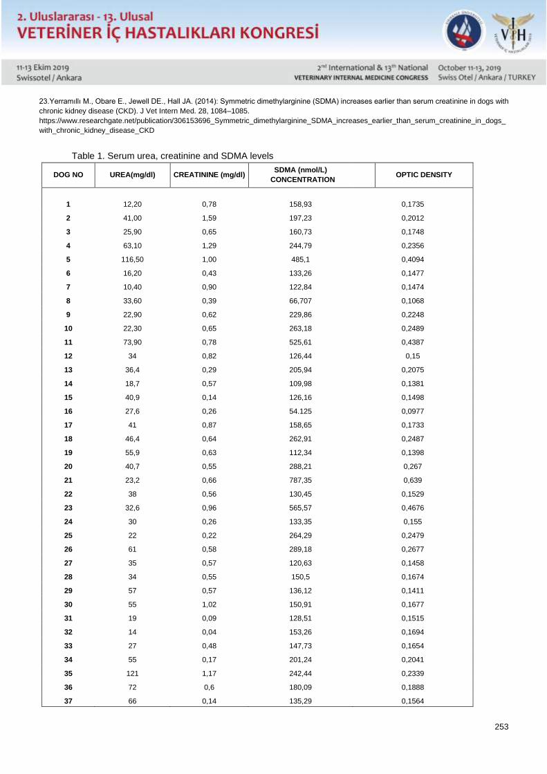
253
23.Yerramıllı M., Obare E., Jewell DE., Hall JA. (2014): Symmetric dimethylarginine (SDMA) increases earlier than serum creatinine in dogs with
chronic kidney disease (CKD). J Vet Intern Med. 28, 1084–1085.
https://www.researchgate.net/publication/306153696_Symmetric_dimethylarginine_SDMA_increases_earlier_than_serum_creatinine_in_dogs_
with_chronic_kidney_disease_CKD
Table 1. Serum urea, creatinine and SDMA levels
DOG NO UREA(mg/dl) CREATININE (mg/dl) SDMA (nmol/L)
CONCENTRATION OPTIC DENSITY
1 12,20 0,78 158,93 0,1735
2 41,00 1,59 197,23 0,2012
3 25,90 0,65 160,73 0,1748
4 63,10 1,29 244,79 0,2356
5 116,50 1,00 485,1 0,4094
6 16,20 0,43 133,26 0,1477
7 10,40 0,90 122,84 0,1474
8 33,60 0,39 66,707 0,1068
9 22,90 0,62 229,86 0,2248
10 22,30 0,65 263,18 0,2489
11 73,90 0,78 525,61 0,4387
12 34 0,82 126,44 0,15
13 36,4 0,29 205,94 0,2075
14 18,7 0,57 109,98 0,1381
15 40,9 0,14 126,16 0,1498
16 27,6 0,26 54.125 0,0977
17 41 0,87 158,65 0,1733
18 46,4 0,64 262,91 0,2487
19 55,9 0,63 112,34 0,1398
20 40,7 0,55 288,21 0,267
21 23,2 0,66 787,35 0,639
22 38 0,56 130,45 0,1529
23 32,6 0,96 565,57 0,4676
24 30 0,26 133,35 0,155
25 22 0,22 264,29 0,2479
26 61 0,58 289,18 0,2677
27 35 0,57 120,63 0,1458
28 34 0,55 150,5 0,1674
29 57 0,57 136,12 0,1411
30 55 1,02 150,91 0,1677
31 19 0,09 128,51 0,1515
32 14 0,04 153,26 0,1694
33 27 0,48 147,73 0,1654
34 55 0,17 201,24 0,2041
35 121 1,17 242,44 0,2339
36 72 0,6 180,09 0,1888
37 66 0,14 135,29 0,1564
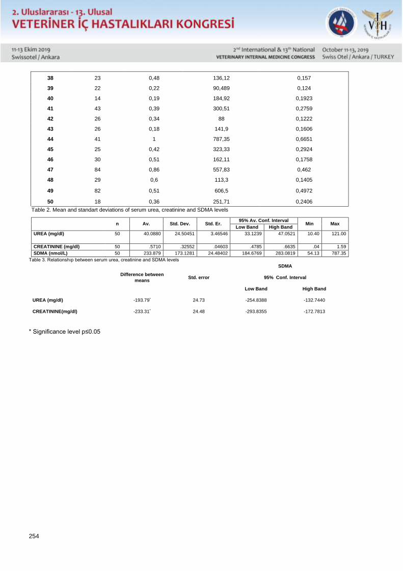
254
38 23 0,48 136,12 0,157
39 22 0,22 90,489 0,124
40 14 0,19 184,92 0,1923
41 43 0,39 300,51 0,2759
42 26 0,34 88 0,1222
43 26 0,18 141,9 0,1606
44 41 1 787,35 0,6651
45 25 0,42 323,33 0,2924
46 30 0,51 162,11 0,1758
47 84 0,86 557,83 0,462
48 29 0,6 113,3 0,1405
49 82 0,51 606,5 0,4972
50 18 0,36 251,71 0,2406
Table 2. Mean and standart deviations of serum urea, creatinine and SDMA levels
n Av. Std. Dev. Std. Er. 95% Av. Conf. Interval
Min Max Low Band High Band
UREA (mg/dl)
50 40.0880 24.50451 3.46546 33.1239 47.0521 10.40 121.00
CREATININE (mg/dl) 50 .5710 .32552 .04603 .4785 .6635 .04 1.59
SDMA (nmol/L) 50 233.879 173.1281 24.48402 184.6769 283.0819 54.13 787.35
Table 3. Relationshıp between serum urea, creatinine and SDMA levels
Difference between
means Std. error
SDMA
95% Conf. Interval
Low Band High Band
UREA (mg/dl) -193.79* 24.73 -254.8388 -132.7440
CREATININE(mg/dl) -233.31* 24.48 -293.8355 -172.7813
* Significance level p≤0.05
255
Poster Sunu / Poster Presentation Tam Metin / Full Text
Köpeklerde İki Farklı Trombositçe Zengin Plazma (PRP) Kit Sisteminin Özellikleri İle
Terapötik Başarı Potansiyellerinin Karşılaştırılması
Duygu Dalgin* , Yucel Meral*, Metin Cenesiz**, Cagatay Esin*, Basar Ulas Sayilkan*, Emre Kulluk*.
*Ondokuz Mayıs Üniversitesi Veteriner Fakültesi İç Hastalıkları Anabilim Dalı, Samsun* Ondokuz Mayıs Üniversitesi
**Veteriner Fakültesi Fizyoloji Anabilim Dalı, Samsun
Özet
Trombositçe zengin plazma (PRP) uygulaması, beşeri hekimlikte pek çok alanda yaygın olarak kullanılmakta,
veteriner sahada ise kullanımı şimdilik sınırlı olmakla birlikte, köpek ve atlarda osteoartritis, dental implantlar, kemik
defektleri ve yara iyileşmelerinde etkinliği ortaya konulmuştur. İnsan ve atlar için ticari kullanıma sunulan pek çok
PRP tüp ve kiti bulunmaktadır. At ve insan için üretilen bu tüplerin köpeklerdeki etkinliği üzerinde sınırlı sayıda
çalışma bulunmakta olup, insan ve at kullanımı için hazırlanmış sistemlerin köpeklerde aynı sonuçları
vermeyebileceği de ortaya konmuştur. Bu projede ülkemizde en sık kullanılan ruhsatlı ve onaylı PRP kit sistemleri
arasında yer alan, biri yerli diğeri ithal ve biri sodyum sitrat bazlı, diğeri ise jelli iki PRP kit sistemi trombosit içeriğ i
açısından karşılaştırılmış ve yerli ve sodyum sitratlı olan kitin, jelli sisteme göre birim düzeyinde istatistiki farklılık
olmamakla birlikte hacmen daha fazla miktarda ürün sağlaması açısından klinik pratikte daha etkin olacağı
gözlenmiştir. PRP uygulamasının rejeneratif tıp dahilinde yerini almış, otolog ve uygulama kolalığına sahip bir yöntem
olup, literatürde bugüne kadar kanıtlanan rejenerasyon kabiliyetine rağmen, veteriner hekimlik alanında yeterince
değerlendirilmemektedir. Yöntemin daha fazla dikkate alınarak endikasyon dahilinde yaygınlaştırılması, ilgili
alanlarda klinik başarıyı arttıracaktır.
Anahtar Kelimeler: Köpek, Trombositçe zengin plazma (PRP), Rejeneratif tedavi
Giriş
Trombositçe zengin plazma (PRP), hastanın kendi kanından elde edilen yüksek konsantrasyonlu bir plazma
solüsyonu olarak tanımlanabilir. Doku iyileşmesi amacıyla bu tekniğin kullanılmasına ait ilk rapor 1998 yılında bir diş
hekimi tarafından insanlarda geniş mandibular defektlerin tedavisi ile ilgili olarak yayınlanmıştır. Bugün oral cerrahi
alanında iyileşmeyi hızlandırıcı amaçla yaygın olarak kullanılmakta olan PRP, ayrıca yine insan hekimliğinde
kompleks yaraların iyileşmesinde, tendinopati ve ligament hasarlarında, kemik hasarlarında, artritiste,
antienflamatuar ve akne tedavisi, yüzde kırışıkların azaltılması, kellik tedavisi gibi kozmetik amaçla estetik
dermatolojide kullanılmaktadır. Metod lokal hücre aktivasyonu bazlı etki gösterdiğinden uygulamalar lokal olmaktadır.
PRP, doku rejenerasyonundaki etkinliğinin ortaya çıkması ile, son 5 yılda rejeneratif tıp alanına dahil edilmiştir ve
kolay sağlanabilmesi, otolog olup immun reaksiyona neden olmaması, hızlı ve kolay uygulanabilirliği, maliyetinin
düşük olması ile yararlılığının yüksek olması nedeniyle yaygın şekilde kullanılmaktadır.
Veteriner sahada çok yeni olan bu uygulama, yaygın olmamakla birlikte köpek ve atlarda osteoartritiste, dental
implantlarda, kemik defektlerinde ve yara iyileşmelerinde kullanılmaktadır.
İnsan ve atlar için ticari kullanıma sunulan pek çok PRP tüp ve kiti bulunmaktadır. At ve insan için üretilen bu tüplerin
köpeklerdeki etkinliği üzerinde sınırlı sayıda çalışma bulunmakta olup, insan ve at kullanımı için hazırlanmış
sistemlerin köpeklerde aynı sonuçları vermeyebileceği de ortaya konmuştur.
En etkin kit kullanılması, aynı miktar kandan daha fazla trombosit elde edilmesi, bu da daha etkin bir tedavi anlamına
geldiğinden, türe göre en etkin kit kullanımı başarı şansını arttıracaktır.
256
Genel Bilgiler
Trombositçe zengin plazma (PRP), hastanın kendi kanından elde edilen yüksek konsantrasyonlu bir plazma
solüsyonu olarak tanımlanabilir. Doku iyileşmesi amacıyla bu tekniğin kullanılmasına ait ilk rapor 1998 yılında bir diş
hekimi tarafından insanlarda geniş mandibular defektlerin tedavisi ile ilgili olarak yayınlanmıştır(1). Bugün oral cerrahi
alanında iyileşmeyi hızlandırıcı amaçla yaygın olarak kullanılmakta olan PRP, ayrıca yine insan hekimliğinde
kompleks yaraların iyileşmesinde(2), tendinopati ve ligament hasarlarında(3), kemik hasarlarında(4), artritiste(5),
antienflamatuar ve akne tedavisi, yüzde kırışıkların azaltılması, kellik tedavisi gibi kozmetik amaçla estetik
dermatolojide(6-9) kullanılmaktadır. Metod lokal hücre aktivasyonu bazlı etki gösterdiğinden, uygulamalar lokal
olmaktadır.
Trombositlerin içinde üç tip granül tanımlanmıştır; Alfa granülleri, yoğun granüller ve lizozomlar. Alfa granülleri;
trombosit kökenli büyüme faktörleri (PGDF), transforme büyüme faktörü beta (TGFb 1 ve 2), epitelyal büyüme faktörü
(EGF) ve vasküler endotelyal büyüme faktörü (VEGF) ile 300 farklı protein içerir(10,11). Bu büyüme faktörleri hücre
proliferasyonu, diferensiasyonu, anjiogenesis ve şemotakseyi modüle eder. Yoğun granüllerin sayısı daha az olup
serotonin, histamin, dopamin, kalsiyum, ADP, ATP, GDP, GTP, magnezyum ve polifosfat gibi biyoaktif faktörler ihtiva
ederler (11,12).
PRP, doku rejenerasyonundaki etkinliğinin ortaya çıkması ile, son 5 yılda rejeneratif tıp alanına dahil edilmiştir(13)
ve kolay sağlanabilmesi, otolog olup immun reaksiyona neden olmaması, hızlı ve kolay uygulanabilirliği, diğer
rejeneratif tıp uygulamalarına kıyasla maliyetinin düşük ve yararlılığının yüksek olması nedeniyle yaygın şekilde
kullanılmaktadır.
Veteriner sahada çok yeni olan bu uygulama(14), yaygın olmamakla birlikte köpek ve atlarda osteoartritiste(15,16),
dental implantlarda(17), kemik defektlerinde(18) ve yara iyileşmelerinde(19) kullanılmaktadır. PRP enjeksiyonları,
özellikle dekübit yaraları, deri ülserleri, yanıklar, operasyon sonrası geniş sahalı yaralar, sistemik hastalıklara bağlı
olarak (ör, diabetes mellitus) yara iyileşmesinde gecikmeler, kuyruk yakalama sendromu gibi davranışsal
bozukluklara bağlı oluşan ve tekrarlayan yaraların tedavisinde yüksek başarı göstermektedir.
Başarılı bir PRP uygulaması için anahtar faktör, işlenen kandan elde edilen PRP içeriğindeki hücre sayısıdır.
Çalışmalar ideal bir PRP ürününde trombosit sayısının normal kandan 4-7 kat fazla olması gerektiğini öne
sürmektedir(20,21,22), fakat trombosit sayısı dışında mononükleer hücre sayısı, nötrofil ve alyuvar sayıları da PRP
enjeksiyonunun yangısal etkileri ve tedavi başarısını önemli ölçüde etkilemektedir(20,21). Genel olarak, inflammatuar
etkilerinden kaçınmak amacıyla alyuvar ve nötrofillerin sayısının mümkün olduğunca düşük olması hedeflenir (23,
24).
İnsan ve atlar için ticari kullanıma sunulan pek çok PRP tüp ve kiti bulunmaktadır. PRP seperasyon sistemleri farklılık
gösteren bu kitlerden elde edilen son ürün içeriğindeki trombosit, akyuvar ve büyüme faktörleri konsantrasyonları da
farklılık göstermektedir(22,25). At ve insan için üretilen bu tüplerin köpeklerdeki etkinliği üzerinde sınırlı sayıda
çalışma bulunmakta olup, insan ve at kullanımı için hazırlanmış sistemlerin köpeklerde aynı sonuçları
vermeyebileceği de ortaya konmuştur(26). Beşeri hekimlik için üretilmiş bazı kit sistemlerinin köpek kanındaki
sonuçlarını kıyaslayan çok az sayıda çalışma yapılmıştır (14,27). Ülkemizde pek çok ithal PRP kit sistemi bulunmakla
birlikte, bunların köpeklerdeki etkinlikleri henüz araştırılmamıştır. Ayrıca yerli markalarda üretilmekte olup, bunların
da köpeklerdeki sonuçları ortaya konmamıştır.
Bu projede, ülkemizde en sık kullanılan ruhsatlı ve onaylı PRP kit sistemleri arasında yer alan, biri yerli diğeri ithal iki
PRP kit sistemi karşılaştırılacaktır. Burada önemli bir karşılaştırma da, tüplerin birinin jelli, diğerinin ise sodyum sitratlı
olmasından ileri gelmektedir. Bazı tüplerde bulunan jelin enjekte edilecek olan PRP kısmına karışmaması
gerekmekte, fakat santrifüj ve PRP nin ayrılması manipülasyonu sırasında karışma olasılığı da bir rahatsızlık olarak
değerlendirilmektedir. Bu nedenle hem iki ayrı sistemin köpeklerdeki etkinliğinin, hem de jel ve sodyum sitrat
kullanımının karşılaştırılması literatürde önemli yer bulacaktır. Ayrıca ülkemizde henüz yeni yeni kullanılmaya
257
başlayan PRP tedavileri için köpeklerdeki uygun kit sistemi seçiminde önemli bir rehber oluşacak ve yaygın kullanım
alanı ortaya çıkacaktır.
Gereç ve Yöntem
Hayvan Materyali
Araştırma kapsamında OMÜ Veteriner Fakültesi Hayvan Hastanesine çeşitli nedenlerle (dekübit yaraları, deri
ülserleri, yanıklar, operasyon sonrası geniş sahalı yaralar, diabetes mellitus gibi sistemik hastalıklar, kuyruk yakalama
sendromu gibi davranışsal bozukluklara bağlı olarak) yara iyileşmesinde gecikmeler ve tekrarlayan yaralarla
başvuran 15 adet sahipli köpek kullanılmıştır. Detaylı anamnez alınması, genel muayene, yara muayenesi, yarada
kontaminasyon ve komplikasyon tanımlanması için dermatolojik analizler ve sistemik hastalıklar açısından
muayeneler uygulandıktan sonra, yara tedavisi amacıyla PRP uygulama endikasyonu olan 15 köpek belirlenmiştir.
Bu nedenle çalışma deneysel olmayıp, klinik tedavi uygulaması dahilinde elde edilen PRP ürününde hücre sayımı
yapılmasından ibarettir.
PRP Kit Sistemleri
PRP elde edilmesi amacıyla iki farklı PRP kit kullanılacaktır. Kullanılacak ilk sistem yerli üretim, klinik kullanım için
ruhsatlı, T-Lab PRP kit (T-Biyoteknoloji Ltd) olup, tüp 10ml hacminde ve 1ml pıhtılaşma önleyici sodyum sitrat içerir
(Birinci sistem). İçerisi gama steril olan tüpler kesinlikle ficoll ya da jel barındırmamaktadır.
İkinci sistem ise yine klinik kullanım ruhsatına sahip, ithal DPG PRP kitleri olacaktır (Dermoaroma Inc., İtalya) (İkinci
sistem). Bu kit tüp içinde alkil akrilat krospolimer bazlı ayrıştırıcı jeli (inert separator jel) ihtiva etmektedir.
PRP Elde Edilmesi
Bu amaçla, her bir köpekten her iki sistem kit tüpü içine venöz kan alınacak ve üreticinin direktifleri doğrultusunda
işlemler gerçekleştirilecektir. Elde edilen ürün ayrıldıktan sonra hücre sayımı yapılacak ve dermal iğne vasıtasıyla
yara yerine uygulama yapılacaktır. Böylece her bir köpekten, her iki sistem için de kan alınmış olacaktır.
Hücre Sayımı
Trombosit sayıları thoma lamı kullanılarak mikroskopta hemositometrik yöntemle belirlenmiştir.
İstatistiksel Değerlendirme
İki farklı sistemde trombosit değerlerinin belirlenmesinin hedeflendiği araştırmada, iki grup için ortalama değer,
standart sapma ve standart hata değerlerinin belirlenmesi ve gruplar arasındaki farkın kontrolünde student t
testinden yararlanılmıştır.
Araştırmada kullanılacak örnek büyüklüğünün yani canlı materyal sayısının belirlenmesinde
formülünden yararlanılmıştır. = 0.05 olarak alınmıştır.
Bulgular
Her iki sistemde elde edilen trombosit sayılar Tablo 1’de, sistemlerin istatistiki karşılaştırılması ise Tablo 2’ verimişti
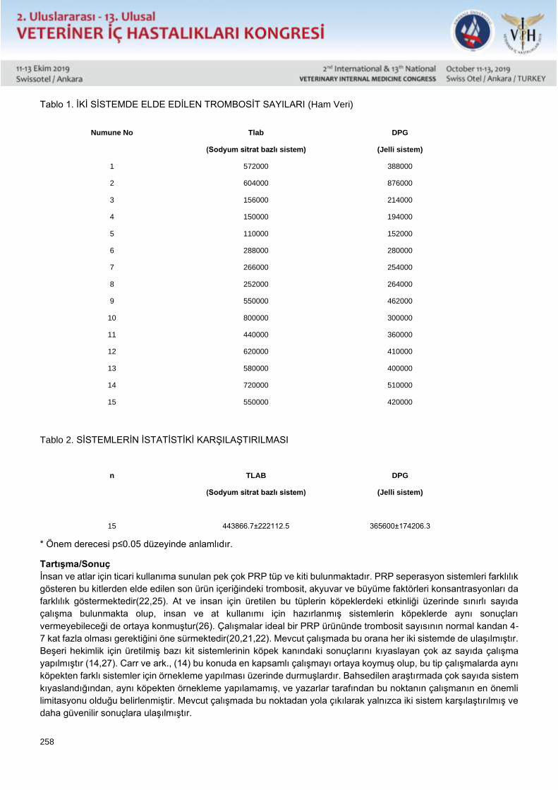
258
Tablo 1. İKİ SİSTEMDE ELDE EDİLEN TROMBOSİT SAYILARI (Ham Veri)
Numune No
Tlab
(Sodyum sitrat bazlı sistem)
DPG
(Jelli sistem)
1 572000 388000
2 604000 876000
3 156000 214000
4 150000 194000
5 110000 152000
6 288000 280000
7 266000 254000
8 252000 264000
9 550000 462000
10 800000 300000
11 440000 360000
12 620000 410000
13 580000 400000
14 720000 510000
15 550000 420000
Tablo 2. SİSTEMLERİN İSTATİSTİKİ KARŞILAŞTIRILMASI
n
TLAB
(Sodyum sitrat bazlı sistem)
DPG
(Jelli sistem)
15
443866.7±222112.5
365600±174206.3
* Önem derecesi p≤0.05 düzeyinde anlamlıdır.
Tartışma/Sonuç
İnsan ve atlar için ticari kullanıma sunulan pek çok PRP tüp ve kiti bulunmaktadır. PRP seperasyon sistemleri farklılık
gösteren bu kitlerden elde edilen son ürün içeriğindeki trombosit, akyuvar ve büyüme faktörleri konsantrasyonları da
farklılık göstermektedir(22,25). At ve insan için üretilen bu tüplerin köpeklerdeki etkinliği üzerinde sınırlı sayıda
çalışma bulunmakta olup, insan ve at kullanımı için hazırlanmış sistemlerin köpeklerde aynı sonuçları
vermeyebileceği de ortaya konmuştur(26). Çalışmalar ideal bir PRP ürününde trombosit sayısının normal kandan 4-
7 kat fazla olması gerektiğini öne sürmektedir(20,21,22). Mevcut çalışmada bu orana her iki sistemde de ulaşılmıştır.
Beşeri hekimlik için üretilmiş bazı kit sistemlerinin köpek kanındaki sonuçlarını kıyaslayan çok az sayıda çalışma
yapılmıştır (14,27). Carr ve ark., (14) bu konuda en kapsamlı çalışmayı ortaya koymuş olup, bu tip çalışmalarda aynı
köpekten farklı sistemler için örnekleme yapılması üzerinde durmuşlardır. Bahsedilen araştırmada çok sayıda sistem
kıyaslandığından, aynı köpekten örnekleme yapılamamış, ve yazarlar tarafından bu noktanın çalışmanın en önemli
limitasyonu olduğu belirlenmiştir. Mevcut çalışmada bu noktadan yola çıkılarak yalnızca iki sistem karşılaştırılmış ve
daha güvenilir sonuçlara ulaşılmıştır.
259
PRP tekniğinde en önemli noktalardan biri steril bir ürün elde edilmesidir ve açık manuel metodlarda hazırlık işlemleri
esnasında bakteriyel kontaminasyon riski bulunur (28). Pek çok ürün içerisinde pıhtılaşma önleyici bulunmamakta,
pıhtılaşma önleyicinin dışarıdan katılması yada kit içinde bulunan preparatın tüp içine verilmesi beklenmektedir. Bu
da hem kontaminasyon riskini, hem de pıhtılaşma önleyici preparasyon miktarında hata riskini arttırmaktadır. Bu
çalışmada incelenen sistemler içinde sitrat bazlı sistemin tüp içinde sodyum sitrat ihtiva etmesi bu riskleri ortadan
kaldırmıştır.
PRP elde etmek için yapılan santrifügasyon işlemi sırasında hücreler 1.500G gibi çok yüksek bir yer çekimi kuvvetine
tabi tutulur ve moleküler ağırlıklarına göre ayrılır. Bu işlem PRP elde edilmesini sağlamakla birlikte, trombosit
kümeleşmesini de beraberinde getirir ki bu risk terapötik başarının azalmasına yol açar (5,10,11). Bu kümelenme
dağıtılmadan yapılacak bir uygulamada her enjeksiyonda farklı miktarda trombosit verilebileceğinden homojen bir
dağılım sağlanamaz. Yine bu çalışmada kullanılan iki sistemden biri olan sitrat bazlı sistem içinde bulunan
resüspansiyon tüpü, kümelenen trombositleri PRP içerisine daha homojen bir şekilde yaymaya yardımcı olduğundan
avantaj teşkil etmektedir.
Çeşitli sistemlerde farklı miktarlarda kan alınması gerekliliği vardır. Fakat alınan kan miktarı ile toplanan trombosit
miktarı her zaman ilişkili olmamaktadır. Örneğin Carr ve ark.,(14), beş farklı sistemi karşılaştırmış, bunlarda toplanan
kan miktarları 50, 16, 50, 9 ve 55 ml iken, elde edilen PRP ürün miktarları 10, 4-7, 4, 4-5, 6-8 ml olmuştur. Fakat
trombosit sayısı, dolayısıyla ürün etkinliği ele alındığında bir ve üçüncü sistemleri takiben ikinci sistemlerin en etkin
ürünü ortaya çıkardığını bildirmişlerdir. Oysa toplanan kan miktarına gören yüksek miktarda ürün elde edilen sistem
dördüncü sıradaki olup, etkinlik açısından bu sistem en son sırada yer almaktadır. Mevcut çalışmada elde edilen
trombosit sayısı açısından her iki sistem arasında istatistiki olarak bir farklılık bulunmamıştır.
Sonuç olarak, bu çalışmada mukayese edilen her iki sistem açısından trombosit sayısı açısından istatistiksel olarak
anlamlı bir farklılık bulunmamakla birlikte, daha fazla ürün elde edilebilmesi, kontaminasyon riskinin daha düşük
olması, resüspansiyon tüpü ile trombosit kümeleşmesi riskinin azaltılması yönlerinden sodyum sitrat bazlı sistemin
klinik etkinlik açısından daha avantajlı olduğu sonucuna varılmıştır. Ayrıca, klinik pratikte uygulanan PRP işlemlerinde
hücre sayımı yapılması, türe göre etkinliği ortaya konulmuş bir ürün kullanılması ve işlem sırasında sterilizasyona
azami dikkat gösterilmesi gereklidir.
Kaynaklar 1-Bosch G, van Schie HTM, Groot MW, Cadby JA, van de Lest, CHA, Barneveld AB, van Weeren PR (2010) Effects of platelet-rich plasma on
the quality of repair of mechanically induced core lesions in equine superficial digital flexor tendons: A placebo-controlled experimental study.
Journal of Orthopaedic Research. 28(2): 211–217.
https://onlinelibrary.wiley.com/doi/full/10.1002/jor.20980
2-Boswell SG, Schnabel LV, Mohammed HO, Sundman EA, Minas T, Fortier LA. (2013) Increasing platelet concentrations in leukocyte-reduced
platelet-rich plasma decrease collagen gene synthesis in tendons. Am J Sports Med. 42(1): 42–9.
https://doi.org/10.1177%2F0363546513507566
3-Carr BJ., Canapp SO., Mason DR., Cox C., Hess T. (2016) Canine Platelet-Rich Plasma Systems: A Prospective Analysis. Front Vet Sci. 2015;
2: 73.
http://dx.doi.org/10.3389/fvets.2015.00073.
4-Castillo TN., Pouliot MA., Kim HJ., Dragoo JL. (2011) Comparison of growth factor and platelet concentrations from commercial platelet-rich
plasma separation systems. Am J Sports Med. 39(2): 266–71.
https://doi.org/10.1177%2F0363546510387517
5-Chahal J, Van Thiel GS, Mall N, Heard W, Bach BR, Cole BJ, Nicholson GP, Verma NN, Whelan DB, Romeo AA. (2012) The Role of Platelet-
Rich Plasma in Arthroscopic Rotator Cuff Repair: A Systematic Review With Quantitative Synthesis. The Journal of Arthroscopic & Related
Surgery. 28(11): 1718–1727.
https://doi.org/10.1016/j.arthro.2012.03.007
6-Dohan Ehrenfest DM, Doglioli P, de Peppo GM, Del Corso M, Charrier JB. (2010) Choukroun’s platelet-rich fibrin (PRF) stimulates in vitro
proliferation and differentiation of human oral bone mesenchymal stem cell in a dose-dependent way. Arch Oral Biol. 55:185–94.
https://doi.org/10.1016/j.archoralbio.2010.01.004
7-Fallouh L, Nakagawa K, Sasho T, Arai M, Kitahara S, Wada Y, Moriya H, Takahashi K. (2010) Effects of autologous platelet-rich plasma on cell
viability and collagen synthesis in injured human anterior cruciate ligament. J Bone Joint Surg Am. 92(18): 2909–2916.
260
https://insights.ovid.com/pubmed?pmid=21159991
8-Foster TE, Puskas BL, Mandelbaum BR, Gerhardt MB, Rodeo SA. (2009) Platelet-rich plasma: from basic science to clinical applications. Am
J Sports Med. 37: 2259-2265.
https://doi.org/10.1177%2F0363546509349921
9-Franklin SP, Cook JL (2013) Prospective trial of autologous conditioned plasma versus hyaluronan plus corticosteroid for treatment of chronic
elbow osteoarthritis in dogs. Can Vet J. 54:881-884.
https://www.ncbi.nlm.nih.gov/pmc/articles/PMC3743576/
10-Franklin SP, Garner BC, Cook JL (2015) Characteristics of canine platelet-rich plasma prepared with five commercially available systems. Am
J Vet Res. 76(9): 822–7.
https://doi.org/10.2460/ajvr.76.9.822
11-Gawdat HI, Hegazy RA, Fawzy MM, Fathy M (2014) Autologous Platelet Rich Plasma: Topical Versus Intradermal After Fractional Ablative
Carbon Dioxide Laser Treatment of Atrophic Acne Scars. Dermatologic Surgery. 40(2): 152–161.
https://www.ncbi.nlm.nih.gov/pubmed/24354616
12-Jeong KI, Kim SG, Oh JS, Lee SY, Cho YS, Yang SS, Park SC, You JS, Lim SC, Jeong MA, Kim JS, Lee SY. (2013) Effect of Platelet-Rich
Plasma and Platelet-Rich Fibrin on Peri-Implant Bone Defects in Dogs. Journal of Biomedical Nanotechnology. 9(3): 535-537.
https://www.ncbi.nlm.nih.gov/pubmed/23621011
13-Karayannopoulou M, Psalla D, Kazakos G, Loukopoulos P, Giannakas N, Savvas I, Kritsepi-Konstantinou M, Chantes A., Papazoglou LG
(2015) Effect of locally injected autologous platelet-rich plasma on second intention wound healing of acute full-thickness skin defects in dogs.
Veterinary and Comparative Orthopaedics and Traumatology (VCOT). 3: 172-178.
https://www.thieme-connect.com/products/ejournals/abstract/10.3415/VCOT-14-06-0088
14-Lee DK, In J, Lee S. (2015) Standard deviation and standard error of the mean. Korean J Anesthesiol. 68:220–223.
https://dx.doi.org/10.4097%2Fkjae.2015.68.3.220
15-Li ZJ, Choi HI, Choi DK, Sohn KC, Im M, Seo YJ, Lee YH, Lee JH, Lee Y. (2012) Autologous platelet-rich plasma: a potential therapeutic tool
for promoting hair growth. Dermatol Surg 38(71): 1040–1046.
https://insights.ovid.com/pubmed?pmid=22455565
16-Lubkowska A, Dolegowska B, Banfi G (2012) Growth factor content in PRP and their applicability in medicine. J Biol Regul Homeost Agents
26(2): 22-25.
https://www.ncbi.nlm.nih.gov/pubmed/23648195
17-Marx RE, Carlson ER, Eichstaedt RM, Schimmele SR, Strauss JE, Georgeff KR. (1998) Platelet rich plasma: growth factor enhancement for
bone grafts. Oral Surg Oral Med Oral Pathol Oral Radiol Endod. 85(6): 638–646.
https://doi.org/10.1016/S1079-2104(98)90029-4
18-McLellan J, Plevin S (2011) Does it matter which platelet-rich plasma we use? Equine Vet Educ. 23(2): 101–4.
https://doi.org/10.1111/j.2042-3292.2010.00185.x
19-Mooren RE, Hendriks EJ, van den Beucken JJ, Merkx MA, Meijer GJ, Jansen JA, Stoelinga PJ (2010) The effect of platelet-rich plasma in vitro
on primary cells: rat osteoblast-like cells and human endothelial cells. Tissue Eng Part A.16(10): 3159–3172.
https://doi.org/10.1089/ten.tea.2009.0832
20-Nicoli F, Balzani A, Lazzeri D, Gentile P, Chilgar RM, Di Pasquali C, Nicoli M, Bocchini I, Agovino A, Cervelli V. (2015) Severe hidradenitis
suppurativa treatment using platelet-rich plasma gel and Hyalomatrix. Int Wound J. 12: 43-44.
https://doi.org/10.1111/iwj.12117
21-Pelletier MH, Malhotra A, Brighton T, Walsh WR, Lindeman R (2013) Platelet function and constituents of platelet rich plasma. Int J Sports
Med. 34: 74–80.
https://www.thieme-connect.com/products/ejournals/abstract/10.1055/s-0032-1316319
22-Sanchez-Gonzalez DJ, Mendez-Bolaina E, Trejo-Bahena NI (2012) Platelet-rich plasma peptides: key for regeneration. Int J Pept. 3: 532-519.
https://dx.doi.org/10.1155%2F2012%2F532519
23-Shin MK, Lee JH, Lee SJ, Kim NI. (2012) Platelet-rich plasma combined with fractional laser therapy for skin rejuvenation. Dermatol Surg. 38:
623–30.
https://insights.ovid.com/pubmed?pmid=22288389
24-Stief M, Gottschalk J, Ionita JC, Einspanier A, Oechtering G, Böttcher P (2011) Concentration of platelets and growth factors in canine
autologous conditioned plasma. Vet Comp Orthop Traumatol 24: 285–90.
https://www.researchgate.net/publication/49745908_Concentration_of_platelets_and_growth_factors_in_canine_autologous_conditioned_plasm
a
25-Streckbein P, Kleis W, Buch RSR, Hansen T, Weibrich G (2014) Bone Healing with or without Platelet-Rich Plasma around Four Different
Dental Implant Surfaces in Beagle Dogs. Clinical Implant Dentistry and Related Research. 16(4): 479–486.
https://doi.org/10.1111/cid.12026
26-Sundman EA, Cole BJ, Fortier LA (2011) Growth factor and catabolic cytokine concentrations are influenced by the cellular composition of
platelet-rich plasma. Am J Sports Med. 39(10): 2135–40.
https://doi.org/10.1177%2F0363546511417792
27-Textor J (2014) Platelet-Rich Plasma (PRP) as a Therapeutic Agent: Platelet Biology, Growth Factors and a Review of the Literature In:
Platelet-Rich Plasma Regenerative Medicine: Sports Medicine, Orthopedics and Recovery of Musculoskeletal Injuries. Lana JFSD, Santana A,
Belangero D, Luzo M (Eds). XVII, Springer Pub.
http://dx.doi.org/10.1007/978-3-642-40117-6_2
261
28-Vasconcelos E, Figueiredo AC, Seghatchian J (2003) Quality of platelet concentrates derived by platelet rich plasma, buffy coat and apheresis.
Transfus Apher Sci. 29:13-16.
https://doi.org/10.1016/S1473-0502(03)00091-0
29-Villela DL, Santos VL. (2010) Evidence on the use of platelet-rich plasma for diabetic ulcer: a systematic review. Growth Factors. 28(2): 111–
116. https://doi.org/10.3109/08977190903468185
262
Poster Sunu / Poster Presentation Tam Metin / Full Text
Comparison of Characteristics and Therapeutic Potentials of two Different Platelet Rich
Plasma (PRP) Kit Systems in Dogs
Duygu Dalgin* , Yucel Meral*, Metin Cenesiz**, Cagatay Esin*, Basar Ulas Sayilkan*, Emre Kulluk*.
*Ondokuz Mayıs University Faculty of Veterinary Medicine, Department of Internal Medicine, Samsun
**Ondokuz Mayıs University Faculty of Veterinary Medicine, Department of Physiology, Samsun
*Correspondence:
Res. Ast. Başar Ulaş SAYILKAN, Ondokuz Mayıs University, Faculty of Veterinary Medicine, Department of Internal
Medicine, Atakum, 55139, Samsun, Turkey.
This research had been supported by Ondokuz Mayıs University Scientific Research Projects comission with
reference number PYO.VET.1901.17.013
Abstract
Platelet Rich Plasma (PRP) applications are widely used in human medicine, however have limited practice in
veterinary medicine. Even so, efficiency of PRP in osteoarthritis, dental implants, bone defects and wound healing in
dogs and horses are well documented. There are many commercially available PRP tube and kits for horses and
humans, but studies on the efficiency of these products for dogs are limited. Researches demonstrated that systems
prepared for humans and horses do not always give the same results for dogs. In the present study, two prevailing
systems, a sodium citrate based system and a gelled system are compared regarding platelet output. Obtained data
revealed that although there was not a statistically significant difference between both systems, sodium citrate based
kit is probably more efficient in practice due to its superiority by volume output. PRP is an important component of
regenerative therapy. It is autologous and practical, though it has not been put into practice sufficiently in veterinary
practice. Consideration of the method within indications will enhance clinical satisfaction.
Key words: Autologous, Dog, Platelet rich plasma (PRP), Regenerative medicine
Introduction
Platelet Rich Plasma (PRP), may be defined as a high concentrated plasma solution derived from the patients own
blood. First report on PRP efficiency in tissue healing was published in 1998 by a dentist, in the treatment of human
mandibular defects (Marx et al., 1998). Today PRP is used widely intending stimulation of healing, in oral surgery,
complex wounds (Villela and Santos., 2010), tendinopathies and ligament injuries (Fallouh et al., 2010), bone
fractures (Mooren et al., 2010), arthritis (Chahal et al., 2012), antinflammatory and acne treatments, esthetical
dermatology such as facial wrinkles and boldness (Li et al., 2012; Shin et al., 2012; Gawdat et al., 2014; Nicoli et al.,
2015). The method acts based on local cell activation, thus applications are local.
There are three types of granules identified in platelets; Alpha granules, dense granules and lysosomes. Alpha
granules; includes platelet derived growth factors (PGDF), transforme growth factor beta (TGFb 1 and 2), epithelial
growth factor (EGF) with vascular endothelial growth factor (VEGF) and 300 different proteins (Lubkowska et al.,
2012; Textor, 2014). These growth factors modulates cell proliferation, differentiation, angiogenesis and chemotaxis.
Dense granules are less in number and include bioactive factors such as serotonine, histamine, dopamine, calcium,
adenosine diphosphate (ADP), ATP, GDP, GTP, magnesium and polyphosphate (Foster et al., 2009; Textor, 2014).
263
PRP is involved in regenerative medicine area with the discovery of its efficiency in tissue regeneration and is widely
used due to availability, its autologous character avoiding immune response, fast and easy application, low cost
compared to other regenerative medicine methods and high efficiency (Sanchez-Gonzalez et al., 2012).
PRP is relatively emergent in veterinary medicine (Carr et al., 2016), yet is occanially utilised in dogs and horses for
osteoarthritis (Bosch et al., 2010; Franklin and Cook, 2013), dental implants (Streckbein et al., 2014), bone defects
(Jeong et al., 2013) and wound healings (Karayannopoulou et al., 2015). PRP injections demonstrate high efficiency
especially in decubitis, dermal ulcers, burns, wide post op wounds, wound healings due to systemic disorders
(diabetes mellitus, etc.), delayed healings, repeating wounds in behavioral disorders such as tail chasing syndrome.
The key factor for a successful PRP application is the cell count in the end product. Studies demonstrate that an
ideal PRP product must include a platelet number of 4-7 times the circulating blood (Ehrenfest et al., 2010; McLellan
and Plevin, 2011; Pelletier et al., 2013). Apart from platelet number, mononuclear cell count, neutrophil and
erythrocyte numbers also effect the antiinflammatory effects and therapeutical success of PRP injections (Ehrenfest
et al., 2010; McLellan and Plevin, 2011). Generally, minimal erythrocyte and leucocyte counts are targeted in order
to avoid their inflammatory effects (Boswell et al., 2013; Sundman et al., 2013).
There are many commercially available PRP kits prepared for human and horse medicine with different PRP
seperation systems, and different systems result with end products with various platelet, leucocyte and growth factor
concentrations (Castillo et al., 2011; Pelletier et al., 2013). There are limited studies on the efficiency of these human
and horse kits for dogs, since research demonstrated that these kits may not create the same results in dogs (Stief
et al., 2011). There are very few studies investigating the results in dog blood with human kits (Franklin et al., 2015;
Carr et al., 2016).
The aim of this study is to compare the end products obtained with two different PRP kit systems mainly used; one
gel based and the other sodium citrate based. Gel basement should not be mixed into the PRP layer, but this
possibility is evaluated as an inconvience as the centrifugation and manipulation processes ease this complication.
Therefore, attention must be paid on the efficiency of these two major kit systems in dogs for optimum therapeutic
success.
Materials And Methods
Ethical Approval
This study was approved by the Ondokuz Mayıs University Animal Studies Local Ethic Committee with
B.30.2.ODM.0.20.09.00-050.04 - approval number.
Animal Material
The study material was composed of 15 owned dogs with recurrent wounds or delayed healing complaints due to
various reasons such as decubitis wounds, skin ulcers, burns, post op wide wounds, systemic diseases like diabetes
mellitus and tail chasing syndrome. PRP application endicated 15 dogs were determined following detailed history,
physical and dermatological examinations including wound inspection, contamination and complication presence and
detailed laboratory examinations for systemic disorders.
PRP Kit Systems
Two different PRP systems were used in the study. First system was T-Lab PRP kit (T-Biyoteknoloji Ltd), a 10 ml
tube containing sodium citrate for the prevention of coagulation and is licensed for clinical applications. Tubes were
gamma sterile and definitely did not contain ficoll or gel.
264
The second system, DPG PRP kit (Dermoaroma Inc., Italy) was also licensed for clinical use and contains alkile
acrilate crosspolymer based seperating gel (inert separator gel).
Preparation of the PRP Product
Venous blood was collected to each of the tubes of both systems and processed according to the producers manual.
Cell counts for each tubes were recorded by cell counter and the products were applied by dermal needles, thus
blood was collected for each dog to both systems.
Statistical Analyses
Standart error, standart deviation and average values for both groups and difference control for both groups were
calculated by student t test (Lee et al 2015).
RESULTS
Platelet counts for both systems and the statistical comparisions of both systems are presented in Table 1 and 2,
respectively.
DISCUSSION
There are considerable numbers of commercially available PRP tubes and kit systems for men and horses. These
kits differ in the cell seperation systems and correspondingly the platelet, white blood cell, red blood cell and growth
factor concentrations of the end products present variations (Castillo et al., 2011; Pelletier et al., 2013). There are
limited studies of the efficiency of these systems prepared for human and horses at canine species, and some studies
demonstrated that these systems may not create the same results on dogs (Stief et al., 2011). Literature suggests
that an ideal PRP product must contain 4-7 fold of the normal platelet count (Ehrenfest et al., 2010; McLellan and
Plevin, 2011; Pelletier et al., 2013). In the present study, optimal number was achieved in both systems, so both
systems are suitable for dogs in that respect.
Few studies were published on the efficiency of commercially available human kit systems on dogs (Franklin et al.,
2015; Carr et al., 2016, Carr et al., 2017) performed the most extensive study and put emphasis on obtaining blood
from the same dog to different systems in these studies. In that study, too many systems were compared, thus
sampling could not be achieved from the same dog and the authors underlined this point as the major limitation of
the study. In the present study, only two systems were compared considering this point, hence confidential results
were obtained.
One of the crucial key points in PRP techniques is acquiring a sterile product and open manuel methods presents a
bacterial contamination risk during preparation procedure (Vasconcelos et al., 2003). Many products do not contain
anticoagulant necessitating addition of an anticoagulant or injection of the commercial preparation within the kit to
the tube. This brings an important contamination risk together with an error risk for the preparation of the exact
amount of the anticoagulant. In the citrate based system studied in the present research, sodium citrate content in
the tubes eliminates these risks which can be considered as an important advantage.
During the centrifugation process, cells are subjected to a gravity force of 1.500G and are seperated according to
their molecular weights. This procedure ensures PRP production, but also brings together the platelet aggregation
which is a risk for therapeutic success (Chahal et al., 2012; Lubkowska et al., 2012; Textor 2014). In an application
with this aggregation, a homogenous distrubution can not be achieved. In the citrate based system in the present
study, the resuspension tube present within the kit, provides a homogenous distrubution of the platelets, bringing out
another theraupetic advantage.
Different amounts of blood must be obtained in different systems, but obtained blood volume and acquired platelet
number do not always correlate. For example, Carr et al. (2016),, compared five different systems; blood sample
volumes were 50, 16, 50, 9 and 55 ml, but derived PRP product volumes were 10, 4-7, 4, 4-5, 6-8 ml, respectively.
265
On the other hand, regarding the platelet numbers achieved, first, third and second systems yielded the most efficient
products, respectively. Yet, according to the obtained blood volume, the highest volume product yielding system was
the third system, where this system was at the worst option regarding efficiency. In the present study, no statistical
differences were recorded between the two systems regarding the derived platelet numbers.
In conclusion, although there were no statistical differences between the two systems regarding the platelet content,
sodium citrate based system appeared more beneficial considering the end product volume, lower contamination risk
and better platelet. Also, in PRP application in practice, performance of cell count procedure, product selection
according to the species and maximum caution to asepsy are the key points for a successful therapy.
REFERENCES
1-Bosch G, van Schie HTM, Groot MW, Cadby JA, van de Lest, CHA, Barneveld AB, van Weeren PR (2010) Effects of platelet-
rich plasma on the quality of repair of mechanically induced core lesions in equine superficial digital flexor tendons: A placebo-
controlled experimental study. Journal of Orthopaedic Research. 28(2): 211–217.
https://onlinelibrary.wiley.com/doi/full/10.1002/jor.20980
2-Boswell SG, Schnabel LV, Mohammed HO, Sundman EA, Minas T, Fortier LA. (2013) Increasing platelet concentrations in
leukocyte-reduced platelet-rich plasma decrease collagen gene synthesis in tendons. Am J Sports Med. 42(1): 42–9.
https://doi.org/10.1177%2F0363546513507566
3-Carr BJ., Canapp SO., Mason DR., Cox C., Hess T. (2016) Canine Platelet-Rich Plasma Systems: A Prospective Analysis.
Front Vet Sci. 2015; 2: 73.
http://dx.doi.org/10.3389/fvets.2015.00073.
4-Castillo TN., Pouliot MA., Kim HJ., Dragoo JL. (2011) Comparison of growth factor and platelet concentrations from commercial
platelet-rich plasma separation systems. Am J Sports Med. 39(2): 266–71.
https://doi.org/10.1177%2F0363546510387517
5-Chahal J, Van Thiel GS, Mall N, Heard W, Bach BR, Cole BJ, Nicholson GP, Verma NN, Whelan DB, Romeo AA. (2012) The
Role of Platelet-Rich Plasma in Arthroscopic Rotator Cuff Repair: A Systematic Review With Quantitative Synthesis. The Journal
of Arthroscopic & Related Surgery. 28(11): 1718–1727.
https://doi.org/10.1016/j.arthro.2012.03.007
6Dohan Ehrenfest DM, Doglioli P, de Peppo GM, Del Corso M, Charrier JB. (2010) Choukroun’s platelet-rich fibrin (PRF)
stimulates in vitro proliferation and differentiation of human oral bone mesenchymal stem cell in a dose-dependent way. Arch Oral
Biol. 55:185–94.
https://doi.org/10.1016/j.archoralbio.2010.01.004
7-Fallouh L, Nakagawa K, Sasho T, Arai M, Kitahara S, Wada Y, Moriya H, Takahashi K. (2010) Effects of autologous platelet-
rich plasma on cell viability and collagen synthesis in injured human anterior cruciate ligament. J Bone Joint Surg Am. 92(18):
2909–2916.
https://insights.ovid.com/pubmed?pmid=21159991
8-Foster TE, Puskas BL, Mandelbaum BR, Gerhardt MB, Rodeo SA. (2009) Platelet-rich plasma: from basic science to clinical
applications. Am J Sports Med. 37: 2259-2265.
https://doi.org/10.1177%2F0363546509349921
9-Franklin SP, Cook JL (2013) Prospective trial of autologous conditioned plasma versus hyaluronan plus corticosteroid for
treatment of chronic elbow osteoarthritis in dogs. Can Vet J. 54:881-884.
https://www.ncbi.nlm.nih.gov/pmc/articles/PMC3743576/
10-Franklin SP, Garner BC, Cook JL (2015) Characteristics of canine platelet-rich plasma prepared with five commercially
available systems. Am J Vet Res. 76(9): 822–7.
https://doi.org/10.2460/ajvr.76.9.822
11-Gawdat HI, Hegazy RA, Fawzy MM, Fathy M (2014) Autologous Platelet Rich Plasma: Topical Versus Intradermal After
Fractional Ablative Carbon Dioxide Laser Treatment of Atrophic Acne Scars. Dermatologic Surgery. 40(2): 152–161.
https://www.ncbi.nlm.nih.gov/pubmed/24354616
12-Jeong KI, Kim SG, Oh JS, Lee SY, Cho YS, Yang SS, Park SC, You JS, Lim SC, Jeong MA, Kim JS, Lee SY. (2013) Effect of
Platelet-Rich Plasma and Platelet-Rich Fibrin on Peri-Implant Bone Defects in Dogs. Journal of Biomedical Nanotechnology. 9(3):
535-537.
https://www.ncbi.nlm.nih.gov/pubmed/23621011
266
13-Karayannopoulou M, Psalla D, Kazakos G, Loukopoulos P, Giannakas N, Savvas I, Kritsepi-Konstantinou M, Chantes A.,
Papazoglou LG (2015) Effect of locally injected autologous platelet-rich plasma on second intention wound healing of acute full-
thickness skin defects in dogs. Veterinary and Comparative Orthopaedics and Traumatology (VCOT). 3: 172-178.
https://www.thieme-connect.com/products/ejournals/abstract/10.3415/VCOT-14-06-0088
14-Lee DK, In J, Lee S. (2015) Standard deviation and standard error of the mean. Korean J Anesthesiol. 68:220–223.
https://dx.doi.org/10.4097%2Fkjae.2015.68.3.220
15-Li ZJ, Choi HI, Choi DK, Sohn KC, Im M, Seo YJ, Lee YH, Lee JH, Lee Y. (2012) Autologous platelet-rich plasma: a potential
therapeutic tool for promoting hair growth. Dermatol Surg 38(71): 1040–1046.
https://insights.ovid.com/pubmed?pmid=22455565
16-Lubkowska A, Dolegowska B, Banfi G (2012) Growth factor content in PRP and their applicability in medicine. J Biol Regul
Homeost Agents 26(2): 22-25.
https://www.ncbi.nlm.nih.gov/pubmed/23648195
17-Marx RE, Carlson ER, Eichstaedt RM, Schimmele SR, Strauss JE, Georgeff KR. (1998) Platelet rich plasma: growth factor
enhancement for bone grafts. Oral Surg Oral Med Oral Pathol Oral Radiol Endod. 85(6): 638–646.
https://doi.org/10.1016/S1079-2104(98)90029-4
18-McLellan J, Plevin S (2011) Does it matter which platelet-rich plasma we use? Equine Vet Educ. 23(2): 101–4.
https://doi.org/10.1111/j.2042-3292.2010.00185.x
19-Mooren RE, Hendriks EJ, van den Beucken JJ, Merkx MA, Meijer GJ, Jansen JA, Stoelinga PJ (2010) The effect of platelet-
rich plasma in vitro on primary cells: rat osteoblast-like cells and human endothelial cells. Tissue Eng Part A.16(10): 3159–3172.
https://doi.org/10.1089/ten.tea.2009.0832
20-Nicoli F, Balzani A, Lazzeri D, Gentile P, Chilgar RM, Di Pasquali C, Nicoli M, Bocchini I, Agovino A, Cervelli V. (2015) Severe
hidradenitis suppurativa treatment using platelet-rich plasma gel and Hyalomatrix. Int Wound J. 12: 43-44.
https://doi.org/10.1111/iwj.12117
21-Pelletier MH, Malhotra A, Brighton T, Walsh WR, Lindeman R (2013) Platelet function and constituents of platelet rich plasma.
Int J Sports Med. 34: 74–80.
https://www.thieme-connect.com/products/ejournals/abstract/10.1055/s-0032-1316319
22-Sanchez-Gonzalez DJ, Mendez-Bolaina E, Trejo-Bahena NI (2012) Platelet-rich plasma peptides: key for regeneration. Int J
Pept. 3: 532-519.
https://dx.doi.org/10.1155%2F2012%2F532519
23-Shin MK, Lee JH, Lee SJ, Kim NI. (2012) Platelet-rich plasma combined with fractional laser therapy for skin rejuvenation.
Dermatol Surg. 38: 623–30.
https://insights.ovid.com/pubmed?pmid=22288389
24-Stief M, Gottschalk J, Ionita JC, Einspanier A, Oechtering G, Böttcher P (2011) Concentration of platelets and growth factors
in canine autologous conditioned plasma. Vet Comp Orthop Traumatol 24: 285–90.
https://www.researchgate.net/publication/49745908_Concentration_of_platelets_and_growth_factors_in_canine_autologous_co
nditioned_plasma
25-Streckbein P, Kleis W, Buch RSR, Hansen T, Weibrich G (2014) Bone Healing with or without Platelet-Rich Plasma around
Four Different Dental Implant Surfaces in Beagle Dogs. Clinical Implant Dentistry and Related Research. 16(4): 479–486.
https://doi.org/10.1111/cid.12026
26-Sundman EA, Cole BJ, Fortier LA (2011) Growth factor and catabolic cytokine concentrations are influenced by the cellular
composition of platelet-rich plasma. Am J Sports Med. 39(10): 2135–40.
https://doi.org/10.1177%2F0363546511417792
27-Textor J (2014) Platelet-Rich Plasma (PRP) as a Therapeutic Agent: Platelet Biology, Growth Factors and a Review of the
Literature In: Platelet-Rich Plasma Regenerative Medicine: Sports Medicine, Orthopedics and Recovery of Musculoskeletal
Injuries. Lana JFSD, Santana A, Belangero D, Luzo M (Eds). XVII, Springer Pub.
http://dx.doi.org/10.1007/978-3-642-40117-6_2
28-Vasconcelos E, Figueiredo AC, Seghatchian J (2003) Quality of platelet concentrates derived by platelet rich plasma, buffy
coat and apheresis. Transfus Apher Sci. 29:13-16.
https://doi.org/10.1016/S1473-0502(03)00091-0
29-Villela DL, Santos VL. (2010) Evidence on the use of platelet-rich plasma for diabetic ulcer: a systematic review. Growth
Factors. 28(2): 111–116. https://doi.org/10.3109/08977190903468185
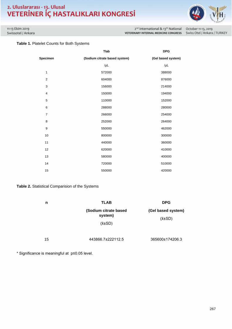
267
Table 1. Platelet Counts for Both Systems
Specimen
Tlab
(Sodium citrate based system)
/µL
DPG
(Gel based system)
/µL
1 572000 388000
2 604000 876000
3 156000 214000
4 150000 194000
5 110000 152000
6 288000 280000
7 266000 254000
8 252000 264000
9 550000 462000
10 800000 300000
11 440000 360000
12 620000 410000
13 580000 400000
14 720000 510000
15 550000 420000
Table 2. Statistical Comparision of the Systems
n
TLAB
(Sodium citrate based
system)
(x±SD)
DPG
(Gel based system)
(x±SD)
15
443866.7±222112.5
365600±174206.3
* Significance is meaningful at p≤0.05 level.
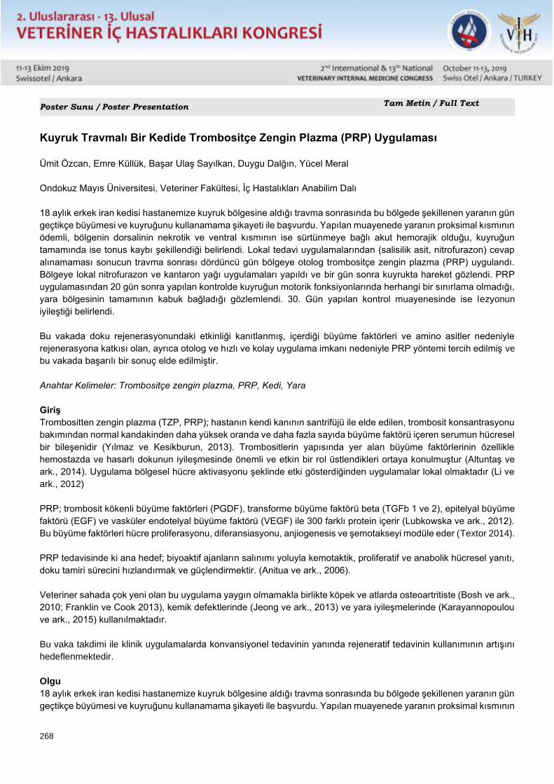
268
Poster Sunu / Poster Presentation Tam Metin / Full Text
Kuyruk Travmalı Bir Kedide Trombositçe Zengin Plazma (PRP) Uygulaması
Ümit Özcan, Emre Küllük, Başar Ulaş Sayılkan, Duygu Dalğın, Yücel Meral
Ondokuz Mayıs Üniversitesi, Veteriner Fakültesi, İç Hastalıkları Anabilim Dalı
18 aylık erkek iran kedisi hastanemize kuyruk bölgesine aldığı travma sonrasında bu bölgede şekillenen yaranın gün
geçtikçe büyümesi ve kuyruğunu kullanamama şikayeti ile başvurdu. Yapılan muayenede yaranın proksimal kısmının
ödemli, bölgenin dorsalinin nekrotik ve ventral kısmının ise sürtünmeye bağlı akut hemorajik olduğu, kuyruğun
tamamında ise tonus kaybı şekillendiği belirlendi. Lokal tedavi uygulamalarından (salisilik asit, nitrofurazon) cevap
alınamaması sonucun travma sonrası dördüncü gün bölgeye otolog trombositçe zengin plazma (PRP) uygulandı.
Bölgeye lokal nitrofurazon ve kantaron yağı uygulamaları yapıldı ve bir gün sonra kuyrukta hareket gözlendi. PRP
uygulamasından 20 gün sonra yapılan kontrolde kuyruğun motorik fonksiyonlarında herhangi bir sınırlama olmadığı,
yara bölgesinin tamamının kabuk bağladığı gözlemlendi. 30. Gün yapılan kontrol muayenesinde ise lezyonun
iyileştiği belirlendi.
Bu vakada doku rejenerasyonundaki etkinliği kanıtlanmış, içerdiği büyüme faktörleri ve amino asitler nedeniyle
rejenerasyona katkısı olan, ayrıca otolog ve hızlı ve kolay uygulama imkanı nedeniyle PRP yöntemi tercih edilmiş ve
bu vakada başarılı bir sonuç elde edilmiştir.
Anahtar Kelimeler: Trombositçe zengin plazma, PRP, Kedi, Yara
Giriş
Trombositten zengin plazma (TZP, PRP); hastanın kendi kanının santrifüjü ile elde edilen, trombosit konsantrasyonu
bakımından normal kandakinden daha yüksek oranda ve daha fazla sayıda büyüme faktörü içeren serumun hücresel
bir bileşenidir (Yılmaz ve Kesikburun, 2013). Trombositlerin yapısında yer alan büyüme faktörlerinin özellikle
hemostazda ve hasarlı dokunun iyileşmesinde önemli ve etkin bir rol üstlendikleri ortaya konulmuştur (Altuntaş ve
ark., 2014). Uygulama bölgesel hücre aktivasyonu şeklinde etki gösterdiğinden uygulamalar lokal olmaktadır (Li ve
ark., 2012)
PRP; trombosit kökenli büyüme faktörleri (PGDF), transforme büyüme faktörü beta (TGFb 1 ve 2), epitelyal büyüme
faktörü (EGF) ve vasküler endotelyal büyüme faktörü (VEGF) ile 300 farklı protein içerir (Lubkowska ve ark., 2012).
Bu büyüme faktörleri hücre proliferasyonu, diferansiasyonu, anjiogenesis ve şemotakseyi modüle eder (Textor 2014).
PRP tedavisinde ki ana hedef; biyoaktif ajanların salınımı yoluyla kemotaktik, proliferatif ve anabolik hücresel yanıtı,
doku tamiri sürecini hızlandırmak ve güçlendirmektir. (Anitua ve ark., 2006).
Veteriner sahada çok yeni olan bu uygulama yaygın olmamakla birlikte köpek ve atlarda osteoartritiste (Bosh ve ark.,
2010; Franklin ve Cook 2013), kemik defektlerinde (Jeong ve ark., 2013) ve yara iyileşmelerinde (Karayannopoulou
ve ark., 2015) kullanılmaktadır.
Bu vaka takdimi ile klinik uygulamalarda konvansiyonel tedavinin yanında rejeneratif tedavinin kullanımının artışını
hedeflenmektedir.
Olgu
18 aylık erkek iran kedisi hastanemize kuyruk bölgesine aldığı travma sonrasında bu bölgede şekillenen yaranın gün
geçtikçe büyümesi ve kuyruğunu kullanamama şikayeti ile başvurdu. Yapılan muayenede yaranın proksimal kısmının
269
ödemli, bölgenin dorsalinin nekrotik ve ventral kısmının ise sürtünmeye bağlı akut hemorajik olduğu, kuyruğun
tamamında ise tonus kaybı şekillendiği belirlendi. Bu tarz olgularda genellikle prognozun olumsuz olması dolayısı ile
hastalara ampütasyon önerilmektedir fakat yapılan detaylı muayene sonrasında kuyruk tonusunun tamamen
kaybolmaması dolayısı ile rejeneratif tedavi yöntemlerinin kullanılması kararlaştırıldı.
Lokal tedavi uygulamalarından (salisilik asit, nitrofurazon) cevap alınamaması sonucun travma sonrası dördüncü gün
bölgeye otolog trombositçe zengin plazma (PRP) uygulandı. Bölgeye lokal nitrofurazon ve kantaron yağı
uygulamaları yapıldı ve bir gün sonra kuyrukta hareket gözlendi. PRP uygulamasından 20 gün sonra yapılan
kontrolde kuyruğun motorik fonksiyonlarında herhangi bir sınırlama olmadığı, yara bölgesinin tamamının kabuk
bağladığı gözlemlendi. 30. Gün yapılan kontrol muayenesinde ise lezyonun iyileştiği belirlendi.
Tartışma
PRP tedavisi son yıllarda veteriner hekimlikte giderek yaygın olarak tercih edilmektedir. Uygulanabilirliği ve maliyeti
açısından diğer rejeneratif tedavi yöntemlerine göre daha avantajlı olduğu görülmektedir. Yapılan çalışmalar
yumuşak ve sert doku iyileşmesine destek olabileceği kanıtlamıştır. Veteriner hekimlikte ise yara iyileşmeleri, eklem
hastalıkları ve kırık iyileşmesi gibi durumlarda kullanılmaktadır.
Travmatik kuyruk yaralanmalarında ilk tedavi bölgesel yangıyı azaltmaya yöneliktir. Ayrıca lokal doku kayıplı yaraların
antibiyotik ve skatrizan ilaçlarla doku rejenerasyonunun başlaması hedeflenir. Bizim vakamızda da PRP uygulaması
yapılana kadar geçen sürede lokal ilaçlar uygulanmış olsa da pozitif bir ilerleme kaydedilememiştir. Bu neden le,
konvansiyonel tedavinin yanı sıra rejenerasyonu hızlandırıcı bir tedavi olan PRP tercih edilmiştir. PRP içeriğinde
bulunan büyüme faktörleri ve aminoasitler nedeniyle rejenerasyona katkısı olacağı bilinen, ayrıca otolog, hızlı ve
kolay uygulama imkanı sunmaktadır.
Sonuç olarak konvansiyonel tedavilerin yanı sıra rejeneratif tıpta yerini almış PRP uygulamasının veteriner hekimlikte
daha birçok alanda uygulanabileceği ve ciddi katkılar sağlayacağı kanaatine varılmıştır.
Kaynaklar 1.Yılmaz B, Kesikburun S. [Platelet-rich plasma applications]. Türk Fiz Tıp Rehab Derg 2013; 59(4):338-44.
2.Altuntaş Z, Gündeşlioğlu AÖ, İnce B, Dadacı M, Savacı N. [Platelet rich plasma (PRP), platelet poor plasma (PPP), plateler rich fibrine (PRF)
concepts, their biological roles on woung healing and clinical applications in plastic surgery]. Turk Plast Surg 2014;22(2): 49-53
3.Li ZJ, Choi HI, Choi DK, et al. Autologous platelet-rich plasma: A potential therapeutic tool for promoting hair growth. Dermatol Surg 2012; 38:
1040-1046.
4.Lubkowska A, Dolegowska B, Banfi G. Growth factor content in PRP and their applicability in medicine. J Biol Regul Homeost Agents 2012; 26:
22-25.
5.Textor J. Platelet-rich plasma (PRP) as a therapeutic agent: Platelet biology, growth factors and a review of the literature In: Lana JFSD, Santana
A, Belangero D, Luzo M (Editors). Platelet-Rich Plasma Regenerative Medicine: Sports Medicine, Orthopedics and Recovery of Musculoskeletal
Injuries”, XVII, 2014, Springer Pub.
6.Anitua E, Sánchez M, Nurden AT, Nurden P, Orive G, Andía I. New insights into and novel applications for platelet-rich fibrin therapies. Trends
Biotechnol 2006;24(5):227-34.
7.Bosch G, van Schie HTM, Groot MW, et al. Effects of platelet-rich plasma on the quality of repair of mechanically induced core lesions in equine
superficial digital flexor tendons: A placebo-controlled experimental study. Journal of Orthopaedic Research 2010; 28: 211-217.
8.Franklin SP, Cook JL. Autologous conditioned plasma versus hyaluronan plus corticosteroid for treatment of chronic elbow osteoarthritis in dogs.
Can Vet J 2013; 54: 881-884.
9.Jeong K, Kim SG, Lee JS, et al. Effect of platelet-rich plasma and platelet-rich fibrin on peri-implant bone defects in dogs. Journal of Biomedical
Nanotechnology 2013; 9: 535-537.
10.Karayannopoulou M, Psalla D, Kazakos G, et al. Effect of locally injected autologous platelet-rich plasma on second intention wound healing
of acute full-thickness skin defects in dogs. Veterinary and Comparative Orthopaedics and Traumatology (VCOT) 2015; 3: 172-178.
270
Poster Sunu / Poster Presentation Tam Metin / Full Text
Platelet Rich Plasma (PRP) Application in a Cat With Tail Trauma
Umit Ozcan, Emre Kulluk, Basar Ulas Sayılkan, Duygu Dalgın, Yucel Meral
Ondokuz Mayıs University, Faculty of Veterinary Medicine, Department of Internal Medicine, Samsun
Summery
An 18-month-old male Iranian cat was presented to our hospital with the complaint of widening wound and inability
to use his tail after an acute trauma at this region. The examination revealed that the proximal part of the wound was
edematous, the ventral part of lesion was necrotic and ventral part was acute hemorrhagic due to friction and tonus
loss was formed in the entire tail. Autologous Platelet-Rich Plasma (PRP) was applied to the region on the fourth
post-traumatic day after the local treatment (salicylic acid, nitrofurazone) was not contribute to healing. Nitrofurazon
and centaural oil were applied to the region locally and the tail movemets were observed the next day. It was observed
that there was no limitation in functions of the tail and the entire wound area was attached to the shell in the control
examination 20 days after PRP application. After 30 days later at the first treatment. On the 30th day, the lesion was
healed.
In this case, it has been proven effective in tissue regeneration, PRP method which contributes to regeneration due
to growth factors and amino acids, and also autologous and fast and easy application has been preferred and a
successful result was obtained in this case.
Keywords: Platelet rich plasma, PRP, cat, wound
Introduction
Platelet rich plasma (PRP); is a cellular component of serum obtained by centrifugation of the patient's own blood,
which contains a higher number of growth factors than normal blood in terms of platelet concentration (Yilmaz ve
Kesikburun, 2013). It has been shown that growth factors in the structure of platelets play an important and effective
role especially in hemostasis and healing of damaged tissue (Altuntas et al., 2014). Applications are regional since
the application acts as local cell activation (Li et al., 2012)
PRP contains platelet-derived growth factors (PGDF), transformed growth factor beta (TGFb 1 and 2), epithelial
growth factor (EGF) and vascular endothelial growth factor (VEGF) and 300 different types of proteins (Lubkowska
et al., 2012). These growth factors modulate cell proliferation, differentiation, angiogenesis and chemotaxis (Textror
2014).
The main purpose of the treatment with PRP; to accelerate and strengthen the chemotactic, proliferative and anabolic
cellular response, tissue repair process through the release of bioactive agents (Anitua et al., 2006). This practice,
which is very new in the veterinary field, is not common, but is used in bone defects (Jeong et al., 2013), wound
healing (Karayannopoulou et al., 2015) and osteoarthritis in dogs and horses (Bosh et al., 2010; Franklin 2013).
This case report aims to increase the use of regenerative therapy in addition to conventional therapy in clinical
practice.
Case
An 18-month-old male Iranian cat was presented to our hospital with the complaint of widening wound and inability
to use his tail after an acute trauma at this region. The examination revealed that the proximal part of the wound was
edematous, the ventral part of lesion was necrotic and ventral part was acute hemorrhagic due to friction and tonus
271
loss was formed in the entire tail. In such cases, amputation is recommended to the patients because of the negative
prognosis, but after detailed examination, it was decided to use regenerative treatment methods because tail tone
did not disappear completely.
Autologous platelet-rich plasma (PRP) was applied to the region on the fourth post-traumatic day due to the lack of
recovery from local treatment (salicylic acid, nitrofurazone). Local nitrofurazon and centaural oil were applied to the
region and the tail movements were observed the next day. It was observed that there was no limitation in motoric
functions of the tail and the entire wound area was attached to the shell in the control performed 20 days after PRP
application. On the 30th day, the lesion was healed completely.
Conclusion
PRP treatment has been increasingly preferred in veterinary medicine in recent years. It is seen to be more
advantageous than other regenerative treatment methods in terms of its applicability and cost. Studies have shown
that it can support soft and hard tissue healing. It is used in veterinary medicine such as wound healing, joint diseases
and fracture healing.
The first treatment for traumatic tail injuries is to reduce regional inflammation. In addition, it is aimed to initiate tissue
regeneration of antibiotic and scatrizant drugs in localized tissue loss wounds.
In our case, although local medications were administered in the period before PRP administration, no positive
progress was made. Therefore, PRP, which is a treatment that accelerates regeneration, is preferred in addition to
conventional treatment. It is known that it will contribute to the regeneration due to the growth factors and amino
acids contained in the PRP content, and also provides an autologous, fast and easy application.
As a result, it has been concluded that PRP, which has taken place in regenerative medicine as well as conventional
treatments, can be applied in many fields in veterinary medicine and will make significant contributions.
References 1.Yılmaz B, Kesikburun S. [Platelet-rich plasma applications]. Türk Fiz Tıp Rehab Derg 2013; 59(4):338-44.
2.Altuntaş Z, Gündeşlioğlu AÖ, İnce B, Dadacı M, Savacı N. [Platelet rich plasma (PRP), platelet poor plasma (PPP), plateler rich fibrine (PRF)
concepts, their biological roles on woung healing and clinical applications in plastic surgery]. Turk Plast Surg 2014;22(2): 49-53
3.Li ZJ, Choi HI, Choi DK, et al. Autologous platelet-rich plasma: A potential therapeutic tool for promoting hair growth. Dermatol Surg 2012; 38:
1040-1046.
4.Lubkowska A, Dolegowska B, Banfi G. Growth factor content in PRP and their applicability in medicine. J Biol Regul Homeost Agents 2012; 26:
22-25.
5.Textor J. Platelet-rich plasma (PRP) as a therapeutic agent: Platelet biology, growth factors and a review of the literature In: Lana JFSD, Santana
A, Belangero D, Luzo M (Editors). Platelet-Rich Plasma Regenerative Medicine: Sports Medicine, Orthopedics and Recovery of Musculoskeletal
Injuries”, XVII, 2014, Springer Pub.
6.Anitua E, Sánchez M, Nurden AT, Nurden P, Orive G, Andía I. New insights into and novel applications for platelet-rich fibrin therapies. Trends
Biotechnol 2006;24(5):227-34.
7.Bosch G, van Schie HTM, Groot MW, et al. Effects of platelet-rich plasma on the quality of repair of mechanically induced core lesions in equine
superficial digital flexor tendons: A placebo-controlled experimental study. Journal of Orthopaedic Research 2010; 28: 211-217.
8.Franklin SP, Cook JL. Autologous conditioned plasma versus hyaluronan plus corticosteroid for treatment of chronic elbow osteoarthritis in dogs.
Can Vet J 2013; 54: 881-884.
9.Jeong K, Kim SG, Lee JS, et al. Effect of platelet-rich plasma and platelet-rich fibrin on peri-implant bone defects in dogs. Journal of Biomedical
Nanotechnology 2013; 9: 535-537.
10.Karayannopoulou M, Psalla D, Kazakos G, et al. Effect of locally injected autologous platelet-rich plasma on second intention wound healing
of acute full-thickness skin defects in dogs. Veterinary and Comparative Orthopaedics and Traumatology (VCOT) 2015; 3: 172-178.
272
Poster Sunu / Poster Presentation Özet / Abstract
Bir Köpekte Enterococcus Faecalis’e Bağlı Gelişen Nadir Septik Perikardit Olgusu
Zeynep Nurselin Kot, Merve Gizem Sezener, Çağatay Esin, Alper Çiftci, Didem Pekmezci
Ondokuz Mayıs Üniversitesi Veteriner Fakültesi İç Hastalıkları Anabilim Dalı, Samsun
Ondokuz Mayıs Üniversitesi Veteriner Fakültesi Mikrobiyoloji Anabilim Dalı, Samsun
Perikardiyal efüzyonlar köpeklerde kardiyak hastalıkların arasında en sık görülen üçüncü hastalık olup tüm kardiyo-
vasküler hastalıkların %10’unu oluşturmaktadır. Üç yaşlı erkek, iri melez bir köpek iştahsızlık ve güçsüzlük şikâyeti
ile hastanemize getirildi. Hastada ileri seviyede abdominal distensiyon ve kardiyak efüzyon tespit edildi. Perikardiyal
sıvı örneğinin 24 saatlik kültürü sonucunda saf olarak üreyen bakterilerin identifikasyonu PZR ile gerçekleştirildi. PZR
sonucunda izolatların Enterococcus faecalis olduğu belirlendi. Septik perikarditis teşhisi sonrası hastaya enjektabl
geniş spektrumlu antibiyotikler ve kardiyak tamponad yönünden tedavi protokolü uygulandı. Bir hafta sonrasında
köpeğin yemeye başlaması ve klinik iyileşmesi sonucunda hasta taburcu edildi. Yazarların bilgisine göre bu rapor
köpekte E. faecalis ilişkili ilk septik perikardit vakasını oluşturmaktadır. Bu vaka köpekte perikarditise neden
olabilecek yabancı cisim penetrasyonu ve diğer nedenlerin olmaması yönüyle ender bir vakayı oluşturmuştur.
273
Poster Sunu / Poster Presentation Özet / Abstract
A Rare Enterococcus Faecalis Associated Pericarditis Case İn A Dog
Zeynep Nurselin Kot, Merve Gizem Sezener, Cagatay Esin, Alper Ciftci, Didem Pekmezci
Ondokuz Mayis University Faculty of Veterinary Medicine Department of Internal Medicine, Samsun
Ondokuz Mayis University Faculty of Veterinary Medicine Department of Microbiology, Samsun
Pericardial effusion is the third most common cardiac disease in dogs and accounts for around 10% of all cardio-
vascular diseases. A 3-year-old male mixed large breed dog was presented for lethargy and anorexia to our hospital.
Severe abdominal distension and pericardial effusion was detected upon referral. After 24 hours culturing the
collected pericardial fluid, pure bacterial growth was identified by PCR. PCR results revealed the isolates as
Enterococcus faecalis. Septic pericarditis was diagnosed and the dog was treated for its cardiac tamponade and
injectable broad spectrum antibiotics treatment was started. After one week later the dog was started to eat and
discharged from the hospital because of the clinical amelioration. To the authors’ knowledge, this is the first report of
E. faecalis associated pericarditis in a dog. This was a rare case in the dog with no evidence of foreign body
penetration or other precipitating cause for the pericarditis.
274
Poster Sunu / Poster Presentation Özet / Abstract
Şap Hastalıklı Buzağılarda Serum Malondialdehit, Koenzim Q10 ve 8-hidroksi-2-
Deoksiguanozin Seviyeleri
Cumali Özkan1, Zübeyir Huyut2, Serkan Yıldırım3, Mustafa Özbek1, Hasan İçen4
* Van Yüzüncü Yıl Üniversitesi, Veteriner Fakültesi, İç Hastalıkları Anabilim Dalı, Van
** Van Yüzüncü Yıl Üniversitesi, Tıp Fakültesi, Biyokimya Anabilim Dalı, Van
*** Atatürk Üniversitesi, Veteriner Fakültesi, Patoloji Anabilim Dalı, Erzurum
**** Dicle Üniversitesi, Veteriner Fakültesi, İç Hastalıkları Anabilim Dalı, Diyarbakır
Şap hastalığı; dünyada ve ülkemizde önemli ekonomik kayıplara sebep olan, akut seyirli, bulaşıcı bir viral hastalıktır.
Bu çalışma şap hastalıklı buzağılarda serum malondialdehit (MDA), koenzim Q10 (CoQ10), 8-hidroksi-2-
dekosiguanozin (8-OHdG) ve deoksiguanozin (dG) düzeylerinde meydana gelen değişimlerin tespit edilmesi ve ölen
hayvanlarda histopatolojik bulguların belirlenmesi amacıyla yapıldı. Çalışmanın hayvan materyalini 20 adet şap
hastalıklı ve 10 adet hastalık olmayan, toplam 30 buzağı oluşturdu. Hayvanların rutin klinik muayeneleri yapılarak,
hayvanlardan kan örnekleri alındı. Alınan kan örneklerinden serum MDA, CoQ10, 8-OHdG ve dG düzeyleri belirlendi.
Ölen şaplı buzağılara ise nekropsi yapılarak patolojik ve histopatolojik incelemeler gerçekleştirildi. MDA ve 8-
OHdG/106dG seviyelerinin şap hastalığı bulunan buzağılarda, kontrol grubuna göre belirgin düzeylerde yüksek
olduğu tespit edildi. CoQ10 düzeyleri ise şaplı buzağılarda kontrol grubuna göre artış göstermesine rağmen, bu
artışın istatisiksel olarak anlamlı olmadığı tespit edildi. Akut miyokarditis şekillenmiş buzağıların kalp dokuları
makroskobik olarak incelendiğinde, kalbin ventriküler duvar kasında solgun, sarımtırak gri beyaz nekrotik kas lifleri
gözlendi. Mikroskobik olarak miyokartta kas liflerinin şişkin, çekirdeklerinin piknotik ve bu bölgelerde yoğun lenfositik
hücre infiltrasyonunun olduğu gözlendi. Longitudinal kesitlerde ise, kas liflerinin çizgisel yapısını kaybettiği, şişkin
homojen pembe renkte ve çekirdeklerinin piknotik olduğu, kas lifleri arasında yoğun mononükler hücre infiltrasyonu
olduğu tespit edildi. Sonuç olarak, şap hastalıklı buzağılarda oksidatif stresin belirgin ölçülerde artış gösterdiği ve şap
hastalığının etiyopatogenezinde lipid peroksidasyonu ve oksidatif DNA hasarının önemli bir rolünün olabileceği
düşünülmektedir. Bu çalışma şap hastalığı bulunan buzağılarda CoQ10 ve 8-OHdG seviyelerinin belirlendiği ilk
çalışma olmasından dolayı, bu konu hakkında ileride yapılacak çalışmalara katkı sağlayacağı kanaatine varıldı.
275
Poster Sunu / Poster Presentation Özet / Abstract
Serum Malondialdehyde, Coenzyme Q10 and 8-hydroxy-2-deoxyguanosine Levels in
Calves with Foot-and-Mouth Disease
Cumali Özkan,, Zübeyir Huyut, Serkan Yıldırım, Mustafa Özbek, Hasan İçen
* Van Yuzuncu Yil University, Faculty of Veterinary Medicine, Department of Internal Medicine, Van
** Van Yuzuncu Yil University, Faculty of Medicine, Department of Biochemistry, Van
*** Ataturk University, Faculty of Veterinary Medicine, Department of Pathology, Erzurum
**** Dicle University, Faculty of Veterinary Medicine, Department of Internal Medicine, Diyarbakır
Foot-and-mouth disease (FMD) is an acute, contagious viral disease in cattle that is associated with enormous
economic losses in Turkey and worldwide. The purpose of this study was to determine changes in serum
malondialdehyde (MDA), coenzyme Q10 (CoQ10), 8-hydroxy-2-deoxyguanosine (8-OHdG) and deoxyguanosine
(dG) and to perform histopathological examinations in calves with FMD. Thirty calves were studied, 20 of which were
infected with FMD and 10 were free of the disease. Following a routine clinical examination, blood samples were
obtained, and serum MDA, CoQ10, 8-OHdG and dG levels were determined. Necropsy and histopathological
examinations were performed on dead calves with FMD. MDA and 8-OHdG/106dG levels were significantly higher
in calves with FMD than in the control group. However, when compared with the control group, the increase in CoQ10
levels in calves with FMD was not statistically significant. Macroscopic examination of the heart tissue of calves with
acute myocarditis revealed the presence of pale, yellowish gray-white necrotic muscle fibers in the ventricular wall
of the heart. The muscle fibers in the myocardium were swollen and exhibited pyknotic nuclei and intense lymphocytic
cell infiltration. In longitudinal sections, the muscle fibers were non-striated, swollen, homogenously pink and
contained pink nuclei. Between muscle fibers, intense mononuclear cell infiltration was observed. The findings of the
present study indicate that oxidative stress is significantly increased in calves with FMD and oxidative DNA damage
may play an important role in the etiopathogenesis of FMD. This is the first study to report CoQ10 and 8-OHdG levels
in calves with FMD, thus its findings may serve as the basis for future studies on this subject.
276
Poster Sunu / Poster Presentation Özet / Abstract
Irak, Erbil Bölgesinde Kronik Solunum Sistemi Hastalığı Olan Koyunlarda Maedi-Visna
Hastalığının Serolojik Olarak Araştırılması
Luqman Awla HAMZA, Cumali ÖZKAN
1Van Yüzüncü Yıl Üniversitesi, Veteriner Fakültesi, İç Hastalıkları Anabilim Dalı, Van
Maedi-Visna erişkin koyunlarda yaygın bir şekilde görülen, kronik ve progresif bir viral hastalıktır. Hastalık progresif
interstisyel pnömoni ve/veya sinirsel semptomlarla karakterizedir. Bu çalışmanın amacı, Irak-Erbil bölgesinde kronik
solunum sistemi problemi olan erişkin koyunlarda Maedi-Visna hastalığının ELISA yöntemiyle prevalansının ortaya
konulmasıdır. Bu çalışmanın hayvan materyalini kronik pnömonili, 2-8 yaş arası, dişi ve erkek, Erbil ilinin farklı
bölgelerinde (Hawler, Dashti Hawler, Maxmur ve Xebat) yaşayan 100 ivesi ırkı koyun oluşturdu. Bu hayvanlardan
Ocak ve Nisan 2016 ayları arasında kan örnekleri alındı ve kan örnekleri laboratuvarda ELISA yöntemine göre Maedi-
Visna hastalığı yönünden analiz edildi. Yapılan serolojik analizler sonucu, 100 örnekten 65’i pozitif, 3’ü şüpheli ve
32’si negatif olarak tespit edildi. Hastalığın seropozitifliğinin yaşla birlikte artış gösterdiği ve dişilerde daha yüksek
oranda olduğu gözlendi. Bölgelere göre en yüksek seropozitiflik ise Hawler’de belirlendi. Klinik bulgu olarak,
hayvanlarda genellikle aşırı kilo kaybı, öksürük, dispne, mastitis ve paralizis belirlendi. Sonuç olarak, Irak-Erbil
bölgesinde kronik solunum sistemi hastalıklı hayvanlarda Maedi-Visna hastalığının yüksek oranda olduğu tespit
edildi. Bu çalışmanın bölgede Maedi-Visna hastalığı ile ilgili yapılan ilk çalışma olmasından ötürü gelecekte yapılacak
çalışmalara ışık tutacağı kanısına varıldı.

277
Poster Sunu / Poster Presentation Özet / Abstract
Serological Investigation of Maedi-Visna in Sheep with Chronic Respiratory Disease in
Erbil, Iraq
Luqman Awla HAMZA, Cumali ÖZKAN
Van Yuzuncu Yil University, Faculty of Veterinary Medicine, Department of Internal Medicine, Van
Maedi-Visna is a chronic and progressive viral disease most commonly seen in adult sheep. Disease is characterized
by progressive interstitial pneumonia and/or neurological manifestations. The aim of this study was to serologically
reveal the presence of Maedi-Visna in adult sheep in Erbil province in Iraq by ELISA method. The animal material of
this study consisted of 100, 2-8 years old, male and female Awassi sheep with chronic pneumonia living in different
regions of Erbil city (Hawler, Dashti Hawler, Maxmur and Xebat). Blood samples were obtained from the animals
between January and April 2016 and blood samples were analysed for Maedi-Visna by using ELISA method in
laboratory. According to the analysis, of 100 samples; positive, suspected and negative results were determined as
65%, 3% and 32%, respectively. Seropositivity increased with regard to age and it was determined higher in ewes.
According to the seropositivity of regions, highest seropositivity was determined in Hawler. Clinical findings such as
emaciation, coughing, dyspnea, mastitis and paralysis were common in the animals. As a result, in animals with
chronic respiratory system disease, Maedi-Visna was observed with high rate in Iraq-Erbil region. As this is the first
study performed in the region related with Maedi-Visna, we believe that this study will provide a basis for the further
studies in the region.
278
Poster Sunu / Poster Presentation Özet / Abstract
Solunum Sistemi Hastalıklı Buzağılarda Farklı Oksijen Uygulama Yöntemlerinin Arteriyel
ve Venöz Kan Gazları Üzerine Etkileri
Sakine Dalğa, Cumali Özkan
Van Yüzüncü Yıl Üniversitesi, Veteriner Fakültesi, İç Hastalıkları Anabilim Dalı, Van
Bu çalışmanın amacı, kan gazı analizi sonuçlarına göre oksijen tedavisi ihtiyacı bulunan solunum sistemi hastalıklı
buzağılarda; rutin solunum sistemi tedavisine ilave olarak maske ve intranazal kateter yöntemiyle oksijen
uygulamalarının, klinik, hematolojik bulgular ile arteriyel ve venöz kan gazları üzerine etkilerini belirlemektir.
Çalışmanın hayvan materyalini 18 adet buzağı oluşturdu. Klinik muayeneler sonucu solunum sistemi problemi olan
ve kan gazları analizlerine göre oksijen tedavisine ihtiyaç duyan buzağılar çalışmaya dâhil edildi. Buzağılar kendi
aralarında 3 ayrı gruba ayrıldı. 1. grup (Kontrol) hayvanlara rutin solunum sistemi hastalığı tedavisi, 2. grup (Maske)
hayvanlara rutin solunum sistemi hastalığı tedavisinin yanı sıra maske ile oksijen tedavisi, 3. grup (İntranazal kateter)
hayvanlara ise rutin solunum sistemi tedavisine ilave olarak intranazal kateter ile oksijen verildi. Çalışma süresince
tüm gruplardaki buzağıların klinik muayeneleri yapıldı ve kayıtlar tutuldu. Kan gazları ölçümleri için çalışmanın 0., 3.,
5. ve 24. saatlerinde arteriol ve venöz, hematolojik analizler için ise 0. ve 24. saatlerde venöz kan örnekleri alındı.
Solunum güçlüğü olan buzağılarda; ven ve arter pH, pCO2, pO2, laktat, sO2 değerleri ile solunum sayılarında
istatistiksel bazı değişimler gözlendi. Genel olarak 1. grup hayvanlarda bu parametrelerde pek bir değişim
gözlenmezken, rutin tedaviye ilave olarak oksijen tedavisi yapılan 2. ve 3. grup hayvanlarda ise özellikle pCO2
seviyelerinin zamanla normale döndüğü, laktat değerlerinin azaldığı, sO2 ve pO2 değerlerinin ise artış gösterdiği
belirlendi. Oksijen uygulaması yapılan 2. ve 3. grup hayvanlarda oksijen uygulamasına paralel olarak tespit edilen
klinik semptomların hafiflediği ve normale döndüğü, laboratuar parametrelerindeki iyileşmenin daha belirgin olduğu
belirlendi. Ayrıca uygulama yöntemlerinden intranazal kateter ile uygulamanın maskeye göre kan gazları üzerinde
daha belirgin değişimler yaptığı ve klinik olarak daha etkili olduğu belirlendi. Sonuç olarak buzağılarda solunum
sistemi hastalıklarında rutin tedaviye ilave olarak oksijen tedavisi uygulanmasının arteriyel ve venöz kan gazları
üzerine etkili olduğu ve solunum güçlüğü olan buzağıların klinik iyileşmesinde faydalı olduğu kanısına varıldı.
279
Poster Sunu / Poster Presentation Özet / Abstract
Effects of Different Oxygen Application Methods on Arterial and Venous Blood Gases in
Calves with Respiratory System Disease
Sakine Dalga, Cumali Ozkan
Van Yuzuncu Yil University, Faculty of Veterinary Medicine, Department of Internal Medicine, Van
The purpose of this study was to evaluate the effect of oxygen applications by mask and intranasal catheter in addition
to routine respiratory system treatment on clinical and hematological findings as well as arterial and venous blood
gases in calves with respiratory system disease that require oxygen therapy according to the results of blood gases
analyses. The animal material consisted of 18 calves. Calves that require oxygen therapy according to blood gases
analyses were included to the study. Calves were grouped into 3 different groups. 1st group received routine
treatment, 2nd group received routine treatment and oxygen therapy by mask, 3rd group received routine treatment
and oxygen therapy by intranasal catheter. Clinical examinations in all groups were performed and clinical findings
were recorded. Arteriol and venous blood samples were obtained in 0th, 3rd, 5th and 24th hours of the study for
blood gas analyses, and for hematological analyses, venous blood samples were obtained in 0th and 24th hours.
Certain statistical alterations were detected in venous and arteriol pH, pCO2, pO2 and lactate values as well as
respiratory rates in calves with dyspnea. Generally, in 1st group these parameters did not have apparent change,
however, in 2nd and 3rd group, particularly pCO2 levels became eventually normal, lactate concentrations decreased
and, sO2 and pO2 concentrations increased. In 2nd and 3rd group; parallel with oxygen administrations, clinical
symptoms alleviated and became normal, and recovery in laboratory parameters were more apparent. Besides,
oxygen application by intranasal catheter was more effective on clinical findings and had marked effect on blood
gases when compared with mask application. As a result, it is concluded that applying oxygen therapy in addition to
routine respiratory disease therapy on calves with respiratory disease was efficient on arterial and venous blood
gases and this application was useful in clinical recovery.
280
Poster Sunu / Poster Presentation Tam Metin / Full Text
Kedilerde Viburnum Opulus Un Antiürolitiatik Etkinliği - Ön Çalışma
Duygu Dalğın, Yücel Meral, Ümit Özcan, Emre Küllük, Zeynep Nurselin Kot, Başar Ulaş Sayılkan
Ondokuz Mayıs Üniversitesi Veteriner Fakültesi İç Hastalıkları Anabilim Dalı, Samsun
Özet
Ürolitiazis, kedilerin en önemli alt üriner sistem hastalıklarından biridir ve %25 gözlenme oranına sahiptir. Kedilerde
görülen ürolitler %45 oranında strüvit ve % 41 kalsiyum okzalat menşelidir. Gilaburu’nun (Viburnum opulus L.)
bileşenlerine bağlı antinosiseptif, relaksan ve spazmolitik, hepatoprotektif, hipoglisemik, antioksidan, antikolinesteraz
ve antikarsinojenik potansiyele sahip olduğu ortaya konulmuştur. Ayrıca çeşitli çalışmalar Gilaburu ekstraktının
antiürolitiatik etkilerini kanıtlamış ve ekstrakt beşeri hekimlikte ürolithiazis olgularında alternatif küratif ve protektif bir
protokol olarak yerini almıştır. Bu çalışmanın amacı kedilerde ürolithiazis olgularında, Viburnum opulus (Gilaburu)
etkinliğinin araştırılmasıdır.
Çalışma kapsamında OMÜ Veteriner Fakültesi Hayvan Hastanesinde strüvit (6- 4 tedavi, 2 kontrol) ve kalsiyum
okzalat ürolithiazis (10- 5 tedavi, 5 kontrol) tanısı alan 16 adet kedi değerlendirilmiş. Kontrol grubu kedilere
konvansiyonel tedavi uygulanmış, çalışma grubu olarak değerlendirilen kedilere iki hafta süresince 285 mg/10 kg
dozda Gilaburu ekstraktı (Gilaburine-Arbis İlaç) günde 3 kez uygulanmıştır. Her hasta için ürolithiazis tanısını takiben,
hemogram, rutin serum biyokimya, idrar muayenesi ve mikroskopisi, ultrasonografik muayene ile klinik skorlamala
yapılmış ve 1. ve 2. haftadaki kontrollerde tekrar edilmiştir.
İdrarın mikroskobik muayenesinde kalsiyum oksalat bulunan ve çalışma kedilerde bir mikroskop sahasında görülen
taş miktarında kontrol grubuna göre azalma olduğu belirlendi. Ayrıca klinik skorlamalarının ve hayat kalitesinin daha
iyi olduğu gözlendi. Fakat struvit urolitiyazis teşhisi konulan kedilerde, çalışma ve kontrol grupları arasında bir farklılık
gözlenmedi.
Sonuç olarak, klinik gözlemlerimiz, tamamlayıcı tedavi olarak Viburnum Opulus kullanımının, kedilerde kalsiyum
okzalat ürolitiyaziste klinik katkı sağlayabileceğini ortaya koymuştur.
Anahtar Kelimeler: Kedi, strüvit, ürolitiazis, Viburnum opulus, Gilaburu
Giriş
Ürolitiazis, tüm evcil hayvanlarda gözlenmekle birlikte, kedilerin alt üriner sistem hastalıklarının %25’ini
oluşturmaktadır (7). Mineral kompozisyonuna göre ürolitler; kalsiyum okzalat, kalsiyum fosfat, strüvit (magnezyum
amonyum fosfat), pürin, sistin ve silika üratları olarak sınıflandırılır (12). 1981’de kedi ürolitlerinin % 80’inden
fazlasının strüvit orijinli olduğu rapor edilirken (9), günümüzde bu oran %45 civarındadır (8). Bu düşüşe yol açan ana
neden magnezyum açısından kısıtlanmış, asidifiye edici diyetlerin yaygınlaşmasıdır. Köpeklerin tersine, kedilerde
strüvit ürolitleri genellikle steril idrarda oluşmaktadır (7). Bu durumun patofizyolojisi tam olarak anlaşılamamakla
birlikte, idrarın alkalileşmesine neden olan diyetsel ve metabolik etkenler ile idrarda yüksek magnezyum, amonyum
ve fosfat konsantrasyonu göz önüne alınmaktadır (8). İdrar pH’sı devamlı olarak yüksek olduğu takdirde strüvit
oluşum ihtimali iki kat artar (8). İdrarın supersature olması koşulunda, kristal ve matriks olarak iki ana elemandan
müteşekkil kalkulus oluşur (13).
Ürolitiazis oluşumuna diyet kompozisyonu, su tüketimi veya çeşitli hastalıklar neden olabilirken, pek çok durumda
etyopatogenezi tanımlamak mümkün olamamaktadır. Ürolithiazis, taşın büyüklüğü, nev’i, eşlik eden hastalık varlığı,
tıkanma durumu gibi etkenlere bağlı olarak, klinik semptom vermeme derecesinden, sistit, idrar yolunun kısmi veya
tam tıkanıklığı ve buna bağlı hidronefroz ve komplikasyona bağlı mortalite gibi ciddi klinik tablolara neden olabilir.
Kedilerin boşaltım alışkanlıkları genellikle mahremiyet çerçevesinde olduğundan ve köpeklerdeki gibi hasta sahibinin
takibi mümkün olmadığından, strongüri, hematüri gibi belirtiler gözden kaçmakta ve tam veya kısmi tıkanıklık
şekillenene kadar veteriner hekime başvurulmamaktadır. Ayrıca kedilerde idrar kesesi hassas bir yapıya sahip
281
olduğundan, tıkanma sonucu idrar kesesinde yırtılma sıklıkla oluşabilmektedir. Dolayısıyla, kedilerde ürolitiazis
çoğunlukla daha ciddi komplikasyonlara yol açar. Bu nedenle hastalık küçük hayvan hekimliğinde önemli bir sorun
haline gelmiştir.
Tedavi hastalığın derecesine göre şekillendirilir. Tıkanma bulunmayan, ürolit veya yoğun kristal varlığında
dissolüsyon terapisi, uzun süreli reçete diyet mama kullanımı, idrar yolu antiseptikleri, enfeksiyon komplikasyonunda
antibiyotik protokolü uygulanırken, kısmi veya tam tıkanma olgularında sonda ile idrar yolunun açılması, idrar
kesesinin yıkanması, rehidrasyon ve destekleyici tedavi ile bazı durumlarda ürolitin cerrahi müdahale ile
uzaklaştırılması seçenekleri değerlendirilmektedir (21). Kedilerde kalsiyum okzalat ürolithiazisin konvansiyonel
tedavisi klinik tablonun derecesine göre şekillenmekle birlikte, sıklıkla nüks izlenen, maliyetli, ayrıca idrar yolu
taşlarını eritmekteki güçlük nedeniyle uzun süreli ve taşın yarattığı tahribat ve muhtemel enfeksiyonlar nedeniyle de
zahmetli bir süreçtir.
Gilaburu (Viburnum opulus L.), Caprofoliaceae familyasından, kışın yaprak döken, 2-4 metre boylanabilen bir türdür
(16). Türkiye’ de doğal olarak yetişen bu meyve “Gilaburu”, “Gülabba”, “Gilaboru”, “Gilebolu”, “Geleboru” ve “Giligili”
gibi değişik isimlerle bilinmektedir. (17). Gilaburunun kabuk, meyve, çiçek ve suyunun geleneksel tıpta ve değişik
alanlarda geniş bir kullanımı bulunmaktadır. Halk arasında Gilaburu suyu böbrek taş ve kumlarının düşürülmesinde,
idrar kesesi, karaciğer, mide ve prostat rahatsızlıklarında ve hipertansiyonda yaygın olarak kullanılmaktadır (19).
Bitkinin RP-HPLC ile yapılan analizlerinde iridoidler, triterpenoidler, diterpenoidler, seskiterpenler, kumarin,
anthosiyanin, fenolik asit ve organik asit içerdiği (2,12) ayrıca iyi bir klorojenik asit kaynağı olduğu rapor edilmiştir (3).
Bu bileşenlere bağlı olarak antinosiseptif (1), relaksan ve spazmolitik (4), hepatoprotektif, hipoglisemik (20),
antioksidan (3, 6, 12), antikolinesteraz (6) ve antikarsinojenik (22) potansiyele sahip olduğu ortaya konulmuştur.
Ayrıca çeşitli çalışmalar Gilaburu ekstraktının antiürolitiatik etkilerini kanıtlamış (1,4,10,11,15,18) ve ekstrakt beşeri
hekimlikte ürolithiazis olgularında alternatif küratif ve protektif bir protokol olarak yerini almıştır.
Bu çalışmada, ürolithiazisli kedilerde, ürolitiazis tedavisinde Gilaburu (Viburnum opulus L.) ekstraktı kullanılacak ve
etkiler kontrol grubundaki konvansiyonel tedavi uygulanan kedilerle kıyaslanarak, etkinliği ortaya konacaktır.
Kedilerde ürolithiazis tedavisi hem aylarca kullanılması gereken reçete mamaların maliyeti açısından, hem de
ürolitleri eritmenin uzun süre alması ve bu arada idrar kesesi epitelinin tahribata maruz kalması nedeniyle uzun,
zahmetli ve pahalı bir süreçtir.
Materyal ve Metotlar
Çalışma kapsamında OMÜ Veteriner Fakültesi Hayvan Hastanesinde strüvit (6- 4 tedavi, 2 kontrol) ve kalsiyum
okzalat ürolithiazis (10- 5 tedavi, 5 kontrol) tanısı alan 16 adet kedi değerlendirilmiş. Kontrol grubu kedilere
konvansiyonel tedavi uygulanmış, çalışma grubu olarak değerlendirilen kedilere iki hafta süresince 285 mg/10 kg
dozda Gilaburu ekstraktı (Gilaburine-Arbis İlaç) günde 3 kez uygulanmıştır. Her hasta için ürolithiazis tanısını takiben,
hemogram, rutin serum biyokimya, idrar muayenesi ve mikroskopisi, ultrasonografik muayene ile klinik skorlamala
yapılmış ve 1 ve 2 haftadaki kontrollerde tekrar edilmiştir.
Bulgular
İdrarın mikroskobik muayenesinde kalsiyum oksalat bulunan ve çalışma kedilerde bir mikroskop sahasında görülen
taş miktarında kontrol grubuna göre azalma olduğu belirlendi. Ayrıca klinik skorlamalarının ve hayat kalitesinin daha
iyi olduğu gözlendi. Fakat struvit urolitiyazis teşhisi konulan kedilerde, çalışma ve kontrol grupları arasında bir farklılık
gözlenmedi.
Tartışma
Gilaburu (Viburnum opulus L.) ekstraktı son 10 yılda araştırma konusu olan ve antinosiseptif, relaksan ve spazmolitik,
hepatoprotektif, hipoglisemik, antioksidan, antikolinesteraz ve antikarsinojenik potansiyele sahip bir bitki olup,
kanıtlanmış antiürolitiatik etkilerini sonucunda son birkaç yılda, beşeri hekimlikte ürolithiazis olgularında alternatif
küratif ve protektif bir protokol olarak yerini almıştır (17, 19). Maalesef kedilerde Gilaburu ekstraktının etkilerinin
incelendiği herhangi bir çalışma literatürde yer almamaktadır. Kedilerde ürolithiazis sıklıkla karşılaşılan ve önemli
komplikasyonlara yol açabilen bir rahatsızlıktır. Kedilerde ürolithiazis olgularının büyük bir çoğunluğu, benzer ürolitler
282
gibi bireysel yatkınlığa ve idrar asiditesine bağlı oluşmaktadır. Bu durumlarda, koruyucu olarak reçete mama
kullanımı dışında hali hazırda bir alternatif bulunmamaktadır. Reçete mama kullanımı ise hem oldukça maliyetli, hem
de kedilerin lezzet açısından tercih etmemesi nedeniyle uygulamada zorluk oluşturan bir seçenektir. Bu ön çalışma
bulguları, Viburnum Opulus kullanımının, kedilerde kalsiyum okzalat ürolitiyaziste klinik katkı sağlayabileceğini
düşündürmekle birlikte, denek sayısının çoğaltılarak verilerin değerlendirilmesi daha güvenilir sonuçlar ortaya
koyacaktır.
Sonuç
Sonuç olarak, klinik gözlemlerimiz, tamamlayıcı tedavi olarak Viburnum Opulus kullanımının, kedilerde kalsiyum
okzalat ürolitiyaziste klinik katkı sağlayabileceğini ortaya koymuştur.
283
Poster Sunu / Poster Presentation Tam Metin / Full Text
Anti-Urolithiatıc Effectiveness of Viburnum Opulus In Cats - Preliminary Study
Duygu Dalğın, Yücel Meral, Ümit Özcan, Emre Küllük, Zeynep Nurselin Kot, Başar Ulaş Sayılkan
Ondokuz Mayis University Faculty of Veterinary Medicine Department of Internal Medicine, Samsun
Abstract
Urolithiasis is one of the most important disorders of cats with 25% incidence. The uroliths seen in cats are 45%
struvite and 41% calcium oxalate. Gilaburu (Viburnum opulus L.) has antinociceptive, relaxant and spasmolytic,
hepatoprotective, hypoglycemic, antioxidant, anticholinesterase and anticarcinogenic potentials. In addition, several
studies have proven that Gilaburu extract has antiurolithiatic effects, so the extract has taken its place as an
alternative curative and protective protocol for urolithiasis in human medicine. The aim of this study is to investigate
the efficacy of Gilaburu in feline urolithiasis. In this study, 16 cats which diagnosed with struvite and calcium oxalate
urolithiasis in OMU Veterinary Faculty Animal Hospital were evaluated (6 struvite- 2 control, 4 study, and 10 oxalate
-5 control, 5 study). Control group received conventional treatment, while the study group received 285 mg/10 kg
Gilaburu extract (Gilaburine-Arbis) 3 times in a day for 2 weeks. Following the diagnosis of urolithiasis for each
patient, hemogram, routine serum biochemistry, urinalysis and microscopy, and clinical scoring were performed by
ultrasonographic examination and repeated at 1. and 2. weeks. Microscopic examination of urine revealed a
decrease in the amount of uroliths found in a microscope site in the study cats with calcium oxalate compared to the
control group. It was also observed that clinical scores and quality of life were better. However, no difference was
observed between the study group and control group in the cats diagnosed with struvite urolithiasis. In conclusion,
our clinical observations suggest that the use of Viburnum Opulus as a complementary therapy may contribute to the
clinical management of calcium oxalate in feline urolithiasis .
Key words:Cat, struvit, urolithiasis, Viburnum opulus, Gilaburu
Introduction
Although urolithiasis is observed in all domestic animals, it accounts for 25% of cats' lower urinary tract diseases (7).
Uroliths according to mineral composition; classified as calcium oxalate, calcium phosphate, struvite (magnesium
ammonium phosphate), purine, cystine and silica urates (12). In 1981, more than 80% of cat uroliths were reported
to be of struvite origin (9), while today this rate is around 45% (8). The main reason for this decrease is the widespread
use of magnesium-restricted, acidifying diets. In contrast to dogs, struvite uroliths in cats usually occur in sterile urine
(7). Although the pathophysiology of this condition is not fully understood, dietary and metabolic factors causing
alkalinization of urine and high concentration of magnesium, ammonium and phosphate in urine are considered (8).
The likelihood of struvite formation is doubled if urine pH is consistently high (8).
While the formation of urolithiasis can be caused by dietary composition, water consumption or various diseases, in
many cases it is not possible to define etiopathogenesis. Due to factors such as urolithiasis, stone size, nevus,
presence of concomitant disease, occlusion, it may cause severe clinical manifestations such as cystitis, partial or
complete obstruction of urinary tract, and associated mortality due to hydronephrosis and complications. Since the
cats' discharge habits are generally within the framework of privacy and it is not possible to follow the owner as in
dogs, symptoms such as stronguria, hematuria are overlooked and the veterinarian is not consulted until complete
or partial obstruction occurs. In addition, because the bladder has a delicate structure in cats, rupture of the bladder
can occur frequently as a result of obstruction. Therefore, urolithiasis in cats often leads to more serious
complications. Therefore, the disease has become a major problem in small animal medicine.
Treatment is shaped according to the degree of the disease. In the absence of obstruction, dissolution therapy for
urolithic or dense crystal includes long-term prescription dietary formula, urinary tract antiseptics, antibiotic protocol
in infection complication; surgical removal of urolithiasis is evaluated(21). Although conventional treatment of calcium
oxalate urolithiasis in cats is shaped by the degree of clinical presentation, it is often costly, relapsed and long-lasting
284
due to the difficulty in dissolving urinary calculi, and is a laborious process due to stone destruction and possible
infections.
Gilaburu (Viburnum opulus L.), Caprofoliaceae family, deciduous in winter, is a species that can grow up to 2-4
meters (16). This fruit which naturally grown in Turkey, is known by different names like "Gilaburu", "Gulabba",
"Gilabori", "Gilebolu" and "Geleboru" and "Giligili" (17). Gilaburu’s shell, fruit, flowers and water has a wide use in
traditional medicine and in various fields. Among the people, Gilaburu juice is widely used for reducing kidney stones
and sands, bladder, liver, stomach and prostate diseases and hypertension (19). RP-HPLC analysis of the plant
reported that Gilaburu contains iridoid, triterpenoid, diterpenoid, sesquiterpenes, coumarin, anthocyanin, phenolic
acid and organic acid and also,it's good source of chlorogenic acid (2,3,12). Antinociceptive (1), relaxant and
spasmolytic (4), hepatoprotective and hypoglycemic (20), antioxidant (3,6,12), anticholinesterase (6) and
anticarcinogenic (22) potential have been shown to be related to these components. In addition, various studies have
proven the antiurolithiatic effects of Gilaburu extract (1,4,10,11,15,18) and the extract has taken its place as an
alternative curative and protective protocol for urolithiasis in human medicine.
In this study, Gilaburu (Viburnum opulus L.) extract will be used in the treatment of urolithiasis in cats with urolithiasis
and the effects will be compared with the conventional cats in the control group and their efficacy will be
demonstrated. Treatment of urolithiasis in cats is a long, laborious and expensive process, both in terms of the cost
of prescription foods that need to be used for months, and also because of the prolonged dissolution of uroliths and
the destruction of the bladder epithelium.
Materials and Methods
In this study, 16 cats which diagnosed with struvite and calcium oxalate urolithiasis in OMU Veterinary Faculty Animal
Hospital were evaluated (6 struvite- 2 control, 4 study, and 10 oxalate -5 control, 5 study). Control group received
conventional treatment, while the study group received 285 mg/10 kg Gilaburu extract (Gilaburine-Arbis) 3 times in
a day for 2 weeks. Following the diagnosis of urolithiasis for each patient, hemogram, routine serum biochemistry,
urinalysis and microscopy, and clinical scoring were performed by ultrasonographic examination and repeated at 1.
and 2. weeks.
Findings
Microscopic examination of urine revealed a decrease in the amount of stones found in a microscope site in the study
cats with calcium oxalate compared to the control group and also observed that clinical scores and quality of life were
better. However, no difference was observed between the study and control groups in cats diagnosed with struvite
urolithiasis.
Discussion
Gilaburu (Viburnum opulus L.) extract has been the subject of research in the last 10 years and has antinociceptive,
relaxant and spasmolytic, hepatoprotective, hypoglycemic, antioxidant, anticholinesterase and anticarcinogenic
potentials as a protective protocol(17,19). Unfortunately, no studies have examined the effects of Gilaburu extract in
cats. Urolithiasis in cats is a common disorder that can cause significant complications. The majority of cases of
urolithiasis in cats are due to individual predisposition and urinary acidity, like similar uroliths. In these cases, there
is currently no alternative except for the use of prescription food as a preservative. The use of recipe food is a very
costly option, and it is a challenging option since cats do not prefer it in terms of flavor. Although the results of these
preliminary studies suggest that the use of Viburnum Opulus may contribute clinically to calcium oxalate urolithiasis
in cats, evaluating the data by increasing the number of subjects will yield more reliable results.
Results
In conclusion, our clinical observations suggest that the use of Viburnum Opulus as a complementary therapy may
contribute to the clinical management of calcium oxalate urolithiasis in cats.
References 1. Akyol E, Ongun K, Kirboga S, Öner M. A kinetic study for calcium oxalate crystallization in the presence of Viburnum opulus extract. Biointerface
Research in Applied Chemistry 2016; 6(1): 1064-9.
285
2. Altun ML, Saltan Çitoǧlu G, Sever Yilmaz B, Özbek H. Antinociceptive and anti-inflammatory activities of Viburnum opulus. Pharmaceutical
Biology 2009; 47(7): 653–658.
3. Altun ML, Yilmaz BS. HPLC method for the analysis of salicin and chlorogenic acid from Viburnum opulus and V. Lantana. Chemistry of Natural
Compounds 2007; 43(2): 205–207.
4. Ashok P, Koti BK, Vishwanathswamy AHM: Antiurolithiatic and antioxidant activity of Mimusops elengi on ethylene glycol-induced urolithiasis
in rats. Indian Journal of Pharmacology 2010; 42(6): 380–383.
5. Cometa MF, Parisi L, Palmery M, Meneguz A, Tomassini L. In vitro relaxant and spasmolytic effects of constituents from Viburnum prunifolium
and HPLC quantification of the bioactive isolated iridoids. Journal of Ethnopharmacology 2009; 123(2): 201–207.
6. Erdoğan-Orhan İ, Altun ML, Sever-Yilmaz B, Saltan Çitoğlu G. Anti-acetylcholinesterase and antioxidant assets of the major components
(salicin, amentoflavone, and chlorogenic acid) and the extracts of Viburnum opulus and Viburnum lantana and their total phenol and flavonoid
contents. Journal of Medicinal Food 2011; 14(4): 434–440.
7. Forrester SD, Kruger JM, Allen TA. Feline lower urinary tract diseases. In Hand MS, Thatcher CD, Remillard RL, et al (eds): Small Animal
Clinical Nutrition, 5th ed. Topeka, KS: Mark Morris Institute, 2010, pp 925-976.
8. Grauer GF. Feline Struvite & Calcium Oxalate Urolithiasis. Today’s Veterinary Practice september/october 2015 ; 5: 14-21.
9. Hunprasit V, Lulich JP, Osborne CA, Nwaokorie E. Canine and feline urolith epidemiology: 1981-2013. DVM360 Magazine August 1, 2014.
http://veterinarynews.dvm360.com/canineand-feline-urolith-epidemiology-1981-2013.
10. İlhan M, Ergene B, Süntar I, Özbilgin S, Saltan Çitoğlu G, Demirel MA, Keleş H, Altun L, Küpeli Akkol E. Preclinical Evaluation of Antiurolithiatic
Activity of Viburnum opulus L. on Sodium Oxalate-Induced Urolithiasis Rat Model. Evidence-Based Complementary and Alternative Medicine
Volume 2014; Article ID 578103, http://dx.doi.org/10.1155/2014/578103.
11. Jarald EE, Kushwah P, Edwin S, Asghar S, Patni SA. Effect of Unex on ethylene glycol-induced urolithiasis in rats. Indian Journal of
Pharmacology 2011; 43(4): 466–468.
12. Kraujalyte V, Venskutonis PR, Pukalskas A, Cesoniene L, Daubaras R. Antioxidant properties and polyphenolic compositions of fruits from
different European cranberrybush (Viburnum opulus L.) genotypes. Food Chemistry 2013; 141: 3695–3702.
13. Lekcharoensuk C, Osborne CA, Lulich JP. Associations between dietary factors in canned food and formation of calcium oxalate uroliths in
dogs. Am J Vet Res. 2002 63: 163-169.
14. Meyer HP, Bečvářová I. Effects of a Urinary Food Supplemented with Milk Protein Hydrolysate and L-tryptophan on Feline Idiopathic Cystitis
– Results of a Case Series in 10
Cats. Intern J Appl Res Vet Med 2016; 14(1):5.
15. Najeeb Q, Masood I, Bhaskar N. Effect of BMI and urinary pH on urolithiasis and its composition. Saudi journal of kidney diseases and
transplantation 2013; 24(1):60–66.
16. Özer E. Gilaburu (Viburnum opulus L.) nun Yeşil Çelikle Çoğaltılma İmkanlarının Araştırılması, Yüksek Lisans Tezi, Selçuk Üniversitesi Fen
Bilimleri Enstitüsü, Konya 2000; ss 2-60.
17. Özer E, Kalyoncu İH. Gilaburu (Viburnum opulus L.)' nun Yeşil Çelikle Çoğaltma İmkanlarının Araştırılması. Selçuk Üniversitesi Ziraat Fakültesi
Dergisi, 2007; 21(43): 46-52.
18. Patel PK, Patel MA, Saralai MG, .Gandhi TR. Antiurolithiatic Effects of Solanum xanthocarpum Fruit Extract on Ethylene-Glycol-Induced
Nephrolithiasis in Rats. Journal of Young Pharmacists 2012; 4(3): 164-170
19. Sagdic O, Ozturk I, Yapar N, Yetim H. Diversity and probiotic potentials of lactic acid bacteria isolated from gilaburu, a traditional Turkish
fermented European cranberrybush (Viburnum opulusL.) fruit drink. Food Research International 2014; 64: 537–545.
20. Sever Yılmaz B, Saltan Çitoğlu G, Altun ML. Hepatoprotective and hypoglycemic activity of Viburnum lantana L. Turkish Journal of
Pharmaceutical Sciences 2006; 3 (3): 151–165.
21. Tion MT, Dvorska J, Saganuwan SA. A A review on urolithiasis in dogs and cats.
Bulgarian Journal of Veterinary Medicine 2015; 18(1): 1 18.
22. Ulger H, Ertekin T, Karaca O. Influence of gilaburu (Viburnum opulus) juice on 1,2-dimethylhydrazine (DMH)-induced colon cancer. Toxicology
and Industrial Health 2013; 29(9): 824–829.
286
Poster Sunu / Poster Presentation Özet / Abstract
Fasciola hepatica ile Doğal Enfekte Koyunlarda serum Amyloid A, Haptoglobin ve
Fibrinogen Düzeylerinin Araştırılması
Vural Denizhan* Süleyman Kozat** Ali Bilgin Yılmaz***
*Van Yüzüncü Yıl Üniversitesi,Özalp Meslek Yüksekokulu,Tıbbi Laboratuvar Teknikliği Bölümü, Zeve Kampus,
65080 Van-Türkiye
**Van Yüzüncü Yıl Üniversitesi, Veteriner Fakültesi, İç hastalıkları Anabilim Dalı, Zeve Kampus, 65080 Van-Türkiye
***Van Yüzüncü Yıl Üniversitesi, Van Sağlık Yüksekokulu, Ebelik Bölümü Zeve Kampus, 65080 Van-Türkiye
Bu çalışmanın amacı; Fasciola hepatica ile enfekte koyunların karaciğer organında oluşan patolojik bozukluklarda
serum haptoglobin, serum amiloid A ve fibrinojen düzeylerindeki değişimlerin ortaya konulması amaçlandı. Bu
çalışmada 50 fascioliasisli ve 10 sağlıklı olmak üzere toplam 60 baş koyun kullanıldı.
İstatistiksel analize göre Fasciola hepatica ile enfekte koyunların SAA, Hp ve fibrinojen konsantrasyonlarının
düzeyleri sağlıklı koyunların SAA, Hp ve fibrinojen konsantrasyonlarının düzeylerine göre yüksek tespit edildi (
P<0.05). Fasciola hepatica ile enfekte koyunların ALT, AST ve GGT değerleri sağlıklı koyunların aynı parametrelerine
yüksek tespit edilirken, serum TP, ALB ve glukoz değerleri düşük saptandı. Hematolojik parametrelerin İstatistiksel
analizde ise Fasciola hepatica ile enfekte koyunların WBC, Neu ve Eo değerleri kontrol grubunun WBC, Neu ve Eo
değerlerine göre yüksek tespit edilirken, Fasciola hepatica ile enfekte koyunların RBC, Hct ve Hb kontrol grubunun
RBC, Hct ve Hb değerlerine göre düşük tespit edildi.
Sonuç olarak; Fasciola hepatica ile enfekte koyunlarda serum haptoglobin, serum amiloid A, ve fibrinojen düzeyleri
sağlıklı grubun aynı parametrelerin değerlerine göre önemli düzeyde yüksek bulundu. Fasciolosisli koyunlarda serum
haptoglobin, serum amiloid A ve fibrinojen düzeylerindeki artışların karaciğerde oluşan bozukluklara bağlı olarak
paralellik arz etmesi; bu parametrelerin karaciğer organındaki hasarının derecesini ve prognozunu belirlemede bu
parametrelerin faydalı olabileceği ve karaciğerdeki bozukluklarıyla ilgili yapılacak çalışmalara ışık tutacağı
öngörülmektedir.
Anahtar Kelimeler: Koyun, Fasciola hepatica, haptoglobin, serum Amyloid A, fibrinogen
*Bu çalışma Van Yüzüncü Yıl Üniversitesi Bilimsel Projeleri Destekleme Koordinatörlüğü tarafından TSA-2018-6797
nolu proje olarak desteklenmiştir.
287
Poster Sunu / Poster Presentation Özet / Abstract
Investigation of serum Amyloid A, haptoglobin and fibrinogen levels in sheep infected
with Fasciola hepatica
Vural Denizhan* Süleyman Kozat** Ali Bilgin Yilmaz***
* Medical Laboratory Techniques Dept., Özalp Vocational School, Van Yuzuncu Yil University, Zeve Campus, 65080
Van-Turkey,
** Department of internal diseases, Faculty of veterinary, Van Yuzuncu Yil University, Zeve Campus, 65080 Van-
Turkey,
*** Midwifery Department, Van Health Vocational School, Van Yuzuncu Yil University, Zeve Campus, 65080 Van-
Turkey,
The present study aimed to determine the changes in serum haptoglobin, serum amyloid A and fibrinogen levels in
pathological disorders of sheep liver with Fasciola hepatica. Sixty sheep (50 with fascioliasis and 10 healthy) were
used in the present study. Statistical analysis results demonstrated that SAA, Hp and fibrinogen concentration levels
of the sheep infected with Fasciola hepatica were higher when compared to SAA, Hp and fibrinogen concentration
levels in healthy sheep (P <0.05). While ALT, AST and GGT values of sheep infected with Fasciola hepatica were
found to be higher than those of the healthy sheep, serum TP, ALB and glucose levels were lower. In statistical
analysis, it was found that WBC, Neu and Eo values of sheep infected with Fasciola hepatica were higher when
compared to WBC, Neu and Eo values in the control group. In conclusion, serum haptoglobin, serum amyloid A, and
fibrinogen levels were significantly higher in sheep infected with Fasciola hepatica when compared to the healthy
group. Due to the fact that increases in serum haptoglobin, serum amyloid A and fibrinogen levels in sheep with
fasciolosis were similar to those observed in liver disorders, it was suggested that these parameters could be useful
in determination of the liver damage levels and prognosis and in studies on liver disorders.
Keywords: Sheep; Fasciola hepatica; haptoglobin; serum Amyloid A; fibrinogen
*This study was financed by Van Yuzuncu Yil University, Scientific Research Projects Coordination Unit, Project
number TSA -2018-6796
288
Poster Sunu / Poster Presentation Özet / Abstract
Nanobiyogümüş Kullanımının Akut, Subakut ve Subkronik Toksik Etkilerinin Rodent
Modellerinde Araştırılması
Yasemin Yeşilören, Buğrahan Bekir Yağcı
Kırıkkale Üniversitesi Veteriner Fakültesi İç Hastalıkları Anabilim Dalı
Bu çalışmanın amacı biyogümüş parçacıklarının kemirgen modeli üzerindeki toksisitesini araştırmaktır. Sistemik
toksisite çalışmalarında 8-12 haftalık fareler kullanıldı. Çalışma grupları akut toksisite testlerinde 5 dişi ve 5 erkek
fareden oluşurken subkronik toksisite testleri için 10 dişi ve 10 erkek çalışma grubu oluşturuldu Kontrol grubu ise 8-
12 haftalık 5 dişi ve 5 erkek fareden oluşturuldu. Negatif kontrol için Phosphate Buffered Saline solüsyonu kullanıldı.
Akut toksisite testleri için tek doz nanobiyogümüş enjekte edildi; subakut ve subkronik toksisite testleri için toplam 7
enjeksiyon yapıldı. Nanobiyogümüş, bir kilogram vücut ağırlığı başına 20 mL'lik bir dozda intraperitonal olarak enjekte
edildi. Çalışma gruplarında biyogümüş uygulamalarının sonunda klinik bulgularda belirgin değişiklikler görülmedi.
Vücut ağırlığı değişiklikleri çalışma gruplarındaki referans aralıklarında hesaplandı. Ayrıca, tüm dokular
biyogüvenliğin sistemik toksisitesini değerlendirmek için histopatolojik olarak incelendi. Çalışma gruplarının vital
dokularında önemli değişiklikler olmadığı belirlendi.
Sonuç olarak, biyogümüş partiküllerinin, kemirgen modeli üzerinde herhangi bir akut, subakut ve subkronik toksisite
etkiye sahip olmadığı belirlendi.
289
Poster Sunu / Poster Presentation Özet / Abstract
Investigation Of Acute Subacute And Subchronic Toxic Effects Of Nanobiosilver In The
Rodent Model
Yasemin Yeşilören, Buğrahan Bekir Yağcı
Kırıkkale Üniversitesi Veteriner Fakültesi İç Hastalıkları Anabilim Dalı
The aim of this study was to investigate the toxicity of biosilver particles on rodent model. 8-12 weeks aged mice
were used for systemic toxicity experiments. Study groups was consist of 5 female and 5 male mice for acute toxicity
tests while 10 female and 10 male mice for subchronical toxicity tests. Control group was consisting of 5 female and
5 male mice aged with 8-12 weeks. Phosphate Buffered Saline extraction solution was used for negative control.
Single dose of nanobiosilver was injected for acute toxicity tests; while totally 7 injections was performed for subacute
and subchronic toxicity tests. Nanobiosilver was injected intraperitonealy at a dosage of 20 mL per kilograms of body
weight. No significant clinical signs changes was observed after the biosilver administrations in all study groups.
Body weight alterations were calcutated in referance ranges in study groups . Furthermore, all the tissues were
examined histopathogicaly for evaluate the systemic toxicity of biosilver. There were no significant changes on vital
tissues of study groups.
In conlusion, biosilver particles have not any acute, subcute and subchronic toxicity effects on rodent model.
290
Poster Sunu / Poster Presentation Tam Metin / Full Text
Köpeklerde Viburnum Opulus'un Antiürolitiatik Etkinliği- Ön Çalışma
Yücel Meral, Duygu Dalğın, Ümit Özcan, Emre Küllük, Çağatay Esin, Başar Ulaş Sayılkan
Ondokuz Mayıs Üniversitesi Veteriner Fakültesi İç Hastalıkları Anabilim Dalı, Samsun
Özet
Köpeklerde ürolithiazis sıklıkla karşılaşılan ve güçlükle tedavi edilen bir hastalıktır. Köpeklerde görülen ürolitler %80-
91 oranında strüvit ve kalsiyum okzalat menşelidir ve tüm ürolitlerin içinde strüvitler %44-45 oranında bildirilirken,
okzalatlar %35-42 oranında rapor edilmektedir. Gilaburu’nun (Viburnum opulus L.) RP-HPLC ile yapılan analizlerinde
iridoidler, triterpenoidler, diterpenoidler, seskiterpenler, kumarin, anthosiyanin, fenolik asit ve organik asit içerdiği
ayrıca iyi bir klorojenik asit kaynağı olduğu rapor edilmiştir. Bu bileşenlere bağlı antinosiseptif, relaksan ve
spazmolitik, hepatoprotektif, hipoglisemik, antioksidan, antikolinesteraz ve antikarsinojenik potansiyele sahip olduğu
ortaya konulmuştur. Ayrıca çeşitli çalışmalar Gilaburu ekstraktının antiürolitiatik etkilerini kanıtlamış ve ekstrakt beşeri
hekimlikte ürolithiazis olgularında alternatif küratif ve protektif bir protokol olarak yerini almıştır. Bu çalışmanın amacı
köpeklerde ürolithiazis olgularında, Viburnum opulus (Gilaburu) etkinliğinin araştırılmasıdır.
Çalışma kapsamında OMÜ Veteriner Fakültesi Hayvan Hastanesinde strüvit (5- 2 kontrol, 3 çalışma) ve kalsiyum
okzalat (4- 2 kontrol, 2 çalışma) ürolithiazis tanısı alan 9 adet köpek değerlendirilmiştir. Hastalar seçilirken kısmi ve
parsiyel tıkanma bulunmayan, klinik durumu aciliyet ve ölümcül komplikasyon tehlikesi arzetmeyen köpekler
alınmıştır.
Kontrol grubu köpeklere konvansiyonel tedavi uygulanırken, çalışma grubuna 2 hafta süresince 285mg/10kg dozda
Gilaburu ekstraktı (Gilaburine-Arbis İlaç) günde 3 kez uygulandı. Haftalık yapılan iki kontrol ziyaretinde klinik
skorlama ile idrar muayenesi ve mikroskobisi yapıldı. İdrarın mikroskobik muayenesinde kalsiyum oksalat bulunan
ve çalışmaya dahil edilen köpeklerde (2) bir mikroskop sahasında görülen taş miktarında kontrol grubuna göre
azalma olduğu ve 3 haftanın sonunda taşların tamamen eridiği belirlendi. Çalışma grubundaki hayvanlarda klinik
skorlamada iyileşme olduğu gözlendi. Struvit urolithiazis çalışma grubunda ise, 2 haftalık periyotta gleburin
kullanımının taşların miktarını değiştirmediği fakat üriner sistem şikayetlerinde azalma olduğu belirlendi.
Sonuç olarak kısıtlı denekle yapılan klinik gözlemlerimiz, köpeklerde ürolitiyazis olgularında Viburnum Opulus’un
tamamlayıcı tedavi olarak göz önüne alınabileceğini ortaya koymuştur.
Anahtar Kelimeler: Köpek, strüvit, ürolitiazis, Viburnum opulus, Gilaburu
Giriş
Ürolitiazis, tüm evcil hayvanlarda gözlenmekle birlikte, köpeklerin en önemli alt üriner sistem hastalıklarından
biridir(8). Mineral kompozisyonuna göre ürolitler; kalsiyum okzalat, kalsiyum fosfat, strüvit (magnezyum amonyum
fosfat), pürin, sistin ve silika üratları olarak sınıflandırılır (12). Ürolitiazis oluşumuna diyet kompozisyonu, su tüketimi
veya çeşitli hastalıklar neden olabilirken, pek çok durumda etiyopatogenezi tanımlamak mümkün olamamaktadır.
Yanısıra, köpeklerde taş oluşumu böbreklerden ziyade idrar kesesi ve üretrada gerçekleşmektedir(8]. Ürolithiazis,
taşın büyüklüğü, nev’i, eşlik eden hastalık varlığı, tıkanma durumu gibi etkenlere bağlı olarak, klinik semptom
vermeme derecesinden, sistit, idrar yolunun kısmi veya tam tıkanıklığı ve buna bağlı hidronefroz ve komplikasyona
bağlı mortalite gibi ciddi klinik tablolara neden olabilir. Köpeklerde ürolithiazisin konvansiyonel tedavisi klinik tablonun
derecesine göre şekillenmekle birlikte, uzun süreli reçete mama kullanımı nedeniyle maliyetli, ayrıca idrar yolu
taşlarını eritmekteki güçlük nedeniyle uzun süreli ve taşın yarattığı tahribat ve muhtemel enfeksiyonlar nedeniyle de
zahmetli bir süreçtir.
Gilaburu (Viburnum opulus L.) ekstraktı son 10 yılda araştırma konusu olan ve antinosiseptif, relaksan ve spazmolitik,
hepatoprotektif, hipoglisemik, antioksidan, antikolinesteraz ve antikarsinojenik potansiyele sahip bir bitki olup,
291
kanıtlanmış antiürolitiatik etkilerini sonucunda son birkaç yılda, beşeri hekimlikte ürolithiazis olgularında alternatif
küratif ve protektif bir protokol olarak yerini almıştır(17, 20).
Bu çalışmanın amacı köpeklerde ürolitiazis olgularında, Viburnum etkinliğini ortaya koymaktır. Bu alanda bir çalışma
literatürde bulunmamaktadır.
Materyal ve Metot
Çalışma kapsamında OMÜ Veteriner Fakültesi Hayvan Hastanesinde strüvit (5- 2 kontrol, 3 çalışma) ve kalsiyum
okzalat (4- 2 kontrol, 2 çalışma) ürolithiazis tanısı alan 9 adet köpek değerlendirilmiştir. Hastalar seçilirken kısmi ve
parsiyel tıkanma bulunmayan, klinik durumu aciliyet ve ölümcül komplikasyon tehlikesi arzetmeyen köpekler
alınmıştır.
Kontrol grubu köpeklere konvansiyonel tedavi uygulanırken, çalışma grubuna 2 hafta süresince 285mg/10kg dozda
Gilaburu ekstraktı (Gilaburine-Arbis İlaç) günde 3 kez uygulandı. Haftalık yapılan iki kontrol ziyaretinde klinik
skorlama ile idrar muayenesi ve mikroskobisi yapıldı.
Bulgular
İdrarın mikroskobik muayenesinde kalsiyum oksalat bulunan ve çalışmaya dahil edilen köpeklerde (2) bir mikroskop
sahasında görülen taş miktarında kontrol grubuna göre azalma olduğu ve 3 haftanın sonunda taşların tamamen
eridiği belirlendi. Çalışma grubundaki hayvanlarda klinik skorlamada iyileşme olduğu gözlendi. Struvit urolithiazis
çalışma grubunda ise, 2 haftalık periyotta gleburin kullanımının taşların miktarını değiştirmediği fakat üriner sistem
şikayetlerinde azalma olduğu belirlendi.
Tartışma
Ürolitiazis, tüm evcil hayvanlarda gözlenmekle birlikte, köpeklerin en önemli alt üriner sistem hastalıklarından biridir
(7). İdrarın supersature olması koşulunda, kristal ve matriks olarak iki ana elemandan müteşekkil kalkulus oluşur
(11). Mineral kompozisyonuna göre ürolitler; kalsiyum okzalat, kalsiyum fosfat, strüvit (magnezyum amonyum fosfat),
pürin, sistin ve silika üratları olarak sınıflandırılır (11). Köpeklerde görülen ürolitler %80-91 oranında strüvit ve
kalsiyum okzalat menşelidir ve tüm ürolitlerin içinde strüvitler %44-45 oranında bildirilirken, okzalatlar %35-42
oranında rapor edilmektedir (6,12).
Ürolitiazis oluşumuna diyet kompozisyonu, su tüketimi veya çeşitli hastalıklar neden olabilirken, pek çok durumda
etyopatogenezi tanımlamak mümkün olamamaktadır. Yanısıra, köpeklerde taş oluşumu böbreklerden ziyade idrar
kesesi ve üretrada gerçekleşmektedir (7]. Ürolithiazis, taşın büyüklüğü, nev’i, eşlik eden hastalık varlığı, tıkanma
durumu gibi etkenlere bağlı olarak, klinik semptom vermeme derecesinden, sistit, idrar yolunun kısmi veya tam
tıkanıklığı ve buna bağlı hidronefroz ve komplikasyona bağlı mortalite gibi ciddi klinik tablolara neden olabilir.
Tedavi hastalığın derecesine göre şekillendirilir. Tıkanma bulunmayan, ürolit veya yoğun kristal varlığında
dissolüsyon terapisi, uzun süreli reçete diyet mama kullanımı, idrar yolu antiseptikleri, enfeksiyon komplikasyonunda
antibiyotik protokolü uygulanırken, kısmi veya tam tıkanma olgularında sonda ile idrar yolunun açılması, idrar
kesesinin yıkanması, rehidrasyon ve destekleyici tedavi ile bazı durumlarda ürolitin cerrahi müdahale ile
uzaklaştırılması seçenekleri değerlendirilmektedir (20).
Gilaburu (Viburnum opulus L.), Caprofoliaceae familyasından, kışın yaprak döken, 2-4 metre boylanabilen bir türdür
(15). Türkiye’ de doğal olarak yetişen bu meyve “Gilaburu”, “Gülabba”, “Gilaboru”, “Gilebolu”, “Geleboru” ve “Giligili”
gibi değişik isimlerle bilinmektedir. (16). Gilaburunun kabuk, meyve, çiçek ve suyunun geleneksel tıpta ve değişik
alanlarda geniş bir kullanımı bulunmaktadır. Halk arasında Gilaburu suyu böbrek taş ve kumlarının düşürülmesinde,
idrar kesesi, karaciğer, mide ve prostat rahatsızlıklarında ve hipertansiyonda yaygın olarak kullanılmaktadır (18).
Bitkinin RP-HPLC ile yapılan analizlerinde iridoidler, triterpenoidler, diterpenoidler, seskiterpenler, kumarin,
anthosiyanin, fenolik asit ve organik asit içerdiği ayrıca iyi bir klorojenik asit kaynağı olduğu rapor edilmiştir. Bu
bileşenlere bağlı antinosiseptif (1), relaksan ve spazmolitik (4), hepatoprotektif, hipoglisemik (19), antioksidan (2, 5,
10), antikolinesteraz (5) ve antikarsinojenik (21 ) potansiyele sahip olduğu ortaya konulmuştur. Ayrıca çeşitli
292
çalışmalar Gilaburu ekstraktının antiürolitiatik etkilerini kanıtlamış (3,8,9,14,17) ve ekstrakt beşeri hekimlikte
ürolithiazis olgularında alternatif küratif ve protektif bir protokol olarak yerini almıştır.
Mevcut çalışma verileri, denek sayısının kısıtlı olması nedeniyle geliştirilmeye muhtaç olmakla birlikte, köpeklerde
ürolitiazis vakalarında Viburnum opulus ekstraktlarının değerlendirilmesi, umut verici ve pek çok açıdan avantajlı bir
tamamlayıcı tedavi olasılığı gibi görünmektedir.
Sonuç
Sonuç olarak kısıtlı denekle yapılan klinik gözlemlerimiz, köpeklerde ürolitiyazis olgularında Viburnum Opulus’un
tamamlayıcı tedavi olarak göz önüne alınabileceğini ortaya koymuştur.
293
Poster Sunu / Poster Presentation Tam Metin / Full Text
Anti-Urolithiatic Effectiveness of Viburnum Opulus in Dogs - Preliminary Study
Yucel Meral, Duygu Dalgin, Umit Ozcan, Emre Kulluk, Cagatay Esin, Basar Ulas Sayilkan
Ondokuz Mayis University Faculty of Veterinary Medicine Department of Internal Medicine, Samsun
Abstract
Urolithiasis in dogs is a common disorder and is difficult to treat. Uroliths in dogs are seen as 80-91% struvite and
calcium oxalate origin. Struvites are reported 44-45% and oxalates are reported 35-42%. RP-HPLC analysis of
Gilaburu has been reported to include iridoids, diterpenoids, sesquiterpenes, coumarin, anthocyanin, phenolic acid
and organic acid and is also good source of chlorogenic acid. It has been demonstrated to have antinociceptive,
relaxant and spasmolytic, hepatoprotective, hypoglycemic, antioxidant, anticholinesterase and anticarcinogenic
potential. In addition, several studies have proven the antiurolithiatic effects of Gilaburu extract and it has taken place
as an alternative curative and protective protocol for urolithiasis in human medicine. The aim of this study is to
investigate the efficacy of Gilaburu in canine urolithiasis. In this study, 9 dogs which diagnosed with struvite and
calcium oxalate urolithiasis in OMU Veterinary Faculty Animal Hospital were evaluated (5struvite -2 control, 3study-
and 4oxalate -2control, 2study). Dogs without partial obstruction and whose clinical status didn’t show urgency and
risk of fatal complications have chosen for the study. Control group received conventional treatment, while the study
group received 285 mg/10 kg Gilaburu extract (Gilaburine-Arbis) 3 times a day for 2 weeks. Clinical scoring,
microscopy and urinary examination has performed weekly for 2 times. Microscopic examination of urine revealed
that the amount of uroliths seen was reduced in dogs with calcium oxalate at study group (2 dogs) compared to the
control group and the uroliths had completely dissolved after 3 weeks. Improvement of clinical scoring was observed
in the study group. In the study group of struvite, it was determined that the amount of urolith haven’t change but
urinary system complaints decreased. In conclusion, our clinical observations with limited subjects showed that
Viburnum can be considered as a complementary therapy in urolithiasis.
Keywords : Dog,struvit,urolithiasis,Viburnum opulus,Gilaburu
Introduction
Although urolithiasis is observed in all domestic animals, it is one of the most important lower urinary tract diseases
of dogs (8). Uroliths according to mineral composition; calcium oxalate, calcium phosphate, struvite (magnesium
ammonium phosphate), purine, cystine and silica urates (12). While the formation of urolithiasis can be caused by
dietary composition, water consumption or various diseases, in many cases it is not possible to define the
etiopathogenesis (8). Due to factors such as urolithiasis, stone size,type, presence of concomitant disease, occlusion,
it may cause severe clinical manifestations such as cystitis, partial or complete obstruction of urinary tract, and related
hydronephrosis and complication-related mortality. Conventional treatment of urolithiasis in dogs is shaped by the
degree of clinical presentation, but is costly due to the use of long-term prescription formula, and is also a long-term
process due to the difficulty in dissolving urinary calculi, and the laborious process caused by stone and possible
infections.
Gilaburu (Viburnum opulus L.) extract has been the subject of research in the last 10 years and has antinociceptive,
relaxant and spasmolytic, hepatoprotective, hypoglycemic, antioxidant, anticholinesterase and anticarcinogenic
potentials. and as a protective protocol (17,20).
The aim of this study was to determine the efficacy of Viburnum in dogs with urolithiasis. A study in this area is not
available in the literature.
Materials and Methods
In this study, 9 dogs which diagnosed with struvite and calcium oxalate urolithiasis in OMU Veterinary Faculty Animal
Hospital were evaluated (5 struvite -2 control, 3 study- and 4 oxalate -2 control, 2 study). Dogs without partial
294
obstruction and whose clinical status didn’t show urgency and risk of fatal complications have chosen for the study.
Control group received conventional treatment, while the study group received 285 mg/10 kg Gilaburu extract
(Gilaburine-Arbis) 3 times a day for 2 weeks. Clinical scoring, microscopy and urinary examination has performed
weekly for 2 times.
Findings
Microscopic examination of urine revealed that the amount of uroliths seen was reduced in dogs with calcium oxalate
at study group (2 dogs) compared to the control group and the uroliths had completely dissolved after 3 weeks.
Improvement of clinical scoring was observed in the study group. In the study group of struvite, it was determined
that the amount of urolith haven’t change but urinary system complaints decreased.
Discussion
Although urolithiasis is observed in all domestic animals, it is one of the most important lower urinary tract diseases
of dogs. When the urine is supersature, calculus composed of two main elements, crystal and matrix, are formed
(11). Uroliths according to mineral composition; classified as calcium oxalate, calcium phosphate, struvite
(magnesium ammonium phosphate), purine, cystine and silica urates (11). The uroliths seen in dogs are 80-91%
struvite and calcium oxalate in origin, and struvites are reported in 44-45% of all uroliths, while oxalates are reported
in 35-42% (6,12).
While the formation of urolithiasis can be caused by dietary composition, water consumption or various diseases, in
many cases it is not possible to define etiopathogenesis. In addition, stone formation in dogs occurs in the urinary
bladder and urethra rather than in the kidneys (7). Depending on factors such as urolithiasis, stone size, nevus,
presence of concomitant disease, occlusion status, cystitis, partial or complete obstruction of the urinary tract and
associated hydronephrosis and complication-related mortality.
Treatment is shaped according to the degree of the disease. In the absence of obstruction, dissolution therapy for
urolithic or dense crystal includes long-term prescription dietary formula, urinary tract antiseptics, antibiotic protocol
in infection complication; surgical removal of urolithiasis is evaluated (20).
Gilaburu (Viburnum opulus L.), Caprofoliaceae family, deciduous in winter, is a species that can grow up to 2-4
meters (15). This fruit which naturally grown in Turkey, is known by different names like "Gilaburu", "Gulabba",
"Gilabori", "Gilebolu" and "Geleboru" and "Giligili" (16). Gilaburu’s shell, fruit, flowers and water has a wide use in
traditional medicine and in various fields. Among the people, Gilaburu juice is widely used for reducing kidney stones
and sands, bladder, liver, stomach and prostate diseases and hypertension (18).
RP-HPLC analysis of the plant reported that Gilaburu contains iridoid, triterpenoid, diterpenoid, sesquiterpenes,
coumarin, anthocyanin, phenolic acid and organic acid and also ,it's good source of chlorogenic acid . Antinociceptive
(1), relaxant and spasmolytic (4), hepatoprotective and hypoglycemic (19), antioxidant (2,5,10), anticholinesterase
(5) and anticarcinogenic (21) potential have been shown to be related to these components. In addition, various
studies have proven the antiurolithiatic effects of Gilaburu extract (3,8,19,14,17) and the extract has taken its place
as an alternative curative and protective protocol for urolithiasis in human medicine.
Although the available study data need to be improved due to the limited number of subjects, the evaluation of
Viburnum opulus extracts in cases of urolithiasis in dogs seems to be a promising and advantageous complementary
treatment in many ways.
Results
In conclusion, our clinical observations with limited subjects showed that Viburnum Opulus can be considered as a
complementary treatment in cases of urolithiasis in dogs.
References 1. Altun ML, Saltan Çitoǧlu G, Sever Yilmaz B, Özbek H. Antinociceptive and anti-inflammatory activities of Viburnum opulus. Pharmaceutical
Biology 2009; 47(7): 653–658.
2. Altun ML, Yilmaz BS. HPLC method for the analysis of salicin and chlorogenic acid from Viburnum opulus and V. Lantana. Chemistry of Natural
Compounds 2007; 43(2): 205–207.
295
3. Ashok P, Koti BK, Vishwanathswamy AHM: Antiurolithiatic and antioxidant activity of Mimusops elengi on ethylene glycol-induced urolithiasis
in rats. Indian Journal of Pharmacology 2010; 42(6): 380–383.
4. Cometa MF, Parisi L, Palmery M, Meneguz A, Tomassini L. In vitro relaxant and spasmolytic effects of constituents from Viburnum prunifolium
and HPLC quantification of the bioactive isolated iridoids. Journal of Ethnopharmacology 2009; 123(2): 201–207.
5. Erdoğan-Orhan İ, Altun ML, Sever-Yilmaz B, Saltan Çitoğlu G. Anti-acetylcholinesterase and antioxidant assets of the major components
(salicin, amentoflavone, and chlorogenic acid) and the extracts of Viburnum opulus and Viburnum lantana and their total phenol and flavonoid
contents. Journal of Medicinal Food 2011; 14(4): 434–440.
6. Houston DM, Moore AEP, Favrin MG, Hoff B. Canine urolithiasis: a look
at over 16000 urolith submissions to the Canadian Veterinary Urolith Centre
from February 1998 to April 2003. Can Vet J 2004;45:225-230.
7. Hoxha Z, Rapti D. Urolithiasis in dogs. Albanian j Agric Sci (Special Edition), 2017; 637-40.
8. İlhan M, Ergene B, Süntar I, Özbilgin S, Saltan Çitoğlu G, Demirel MA, Keleş H, Altun L, Küpeli Akkol E. Preclinical Evaluation of Antiurolithiatic
Activity of Viburnum opulus L. on Sodium Oxalate-Induced Urolithiasis Rat Model. Evidence-Based Complementary and Alternative Medicine
Volume 2014; Article ID 578103, http://dx.doi.org/10.1155/2014/578103.
9. Jarald EE, Kushwah P, Edwin S, Asghar S, Patni SA. Effect of Unex on ethylene glycol-induced urolithiasis in rats. Indian Journal of
Pharmacology 2011; 43(4): 466–468.
10. Kraujalyte V, Venskutonis PR, Pukalskas A, Cesoniene L, Daubaras R. Antioxidant properties and polyphenolic compositions of fruits from
different European cranberrybush (Viburnum opulus L.) genotypes. Food Chemistry 2013; 141: 3695–3702.
11. Lekcharoensuk C, Osborne CA, Lulich JP. Associations between dietary factors in canned food and formation of calciumoxalate uroliths in
dogs. Am J Vet Res. 2002 63: 163-169.
12. Ling GV, Thurmond MC, Choi YK. Changes in proportion of canine urinary calculi composed of calcium oxalate or struvite in specimens
analyzed from 1981 through 2001. J Vet Intern Med 2003;17(6):817-823.
13. Meyer HP, Bečvářová I. Effects of a Urinary Food Supplemented with Milk Protein Hydrolysate and L-tryptophan on Feline Idiopathic Cystitis
– Results of a Case Series in 10
Cats. Intern J Appl Res Vet Med 2016; 14(1):5.
14. Najeeb Q, Masood I, Bhaskar N. Effect of BMI and urinary pH on urolithiasis and its composition. Saudi journal of kidney diseases and
transplantation 2013; 24(1):60–66.
15. Özer E. Gilaburu (Viburnum opulus L.) nun Yeşil Çelikle Çoğaltılma İmkanlarının Araştırılması, Yüksek Lisans Tezi, Selçuk Üniversitesi Fen
Bilimleri Enstitüsü, Konya 2000; ss 2-60.
16. Özer E, Kalyoncu İH. Gilaburu (Viburnum opulus L.)' nun Yeşil Çelikle Çoğaltma İmkanlarının Araştırılması. Selçuk Üniversitesi Ziraat Fakültesi
Dergisi, 2007; 21(43): 46-52.
17. Patel PK, Patel MA, Saralai MG, .Gandhi TR. Antiurolithiatic Effects of Solanum xanthocarpum Fruit Extract on Ethylene-Glycol-Induced
Nephrolithiasis in Rats. Journal of Young Pharmacists 2012; 4(3): 164-170
18. Sagdic O, Ozturk I, Yapar N, Yetim H. Diversity and probiotic potentials of lactic acid bacteria isolated from gilaburu, a traditional Turkish
fermented European cranberrybush (Viburnum opulusL.) fruit drink. Food Research International 2014; 64: 537–545.
19. Sever Yılmaz B, Saltan Çitoğlu G, Altun ML. Hepatoprotective and hypoglycemic activity of Viburnum lantana L. Turkish Journal of
Pharmaceutical Sciences 2006; 3 (3): 151–165.
20. Tion MT, Dvorska J, Saganuwan SA. A A review on urolithiasis in dogs and cats.
Bulgarian Journal of Veterinary Medicine 2015; 18(1): 1 18.
21. Ulger H, Ertekin T, Karaca O. Influence of gilaburu (Viburnum opulus) juice on 1,2-dimethylhydrazine (DMH)-induced colon cancer. Toxicology
and Industrial Health 2013; 29(9): 824–829.
296
Poster Sunu / Poster Presentation Özet / Abstract
Bir Köpekte Otoimmun Poliglandüler Endokrinopati (Schmidt S Sendrom)
Yücel Meral, Duygu Dalğın B.Ulaş Sayılkan, Emre Küllük
Ondokuz Mayıs Üniversitesi Veteriner Fakültesi İç Hastalıkları AbD. Samsun- Türkiye
Köpeklerde komorbid endokrinopatiler, özellikle de otoimmun müdahale ile birlikte, çok nadir teşhis edilir. Tebliğ
konusu daha önce addison hastalığı ve pankreatit şüphesi tanısı alan, 12 yaşlı, kısırlaştırılmış, dişi, karışık ırk köpek,
letarji, kilo kaybı, iştahsızlık ve zaman zaman bayılma şikayetleri ile sahipleri tarafından kliniğimize getirildi. Köpeğin
düşük doz prednizolon ve konvansiyonel pankreatit tedavisi görmekte olduğu beyan edildi. Bayılmaların addison
hastalığına bağlandığı ifade edildi. Yapılan laboratuar analizlerde, hipotiroidizm, hipoglisemi ve hipoadrenokortisizmi
kapsayan üçlü endokrinopati, yüksek pankreatik ve hepatik enzim seviyeleri ve yüksek titrede Antinükleer Antikor
saptandı. Bayılmalar hipoglisemi ile ilişkilendirildi. Tedavide levothyroxine, prednizolon ile hepatit ve pankreatit için
konvansiyonel protokoller başlandı. 10 gün içinde istikrarlı klinik iyileşme elde edildi. Araştırmalarımız dahilinde, bu
vaka, insanlarda daha sık olarak gözlenen Schmidt's sendromun köpeklerde üçüncü kez rapor edilen vakasıdır.
Anahtar Kelimeler: Köpek, Schmidt's sendrom, hipotiroidizm, Addison hastalığı

297
Poster Sunu / Poster Presentation Özet / Abstract
Autoimmune Polyglandular Endocrınopathy In A Dog (Schmidt S Syndrome)
Yücel Meral, Duygu Dalğın B.Ulaş Sayılkan, Emre Küllük
Ondokuz Mayıs Üniversitesi Veteriner Fakültesi İç Hastalıkları AbD. Samsun- Türkiye
Comorbid endocrinopathies, especially with autoimmune intervention in dogs are diagnosed extremely rare. A 12
year old neutered female mix breed dog, with previously diagnosed addison disease and suspected pancreatitis,
was presented with lethargy, weight gain, loss of appetite and episodic fainting. The dog was already on low dose
prednisolone therapy and conventional therapy for pancreatitis. Episodic faintings were thought to result of Addison
disease. Laboratory examinations revealed triple endocrinopahy including hypothyroidism, hipoglycemia and
hypoadrenocorticism, together with high pancreatic and hepatic enzyme levels. Etiology of faintings were attributed
to hypoglycemia. The striking laboratory finging was high titer positive Antinuclear Antibody results. The dog was
started on levothyroxine, prednisolone and conventional therapy for pancreatitis and hepatitis. Consistant clinical
recovery was achieved in 10 days with normal endocrine and enzyme levels. As far as our knowledge, this is the
third case of Schmidt's syndrome in dogs, which is rather frequently diagnosed in men.
Key Words: Dog, Schmidt s syndrome, hypothyroidism, Addisons disease
298
Poster Sunu / Poster Presentation Özet / Abstract
Bir Buzağıda Kistik Urakal Kalıntı Sonucu Şekillenen Üretral Tıkanıklık Ve Hidronefroz
Olgusu
Ümit ÖZCAN, Emre KÜLLÜK, Özlem NİSBET, Murat GÜZEL
Ondokuz Mayıs Üniversitesi, Veteriner Fakültesi, İç Hastalıkları AD, Samsun, Türkiye
Ondokuz Mayıs Üniversitesi, Veteriner Fakültesi, Cerrahi AD, Samsun, Türkiye
Kistik urakal kalıntı, doğuştan veya edinsel olarak idrar kesesi ile allantoik kese arasındaki embriyonik bağlantının
kalıntı şeklinde bulunması ile ortaya çıkan bir bozukluktur. Bu durumun buzağılardaki prevelansı düşüktür. Bu olgu
sunumunda hastanemize sancı şikayeti ile gelen 28 günlük jersey ırkı buzağıda kistik urakal kalıntı sonucunda
şekillenen üretral tıkanıklık ve hidronefroz olgusu anlatıldı. Yapılan muayenede idrar çıkışının olmadığı tespit edildi.
Ultrasonografik muayenede hidronefroz ve idrar kesesi mukozasının kalınlaşmasının yanısıra idrar kesesinin
arkasında idrar kesesi ile bitişik olan, ortası hipoekojenik çevresi ise hiperekojenik bir yapı gözlendi. Şüpheli yapının
belirlenmesi amacıyla lapararomi yapılan hayvanda urakal kalıntı tespit edilidi. Kalıntının idrar kesesi ile olan bağı
uzaklaştırılıp, üretrostomi yapıldı. Preoperatif serum biyokimyasal analizleri; kreatinin 7,56 mg/dl, üre 168 mg/dl idi.
Postoperatif 3. gün sonuçları ise kreatinin 0,74 mg/dl ve üre 23 mg/dl olduğu belirlendi. Preoperatif hematoloj i
sonuçları ise nötrofiliye bağlı lökositozis ve lenfopeni görüldü. Postoperatif hematolojik muayene sonuçları fizyolojik
sınırlar içerisindeydi. Buzağıların urakal anormalliklerinin ortaya konmasında ultrasonografi değerli bir yardımcı tanı
yöntemidir.
Anahtar Kelimeler: Buzağı, Urakal kalıntı, ultrasonografi.
299
Poster Sunu / Poster Presentation Özet / Abstract
A Case Of Urethral Obstructıon And Hydronephrosıs Caused By A Cystıc Urachal
Remnant In A Calf
Umit OZCAN, Emre KULLUK, Ozlem NISBET, Murat GUZEL
Ondokuz Mayıs University, Faculty of Veterinary Medicine, Department of Internal Medicine, Samsun, Turkey
Ondokuz Mayıs University, Faculty of Veterinary Medicine, Department of Surgery, Samsun, Turkey.
Cystic urachal remnant is a disorder that is congenital or acquired as a remnant of the embryonic connection between
the bladder and the allantoic sac. The prevalence of this condition in calves is low. In this case, we report of an
urethral obstruction and hydronephrosis due to cystic uracal remnant in a 28-day-old jersey calf which presented to
our hospital with pain. Clinical examination determined that there was no urine output. Ultrasonographic examination
revealed hydronephrosis and thickening of the bladder mucosa, as well as a hypoechogenic structure adjacent to
the bladder behind the bladder and a hyperechogenic center in the middle.
In order to determine the suspicious structure, the animal underwent laparotomy and urachal remnant was detected.
Ligament between ucahal remnant and urinary bladder was removed and urethrostomy was performed. Preoperative
serum biochemical analysis; creatinine was 7.56 mg/dl and urea was 168 mg/dl. On postoperative day 3, creatinine
level was 0.74 mg/dl and urea 23 mg/dl. Preoperative hematology results showed neutrophil-induced leukocytosis
and lymphopenia. Postoperative hematological examination results were within physiological limits. Ultrasonography
is a valuable adjunct diagnostic method in detecting urachal abnormalities in calves.
Keywords: Calf, Urachal remnant, ultrasonography.
300
Poster Sunu / Poster Presentation Özet / Abstract
Köpeklerde Atopik Dermatit Tedavisinde Prebiyotik, Probiyotik ve Postbiyotik Kullanımı
Gizem GÜL, Kerem URAL
Adnan Menderes Üniversitesi Veteriner Fakültesi İç Hastalıkları Anabilim dalı, Aydın- Türkiye
Bu derlemenin amacı, prebiyotikler, probiyotikler ve postbiyotiklerin güncel bir incelemesini ve bunların kanin atopik
dermatitisin tedavisindeki uygulamalarını incelemektir. Kanin Atopik Dermatitisi (CAD) veteriner dermatolojide en sık
rastlanan patolojilerden biridir. Günümüzde prebiyotikler, probiyotikler ve postbiyotikler kaşıntı ve iltihaplanmayı
azaltma üzerine yapılan bazı çalışmalarda faydalı etkiler göstermiştir. Bu nedenle, kanin atopik dermatitisin
gelecekteki tedavisinde önemli bir rol oynayabilirler. Özellikle de beyin-bağırsak-deri ekseni göz önünde
bulundurulursa, probiyoterapinin oldukça önem kazanacağı muhakkaktır.
301
Poster Sunu / Poster Presentation Özet / Abstract
Prebiotic, Probiotic And Postbiotic Usage in The Treatment of Atopic Dermatitis in Dogs
Gizem GÜL, Kerem URAL
Adnan Menderes University Faculty of Veterinary Medicine Department of Internal Medicine, Aydın Turkey
The aim of this review is to examine a current review of prebiotics, probiotics and postbiotics and their applications
in the treatment of canine atopic dermatitis. Canine Atopic Dermatitis (CAD) is one of the most common pathologies
in veterinary dermatology. Today, prebiotics, probiotics and postbiotics have shown beneficial effects in some studies
on itching and inflammation reduction. Therefore, canine may play an important role in the future treatment of atopic
dermatitis. It is certain that probiotherapy will gain considerable importance, especially when the brain-intestine-skin
axis is considered.
302
Poster Sunu / Poster Presentation Özet / Abstract
Kriptosporodiozisli Buzağılarda D-Dimer Seviyelerinin Belirlenmesi
C. Çağrı Cıngı, Mustafa Kabu, Hasan Erdoğan, Ali Evren Haydardedeoğlu, Kerem Ural
Afyon Kocatepe Üniversitesi, Veteriner Fakültesi, İç Hastalıklar Anabilim Dalı, Afyon
Adnan Menderes Üniversitesi Veteriner Fakültesi, İç Hastalıklar Anabilim Dalı, Aydın
Aksaray Üniversitesi Veteriner Fakültesi İç Hastalıklar Anabilim Dalı, Aksaray
Bu çalışmada kriptosporodiyozisli buzağılarda kan D-dimer seviyelerinin belirlenmesi amaçlanmıştır. Çalışma 1-3
haftalık , Cryptosporidiozis belirlenen 11 Holstein buzağı ile (çalışma grubu) ve 1-3 haftalık on sağlıklı Holstein buzağı
(kontrol grubu) üzerinde yürütülmüştür. Cryptosporidium tanısı hızlı test kitleri konulmuştur. Her buzağının, v.
jugularis'lerinden alınan kan örnekleri antikoagülansız tüplere toplanmıştır. D-dimer konsantrasyonları, hem çalışma
hem de kontrol grubu buzağılarda floresan immünoassey tekniği ile belirlenmiştir. Sonuç olarak, D-dimer seviyeleri,
kontrol grubu hayvanlara kıyasla enfekte buzağılarda önemli ölçüde yüksek saptanmıştır.

303
Poster Sunu / Poster Presentation Özet / Abstract
Determination of D-Dimer Levels in Calves with Cryptosporidiosis
C. Çagri Cingi, Mustafa Kabu, Hasan Erdogan, Ali Evren Haydardedeoglu, Kerem Ural
Afyon Kocatepe University, Faculty of Veterinary Medicine, Department of Internal Medicine, Afyon
Adnan Menderes University, Faculty of Veterinary Medicine, Department of Internal Medicine, Aydin
Aksaray University, Faculty of Veterinary Medicine, Department of Internal Medicine,Aksaray
The aim of the present study was emphasized to estimation of D-dimer levels among calves with cryptosporodiosis.
The study was conducted on 1-3 weeks old, 11 Holstein calves with Cryptosporidiosis (study group) and on 1-3
weeks old ten healthy Holstein calves (control group). Diagnosis of Cryptosporidiosis made by rapid test kits. Blood
samples were taken from each animal, by puncture of the jugular vein and were collected into plain tubes without
anticoagulant. D-dimer concentrations were detected by fluorescent immunoassay techhnics in both study and
control group calves. As a result, D-dimer values were significantly increased in infected calves when compared with
the control group animals.
304
Poster Sunu / Poster Presentation Özet / Abstract
Babesia canis ile Enfekte Köpeklerde Serum Amiloid-A, Haptoglobin, Seruloplazmin ve
Albumin Seviyeleri*
Ali Haydar KIRMIZIGÜL* Ekin Emre ERKILIÇ* Oğuz MERHAN** Metin ÖĞÜN** Neslihan ÖLMEZ***
Gencay Taşkın TAŞÇI**** Zati VATANSEVER****
* Kafkas Üniversitesi, Veteriner Fakültesi, İç Hastalıkları Anabilim Dalı, , TR-36100, Kars, TÜRKİYE
** Kafkas Üniversitesi, Veteriner Fakültesi, Biyokimya Anabilim Dalı, , TR-36100, Kars, TÜRKİYE
*** Kafkas Üniversitesi, Kars Meslek Yüksekokulu, , TR-36100, Kars, TÜRKİYE
**** Kafkas Üniversitesi, Veteriner Fakültesi, Parazitoloji Anabilim Dalı, , TR-36100, Kars, TÜRKİYE
*Bu çalışma Kafkas Üniversitesi Bilimsel Araştırma Projeleri Komisyonu tarafından 2017-TS-40 Numarası ile
desteklenmiştir.
Bu çalışmada Babesia canis ile doğal enfekte köpeklerde serum haptoglobin (Hp), serum amiloid-A (SAA),
seruloplazmin (Cp) ve albümin (Alb) seviyelerinin belirlenmesi amaçlanmıştır. Çalışmanın materyalini, Kafkas
Üniversitesi Veteriner Fakültesi İç Hastalıkları Anabilim Dalı kliniklerine getirilen, kan frotilerinin giemsa yöntemi ile
boyanması sonucunda B. canis tanısı konulan 20 hasta köpek oluşturdu. Kontrol amacı ile 10 sağlıklı köpek kullanıldı.
Hasta hayvanların klinik muayenesinde anoreksi, ikterus, anemi ve hemolobinüri olduğu belirlendi. Hasta ve sağlıklı
hayvanların V. cephalica accecorius’larından serum tüplerine 5’er ml kan alındı. Bu kanlar 3000 devirde 10 dakika
santrifüj edildikten sonra serumları ayrıştırıldı. Serumlar Hp, SAA, Cp ve Alb değerleri ölçülene kadar -20 ºC’de
muhafaza edildi. Hasta hayvanların serum Hp, SAA, Cp ve Alb seviyeleri sırasıyla 0.44±0.12 mg/mL, 43.18±13.77
µg/mL, 10.75±2.45 mg/dL, 2.74±0.15 g/dL olarak belirlendi. Sağlıklı hayvanların serum Hp, SAA, Cp ve Alb değerleri
ise sırasıyla 1.84±0.29 mg/mL, 1.18±0.40 µg/mL, 4.80±0.53 mg/dL, 3.01±0.34 g/dL olarak ölçüldü. Hasta hayvanların
SAA ve Cp değerleri sağlıklı hayvanlara göre daha yüksek bulunurken (P<0.001), Hp (P<0.001) ve Alb (P<0.01)
seviyelerinin daha düşük olduğu belirlendi. Sonuç olarak köpeklerde B. canis enfeksiyonunda serum SAA ve Cp
değerleri yükselirken, Hp ve Alb seviyelerinin düştüğü belirlendi.
Anahtar Sözcükler: Albumin, Babesia canis, Seruloplazmin, Köpek, Haptoglobin, Serum Amiloid-A
305
Poster Sunu / Poster Presentation Özet / Abstract
The Serum Amyloid-A, Haptoglobin, Ceruloplasmin and Albumin Levels in the Dogs
Which are Infected with Babesia Canis*
Ali Haydar KIRMIZIGUL*, Ekin Emre ERKILIC*, Oguz MERHAN**, Metin OGUN**, Neslihan OLMEZ***,
Gencay Taskin TASCI****, Zati VATANSEVER****
* Kafkas University, Faculty of Veterinary Medicine, Department of Internal Medicine, TR-36100 Kars - TURKEY
** Kafkas University, Faculty of Veterinary Medicine, Department of Biochemistry, TR-36100 Kars - TURKEY
*** Kafkas University, Kars Vocational School, TR-36100 Kars - TURKEY
**** Kafkas University, Faculty of Veterinary Medicine, Department of Parasitology, TR-36100 Kars - TURKEY
*This study was supported by Kafkas University Scientific Research Projects Commission with the number
2017-TS-40.
In this study, we aimed to determine the serum haptoglobin (Hp), serum amyloid-A (SAA), ceruloplasmin (Cp) and
albümin (Alb) levels in the dogs which are naturally infected with Babesia canis. 20 patient dogs which were
diagnosed as B. canis by staining of their blood smears with giemsa method, which were brought to Kafkas University
Veterinary Faculty Internal Medicine Department clinics formed the material of the study. 10 Healthy dogs were used
for control. Anorexia, icterus, anemia and hemolobinuria were detected in the clinical examination of the sick animals.
5 ml of blood was taken from V. cephalica accecorius of the sick and healthy animals into serum tubes. These bloods
were centrifuged at 3000 rpm for 10 minutes and than their serums were separated. The serums were conserved at
-20 ºC until Hp, SAA, Cp and Alb values were measured. The serum Hp, SAA, Cp and Alb levels of the sick animals
were determined as 0.44±0.12 mg/mL, 43.18±13.77 µg/mL, 10.75±2.45 mg/dL, 2.74±0.15 g/dL, respectively.
Whereas, the serum Hp, SAA, Cp and Alb values of the healthy animals were measured as 1.84±0.29 mg/mL,
1.18±0.40 µg/mL, 4.80±0.53 mg/dL, 3.01±0.34 g/dL, respectively. While the SAA and Cp values of the sick animals
were determined higher than that of the healthy animals, the (P <0.001), the Hp (P <0.001) and the Alb (P <0.01)
levels were found to be lower. As a result, it was determined that the serum SAA and Cp levels increased, and the
Hp and Alb levels decreased in the dogs which are infected with B. canis.
Keywords: Albumin, Babesia canis, Ceruloplasmin, Dog, Haptoglobin, Serum Amyloid-A
306
Poster Sunu / Poster Presentation Özet / Abstract
Neonatal İshalli Buzağılarda 25 (Oh) D3 Konsantrasyonlarının Araştırılması
Tahir Özalp, Hasan Erdoğan
Aydın Adnan Menderes Üniversitesi, Veteriner Fakültesi, İç Hastalıkları Anabilim Dalı,09100,Aydın,Türkiye
Bu araştırma ile neonatal dönemde ishal semptomu gösteren buzağıların plazma vitamin D3 ve fibrinojen
konsantrasyonları arasındaki ilişkinin belirlenmesi amaçlandı. Bu kapsamda neonatal ishalli (n=100) ve sağlıklı
(n=20) buzağılar araştırmaya dahil edildi. İshalli buzağılar kendi içerisinde mono enfekte ve ko enfeksiyon gruplarına
göre iki ana gruba, mono-enfekte buzağılar kendi içerisinde E. coli, Rotavirus, Coronavirus, Cryptosporidium sp.,
veGiardia sp., ko-enfekte buzağılar ise Rotavirus + Cryptosporidium sp. veE. coli + Cryptosporidium sp. olacak
şekilde alt gruplara ayrıldı.Buzağıların %6’sının E. coli, %19’u Cryptosporidiumsp., %13’ü Rotavirus, %6’sı
Coronovirus, %6’sı Giardia ile mono-enfekte, ko-enfekte buzağıların ise %11’i E. coli+ Cryptosporidium sp., ve
%22’sinin Rotavirüs+ Cryptosporidium sp.,ile enfekte ve %17’nin sağlıklı olduğu belirlendi. Sağlıklı buzağılara göre
mono enfekte ve ko-enfekte buzağıların fibrinojen konsantrasyonlarının anlamlı düzeyde yüksek olduğu buna karşın
25 (OH) D3seviyelerinin ise her iki grupta sağlıklı buzağılara göre anlamlı derecede düşük olduğu belirlendi. İshalli
buzağılarda fibrinojen ve 25 (OH) D3konsantrasyonları arasında (r= - 403, p<0,05) negatif korelasyon bulunduğu
belirlendi. Sonuç olarak 25 (OH) D3konsantrasyonlarının ishalli buzağılarda enfeksiyonun varlığı ve şiddetine bağlı
olarak azaldığı tespit edildi.
Anahtar Kelimeler: Buzağı, Fibrinojen, İshal, Vitamin D3
*Bu çalışma Aydın Adnan Menderes Üniversitesi Bilimsel Araştırmalar Proje Ofisi (ADU-BAP) tarafından VTF-18023
proje numarası ile Yüksek Lisans Tezi olarak desteklenmiştir ve Kocatepe Veteriner Dergisi' de yayımlanmıştır.
307
Poster Sunu / Poster Presentation Özet / Abstract
Evaluation of 25 (Oh) D3 Concentrations in Neonatal Calves with Diarrhea
Tahir Özalp, Hasan Erdoğan
Aydin Adnan Menderes University, Veterinary Faculty, Internal Medicine Department, 09100, Aydin, Turkey
In the present study the aim was to determine the relationship between plasma vitamin D3 and fibrinogen
concentrations of calves with diarrhea. İn this context, calves neonatal diarrhea (n = 100) and healthy (n = 20) ones
were enrolled. Diarrheic calves were enrolled into two intra-groups of mono and co-infected, then mono-infected
calves and co-infected calves were divided into subgroups according to E. coli, Rotavirus, Coronavirus,
Cryptosporidium sp., and Giardia sp., and Rotavirus + Cryptosporidium sp. and E. Coli + Cryptosporidium sp. It was
determined that 6% of the calves were infected with and E.coli, 13% Rotavirus, 6% Coronavirus, 6% Giardia in mono-
infected ones and 11% E.coli + Cryptosporidium sp., and 22% Rotavirus + Cryptosporidium sp., with co-infection and
17% healthy. According to healthy calves, mono-infected and co-infected calves had significantly higher fibrinogen
concentrations, whereas 25 (OH) D3 levels were significantly lower in both groups than healthy calves. There was a
negative correlation between fibrinogen and 25 (OH) D3 concentrations (r = - 403, p <0.05) in calves with diarrhea.
As a result, 25 (OH) D3 concentrations decreased in diarrhea calves due to the presence and severity of the infection.
Keywords: Calf, fibrinogen, diarrhea, vitamin D3
*This study was supported by Aydin Adnan Menderes University Scientific Research Project Office (ADU-BAP) as a
master's thesis with Project number VTF-18023 and was published in the Kocatepe Veterinary Journal.
308
Poster Sunu / Poster Presentation Özet / Abstract
Buzağılarda Boynuzsuzlaştırma, Plazma Kortizol Ve Vitamin D3 Seviyeleri Arasındaki
İlişkinin Belirlenmesi
İsmail Günal, Hasan Erdoğan
Aydın Adnan Menderes Üniversitesi Veteriner Fakültesi İç Hastalıkları Anabilim Dalı, Aydın, Turkiye
Bu araştırma ile buzağılarda boynuz yakma işlemi ve buna bağlı olarak oluşan stres (kortizol) düzeyi ile negatif akut
faz reaktanı olarak da tanımlanan vitamin D3 arasındaki ilişkinin belirlenmesi amaçlandı. Bu amaçla, 30-55 günlük
yaş aralığında toplam 21 buzağı boynuzsuzlaştırma işlemine tabi tutuldu. Buzağılar üç eşit gruba (n=7) ayrılarak
sıcak koterizasyon uygulaması ile boynuzsuzlaştırıldı. Grup-I’ de bulunan buzağılara xylazin hidroklörür, Grup-II’ de
xylazin hidroklörür ve lidokain, Grup-III’ te bulunan buzağılara ise xylazin hidroklörür, lidokain ve ketoprofen
uygulamaları anestezi ve analjezi protokollerini sağlamak amacı ile uygulanarak boynuzsuzlaştırma işlemi
gerçekleştirildi. Kortizol ölçümleri için kan örnekleri 0., 30., 60., 90. ve 120. dakikalarda Vitamin D ölçümleri ise 0.,
24., 48., ve 72. saatlerde olacak şekilde v. jugularis’ ten alındı. Kortizol konsantrasyonlarının gruplar arasında anlamlı
farklılıklar bulunmamasına rağmen gruplar içerisinde zamana bağlı anlamlı değişimlerin olduğu belirlendi. Vitamin
D3düzeylerinin uygulamanın 48. saatinde uygulama öncesine kıyasla anlamlı düzeyde arttığı belirlendi. Sonuç olarak
buzağılarda erken yaş döneminde uygulanan sıcak koterizasyon işleminin uygun analjezi ve anestezi protokolleri
altında vitamin D3seviyelerinde önemli değişimlere neden olmadığı belirlendi.
Anahtar kelimeler:Analjezi, Boynuzsuzlaştırma, Buzağı, Kortizol, Vitamin D3
*Bu çalışma Aydın Adnan Menderes Üniversitesi Bilimsel Araştırmalar Proje Ofisi (ADU-BAP) tarafından VTF-17063
proje numarası ile Yüksek Lisans Tezi olarak desteklenmiştir.
309
Poster Sunu / Poster Presentation Özet / Abstract
Evaluation of Plasma Cortisol and Vitamin D3 Levels in Calves Undergoing Disbudding
Ismail Günal, Hasan Erdogan Aydin Adnan Menderes University, Veterinary Faculty, Internal Medicine Department, Aydin, Turkey The aim of this study was to determine the relationship between the horn burning associated stress (cortisol) and vitamin D3levels, also known as negative acute phase reactant in calves. For this purpose, a total of 21 calves were enrolled into dehorning at the age of 30-55 days. Calves were allocated into three equal groups (n=7) and disbudded by hot cauterization. In order to provide anesthesia and analgesia protocols, xylazin hydrochloride in Group-I calves, xylazin hydrochloride and lidocaine in Group-II and calves in Group-III within xylazin hydrochloride, lidocaine and ketoprofen applications and disbudding applications were performed. Blood samples for cortisol measurements were withdraw from v. jugularisat 0., 30., 60., 90. and 120. minutes and 0., 24., 48., ve 72. hours for vitamin D3. There were no significant differences in the cortisol concentrations between the groups, whereas it was determined that there were significant time-dependent changes in the groups. Vitamin D3levels were found to be increased significantly in 48. hours compared to prior the application. As a result, it was determined that the hot coaterization of calves during the early age did not cause significant alterations in Vitamin D3levels under proper analgesia and anesthesia protocols. Keywords:Analgesia, Calf, Cortisol, Disbudding, Vitamin D3 *This study was supported by Aydin Adnan Menderes University Scientific Research Project Office (ADU-BAP) as a master's thesis with Project number VTF-17063.
310
Poster Sunu / Poster Presentation Özet / Abstract
Burdur ve Yöresinde Yaşayan Yaban Domuzların Mide ve Barsaklarında Bulunan
Parazitler Üzerine Araştırma
Alperen URAL1, Serkal GAZYAĞCI1, Aycan GAZYAĞCI2
Kırıkkale Üniversitesi Veteriner Fakültesi İç Hastalıklar Anabilim Dalı
Kırıkkale Üniversitesi Veteriner Fakültesi Parazitoloji Anabilim Dalı
Burdur ili ve yöresinde yaşayan yaban domuzlarının (Sus scrofa) sindirim sisteminde bulunana parazitlerinin durumu
araştırıldı. 2015-2018 yılları arasında her iki cinsiyetten 30 yaban domuzu çalışmayı oluşturdu. Ölen hayvanların
mide ve barsakları nekropside alındı ve çıkan organlar çalışma yapılana kadar -20 °C de saklandı. Laboratuar
çalışmaları Kırıkkale Üniversitesi Veteriner Fakültesi Parazitoloji Anabilim dalında yapıldı. Organlardan ve
barsakların son kısmında yer alan dışkıdan natif ve flotasyon yöntemiyle bakıları yapıldı. 5 yaban domuzun
dışkılarında helmint yumurtasına (Trichuris suis, Dicrocoelium dendriticum.) rastlandı. Küçük ve yavru hayvanlar
avlanmadığı için parazit saptanan yaban domuzların ortalama yaşı 3 olarak belirlendi. Bu çalışma ile Burdur ve
yöresinde yaşayan yaban domuzların sindirim siteminde yaşayan parazitlerin varlığı ve türü belirlendi.
311
Poster Sunu / Poster Presentation Özet / Abstract
Burdur ve Yöresinde Yaşayan Yaban Domuzların Mide ve Barsaklarında Bulunan
Parazitler Üzerine Araştırma
Alperen URAL1, Serkal GAZYAĞCI1, Aycan GAZYAĞCI2
Kirikkale University, Veterinary Faculty, Department of Internal Medicine
Kirikkale University, Veterinary Faculty, Department of Parasitology
The status of parasites found by digestion of wild boars (Sus scrofa) living in Burdur province were investigated.
Between 2015 and 2018, 30 wild boars from both sexes were constituted. The stomachs and intestines of the killed
animals were necrophed and the organs were stored at -20 ° C until study. Laboratory studies were done at Kırıkkale
University Faculty of Veterinary Department of Parasitology. From the organs and the end of the intestines, were
performed by native and flotation methods. Helminth eggs were found (Trichuris suis, Dicrocoelium dendriticum) in
five wild boars. Small and younger wild boars were not hunted if prohibited by law so the average age was about 3,
infected with parasites. The presence and typing of parasites living in the digestive system of wild boars living in
Burdur region was determined in this study.
312
Poster Sunu / Poster Presentation Özet / Abstract
Parvovirüs İle Enfekte Köpeklerde Serum C Reaktif Protein Değerlerinin Belirlenmesi
Erdal Şimşek, Serkal Gazyağcı
Kırıkkale Üniversitesi Veteriner Fakültesi İç Hastalıkları Anabilim Dalı, Kırıkkale
Bu çalışmada; 30 parvoviral enteritisli köpek çalışma grubunu, 12 sağlıklı köpek ise kontrol grubunu oluşturdu. Doğal
yollardan enfekte parvoviral enteritisli köpeklerin ve sağlıklı köpeklerin fiziksel muayeneleri yapıldıktan sonra kan
alınarak tam kan muayeneleri ile CRP düzeyleri belirlendi.
Çalışma grubundaki köpeklerin kan sonuçları tablosundaki monosit ve granülosit değerlerinin kontrol grubundaki
köpeklerin kan değerlerinin sayılarının ortalamasından oldukça düşük olduğu halde, CRP değerlerinin oldukça
yüksek olduğu ve grup aralarındaki bu değişikliklerin istatistiki yönden önemli olduğu tespit edildi.
Sonuç olarak Parvoviral enteritli köpeklerde değerlendirme kriterlerinde farklılıklar göstermekle beraber yapılan bu
çalışmada oluşan yangısal reaksiyon sonucu sistemik yangısal cevaba bağlı olarak CRP miktarı anlamlı bir şekilde
artış göstermiştir. Yapılan prognoz değerlendirmesinde lökosit oranları ile birlikte aralarında bir korealasyon tespit
edilmemiş olup bu konuda yapılacak birçok çalışma ile birlikte hastalığın erken tanısı, prognoz hakkında bilgi edinimi
ve yapılan tedaviye verilen cevap durumlarında kullanılabilecek bir parametre olduğu kanısına varılmıştır.
Bu tezde; Akut enfeksiyonların tespitinde ve sağaltımının yönlendirilmesinde belirteç olan CRP nin öneminin
araştırılması amaçlanmıştır.
313
Poster Sunu / Poster Presentation Özet / Abstract
Determination of Serum C Reactive Protein Levels In Dogs Infected With Parvovirus
Erdal Simsek, Serkal Gazyagci
Kirikkale University Faculty of Veterinary Medicine Department of Internal Medicine, Kirikkale
In this study; The study group consisted of 30 parviral enteritis dogs and the control group consisted of 12 healthy
dogs. After physical examination of dogs with parvoviral enteritis by naturally infected, Taken whole blood tests were
determined for CRP levels.
Although the monocyte and granulocyte values of the dogs in the study group were significantly lower than the mean
values of the dogs in the control group, it was found that the CRP values were quite high and these changes in the
groups were statistically significant.
In conclusion, although there were differences in the evaluation criteria in dogs with parvoviral enteritis, the amount
of CRP increased significantly due to the inflammatory response in this study. In the evaluation of prognosis, no
correlation was detected with leukocyte ratios and it was concluded that there are many studies to be performed in
this subject, which can be used in early diagnosis of the disease, gaining information about prognosis and response
to treatment.
In this thesis; The aim of this study was to investigate the importance of CRP as a marker for the detection and
treatment of acute infections.
314
Poster Sunu / Poster Presentation Özet / Abstract
Köpeklerde Parvoviral Gastroenteritiste Rektal Yolla Enteroccocus faecium SF68
Probiyotik Sağaltımı
Kerem Ural, Songül Erdoğan, Mehmet Gültekin, Hasan Erdoğan, Gizem Gül, Nilay Arslan, Gözde İçaçan
Aydın Adnan Menderes Üniversitesi, Veteriner Fakültesi, İç Hastalıkları Anabilim Dalı, Aydın
Bu çalışmada parvoviral enteritis tanısı konulan köpeklerde ticari toprak bazlı bir probiyotik olan Enteroccocus
faecium SF 68 (Nestle Purina Pro Plan Veterinary Diets, Fortiflora, Canine Probiotic Complex)’ in rektal yolla
uygulanması amaçlandı. Aydın Adnan Menderes Üniversitesi, Veteriner Fakültesi, İç Hastalıkları Bölümü’ ne getirilen
9 köpekte Canine Hemorajik İshal Şiddet İndeksi ile hızlı tanı test kitleri aracılığıyla tanımlanan parvoviral enteritisin
şiddeti belirlendi. Sağaltım yaklaşımını yalnızca uygun sıvı sağaltımı eşliğinde günde bir kez rektal yolla probiyotik
uygulaması kapsamaktaydı. Canlılık, iştah, kusma, dışkı kıvamı, defekasyon ve dışkıdaki kan durumu 0-3 arasında
skorlanırken toplam puanlama azaldı ve tüm vakalarda 2-4 gün içerisinde iyileşme sağlandı. Son çalışmalar
Enteroccocus faecium suşunun köpeklerdeki antiviral etkinliğini desteklemekle birlikte bu durum antiviral pediocin
benzeri bakteriyosin üretimi ile kısaca ele alınmalıdır.
Anahtar Keilmeler: Antiviral, hemoraji, sağaltım, skorlama.
315
Poster Sunu / Poster Presentation Özet / Abstract
Enteroccocus faecium SF68 Probiotic Usage Via Rectal Route Against Parvoviral
Gastroenteritis Among Dogs
Kerem Ural, Songul Erdogan, Mehmet Gultekin, Hasan Erdogan, Gizem Gul, Nilay Arslan, Gozde Icacan
Aydın Adnan Menderes University, Veterinary Faculty, Department of Internal Medicine, Aydin
In the present study the aim was to administer a commercially available soil based probiotic, Enteroccocus faecium
SF 68 (Nestle Purina Pro Plan Veterinary Diets, Fortiflora, Canine Probiotic Complex) via rectal route to those of
dogs with proofed parvoviral gastroenteritis. To those of 9 dogs administered to Aydin Adnan Menderes University,
Faculty of Veterinary, Department of Internal Medicine Canine Hemorrhagic Diarrhea Severity Index was used for
determining the severity of parvoviral gastroenteritis which was diagnosed based on point of care rapid diagnostic
test kits. Treatment modality involved rectal usage of the probiotic complex once a day along with appropriate fluid
administration solely. Activity, appetite, vomiting, fecal consistency, defecation and blood admixtures were scored
between 0 to 3, in which total scores were declined and recovery was evident between 2 to 4 days in all cases.
Recent evidence supports antiviral effects of Enteroccocus faecium strain against parvovirus in dogs, which should
be briefly discussed with its antiviral pediocin like bacteriocin production.
Keywords: Antiviral, hemorrhagic, index, treatment.
Introduction
Canine parvovirus enteritis, prone puppies younger than 6 months age (1), processes with acute hemorrhagic
gastroenteritis and virus impends to other susceptible dogs due to environmental resistant (2). Deep intestinal crypt
degeneration by virus causes mucosal barrier disruption and bacterial dysbiosis (3).
Probiotics are mostly used in human medicine for prevention different gastrointestinal disease (4) contrary to
systematic review related to influence of probiotics on canine gastrointestinal disease was reported that probiotic not
only improve healthy, also strengthen immunity (5). It was stated that Enterococcus faecium increased immune
function (6) and clinical improvement in diarrhoetic shelter dogs with metronidazole (7).
Interestingly, although commercially available E. faecium supplement commonly used in small animals, there are few
studies about its efficiency (8), especially regarding its antiviral properties in veterinary gastoenterology. For this
purpose, we aimed to administer, Enteroccocus faecium SF 68 (Nestle Purina Pro Plan Veterinary Diets, Fortiflora,
Canine Probiotic Complex) via rectal route to dogs with proofed parvoviral gastroenteritis.
Material and Method
Nine dogs with hemorrhagic diarrhea administered to Aydin Adnan Menderes University, Faculty of Veterinary,
Department of Internal Medicine and foloowing clinical examination, parvoviral gastroenteritis was diagnosed with
rapid test kits (Genbody test kits). Canine Hemorrhagic Diarrhea Severity Index was used for determining the severity
of parvoviral gastroenteritis (Table 1).
Treatment modality involved rectal usage of the probiotic complex once a day along with appropriate fluid
administration including, Ringer’s lactate, %0.9 NaCl for 5 days.
Results
Result of Canine Hemorrhagic Diarrhea Severity Index was shown in Table 2. Total scores of all dogs were declined
and recovery was evident between 2 to 4 days.
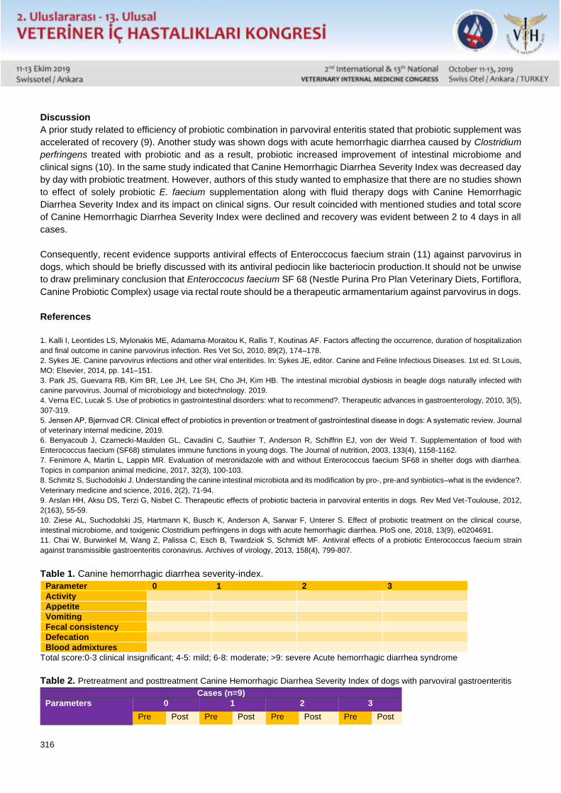
316
Discussion
A prior study related to efficiency of probiotic combination in parvoviral enteritis stated that probiotic supplement was
accelerated of recovery (9). Another study was shown dogs with acute hemorrhagic diarrhea caused by Clostridium
perfringens treated with probiotic and as a result, probiotic increased improvement of intestinal microbiome and
clinical signs (10). In the same study indicated that Canine Hemorrhagic Diarrhea Severity Index was decreased day
by day with probiotic treatment. However, authors of this study wanted to emphasize that there are no studies shown
to effect of solely probiotic E. faecium supplementation along with fluid therapy dogs with Canine Hemorrhagic
Diarrhea Severity Index and its impact on clinical signs. Our result coincided with mentioned studies and total score
of Canine Hemorrhagic Diarrhea Severity Index were declined and recovery was evident between 2 to 4 days in all
cases.
Consequently, recent evidence supports antiviral effects of Enteroccocus faecium strain (11) against parvovirus in
dogs, which should be briefly discussed with its antiviral pediocin like bacteriocin production.It should not be unwise
to draw preliminary conclusion that Enteroccocus faecium SF 68 (Nestle Purina Pro Plan Veterinary Diets, Fortiflora,
Canine Probiotic Complex) usage via rectal route should be a therapeutic armamentarium against parvovirus in dogs.
References
1. Kalli I, Leontides LS, Mylonakis ME, Adamama-Moraitou K, Rallis T, Koutinas AF. Factors affecting the occurrence, duration of hospitalization
and final outcome in canine parvovirus infection. Res Vet Sci, 2010, 89(2), 174–178.
2. Sykes JE. Canine parvovirus infections and other viral enteritides. In: Sykes JE, editor. Canine and Feline Infectious Diseases. 1st ed. St Louis,
MO: Elsevier, 2014, pp. 141–151.
3. Park JS, Guevarra RB, Kim BR, Lee JH, Lee SH, Cho JH, Kim HB. The intestinal microbial dysbiosis in beagle dogs naturally infected with
canine parvovirus. Journal of microbiology and biotechnology. 2019.
4. Verna EC, Lucak S. Use of probiotics in gastrointestinal disorders: what to recommend?. Therapeutic advances in gastroenterology, 2010, 3(5),
307-319.
5. Jensen AP, Bjørnvad CR. Clinical effect of probiotics in prevention or treatment of gastrointestinal disease in dogs: A systematic review. Journal
of veterinary internal medicine, 2019.
6. Benyacoub J, Czarnecki-Maulden GL, Cavadini C, Sauthier T, Anderson R, Schiffrin EJ, von der Weid T. Supplementation of food with
Enterococcus faecium (SF68) stimulates immune functions in young dogs. The Journal of nutrition, 2003, 133(4), 1158-1162.
7. Fenimore A, Martin L, Lappin MR. Evaluation of metronidazole with and without Enterococcus faecium SF68 in shelter dogs with diarrhea.
Topics in companion animal medicine, 2017, 32(3), 100-103.
8. Schmitz S, Suchodolski J. Understanding the canine intestinal microbiota and its modification by pro‐, pre‐and synbiotics–what is the evidence?.
Veterinary medicine and science, 2016, 2(2), 71-94.
9. Arslan HH, Aksu DS, Terzi G, Nisbet C. Therapeutic effects of probiotic bacteria in parvoviral enteritis in dogs. Rev Med Vet-Toulouse, 2012,
2(163), 55-59.
10. Ziese AL, Suchodolski JS, Hartmann K, Busch K, Anderson A, Sarwar F, Unterer S. Effect of probiotic treatment on the clinical course,
intestinal microbiome, and toxigenic Clostridium perfringens in dogs with acute hemorrhagic diarrhea. PloS one, 2018, 13(9), e0204691.
11. Chai W, Burwinkel M, Wang Z, Palissa C, Esch B, Twardziok S, Schmidt MF. Antiviral effects of a probiotic Enterococcus faecium strain
against transmissible gastroenteritis coronavirus. Archives of virology, 2013, 158(4), 799-807.
Table 1. Canine hemorrhagic diarrhea severity-index.
Parameter 0 1 2 3
Activity
Appetite
Vomiting
Fecal consistency
Defecation
Blood admixtures
Total score:0-3 clinical insignificant; 4-5: mild; 6-8: moderate; >9: severe Acute hemorrhagic diarrhea syndrome
Table 2. Pretreatment and posttreatment Canine Hemorrhagic Diarrhea Severity Index of dogs with parvoviral gastroenteritis
Cases (n=9)
Parameters 0 1 2 3
Pre Post Pre Post Pre Post Pre Post
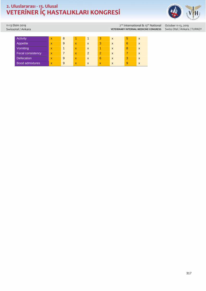
317
Activity x 8 1 1 3 x 5 x
Appetite x 9 x x 3 x 6 x
Vomiting x 1 x x 1 x 8 x
Fecal consistency x 7 x 2 2 x 7 x
Defecation x 9 x x 6 x 3 x
Bood admixtures x 9 x x x x 9 x