Embed Size (px)
Citation preview
By Dr Riaz MohammadRadiology of Thyroid and
parathyroid
Thyroid and parathyroidNormal Anatomy.Radiological test. Disease
Thyroid and parathyroidNormal Anatomy
Embryology: Thyroid gland is derived from a tubular structure at the base of tongue called
foramen cecum
AnatomyThyroid gland has two elongated lateral
lobes.United in the middle by a median isthmusIt is 4-6 cm in L, 1.3-18cm thick. Isthmus is
6mm thickSome time a pyramidal lobe is present which
extend upward from the isthmus.Thyroid is supplied by superior and inferior
thyroid arteries. It is extremely vascular
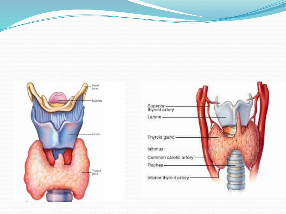

Thyroid AnatomyPosterior aspect Anterior and posterior
aspect
Thyroid produce T3, T4, and calcitonin.Parathyroid are two pairs(4 in number)
located on the back of thyroid. The superior pair is located near the middle of thyroid and the inferior pair at the lower pole on both side.
Parathyroid are the size of a grain of rice(3-5mm). It produce parathyroid hormone.


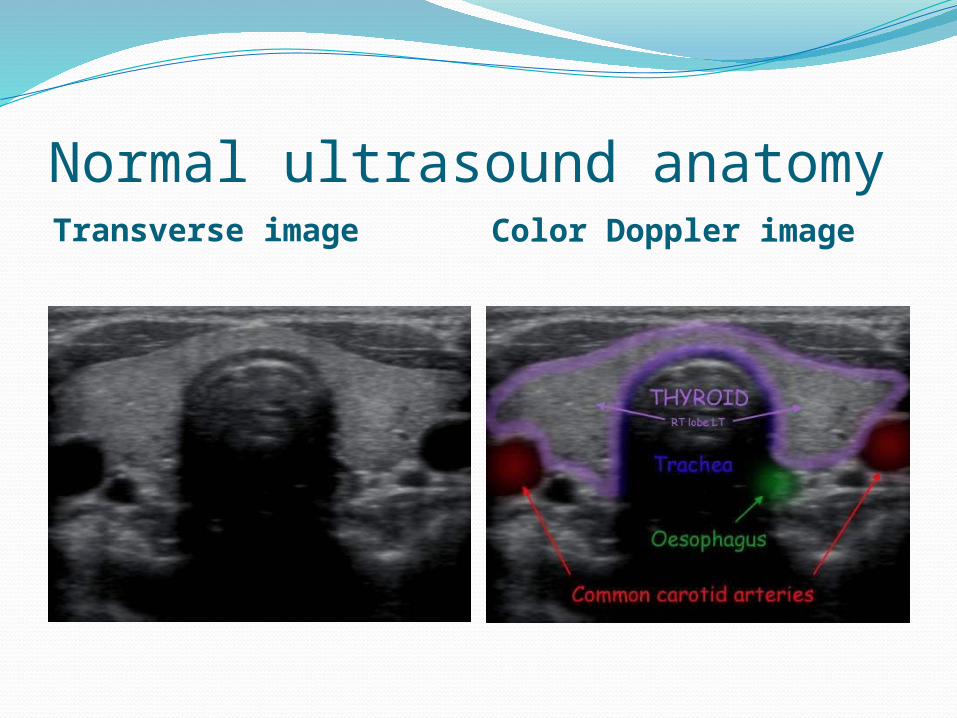
Normal ultrasound anatomy Transverse image Color Doppler image
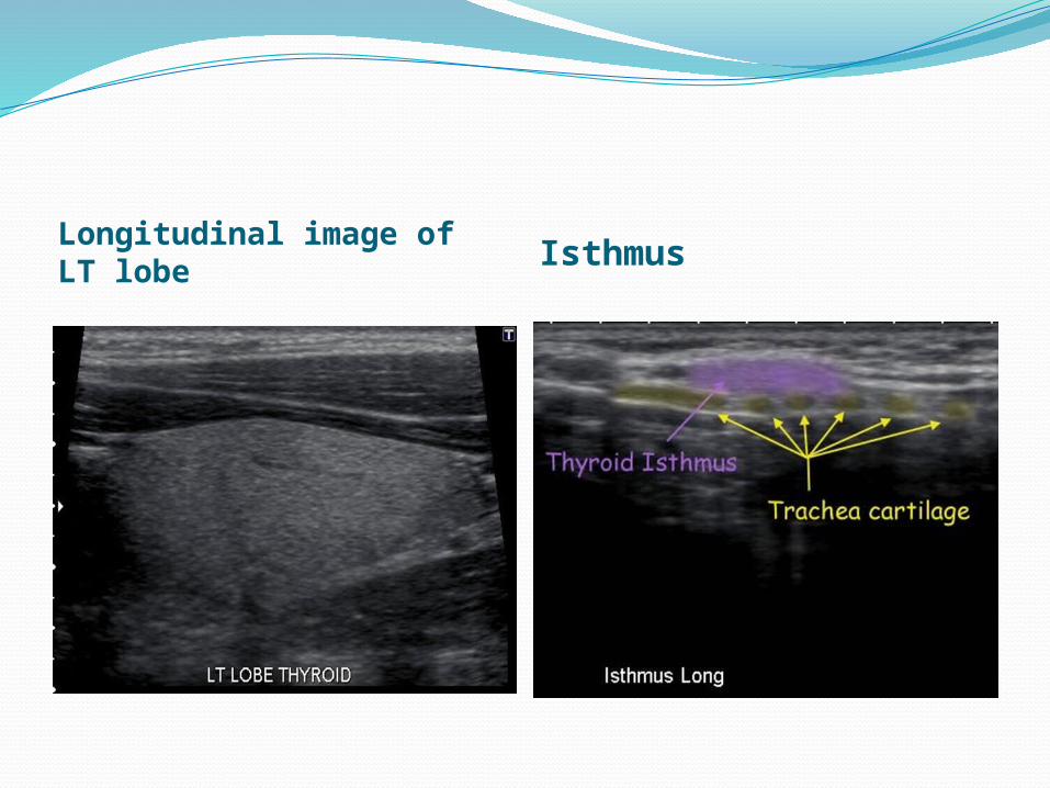
Longitudinal image of LT lobe
Isthmus
Imaging modalities used for evaluation of thyroid and parathyroid.
UltrasoundRadionuclide studiesCT scanBiopsy Ultrasound/ CT guided
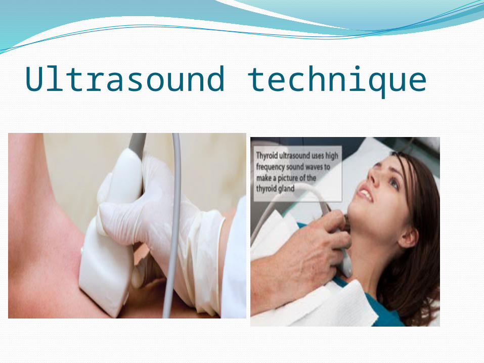
Ultrasound technique
High frequency sound wave are usedPatient is supine with hyperextended neckBoth right ,left lobes and isthmus are examinedColor Doppler is used to see vascularityTransverse and longitudinal images are takenUltrasound shows anatomical detail of thyroid
and adjacent blood vessels and structures.Normal parathyroid can not be seen with U/S,
however parathyroid adenoma and cervical nodes involved in thyroid cancer can be sampled
Radioisotopes studyA radionuclide is injected or given orally.Radioisotope taken up by
thyroid/parathyroidPatient is put under the gamma camera to
see the uptakeSeries of images are taken in different
projectionsRadionuclide studies show function.
Anatomical details are poor.
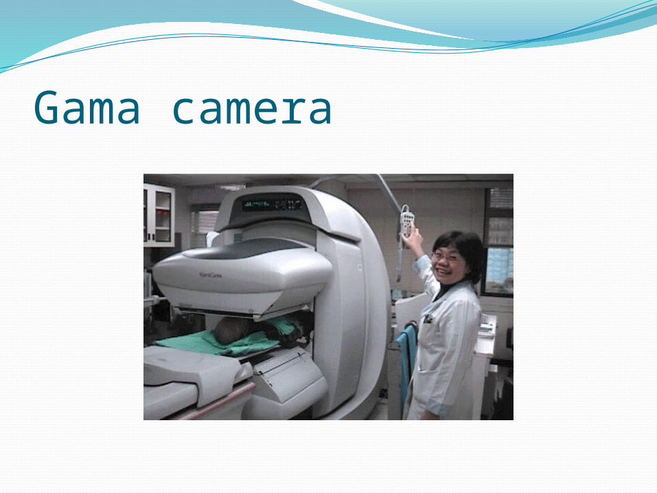
Gama camera
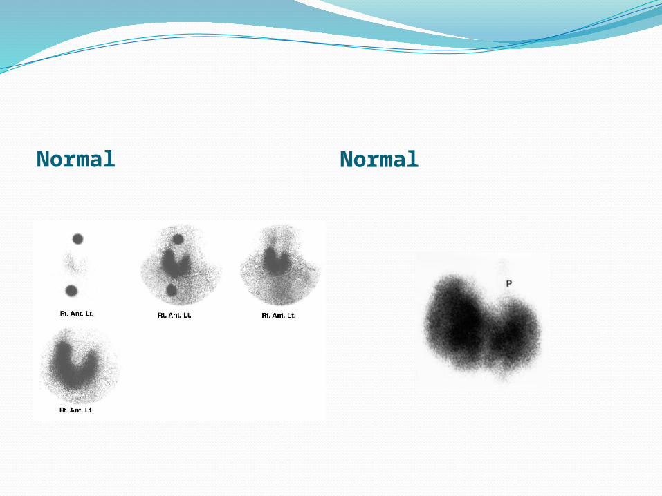
Normal Normal

CT images of thyroidCT shows clear anatomy of thyroid gland. Test is done without and with contrast
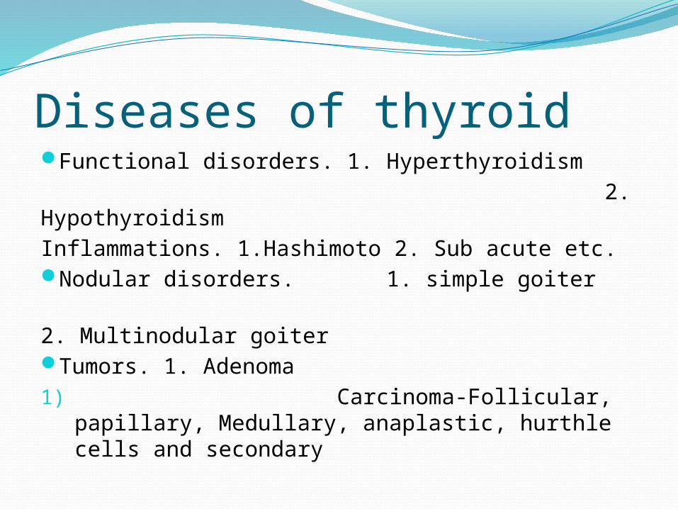
Diseases of thyroidFunctional disorders. 1. Hyperthyroidism 2. HypothyroidismInflammations. 1.Hashimoto 2. Sub acute etc.Nodular disorders. 1. simple goiter 2. Multinodular goiterTumors. 1. Adenoma1) Carcinoma-Follicular, papillary,
Medullary, anaplastic, hurthle cells and secondary
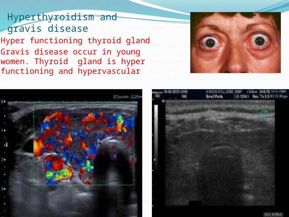
Hyperthyroidism and gravis disease
Hyper functioning thyroid glandGravis disease occur in young women. Thyroid gland is hyper functioning and hypervascular

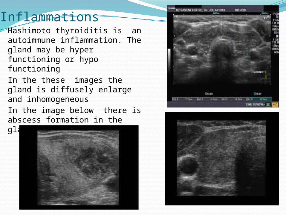
InflammationsHashimoto thyroiditis is an autoimmune inflammation. The gland may be hyper functioning or hypo functioningIn the these images the gland is diffusely enlarge and inhomogeneousIn the image below there is abscess formation in the gland
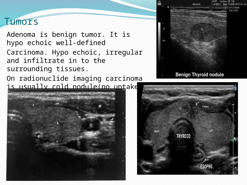
TumorsAdenoma is benign tumor. It is hypo echoic well-definedCarcinoma. Hypo echoic, irregular and infiltrate in to the surrounding tissues.On radionuclide imaging carcinoma is usually cold nodule(no uptake)
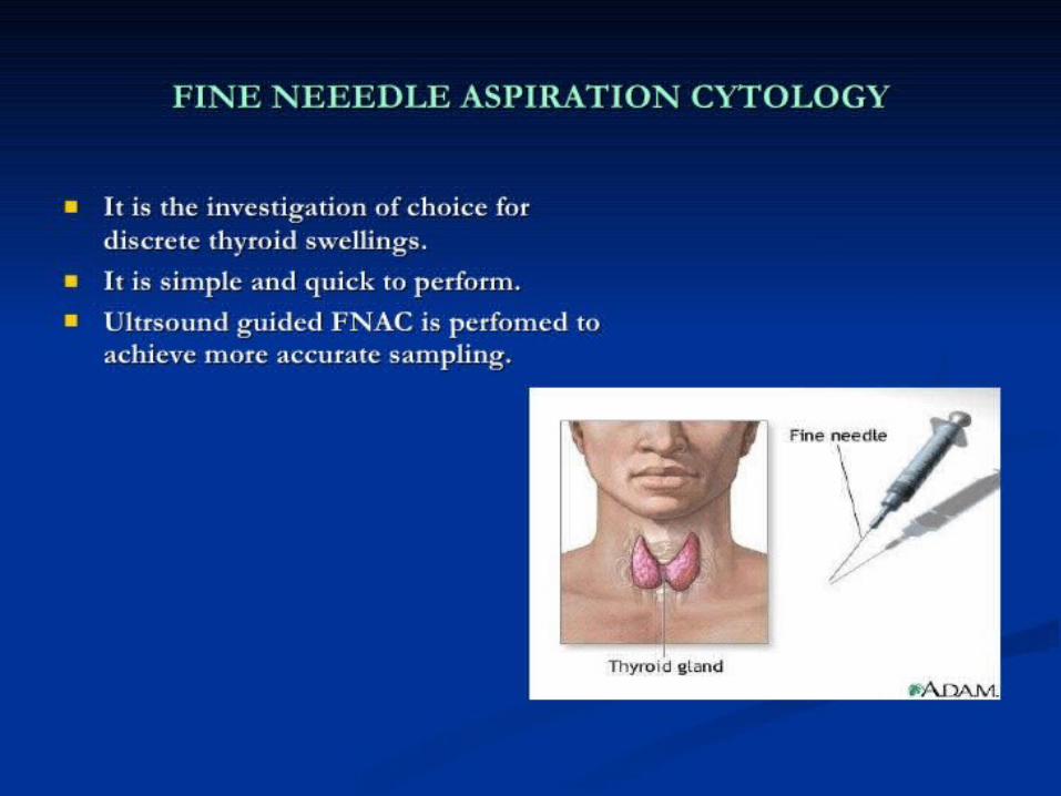

Ultrasound guided biopsy
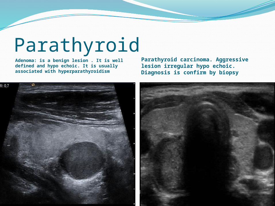
Parathyroid Adenoma: is a benign lesion . It is well defined and hypo echoic. It is usually associated with hyperparathyroidism
Parathyroid carcinoma. Aggressive lesion irregular hypo echoic. Diagnosis is confirm by biopsy
Thank you