Embed Size (px)
Citation preview
1
Respiratory
System
For the body to
survive, there must be
a constant supply of
O2 and a constant
disposal of CO2
Respiratory System
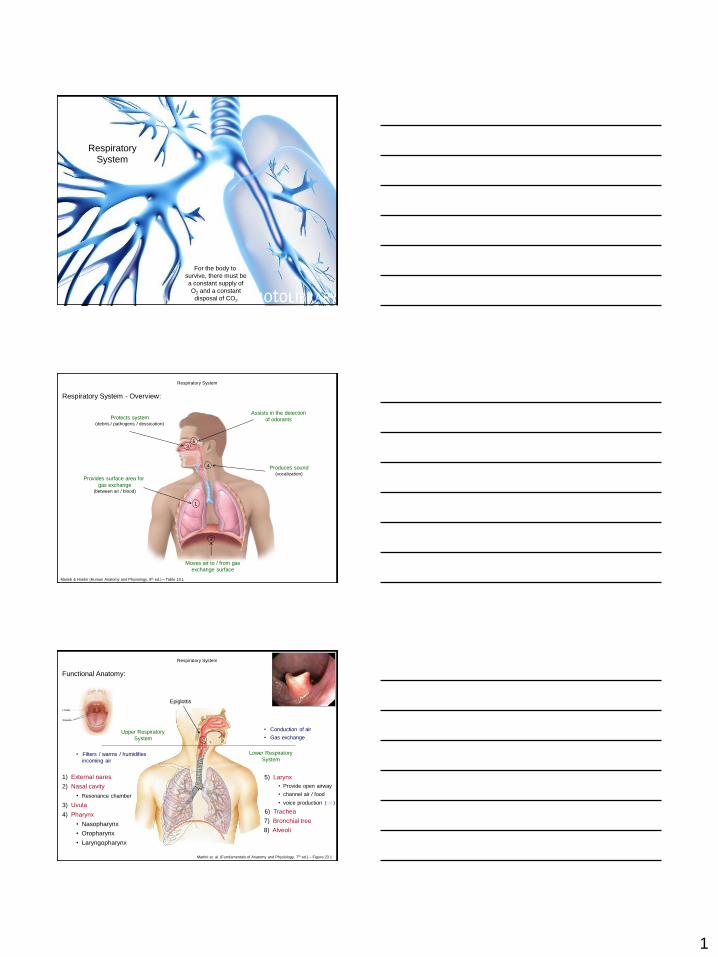
Respiratory System - Overview:
Marieb & Hoehn (Human Anatomy and Physiology, 8th ed.) – Table 19.1
1
Provides surface area for
gas exchange (between air / blood)
Moves air to / from gas
exchange surface
3
2
Protects system (debris / pathogens / dessication)
4 Produces sound (vocalization)
Assists in the detection
of odorants
5
Upper Respiratory
System
Lower Respiratory
System • Filters / warms / humidifies
incoming air
• Conduction of air
• Gas exchange
1) External nares
2) Nasal cavity
• Resonance chamber
3) Uvula
5) Larynx
• Provide open airway
• channel air / food
Epiglottis
4) Pharynx
• Nasopharynx
• Oropharynx
• Laryngopharynx
7) Bronchial tree
• voice production
8) Alveoli
Respiratory System
Functional Anatomy:
(link)
6) Trachea
Martini et. al. (Fundamentals of Anatomy and Physiology, 7th ed.) – Figure 23.1
2
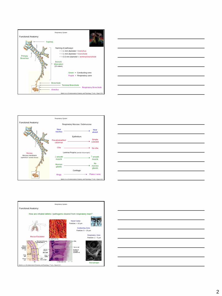
Trachea
Primary
Bronchus
Bronchi
bifurcation (23 orders)
Naming of pathways:
• > 1 mm diameter = bronchus
• < 1 mm diameter = bronchiole
• < 0.5 mm diameter = terminal bronchiole
Bronchiole Terminal Bronchiole
Respiratory Bronchiole Alveolus
Green = Conducting zone
Purple = Respiratory zone
Respiratory System
Functional Anatomy:
Martini et. al. (Fundamentals of Anatomy and Physiology, 7th ed.) – Figure 23.9
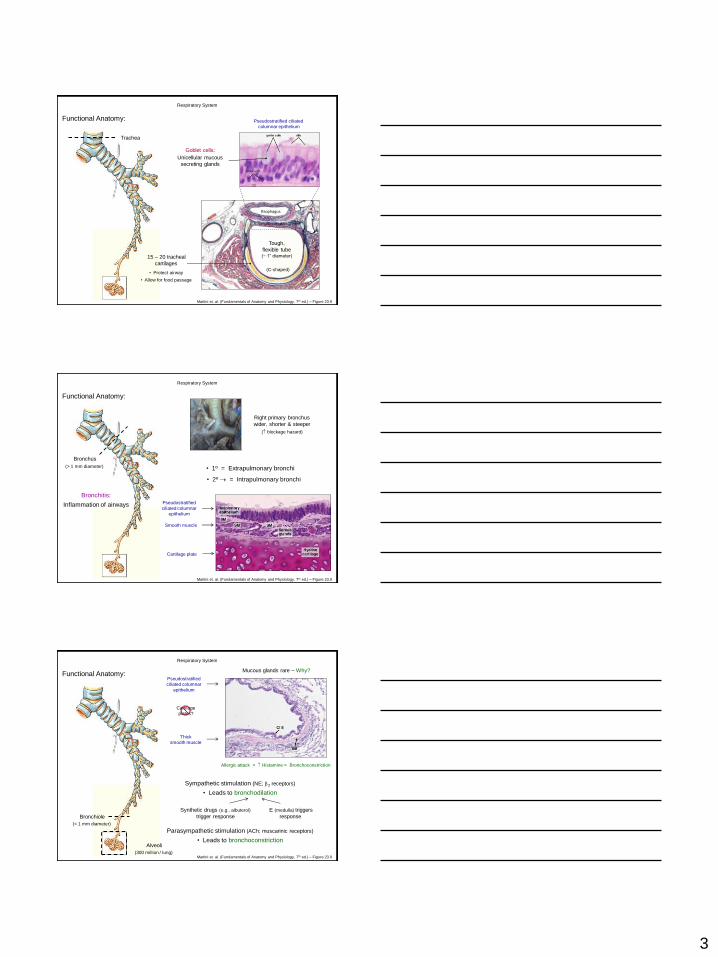
Respiratory Mucosa / Submucosa:
Near
trachea Near
alveoli
Epithelium:
Pseudostratified
columnar
Simple
cuboidal
Lamina Propria (areolar tissue layer):
Cilia No cilia
smooth
muscle
smooth
muscle
Cartilage:
Rings Plates / none
Mucosa:
Mucous membrane
(epithelium / areolar tissue)
Mucous
glands
No
mucous
glands
Respiratory System
Functional Anatomy:
Martini et. al. (Fundamentals of Anatomy and Physiology, 7th ed.) – Figure 23.9
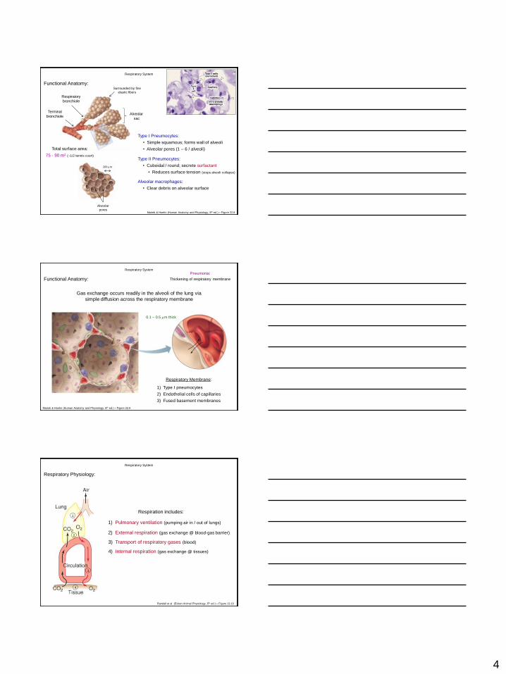
How are inhaled debris / pathogens cleared from respiratory tract?
Macrophages
Nasal Cavity:
Particles > 10 µm
Conducting Zone:
Particles 5 – 10 µm
Respiratory Zone:
Particles 1 – 5 µm Mucus Escalator
Respiratory System
Functional Anatomy:
Martini et. al. (Fundamentals of Anatomy and Physiology, 7th ed.) – Figure 23.2
3
Respiratory System
Functional Anatomy:
Martini et. al. (Fundamentals of Anatomy and Physiology, 7th ed.) – Figure 23.9
Esophagus
Tough,
flexible tube (~ 1” diameter) 15 – 20 tracheal
cartilages
• Protect airway
Pseudostratified ciliated
columnar epithelium
Goblet cells:
Unicellular mucous
secreting glands
(C-shaped)
Trachea
• Allow for food passage
Respiratory System
Functional Anatomy:
Martini et. al. (Fundamentals of Anatomy and Physiology, 7th ed.) – Figure 23.9
Right primary bronchus
wider, shorter & steeper
( blockage hazard)
• 1º = Extrapulmonary bronchi
• 2º = Intrapulmonary bronchi
Bronchus
Pseudostratified
ciliated columnar
epithelium
Smooth muscle
Cartilage plate
Bronchitis:
Inflammation of airways
(> 1 mm diameter)
Respiratory System
Functional Anatomy:
Martini et. al. (Fundamentals of Anatomy and Physiology, 7th ed.) – Figure 23.9
Bronchiole
(< 1 mm diameter)
Pseudostratified
ciliated columnar
epithelium
Thick
smooth muscle
Cartilage
plates?
Mucous glands rare – Why?
Sympathetic stimulation (NE; 2 receptors)
• Leads to bronchodilation
Parasympathetic stimulation (ACh; muscarinic receptors)
• Leads to bronchoconstriction
E (medulla) triggers
response
Allergic attack = Histamine = Bronchoconstriction
Synthetic drugs (e.g., albuterol)
trigger response
Alveoli
(300 million / lung)
4
Respiratory System
Functional Anatomy:
200 m
Type I Pneumocytes:
Type II Pneumocytes:
Alveolar
sac
Total surface area:
75 - 90 m2 (~1/2 tennis court)
Alveolar
pores
Terminal
bronchiole
Respiratory
bronchiole
Alveolar macrophages:
Marieb & Hoehn (Human Anatomy and Physiology, 8th ed.) – Figure 22.8
Surrounded by fine
elastic fibers
• Simple squamous; forms wall of alveoli
• Alveolar pores (1 – 6 / alveoli)
• Cuboidal / round; secrete surfactant
• Reduces surface tension (stops alveoli collapse)
• Clear debris on alveolar surface
Respiratory Membrane:
1) Type I pneumocytes
2) Endothelial cells of capillaries
3) Fused basement membranes
0.1 – 0.5 m thick
Pneumonia:
Thickening of respiratory membrane
Respiratory System
Functional Anatomy:
Gas exchange occurs readily in the alveoli of the lung via
simple diffusion across the respiratory membrane
Marieb & Hoehn (Human Anatomy and Physiology, 8th ed.) – Figure 22.9
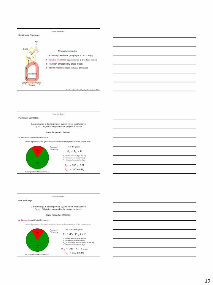
Respiratory Physiology:
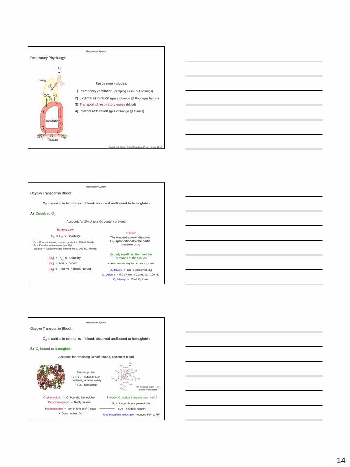
1) Pulmonary ventilation (pumping air in / out of lungs)
2) External respiration (gas exchange @ blood-gas barrier)
3) Transport of respiratory gases (blood)
4) Internal respiration (gas exchange @ tissues)
Respiratory System
Respiration includes: 1
2
3
4
Randall et al. (Eckert Animal Physiology, 5th ed.) – Figure 13.19
5
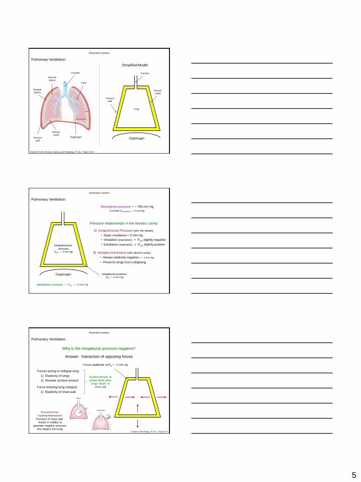
Pulmonary Ventilation:
Trachea
Thoracic
wall
Visceral
pleura
Parietal
pleura
Pleural
cavity
Lung
Diaphragm Diaphragm
Simplified Model:
Trachea
Lung
Pleural
cavity
Thoracic
wall
Respiratory System
Marieb & Hoehn (Human Anatomy and Physiology, 8th ed.) – Figure 22.12
Pressure relationships in the thoracic cavity:
Atmospheric pressure = ~ 760 mm Hg
(Consider Patmospheric = 0 mm Hg)
1) Intrapulmonary Pressure (w/in the alveoli):
• Static conditions = 0 mm Hg
2) Intrapleural pressure (w/in pleural cavity):
• Always relatively negative (~ - 4 mm Hg)
atmospheric pressure = Patm = 0 mm Hg
Intrapleural pressure (Pip = - 4 mm Hg)
Diaphragm
Intrapulmonary
pressure (Ppul = 0 mm Hg)
• Inhalation (inspiration) = Ppul slightly negative
• Exhalation (expiration) = Ppul slightly positive
• Prevents lungs from collapsing
Respiratory System
Pulmonary Ventilation:
Why is the intrapleural pressure negative?
Answer: Interaction of opposing forces
Forces acting to collapse lung:
1) Elasticity of lungs
2) Alveolar surface tension
Force resisting lung collapse:
1) Elasticity of chest wall
Surface tension of
serous fluids keep
lungs “stuck” to
chest wall
Forces equilibrate at Pip = - 4 mm Hg
Respiratory System
Pneumothorax:
(“sucking chest wound”)
Puncture of chest wall;
results in inability to
generate negative pressure
and expand the lungs Costanzo (Physiology, 4th ed.) – Figure 5.9
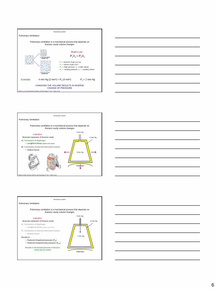
Pulmonary Ventilation:
6
CHANGING THE VOLUME RESULTS IN INVERSE
CHANGE OF PRESSURE
P = pressure of gas (mm Hg)
V = volume of gas (mm3)
P1 = initial pressure; V1 = initial volume
P2 = resulting pressure; V2 = resulting volume
Boyle’s Law:
P1V1 = P2V2
Example: 4 mm Hg (2 mm3) = P2 (4 mm3) P2 = 2 mm Hg
Pulmonary ventilation is a mechanical process that depends on
thoracic cavity volume changes
Respiratory System
Martini et. al. (Fundamentals of Anatomy and Physiology, 7th ed.) – Figure 23.13
Pulmonary Ventilation:
Inspiration:
Muscular expansion of thoracic cavity
Diaphragm
0 mm Hg
0 mm Hg
- 4 mm Hg
A) Contraction of diaphragm
• Lengthens thorax (pushes liver down)
B) Contraction of external intercostal muscles
• Widens thorax
Respiratory System
Pulmonary ventilation is a mechanical process that depends on
thoracic cavity volume changes
Marieb & Hoehn (Human Anatomy and Physiology, 8th ed.) – Figure 22.13
Pulmonary Ventilation:
Inspiration:
Muscular expansion of thoracic cavity
Diaphragm
- 1 mm Hg
0 mm Hg
- 6 mm Hg
A) Contraction of diaphragm
• Lengthens thorax (pushes liver down)
B) Contraction of external intercostal muscles
• Widens thorax
Respiratory System
Pulmonary ventilation is a mechanical process that depends on
thoracic cavity volume changes
Results in:
• Reduced intrapleural pressure (Pip)
• Reduced intrapulmonary pressure (Ppul)
Results in decreased pressure in thoracic
cavity and air enters
Pulmonary Ventilation:
7
0 mm Hg
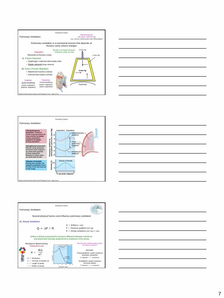
Respiratory System
Pulmonary ventilation is a mechanical process that depends on
thoracic cavity volume changes
Expiration:
Retraction of thoracic cavity
A) Passive Expiration
• Diaphragm / external intercostals relax
• Elastic rebound (lungs rebound)
- 4 mm Hg
0 mm Hg +1 mm Hg
Diaphragm
Results in increased pressure
in thoracic cavity; air exits
B) Active (“Forced”) Expiration
• Abdominal muscles contract
• Internal intercostals contract
Marieb & Hoehn (Human Anatomy and Physiology, 8th ed.) – Figure 22.13
Eupnea:
Quiet breathing
(active inspiration;
(passive expiration)
Hyperpnea:
Forced breathing
(active inspiration;
(active expiration)
Internal pressure
can reach +100 mm Hg
(e.g., why you should exhale when lifting weights)
Pulmonary Ventilation:
Respiratory System
Marieb & Hoehn (Human Anatomy and Physiology, 8th ed.) – Figure 22.14
Pulmonary Ventilation:
A) Airway resistance
Respiratory System
Pulmonary Ventilation:
Several physical factors exist influence pulmonary ventilation
Q = P / R
Q = Airflow (L / min)
P = Pressure gradient (mm Hg)
R = Airway resistance (mm Hg / L / sec)
Airflow is directly proportional to pressure difference between outside air
and alveoli and inversely proportional to resistance of the airway
Resistance determined by
Poiseuille’s Law:
R = 8Lη
r4
R = Resistance
η = Viscosity of inspired air
L = Length of airway
r = Radius of airway
Why don’t the smallest airways provide
the highest resistance?
Reminder:
Parasympathetic system produces
bronchial constriction
Sympathetic system produces
bronchial dilation
( diameter = resistance)
( diameter = resistance)
Resis
tan
ce
Bifurcation stage
Medium-sized
bronchi
Terminal
bronchioles
8
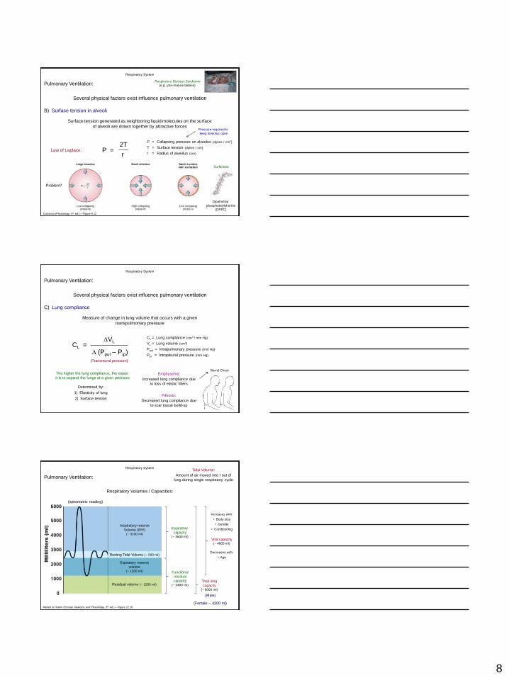
B) Surface tension in alveoli
Respiratory System
Pulmonary Ventilation:
Several physical factors exist influence pulmonary ventilation
Surface tension generated as neighboring liquid molecules on the surface
of alveoli are drawn together by attractive forces
P = 2T
r Law of Laplace:
P = Collapsing pressure on alveolus (dynes / cm2)
T = Surface tension (dynes / cm)
r = Radius of alveolus (cm)
Pressure required to
keep alveolus open
Problem?
Respiratory Distress Syndrome
(e.g., pre-mature babies)
Dipalmitoyl
phosphatidylchorine
(DPPC)
Surfactant
Costanzo (Physiology, 4th ed.) – Figure 5.12
C) Lung compliance
Respiratory System
Pulmonary Ventilation:
Several physical factors exist influence pulmonary ventilation
Measure of change in lung volume that occurs with a given
transpulmonary pressure
CL = VL
(Ppul – Pip)
CL = Lung compliance (cm3 / mm Hg)
VL = Lung volume (cm3)
Ppul = Intrapulmonary pressure (mm Hg)
Ppi = Intrapleural pressure (mm Hg)
(Transmural pressure)
The higher the lung compliance, the easier
it is to expand the lungs at a given pressure
Determined by:
1) Elasticity of lung
2) Surface tension
Emphysema:
Increased lung compliance due
to loss of elastic fibers
Barrel Chest
Fibrosis:
Decreased lung compliance due
to scar tissue build-up
Respiratory Volumes / Capacities:
Tidal Volume:
Amount of air moved into / out of
lung during single respiratory cycle
(spirometric reading)
Resting Tidal Volume (~ 500 ml)
Inspiratory reserve
Volume (IRV) (~ 3100 ml)
Expiratory reserve
volume (~ 1200 ml)
Residual volume (~ 1200 ml)
Vital capacity (~ 4800 ml)
Total lung
capacity (~ 6000 ml)
(Male)
(Female ~ 4200 ml)
Respiratory System
Pulmonary Ventilation:
Marieb & Hoehn (Human Anatomy and Physiology, 8th ed.) – Figure 22.16
Inspiratory
capacity (~ 3600 ml)
Functional
residual
capacity (~ 2400 ml)
Increases with:
• Body size
• Gender
• Conditioning
Decreases with:
• Age
9
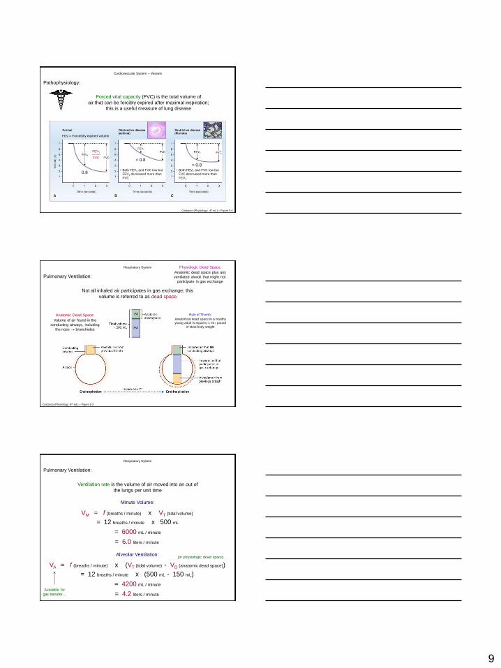
Pathophysiology:
Cardiovascular System – Vessels
Forced vital capacity (FVC) is the total volume of
air that can be forcibly expired after maximal inspiration;
this is a useful measure of lung disease
Costanzo (Physiology, 4th ed.) – Figure 5.6
FEV = Forcefully expired volume
0.8
FEV1
FVC
• Both FEV1 and FVC low but
FEV1 decreased more than
FVC
< 0.8
• Both FEV1 and FVC low but
FVC decreased more than
FEV1
> 0.8
Respiratory System
Pulmonary Ventilation:
Not all inhaled air participates in gas exchange; this
volume is referred to as dead space
Costanzo (Physiology, 4th ed.) – Figure 5.3
Rule of Thumb:
Anatomical dead space in a healthy
young adult is equal to 1 ml / pound
of ideal body weight
Physiologic Dead Space:
Anatomic dead space plus any
ventilated alveoli that might not
participate in gas exchange
Anatomic Dead Space:
Volume of air found in the
conducting airways, including
the nose bronchioles
VM = f (breaths / minute) x VT (tidal volume)
= 12 breaths / minute x 500 mL
= 6000 mL / minute
Minute Volume:
VA = f (breaths / minute) x (VT (tidal volume) - VD (anatomic dead space))
= 12 breaths / minute x (500 mL - 150 mL)
= 4200 mL / minute
Alveolar Ventilation:
Available for
gas transfer…
= 6.0 liters / minute
= 4.2 liters / minute
Respiratory System
Pulmonary Ventilation:
Ventilation rate is the volume of air moved into an out of
the lungs per unit time
(or physiologic dead space)
10
Respiratory Physiology:
1) Pulmonary ventilation (pumping air in / out of lungs)
2) External respiration (gas exchange @ blood-gas barrier)
3) Transport of respiratory gases (blood)
4) Internal respiration (gas exchange @ tissues)
Respiratory System
Respiration includes: 1
2
3
4
Randall et al. (Eckert Animal Physiology, 5th ed.) – Figure 13.19
Respiratory System
Pulmonary Ventilation:
Gas exchange in the respiratory system refers to diffusion of
O2 and CO2 in the lung and in the peripheral tissues
Basic Properties of Gases:
A) Dalton’s Law of Partial Pressures:
The total pressure of a gas is equal to the sum of the pressure of its constituents
% Composition of Atmospheric Air
21%
O2
79%
N2
Patmosphere:
760 mm Hg
For dry gases:
PX = PB x F
PX = Partial pressure of gas (mm Hg)
PB = Barometric pressure (mm Hg)
F = Fractional concentration of gas
PO2 = 760 x 0.21
PO2 = 160 mm Hg
Respiratory System
Gas Exchange:
Gas exchange in the respiratory system refers to diffusion of
O2 and CO2 in the lung and in the peripheral tissues
Basic Properties of Gases:
A) Dalton’s Law of Partial Pressures:
The total pressure of a gas is equal to the sum of the pressure of its constituents
% Composition of Atmospheric Air
21%
O2
79%
N2
Patmosphere:
760 mm Hg
For humidified gases:
PX = (PB – PH2O) x F
PX = Partial pressure of gas (mm Hg)
PB = Barometric pressure (mm Hg)
PH2O = Water vapor pressure at 37C (47 mm Hg)
F = Fractional concentration of gas
PO2 = (760 – 47) x 0.21
PO2 = 150 mm Hg
11
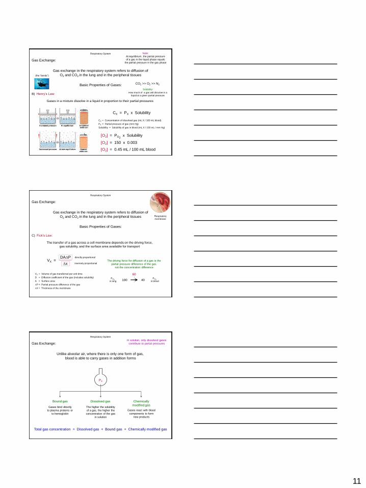
Respiratory System
Gas exchange in the respiratory system refers to diffusion of
O2 and CO2 in the lung and in the peripheral tissues
Basic Properties of Gases:
B) Henry’s Law:
Gases in a mixture dissolve in a liquid in proportion to their partial pressures
Note:
At equilibrium, the partial pressure
of a gas in the liquid phase equals
the partial pressure in the gas phase
CX = PX x Solubility
CX = Concentration of dissolved gas (mL X / 100 mL blood)
PX = Partial pressure of gas (mm Hg)
Solubility = Solubility of gas in blood (mL X / 100 mL / mm Hg)
Solubility:
How much of a gas will dissolve in a
liquid at a given partial pressure
[O2] = PO2 x Solubility
[O2] = 150 x 0.003
[O2] = 0.45 mL / 100 mL blood
CO2 >> O2 >> N2
(the “bends”)
Gas Exchange:
Respiratory System
Basic Properties of Gases:
C) Fick’s Law:
The transfer of a gas across a cell membrane depends on the driving force,
gas solubility, and the surface area available for transport
VX = Volume of gas transferred per unit time
D = Diffusion coefficient of the gas (includes solubility)
A = Surface area
P = Partial pressure difference of the gas
X = Thickness of the membrane
VX = DAP
x The driving force for diffusion of a gas is the
partial pressure difference of the gas,
not the concentration difference
PO2
in lung
PO2
in blood 100 40
60
Gas Exchange:
directly proportional
inversely proportional
Respiratory
membrane
Gas exchange in the respiratory system refers to diffusion of
O2 and CO2 in the lung and in the peripheral tissues
Respiratory System
Gas Exchange:
Unlike alveolar air, where there is only one form of gas,
blood is able to carry gases in addition forms
PX
Dissolved gas Bound gas Chemically
modified gas The higher the solubility
of a gas, the higher the
concentration of the gas
in solution
Gases bind directly
to plasma proteins or
to hemoglobin
Gases react with blood
components to form
new products
Total gas concentration = Dissolved gas + Bound gas + Chemically modified gas
In solution, only dissolved gases
contribute to partial pressures
12
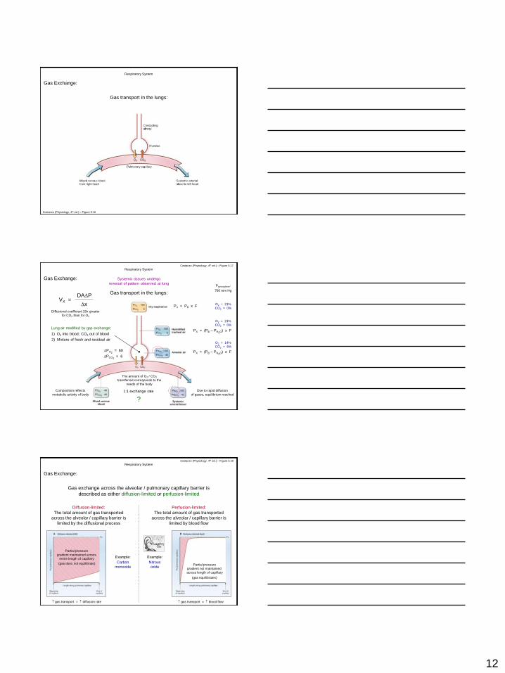
Respiratory System
Gas Exchange:
Gas transport in the lungs:
Costanzo (Physiology, 4th ed.) – Figure 5.16
Respiratory System
Gas Exchange:
Gas transport in the lungs:
Costanzo (Physiology, 4th ed.) – Figure 5.17
PX = PB x F
PX = (PB – PH2O) x F Lung air modified by gas exchange:
O2 = 21%
CO2 = 0%
O2 = 21%
CO2 = 0%
O2 = 14%
CO2 = 6%
PX = (PB – PH2O) x F
Composition reflects
metabolic activity of body
Due to rapid diffusion
of gases, equilibrium reached
Patmosphere:
760 mm Hg
The amount of O2 / CO2
transferred corresponds to the
needs of the body
1:1 exchange rate
PO2 = 60
PCO2 = 6
?
VX = DAP
x
Diffusional coefficient 20x greater
for CO2 than for O2
Systemic tissues undergo
reversal of pattern observed at lung
1) O2 into blood; CO2 out of blood
2) Mixture of fresh and residual air
Respiratory System
Gas Exchange:
Gas exchange across the alveolar / pulmonary capillary barrier is
described as either diffusion-limited or perfusion-limited
Diffusion-limited:
The total amount of gas transported
across the alveolar / capillary barrier is
limited by the diffusional process
Partial pressure
gradient maintained across
entire length of capillary
(gas does not equilibrate)
Perfusion-limited:
The total amount of gas transported
across the alveolar / capillary barrier is
limited by blood flow
Example:
Carbon
monoxide
Example:
Nitrous
oxide Partial pressure
gradient not maintained
across length of capillary
(gas equilibrates)
Costanzo (Physiology, 4th ed.) – Figure 5.18
gas transport = diffusion rate gas transport = blood flow
13
Respiratory System
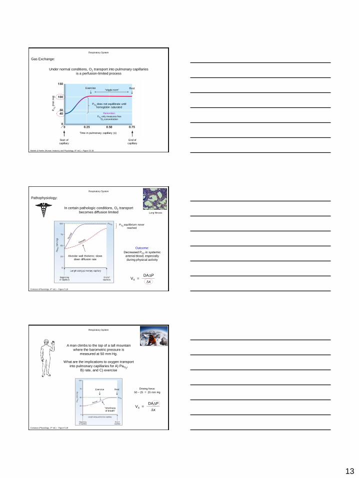
Gas Exchange:
Under normal conditions, O2 transport into pulmonary capillaries
is a perfusion-limited process
Marieb & Hoehn (Human Anatomy and Physiology, 8th ed.) – Figure 22.18
Rest Exercise “wiggle room”
End of
capillary
Start of
capillary
Time in pulmonary capillary (s)
PO
2 (
mm
Hg)
PO2 does not equilibrate until
hemoglobin saturated
Remember:
PO2 only measures free
O2 concentration
Respiratory System
In certain pathologic conditions, O2 transport
becomes diffusion limited
Pathophysiology:
Lung fibrosis
Costanzo (Physiology, 4th ed.) – Figure 5.19
Alveolar wall thickens; slows
down diffusion rate
PO2 equilibrium never
reached
Outcome:
Decreased PO2 in systemic
arterial blood, especially
during physical activity
VX = DAP
x
Respiratory System
Costanzo (Physiology, 4th ed.) – Figure 5.19
VX = DAP
x
A man climbs to the top of a tall mountain
where the barometric pressure is
measured at 50 mm Hg.
What are the implications to oxygen transport
into pulmonary capillaries for A) PaO2,
B) rate, and C) exercise
Rest Exercise
“shortness
of breath”
Driving force:
50 – 25 = 25 mm Hg
14
Respiratory Physiology:
1) Pulmonary ventilation (pumping air in / out of lungs)
2) External respiration (gas exchange @ blood-gas barrier)
3) Transport of respiratory gases (blood)
4) Internal respiration (gas exchange @ tissues)
Respiratory System
Respiration includes: 1
2
3
4
Randall et al. (Eckert Animal Physiology, 5th ed.) – Figure 13.19
Respiratory System
Oxygen Transport in Blood:
O2 is carried in two forms in blood: dissolved and bound to hemoglobin
A) Dissolved O2:
CX = PX x Solubility
CX = Concentration of dissolved gas (mL X / 100 mL blood)
PX = Partial pressure of gas (mm Hg)
Solubility = Solubility of gas in blood (mL X / 100 mL / mm Hg)
Accounts for 2% of total O2 content of blood
Recall:
The concentration of dissolved
O2 is proportional to the partial
pressure of O2
Henry’s Law:
[O2] = PO2 x Solubility
[O2] = 100 x 0.003
[O2] = 0.30 mL / 100 mL blood
Grossly insufficient to meet the
demands of the tissues
At rest, tissues require 250 mL O2 / min
O2 delivery = CO x [dissolved O2]
O2 delivery = 5.0 L / min x 0.3 mL O2 / 100 mL
O2 delivery = 15 mL O2 / min
Respiratory System
Oxygen Transport in Blood:
O2 is carried in two forms in blood: dissolved and bound to hemoglobin
B) O2 bound to hemoglobin:
Accounts for remaining 98% of total O2 content of blood
Globular protein:
2 & 2 subunits each
containing a heme moiety
• 4 O2 / hemoglobin
Oxyhemoglobin = O2 bound to hemoglobin
Iron (ferrous state – Fe2+)
bound to nitrogens
Deoxyhemoglobin = No O2 present
Shouldn’t O2 oxidize iron (ferric state – Fe3+ )?
No – nitrogen bonds prevent this…
Methemoglobin reductase – reduces Fe3+ to Fe2+
BUT – if it does happen: Methemoglobin = Iron in ferric (Fe3+) state
• Does not bind O2
15
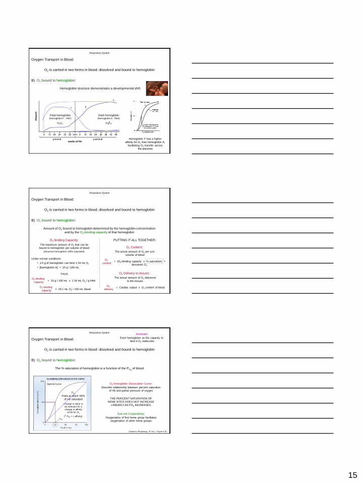
Respiratory System
Oxygen Transport in Blood:
O2 is carried in two forms in blood: dissolved and bound to hemoglobin
B) O2 bound to hemoglobin:
Hemoglobin structure demonstrates a developmental shift
Adult hemoglobin (hemoglobin A - HbA)
22
Fetal hemoglobin (hemoglobin F - HbF)
22
Hemoglobin F has a higher
affinity for O2 than hemoglobin A,
facilitating O2 transfer across
the placenta
Respiratory System
Oxygen Transport in Blood:
O2 is carried in two forms in blood: dissolved and bound to hemoglobin
B) O2 bound to hemoglobin:
Amount of O2 bound to hemoglobin determined by the hemoglobin concentration
and by the O2-binding capacity of that hemoglobin
O2-binding Capacity:
The maximum amount of O2 that can be
bound to hemoglobin per volume of blood
(Assumes hemoglobin 100% saturated)
Under normal conditions:
• 1.0 g of hemoglobin can bind 1.34 mL 02
• [hemoglobin A] = 15 g / 100 mL
THUS
O2-binding
capacity = 15 g / 100 mL x 1.34 mL O2 / g HbA
O2-binding
capacity = 20.1 mL O2 / 100 mL blood
O2 Content:
The actual amount of O2 per unit
volume of blood
O2
content = (O2-binding capacity x % saturation) +
dissolved O2
O2 Delivery to tissues:
The actual amount of O2 delivered
to the tissues
O2
delivery = Cardiac output x O2 content of blood
PUTTING IT ALL TOGETHER:
Respiratory System
Oxygen Transport in Blood:
O2 is carried in two forms in blood: dissolved and bound to hemoglobin
B) O2 bound to hemoglobin:
The % saturation of hemoglobin is a function of the PO2 of blood
Reminder:
Each hemoglobin as the capacity to
bind 4 O2 molecules
O2-hemoglobin Dissociation Curve:
Describe relationship between percent saturation
of Hb and partial pressure of oxygen
Sub-unit Cooperativity:
Oxygenation of first heme group facilitates
oxygenation of other heme groups
Sigmoid curve
THE PERCENT SATURATION OF
HEME SITES DOES NOT INCREASE
LINEARLY AS PO2 INCREASES
P50
Point at which 50%
of Hb saturated
Change in value is
an indicator for a
change in affinity
of Hb for O2
( P50 = affinity)
Costanzo (Physiology, 4th ed.) – Figure 5.20
16
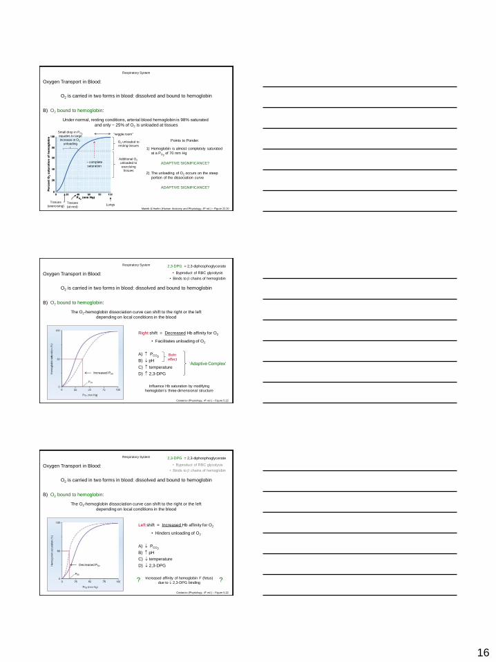
Respiratory System
Oxygen Transport in Blood:
O2 is carried in two forms in blood: dissolved and bound to hemoglobin
B) O2 bound to hemoglobin:
Under normal, resting conditions, arterial blood hemoglobin is 98% saturated
and only ~ 25% of O2 is unloaded at tissues
Marieb & Hoehn (Human Anatomy and Physiology, 8th ed.) – Figure 22.20
Points to Ponder:
1) Hemoglobin is almost completely saturated
at a PO2 of 70 mm Hg
~ complete
saturation
“wiggle room”
ADAPTIVE SIGNIFICANCE?
2) The unloading of O2 occurs on the steep
portion of the dissociation curve
Lungs Tissues
(at rest)
ADAPTIVE SIGNIFICANCE?
O2 unloaded to
resting tissues
Additional O2
unloaded to
exercising
tissues
Tissues
(exercising)
Small drop in PO2 equates to large
increase in O2
unloading
Respiratory System
Oxygen Transport in Blood:
O2 is carried in two forms in blood: dissolved and bound to hemoglobin
B) O2 bound to hemoglobin:
The O2-hemoglobin dissociation curve can shift to the right or the left
depending on local conditions in the blood
Costanzo (Physiology, 4th ed.) – Figure 5.22
Right shift = Decreased Hb affinity for O2
Increased P50
• Facilitates unloading of O2
A) PCO2
B) pH ‘Adaptive Complex’
2,3-DPG = 2,3-diphosphoglycerate
• Byproduct of RBC glycolysis
• Binds to chains of hemoglobin
Influence Hb saturation by modifying
hemoglobin’s three-dimensional structure
Bohr
effect
C) temperature
D) 2,3-DPG
Respiratory System
Oxygen Transport in Blood:
O2 is carried in two forms in blood: dissolved and bound to hemoglobin
B) O2 bound to hemoglobin:
The O2-hemoglobin dissociation curve can shift to the right or the left
depending on local conditions in the blood
Costanzo (Physiology, 4th ed.) – Figure 5.22
Left shift = Increased Hb affinity for O2
Decreased P50
• Hinders unloading of O2
A) PCO2
B) pH
C) temperature
D) 2,3-DPG
2,3-DPG = 2,3-diphosphoglycerate
• Byproduct of RBC glycolysis
• Binds to chains of hemoglobin
Increased affinity of hemoglobin F (fetus)
due to 2,3-DPG binding ? ?
17
Respiratory System
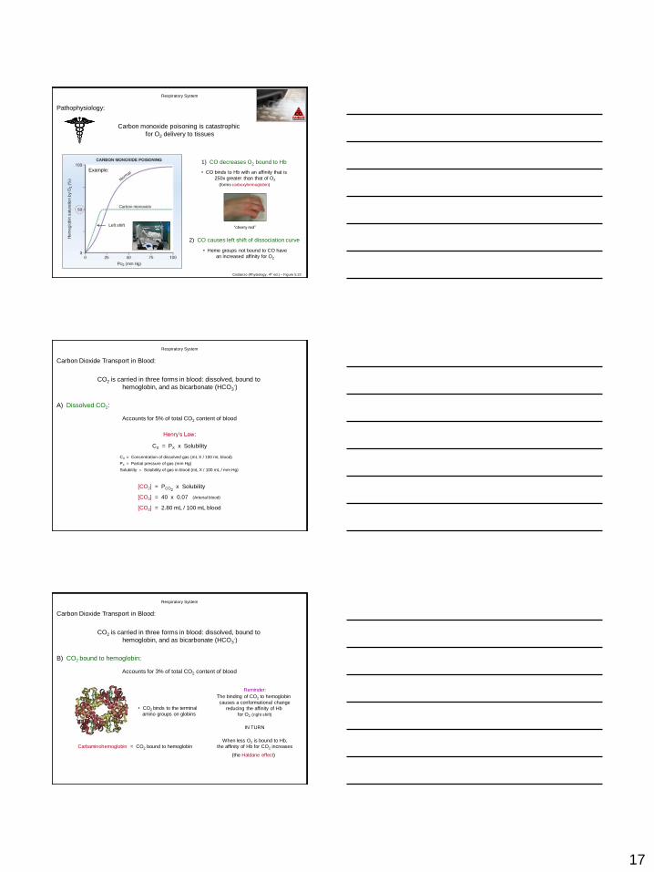
Pathophysiology:
Carbon monoxide poisoning is catastrophic
for O2 delivery to tissues
Costanzo (Physiology, 4th ed.) – Figure 5.22
1) CO decreases O2 bound to Hb
• CO binds to Hb with an affinity that is
250x greater than that of O2
(forms carboxyhemoglobin)
Example:
“cherry red”
2) CO causes left shift of dissociation curve
• Heme groups not bound to CO have
an increased affinity for O2
Left shift
Respiratory System
Carbon Dioxide Transport in Blood:
CO2 is carried in three forms in blood: dissolved, bound to
hemoglobin, and as bicarbonate (HCO3-)
A) Dissolved CO2:
CX = PX x Solubility
CX = Concentration of dissolved gas (mL X / 100 mL blood)
PX = Partial pressure of gas (mm Hg)
Solubility = Solubility of gas in blood (mL X / 100 mL / mm Hg)
Accounts for 5% of total CO2 content of blood
Henry’s Law:
[CO2] = PCO2 x Solubility
[CO2] = 40 x 0.07
[CO2] = 2.80 mL / 100 mL blood
(Arterial blood)
Respiratory System
Carbon Dioxide Transport in Blood:
CO2 is carried in three forms in blood: dissolved, bound to
hemoglobin, and as bicarbonate (HCO3-)
B) CO2 bound to hemoglobin:
Accounts for 3% of total CO2 content of blood
• CO2 binds to the terminal
amino groups on globins
Carbaminohemoglobin = CO2 bound to hemoglobin
Reminder:
The binding of CO2 to hemoglobin
causes a conformational change
reducing the affinity of Hb
for O2 (right shift)
IN TURN
When less O2 is bound to Hb,
the affinity of Hb for CO2 increases
(the Haldane effect)
18
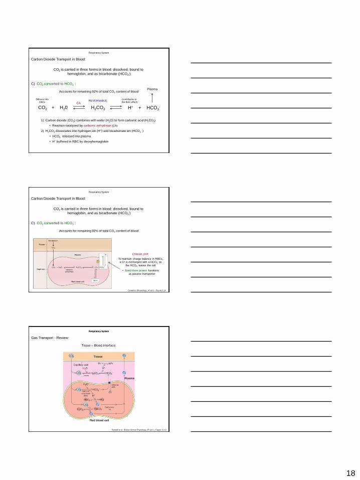
Respiratory System
Carbon Dioxide Transport in Blood:
CO2 is carried in three forms in blood: dissolved, bound to
hemoglobin, and as bicarbonate (HCO3-)
C) CO2 converted to HCO3- :
Accounts for remaining 92% of total CO2 content of blood
+ CO2 H20 H2CO3 H+ + HCO3-
CA
Plasma
1) Carbon dioxide (CO2) combines with water (H2O) to form carbonic acid (H2CO3)
• Reaction catalyzed by carbonic anhydrase (CA)
2) H2CO3 dissociates into hydrogen ion (H+) and bicarbonate ion (HCO3- )
• HCO3- released into plasma
Diffuses into
RBCs
• H+ buffered in RBC by deoxyhemaglobin
(contributes to
the Bohr effect) REVERSABLE
Respiratory System
Carbon Dioxide Transport in Blood:
CO2 is carried in three forms in blood: dissolved, bound to
hemoglobin, and as bicarbonate (HCO3-)
C) CO2 converted to HCO3- :
Accounts for remaining 92% of total CO2 content of blood
Costanzo (Physiology, 4th ed.) – Figure 5.24
To maintain charge balance in RBCs,
a Cl- is exchanged with a HCO3- as
the HCO3- leaves the cell
Chloride shift:
• Band three protein functions
as passive transporter
Respiratory System Respiratory System
Gas Transport - Review:
Tissue – Blood Interface:
Randall et al. (Eckert Animal Physiology, 5th ed.) – Figure 13.10
19
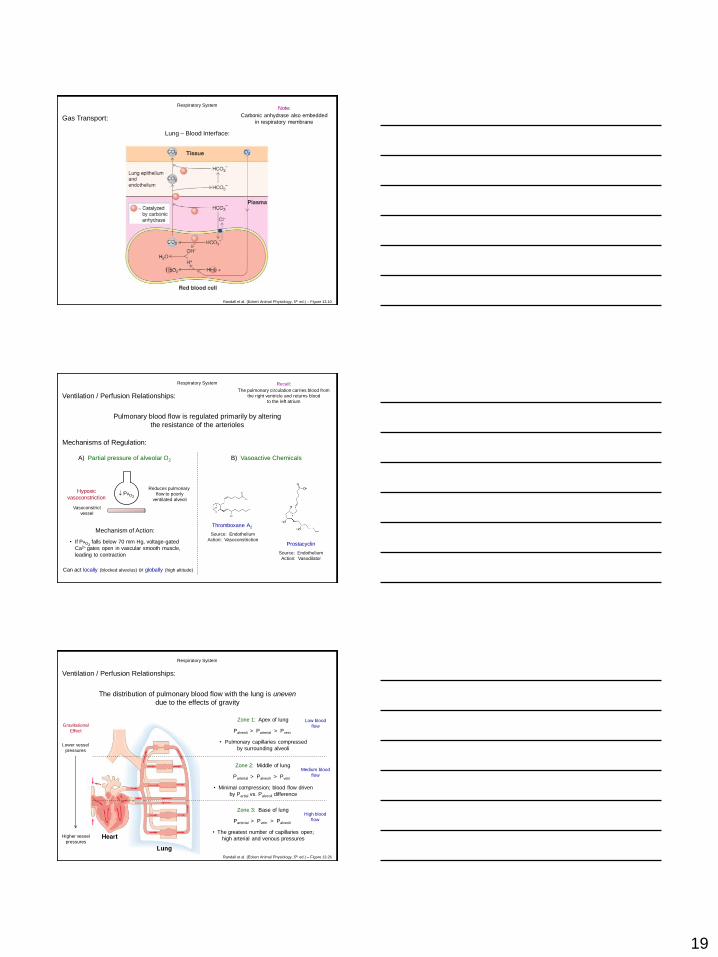
Respiratory System
Gas Transport:
Lung – Blood Interface:
Randall et al. (Eckert Animal Physiology, 5th ed.) – Figure 13.10
Note:
Carbonic anhydrase also embedded
in respiratory membrane
Respiratory System
Ventilation / Perfusion Relationships:
Pulmonary blood flow is regulated primarily by altering
the resistance of the arterioles
Recall:
The pulmonary circulation carries blood from
the right ventricle and returns blood
to the left atrium
Mechanisms of Regulation:
A) Partial pressure of alveolar O2
PAO2
Vasoconstrict
vessel
Hypoxic
vasoconstriction
Reduces pulmonary
flow to poorly
ventilated alveoli
Mechanism of Action:
• If PAO2 falls below 70 mm Hg, voltage-gated
Ca2+ gates open in vascular smooth muscle,
leading to contraction
Can act locally (blocked alveolus) or globally (high altitude)
B) Vasoactive Chemicals
Thromboxane A2
Source: Endothelium
Action: Vasoconstriction Prostacyclin
Source: Endothelium
Action: Vasodilator
Respiratory System
Ventilation / Perfusion Relationships:
The distribution of pulmonary blood flow with the lung is uneven
due to the effects of gravity
Zone 1: Apex of lung
Palveoli > Parterial > Pvein Gravitational
Effect
Lower vessel
pressures
Higher vessel
pressures
• Pulmonary capillaries compressed
by surrounding alveoli
Zone 2: Middle of lung
Parterial > Palveoli > Pvein
• Minimal compression; blood flow driven
by Partial vs. Palveoli difference
Low blood
flow
Medium blood
flow
Zone 3: Base of lung
Parterial > Pvein > Palveoli
• The greatest number of capillaries open;
high arterial and venous pressures
High blood
flow
Randall et al. (Eckert Animal Physiology, 5th ed.) – Figure 13.26
20
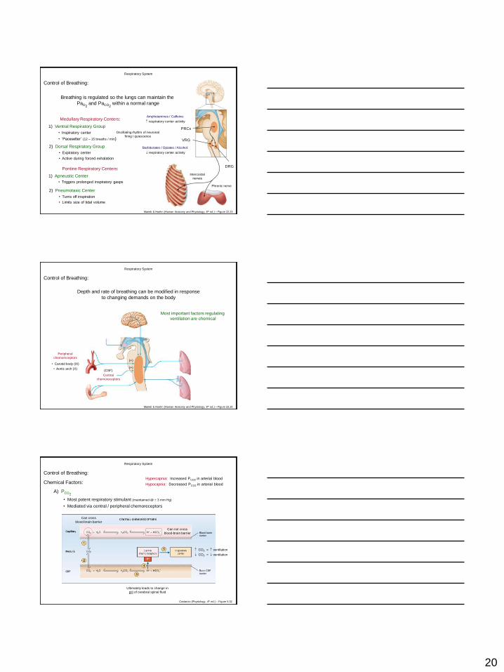
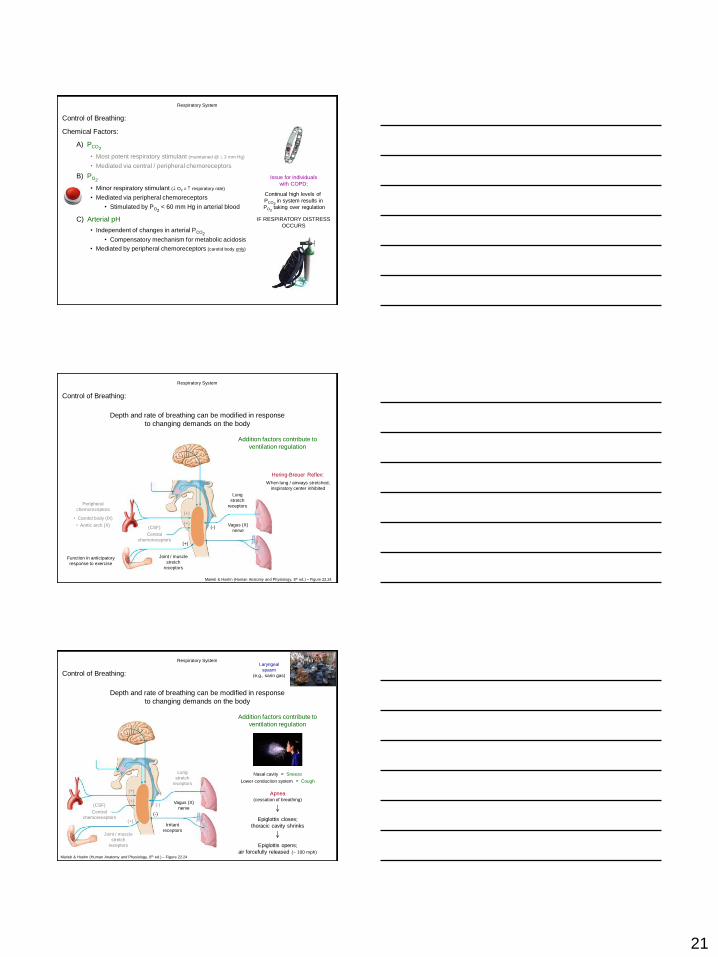
Respiratory System
Control of Breathing:
Breathing is regulated so the lungs can maintain the
PaO2 and PaCO2
within a normal range
1) Ventral Respiratory Group
2) Dorsal Respiratory Group
VRG
Medullary Respiratory Centers:
Oscillating rhythm of neuronal
firing / quiescence
DRG
Barbiturates / Opiates / Alcohol:
respiratory center activity
1) Apneustic Center
2) Pneumotaxic Center
Pontine Respiratory Centers:
Amphetamines / Caffeine:
respiratory center activity
PRCs
Phrenic nerve
Intercostal
nerves
Marieb & Hoehn (Human Anatomy and Physiology, 8th ed.) – Figure 22.23
• Inspiratory center
• ‘Pacesetter’ (12 – 15 breaths / min)
• Expiratory center
• Active during forced exhalation
• Triggers prolonged inspiratory gasps
• Turns off inspiration
• Limits size of tidal volume
Respiratory System
Control of Breathing:
Depth and rate of breathing can be modified in response
to changing demands on the body
Marieb & Hoehn (Human Anatomy and Physiology, 8th ed.) – Figure 22.24
Most important factors regulating
ventilation are chemical
• Carotid body (IX)
• Aortic arch (X)
Peripheral
chemoreceptors
Central
chemoreceptors
(CSF)
(+)
(+)
• Most potent respiratory stimulant (maintained @ 3 mm Hg)
Chemical Factors:
Respiratory System
Control of Breathing: Hypercapnia: Increased PCO2 in arterial blood
Hypocapnia: Decreased PCO2 in arterial blood
Can not cross
blood-brain barrier
Can cross
blood-brain barrier
Ultimately leads to change in
pH of cerebral spinal fluid
CO2 = ventilation
CO2 = ventilation
A) PCO2
Costanzo (Physiology, 4th ed.) – Figure 5.32
• Mediated via central / peripheral chemoreceptors
21
• Most potent respiratory stimulant (maintained @ 3 mm Hg)
• Mediated via central / peripheral chemoreceptors
A) PCO2
Chemical Factors:
Respiratory System
Control of Breathing:
B) PO2
• Minor respiratory stimulant ( O2 = respiratory rate)
• Mediated via peripheral chemoreceptors
• Stimulated by PO2 < 60 mm Hg in arterial blood
C) Arterial pH
• Independent of changes in arterial PCO2
• Compensatory mechanism for metabolic acidosis
Issue for individuals
with COPD:
Continual high levels of
PCO2 in system results in
PO2 taking over regulation
IF RESPIRATORY DISTRESS
OCCURS
• Mediated by peripheral chemoreceptors (carotid body only)
Respiratory System
Control of Breathing:
Depth and rate of breathing can be modified in response
to changing demands on the body
Marieb & Hoehn (Human Anatomy and Physiology, 8th ed.) – Figure 22.24
• Carotid body (IX)
• Aortic arch (X)
Peripheral
chemoreceptors
Central
chemoreceptors
(CSF)
Lung
stretch
receptors
Hering-Breuer Reflex:
When lung / airways stretched,
inspiratory center inhibited
(-)
(+)
Joint / muscle
stretch
receptors
Function in anticipatory
response to exercise
Addition factors contribute to
ventilation regulation
(+)
(+) Vagus (X)
nerve
Respiratory System
Control of Breathing:
Depth and rate of breathing can be modified in response
to changing demands on the body
Marieb & Hoehn (Human Anatomy and Physiology, 8th ed.) – Figure 22.24
Addition factors contribute to
ventilation regulation
Central
chemoreceptors
(CSF)
Lung
stretch
receptors
(-)
(+)
Joint / muscle
stretch
receptors
(+)
(+)
Apnea (cessation of breathing)
Epiglottis closes;
thoracic cavity shrinks
Nasal cavity = Sneeze
Lower conduction system = Cough
Epiglottis opens;
air forcefully released (~ 100 mph)
Irritant
receptors
(-)
Vagus (X)
nerve
Laryngeal
spasm
(e.g., sarin gas)
22
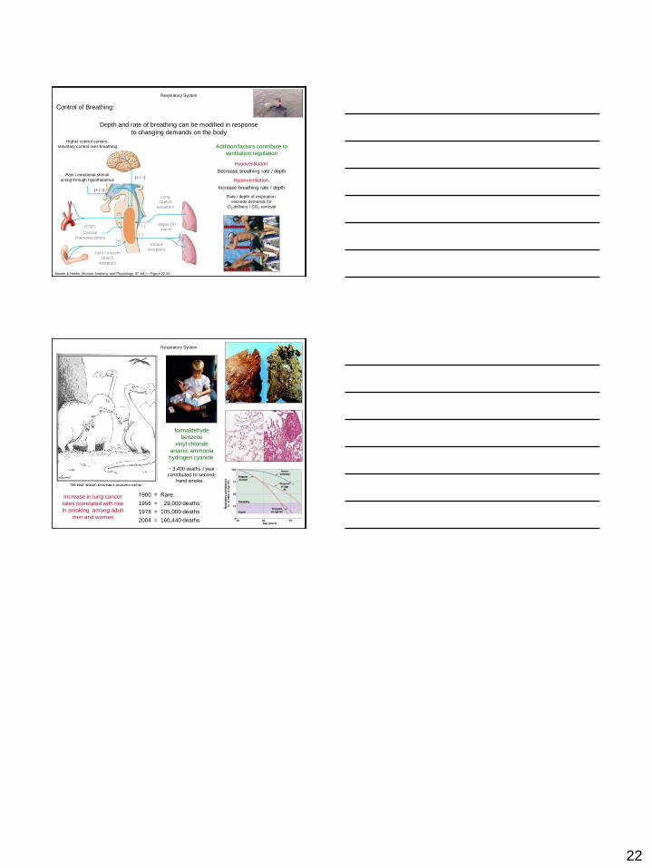
Respiratory System
Control of Breathing:
Depth and rate of breathing can be modified in response
to changing demands on the body
Marieb & Hoehn (Human Anatomy and Physiology, 8th ed.) – Figure 22.24
Addition factors contribute to
ventilation regulation
Central
chemoreceptors
(CSF)
Lung
stretch
receptors
(-)
(+)
Joint / muscle
stretch
receptors
(+)
(+)
Irritant
receptors
(-)
Vagus (X)
nerve
Pain / emotional stimuli
acting through hypothalamus
(+ / -)
Higher control centers;
Voluntary control over breathing
(+ / -)
Hypoventilation:
Decrease breathing rate / depth
Hyperventilation:
Increase breathing rate / depth
Rate / depth of respiration
exceeds demands for
O2 delivery / CO2 removal
1900 = Rare…
1956 = 29,000 deaths
1978 = 105,000 deaths
2004 = 160,440 deaths
Increase in lung cancer
rates correlated with rise
in smoking among adult
men and women
formaldehyde
benzene
vinyl chloride
arsenic ammonia
hydrogen cyanide
~ 3,400 deaths / year
contributed to second-
hand smoke…
Respiratory System


![Respiratory system roadmap.pptx [Repaired] - Loginanatomical-sciences.health.wits.ac.za/roadmaps/Respiratory system... · DIVISION OF THE RESPIRATORY SYSTEM CONDUCTING PORTION Nasal](https://img.pdfslide.net/doc/110x75/5a78c3d87f8b9ae6228c9db0/respiratory-system-repaired-loginanatomical-scienceshealthwitsaczaroadmapsrespiratory.jpg)


![Anatomy and Physiology Respiratory System [Tab 2] Respiratory System](https://img.pdfslide.net/doc/110x75/56649ebd5503460f94bc631f/anatomy-and-physiology-respiratory-system-tab-2-respiratory-system.jpg)


