Embed Size (px)
Citation preview
12 Arylstibonic acids that inhibit the DNA binding of five B-ZIP dimers
Vikas Rishi a, Won-Jun Oh a, Sarah L. Heyerdahl a, Jianfei Zhao a, Dominic Scudiero b,Robert H. Shoemaker c, Charles Vinson a,*
a Laboratory of Metabolism, National Cancer Institute, National Institutes of Health, Building 37, Room 3128, Bethesda, MD 20892, USAb Science Applications International Corporation (SAIC), Frederick, MD 21702, USAcDevelopmental Therapeutics Program, DCTD, National Cancer Institute, Frederick, MD 21702, USA
a r t i c l e i n f o
Article history:Received 3 November 2009Received in revised form 16 February 2010Accepted 16 February 2010Available online 20 February 2010
Keywords:Arylstibonic acidsDNA bindingB-ZIP domainLeucine zipperAntimonySmall-molecule inhibitors
a b s t r a c t
Previously, we identified an arylstibonic acid, NSC13778 that specifically binds to the basic region of theC/EBPa B-ZIP domain and disrupts DNA binding. We now examine a panel of 14 additional arylstibonicacid derivatives of NSC13778 for their ability to inhibit the DNA binding of five B-ZIP dimers (c-Fos|JunD,VBP, C/EBPa, C/EBPb, and CREB). They show various specificities at inhibiting the DNA binding of five B-ZIP domains. NSC13746 inhibits the DNA binding of C/EBPb and CREB at 100 nM and promiscuouslyinhibiting the DNA binding of all five proteins in the 1 lM range. Dialysis experiments indicate thatNSC 13746 binding to the B-ZIP domain is reversible. Thermal denaturation studies indicate thatNSC13746 binds the B-ZIP domain. Some compounds specifically inhibit DNA binding, with VBP and c-Fos|JunD being most easily disrupted. These compounds inhibit, with similar specificities to the pureB-ZIP domains, the DNA binding of nuclear extract to the AP1 DNA sequence but no inhibition is observedto SP1 containing oligonucleotide. Transient transfection assays indicate that NSC13746 can inhibit theTPA induced activation of two B-ZIP dependent reporters. These experiments suggest that arylstibonicacids are promising leads for inhibiting the DNA binding of a group of B-ZIP proteins in cells.
Published by Elsevier Inc.
1. Introduction
The B-ZIP domain dimerizes and binds to DNA in a sequencespecific manner to regulate gene transcription. These transcriptionfactors occur in approximately 55 genes in the human genome thathave been grouped into 13 families with similar dimerizationproperties (Vinson et al., 2002; Newman and Keating, 2003). Manyof these B-ZIP domain containing genes are immediate early genes(IEGs) that are induced in a variety of pathologies including cancer(Darnell, 2002). For example, c-Jun, a member of the AP1 family ofB-ZIP transcription factors, was the first nuclear transcription fac-tor described as an oncogene (Vogt et al., 1987; Vogt, 2001). How-ever, no inhibitors of the DNA binding of these proteins are inclinical trials, and only one compound has been described thatcan bind to the B-ZIP domain to inhibit DNA binding (Rishi et al.,2005).
Targeting the DNA binding of the B-ZIP motif is a formidabletask because of (a) the absence of any enzymatic pocket thatmay be targeted specifically and (b) the presence of a large andshallow interface (1000–3000 Å) that gets buried when B-ZIP pro-teins form homo- or heterodimers (Lo Conte et al., 1999; Jones andThornton, 1996; Hu et al., 2000). Potentially, binding of a B-ZIP pro-
tein to DNA can be disrupted in two ways: (1) targeting moleculesthat bind to the major or minor grooves of DNA causing changes inits topology and steric hindrances such that the transcription factorcannot bind to DNA (Olenyuk et al., 2004; Dervan, 2001); or (2) tar-geting the B-ZIP domain itself. The B-ZIP class of transcription fac-tors binds to the major groove of DNA as homo- and/orheterodimers via their basic DNA binding domain. The dimeriza-tion domains of the B-HLH-ZIP domains, which are structurallysimilar to the B-ZIP domain, have been successfully targeted. Asynthetic peptidomimetic that inhibits the protein–protein inter-action between Myc and Max heterodimer has been described(Berg et al., 2002). But due to poor cell permeability and high sus-ceptibility to degradation, these peptides are poor pharmacologicalagents.
The potential clinical value of inhibiting the DNA binding ofspecific B-ZIP protein families has been demonstrated using trans-genic mice (Oh et al., 2007; Gerdes et al., 2006). Previously, we de-signed potent dominant negatives for various B-ZIP and B-HLH-ZIPtranscription factor families that dimerize with their wild type pro-tein partners with high affinity and inhibit DNA binding (Krylovet al., 1995; Ahn et al., 1998; Moll et al., 2000; Rishi et al., 2004).These dominant negative proteins have been expressed in trans-genic mice and shown to inhibit and/or regress papillomas in skin.For example, expression in the mouse epidermis of A-C/EBP, adominant negative that inhibits the DNA binding of all C/EBP
1047-8477/$ - see front matter Published by Elsevier Inc.doi:10.1016/j.jsb.2010.02.013
* Corresponding author. Fax: +1 301 496 8419.E-mail address: [email protected] (C. Vinson).
Journal of Structural Biology 170 (2010) 216–225
Contents lists available at ScienceDirect
Journal of Structural Biology
journal homepage: www.elsevier .com/ locate/yjsbi
family members, prevents papilloma formation and can causeestablished papillomas to regress if expressed after papilloma for-mation (Oh et al., 2007). Expression in the mouse epidermis of A-Fos, a dominant negative that inhibits AP1 DNA binding, convertspapillomas into benign sebaceous adenomas that are not able toconvert into carcinomas (Gerdes et al., 2006). Additionally, inhibit-ing CREB by expressing A-CREB is able to prevent papilloma forma-tion (Rozenberg et al., 2009). Together, these results suggest thatinhibition of DNA-binding activity of B-ZIP transcription factors of-fers a promising molecular target to develop an effective anti-can-cer therapy.
In a previous study, we used a high-throughput fluorescenceanisotropy screen and three secondary screens to identify small-molecule inhibitors that disrupt the DNA binding of the B-ZIP do-mains (Rishi et al., 2005). From a diverse set of 1990 molecules,an arylstibonic acid, NSC13778, containing an antimony element,was identified that preferentially binds the C/EBPa basic regionand inhibits DNA binding. Here we report the effect of 14 arylsti-bonic acid derivatives of the lead compound NSC13778 on theDNA-binding activity of five B-ZIP dimers: c-Fos|JunD (AP1), VBP,C/EBPa, C/EBPb, and CREB. In the present study, two in vitro andan in vivo assay were used to characterize these compounds. Elec-trophoretic Mobility Shift Assay (EMSA) was used to screen 14derivatives of NSC13778 for their ability to inhibit B-ZIP–DNAbinding in a dose-dependent manner. The two water-soluble com-pounds that were positive by EMSA were further characterizedusing isothermal circular dichroism (CD) spectroscopy and CDthermal denaturation studies. NSC13746 was also evaluatedin vivo by a transient transfections luciferase reporter assay andshown to inhibit B-ZIP mediated transcriptional activity.
2. Materials and methods
2.1. Compounds
NSC13778 and 14 arylstibonic acid derivatives were obtainedfrom the Drug Synthesis and Chemistry Branch, DevelopmentalTherapeutics Program, National Cancer Institute, Bethesda, MD.
2.2. Protein expression and purification
Amino acid sequences of B-ZIP domain proteins used in thisstudy are given elsewhere: c-Fos (Olive et al., 1997), JunD (Rishiet al., 2005), VBP (Moll et al., 2000), C/EBPa and C/EBPb (Krylovet al., 1995), CREB and its dominant negative A-CREB (Ahn et al.,1998). All recombinant proteins were expressed in Escherichia coliBL21(LysE) strain and purified as described previously (Rishi et al.,2004). Final protein purification step involved HPLC using VydacC14 reverse phase column. A linear gradient from 0% to 100% ace-tonitrile containing 0.01% trifluoroacetic acid over 45 min with aflow rate of 1 ml/min was used to elute the proteins. All proteinshave a 13 amino acid N-terminal u10 epitope that was used for im-muno detection of proteins in Western blots.
2.3. Electrophoretic Mobility Shift Assay (EMSA)
28 mer oligonucleotides were purchased from Sigma–Genosysand were HPLC purified. Single strand oligonucleotide was end la-beled with [c32P]ATP using T4 phage polynucleotide kinase. La-beled oligo was purified using a G-50 column (GE Healthcare,UK) according to manufacturer instructions and annealed to com-plementary unlabeled oligo resulting in radiolabeled doublestranded DNA. Sequences of oligonucleotides used for EMSA exper-iments were: AP1: 50-GTCAGTCAGAATGACTCATATCGGTCAG-30;VBP: 50-GTCAGTCAGATTACGTAATATCGGTCAG-30; C/EBP: 50-
GTCAGTCAGATTGCGCAATATCGGTCAG-30; CREB: 50-GTCAGTCA-GATGACGTCATATCGGTCAG-30. Underlined nucleotides are thebinding sequences for B-ZIP proteins. Protein–DNA interaction inpresence and absence of a small-molecule was studied usingEMSA. Six HPLC purified proteins, four homodimers (VBP, C/EBPa,C/EBPb, CREB) and one heterodimer (c-Fos|JunD form AP1) wereused in band shift experiments. Before EMSA protein stock solu-tions (10 lM) in CD buffer were heated to 50 �C for 5 min andcooled to room temperature. The heterodimer between c-Fos andJunD domains was generated by heating equimolar concentrationsof c-Fos and JunD proteins at 65 �C in presence of 1 mM DTT for15 min and cooling the mixture to room temperature. For EMSA,samples were prepared by mixing 10 nM dimer protein with therequired concentration of small-molecule (0.1, 1.0 and 10 lM) in15 ll gel shift reaction buffer (25 mM Tris; pH 8.0, 50 mM KCl,0.5 mM EDTA, 2.5 mM DTT, 1% bovine serum albumin and 10%glycerol) and incubated for 5 min at 37 �C. 7 pM 32P-radiolabeleddouble stranded oligonucleotide was added and final volume wasmade up to 20 ll with gel shift reaction buffer and equilibratedat 37 �C for 15 min. Finally, the mixture was incubated for addi-tional 5 min at room temperature and DNA|protein complexeswere resolved on a 7.5% native PAGE using 25 mM Tris–borate run-ning buffer (pH 8.0) with 0.3 mM EDTA at 150 V potential (11 mAcurrent) for 90 min. Gels were dried and autoradiographed after18 h exposure using a Kodak MR X-ray film. The shifted bands rep-resent DNA|protein complexes.
2.4. Dialysis experiments
2 lM CREB B-ZIP dimer protein in absence and presence of100 lM NSC13746 was dialyzed against CD buffer for 24 h at4 �C using a Slide-a-Layer dialysis tubing (Pierce) with a molecularweight cut off of 8000. Dialyzed samples without heating and afterheating to 50 �C for 5 min were mixed with radioactive probe andran on 7.5% native PAGE as described above. Gel was dried and ex-posed to X-ray film for 5 h and developed subsequently.
2.5. Nuclear extracts
Nuclear extracts were made from mouse liver (Gorski et al.,1986) with somemodifications. Mouse liver (10–15 g) was homog-enized in 30 ml of homogenization buffer (HB) (10 mM Hepes (pH7.6), 25 mM KCl, 0.15 mM spermine, 0.5 mM spermidine, 1 mMEDTA, 10% glycerol, and 2 M sucrose) using a Teflon-glass homog-enizer. Fifteen strokes of homogenizer were enough to get 90% ofcells lysed. 50 ml of HB buffer was added to make the final volumeto 85 ml. 28 ml of homogenate was overlaid on 10 ml HB in threecentrifuge tubes and spun at 24,000 rpm for 30 min at �2 �C. Indi-vidual pellets containing nuclei from three tubes were pooled andre-suspended in 50 ml of 9:1 v/v HB and glycerol, divided intothree parts and layered on 10 ml of HB buffer in three tubes andcentrifugation step was repeated. Pellets were pooled again andsuspended in nuclei lysis buffer (10 mM Hepes (pH 7.9), 100 mMKCl, 3 mM MgCl2, 0.1 mM EDTA, 1 mM DTT, 0.1 mM PMSF, and10% glycerol). Nuclei lysis was enhanced by 10 strokes in a glassDounce homogenizer kept on ice and incubated for 40 min. Finalcentrifugation was performed for 30 min at 35,000 rpm and 4 �C.The pellet was discarded and supernatant was aliquoted and storedat �70 �C. Protein concentration of nuclear extract was measuredby Bradford method (Bio-Rad, CA). For EMSA 5 lg of nuclear ex-tracts were incubated with or without compounds (37 �C for15 min) in gel shift buffer that contained 10 mM Hepes (pH 8.0),6% glycerol, 80 mM KCl, 0.05 mM EDTA, 1 mM MgCl2 and 1 mMDTT. In addition, 1 lg of polydI–dC was added to each reactionmixture to inhibit non-specific binding. 70 pM of radiolabeledDNA was added, mixture was incubated at room temperature
V. Rishi et al. / Journal of Structural Biology 170 (2010) 216–225 217
and DNA|protein complexes were resolved using a 6% native PAGEunder the same condition as described for pure proteins. Gels weredried and autoradiographed. The following sequence was used forSP1 gel shifts: 50-GTCAGTCAGGGGGCGGGGCATCGGTCAG-30.Underlined nucleotides are the consensus binding site for SP1.
2.6. Circular dichroism spectroscopy
Water-soluble NSC13746 and NSC13782 were used in circulardichroism (CD) studies. Samples for CD were prepared in phos-phate buffer (12.5 mM phosphate buffer, pH 7.4, 150 mM KCl,0.25 mM EDTA, 1 mM DTT). 2 lM dimer of each protein with re-quired concentrations of compounds were incubated for 2 h at37 �C. CD spectra were recorded at 6 �C on JASCO J-720 spectropo-larimeter in 5.0 mm quartz cuvette using a scanning rate of100 nm/min at the spectral bandwidth of 2.0 nm. Each spectrumwas an average of 10 individual spectra. Thermal denaturationstudies of the B-ZIP domains in the presence and absence of com-pounds were carried out by heating the samples from 6 to 85/90 �C with a heating rate of 1 �C/min and ellipticity (h) changeswere recorded at 222 nm. The raw CD data were normalized tomean residue ellipticity (deg cm2/dmol) using the followingrelation
½h� ¼ h=10Cl ð1Þwhere h is the observed ellipticity in millidegrees, C is the mean res-idue molar concentration of the protein, and l is the pathlength incentimeters.
Each thermal denaturation curve was analyzed as describedearlier (Krylov et al., 1994). All data points ([h]222, T) were fittedto the following equation
½h�222ðTÞ ¼ ðN � DÞ � ð1� T=INTRÞ�ð1þ ð1� SQRTð8 � ExpðDHm=R
�ð1=ðTm þ 273:3Þ � 1=ðTþ 273:3ÞÞÞ þ 1ÞÞ=ð4 � ExpðDHm=R
�ð1=ðTm þ 273:3Þ� 1=ðT þ 273ÞÞÞÞÞ þ D ð2Þ
where T is temperature in �C, N and D are the values of h222 ofmonomer and dimer, respectively, at temperature 0 �C, INTR isthe temperature at which linear temperature-dependencies of di-mer and monomer intercepts, Tm is the midpoint of thermal dena-turation, DHm is the enthalpy change at Tm and R is the Gasconstant. Values of Tm, DHm with predetermined DCp (heat capacitychange on dimer formation) value of �1.2 kcal mol�1 dimer�1 K�1
(Moll et al., 2000) were used to obtain DGDi (Gibbs free energychange on dimer formation) according to the Gibbs–Helmholtzequation.
Binding constant for B-ZIP protein–small-molecule interactionat Tm is calculated according to Waldron and Murphy (2003) usingthe following equation
KaðTmÞ ¼ Expðð�DHm=RÞ � ð1=Tm � 1=ToÞþ ðDCp=RÞ � ðlnðTm=ToÞ þ ðTo=TmÞ � 1ÞÞ � 1=½L�Tm ð3Þ
where To and Tm are the midpoints of thermal denaturations of B-ZIP protein in absence and presence of small-molecule, respectively.DHm and DCp are the protein’s unfolding energetic parameters inabsence of a small-molecule. R is the Gas constant and L is the freesmall-molecule concentration at Tm.
2.7. Transient transfections
Thirty thousand HepG2 cells were plated in each well in 24 wellplates. Cells were co-transfected with Luc pGL3 basic vector(0.4 lg/well) with multiple binding sites for the AP1 and CREB B-
ZIP proteins (AP1(7�), CRE(3�), and IL-2(5�) and b-galactosidaseexpressing plasmids (0.1 lg/well) using lipofectamine reagentaccording to manufacturer’s instructions (Invitrogen, USA). Cellswere allowed to attach (day 0). After 17 h, NSC13746 that was dis-solved in water was added to the wells at the final concentration of10 lM (day 2). Cells were incubated for 24 h in 37 �C incubatorwith 5% CO2 flow (day 3). The next day, half of the cells with orwithout compound, were induced by adding TPA (50 ng/well).Cells were further incubated for 4 h at 37 �C, 5% CO2 environment.Finally, cells from each well were lysed in 100 ll of lysis buffer andanalyzed for luciferase and b-galactosidase activities (Galacto-Light Plus™ System, AB Applied Biosystem, MA).
3. Results
3.1. Arylstibonic acid compounds
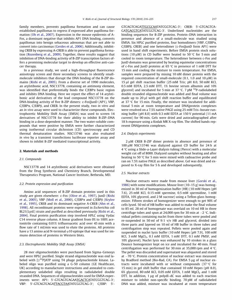
14 Arylstibonic acid derivatives of NSC13778 with molecularweights ranging from 293 to 363 Da were examined for their abil-ity to prevent the DNA binding of five B-ZIP dimers (AP1, VBP, C/EBPa, C/EBPb, and CREB). The calculated log P values (log of the ra-tio of compound’s solubility in organic solvent to water) identifiedfour compounds; NSC13746, NSC13748, NSC13776, and NSC13782that could be dissolved in water and the 11 remaining compoundswere dissolved in 100% DMSO. Details of chemical and physicalproperties of the compounds can be found at http://129.43.27.140/ncidb2/. The chemical structures of the 12 active compoundsand three inactive compounds are presented in Fig. 1.
3.2. Interactions of 15 arylstibonic acids with five B-ZIP domainsassayed by EMSA
Fig. 2 presents EMSA used to evaluate which of the 15 antimonycompounds at three concentrations (0.1 lM, 1 lM and 10 lM)could inhibit the formation of five DNA|B-ZIP complexes (AP1,VBP, C/EBPa, C/EBPb and CREB). All five B-ZIP proteins used herebind with KD of pM–nM (KD, the dissociation constant is the B-ZIP protein concentration at which bound and free probe are ofequal intensities) (Moll et al., 2002). Each sample contained10 nM dimer of pure protein mixed with 7 pM of radioactiveDNA in the absence and presence of compound. The absence or de-crease in the intensity of the DNA|B-ZIP complex indicates that thecompound is preventing formation of the DNA|B-ZIP complex andtherefore is active.
At 0.1 lM, most arylstibonic acid derivatives were inactive(Fig. 2A). However, as demonstrated in our previous study,NSC13778 inhibited the DNA-binding activity of C/EBPa (Rishiet al., 2005) while NSC13746, a sulfonic acid derivative ofNSC13778, disrupted the DNA binding of C/EBPb and CREB.Increasing the concentration of the compounds to 1 lM generateda complex pattern of DNA-binding inhibition (Fig. 2B). Two com-pounds, NSC13742 and NSC13782, showed the highest specificityas they completely disrupted the DNA binding of the AP1 B-ZIP tran-scription factor only. Four compounds (NSC13743, NSC13759,NSC13760, and NSC13793) displayed similar binding patterns beingmost active against AP1 and VBP, only weakly active against C/EBPa,andhadnoeffect on theDNAbindingofC/EBPbandCREB. CompoundNSC13765 was active for AP1 and C/EBPb only. Consistent with pre-vious results, NSC13778 disrupted the DNA|AP1 and DNA|C/EBPbcomplex at 1 lM (Rishi et al., 2005). Only three compounds wereinactive against all B-ZIP proteins (NSC13748, NSC13776,NSC13794), while two compounds disrupted the DNA binding ofall the B-ZIP transcription factors (NSC13746 and NSC13755).
Raising compound concentrations to 10 lM produced moreinhibition of DNA binding, again with a complex pattern. Eight
218 V. Rishi et al. / Journal of Structural Biology 170 (2010) 216–225
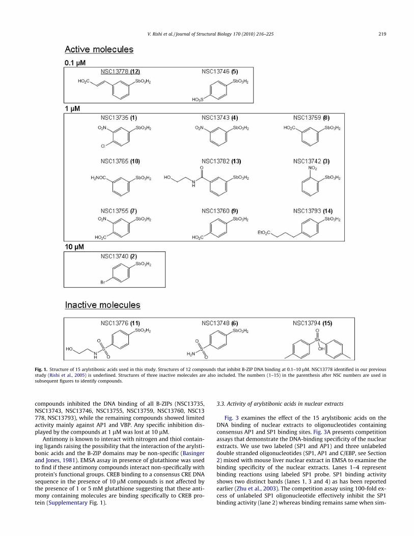
compounds inhibited the DNA binding of all B-ZIPs (NSC13735,NSC13743, NSC13746, NSC13755, NSC13759, NSC13760, NSC13778, NSC13793), while the remaining compounds showed limitedactivity mainly against AP1 and VBP. Any specific inhibition dis-played by the compounds at 1 lM was lost at 10 lM.
Antimony is known to interact with nitrogen and thiol contain-ing ligands raising the possibility that the interaction of the arylsti-bonic acids and the B-ZIP domains may be non-specific (Basingerand Jones, 1981). EMSA assay in presence of glutathione was usedto find if these antimony compounds interact non-specifically withprotein’s functional groups. CREB binding to a consensus CRE DNAsequence in the presence of 10 lM compounds is not affected bythe presence of 1 or 5 mM glutathione suggesting that these anti-mony containing molecules are binding specifically to CREB pro-tein (Supplementary Fig. 1).
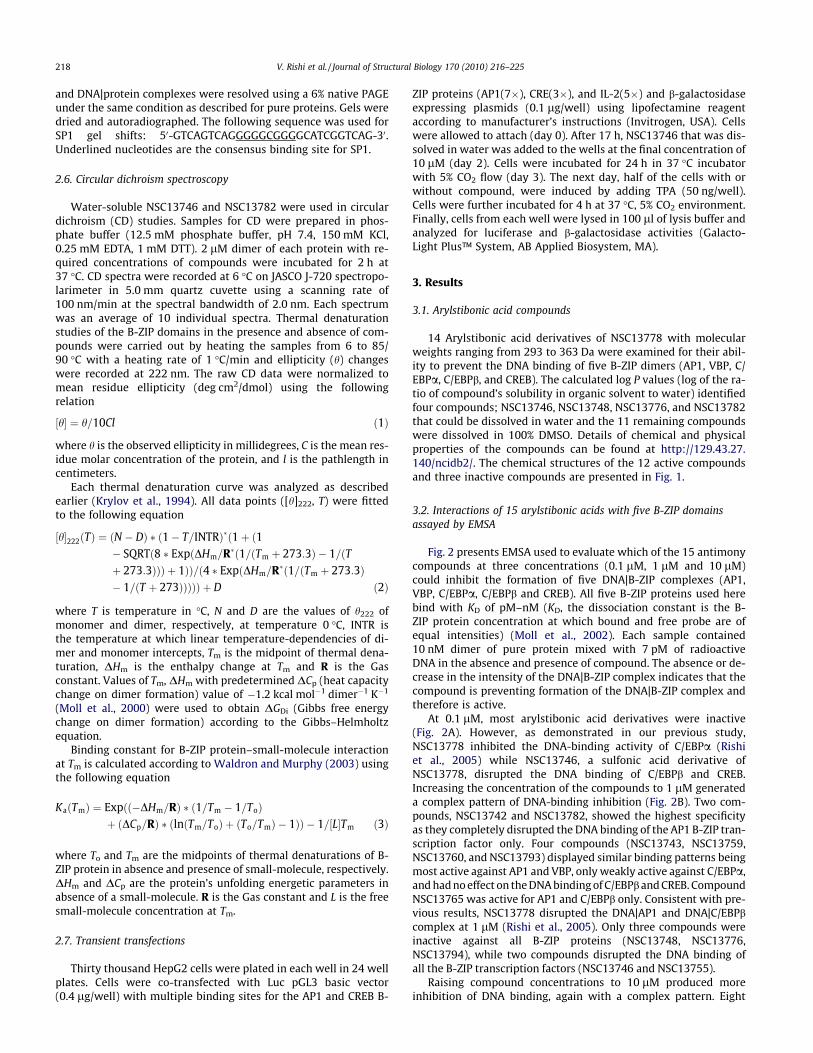
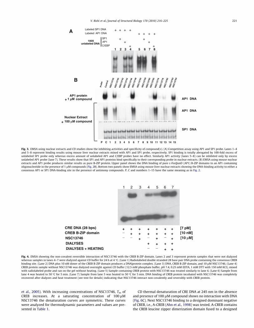
3.3. Activity of arylstibonic acids in nuclear extracts
Fig. 3 examines the effect of the 15 arylstibonic acids on theDNA binding of nuclear extracts to oligonucleotides containingconsensus AP1 and SP1 binding sites. Fig. 3A presents competitionassays that demonstrate the DNA-binding specificity of the nuclearextracts. We use two labeled (SP1 and AP1) and three unlabeleddouble stranded oligonucleotides (SP1, AP1 and C/EBP, see Section2) mixed with mouse liver nuclear extract in EMSA to examine thebinding specificity of the nuclear extracts. Lanes 1–4 representbinding reactions using labeled SP1 probe. SP1 binding activityshows two distinct bands (lanes 1, 3 and 4) as has been reportedearlier (Zhu et al., 2003). The competition assay using 100-fold ex-cess of unlabeled SP1 oligonucleotide effectively inhibit the SP1binding activity (lane 2) whereas binding remains same when sim-
Fig. 1. Structure of 15 arylstibonic acids used in this study. Structures of 12 compounds that inhibit B-ZIP DNA binding at 0.1–10 lM. NSC13778 identified in our previousstudy (Rishi et al., 2005) is underlined. Structures of three inactive molecules are also included. The numbers (1–15) in the parenthesis after NSC numbers are used insubsequent figures to identify compounds.
V. Rishi et al. / Journal of Structural Biology 170 (2010) 216–225 219
ilar amount of unlabeled AP1 or C/EBP oligonucleotides were used(lane 3 and 4) suggesting the bound activity is specific for SP1.Lanes 5–8 show nuclear extract binding activity when labeledAP1 probe was used. 100-fold excess of only unlabeled AP1 oligo-nucleotide effectively competes out the binding (lane 7). In con-trast, 100-fold excess of unlabeled SP1 or C/EBP oligonucleotides(lanes 6 and 8) do not affect the binding.
Fig. 3B initially presents EMSA binding patterns generated byincubating 1 lM compound with AP1 pure B-ZIP domains (c-Fos|-JunD) (Fig. 2B). This pattern is reproduced when 100 lM com-pound is incubated with nuclear extracts. The similarity incompound activity suggests that these compounds retain theirspecificity of action in the complex nuclear extract environment.In contrast, the arylstibonic acids did not affect the ability of nucle-ar extracts to bind to an oligonucleotide containing a consensusSP1 binding site. SP1 belongs to the zinc finger family of transcrip-tion factors and does not share structural homology with B-ZIPtranscription factors.
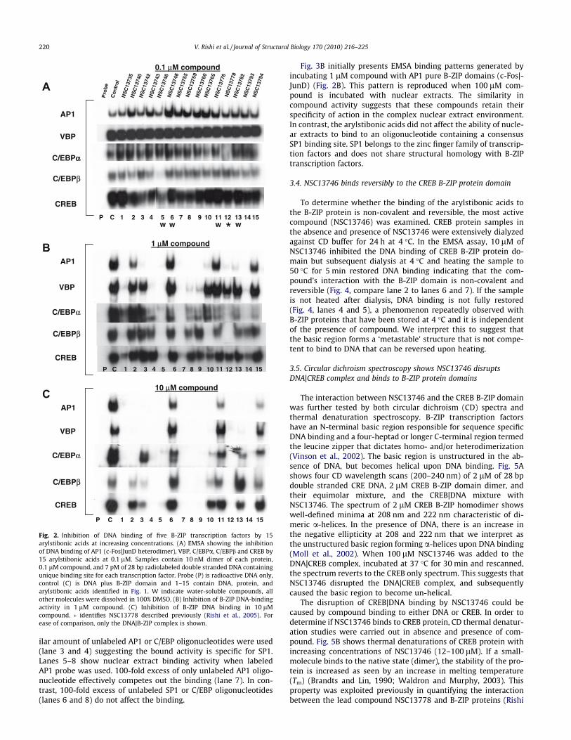
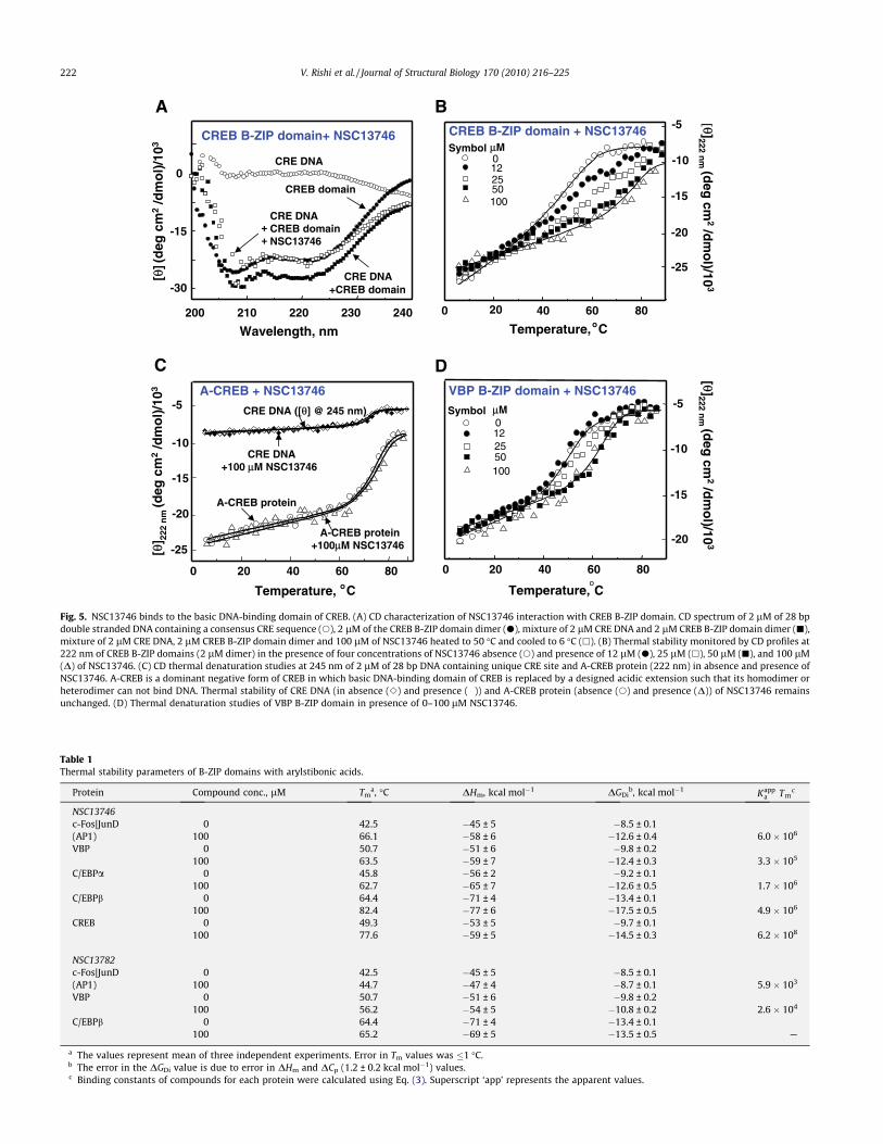
3.4. NSC13746 binds reversibly to the CREB B-ZIP protein domain
To determine whether the binding of the arylstibonic acids tothe B-ZIP protein is non-covalent and reversible, the most activecompound (NSC13746) was examined. CREB protein samples inthe absence and presence of NSC13746 were extensively dialyzedagainst CD buffer for 24 h at 4 �C. In the EMSA assay, 10 lM ofNSC13746 inhibited the DNA binding of CREB B-ZIP protein do-main but subsequent dialysis at 4 �C and heating the sample to50 �C for 5 min restored DNA binding indicating that the com-pound’s interaction with the B-ZIP domain is non-covalent andreversible (Fig. 4, compare lane 2 to lanes 6 and 7). If the sampleis not heated after dialysis, DNA binding is not fully restored(Fig. 4, lanes 4 and 5), a phenomenon repeatedly observed withB-ZIP proteins that have been stored at 4 �C and it is independentof the presence of compound. We interpret this to suggest thatthe basic region forms a ‘metastable’ structure that is not compe-tent to bind to DNA that can be reversed upon heating.
3.5. Circular dichroism spectroscopy shows NSC13746 disruptsDNA|CREB complex and binds to B-ZIP protein domains
The interaction between NSC13746 and the CREB B-ZIP domainwas further tested by both circular dichroism (CD) spectra andthermal denaturation spectroscopy. B-ZIP transcription factorshave an N-terminal basic region responsible for sequence specificDNA binding and a four-heptad or longer C-terminal region termedthe leucine zipper that dictates homo- and/or heterodimerization(Vinson et al., 2002). The basic region is unstructured in the ab-sence of DNA, but becomes helical upon DNA binding. Fig. 5Ashows four CD wavelength scans (200–240 nm) of 2 lM of 28 bpdouble stranded CRE DNA, 2 lM CREB B-ZIP domain dimer, andtheir equimolar mixture, and the CREB|DNA mixture withNSC13746. The spectrum of 2 lM CREB B-ZIP homodimer showswell-defined minima at 208 nm and 222 nm characteristic of di-meric a-helices. In the presence of DNA, there is an increase inthe negative ellipticity at 208 and 222 nm that we interpret asthe unstructured basic region forming a-helices upon DNA binding(Moll et al., 2002). When 100 lM NSC13746 was added to theDNA|CREB complex, incubated at 37 �C for 30 min and rescanned,the spectrum reverts to the CREB only spectrum. This suggests thatNSC13746 disrupted the DNA|CREB complex, and subsequentlycaused the basic region to become un-helical.
The disruption of CREB|DNA binding by NSC13746 could becaused by compound binding to either DNA or CREB. In order todetermine if NSC13746 binds to CREB protein, CD thermal denatur-ation studies were carried out in absence and presence of com-pound. Fig. 5B shows thermal denaturations of CREB protein withincreasing concentrations of NSC13746 (12–100 lM). If a small-molecule binds to the native state (dimer), the stability of the pro-tein is increased as seen by an increase in melting temperature(Tm) (Brandts and Lin, 1990; Waldron and Murphy, 2003). Thisproperty was exploited previously in quantifying the interactionbetween the lead compound NSC13778 and B-ZIP proteins (Rishi
P C 1 2 3 4 5 6 7 8 9 10 11 12 13 14 15
VBP
C/EBPαα
C/EBPβ
CREB
AP1
VBP
C/EBPα
C/EBPβ
CREB
AP1
VBP
C/EBPα
C/EBPβ
CREB
AP1
10 μM compound
1 μM compound
0.1 μM compound
P C 1 2 3 4 5 6 7 8 9 10 11 12 13 14 15
P C 1 2 3 4 5 6 7 8 9 10 11 12 13 14 15w w ww
A
B
C
*
NS
C13
735
NS
C13
740
NS
C13
742
NS
C13
743
NS
C13
746
NS
C13
748
NS
C13
755
NS
C13
759
NS
C13
760
NS
C13
765
NS
C13
776
NS
C13
778
NS
C13
782
NS
C13
793
NS
C13
794
Con
trol
Pro
be
Fig. 2. Inhibition of DNA binding of five B-ZIP transcription factors by 15arylstibonic acids at increasing concentrations. (A) EMSA showing the inhibitionof DNA binding of AP1 (c-Fos|JunD heterodimer), VBP, C/EBPa, C/EBPb and CREB by15 arylstibonic acids at 0.1 lM. Samples contain 10 nM dimer of each protein,0.1 lM compound, and 7 pM of 28 bp radiolabeled double stranded DNA containingunique binding site for each transcription factor. Probe (P) is radioactive DNA only,control (C) is DNA plus B-ZIP domain and 1–15 contain DNA, protein, andarylstibonic acids identified in Fig. 1. W indicate water-soluble compounds, allother molecules were dissolved in 100% DMSO. (B) Inhibition of B-ZIP DNA-bindingactivity in 1 lM compound. (C) Inhibition of B-ZIP DNA binding in 10 lMcompound. � identifies NSC13778 described previously (Rishi et al., 2005). Forease of comparison, only the DNA|B-ZIP complex is shown.
220 V. Rishi et al. / Journal of Structural Biology 170 (2010) 216–225
et al., 2005). With increasing concentrations of NSC13746, Tm ofCREB increases. At a saturating concentration of 100 lMNSC13746 the denaturation curves are symmetric. These curveswere analyzed for thermodynamic parameters and values are pre-sented in Table 1.
CD thermal denaturation of CRE DNA at 245 nm in the absenceand presence of 100 lM compound shows no interaction with DNA(Fig. 6C). Next NSC13746 binding to a designed dominant negativeof CREB, i.e., A-CREB (Ahn et al., 1998) was tested. A-CREB containsthe CREB leucine zipper dimerization domain fused to a designed
A
AP1 protein1 μμM AP1 DNA
SP1 DNA
compound+
Nuclear Extract100 μM compound+
AP1 DNAN
SC
1373
5N
SC
1374
0N
SC
1374
2N
SC
1374
3N
SC
1374
6N
SC
1374
8N
SC
1375
5N
SC
1375
9N
SC
1376
0N
SC
1376
5N
SC
1377
6N
SC
1377
8N
SC
1378
2N
SC
1379
3N
SC
1379
4
Con
trol
Pro
beB
Labeled SP1 DNA
Labeled AP1 DNA
SP1AP1C/EBP
100Xunlabeled DNA
+ + + ++ + + +
+ +++
+ +
P C 1 2 3 4 5 6 7 8 9 10 11 12 13 14 15
1 2 3 4 5 6 7 8
Fig. 3. EMSA using nuclear extracts and CD studies show the inhibiting activities and specificity of compound(s). (A) Competition assay using AP1 and SP1 probe. Lanes 1–4and 5–8 represent binding results using mouse liver nuclear extracts mixed with AP1 and SP1 probe, respectively. SP1 binding is totally abrogated by 100-fold excess ofunlabeled SP1 probe only whereas excess amount of unlabeled AP1 and C/EBP probes have no effect. Similarly AP1 activity (lanes 5–8) can be inhibited only by excessunlabeled AP1 probe (lane 7). These results show that SP1 and AP1 proteins bind specifically to their corresponding probe in nuclear extracts. (B) EMSA using mouse nuclearextracts and AP1 probe produces similar results as pure B-ZIP protein. Upper panel shows the DNA binding of pure c-Fos|JunD (AP1) B-ZIP domains to an AP1 containingoligonucleotide in the presence of 1 lM compounds (Fig. 2B). Bottom two panels show EMSA using mouse liver nuclear extracts showing the DNA-binding activity to either aconsensus AP1 or SP1 DNA-binding site in the presence of antimony compounds. P, C and numbers 1–15 have the same meaning as in Fig. 2.
CRE DNA (28 bps)CREB B-ZIP domainNSC13746
++ +++++
+__ _
_
[10 μM][10 nM][7 pM]+
+
DIALYSESDIALYSES + HEATING
+_
1 2 3 4 5 6 7
++_
+
++
++ + +
_
+ +
Fig. 4. EMSA showing the non-covalent reversible interaction of NSC13746 with the CREB B-ZIP domain. Lanes 2 and 3 represent protein samples that were not dialyzedwhereas samples in lanes 4–7 were dialyzed against CD buffer for 24 h at 4 �C. (Lane 1) Radiolabeled double stranded 28 base pair DNA probe containing the consensus CREBbinding site. (Lane 2) DNA plus 10 nM dimer of the CREB B-ZIP domain produces a DNA|protein complex. (Lane 3) DNA, CREB B-ZIP domain, and 10 lM NSC13746. (Lane 4)CREB protein sample without NSC13746 was dialyzed overnight against CD buffer (12.5 mM phosphate buffer, pH 7.4, 0.25 mM EDTA, 1 mM DTT with 150 mM KCl), mixedwith radiolabeled probe and ran on the gel without heating. (Lane 5) Sample containing CREB protein with NSC13746 was treated similarly to lane 4. (Lane 6) Sample fromlane 4 was heated to 50 �C for 5 min. (Lane 7) Sample from lane 5 was heated to 50 �C for 5 min. DNA binding of CREB protein incubated with NSC13746 was completelyrecovered after dialyzes and heat treatment (see text for details) indicating that NSC13746 interact non-covalently and reversibly with CREB protein.
V. Rishi et al. / Journal of Structural Biology 170 (2010) 216–225 221
A
Wavelength, nm Temperature, oC
B
C
Temperature, oC
D
Temperature,oC
0 20 40 60 80
-15
-5VBP B-ZIP domain + NSC13746
μMSymbol012
50100
25
[θ ]222 nm
(deg
cm2
/dm
ol)/10
3
[ θ] 22
2 n
m (d
eg c
m2
/dm
ol)
/103
[ θ]222 nm
(deg
cm2
/dm
ol)/10
3
[θ](
deg
cm
2/d
mo
l)/1
03
200 210 220 230 240
-30
-15
0CRE DNA
CRE DNA+CREB domain
CREB domain
CRE DNACREB domainNSC13746
++
CREB B-ZIP domain+ NSC13746
0 20 40 60 80
-25
-20
-15
-10
CREB B-ZIP domain + NSC13746μMSymbol012
50100
25
-5
0 20 40 60 80
-25
-20
-10
-5
A-CREB protein
A-CREB protein+100μM NSC13746
CRE DNA ([θ] @ 245 nm)
CRE DNA +100 μM NSC13746
A-CREB + NSC13746
-15
-20
-10
Fig. 5. NSC13746 binds to the basic DNA-binding domain of CREB. (A) CD characterization of NSC13746 interaction with CREB B-ZIP domain. CD spectrum of 2 lM of 28 bpdouble stranded DNA containing a consensus CRE sequence (s), 2 lM of the CREB B-ZIP domain dimer (d), mixture of 2 lM CRE DNA and 2 lM CREB B-ZIP domain dimer (j),mixture of 2 lM CRE DNA, 2 lM CREB B-ZIP domain dimer and 100 lM of NSC13746 heated to 50 �C and cooled to 6 �C (h). (B) Thermal stability monitored by CD profiles at222 nm of CREB B-ZIP domains (2 lM dimer) in the presence of four concentrations of NSC13746 absence (s) and presence of 12 lM (d), 25 lM (h), 50 lM (j), and 100 lM(D) of NSC13746. (C) CD thermal denaturation studies at 245 nm of 2 lM of 28 bp DNA containing unique CRE site and A-CREB protein (222 nm) in absence and presence ofNSC13746. A-CREB is a dominant negative form of CREB in which basic DNA-binding domain of CREB is replaced by a designed acidic extension such that its homodimer orheterodimer can not bind DNA. Thermal stability of CRE DNA (in absence (e) and presence (�)) and A-CREB protein (absence (s) and presence (D)) of NSC13746 remainsunchanged. (D) Thermal denaturation studies of VBP B-ZIP domain in presence of 0–100 lM NSC13746.
Table 1Thermal stability parameters of B-ZIP domains with arylstibonic acids.
Protein Compound conc., lM Tma, �C DHm, kcal mol�1 DGDi
b, kcal mol�1 Kappa Tm
c
NSC13746c-Fos|JunD 0 42.5 �45 ± 5 �8.5 ± 0.1(AP1) 100 66.1 �58 ± 6 �12.6 ± 0.4 6.0 � 106
VBP 0 50.7 �51 ± 6 �9.8 ± 0.2100 63.5 �59 ± 7 �12.4 ± 0.3 3.3 � 105
C/EBPa 0 45.8 �56 ± 2 �9.2 ± 0.1100 62.7 �65 ± 7 �12.6 ± 0.5 1.7 � 106
C/EBPb 0 64.4 �71 ± 4 �13.4 ± 0.1100 82.4 �77 ± 6 �17.5 ± 0.5 4.9 � 106
CREB 0 49.3 �53 ± 5 �9.7 ± 0.1100 77.6 �59 ± 5 �14.5 ± 0.3 6.2 � 108
NSC13782c-Fos|JunD 0 42.5 �45 ± 5 �8.5 ± 0.1(AP1) 100 44.7 �47 ± 4 �8.7 ± 0.1 5.9 � 103
VBP 0 50.7 �51 ± 6 �9.8 ± 0.2100 56.2 �54 ± 5 �10.8 ± 0.2 2.6 � 104
C/EBPb 0 64.4 �71 ± 4 �13.4 ± 0.1100 65.2 �69 ± 5 �13.5 ± 0.5 —
a The values represent mean of three independent experiments. Error in Tm values was �1 �C.b The error in the DGDi value is due to error in DHm and DCp (1.2 ± 0.2 kcal mol�1) values.c Binding constants of compounds for each protein were calculated using Eq. (3). Superscript ‘app’ represents the apparent values.
222 V. Rishi et al. / Journal of Structural Biology 170 (2010) 216–225
acidic extension that replaces the DNA-binding region. The Tm ofthe A-CREB protein does not change in presence of 100 lMNSC13746.
The ability of NSC13746 to stabilize the VBP B-ZIP protein do-main was also evaluated by CD thermal denaturation experiments(Fig. 5D). Tm of VBP B-ZIP protein domain increases in presence ofNSC13746 compound suggesting that the compound is binding tothe VBP protein dimer.
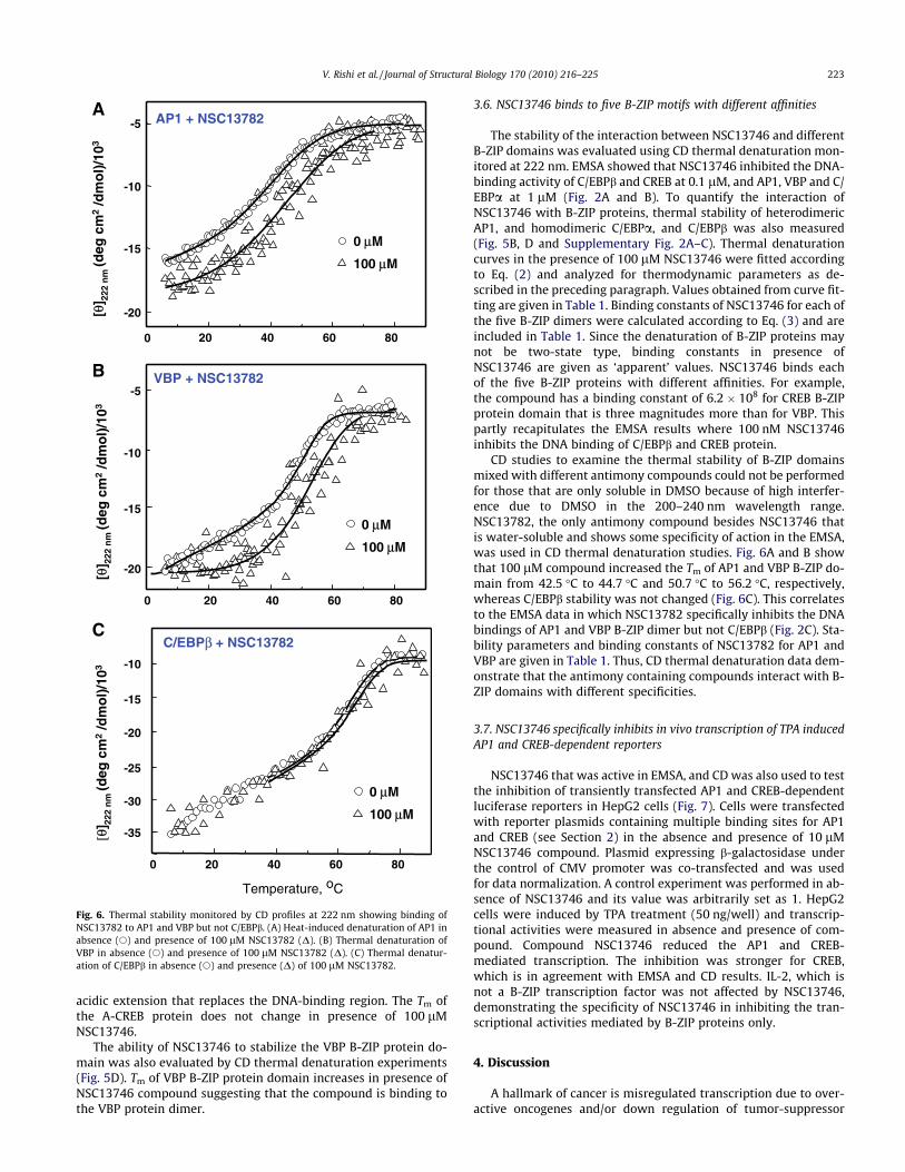
3.6. NSC13746 binds to five B-ZIP motifs with different affinities
The stability of the interaction between NSC13746 and differentB-ZIP domains was evaluated using CD thermal denaturation mon-itored at 222 nm. EMSA showed that NSC13746 inhibited the DNA-binding activity of C/EBPb and CREB at 0.1 lM, and AP1, VBP and C/EBPa at 1 lM (Fig. 2A and B). To quantify the interaction ofNSC13746 with B-ZIP proteins, thermal stability of heterodimericAP1, and homodimeric C/EBPa, and C/EBPb was also measured(Fig. 5B, D and Supplementary Fig. 2A–C). Thermal denaturationcurves in the presence of 100 lM NSC13746 were fitted accordingto Eq. (2) and analyzed for thermodynamic parameters as de-scribed in the preceding paragraph. Values obtained from curve fit-ting are given in Table 1. Binding constants of NSC13746 for each ofthe five B-ZIP dimers were calculated according to Eq. (3) and areincluded in Table 1. Since the denaturation of B-ZIP proteins maynot be two-state type, binding constants in presence ofNSC13746 are given as ‘apparent’ values. NSC13746 binds eachof the five B-ZIP proteins with different affinities. For example,the compound has a binding constant of 6.2 � 108 for CREB B-ZIPprotein domain that is three magnitudes more than for VBP. Thispartly recapitulates the EMSA results where 100 nM NSC13746inhibits the DNA binding of C/EBPb and CREB protein.
CD studies to examine the thermal stability of B-ZIP domainsmixed with different antimony compounds could not be performedfor those that are only soluble in DMSO because of high interfer-ence due to DMSO in the 200–240 nm wavelength range.NSC13782, the only antimony compound besides NSC13746 thatis water-soluble and shows some specificity of action in the EMSA,was used in CD thermal denaturation studies. Fig. 6A and B showthat 100 lM compound increased the Tm of AP1 and VBP B-ZIP do-main from 42.5 �C to 44.7 �C and 50.7 �C to 56.2 �C, respectively,whereas C/EBPb stability was not changed (Fig. 6C). This correlatesto the EMSA data in which NSC13782 specifically inhibits the DNAbindings of AP1 and VBP B-ZIP dimer but not C/EBPb (Fig. 2C). Sta-bility parameters and binding constants of NSC13782 for AP1 andVBP are given in Table 1. Thus, CD thermal denaturation data dem-onstrate that the antimony containing compounds interact with B-ZIP domains with different specificities.
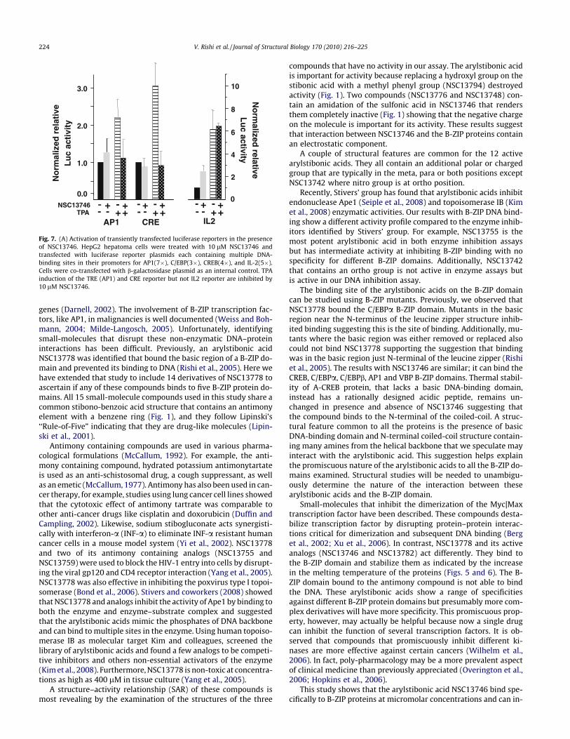
3.7. NSC13746 specifically inhibits in vivo transcription of TPA inducedAP1 and CREB-dependent reporters
NSC13746 that was active in EMSA, and CD was also used to testthe inhibition of transiently transfected AP1 and CREB-dependentluciferase reporters in HepG2 cells (Fig. 7). Cells were transfectedwith reporter plasmids containing multiple binding sites for AP1and CREB (see Section 2) in the absence and presence of 10 lMNSC13746 compound. Plasmid expressing b-galactosidase underthe control of CMV promoter was co-transfected and was usedfor data normalization. A control experiment was performed in ab-sence of NSC13746 and its value was arbitrarily set as 1. HepG2cells were induced by TPA treatment (50 ng/well) and transcrip-tional activities were measured in absence and presence of com-pound. Compound NSC13746 reduced the AP1 and CREB-mediated transcription. The inhibition was stronger for CREB,which is in agreement with EMSA and CD results. IL-2, which isnot a B-ZIP transcription factor was not affected by NSC13746,demonstrating the specificity of NSC13746 in inhibiting the tran-scriptional activities mediated by B-ZIP proteins only.
4. Discussion
A hallmark of cancer is misregulated transcription due to over-active oncogenes and/or down regulation of tumor-suppressor
-30
-20
-15
-10
-10
-5
0
-20
-15
-10
-5
0 20 40 60 80
0 20 40 60 80
20 40 60 80
Temperature, oC
A
B
C
AP1 + NSC13782
0 μM
100 μM
VBP + NSC13782
C/EBPβ + NSC13782
[θ] 22
2 n
m (d
eg c
m2
/dm
ol)
/103
[θ] 22
2 n
m (d
eg c
m2
/dm
ol)
/103
[θ] 22
2 n
m (d
eg c
m2
/dm
ol)
/103
0 μM
100 μM
0 μM
100 μM
-15
-20
-25
-35
Fig. 6. Thermal stability monitored by CD profiles at 222 nm showing binding ofNSC13782 to AP1 and VBP but not C/EBPb. (A) Heat-induced denaturation of AP1 inabsence (s) and presence of 100 lM NSC13782 (D). (B) Thermal denaturation ofVBP in absence (s) and presence of 100 lM NSC13782 (D). (C) Thermal denatur-ation of C/EBPb in absence (s) and presence (D) of 100 lM NSC13782.
V. Rishi et al. / Journal of Structural Biology 170 (2010) 216–225 223
genes (Darnell, 2002). The involvement of B-ZIP transcription fac-tors, like AP1, in malignancies is well documented (Weiss and Boh-mann, 2004; Milde-Langosch, 2005). Unfortunately, identifyingsmall-molecules that disrupt these non-enzymatic DNA–proteininteractions has been difficult. Previously, an arylstibonic acidNSC13778 was identified that bound the basic region of a B-ZIP do-main and prevented its binding to DNA (Rishi et al., 2005). Here wehave extended that study to include 14 derivatives of NSC13778 toascertain if any of these compounds binds to five B-ZIP protein do-mains. All 15 small-molecule compounds used in this study share acommon stibono-benzoic acid structure that contains an antimonyelement with a benzene ring (Fig. 1), and they follow Lipinski’s‘‘Rule-of-Five” indicating that they are drug-like molecules (Lipin-ski et al., 2001).
Antimony containing compounds are used in various pharma-cological formulations (McCallum, 1992). For example, the anti-mony containing compound, hydrated potassium antimonytartateis used as an anti-schistosomal drug, a cough suppressant, as wellas an emetic (McCallum, 1977). Antimony has also been used in can-cer therapy, for example, studies using lung cancer cell lines showedthat the cytotoxic effect of antimony tartrate was comparable toother anti-cancer drugs like cisplatin and doxorubicin (Duffin andCampling, 2002). Likewise, sodium stibogluconate acts synergisti-cally with interferon-a (INF-a) to eliminate INF-a resistant humancancer cells in a mouse model system (Yi et al., 2002). NSC13778and two of its antimony containing analogs (NSC13755 andNSC13759)were used to block the HIV-1 entry into cells by disrupt-ing the viral gp120 and CD4 receptor interaction (Yang et al., 2005).NSC13778 was also effective in inhibiting the poxvirus type I topoi-somerase (Bond et al., 2006). Stivers and coworkers (2008) showedthatNSC13778 and analogs inhibit the activity of Ape1 by binding toboth the enzyme and enzyme–substrate complex and suggestedthat the arylstibonic acids mimic the phosphates of DNA backboneand can bind tomultiple sites in the enzyme. Using human topoiso-merase IB as molecular target Kim and colleagues, screened thelibrary of arylstibonic acids and found a few analogs to be competi-tive inhibitors and others non-essential activators of the enzyme(Kimet al., 2008). Furthermore,NSC13778 is non-toxic at concentra-tions as high as 400 lM in tissue culture (Yang et al., 2005).
A structure–activity relationship (SAR) of these compounds ismost revealing by the examination of the structures of the three
compounds that have no activity in our assay. The arylstibonic acidis important for activity because replacing a hydroxyl group on thestibonic acid with a methyl phenyl group (NSC13794) destroyedactivity (Fig. 1). Two compounds (NSC13776 and NSC13748) con-tain an amidation of the sulfonic acid in NSC13746 that rendersthem completely inactive (Fig. 1) showing that the negative chargeon the molecule is important for its activity. These results suggestthat interaction between NSC13746 and the B-ZIP proteins containan electrostatic component.
A couple of structural features are common for the 12 activearylstibonic acids. They all contain an additional polar or chargedgroup that are typically in the meta, para or both positions exceptNSC13742 where nitro group is at ortho position.
Recently, Stivers’ group has found that arylstibonic acids inhibitendonuclease Ape1 (Seiple et al., 2008) and topoisomerase IB (Kimet al., 2008) enzymatic activities. Our results with B-ZIP DNA bind-ing show a different activity profile compared to the enzyme inhib-itors identified by Stivers’ group. For example, NSC13755 is themost potent arylstibonic acid in both enzyme inhibition assaysbut has intermediate activity at inhibiting B-ZIP binding with nospecificity for different B-ZIP domains. Additionally, NSC13742that contains an ortho group is not active in enzyme assays butis active in our DNA inhibition assay.
The binding site of the arylstibonic acids on the B-ZIP domaincan be studied using B-ZIP mutants. Previously, we observed thatNSC13778 bound the C/EBPa B-ZIP domain. Mutants in the basicregion near the N-terminus of the leucine zipper structure inhib-ited binding suggesting this is the site of binding. Additionally, mu-tants where the basic region was either removed or replaced alsocould not bind NSC13778 supporting the suggestion that bindingwas in the basic region just N-terminal of the leucine zipper (Rishiet al., 2005). The results with NSC13746 are similar; it can bind theCREB, C/EBPa, C/EBPb, AP1 and VBP B-ZIP domains. Thermal stabil-ity of A-CREB protein, that lacks a basic DNA-binding domain,instead has a rationally designed acidic peptide, remains un-changed in presence and absence of NSC13746 suggesting thatthe compound binds to the N-terminal of the coiled-coil. A struc-tural feature common to all the proteins is the presence of basicDNA-binding domain and N-terminal coiled-coil structure contain-ing many amines from the helical backbone that we speculate mayinteract with the arylstibonic acid. This suggestion helps explainthe promiscuous nature of the arylstibonic acids to all the B-ZIP do-mains examined. Structural studies will be needed to unambigu-ously determine the nature of the interaction between thesearylstibonic acids and the B-ZIP domain.
Small-molecules that inhibit the dimerization of the Myc|Maxtranscription factor have been described. These compounds desta-bilize transcription factor by disrupting protein–protein interac-tions critical for dimerization and subsequent DNA binding (Berget al., 2002; Xu et al., 2006). In contrast, NSC13778 and its activeanalogs (NSC13746 and NSC13782) act differently. They bind tothe B-ZIP domain and stabilize them as indicated by the increasein the melting temperature of the proteins (Figs. 5 and 6). The B-ZIP domain bound to the antimony compound is not able to bindthe DNA. These arylstibonic acids show a range of specificitiesagainst different B-ZIP protein domains but presumably more com-plex derivatives will have more specificity. This promiscuous prop-erty, however, may actually be helpful because now a single drugcan inhibit the function of several transcription factors. It is ob-served that compounds that promiscuously inhibit different ki-nases are more effective against certain cancers (Wilhelm et al.,2006). In fact, poly-pharmacology may be a more prevalent aspectof clinical medicine than previously appreciated (Overington et al.,2006; Hopkins et al., 2006).
This study shows that the arylstibonic acid NSC13746 bind spe-cifically to B-ZIP proteins at micromolar concentrations and can in-
0.0
1.0
2.0
3.0
No
rmal
ized
rel
ativ
eL
uc
acti
vity
---+
+- +
+NSC13746
TPA-- +
- ++-
+
AP1 CRE
0
2
4
6
8
10
No
rmalized
relativeL
uc
activity
-- +
- ++-
+
IL2
Fig. 7. (A) Activation of transiently transfected luciferase reporters in the presenceof NSC13746. HepG2 hepatoma cells were treated with 10 lM NSC13746 andtransfected with luciferase reporter plasmids each containing multiple DNA-binding sites in their promoters for AP1(7�), C/EBP(3�), CREB(4�), and IL-2(5�).Cells were co-transfected with b-galactosidase plasmid as an internal control. TPAinduction of the TRE (AP1) and CRE reporter but not IL2 reporter are inhibited by10 lM NSC13746.
224 V. Rishi et al. / Journal of Structural Biology 170 (2010) 216–225
hibit their DNA-binding activity both in vitro and in vivo. The inhi-bition of the DNA binding of B-ZIP domains by these compoundssuggests this molecular scaffold is a promising avenue to exploreadditional more diverse compounds.
Acknowledgments
This research was supported by the Intramural Research Pro-gram of the Center for Cancer Research, National Cancer Institute,National Institutes of Health. The content of this publication doesnot necessarily reflect the views or policies of the Department ofHealth and Human Services, nor does mention of trade names,commercial products, or organizations imply endorsement by theU.S. Government.
Appendix A. Supplementary data
Supplementary data associated with this article can be found, inthe online version, at doi:10.1016/j.jsb.2010.02.013.
References
Ahn, S., Olive, M., Aggarwal, S., Krylov, D., Ginty, D.D., Vinson, C., 1998. A dominant-negative inhibitor of CREB reveals that it is a general mediator of stimulus-dependent transcription of c-fos. Mol. Cell. Biol. 18, 967–977.
Basinger, M.A., Jones, M.M., 1981. Structural requirements for chelate antidotalefficacy in acute antimony(III) intoxication. Res. Commun. Chem. Pathol.Pharmacol. 32, 355–363.
Berg, T., Cohen, S.B., Desharnais, J., Sonderegger, C., Maslyar, D.J., Goldberg, J., Boger,D.L., Vogt, P.K., 2002. Small-molecule antagonists of Myc/Max dimerizationinhibit Myc-induced transformation of chicken embryo fibroblasts. Proc. Natl.Acad. Sci. USA 99, 3830–3835.
Bond, A., Reichert, Z., Stivers, J.T., 2006. Novel and specific inhibitors of a poxvirustype I topoisomerase. Mol. Pharmacol. 69, 547–557.
Brandts, J.F., Lin, L.N., 1990. Study of strong to ultratight protein interactions usingdifferential scanning calorimetry. Biochemistry 29, 6927–6940.
Darnell Jr., J.E., 2002. Transcription factors as targets for cancer therapy. Nat. Rev.Cancer 2, 740–749.
Dervan, P.B., 2001. Molecular recognition of DNA by small molecules. Bioorg. Med.Chem. 9, 2215–2235.
Duffin, J., Campling, B.G., 2002. Therapy and disease concepts: the history (andfuture?) of antimony in cancer. J. Hist. Med. Allied Sci. 57, 61–78.
Gerdes, M.J., Myakishev, M., Frost, N.A., Rishi, V., Moitra, J., Acharya, A., Levy, M.R.,Park, S.W., Glick, A., Yuspa, S.H., Vinson, C., 2006. Activator protein-1 activityregulates epithelial tumor cell identity. Cancer Res. 66, 7578–7588.
Gorski, K., Carneiro, M., Schibler, U., 1986. Tissue-specific in vitro transcription fromthe mouse albumin promoter. Cell 47, 767–776.
Hopkins, A.L., Mason, J.S., Overington, J.P., 2006. Can we rationally designpromiscuous drugs? Curr. Opin. Struct. Biol. 16, 127–136.
Hu, Z., Ma, B., Wolfson, H., Nussinov, R., 2000. Conservation of polar residues as hotspots at protein interfaces. Proteins 39, 331–342.
Jones, S., Thornton, J.M., 1996. Principles of protein–protein interactions. Proc. Natl.Acad. Sci. USA 93, 13–20.
Kim, H., Cardellina 2nd, J.H., Akee, R., Champoux, J.J., Stivers, J.T., 2008. Arylstibonicacids: novel inhibitors and activators of human topoisomerase IB. Bioorg. Chem.36, 190–197.
Krylov, D., Mikhailenko, I., Vinson, C., 1994. A thermodynamic scale for leucinezipper stability and dimerization specificity: e and g interhelical interactions.EMBO J. 13, 2849–2861.
Krylov, D., Olive, M., Vinson, C., 1995. Extending dimerization interfaces: the bZIPbasic region can form a coiled coil. EMBO J. 14, 5329–5337.
Lipinski, C.A., Lombardo, F., Dominy, B.W., Feeney, P.J., 2001. Experimental andcomputational approaches to estimate solubility and permeability in drugdiscovery and development settings. Adv. Drug Deliv. Rev. 46, 3–26.
Lo Conte, L., Chothia, C., Janin, J., 1999. The atomic structure of protein–proteinrecognition sites. J. Mol. Biol. 285, 2177–2198.
McCallum, R.I., 1977. President’s address. Observations upon antimony. Proc. R. Soc.Med. 70, 756–763.
McCallum, R.I., 1992. Antimony in medicine. Rep. Proc. Scott. Soc. Hist. Med. 93–94,1–16.
Milde-Langosch, K., 2005. The Fos family of transcription factors and their role intumourigenesis. Eur. J. Cancer 41, 2449–2461.
Moll, J.R., Olive, M., Vinson, C., 2000. Attractive interhelical electrostatic interactionsin the proline- and acidic-rich region (PAR) leucine zipper subfamily precludeheterodimerization with other basic leucine zipper subfamilies. J. Biol. Chem.275, 34826–34832.
Moll, J.R., Acharya, A., Gal, J., Mir, A.A., Vinson, C., 2002. Magnesium is required forspecific DNA binding of the CREB B-ZIP domain. Nucleic Acids Res. 30, 1240–1246.
Newman, J.R., Keating, A.E., 2003. Comprehensive identification of human bZIPinteractions with coiled-coil arrays. Science 300, 2097–2101.
Oh, W.J., Rishi, V., Orosz, A., Gerdes, M.J., Vinson, C., 2007. Inhibition of CCAAT/enhancer binding protein family DNA binding in mouse epidermis prevents andregresses papillomas. Cancer Res. 67, 1867–1876.
Olenyuk, B.Z., Zhang, G.J., Klco, J.M., Nickols, N.G., Kaelin Jr., W.G., Dervan, P.B., 2004.Inhibition of vascular endothelial growth factor with a sequence-specifichypoxia response element antagonist. Proc. Natl. Acad. Sci. USA 101, 16768–16773.
Olive, M., Krylov, D., Echlin, D.R., Gardner, K., Taparowsky, E., Vinson, C., 1997. Adominant negative to activation protein-1 (AP1) that abolishes DNA bindingand inhibits oncogenesis. J. Biol. Chem. 272, 18586–18594.
Overington, J.P., Al-Lazikani, B., Hopkins, A.L., 2006. How many drug targets arethere? Nat. Rev. Drug Discov. 5, 993–996.
Rishi, V., Gal, J., Krylov, D., Fridriksson, J., Boysen, M.S., Mandrup, S., Vinson, C.,2004. SREBP-1 dimerization specificity maps to both the helix-loop-helix andleucine zipper domains: use of a dominant negative. J. Biol. Chem. 279,11863–11874.
Rishi, V., Potter, T., Laudeman, J., Reinhart, R., Silvers, T., Selby, M., Stevenson, T.,Krosky, P., Stephen, A.G., Acharya, A., Moll, J., Oh, W.J., Scudiero, D., Shoemaker,R.H., Vinson, C., 2005. A high-throughput fluorescence-anisotropy screen thatidentifies small molecule inhibitors of the DNA binding of B-ZIP transcriptionfactors. Anal. Biochem. 340, 259–271.
Rozenberg, J., Rishi, V., Orosz, A., Moitra, J., Glick, A., Vinson, C., 2009. Inhibition ofCREB function in mouse epidermis reduces papilloma formation. Mol. CancerRes. 7, 654–664.
Seiple, L.A., Cardellina 2nd, J.H., Akee, R., Stivers, J.T., 2008. Potent inhibition ofhuman apurinic/apyrimidinic endonuclease 1 by arylstibonic acids. Mol.Pharmacol. 73, 669–677.
Vinson, C., Myakishev, M., Acharya, A., Mir, A.A., Moll, J.R., Bonovich, M., 2002.Classification of human B-ZIP proteins based on dimerization properties. Mol.Cell. Biol. 22, 6321–6335.
Vogt, P.K., 2001. Jun, the oncoprotein. Oncogene 20, 2365–2377.Vogt, P.K., Bos, T.J., Doolittle, R.F., 1987. Homology between the DNA-binding
domain of the GCN4 regulatory protein of yeast and the carboxyl-terminalregion of a protein coded for by the oncogene jun. Proc. Natl. Acad. Sci. USA 84,3316–3319.
Waldron, T.T., Murphy, K.P., 2003. Stabilization of proteins by ligand binding:application to drug screening and determination of unfolding energetics.Biochemistry 42, 5058–5064.
Weiss, C., Bohmann, D., 2004. Deregulated repression of c-Jun provides a potentiallink to its role in tumorigenesis. Cell Cycle 3, 111–113.
Wilhelm, S., Carter, C., Lynch, M., Lowinger, T., Dumas, J., Smith, R.A., Schwartz, B.,Simantov, R., Kelley, S., 2006. Discovery and development of sorafenib: amultikinase inhibitor for treating cancer. Nat. Rev. Drug Discov. 5, 835–844.
Xu, Y., Shi, J., Yamamoto, N., Moss, J.A., Vogt, P.K., Janda, K.D., 2006. A credit-cardlibrary approach for disrupting protein–protein interactions. Bioorg. Med.Chem. 14, 2660–2673.
Yang, Q.E., Stephen, A.G., Adelsberger, J.W., Roberts, P.E., Zhu, W., Currens, M.J.,Feng, Y., Crise, B.J., Gorelick, R.J., Rein, A.R., Fisher, R.J., Shoemaker, R.H., Sei,S., 2005. Discovery of small-molecule human immunodeficiency virus type 1entry inhibitors that target the gp120-binding domain of CD4. J. Virol. 79,6122–6133.
Yi, T., Pathak, M.K., Lindner, D.J., Ketterer, M.E., Farver, C., Borden, E.C., 2002.Anticancer activity of sodium stibogluconate in synergy with IFNs. J. Immunol.169, 5978–5985.
Zhu, W.G., Srinivasan, K., Dai, Z., Duan, W., Druhan, L.J., Ding, H., Yee, L., Villalona-Calero, M.A., Plass, C., Otterson, G.A., 2003. Methylation of adjacent CpG sitesaffects Sp1/Sp3 binding and activity in the p21(Cip1) promoter. Mol. Cell. Biol.23, 4056–4065.
V. Rishi et al. / Journal of Structural Biology 170 (2010) 216–225 225