Embed Size (px)
Citation preview
TITLE
3D-Image Analysis of the Impact of Toothpaste Abrasivity on the Progression of Simulated Non-
Carious Cervical Lesions
SHORT TITLE
Impact of toothpaste on NCCL progression
AUTHORS
Alaa H. Sabraha, Cecilia P. Turssib, Frank Lippertc, George J. Eckertd, Adam B. Kellye, Anderson
T. Haraf
a Assistant Professor, Department of Conservative Dentistry, Faculty of Dentistry, University of
Jordan, Amman, Jordan. e-mail: [email protected]
b Assistant professor, Division of Cariology and Restorative Dentistry, São Leopoldo Mandic
Institute and Dental Research Center, Campinas, SP, Brazil. e-mail: [email protected]
c Associate Research Professor, Department of Cariology, Operative Dentistry and Dental Public
Health, Indiana University School of Dentistry, Indianapolis, IN, USA. e-mail: [email protected]
d Biostatistician supervisor, Department of Biostatistics, Indiana University School of Medicine.
e-mail: [email protected]
e Erosive Tooth Wear Laboratory Supervisor, Oral Health Research Institute, Indiana University
School of Dentistry. e-mail: [email protected]
f Associate Professor, Department of Cariology, Operative Dentistry and Dental Public Health,
Indiana University School of Dentistry, Indianapolis, IN, USA. e-mail: [email protected]
CORRESPONDING AUTHOR
Anderson T. Hara, Department of Cariology, Operative Dentistry and Dental Public Health,
Indiana University School of Dentistry, 415 N. Lansing Street, Indianapolis, 46202-2876, IN,
USA. Tel. +1 317 278 0577, e-mail: [email protected] ACCEPTED M
ANUSCRIPT
___________________________________________________________________
This is the author's manuscript of the article published in final edited form as:
Sabrah, A. H., Turssi, C. P., Lippert, F., Eckert, G. J., Kelly, A. B., & Hara, A. T. (2018). 3D-Image analysis of the impact of toothpaste abrasivity on the progression of simulated non-carious cervical lesions. Journal of Dentistry. https://doi.org/10.1016/j.jdent.2018.03.012
ABSTRACT
Objectives: To investigate the effect of toothpaste abrasive level on the progression of non-
carious cervical lesions (NCCLs) using 3D-image subtraction. Methods: Upper first premolars
were allocated into seven groups (n=16) of toothpaste/abrasive slurries: A-Zeodent113/5%, B-
Zeodent124/10%, C-Zeodent103/15%, D-Sensodyne Pronamel, E-Crest Cavity-Protection, F-
Crest Pro-Health-Whitening, and G-Deionized water (DIW). Teeth were mounted on acrylic
blocks, and their root surfaces covered with acrylic resin, except for 2-mm near the cemento-
enamel junction that was exposed to toothbrushing. Specimens were brushed with the slurries for
5,000-, 15,000-, 35,000- and 65,000-strokes. Impressions were taken at baseline and after each
brushing time, and then scanned by a 3D optical profilometer. Dentine volume loss was
calculated by image subtraction software and subjected to mixed-model ANOVA and multiple
comparison tests (=0.05). Results: No significant differences among slurries were observed at
5,000 and 15,000. At 35,000, F showed higher loss than all other groups except C, which did not
differ from the others. At 65,000, F (4.19±3.29 mm3) showed the highest loss, followed by C
(2.33±1.47 mm3), which differed from all the other groups except B (1.85±0.91 mm3). Groups B,
A (1.35±0.65 mm3), D (1.17±0.48 mm3), E (1.40±0.68 mm3) and G (1.12±0.73 mm3) did not
differ from each other. Groups F and C showed significant increase of volume loss starting at
35,000, while B, A, D and E only at 65,000; no increase loss was observed for G. Conclusions:
3D-image subtraction was able to quantify and differentiate tooth loss, but only at advanced
stages. The progression of NCCLs was more evident and faster for highly abrasive slurries.
Clinical significance: Upon root dentin exposure, brushing with lower abrasive dentifrices is
advisable to reduce the risk for NCCLs development.
KEYWORDS
Non carious cervical lesion, toothpaste, abrasivity, non-contact profilometry
ACCEPTED MANUSCRIP
T
INTRODUCTION
Non-carious cervical lesions (NCCLs) have long been described as highly prevalent [1-5] but
their etiological and physiopathological mechanisms remain unclear [2,6]. In advanced stages,
they result in functional and aesthetic problems, dentine hypersensitivity and often require
extensive restorative work. Therefore, the management of NCCLs should focus on the early
identification of these lesions and adoption of preventive measures.
NCCLs initiate and progress due to the interplay of different wear processes [3,5,7-11], including
abrasion (abrasive wear), erosion (corrosive wear) and abfraction (fatigue wear), which may act
independently or in association. Abrasion results from either the contact of the tooth with harder
asperities or the entrainment of harder particulates on its surface; while erosion develops by the
contact of dental surfaces with non-bacterial acids and/or chelating agents [12-13]. The existence
of abfraction remains controversial and systematic reviews cannot confirm or reject an
association between occlusal stress and NCCLs [14, 15].
Toothbrushing abrasion is probably the most relevant abrasive wear mechanisms affecting the
cervical area, with a clearly established relationship [16]. However, it has become more relevant
recently due to the increasing tooth retention rates [17], growing emphasis on oral hygiene
procedures including toothbrushing for oral diseases prevention, and widespread availability of
abrasive whitening toothpastes. Laboratory simulations have reported that toothbrushing can
induce wedge-shaped NCCLs independently of other factors [18]. Relevant aspects of
toothbrushing include: toothpaste (abrasivity, composition), filament stiffness (soft, medium,
firm), and behavioral aspects (brushing movement, force, frequency, duration) [19]. A meta-
ACCEPTED MANUSCRIP
T
analysis showed the association between NCCLs and frequency, method of toothbrushing and
toothbrush stiffness [9].
Although more abrasive toothpastes can hasten dental abrasion [20], especially of exposed
cervical dentine, no studies have yet clearly determined their impact on the initiation and
progression of NCCLs over time. Clinical trials, while ideal, suffer from the lack of control of
many variables to isolate abrasivity and brushing time effects. In addition, the majority of trials
rely on subjective qualitative methods for the evaluation of NCCL progression. In this respect,
the quantitative analysis of tridimensional images acquired by non-contact profilometry from
NCCLs seems to be a promising evaluation method, in both the laboratory and clinical setting.
The aims of this in vitro study were to investigate the suitability of a subtraction analysis of
profilometric 3D-images to measure the progression of NCCLs; and to use this method to
investigate the influence of toothpaste abrasivity on the progression of NCCLs. The null
hypotheses tested in this paper were: (1) the use of 3D-subtraction analysis could not detect and
monitor the progression of simulated NCCLs over time; (2) the abrasive level of toothpastes did
not affect the initiation and progression of NCCLs.
MATERIALS AND METHODS
Experimental design
The experiment followed a factorial 7×4 design with two factors: 1. slurries, at 7 levels, prepared
from either abrasives: higher (Zeodent 103/15%), medium (Zeodent 124/10%) and lower
(Zeodent 113/5%) or marketed toothpastes: lower (Sensodyne Pronamel), medium (Crest Cavity
Protection) and higher (Crest Pro-Health-Whitening) abrasive (Table 1); deionized water (DIW)
ACCEPTED MANUSCRIP
T
was used as negative control; 2. Brushing time, at 4 levels: 5,000, 15,000, 35,000 and 65,000
strokes. The main response variable was dentine volume loss, in mm3. Secondary outcomes were
lesion shape classification (%) and lesion angle (degrees).
Specimen preparation
A total of 112 out of 200 upper 1st premolars were selected with no restorations, stains or any
type of enamel and root defects. The use of human teeth was reviewed and approved by the local
Institutional Review Board under the number # NS0911-07. Teeth were cleaned with a
periodontal scaler and allocated into seven groups each with 16 teeth of similar anatomy and
dimensions (mesio-distal 9 ± 0.5 mm and bucco-lingual 11 ± 0.5 mm) at the cemento-enamel
junction (CEJ). Teeth were mounted on acrylic blocks in which each block carried two teeth,
resulting in a total of eight pairs for each group. Pairing was done in order to better simulate the
horizontal toothbrushing technique with concentration of filament stress in the cervical area.
Root portions were covered by a light-polymerizing acrylic resin sheet (Triad, VLC material,
Dentsply Int., York, PA, USA), with the exception of the root surface area 2-mm near the CEJ
(Figure 1). After contouring and exposing the root surface area with the aid of a scalpel, the
specimen set was light-cured for 5 min.
Reference areas apical and occlusal to the brushing surfaces were determined and protected from
the brushing abrasion by fabrication of a protective bleaching tray. Briefly, 1-mm thick plastic
tray sheets were molded against each pair of teeth using a bleaching tray vacuum machine
(ECONO-VAC, Buffalo Dental Mfg Co, Syosset, NY, USA). After that, a window was cut in the
plastic tray material in the area of the CEJ and 2 mm of the root surface above it, leaving the
reference areas protected by the plastic tray. Reference areas were used to aid in the positioning
of the images for the 3D image subtraction analysis.
ACCEPTED MANUSCRIP
T
Impression
Impressions of the specimens were taken at baseline and after each brushing period (5,000,
15,000, 35,000, 65,000 strokes), with the aid of a petri dish. The eight specimen pairs for each
group were mounted on the dish and the impression material (Hydrophilic Vinyl Polysiloxan,
Examix NDS Injection Type; GC America, Alsip, IL, USA) loaded on the lid and positioned
against the specimens. This guided method allowed the impressions to be taken at similar angles
and directions, facilitating the alignment of scans for the subtraction analysis.
Toothbrushing
The specimens were positioned in a V-8 toothbrushing machine, with their long-axis
perpendicular to the long-axis of the toothbrushes (Oral B-40, Procter and Gamble, Cincinnati,
OH, USA). A brushing load of 200 g was used during the experiment. Slurries were prepared
using the different abrasives or toothpastes (Table 1). A volume of 60 ml was used for each
specimen block. The reference areas were protected using plastic trays and specimens brushed
for 5,000, 15,000, 35,000, and 65,000 double strokes. After finishing each brushing period, the
specimens were thoroughly rinsed in deionized water and impressions were taken as described
above.
Optical profilometry
An area 20-mm long (X) × 25-mm wide (Y) of each impression was scanned with an optical
profilometer (Proscan 2000, Scantron, Taunton, UK). The sensor used was the 10-mm S65/10a
(04.41.1665 -10 mm), at 300 Hz and with 100 repetitions in X axis direction and 125 repetitions
in Y axis direction. The step size was set at 0.2 mm for both X and Y directions. Each scan had
ACCEPTED MANUSCRIP
T
the reference points automatically identified in the highest location of the protected areas (one in
each of the two paired crowns and one in the acrylic area). Using a dedicated software (Proform,
Scantron, Taunton, UK), the reference points guided the superimposition of the scans (each
brushing period vs. baseline), allowing the natural curvature of the specimen to be considered in
the evaluation. For the subtraction analysis, an area of 3×6 mm was selected in each tooth,
covering the exposed dentine area (lesion) as well as the adjacent references (occlusal and
apical). Dentine volume loss after each brushing time was calculated by a trained examiner (AS),
previously checked for intra and inter-examiner agreement.
For agreement, three trained examiners (AS, AH and AK) evaluated the dentine loss of six
specimens (2 per group) from 3 different groups (C, F, and G) over different brushing strokes
(5,000; 15,000; 35,000 and 65,000 strokes). Each examiner performed this evaluation three
independent times, in random sequence and blind conditions to calculate the intra- and inter-
examiner agreement. In addition, the within- and between examiner absolute errors were also
determined.
Determination of lesion shape
After each brushing cycle, each specimen was photographed using Nikon SMZ 1500 (Nikon,
Tokyo, Japan) and its associated software. Visual assessment of the lesion shape was done after
the last brushing cycle by a single, blinded examiner, who classified the lesions into flat, cupped
and wedged shape. The frequencies of each shape were then recorded for the different
abrasive/toothpastes groups.
ACCEPTED MANUSCRIP
T
Determination of lesion angle
The internal angle between the occlusal and apical walls of the NCCLs was calculated using the
two-dimensional function of dedicated software (Proscan, Scantron, Taunton, UK). After
determining the deepest part of the lesion on the tooth long-axis direction, the two inclines were
drawn (following the lesion walls) at a distance of approximately 1.5 mm from the identified
deepest point. The angle between the inclines was measured by the software.
Statistical analysis
Intra-examiner and inter-examiner agreement were assessed using intraclass correlation
coefficients. The within- and between examiner absolute errors were calculated based on the
average lesion size that was 0.94 mm3.
Volume loss and lesion angle data were examined for normality using Shapiro-Wilk tests.
Volume loss was summarized (mean and standard deviation) for each level of group (A, B, C, D,
E, F, and G) and brushing stroke (5,000, 15,000, 35,000 and 65,000) and mixed-model ANOVA
was used to evaluate the effects of group, brushing stroke, and their interaction. Angle
calculations were summarized (mean and standard deviation) for each level of group (A, B, C, D,
E, F, and G) and mixed-model ANOVA was used to evaluate the effects of group. The absolute
and relative frequencies of lesion shape were summarized. Pearson correlations were used to
investigate associations between slurry abrasivity level, dentine volume loss and lesion angle.
Statistical significance was set at 5%. All statistical analyses were performed using SAS version
9.4 (SAS Institute Inc., Cary, NC, USA).
ACCEPTED MANUSCRIP
T
RESULTS
The intra-class correlation coefficients showed that the intra-examiner agreements were 0.99
(AS), 0.98 (AH) and 0.92 (AK), while the inter-examiner agreement was 0.98. Overall within
examiner error was 0.22 mm3 (AH: 0.15 mm3, AK: 0.33 mm3, and AS: 0.15 mm3).
Between examiner error was 0.09 mm3. And the combined between and within examiner error
was 0.24 mm3.
Table 2 shows the summary statistics (mean and standard deviation) for each groups (A, B, C, D,
E, F, and G) and brushing stroke (5,000, 15,000, 35,000, and 65,000). ANOVA showed that the
interaction between group and brushing strokes (p < 0.001) had significant impact on volume
loss. Dentine volume losses at 5,000, and 15,000 were not significant for all tested groups.
Significant difference in volume loss started at 35,000 brushing strokes where volume loss of
group F (Crest Pro-Health Whitening) was significantly higher than groups A (Zeodent 113), D
(Sensodyne Pronamel), E (Crest Cavity Protection), and G (DIW). After 65,000 brushing
strokes, group F (Crest Pro-Health Whitening) volume loss was significantly higher than all
other groups. Group C (Zeodent 103) volume loss was significantly higher than groups A
(Zeodent 113), D (Sensodyne Pronamel), E (Crest Cavity Protection), and G (DIW). Groups A
(Zeodent 113), B (Zeodent 124), D (Sensodyne Pronamel), E (Crest Cavity Protection), and G
(DIW) were not significantly different from each other.
When testing the brushing stroke effect, the dentine volume loss of group A (Zeodent 113) was
significantly higher at 65,000 than at 5,000. The loss of group B (Zeodent 124) was significantly
higher at 65,000 compared with other brushing strokes. For groups C (Zeodent 103) and F (Crest
ACCEPTED MANUSCRIP
T
Pro-Health Whitening) the loss was significantly higher at 65,000 than other brushing strokes,
with 35,000 being significantly higher than 5,000 and 15,000. For group D (Sensodyne
Pronamel), dentine loss was significantly higher at 65,000 compared to 15,000. The loss of group
E (Crest Cavity Protection) was significantly higher at 65,000 than both 5,000 and 15,000.
Finally, no change was observed on the dentine loss of group G (DIW) over the tested brushing
strokes.
The frequencies of different shapes of the lesions and calculated lesion angles are summarized in
Table 3. The angles for the higher abrasive toothpaste (Crest Pro-Health Whitening) resulted in
acute angles compared to less abrasive toothpastes that led to obtuse angles.
Table 4 summarizes the results from Pearson correlations and shows that slurry abrasivity level
was strongly positive correlated with dentin volume loss after 35,000 and 65,000 brushing
strokes. At the same time points a strong negative correlation was seen between slurry abrasivity
level and lesion angle.
DISCUSSION
The present study simulated the cumulative nature of tooth wear due to toothbrushing abrasion.
In a simplified theoretical exercise, and considering that adequate plaque removal may require
toothbrushing for about 2 min or 20 s per sextant [21], each tooth surface (buccal, lingual and
occlusal) would need approximately 6 s of toothbrushing. This equates to 15 about brushing
strokes per surface, per day. As result, a year would represent a total of 5,475 strokes.
Extrapolating, the number of brushing strokes of 5,000, 15,000, 35,000, and 65,000 performed in
ACCEPTED MANUSCRIP
T
this study would represent approximately 1, 3, 7 and 13 years, respectively, which might be
reasonably in line with the time expected for NCCLs to develop clinically.
To the authors’ knowledge, no controlled studies with quantitative outcomes have focused on
how the abrasive level of toothpastes affects the initiation and progression of NCCLs to date.
Previous reports on the effect of toothpaste abrasivity and toothbrush stiffness progression on
NCCLs were limited to qualitative descriptions [22]. The subtraction analysis of profilometric
3D-images used in this study allows for quantitative measurements [10]. Based on the intra- and
inter-examiner agreement and on the within- and between examiner error and on the capacity to
differentiate the abrasive level of the slurries (discussed later), 3D-subtraction analysis can be
considered a suitable tool to measure the progression of NCCLs, thereby leading to the rejection
of the first null hypothesis. However, for early-stage lesions, the results may not be
straightforward, as surface loss may be below the method`s detection threshold. The volume loss
values observed after 5,000 brushing strokes most likely include errors resulting from
quantification limits of the method. This became apparent in the DIW group, where loss values
were reported, although no further increases in dentine volume loss were noted with the
increasing number of brushing strokes. A potential explanation could be the low resolution of the
scan measurements. A relatively high step-size (0.2 mm) was used in the present study to cover
the area of interest in a reasonable amount of time; however, this could probably influence the
ability of the profilometer to detect smaller changes. Another artificial aspect of this study was
the use of slurries at a fixed dilution throughout the toothbrushing procedure, as the slurry
gradually dilutes in the presence of saliva [23].
The lack of lesion progression in specimens brushed with DIW seems to suggest that toothpaste
carries more relevance for the progression of the NCCLs compared to the toothbrush. This
ACCEPTED MANUSCRIP
T
observation corroborates previous reports showing that NCCLs developed only when brushing
was performed with toothpastes, but not with water [22]. The toothbrushes used presented soft
stiffness. Approximately a 200 g load was applied on the toothbrush head. Although filament
stiffness likely follows toothpaste abrasivity in terms of relevance in the abrasion process for
dentine [24], the controversial findings in the literature with some authors finding more abrasion
with increased bristle stiffness [25, 26], whereas others the opposite [27, 28], call for further
studies.
The test of three different standard slurries (Zeodent 113, Zeodent 124, and Zeodent 103)
determined the effect of abrasives on the NCCLs progression. At 35,000 and 65,000 brushing
strokes there was a trend toward increased dentine volume loss with increasing slurry abrasivity.
This is in line with the strong positive correlation found between slurry abrasivity and dentine
volume loss after 35,000 and 65,000 brushing strokes.
The effect of slurry abrasivity and the rejection of the second null hypothesis becomes clear
when comparing the volume loss for each tested group as a function of brushing strokes. Worth
mentioning is that for Zeodent 103 and Crest Pro-Health Whitening (most abrasive) dentine
volume loss increased exponentially with brushing strokes. This finding may be explained by the
fact that as dentine wears away, a softer and therefore less resistant dentine is exposed, rapidly
increasing its volume loss.
Both DIW and Sensodyne Pronamel caused flat lesions on all specimens. On the other hand, the
higher abrasives/toothpastes caused more wedge-shaped lesions. Compared to visual assessment
of the lesion shape, the use of non-contact profilometry enabled us to quantify the lesion angle,
providing a better estimate of the lesion shape. A previous report showed no relationship
between the shape of the formed lesion and the abrasivity of the toothpaste when evaluated
ACCEPTED MANUSCRIP
T
visually [22]. However, when using non-contact profilometry the higher abrasive toothpaste
(Crest Pro-Health Whitening) caused significantly more acute angles (mean: 61.8 ͦ) compared to
the lower abrasive toothpastes (Sensodyne Pronamel, Zeodent 113, Crest Cavity Protection and
Zeodent 124), which in turn forms more obtuse (means ranging from 139.1o to 107.1o) angle
lesions. This clearly illustrates that non-contact profilometry angle measurements can
significantly differentiate the groups according to the abrasive level of the toothpastes/abrasives.
However, the qualitative visual assessment of the lesion shape could not predict such distinction
[21].
CONCLUSION
3D-image subtraction could quantify and differentiate dentine volume loss, but only at simulated
advanced stages. The use of 3D- image subtraction showed potential to properly monitor the
long-term progression of NCCLs that developed faster and more markedly, due to brushing with
higher abrasive slurries. Higher abrasive slurries produced lesions with acute angles which
correlated well with the toothpaste abrasivity.
DECLARATION OF INTEREST
The authors of this paper have no interest to declare.
ACKNOWLEDGEMENTS
This project was supported by the Erosive Tooth Wear Research Program, of the Indiana
University School of Dentistry, Indianapolis, IN, USA.
ACCEPTED MANUSCRIP
T
REFERENCES
[1] DW Bartlett, P Shah P, A critical review of non-carious cervical (wear) lesions and the role
of abfraction, erosion, and abrasion, J. Dent. Res. 85 (2006) 306-312.
[2] I Wood, Z Jawad, C Paisley, P Brunton, Non-carious cervical tooth surface loss: a literature
review, J. Dent. 36 (2008) 759-766.
[3] K Que, B Guo, Z Jia, Z Chen, J Yang, P Gao, A cross-sectional study: non-carious cervical
lesions, cervical dentine hypersensitivity and related risk factors, J. Oral. Rehabil. 40 (2013)
24-32.
[4] J Yang, D Cai, F Wang, D He, L Ma, Y Jin, K Que, Non-carious cervical lesions (NCCLs)
in a random sampling community population and the association of NCCLs with occlusive
wear, J. Oral. Rehabil. 43 (2016) 960-966.
[5] KT Yoshizaki, LF Francisconi-Dos-Rios, MA Sobral, AC Aranha, FM Mendes, T
Scaramucci, Clinical features and factors associated with non-carious cervical lesions and
dentin hypersensitivity, J. Oral. Rehabil. 44 (2017) 112-118.
[6] JD Bader, F McClure, MS Scurria, DA Shugars, HO Heymann, Case-control study of non-
carious cervical lesions, Community Dent. Oral Epidemiol. 24 (1996) 286-291.
[7] J Takehara, T Takano, R Akhter, M Morita, Correlations of noncarious cervical lesions and
occlusal factors determined by using pressure-detecting sheet, J. Dent. 36 (2008) 774-779.
[8] RP Shellis, M Addy, The interactions between attrition, abrasion and erosion in tooth wear,
Monogr. Oral Sci. 25 (2014) 32-45.
[9] PA Heasman, R Holliday, A Bryant, PM Preshaw, Evidence for the occurrence of gingival
recession and non-carious cervical lesions as a consequence of traumatic toothbrushing, J.
Clin. Periodontol. 42 (2015) S237-255.
[10] K Sawlani, NC Lawson, JO Burgess, JE Lemons, KE Kinderknecht, DA Givan, L Ramp,
Factors influencing the progression of noncarious cervical lesions: A 5-year prospective
clinical evaluation, J. Prosthet. Dent. 115 (2016) 571-577.
[11] R Abdalla, RJ Mitchell, YF, Ren Non-carious cervical lesions imaged by focus variation
microscopy, J. Dent. 63 (2017) 14-20.
[12] LH Mair, P Padipatvuthikul, Wear mechanisms in the mouth, Proc. Inst. Mech. Eng. J J. Eng.
Tribol. 224 (2010) 569-575.
[13] E d'Incau, C Couture, B Maureille, Human tooth wear in the past and the present: tribological
mechanisms, scoring systems, dental and skeletal compensations, Arch. Oral Biol. 57 (2012)
214-229.
[14] D Duangthip, A Man, PH Poon, ECM Lo, CH Chu CH, Occlusal stress is involved in the
formation of non-carious cervical lesions. A systematic review of abfraction, Am. J. Dent.
30 (2017) 212-220.
[15] AG Silva, CC Martins, LG Zina, NA Moreira, SM Paiva, IA Pordeus IA, CS Magalhães, The
association between occlusal factors and noncarious cervical lesions: a systematic review, J.
Dent. 41 (2013) 9-16.
ACCEPTED MANUSCRIP
T
[16] WB Davis, Winter PJ, Measurement in vitro of enamel abrasion by dentifrice, J. Dent. Res.
55 (1976) 970-975.
[17] NJ Kassebaum, E Bernabé, M Dahiya, B Bhandari, CJ Murray, W Marcenes, Global burden
of severe tooth loss: a systematic review and meta-analysis, J. Dent. Res. 93 (2014) 20S-28S.
[18] LA Litonjua, S Andreana, PJ Bush, TS Tobias, RE Cohen, Wedged cervical lesions produced
by toothbrushing, Am. J. Dent. 17 (2004) 237-240.
[19] A Wiegand, N Schlueter, The role of oral hygiene: does toothbrushing harm? Monogr. Oral
Sci. 25 (2014) 215-219.
[20] A Giles, NC Claydon, M Addy, N Hughes, F Sufi, NX West, Clinical in situ study
investigating abrasive effects of two commercially available toothpastes, J. Oral Rehabil. 36
(2009) 498-507.
[21] GA Van der Weijden, MF Timmerman, A Nijboer, MA Lie, U Van der Velden, A
comparative study of electric toothbrushes for the effectiveness of plaque removal in relation
to toothbrushing duration. Timerstudy, J. Clin. Periodontol. 20 (1993) 476-481.
[22] JJ Dzakovich, RR Oslak RR, In vitro reproduction of noncarious cervical lesions, J. Prosthet.
Dent. 100 (2008) 1-10.
[23] SA Duke, GC Forward, The conditions occurring in vivo when brushing with toothpastes, Br.
Dent. J. 152 (1982) 52-54.
[24] F Lippert, MA Arrageg, GJ Eckert, AT Hara, Interaction between toothpaste abrasivity and
toothbrush filament stiffness on the development of erosive/abrasive lesions in vitro, Int.
Dent. J. (in press).
[25] P De Boer, AS Duinkerke, J Arends, Influence of tooth paste particle size and tooth brush
stiffness on dentine abrasion in vitro, Caries Res. 19 (1985) 232-239.
[26] DA Brandini, AL de Sousa, CI Trevisan, LA Pinelli, SC do Couto Santos, D Pedrini, SR
Panzarini, Noncarious cervical lesions and their association with toothbrushing practices: in
vivo evaluation, Oper. Dent. 36 (2011) 581-589.
[27] A Wiegand, M Kuhn, B Sener, M Roos, T Attin, Abrasion of eroded dentin caused by
toothpaste slurries of different abrasivity and toothbrushes of different filament diameter, J.
Dent. 37 (2009) 480-484.
[28] M Bizhang, K Riemer, WH Arnold, J Domin, S Zimmer, Influence of bristle stiffness of
manual toothbrushes on eroded and sound human dentin - an in vitro study, PLoS One. 11
(2016) e0153250.
ACCEPTED MANUSCRIP
T
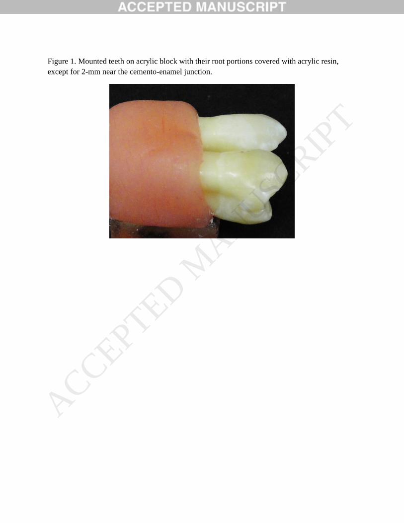
Figure 1. Mounted teeth on acrylic block with their root portions covered with acrylic resin,
except for 2-mm near the cemento-enamel junction.
ACCEPTED MANUSCRIP
T
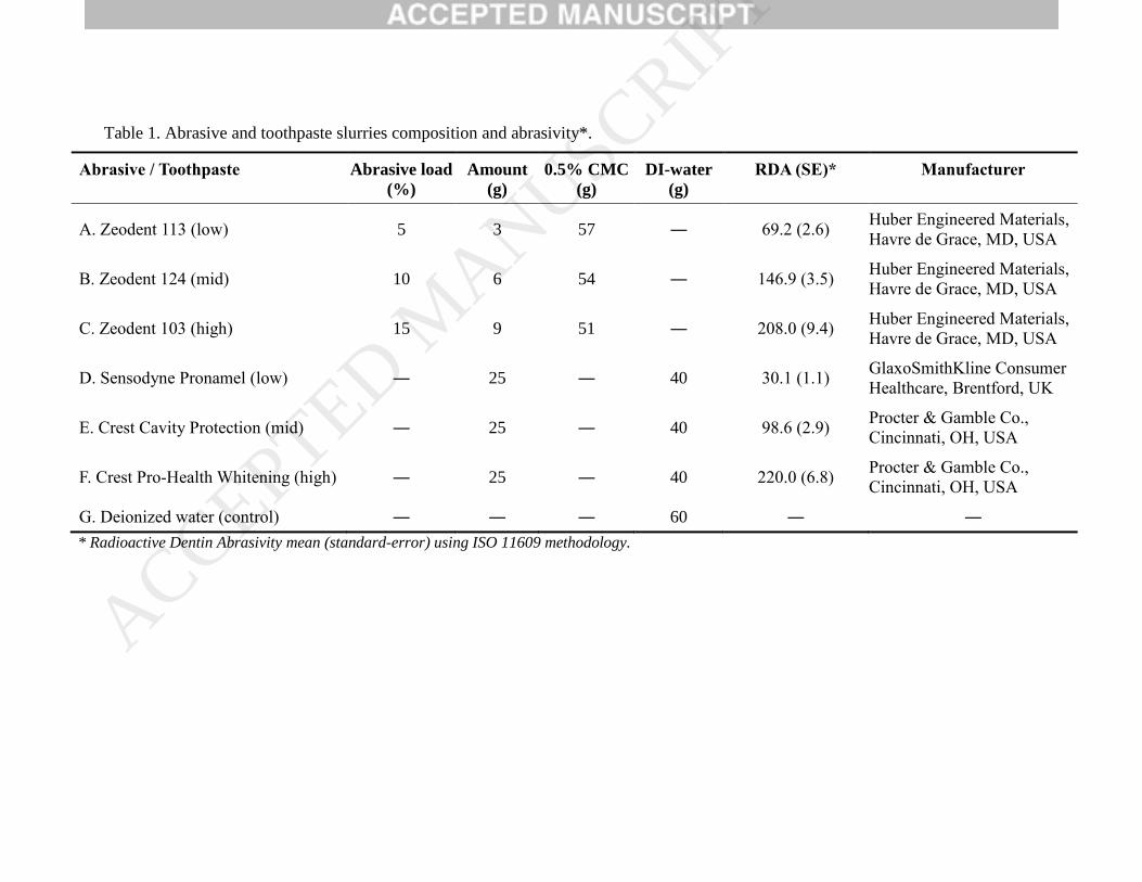
Table 1. Abrasive and toothpaste slurries composition and abrasivity*.
Abrasive / Toothpaste Abrasive load
(%)
Amount
(g)
0.5% CMC
(g)
DI-water
(g)
RDA (SE)* Manufacturer
A. Zeodent 113 (low) 5 3 57 ― 69.2 (2.6) Huber Engineered Materials,
Havre de Grace, MD, USA
B. Zeodent 124 (mid) 10 6 54 ― 146.9 (3.5) Huber Engineered Materials,
Havre de Grace, MD, USA
C. Zeodent 103 (high) 15 9 51 ― 208.0 (9.4) Huber Engineered Materials,
Havre de Grace, MD, USA
D. Sensodyne Pronamel (low) ― 25 ― 40 30.1 (1.1) GlaxoSmithKline Consumer
Healthcare, Brentford, UK
E. Crest Cavity Protection (mid) ― 25 ― 40 98.6 (2.9) Procter & Gamble Co.,
Cincinnati, OH, USA
F. Crest Pro-Health Whitening (high) ― 25 ― 40 220.0 (6.8) Procter & Gamble Co.,
Cincinnati, OH, USA
G. Deionized water (control) ― ― ― 60 ― ―
* Radioactive Dentin Abrasivity mean (standard-error) using ISO 11609 methodology.
ACCEPTED MANUSCRIP
T
Table 2. Means (standard deviation) of dentine volume loss (mm3) by abrasive/toothpaste, at different brushing strokes.
Abrasive / Toothpaste Dentine volume loss mean (mm3)
5,000 15,000 35,000 65,000
A. Zeodent 113 (low) 0.82 (0.35) Aa 0.97(0.47) Aab 1.07 (0.41) Bab 1.35 (0.65) Cb
B. Zeodent 124 (mid) 0.98 (0.34) Aa 0.81 (0.33) Aa 1.18 (0.38) ABa 1.85 (0.91) BCb
C. Zeodent 103 (high) 0.72 (0.25) Aa 0.80 (0.29) Aa 1.31 (0.57) ABb 2.33 (1.47) Bc
D. Sensodyne Pronamel (low) 0.79 (0.47) Aab 0.71 (0.34) Aa 0.97 (0.39) Bab 1.17 (0.48) Cb
E. Crest Cavity Protection (mid) 0.80 (0.32) Aa 0.83 (0.39) Aa 1.03 (0.50) Bab 1.40 (0.68) Cb
F. Crest Pro-Health Whitening (high) 1.25 (1.32) Aa 1.20 (1.07) Aa 1.93 (1.38) Ab 4.19 (3.29) Ac
G. Deionized water (control) 0.88 (0.51) Aa 0.83 (0.43) Aa 1.02 (0.72)Ba 1.12 (0.73) Ca
Means followed by the same uppercase letters indicate no difference between groups within each column. Means followed by the same
lowercase letters did not differ from each other within each row.
ACCEPTED MANUSCRIP
T
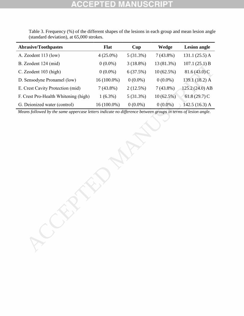
Table 3. Frequency (%) of the different shapes of the lesions in each group and mean lesion angle
(standard deviation), at 65,000 strokes.
Abrasive/Toothpastes Flat Cup Wedge Lesion angle
A. Zeodent 113 (low) 4 (25.0%) 5 (31.3%) 7 (43.8%) 131.1 (25.5) A
B. Zeodent 124 (mid) 0 (0.0%) 3 (18.8%) 13 (81.3%) 107.1 (25.1) B
C. Zeodent 103 (high) 0 (0.0%) 6 (37.5%) 10 (62.5%) 81.6 (43.0) C
D. Sensodyne Pronamel (low) 16 (100.0%) 0 (0.0%) 0 (0.0%) 139.1 (18.2) A
E. Crest Cavity Protection (mid) 7 (43.8%) 2 (12.5%) 7 (43.8%) 125.2 (24.0) AB
F. Crest Pro-Health Whitening (high) 1 (6.3%) 5 (31.3%) 10 (62.5%) 61.8 (29.7) C
G. Deionized water (control) 16 (100.0%) 0 (0.0%) 0 (0.0%) 142.5 (16.3) A
Means followed by the same uppercase letters indicate no difference between groups in terms of lesion angle.
ACCEPTED MANUSCRIP
T
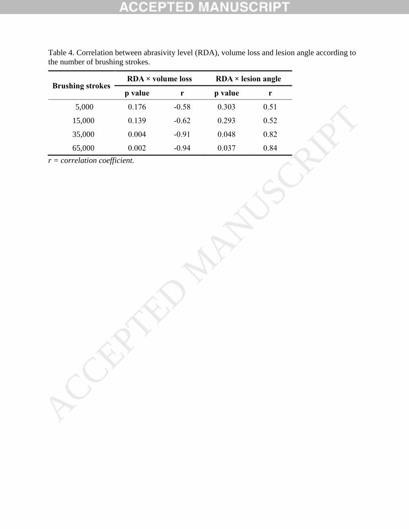
Table 4. Correlation between abrasivity level (RDA), volume loss and lesion angle according to
the number of brushing strokes.
Brushing strokes RDA × volume loss RDA × lesion angle
p value r p value r
5,000 0.176 -0.58 0.303 0.51
15,000 0.139 -0.62 0.293 0.52
35,000 0.004 -0.91 0.048 0.82
65,000 0.002 -0.94 0.037 0.84
r = correlation coefficient.
ACCEPTED MANUSCRIP
T