Embed Size (px)
Citation preview

Sao Paulo Med J. 2015; 133(5):445-9 445
CASE REPORTDOI: 10.1590/1516-3180.2014.7832407
A case of tumor-like inflammatory demyelinating disease with progressive brain and spinal cord involvementUm caso de doença desmielinizante inflamatória semelhante a tumor com envolvimento progressivo de cérebro e medula espinhalXu Zhi PengI, Li Hong HuaII, Sun Zhi QiangIII, Wu QiangII
Department of Neurology, Wuhan General Hospital of Guangzhou Command, Wuhan, China
ABSTRACTCONTEXT: Tumor-like inflammatory demyelinating disease (TIDD) usually occurs in the brain and rarely occurs in the spinal cord. TIDD appears to be very similar to tumors such as gliomas on imaging, which may lead to incorrect or delayed diagnosis and treatment. CASE REPORT: Because of headache and incoherent speech, a 24-year-old Chinese male presented to our hospital with a two-week history of respiratory infections. After dexamethasone treatment, his symptoms still got worse and surgery was performed for diagnostic purposes. Histological examination revealed that the lesion was inflammatory. Further lesions appeared in the spine (T3 and T4 levels) after two months and in the right occipital lobe after three months. After intravenous immunoglobulin (IVIG) and methylpred-nisolone treatment, his symptoms improved. CONCLUSION: Progressive lesions may damage the brain and spinal cord, and long-term prednisolone and IVIG therapy are beneficial in TIDD patients.
RESUMOCONTEXTO: A doença desmielinizante inflamatória tumoral (DDIT) geralmente ocorre no cérebro e ra-ramente na medula espinhal. A DDIT é muito semelhante a tumores tais como gliomas em exames de imagem, o que pode conduzir a diagnóstico e tratamento tardios e incorretos.RELATO DO CASO: Por causa de dor de cabeça e discurso incoerente, um homem chinês de 24 anos de idade foi ao hospital com história de duas semanas de infecções respiratórias. Após o tratamento com de-xametasona, seus sintomas ficaram ainda piores e a cirurgia foi realizada para fins de diagnóstico. O exame histológico revelou que a lesão era inflamatória. Mais lesões apareceram na coluna vertebral (níveis T3 e T4) após dois meses, e no lobo occipital direito depois de três meses. Depois de tratamento com imuno-globulina intravenosa (IGIV) e metilprednisolona, seus sintomas melhoraram.CONCLUSÃO: Lesões progressivas podem danificar o cérebro e a medula espinhal, e prednisolona a lon-go prazo e terapia de IGIV são benéficas em pacientes DDIT.
IMD, MSc. Attending Physician, Department of Neurology, Wuhan General Hospital of Guangzhou Command, Wuhan, China.IIMD, PhD. Professor, Department of Neurology, Wuhan General Hospital of Guangzhou Command, Wuhan, China.IIIMD, MSc. Attending Physician, Department of Radiology, Wuhan General Hospital of Guangzhou Command, Wuhan, China.
KEY WORDS:Demyelinating diseases.Brain.Spinal cord. Radiology. Pathology.
PALAVRAS-CHAVE:Doenças desmielinizantes.Cérebro.Medula espinhal.Radiologia. Patologia.
CASE REPORT | Peng XZ, Hua LH, Qiang SZ, Qiang W
446 Sao Paulo Med J. 2015; 133(5):445-9
INTRODUCTIONTumor-like inflammatory demyelinating disease (TIDD) is a rare central nervous system (CNS) demyelinating disorder affecting the cerebral hemispheres or the spinal cord.1 TIDD can be dif-ficult to diagnose because the inflammatory lesion clinically and radiologically mimics a tumor in the early stages.2 Its exact inci-dence and etiology remain unknown.3 Despite its rarity, aware-ness of this entity is important for guiding clinical practice, since a delayed diagnosis or misdiagnosis may deny patients the full benefit of a complete recovery or result in unnecessary aggressive management, thereby having a deleterious effect.4
TIDD usually occurs in the brain and rarely occurs in the spi-nal cord. TIDD with both brain and spinal cord involvement is even rarer, with only two reported cases.1,5 Because of the presence of the demyelinating mechanism, corticosteroids may provide effective treatment. Only a small percentage of TIDD patients do not respond to corticosteroid therapy. The following case report is an example of TIDD that was diagnosed in our hospital. Because the patient was not corticosteroid-sensitive, progressive lesions occurred in his brain and spinal cord. The report focuses on the difficulties in diag-nosis and treatment, and also presents a review of the literature.
CASE REPORTA 24-year-old right-handed male was admitted to the Neurology Department of Wuhan General Hospital of Guangzhou Command, complaining of headache and incoherent speech for two days.
The patient had reported a history of cold with coughing and fever about two weeks earlier. He had been treated with anti-cold medicine for several days and these symptoms had resolved one week prior to the admission of this report. He was a college student, and unmarried. He did not smoke and did not use alcohol. Prior to these occurrences, he had been in good health, with no relevant past medi-cal or family history. He had received routine immunizations at an early age without any side effects. He did not have any history of travel, exposure to animals or contact with sick people over recent months.
On arrival at our center, he reported experiencing severe head-ache. The physical examination was unremarkable, with normal blood pressure of 120/80 mmHg, heart rate of 85 bpm, respiratory rate of 18/min and temperature of 36.8 °C. Neurological examina-tion revealed cognitive impairment, neck rigidity, positive Kernig’s sign and negative upper and lower Brudzinski’s signs. All of the following blood test parameters were within the reference range, namely: white blood cell and platelet counts, liver function tests, renal function, electrolytes, C-reactive protein and erythrocyte sedimentation rate. Autoimmune antibody and serological screen-ing for HIV, syphilis and hepatitis B and C were all negative.
A lumbar puncture was subsequently performed, and analy-sis of the cerebrospinal fluid (CSF) revealed elevated opening pres-sure (250 mmH2O; normal < 180 mmH2O) and elevated total pro-tein (51 mg/dl; normal < 40 mg/dl). White blood cell and red blood
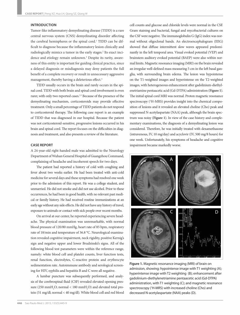
cell counts and glucose and chloride levels were normal in the CSF. Gram staining and bacterial, fungal and mycobacterial cultures on the CSF were negative. The immunoglobulin G (IgG) index was nor-mal without oligoclonal bands. An electroencephalogram (EEG) showed that diffuse intermittent slow waves appeared predomi-nantly in the left temporal area. Visual evoked potential (VEP) and brainstem auditory evoked potential (BAEP) were also within nor-mal limits. Magnetic resonance imaging (MRI) on the brain revealed an irregular well-defined mass measuring 5 cm in the left basal gan-glia, with surrounding brain edema. The lesion was hypointense on the T1-weighted images and hyperintense on the T2-weighted images, with heterogeneous enhancement after gadolinium-diethyl-enetriamine pentaacetic acid (Gd-DTPA) administration (Figure 1). The initial spinal cord MRI was normal. Proton magnetic resonance spectroscopy (1H-MRS) provides insight into the chemical compo-sition of lesions and it revealed an elevated choline (Cho) peak and suppressed N-acetylaspartate (NAA) peak, although the brain spec-trum was noisy (Figure 1). In view of the case history and comple-mentary examinations, the diagnosis of a demyelinating lesion was considered. Therefore, he was initially treated with dexamethasone (intravenous, IV, 10 mg/day) and acyclovir (IV, 500 mg/8 hours) for one week. Unfortunately, his symptoms of headache and cognitive impairment became markedly worse.
A B
C D
Figure 1. Magnetic resonance imaging (MRI) of brain on admission, showing: hypointense image with T1 weighting (A); hyperintense image with T2 weighting (B); enhancement after gadolinium-diethylenetriamine pentaacetic acid (Gd-DTPA) administration, with T1 weighting (C); and magnetic resonance spectroscopy (1H-MRS) with increased choline (Cho) and decreased N-acetylaspartate (NAA) peaks (D).
A case of tumor-like inflammatory demyelinating disease with progressive brain and spinal cord involvement | CASE REPORT
Sao Paulo Med J. 2015; 133(5):445-9 447
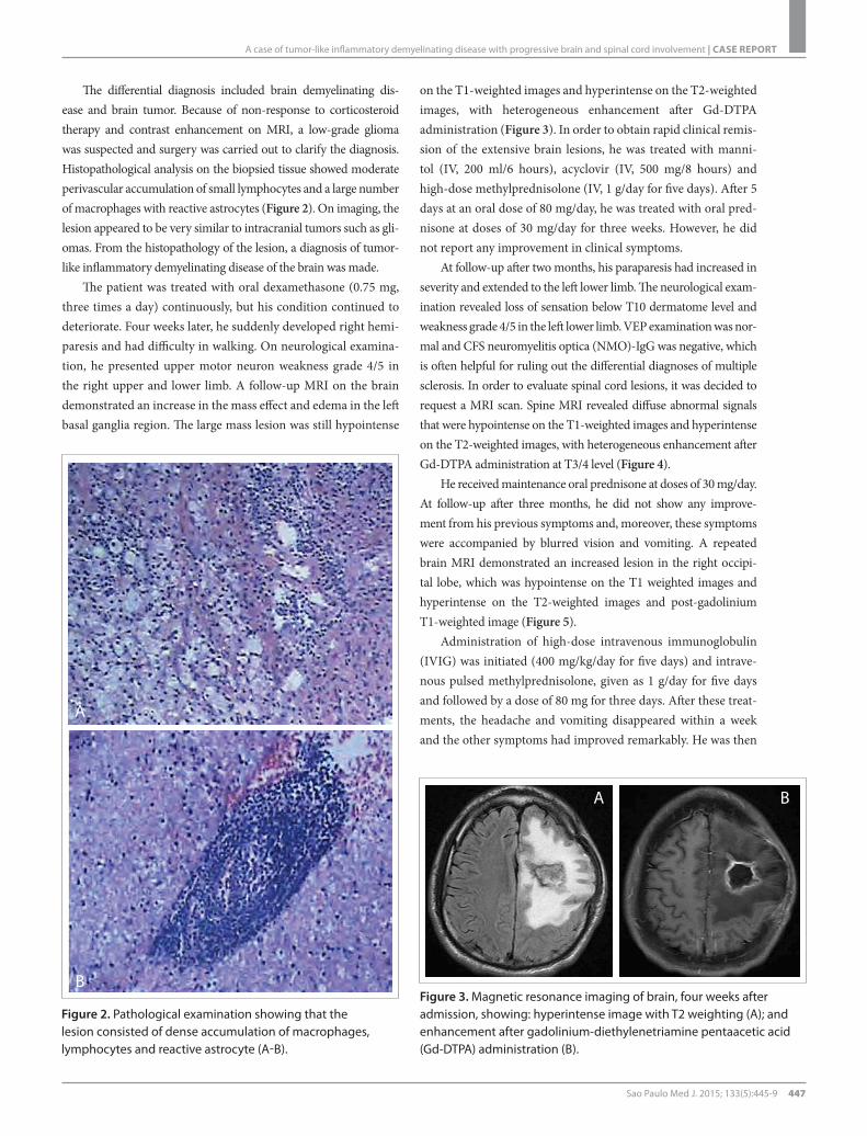
The differential diagnosis included brain demyelinating dis-ease and brain tumor. Because of non-response to corticosteroid therapy and contrast enhancement on MRI, a low-grade glioma was suspected and surgery was carried out to clarify the diagnosis. Histopathological analysis on the biopsied tissue showed moderate perivascular accumulation of small lymphocytes and a large number of macrophages with reactive astrocytes (Figure 2). On imaging, the lesion appeared to be very similar to intracranial tumors such as gli-omas. From the histopathology of the lesion, a diagnosis of tumor-like inflammatory demyelinating disease of the brain was made.
The patient was treated with oral dexamethasone (0.75 mg, three times a day) continuously, but his condition continued to deteriorate. Four weeks later, he suddenly developed right hemi-paresis and had difficulty in walking. On neurological examina-tion, he presented upper motor neuron weakness grade 4/5 in the right upper and lower limb. A follow-up MRI on the brain demonstrated an increase in the mass effect and edema in the left basal ganglia region. The large mass lesion was still hypointense
on the T1-weighted images and hyperintense on the T2-weighted images, with heterogeneous enhancement after Gd-DTPA administration (Figure 3). In order to obtain rapid clinical remis-sion of the extensive brain lesions, he was treated with manni-tol (IV, 200 ml/6 hours), acyclovir (IV, 500 mg/8 hours) and high-dose methylprednisolone (IV, 1 g/day for five days). After 5 days at an oral dose of 80 mg/day, he was treated with oral pred-nisone at doses of 30 mg/day for three weeks. However, he did not report any improvement in clinical symptoms.
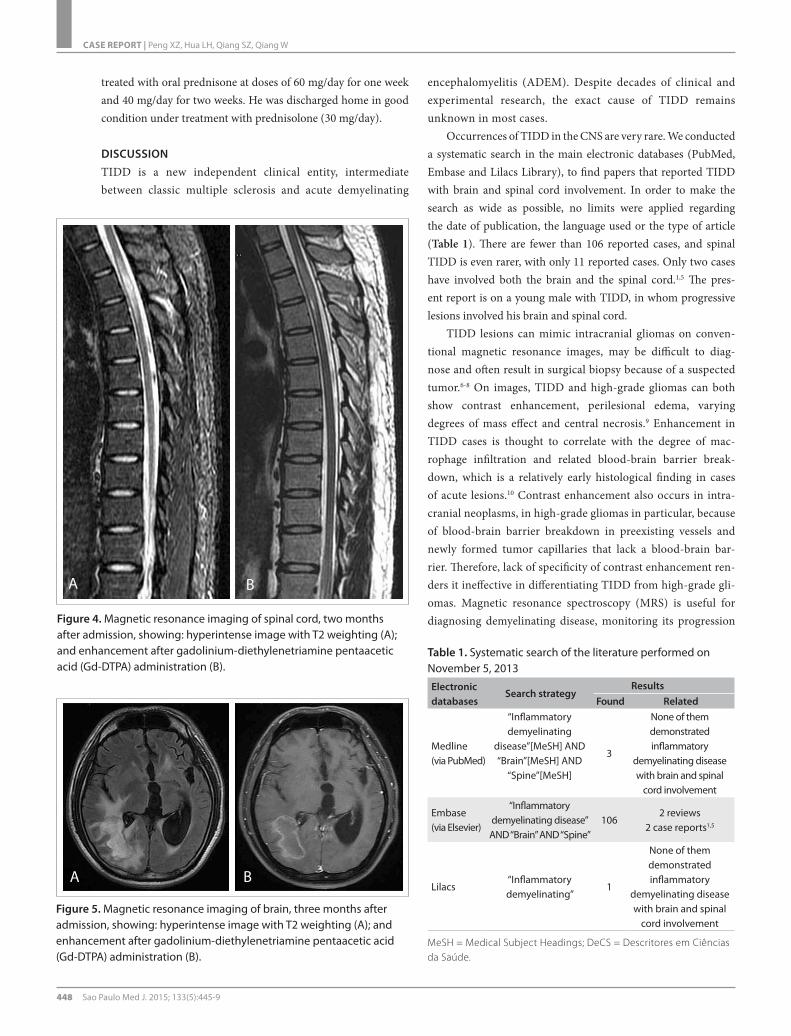
At follow-up after two months, his paraparesis had increased in severity and extended to the left lower limb. The neurological exam-ination revealed loss of sensation below T10 dermatome level and weakness grade 4/5 in the left lower limb. VEP examination was nor-mal and CFS neuromyelitis optica (NMO)-IgG was negative, which is often helpful for ruling out the differential diagnoses of multiple sclerosis. In order to evaluate spinal cord lesions, it was decided to request a MRI scan. Spine MRI revealed diffuse abnormal signals that were hypointense on the T1-weighted images and hyperintense on the T2-weighted images, with heterogeneous enhancement after Gd-DTPA administration at T3/4 level (Figure 4).
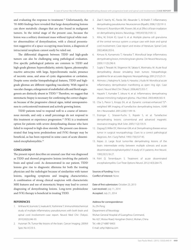
He received maintenance oral prednisone at doses of 30 mg/day. At follow-up after three months, he did not show any improve-ment from his previous symptoms and, moreover, these symptoms were accompanied by blurred vision and vomiting. A repeated brain MRI demonstrated an increased lesion in the right occipi-tal lobe, which was hypointense on the T1 weighted images and hyperintense on the T2-weighted images and post-gadolinium T1-weighted image (Figure 5).
Administration of high-dose intravenous immunoglobulin (IVIG) was initiated (400 mg/kg/day for five days) and intrave-nous pulsed methylprednisolone, given as 1 g/day for five days and followed by a dose of 80 mg for three days. After these treat-ments, the headache and vomiting disappeared within a week and the other symptoms had improved remarkably. He was then
Figure 2. Pathological examination showing that the lesion consisted of dense accumulation of macrophages, lymphocytes and reactive astrocyte (A-B).
A
BFigure 3. Magnetic resonance imaging of brain, four weeks after admission, showing: hyperintense image with T2 weighting (A); and enhancement after gadolinium-diethylenetriamine pentaacetic acid (Gd-DTPA) administration (B).
A B
CASE REPORT | Peng XZ, Hua LH, Qiang SZ, Qiang W
448 Sao Paulo Med J. 2015; 133(5):445-9
treated with oral prednisone at doses of 60 mg/day for one week and 40 mg/day for two weeks. He was discharged home in good condition under treatment with prednisolone (30 mg/day).
DISCUSSIONTIDD is a new independent clinical entity, intermediate between classic multiple sclerosis and acute demyelinating
encephalomyelitis (ADEM). Despite decades of clinical and experimental research, the exact cause of TIDD remains unknown in most cases.
Occurrences of TIDD in the CNS are very rare. We conducted a systematic search in the main electronic databases (PubMed, Embase and Lilacs Library), to find papers that reported TIDD with brain and spinal cord involvement. In order to make the search as wide as possible, no limits were applied regarding the date of publication, the language used or the type of article (Table 1). There are fewer than 106 reported cases, and spinal TIDD is even rarer, with only 11 reported cases. Only two cases have involved both the brain and the spinal cord.1,5 The pres-ent report is on a young male with TIDD, in whom progressive lesions involved his brain and spinal cord.
TIDD lesions can mimic intracranial gliomas on conven-tional magnetic resonance images, may be difficult to diag-nose and often result in surgical biopsy because of a suspected tumor.6-8 On images, TIDD and high-grade gliomas can both show contrast enhancement, perilesional edema, varying degrees of mass effect and central necrosis.9 Enhancement in TIDD cases is thought to correlate with the degree of mac-rophage infiltration and related blood-brain barrier break-down, which is a relatively early histological finding in cases of acute lesions.10 Contrast enhancement also occurs in intra-cranial neoplasms, in high-grade gliomas in particular, because of blood-brain barrier breakdown in preexisting vessels and newly formed tumor capillaries that lack a blood-brain bar-rier. Therefore, lack of specificity of contrast enhancement ren-ders it ineffective in differentiating TIDD from high-grade gli-omas. Magnetic resonance spectroscopy (MRS) is useful for diagnosing demyelinating disease, monitoring its progression
Table 1. Systematic search of the literature performed on November 5, 2013Electronic databases
Search strategyResults
Found Related
Medline (via PubMed)
“Inflammatory demyelinating
disease”[MeSH] AND “Brain”[MeSH] AND
“Spine”[MeSH]
3
None of them demonstrated inflammatory
demyelinating disease with brain and spinal
cord involvement
Embase (via Elsevier)
“Inflammatory demyelinating disease”
AND “Brain” AND “Spine”106
2 reviews2 case reports1,5
Lilacs“Inflammatory demyelinating”
1
None of them demonstrated inflammatory
demyelinating disease with brain and spinal
cord involvement
MeSH = Medical Subject Headings; DeCS = Descritores em Ciências da Saúde.
Figure 4. Magnetic resonance imaging of spinal cord, two months after admission, showing: hyperintense image with T2 weighting (A); and enhancement after gadolinium-diethylenetriamine pentaacetic acid (Gd-DTPA) administration (B).
BA
Figure 5. Magnetic resonance imaging of brain, three months after admission, showing: hyperintense image with T2 weighting (A); and enhancement after gadolinium-diethylenetriamine pentaacetic acid (Gd-DTPA) administration (B).
BA
A case of tumor-like inflammatory demyelinating disease with progressive brain and spinal cord involvement | CASE REPORT
Sao Paulo Med J. 2015; 133(5):445-9 449
and evaluating the response to treatment.11 Unfortunately, the 1H-MRS findings here revealed that large demyelinating lesions can show metabolic changes that are also found in malignant tumors. In the initial stage of the present case, because the lesion was a solitary dominant mass without typical white mat-ter abnormalities of demyelination, with a clinical presenta-tion suggestive of a space-occupying mass lesion, a diagnosis of intracranial neoplasm cannot easily be ruled out.
The differential diagnosis between TIDD and high-grade tumors can also be challenging using pathological evaluation. Five specific pathological patterns are common to TIDD and high-grade gliomas: hypercellularity, mitotic figures, pleomorphic reactive astrocytes with large, hyperchromatic nuclei, presence of necrotic areas, and areas of cystic degeneration or cavitation. Despite some similar histopathological features, TIDD and high-grade gliomas are different regarding vascularity. With regard to vascular changes, enlargement of endothelial cells and florid angio-genesis are distinctly absent in TIDD.12 Therefore, we suggest that stereotactic biopsy is necessary for confirming the correct diagno-sis because of the progressive clinical signs, initial unresponsive-ness to corticosteroid treatment and actively growing lesion.
TIDD patients tend to respond well to a course of intrave-nous steroids, and only a small percentage do not respond to this treatment or experience progression.13 IVIG is a treatment option for patients with severe demyelinating disease who have failed to respond to high-dose steroids. The present case demon-strated that long-term prednisolone and IVIG therapy may be beneficial, as has been reported in some cases of acute dissemi-nated encephalomyelitis.14
CONCLUSIONThe present report describes an unusual case that was diagnosed as TIDD and showed progressive lesions involving the patient’s brain and spinal cord. As demonstrated in our patient, TIDD lesions give rise to diagnostic dilemma for both the treating physician and the radiologist because of similarities with tumor lesions, regarding symptoms and imaging characteristics. A combination of strong clinical suspicion with characteristic MRI features and use of stereotactic biopsy may lead to correct diagnosing of demyelinating lesions. Long-term prednisolone and IVIG therapy is beneficial in treating TIDD.
REFERENCES1. Ishihara M, Izumoto S, Iwatsuki K, Yoshimine T. Immunohistochemical
study of multiple inflammatory pseudotumors with both brain and
spinal cord involvement--case report. Neurol Med Chir (Tokyo).
2010;50(3):246-50.
2. Huisman TA. Tumor-like lesions of the brain. Cancer Imaging. 2009;9
Spec No A:S10-3.
3. Ziad F, Katchy KC, Panda SM, Alexander S, Al-Sheikh T. Inflammatory
demyelinating pseudotumor. Neurosciences (Riyadh). 2006;11(3):191-3.
4. Peterson K, Rosenblum MK, Powers JM, et al. Effect of brain irradiation
on demyelinating lesions. Neurology. 1993;43(10):2105-12.
5. Kilinç M, Ertürk IO, Uysal H, et al. Multiple plasma cell granuloma
of the central nervous system: a unique case with brain and spinal
cord involvement. Case report and review of literature. Spinal Cord.
2002;40(4):203-6.
6. Kimura N, Kumamoto T, Hanaoka T. Monofocal large inflammatory
demyelinating lesion, mimicking brain glioma. Clin Neurol Neurosurg.
2009;111(3):296-9.
7. Sugita Y, Terasaki M, Shigemori M, Sakata K, Morimatsu M. Acute focal
demyelinating disease simulating brain tumors: histopathologic
guidelines for an accurate diagnosis. Neuropathology. 2001;21(1):25-31.
8. Akimoto J, Nakajima N, Saida A, Haraoka J, Kudo M. Monofocal acute
inflammatory demyelination manifesting as open ring sign. Case
report. Neurol Med Chir (Tokyo). 2006;46(7):353-7.
9. Hayashi T, Kumabe T, Jokura H, et al. Inflammatory demyelinating
disease mimicking malignant glioma. J Nucl Med. 2003;44(4):565-9.
10. Cha S, Pierce S, Knopp EA, et al. Dynamic contrast-enhanced T2*-
weighted MR imaging of tumefactive demyelinating lesions. AJNR
Am J Neuroradiol. 2001;22(6):1109-16.
11. Enzinger C, Strasser-Fuchs S, Ropele S, et al. Tumefactive
demyelinating lesions: conventional and advanced magnetic
resonance imaging. Mult Scler. 2005;11(2):135-9.
12. Zagzag D, Miller DC, Kleinman GM, et al. Demyelinating disease versus
tumor in surgical neuropathology. Clues to a correct pathological
diagnosis. Am J Surg Pathol. 1993;17(6):537-45.
13. Kepes JJ. Large focal tumor-like demyelinating lesions of the
brain: intermediate entity between multiple sclerosis and acute
disseminated encephalomyelitis? A study of 31 patients. Ann Neurol.
1993;33(1):18-27.
14. Pohl D, Tenembaum S. Treatment of acute disseminated
encephalomyelitis. Curr Treat Options Neurol. 2012;14(3):264-75.
Sources of funding: None
Conflict of interest: None
Date of first submission: October 23, 2013
Last received: July 11, 2014
Accepted: July 21, 2014
Address for correspondence:
Xu Zhi Peng
Department of Neurology
Wuhan General Hospital of Guangzhou Command,
No. 627, Wuluo Road, Hongshan District, Wuhan, China
Tel. +86-27-6887-8403
E-mail: [email protected]